Preprint
Article

Whole Exome Sequencing of Meningioma in Sudanese Patients

Altmetrics

Downloads

62

Views

30

Comments

0

Submitted:

14 September 2024

Posted:

18 September 2024

You are already at the latest version

Alerts
Abstract
Introduction Meningioma is the second most common primary intracranial tumor of the central nervous system. in Sudan, meningioma is the most common primary brain tumor, among Sudanese patients, Surgical total excision of meningioma offers a better survival, however, Chemotherapy has largely been unsuccessful for meningioma treatment, and therefore, refractory and recurrence meningioma treated with palliative surgery and radiotherapy. Monosomy of chromosome 22 is the utmost common genetic alteration among meningioma and many genes were investigated and described in the development of this tumor of which , NF2 hSNF5/INI1gene, P53. In addition to this, Molecular signal pathways in meningioma, and that contribute to the survival, proliferation, self-renewal, and differentiation properties of normal stem cells which are abnormally activated and nominated as cancerous stem cell (CSCs). Objectives 1/ To identify the possible pathway of candidate genes in both setting and whole genes hypothesis free approach 2/ To assess the presence of mutations in candidate genes among histological subtypes of meningioma in Sudan. Material and methods This is a prospective cross-sectional study done at the National Center of Neurological Sciences (NCNS) Khartoum, from 2018 to 2021. The study included all cerebral tumors that were radio- logically diagnosed preoperatively as meningioma. Ethical approval was taken from Institute of Endemic disease, Khartoum University, and written consent was obtained Three DNA extracts were randomly selected from 20 meningioma tissue samples and processed for WES. All genes harboring exonic and splice site variations stratified into SNVs and INDELs sets were prepared. The 1000G online database was used to explore novel mutations per 3 samples. SIFT and PolyPhen-2 programs were used to predict possible impact of amino acids substitution. To address meticulous genes interaction and particular centrality, all shared genes within the class INDEL and SNVs were used as input on Network Analysis to predict protein- protein interaction and functional gene ontology. Finally, shared mutations within genes of centrality were detected via Sanger sequencing and mutations were correlated between histological sub-types and recurrenceThree non meningioma brain tumor used as control. Results WES analysis revealed high number for mutational signature within 2 classes (INDEL and SNVs) per 3 samples, the findings of 1000 genome online database showed number of novel mutations; 5618 in fibrous , 6032 in meningothelial while in recurrent fibrous sample 5930. Network analysis of shared genes per each class (INDEL, SNVs), showed UBC is the most protein of remarkable degree centrality over thousand of genes. Whereas, all shared genes per 3 samples within the 2 classes (INDEL and SNVs) revealed Akt1 with the highest degree of centrality and betweenness. Akt1 rs17846829was detected in 20% of the samples, while CD44 stemness gene revealed mutation A>G (rs9666607) in all meningioma samples (100%), this mutation was associated with 60% of the recurrence. Conclusions Nevertheless, the present study, highlight how such complex molecular processes might contribute to tumorogenesis, in attempt to shed light over thousands of genes and multifaceted network analysis rather than a solitary gene which is challenging question.
Keywords: 
Subject: Medicine and Pharmacology  -   Other

Introduction

Meningioma is the second most common primary intracranial tumor of the central nervous system, comprising nearly 30% of all primary brain tumors. [1,2] in Sudan, meningioma is the most common primary brain tumor, among Sudanese patients.[3]Surgical total excision of meningioma offers a better survival and clinical outcome to patients; however, up to 18% of benign meningioma, 40% of atypical meningioma, and 80% of malignant meningioma recur within 5 years despite of complete excision. [4] Chemotherapy has largely been unsuccessful for meningioma treatment, and therefore, refractory and recurrence meningioma treated with palliative surgery and radiotherapy.[5,6]In this context need for definite therapeutic approaches based on effective molecular targets in order to improve outcome and or long-term control of meningioma is urgent. However; still, in-adequate of clinical predictive power remains one of the most critical impediments in the development of novel study models. [7]

Genetic of Meningioma:

Monosomy of chromosome 22 is the utmost common genetic alteration among meningioma and was one of thefirst cytogenetic alterations described in solid tumors. Mutations within the NF2 gene were detected in up to 60% of sporadic meningioma and are typically associated with loss of heterozygosity(LOH) 22q.[8]
The majorityNF2mutations have a shortening effect, which indicate inactivation of the NF2gene product Merlin(schwannomin), this gene is functionally important inmeningioma pathogenesis, absent or reduced immune- reactivity in some studiesforMerlin was found in the bulk of meningioma,[9] and was strongly associated with loss of heterozygosis 22q.Although the suppressive activity of this gene is poorly understood, the signaling cascade disruption is considered in tumor formation. One study showed that, molecular genetics and protein of NF2 was differ among fibroblastic, transitional and meningiothelial meningioma which are the common benignvariant, merlin reduction was observed in the first two variant but rarely in the last one, this observation may suggests the genetic origin of this tumor is independent of NF2 gene alteration. Similarly study done in Sudan at the National center for neurological sciences (unpublished data), in meningioma among Sudanese patients, revealed mutations in NF2 gene is not involved in meningioma progression.
In addition to this, some candidate tumor suppressor genes on 22q were screened for mutations in meningioma, one of these was hSNF5/INI1gene, was frequently mutated in atypical teratoid/rhabdoidtumors. [10], LARGE gene which maps on (22q12.3) and denotes another candidate gene.[11]
Nevertheless, the well-known tumor suppressor gene, P53, which is located on chromosome 17 short arm, is one of most frequent mutated gene in human cancer and brain astrocytoma, however, mutation in this gene are rarely seen in meningioma.[12] Several large studies reported some mutations, among which study done in Sudan by Gassoum et al. [13]

Molecular Signaling Pathways

Molecular signal pathways in meningioma, are many and among which is mitogenic signals transduction, approximately all growth factor receptors and kinases are recognized to be involved in tumor growth and being contributing factors in meningioma cell lines, of which epidermal growth factor receptor (EGFR), platelet-derived growth factor receptor β (PDGFRβ), vascular endothelial growth factor receptor (VEGFR) and insulin-like growth factor receptor (IGFR).[14]
Moreover, there are many signaling pathways that contribute to the survival, proliferation, self-renewal, and differentiation properties of normal stem cells which are abnormally activated and nominated as cancerous stem cell (CSCs) and thus causing tumorigenesis. In this context, various endogenous or exogenous genes and microRNAs regulate these complex pathways. Eventually, these signaling pathways can induce downstream gene expression, such as cytokines, growth factors, apoptosis genes, anti - apoptotic genes, proliferation genes, and metastasis genes in CSCs. The importance of these signaling pathways regulates and linked networks of signaling mediators to regulate cancerous stem cell growth.
These signaling pathways include, Wnts pathway which include large protein ligands, plays an important role in the dedifferentiation of CSCs, metastasis and regulating CSC apoptosis,[15]It affects miscellaneous processes, such as the generation of cell polarity, and cells fate, Aberrant Wnt signaling is found in many aggressive cancers,[16] beside this, activation of Wnt induce the transformation of dormant CSCs into active CSCs which eventually enhancing cell cycle progression through β-catenin.[17]In addition, long noncoding RNAs and microRNAs also promote self-renewal of CSCs through Wnt signaling pathway, this occurred in many cancers such as gastric. [18]

Objectives

1/ To identify the possible pathway of candidate genes in both setting and whole genes hypothesis free approach
2/ To assess the presence of mutations in candidate genes among histological subtypes of meningioma in Sudan

Material and Methods

This is a prospective cross-sectional study done at the National Center of Neurological Sciences (NCNS) Khartoum, from 2018 to 2021. The study included all cerebral tumors that were radio- logically diagnosed preoperatively as meningioma. Ethical approval was taken from Institute of Endemic disease, Khartoum University, and written consent was obtained from each patient.
The tumor specimens were obtained immediately from the operating room, before surgery was completed, and sent to the research laboratory in the center within 2-5 minutes. At arrival to the laboratory, small portion from 20 tissue samples were processed for DNA extraction, three of which were selected for whole exome analysis and each was processed further for detection of gene of centrality by using PCR and thus sequencing analysis. Three non meningioma brain tumor were used as control

Whole Exome Sequencing (WES)

Sample Preparation

After evaluating the detailed histopathology of our tumor specimens, 3 samples were selected for DNA extraction for whole exome analysis and each was proceeded further for detection genes of centrality.DNA quality and quantity were assessed using Nano-Drop ND-1000 (Nano-Drop Products, Wilmington, DE, USA) spectrophotometer at wavelength spectrum of 220-750nm and standard 1% agarose gel electrophoresis. Each sample was labeled with a unique accession number, and DNA was sent to Beijing Genomics Institute (BGI) for whole exome sequencing.

DNA Extraction Method

DNA was extracted from tissues obtained from meningioma tumors using Guanidine chloride method. (According to the National Center of Neurological sciences standard protocol)

Exploratory Analysis

Genes set were prepared by filtering for all genes harboring exonic and splice site variations stratified into SNVs and INDELs set genes for each sample. In addition to that, we used 1000G online database to explore novel mutations. And also we explored the excess variant of allele frequency per 3 samples.
To predict possible impact of an amino acid substitution on the structure and function of a human protein, SIFT and PolyPhen-2 programs were used.
In addition to this, shared genes filtering for all genes which stratified into two classes (SNVs and INDELs) per 3 sample, were constructed by utilizing R software (version 4.1.0, 2021-05-18)

Network Analysis Using Network Analyst 2021

In this study, Network analyst 2021 was used to put up individual functional interaction network of genes per each sample, and for gene set which harboring damaging effect. Although there was number of mutated genes, they were not all included in network analysis, we managed shared gene analysis in both class (INDEL and SNVs) per 3 sample, when needed , high and moderate, low and modifier impact were defined in SNVs. On topography of network analysis, genes of centrality can be conveyed.
To address meticulous genes interaction, and whether these interactions with particular centrality, all shared genes within the class INDEL and SNVs were used as input on Network Analyst (GenMania, Reactome and STRING) to predict protein- protein interaction and functional gene onto

Pathway and GO Enrichment Analysis

Based on enrichment analysis online tools (Network Analyst 2021), which was used to examine pathways enrichment for sets of genes in various data base, and application to forecast diseases associated.
Additionally, shared mutations within genes of centrality were detected using polymerase chain reaction by means of specific forward and reverse primers.
The primers were designed using Primer 3 online program. The table below is containing the list of primers used in this study.
AKT1 F CCCCTCAGATGATCTCTCCA
R TGAAGAATTTGGAGGGAAGG
CD44 F AGGAACAGTGGTTTGGCAAC
R AACTGGCTTGTATCCATTCCT

Results

In this study, we present the result of genetic whole exome sequence analysis of 3 samples from our patients. DNA was labeled as: sample 1B denote fibrous subtype, sample 2B indicates meningiothelial subtype while 4A is fibrous recurrence.

Result of Exploratory Analysis

There was high number for mutational signature within 2 classes is shown in Table 3 and Table 4.
Moreover, when we utilized 1000 genome online database to categorize novel mutations, our results showed that, number of mutations in fibrous were 5618, in meningiothelialwere 6032 while in fibrous recurrence were 5930. The result of excess transition C>T, in fibrous recurrencewas 1063, whereas in fibrouswas 1049 and meningiothelialwas 1127, the low allele frequency was T>A, in fibrous 189, meningiothelial 204and fibrous recurrence 213 (Table 5).
Table 3. number of single nucleotide variations and insertion deletion per 3 samples in each class.
Class Sample All Novel Miss Intronic 3UTR 5UTR Splicing
SNVs 1B 116490 930 11323 74276 3481 2125 2560
2B 119465 6032 11512 75940 3734 379 2386
4A 118987 5930 10719 77996 3689 369 2157
INDEL Frs
1B 15103 4884 662 11638 554 282 660
2B 15579 5007 94 3743 173 76 278
4A 17114 5745 627 13269 660 336 677
1B is fibrous type, 2B is meningiothelial and 4A is fibrous recurrence, Miss=missense, Frs=frame-shift.
Table 4. number of single nucleotide variations and insertion deletion with high and moderate impact per 3 samples.
Sample Total high impact SNVs/INDEL Total moderate impact SNVs/INDEL P value
1B 17 253 <0.001
2B 17 267 <0.001
4A 11 242 <0.001
1B is fibrous type, 2B is meningiothelial and 4A is fibrous recurrence.
As shown in Table 6, the total number of missense variants with high and moderate impact in class (SNVs) was 253 in fibrous meningioma. SIFT deleterious effect was detected in 54 mutations, among which 16 variants showed damaging effect by PolyPhen-2, as the same time, meningiothelial subtype which included 267 missense variants, by SIFT 63 were deleterious, among which, 18 variants were damaging. SIFT mutations in fibrous recurrence revealed that, 48 mutations were deleterious, among which 30 were damaging by PolyPhen-2.
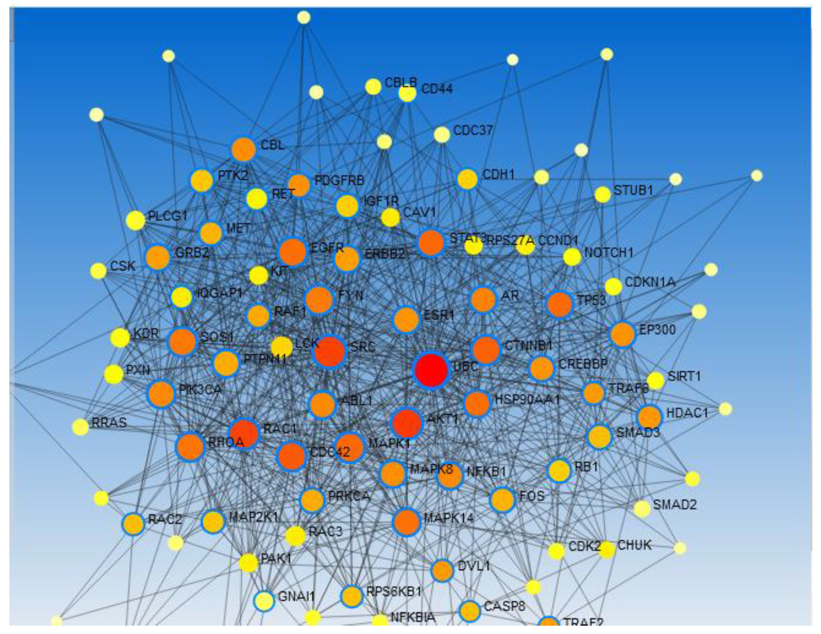
Network analysis results of class insertion/ deletion (INDEL), the most fabulous features is UBC protein of remarkable degree centrality over thousand of genes, the centrality was considered using degree of centrality and betweenness. Furthermore, other genes of centrality AKt1 and CD44 were detected.
When we used all shared genes in class SNVs, the network analysis, revealed that, UBC showed the highest degree of centrality, this is with other central hub genes of stemness and non stemenesswhich includedSTAT3, CD44, GLI1, KDR, KITTP53, Akt1 andMAPK1,these genes interacting with nearly 2000 of proteins, direct interaction of gene is shown in
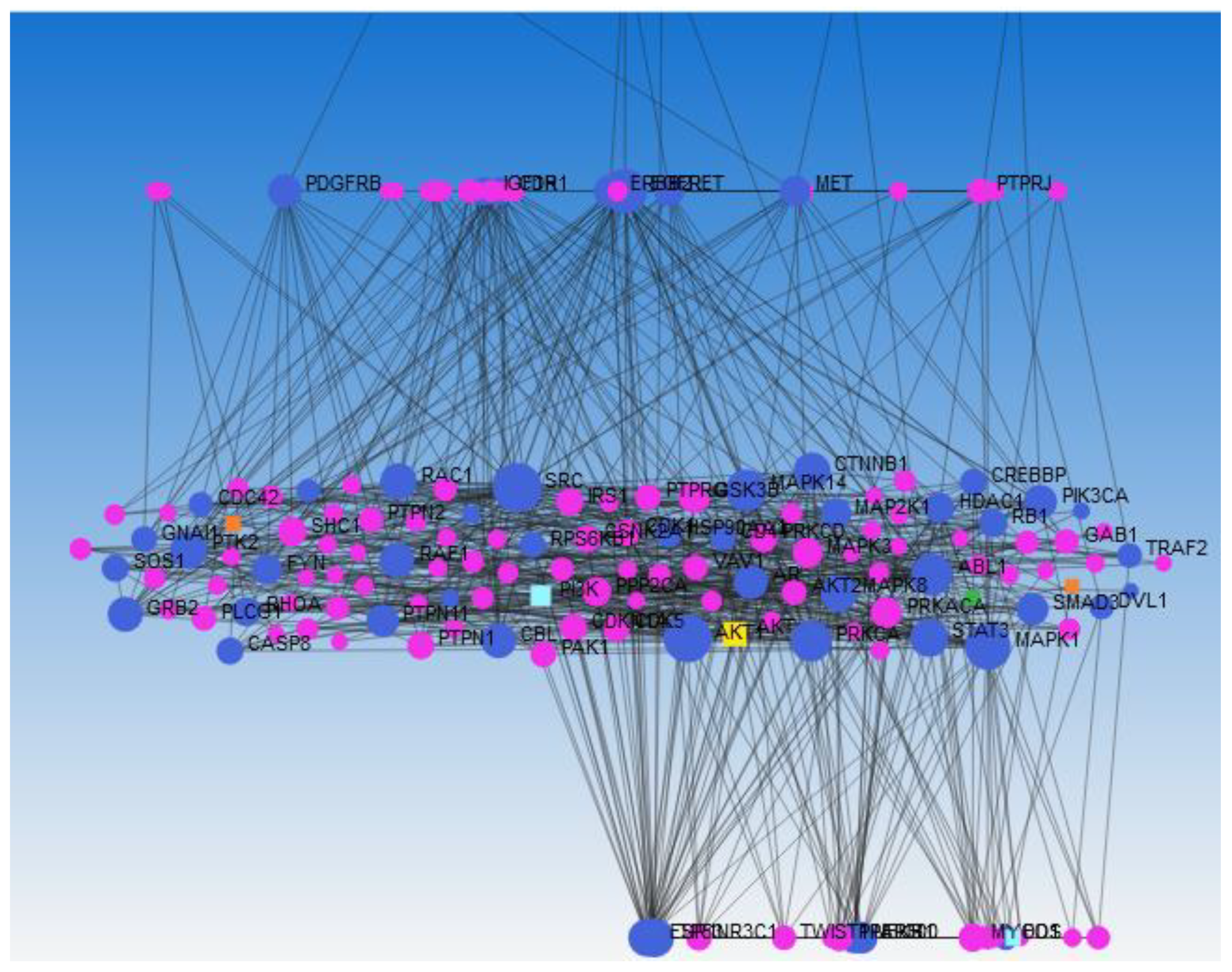
Despite cancer is multiple complex processes, we aimed to verify number of mutations that might be related to cancer. In this context we used all shared gene in 2 classes (INDEL and SNVs), and based on Network Analyst, we demonstrated that, 172 number of proteins were interacting, AKt1 protein showed the highest degree centrality and betweenness (degree=52, betweenness=1657.89) flowed by EGFR, PIK3CA, UBC, SOS1 and varying number of stemness proteins among which were STAT3, MAPK, CD44, NOTCH and SMO (Figure 8a).Additionally, to take in hand whether these interaction were true particularly of centrality among huge partner, we utilized random selection of genes set, striking finding Akt1 and UBC showed the highest centrality and betweeness (degree= 52, betweenness=1657.89) and (degree=42 betweenness=1317.9) respectively, along with other stemness genes (Figure 8b).
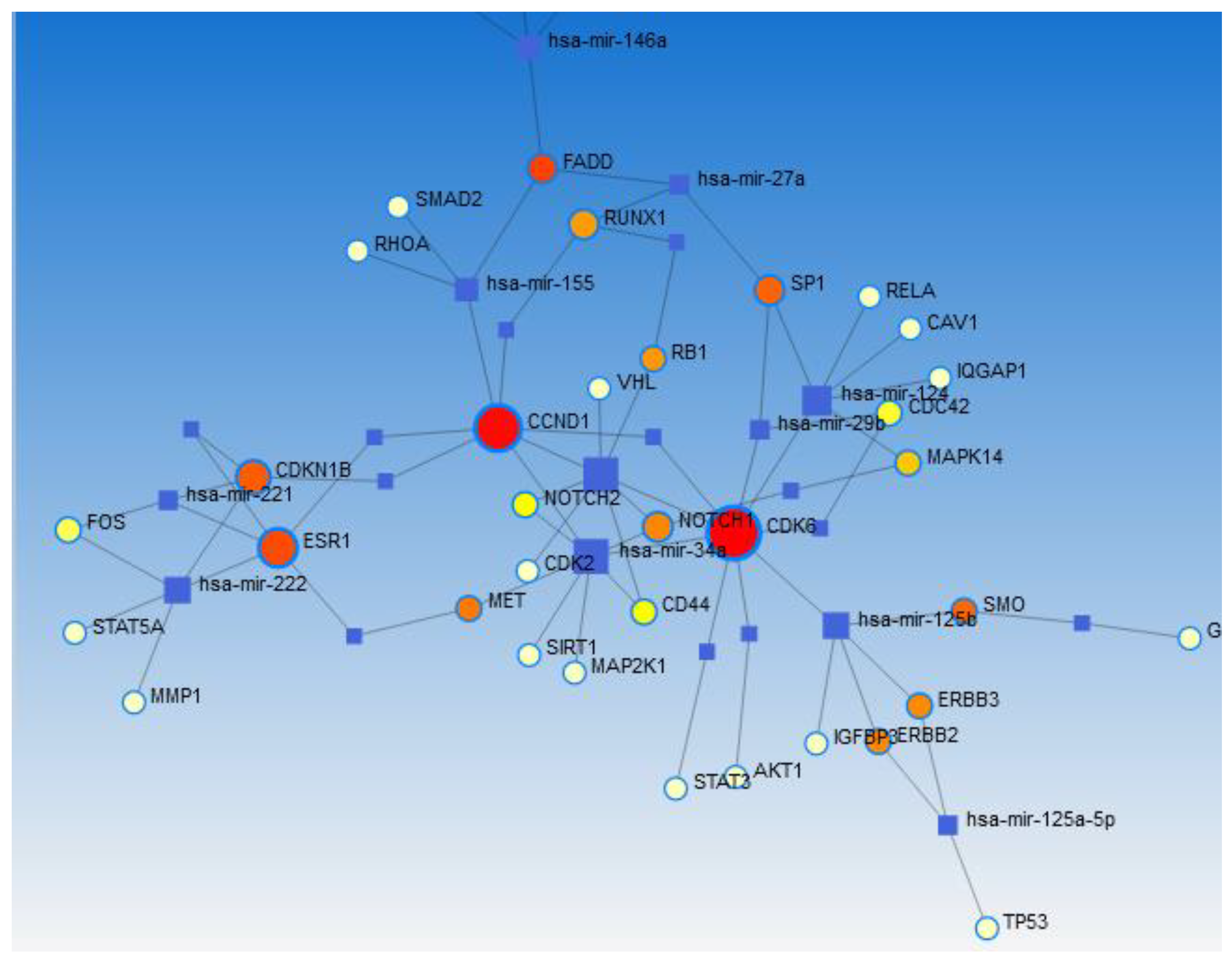
To figure out, physiological functions, as in put, all shared genes within 2 classes were used, our results by using signaling network, Akt1 showed high degree centrality and connected to PI3K pathway and yelled its proteins family (Figure 9). Nevertheless, when we utilized gene regularity network (GRN) Aug 2020, to predict miRNA, our findings for instance, revealed number of stemness proteins and non stemness interact with each other(Figure 10).
Our result after genes application provided significant pathways related to cancer (Table7). Striking finding, enrichment analysis of gene/disease association revealed that, most of shared genes per 3 samples in 2 classes are related to cancers development, among which is meningioma brain tumor, white test was used to define p value (Table 8).
Table 7. Enriched pathways and p value in mutated shared genes per 3 meningioma samples.
Pathway P value
Pathways in cancer 1.61E-70
Proteoglycans in cancer 2.83E-40
ErbB signaling pathway 2.14E-28
Focal adhesion 6.22E-28
MAPK signaling pathway 2.03E-27
Prostate cancer 1.71E-26
Pancreatic cancer 0.02E-25
Breast cancer 1.21E-25
Colorectal cancer 2.4E-25
Glioma 4.72E-20
Wnt signaling pathway 8.79E-18
MicroRNAs in cancer 2.03E-17
PI3K-Akt signaling pathway 9.42E-17
Signaling pathways regulating pluripotency of stem cells 5.32E-11
mTOR signaling pathway 9.57E-08
Hedgehog signaling pathway 4.05E-07
Phosphatidylinositol signaling system 5.85E-06
P value is written using White test.
Table 8. Disease in shared genes in 2 classes per 3 samples Enrichr.
Disease Name P value
Malignant neoplasm of thyroid 2.060e-20
Ovarian Carcinoma 2.770e-26
Transitional Meningioma 0.00001274
Rhabdomyosarcoma 1.382e-18
Neuroectodermal Tumors 9.353e-8
GlioblastomaMultiforme 2.024e-22
Endometrial Neoplasms 1.741e-15
Sphenoid Wing Meningioma 0.00001588
Clear Cell Meningioma 0.00001588
Squamous cell carcinoma of the head and neck 1.458e-22
Carcinoma of bladder 2.235e-23
Malignant Peripheral Nerve Sheath Tumor 4.794e-14
P value is written using White test.

PCR and Sanger Sequencing Results of Genes of Centrality (CD44 and Akt1)

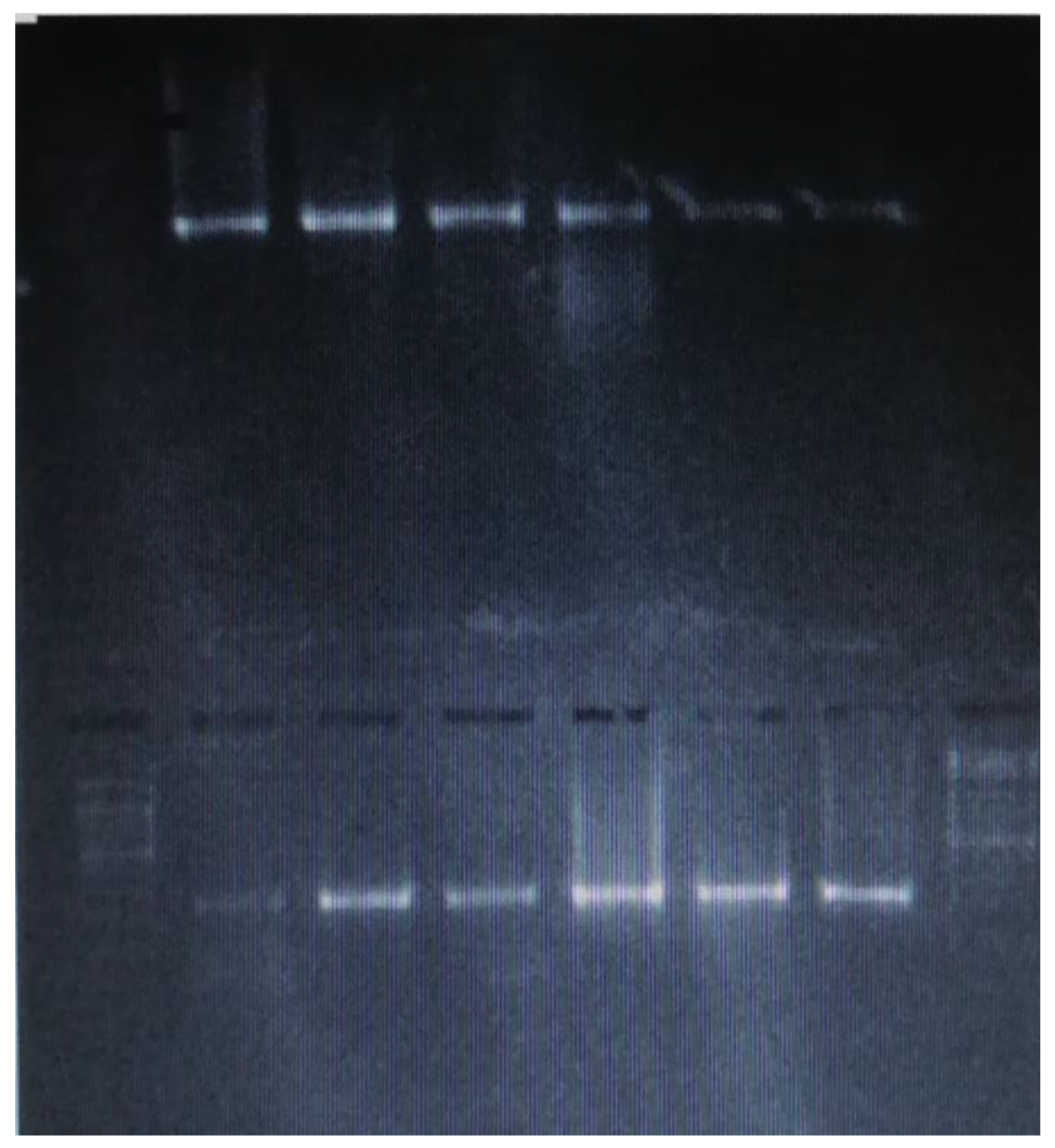
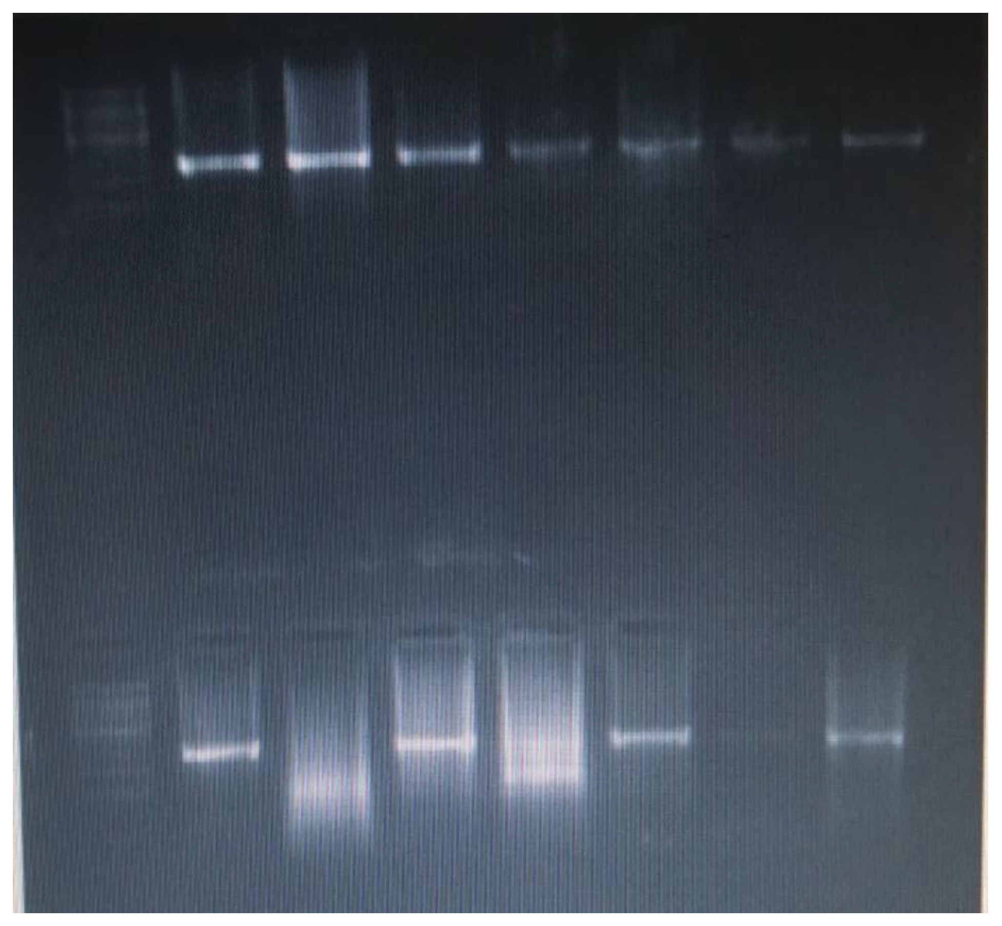
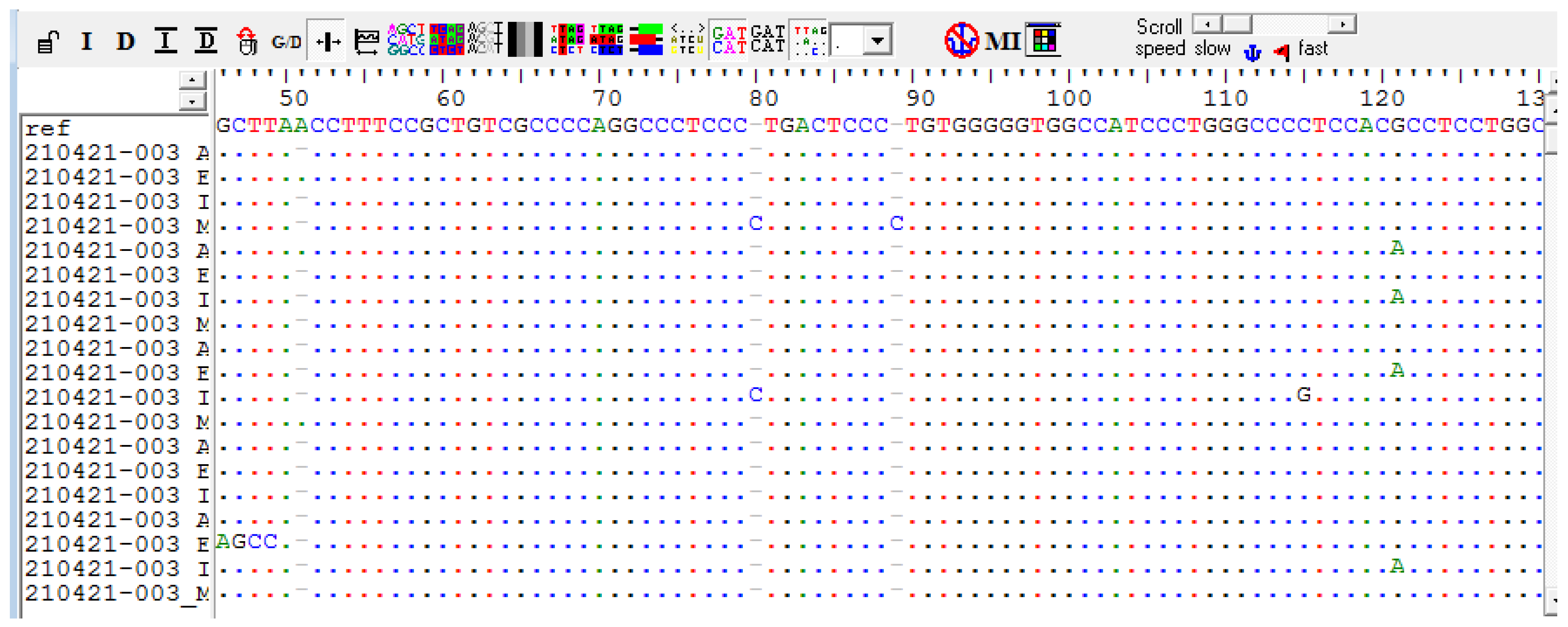
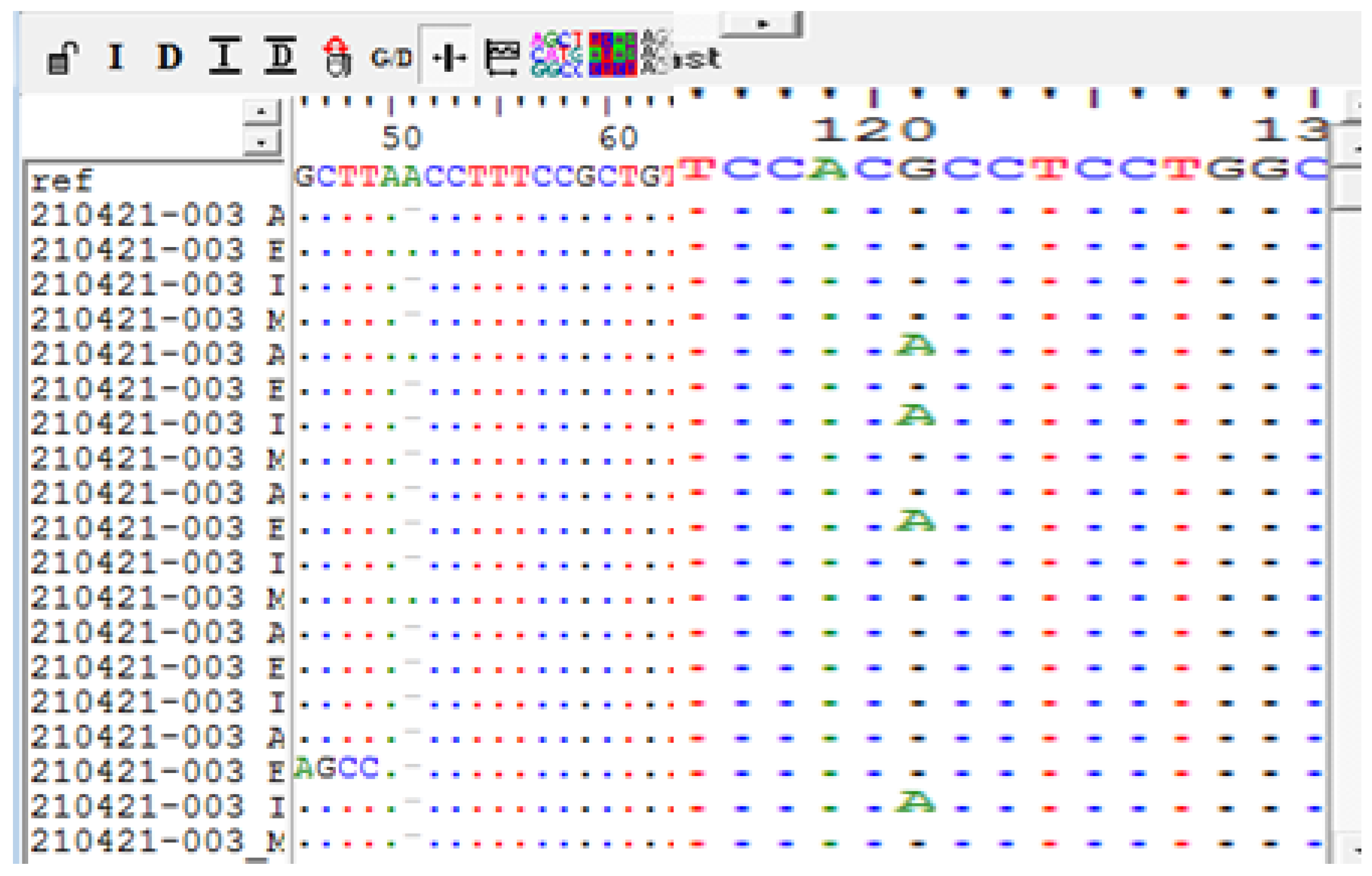
In the present study, 20 meningioma samples (11 fibrous, 4 meningiothelial, 2 atypical and 3 clear cell meningioma) were amplified for Akt1 and CD44 genes, 40 PCR product (20 Akt1 and 20 CD44) were analyzed by standard sequencing, the findings of Akt1 gene showed that, the rs17846829 was detected in 20% of the samples (2 fibrous and 2 atypical meningioma), this mutation (rs17846829) was found in sample 1B (fibrous meningioma) only among our WES data.In addition to that, deletion A at position (chr14:105235886_105235886delT) in Akt1 was detected in 85% of our samples (8 fibrous, 4 meningiothelial, 3 atypical and 2 clear cell meningioma), moreover rs58565216 was seen in 63% of the fibrous meningioma.
The findings of CD44 gene revealed that, A>G (rs9666607) was detected in all our meningioma samples (100%), interestingly, this mutation A>G (rs9666607) was sharedper 3 samples ofour WES data, frame shift mutation (D419Tfs*61) was detected in 45% of the samples (6 fibrous, 2 clear cell and 1 meningiothelial meningioma) (Table 9 and Figure 11, Figure 12, Figure 13a, Figure 13b, Figure 14, 15a, 15b).

Discussion:

Meningioma is the most common brain tumor in Sudan and mostly reported in female, [3] similarly, in this current study, there was clear cut towards female predominance. Most of these tumors are slowly growing and associated with symptoms of gradual increase in intracranial pressure, in this context, headache was the main cardinal feature among our patients. Understanding of the normal development of the nervous system has noticeably improved in recent years, it has a complex differentiation hierarchy ranging from a neural stem cell that can give rise to all of the major lineages in the brain parenchyma to lineage committed progenitors that have a more restricted differentiation potential to terminally differentiated cells. Stem cell concepts can influence the understanding of brain cancer. The cancer stem cell hypothesis proposes that established tumors consist of a cellular hierarchy with a subpopulation of tumor cells able to maintain and propagate the tumor. In this current study, thousands of mutations were predicted by whole exome sequencing analysis, of these high C>T of transversions mutations followed by G>A were observed per fibrous, meningiothelial and fibrous recurrence, our results goes with international results, that different cancer type have different mutational signature similar to gastric cancer in which the prevalence of C>T and C>A transversions were high.[19]What is more, among our samples, numerous mutations that harboring deleterious poly phen 2 effects were found, which highlight the genetic complexity on this type of brain tumor. Are these thousands mutation are being claimed with development of tumorigenesis process on this type of brain tumor, a question is not merely answered. Despite the similarity, further investigations are considered necessary in our study to investigate prospective functions.
In recent years, the availability of large-scale network and genomic data sets allow studying the association between the position of proteins within molecular networks and their patterns of molecular evolution,[20] the consequence of these studies have shown the individual genes are affected by the position that their encoded products occupy in molecular networks. Genes acting at the center of protein–protein interaction networks and metabolic networks evolve under higher levels of purifying selection than those acting at fundamentals of a network periphery, and those genes are so named genes/ proteins of Centrality. Hence Protein interactions play important roles in vital biological processes such as cell cycle control, metabolic and signaling pathways and disease pathways. These interactions can be represented as complex networks, where the nodes are the proteins and the edges represent the interactions between the pairs of proteins they connect.[21]
With regard to the potential of our datasets to guide precision analysis, genes/ protein of centrality are Akt1, UBC and numerous stemness genes like CD44, which emphasize the significance of network.The significance of Akt1 gene encodes one of the three members of the human Akt serine-threonine protein kinase family which are often referred to as protein kinase B alpha, beta, and gamma. These proteins are phosphorylated by phosphoinositide 3-kinase (PI3K), AKT/PI3K forms a key in component of many signaling pathways that involve the binding of membrane-bound ligands resembling receptor tyrosine kinases, G-protein coupled receptors, and integrin-linked kinase. Therefore, these Akt proteins regulate wide variety of cellular functions of which cell proliferation, survival, metabolism, and angiogenesis together in normal and malignant cells, this is along with, Akt proteins are recruited to the cell membrane by phosphatidylinositol 3, 4, 5-trisphosphate (PIP3, 4, 5).Mutations in this gene are associated with multiple types of cancer like breast, colorectal, and ovarian cancers.[22]
UBC genewhich is located on 12q24.3, consisting of 2 exons,the promoter of it contains heat shock element which renders and mediates UBC stimulation upon stress. It also plays key role in maintaining cellular ubiquitin level under stress condition.[23]
Ubiquitin (Ub), was first identified by Gideon Goldstein et al. in 1975 and further confirmed over the next several decades in the human genome. It is highly conserved regulatory protein containing 76 amino acids, can be covalently tagged to target proteins via cascade of enzymatic reactions and regulates numerous physiological and pathological functions.[24,25]
Moreover, this gene is associated with various signaling pathways, of which important nutrient and environmental stimulus. The amino acid plays critical role in the mechanistic target of rapamycin complex 1 (mTORC1) signaling pathway. [26,27] In cancerous stem cell, it regulates and maintains stemness of genes expression likewise Oct4, Sox2 and Nanog.[28]It also plays fundamental role in CSC characteristics, such as self-renewal, maintenance, differentiation and tumor-genesis.[29]
Notably, this gene is up regulated in several cancer, meanwhile the role of UBC in meningioma should be addressed, in this current study, UBC per 2 classes is mutated and represented gene of centrality within each class individually, however, it is not the case, when all shared being the case.
Tumor is the disease which is characterized by multiple phenotypes of which is abnormal cell growth and miscellaneous biological progression. The interaction of biological entities of genes, proteins and metabolites is of great interest in cancer development, as shown in our results, signaling pathway that yelled protein of Akt family. Recently, our results on using next generation sequencing, identified multiple genetic signaling pathways of which is Pathways in cancer, Proteoglycans in cancer and PI3K-Akt signaling pathway.
It is known that signaling pathways control cell cycle, however, the mechanism and interactions among these pathways vary between individual tumor and tumor types. For instance, our results among meningioma histological subtypes, taking into account pathways, more than 10 pathways are significantly co expressed and have been compared in a number of pathway data base. Interestingly, these pathways are of considerable similarity between our 3 meningioma sample, and this might denote genetic resemblance, since most of input genes did not show variation. Furthermore, our exome analysis highlight number of pathways that related to cancer development; of these, proteoglycan pathway which provided connection between cell membrane and surrounding extra cellular membrane, thus playing vital role in regulating cell cancer, adhesion and migration. Another detected pathway in our sample was miRNA signaling pathway, miRNAs are family of small non coding RNAs which regulate wide range of biological processes that encoding carcinogenesis, and thus have been known dysregulated in cancer. Generously, abnormal expression of miRNA in tumor, it believed that this tumor dysregulation might affect several cancers hallmarks on other words, initiation, and progression. Besides, functionally these miRNAs incorporate into cell cycle proliferation pathway, and the dysregulated miRNAs are accountable for evading growth suppresser and hence sustaining proliferating signaling pathway in cancer.
Nevertheless, Akt pathway is intracellular pathway promoted metabolism, proliferation, cell survival growth and angiogenesis in response to extracellular signals. Yet, this pathwayregulates different cell functions and play conservative role through mTORcomplex.
Remarkably, stemness is regarded as the key factor of carcinogenesis and resistance to chemotherapy. [30]Although the cancer stem cells were yet to be isolated from meningioma, cancer stem cell and embryonic stem cell markers have been widely identified from meningioma and strongly associated withpatients’ outcomes. [31]
The majority of exome analysis focused on analyzing cancer etiology depending on the mutations, however with no or little look over nature of these genes, few of these studies questioned stem cell genes.
Outstandingly, from our results, stem cell genes were mutated equally per our 3 meningioma samples which might suggest that, cancerous stem cell were probably drive the force of meningioma development, and taken together the distinct histological variant of meningioma ( fibrous, meningiothelial and fibrous recurrence) which are the most common variant, suggesting the biological behavior other than recurrence. In absolute numbers of meningioma, most recurrence meningiomaare corresponded to histological benign variant (WHO grade1), however, their potential aggressive behavior still need to be evaluated.
Various cancers are associated with mutations on Akt1 gene, and some mutations have been associated with the meningioma recurrence. Interestingly, in this current study, Akt1 has shown number of mutations among 20 meningioma variants. While Akt1 plays role in modulating synaptic plasticity and signal transduction pathways,[32] deficiency in Akt1 may lead to abnormal prefrontal cortical structure and deficits in cognitive functions and risk for schizophrenia.[33]This might be claimed to behavioral changes among our patients with this kind of brain tumor.
Overall, our finding implicated rs17846829 which was seen in fibrous and atypical meningioma variant and rs58565216 that was seen within the fibrous variant might explain cognitive decline in meningioma patients. A recent robust study using array comparative genomic hybridization (aCGH), exposed that, identification of SMO and Akt1 in meningioma increased the possibility of targeting therapy among meningioma patients.[34]Another German a study, concluded that, mutation of Akt1 in skull base meningioma designate shorter time of recurrence and activate mTOR and ERK1/2 signaling and targeting mTOR pathway might be of benefit to patients with recurrent skull base meningioma.[35] Furthermore, case study from Switzerland exhibited presence of activated mutations of Akt1in sub-population of meningioma by using sequencing, and culturing of meningioma discovered sensitivity to AZD5363, which is a selective inhibitor to kinase activity. Thus clinical therapy of AZD5363 was given to the patients for tumor control. [36] Our study promotes the view, that mutations in Akt1 denote recurrence and might be future challenges in clinical therapy; however, big data might be needed to consolidate our findings. Moreover, CD44 A>G (rs9666607 mutation was detected in all our tumor samples, this mutation is located within stretch of residues annotated in UniProt as a distinct region, and the differences in amino acid properties can disturb this region and eventually disturb its function. Furthermore, this mutation is located in a region with known splice variants, and is described K > R (in dbSNP: rs9666607). Despite the mutant residue among the observed residue types at this position in other homologous sequences, and sometimes suggests that, this variant was not damaging for the protein’s structure and function, however, this mutation was already established as having harmful effect.[37]In one study which analyzed SNPs within stemness-related genes for association with overall survival in the prostate, lung, colorectal and ovarian cancer screening and thus identified SNPs of CD44 (rs9666607)were evaluated for association with gene expression.[38]In light of the above this rs9666607 in CD44among our meningioma samples was detected in CD44 involves with Akt1 gene in a proteoglycan pathway in cancer.[39]Nevertheless, CD44 frame shift mutation (D419Tfs*61) in our samples, which is located within stretch of residues annotated in UniProt as special region consequently differences in amino acid properties can disturb this region and disturb its function, and as well this mutation showed difference in charge between the wild-type and mutant amino acid. Additionally, the charge of the wild-type residue was lost, this be able to cause loss of interactions with other molecules or residues.[40]

Conclusion

Nevertheless, the present study, highlight how such complex molecular processes might contribute to tumorogenesis, in attempt to shed light over thousands of genes and multifaceted network analysis rather than a solitary gene which is challenging question.

Funding

This research was self-funded.

Conflict of interest

Authors declare there is no conflict of interest.

References

  1. Baldi I, Engelhardt J, Bonnet C, Bauchet L, Berteaud E, Grüber A, Loiseau H. Epidemiology of meningiomas. Neurochirurgie. 2018 Mar 1;64(1):5-14. [CrossRef]
  2. Holleczek B, Zampella D, Urbschat S, Sahm F, von Deimling A, Oertel J, Ketter R. Incidence, mortality and outcome of meningiomas: a population-based study from Germany. Cancer epidemiology. 2019 Oct 1;62:101562. [CrossRef]
  3. Mohamed Abdel RahmanArbab, Sawsan.A.H.Aldeaf, Lamyaa. A.Elhasan, AhmedElhasan, Cranial meningioma in Sudanese patients: clinical and pathological characteristic. International Journal of Research (IJR) 2014; Vol-1, Issue-7 ISSN 2348-684.
  4. van Alkemade H, de Leau M, Dieleman EM, Kardaun JW, van Os R, Vandertop WP, van Furth WR, Stalpers LJ. Impaired survival and long-term neurological problems in benign meningioma.Neuro-oncology. 2012 May 1;14(5):658-66. [CrossRef]
  5. Wen PY, Quant E, Drappatz J, Beroukhim R, Norden AD. Medical therapies for meningiomas.JNeurooncol.2010;99(3):365–378. [CrossRef]
  6. Moazzam AA, Wagle N, Zada G. Recent developments in chemotherapy for meningiomas: a review. Neurosurg Focus. 2013; 35(6):E18. [CrossRef]
  7. Choy W, Kim W, Nagasawa D, Stramotas S, Yew A, Gopen Q, Parsa AT, Yang I. The molecular genetics and tumor pathogenesis of meningiomas and the future directions of meningioma treatments.Neurosurgical focus. 2011 May 1;30(5):E6. [CrossRef]
  8. AbdElmontalab FY, Fadl EI, Abushama HM, Kreskowski K, Liehr T. Molecular cytogenetic study of the NF2 gene deletion in meningioma in sudanese patients. Balkan journal of medical genetics: BJMG. 2013 Dec;16(2):29. [CrossRef]
  9. Evans JJ, Jeun SS, Lee JH, Harwalkar JA, Shoshan Y, Cowell JK, Golubic M. Molecular alterations in the neurofibromatosis type 2 gene and its protein rarely occurring in meningothelialmeningiomas. Journal of neurosurgery. 2001 Jan 1;94(1):111-7.
  10. Schmitz U, Mueller W, Weber M, Sevenet N, Delattre O, vonDeimling A. INI1 mutations in meningiomas at a potential hotspotin exon 9. Br J Cancer 2001;84:199–201.
  11. Lamszus K. Meningioma pathology, genetics, and biology. Journal of Neuropathology & Experimental Neurology. 2004 Apr 1;63(4):275-86.
  12. Malmer BS, Feychting M, Lönn S, Lindström S, Grönberg H, Ahlbom A, Schwartzbaum J, Auvinen A, Collatz-Christensen H, Johansen C, Kiuru A. Genetic variation in p53 and ATM haplotypes and risk of glioma and meningioma. Journal of neuro-oncology. 2007 May;82(3):229-37. [CrossRef]
  13. Gassoum A, Arbab MA, Aldeaf SA, Elhassan LA, Elhassan AM. P53 Gene Allele Frequency in Linguistic Affiliation of Different Sudanese Tribes.International Journal of Research.2014;1:880-5.
  14. Hilton DA, Shivane A, Kirk L, Bassiri K, Enki DG, Hanemann CO. Activation of multiple growth factor signalling pathways is frequent in meningiomas. Neuropathology. 2016; Jun;36(3):250-61. [CrossRef]
  15. Mu J, Hui T, Shao B, Li L, Du Z, Lu L, Ye L, Li S, Li Q, Xiao Q, Qiu Z. Dickkopf-related protein 2 induces G0/G1 arrest and apoptosis through suppressing Wnt/β-catenin signaling and is frequently methylated in breast cancer. Oncotarget. 2017 Jun 13;8(24):39443.
  16. Ali MM. Aberrant expression of β-catenin in invasive ductal breast carcinomas.Journal of the Egyptian National Cancer Institute. 2009 Jun 1;21(2):185-95.
  17. Mao J, Fan S, Ma W, Fan P, Wang B, Zhang J, Wang H, Tang B, Zhang Q, Yu X, Wang L. Roles of Wnt/β-catenin signaling in the gastric cancer stem cells proliferation and salinomycin treatment. Cell death & disease. 2014 Jan;5(1):e1039-. [CrossRef]
  18. Wu K, Ma L, Zhu J. miR-483-5p promotes growth, invasion and self-renewal of gastric cancer stem cells by Wnt/β-catenin signaling. Molecular medicine reports. 2016 Oct 1;14(4):3421-8.
  19. Nagarajan N, Bertrand D, Hillmer AM, Zang ZJ, Yao F, Jacques PÉ, et al. Whole-genome reconstruction and mutational signatures in gastric cancer. Genome Biol. 2012; 13: R115. [CrossRef]
  20. Davila-Velderrain J, Servin-Marquez A, Alvarez-Buylla ER. Molecular evolution constraints in the floral organ specification gene regulatory network module across 18 angiosperm genomes. Molecular biology and evolution. 2014 Mar 1;31(3):560-73. [CrossRef]
  21. Mathivanan S, Periaswamy B, Gandhi TK, Kandasamy K, Suresh S, Mohmood R, Ramachandra YL, Pandey A. An evaluation of human protein-protein interaction data in the public domain.BMC bioinformatics. 2006 Dec;7(5):1-4. [CrossRef]
  22. Carpten JD, Faber AL, Horn C, Donoho GP, Briggs SL, Robbins CM, Hostetter G, Boguslawski S, Moses TY, Savage S, Uhlik M. A transforming mutation in the pleckstrin homology domain of AKT1 in cancer.Nature. 2007 Jul;448(7152):439-44. [CrossRef]
  23. Bianchi M, Giacomini E, Crinelli R, Radici L, Carloni E, Magnani M. Dynamic transcription of ubiquitin genes under basal and stressful conditions and new insights into the multiple UBC transcript variants. Gene. 2015 Nov 15;573(1):100-9. [CrossRef]
  24. Clague MJ, Heride C, Urbé S. The demographics of the ubiquitin system.Trends in cell biology. 2015 Jul 1;25(7):417-26. [CrossRef]
  25. Husnjak, K. &Dikic, I. Ubiquitin-binding proteins: decoders of ubiquitin-mediated cellular functions. Annu. Rev. Biochem.2012; 81, 291–322.
  26. Jewell JL, Russell RC, Guan KL. Amino acidsignaling upstream of mTOR.Nature reviews Molecular cell biology. 2013 Mar;14(3):133-9.
  27. Saxton RA, Sabatini DM. mTOR signaling in growth, metabolism, and disease. Cell. 2017 Mar 9;168(6):960-76.
  28. Buckley SM, Aranda-Orgilles B, Strikoudis A, Apostolou E, Loizou E, Moran-Crusio K, Farnsworth CL, Koller AA, Dasgupta R, Silva JC, Stadtfeld M. Regulation of pluripotency and cellular reprogramming by the ubiquitin-proteasome system. Cell stem cell. 2012 Dec 7;11(6):783-98. [CrossRef]
  29. Buckley SM, Aranda-Orgilles B, Strikoudis A, Apostolou E, Loizou E, Moran-Crusio K, Farnsworth CL, Koller AA, Dasgupta R, Silva JC, Stadtfeld M. Regulation of pluripotency and cellular reprogramming by the ubiquitin-proteasome system. Cell stem cell. 2012 Dec 7;11(6):783-98. [CrossRef]
  30. Malta TM, Sokolov A, Gentles AJ, Burzykowski T, Poisson L, Weinstein JN, Kamińska B, Huelsken J, Omberg L, Gevaert O, Colaprico A. Machine learning identifies stemness features associated with oncogenic dedifferentiation. Cell. 2018 Apr 5;173(2):338-54. [CrossRef]
  31. Shivapathasundram, G.;Wickremesekera, A.C.; Brasch, H.D.; Marsh, R.; Tan, S.T.; Itinteang, T. Expression of Embryonic StemCell Markers on the Microvessels of WHO Grade I Meningioma. Front. Surg. 2018; 5, 65.
  32. Li M, Dai FR, Du XP, Yang QD, Zhang X, Chen Y. Infusion of BDNF into the nucleus accumbens of aged rats improves cognition and structural synaptic plasticity through PI3K-ILK-Akt signaling. Behavioural brain research. 2012 May 16;231(1):146-53. [CrossRef]
  33. Balu DT, Carlson GC, Talbot K, Kazi H, Hill-Smith TE, Easton RM, Birnbaum MJ, Lucki I. Akt1 deficiency in schizophrenia and impairment of hippocampal plasticity and function. Hippocampus. 2012 Feb;22(2):230-40. [CrossRef]
  34. .Brastianos PK, Horowitz PM, Santagata S, Jones RT, McKenna A, Getz G, Ligon KL, Palescandolo E, Van Hummelen P, Ducar MD, Raza A. Genomic sequencing of meningiomas identifies oncogenic SMO and AKT1 mutations. Nature genetics. 2013 Mar;45(3):285-9. [CrossRef]
  35. Yesilöz Ü, Kirches E, Hartmann C, Scholz J, Kropf S, Sahm F, Nakamura M, Mawrin C. Frequent AKT1 E17K mutations in skull base meningiomas are associated with mTOR and ERK1/2 activation and reduced time to tumor recurrence. Neuro-oncology. 2017 Aug 1;19(8):1088-96.
  36. Weller M, Roth P, Sahm F, Burghardt I, Schuknecht B, Rushing EJ, Regli L, Lindemann JP, Von Deimling A. Durable control of metastatic AKT1-mutant WHO grade 1 meningothelialmeningioma by the AKT inhibitor, AZD5363. Journal of the National Cancer Institute. 2017 Mar 1;109(3):djw320.
  37. Boehm M and Slack FJ: microRNA control of lifespan and metabolism. Cell Cycle. 5:837–840. 2006.
  38. Freedman JA, Wang Y, Li X, Liu H, Moorman PG, George DJ, Lee NH, Hyslop T, Wei Q, Patierno SR. Single-nucleotide polymorphisms of stemness genes predicted to regulate RNA splicing, microRNA and oncogenic signaling are associated with prostate cancer survival. Carcinogenesis. 2018 Jul 3;39(7):879-88. [CrossRef]
  39. Iozzo RV, Sanderson RD. Proteoglycans in cancer biology, tumour microenvironment and angiogenesis.Journal of cellular and molecular medicine. 2011 May;15(5):1013-31.
  40. Venselaar, H., teBeek, T.A.H., Kuipers, R.K.P., Hekkelman, M.L. and Vriend, G. Protein Structure Analysis of Mutations Causing Inheritable Diseases. An e-Science Approach with Life Scientist Friendly Interfaces. BMC Bioinformatics.2010 11, 548. [CrossRef]
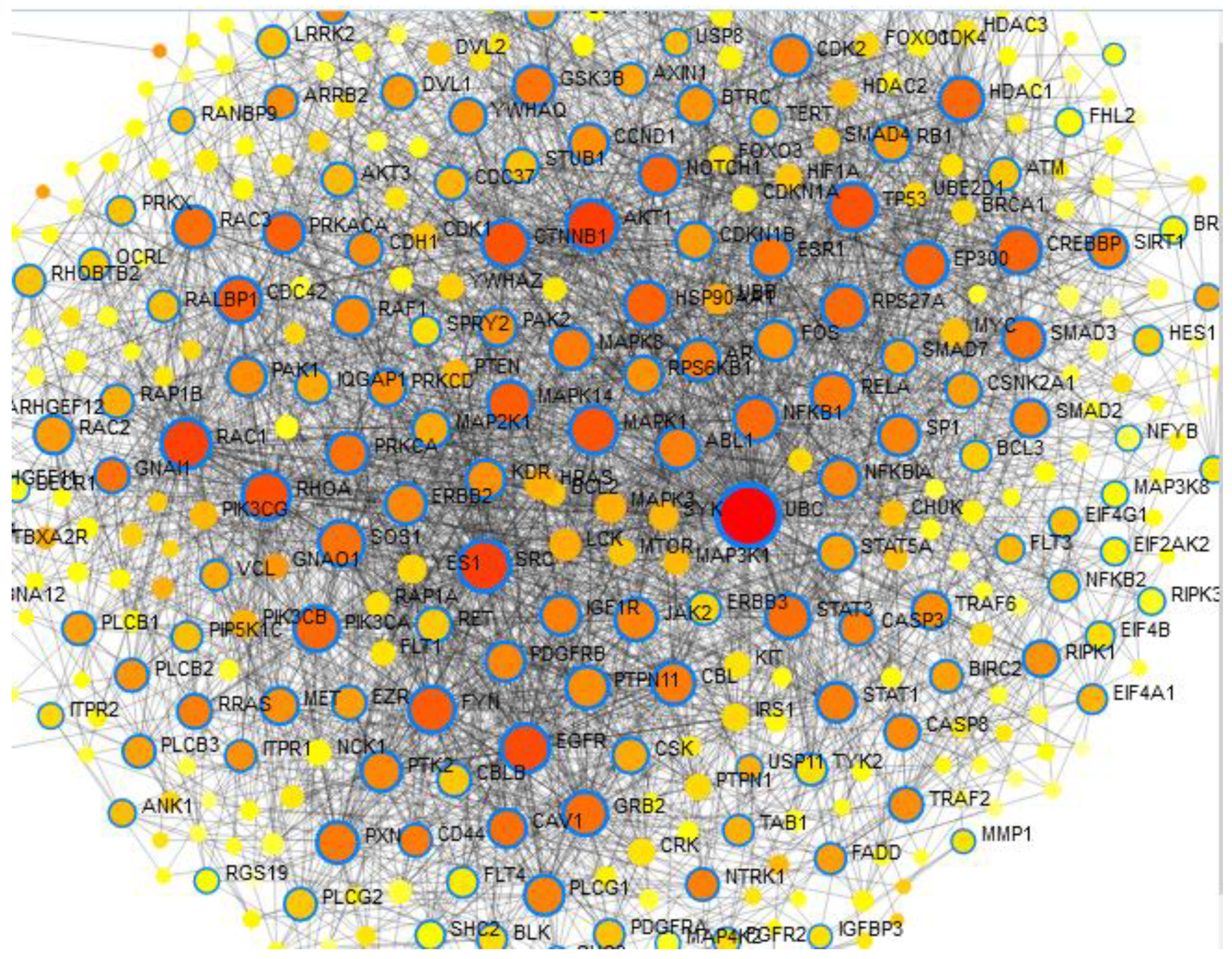
Figure 8a. This figure shows UBC, Akt1 and CD44 network interaction together with other hub genes per 3 samples, input included all shared genes harboring effect on both classes ( SNVs and INDEL), UBC and Akt1 are red in color while CD44 is brown and highlighted with blue circle color, the interacted proteins are present in yellow color.
Preprints 118271 g001
Figure 8b. random selection shows UBC, Akt1 and CD44 network interaction together with other hub genes per 3 samples, input included random selection of shared genes harboring effect on both classes ( SNVs and INDEL), UBC and Akt1 are red in color while CD44 is yellow and highlighted with blue circle color.
Preprints 118271 g002
Figure 9. signaling pathway shows physiological function of shared genes per 2 classes. Akt1 protein and protein family shows high degree centrality.Violet: protein, blue: chemical, green: complex, yellow: protein family, dark brown: small molecule, brown: undefined.
Preprints 118271 g003
Figure 10. Gene regulatory network (GRN) showed Gene-miRNA-interactions. MicroRNA (miRNA)–gene interactions are well-recognized as involved in the progression of almost all cancer types. Red= nodes, purple=miRna.
Preprints 118271 g004
Figure 11. Gel electrophoresis of Akt1gene, lanes 1-6 shows band size 232 bp.
Preprints 118271 g005
Figure 12. Gel electrophoresis of CD44 gene, lanes 1-7 shows band size 286 bp.
Preprints 118271 g006
Figure 13a. alignment of Akt1gene sequencing shows, deletion A at position (chr14:105235886_105235886delT) and G>A (rs17846829).
Preprints 118271 g007
Figure 13b. magnified alignment of Akt1gene sequencing shows, deletion A at position (chr14:105235886_105235886delT) and G>A (rs17846829).
Preprints 118271 g008
Figure 14. Chromogram of Akt1gene sequencing shows, polymorphism C>G (rs58565216).
Preprints 118271 g009
Table 6. number of single nucleotide variations and the prediction effect of SIFT and PolyPhen-2 per 3 samples.
Sample SIFT/Total deleterious
effect
PolyPhen-2/total D PolyPhen-2/total P/D PolyPhen-2/total/B
1B/253SNVs 54 16 10 21
2B/267SNVs 63 18 18 20
4A/242SNVs 48 30 16 25
1B is fibrous type, 2B is meningiothelial and 4A is fibrous recurrence, D=damaging, P/D=probably damaging, B=benign. SIFT score <0.05=deleterious, PolyPhen p=>0. 5 (probably damaging/ damaging).
Table 9. | histopathology, WHO grades, recurrence and mutations of Akt1 and CD44 genes in our meningioma samples.
NO Histopathology variant WHO Grades Recurrence Mutation Akt1 Mutation CD44
1 Meningiothelial 1 NO Del A A>G (rs9666607)
2 Clear cell 11 NO A>G (rs9666607)
D419Tfs*61
InsC
3 Meningiothelial 1 NO Del A A>G (rs9666607)
4 Meningiothelial 1 NO Del A/
InsC
A>G (rs9666607)
5 Fibrous 1 NO G>A (rs17846829) A>G (rs9666607)
D419Tfs*61
6 Clear cell 11 Yes Del A A>G (rs9666607)
7 Fibrous 1 Yes Del A G>A(rs17846829) A>G (rs9666607)
D419Tfs*61
InsC
8 Fibrous 1 Yes Del A A>G (rs9666607)
D419Tfs*61
9 Fibrous 1 Yes Del A A>G (rs9666607)
10 Atypical 11 Yes Del A
G>A (rs17846829)
A>G (rs9666607)
11 Fibrous 1 Yes Del A
InsC
A>G (rs9666607)
InsC
12 Fibrous 1 NO A>G (rs9666607)
D419Tfs*61
13 Fibrous 1 Yes Del A A>G (rs9666607)
14 Fibrous 1 Yes Del A A>G (rs9666607)
D419Tfs*61
15 Fibrous 1 NO Del A A>G (rs9666607)
InsC
16 Fibrous 1 NO A>G (rs9666607)
D419Tfs*61
17 Clear cell 11 Yes Del A A>G (rs9666607)
D419Tfs*61
18 Fibrous 1 Yes Del A A>G (rs9666607)
19 Atypical 11 Yes Del A G>A(rs17846829) A>G (rs9666607)
20 Meningiothelial 1 Yes Del A A>G (rs9666607)
D419Tfs*61
Disclaimer/Publisher’s Note: The statements, opinions and data contained in all publications are solely those of the individual author(s) and contributor(s) and not of MDPI and/or the editor(s). MDPI and/or the editor(s) disclaim responsibility for any injury to people or property resulting from any ideas, methods, instructions or products referred to in the content.
Copyright: This open access article is published under a Creative Commons CC BY 4.0 license, which permit the free download, distribution, and reuse, provided that the author and preprint are cited in any reuse.
Prerpints.org logo

Preprints.org is a free preprint server supported by MDPI in Basel, Switzerland.

Subscribe

© 2024 MDPI (Basel, Switzerland) unless otherwise stated