1. Introduction
Limoncella is the common name for a fruit tree of the Rutaceae family, considered a rare and ancient Mediterranean variety belonging to the
Citrus genus. This family comprises 150 genera with approximately 2.000 species, 70 of which are
Citrus. The taxonomy of
Citrus still stirs up severe doubts in the scientific community because of the intergeneric sexual compatibility, the high frequency of bud mutations, and the long history of cultivation and spread [
1]. Despite the difficulties in establishing a consensual classification of edible citrus, most authors now agree on the origin of cultivated forms. The use of molecular markers and genome sequencing have contributed to identifying four primary taxa:
C. maxima (pummelos),
C. medica (citrons),
C. reticulata (mandarins), and
C. micrantha (a wild Papeda species) [
2].
Citrus species are economically and culturally significant, with their fruits widely consumed fresh, juiced, or used as flavorings in various culinary dishes. Italy is one of the world’s largest producers of citrus fruit and is in second place, after Spain, at the European level [
3]. In 2022, Italy produced 3.09 tons of citrus fruits. The Italian citrus distribution is higher in southern regions, including Sicily and Calabria, with a value above 80%, as reported by ISTAT [
4].
Citrus fruits also have a long history of use in pharmacology due to their rich content of bioactive compounds, particularly flavonoids, essential oils, and vitamin C. These components offer a range of therapeutic benefits and are utilized in various medicinal and health applications. Some species of
Citrus have a broad spectrum of biological activities, including antibacterial, antiviral, antioxidant, antifungal, analgesic, and anti-inflammatory [
5,
6,
7,
8]. Citrus fruit peel (epicarp) is composed of flavedo (exocarp), the pigmented more external region, and albedo (mesocarp), the white layer before the pulp.
Many scientific studies refer to the citrus peel as a whole but must distinguish between the flavedo and albedo layers separately. The peel is often treated as a single entity in research, which can sometimes overlook the unique properties and functions of these individual components. Different studies conducted on various
Citrus species have shown that peels have antioxidant properties, but less is known about the albedo fraction [
9,
10,
11,
12,
13,
14]. It is known that the albedo is characterized by high nutritional value due to the presence of functional compounds: phenolic acid, flavanones, and flavones [
15]. In this study, we thoroughly characterized the flavedo and albedo extracts from sweet citrus popularly named
Limoncella of Mattinata, a location on the Southern Italian coast of the Gargano in the Puglia Region. We investigated properties, including antioxidant and genoprotective activities, to valorize its flavedo and albedo as a natural resource and promote the cultivation of this traditional citrus tree on the verge of extinction.
2. Materials and Methods
2.1. Materials
Cyanidin-3-glucoside chloride, delphinidin-3,5-diglucoside chloride, delphinidin-3-galactoside chloride, petunidin-3-glucoside chloride, malvidin-3-galactoside chloride, quercetin-3-glucoside and kaempferol-3-glucoside were purchased from PhytoLab (Vestenbergsgreuth, Germany). The remaining 31 analytical standards of the 38 phenolic compounds were supplied by Sigma-Aldrich (Milan, Italy). Formic acid (99%) was obtained from Merck (Darmstadt, Germany). Analytical-grade hydrochloric acid (37%) was obtained from Carlo Erba Reagents (Milan, Italy). HPLC-grade methanol was supplied by Sigma-Aldrich (Milano, Italy). Deionized water (> 18 MΩ cm resistivity) was further purified using a Milli-Q SP Reagent Water System (Millipore, Bedford, MA, USA). All solvents and solutions were filtered through a 0.2 μm polyamide filter from Sartorius Stedim (Goettingen, Germany). Before HPLC analysis, all samples were dissolved in methanol and filtered with Phenex™ RC 4 mm 0.2 μm syringeless filter, Phenomenex (Castel Maggiore, BO, Italy). Reagents for sample preparation, antioxidant activity experiments, and genoprotective properties were purchased from Sigma Aldrich. Cell culture materials and reagents were from VWR International (Milan, Italy).
2.2. PCR Amplification of ITS Region
The molecular characterization was performed by amplification and sequencing the ITS (Internal Transcribed Spacer) region. According to the manufacturer's recommendation, the genomic DNA was obtained from 100 mg plant leaf using a QIAamp DNA mini kit (Qiagen, Milan, Italy). Universal primers ITS1F-(5'-TCCGTAGGTGAACCTGCGG-3’) and ITS4 R-(5'-TCCTCCGCTTATTGATATGC-3’), located at 3’ and 5’of 18S and 28 S of ribosomal genes, respectively were employed. PCR reactions were performed in 25 μl volume containing 50 ng of the genomic DNA, 0.4 μM of each of the above primers, 200 μM dNTP’s, 2.5 μl of 10x buffer, and 0.74 U of Taq DNA polymerase (Takara). Reaction conditions were: denaturation at 95 °C for 10 min followed by 35 cycles of 95 °C for 30 sec, 55 °C for 30 sec, and 72 °C for 30 sec, with final extension at 72 °C for 10 min. The amplification products were electrophoresed on 0.8 % agarose TBE gel and stained with ethidium bromide (0.3 μg/ml). PCR products were purified using the Gel Extraction Kit (Qiagen, Milano, Italy). Both strands of the products were sequenced using the pair of primers used in the PCR amplification. Sequences were run on an ABI 3700 automated sequencer, and the obtained sequences were aligned using BLAST as described in
Supplementary Material.
2.3. Light and Transmission Electron Microscopy
For Light and Transmission Electron Microscopy (TEM), the samples were cut into small pieces, fixed in 2.5% glutaraldehyde, and post-fixed in 1% aqueous osmium tetroxide, both in phosphate buffer for one hour. The samples were rinsed in phosphate buffer, dehydrated in an increasing ethanol series (50, 70, 80, 90, 95, 100%, 15 min each), further dehydrated twice with propylene oxide for 15 min, and then incubated in epoxy resin at 60 °C for three days. Semi-thin sections were cut with an ultra-microtome (Ultratome LKB) into 1-mm semi-thin sections and stained with 1% Toluidine blue in distilled water, 0.2% Basic fuchsin in ethanol, Safranin O in 50% alcohol, 0.13 % toluidine blue + 0.02% Azur II. Basic fuchsin, Safranin O, and Azur II staining were performed on etched resin sections with sodium meta-periodate. Different dyes were used to highlight the various parenchyma morphological components. From the same sample block, sections were cut into 70-80 nm ultra-thin sections and placed on 400 mesh grids. Ultra-thin sections were stained for 45 min each with Uranyless followed by 30 min lead citrate and observed by Philips CM10 at 80 kV.
2.4. Scanning Electron Microscopy (SEM) and Environmental Scanning Electron Microscopy (ESEM)
Sample preparation for conventional SEM analysis involved fixation in 2.5% glutaraldehyde (Sigma-Aldrich) for 1 h at 4 °C and dehydration using 50%, 70%, 80%, 90%, and twice with 100% ethanol (Sigma-Aldrich) for 5 minutes at each concentration. The samples were treated with Hexamethyldisilazane (HMDS), transferred on aluminum stubs, and sputter-coated with an S-thin layer of gold, approximately 10 nm. Images were acquired using a Philips 515. ESEM technology represents an upgrade of the conventional scanning electron microscope (SEM), allowing the observation of samples at different vacuum levels, either prepared according to the conventional SEM method or natural, without dehydration or conductive coating. The latter opportunity is advantageous for biological samples, allowing morphological analysis without pre-treatment before observation. If equipped with a spectrometer for the dispersion of energy (EDS), ESEM allows the semiquantitative detection of chemical elements constituting the ultrastructural components of the sample through point or areal analysis. An FEI Quanta 200 FEG Environmental Scanning Electron Microscope (FEI, Hillsboro, OR, USA) was used with an energy-dispersive X-ray spectrometer (EDAX Inc., Mahwah, NJ, USA). The analyses used a focalized electron beam in a vacuum electron gun pressure of 5.0 e-6 bar. The ESEM was utilized in low vacuum mode, with a specimen chamber pressure set at about 0.80 mbar, an accelerating voltage of 25 kV, a working distance of 10 mm, a tilt angle of 0, a variable beam diameter, and a magnification between 100 and 40,000 x. The images were obtained utilizing the back-scattered electron detector to highlight the presence of particles or aggregates or the secondary electron detector to highlight the morphological features. The spectrometric unit has an ECON (Edax Carbon Oxygen Nitrogen) 6 UTW X-ray detector and Genesis Analysis software. Each sample was analyzed with a time count of 100 sec and an Amp Time of 51, while the probe current was 290 μA.
2.5. Preparation of Albedo and Flavedo Extracts
Albedo and flavedo of Limoncella lemons from Mattinata were separated and subject to drying at 60 °C overnight. After drying, albedo and peel were triturated in a mortar with liquid nitrogen. This procedure was repeated three times to reduce samples in a fine powder. Subsequently, 3 g of ground flavedo and albedo samples were extracted with 30 ml of 80% ethanol/water (80:20 v/v) at 4 °C under stirring. The albedo and flavedo extracts were then centrifuged at 400 g for 15 min, and the residues were extracted using two additional portions of the ethanol/water mixture. All supernatants were pooled, concentrated, and kept at -20 °C until use.
2.6. Quantification of Total Phenolics by Folin-Ciocalteu Method
Total polyphenol content was determined using the Folin-Ciocalteu method described by Singleton et al. [
16]. The total volume of the reaction mixture was minimized to 1 ml. The extract solutions (10 μl) were mixed with Folin-Ciocalteu reagent (50 μl) and deionized water (90 μl). Three minutes later, 300 μl of 20% (w/v) sodium carbonate was added, and the mixture was brought up to 1 ml with distilled water. The tubes were vortex-mixed for 15 sec and allowed to stand for 30 min at room temperature for color development. Absorbance was then measured at 725 nm using a UV Beckman spectrophotometer. The amount of total phenolics was expressed as caffeic acid equivalents through the calibration curve of caffeic acid. The calibration curve ranged from 1 to 15 μg/ml (R
2 = 0.9973).
2.7. HPLC-ESI-MS/MS Analysis
HPLC-MS/MS studies were performed using an Agilent 1290 Infinity series and a Triple Quadrupole 6420 from Agilent Technology (Santa Clara, CA) equipped with an electrospray ionization (ESI) source operating in negative and positive ionization modes by following a previously published method [
17,
18,
19]. The separation of target compounds was achieved on a Synergy Polar–RP C18 analytical column (250 mm x 4.6 mm, 4 µm) from Phenomenex (Chesire, UK). The column was preceded by a Polar RP security guard cartridge (4 mm x 3 mm ID). The mobile phase was a mixture of water (A) and methanol (B), with formic acid 0.1% at a flow rate of 0.8 ml min
−1 in gradient elution mode. The composition of the mobile phase varied as follows: 0–1 min, isocratic condition, 20% B; 1–25 min, 85% B; 25–26 min, isocratic condition, 85% B; 26–32 min, 20% B. The injection volume was 2 μl. The column temperature was 30 °C, and the drying gas temperature in the ionization source was 350 °C. The gas flow was 12 l/min, the nebulizer pressure was 55 psi, the capillary voltage was 4000 V, and a gas flow rate was maintained at 1,2000 ml/min. Detection was performed in the “dynamic-multiple reaction monitoring” (dynamic-MRM) mode and the dynamic-MRM peak areas were integrated for quantification. The most abundant product ion was used for quantitation, and the other for quantification. The selected ion transitions and the mass spectrometer parameters, including the specific time window for each compound (Δ retention time), are reported in
Table S2 (
Supplementary Materials).
2.8. Antioxidant Activity
2.8.1. DPPH (2,2-Diphenyl-1-picrylhydrazyl) Assay
The DPPH radical scavenging assay was conducted using the procedure previously described by Saltarelli et al. [
20]. Fresh DPPH ethanol solution (850 µl, 100 μM) was mixed with the sample at different concentrations (150 µl, 0.2-3.5 mg/ml). The decreased absorbance at 517 nm was recorded after 10 min at room temperature. The scavenging activity was calculated as % = [(A
0 – A)/A
0] × 100, where A
0 is the absorbance of the control reaction, and A is the absorbance in the presence of samples. The EC
50 value was calculated from the plots as concentration extracts required to provide 50% free radical scavenging activity.
2.8.2. ABTS, 2,2′-Azino-bis(3-ethylbenzothiazoline-6-sulphonic acid) Assay
Antioxidant activity against ABTS radical was performed as described by Loizzo et al. with some modifications [
21]. Briefly, the reaction mixture was prepared by mixing 7 mM ABTS solution and 2.45 mM potassium persulphate followed by 12–16 h incubation in the dark at room temperature to produce ABTS radical. Before use, the solution was diluted with ethanol to absorb 0.80 ± 0.05 at 734 nm. Aliquots of the sample at concentrations ranging from 0.01 to 0.4 mg/ml were added to 1 ml of ABTS ethanolic solution and incubated in the dark at room temperature for 6 min. The absorbance was then recorded at 734 nm using a UV Beckman spectrophotometer. The ABTS radical scavenging activity was calculated following the equation: ABTS scavenging activity (%) = [(A
734 nm of blank – A
734 nm of the sample)/A
734 nm of blank] × 100. Results are reported as EC
50 values (µg/mL). Trolox was a reference compound (0.5–5 μg/ml).
2.8.3. Lipoxygenase Inhibition Assay
Soybean 5-lipoxygenase was used for the assay, according to Saltarelli et al. [
20]. Inhibition experiments were performed by measuring the loss of 5-lipoxygenase activity (0.18 µg/ml) with 100 µM linoleic acid as the substrate in 50 mM sodium phosphate, pH = 6.8. The reaction mixture was pre-equilibrated at 20 °C for 20 min without the enzyme. Inhibition studies in the presence of various extract concentrations (0.2-0.7 mg/ml) were recorded at 235 nm at 20 °C using a UV Beckman spectrophotometer. The lipoxygenase activity was calculated as % = 100-{[(Δ
235 nm of blank - Δ
235 nm of sample)/Δ
235 nm of blank] x 100}. EC
50 was determined by plotting the graph with the concentration of extracts versus the percentage of inhibition of linoleic acid peroxidation.
2.8.4. Chelating Capacity on Fe2+
Fe
2+ chelating capacity was evaluated as described by Saltarelli et al. [
20] with some modifications. Briefly, an aliquot of 20 μl of FeSO
4 solution (2 mM) was added to 200 μl of sample (range 20-125 μg/ml) and incubated at room temperature for 5 min. After that, an aliquot of 40 μl of ferrozine solution (4 mM) was added to the reaction mixture, and the sample volume was diluted to 1 ml with deionized water, mixed, and incubated for 10 min in the dark at room temperature. The absorbance at 562 nm was then spectrophotometrically determined. The chelating activity was calculated as % = [(A562 nm of blank - A562 nm of the sample)/A
562 nm of blank] × 100. EC
50 is the concentration at which ferrous ions are chelated by 50% and was evaluated by plotting the sample concentration versus the chelating activity.
2.9. DNA Nicking Assay
The DNA nicking assay evaluated the protective effect of hydroalcoholic albedo and Limoncella flavedo extracts against oxidative DNA damage, which employs ferrous ions and dioxygen (Fe
2+ + O
2) to generate DNA strand breaks induced from free radicals [
22]. The assay consists of a cell-free system composed of plasmid DNA (pEMBL8), which resembles the structure of mtDNA [
23]. The hydroalcoholic solutions were sequentially diluted with water to obtain the final concentrations of 250 µg/ml, 125 µg/ml, 63 µg/ml and 31 µg/ml. Each extract was assayed in a final volume of 72 µl, consisting of PBS (Phosphate Buffer Solution), 7 µg/mL of pEMBL8, and 20 µl of the related dilution. The mixtures were added 8 μl of 3 mM FeSO
4 freshly prepared and kept on ice. The tubes were incubated for 10 minutes at 37 °C, and the reaction stopped with 20 µl of loading buffer. The disappearance of the supercoiled form of the plasmid (CCC) was assessed on an ethidium bromide-stained agarose gel electrophoresis followed by quantification using Gel Doc 2000 and Quantity One software (Bio-Rad). In detail, 20 µl of each sample were loaded onto a 1.2% w/v agarose gel in TAE buffer (40 mM Tris-acetate and 1 mM EDTA, pH 8.0), run at 80 V for 30 min in a small electrophoresis chamber (Mini-Sub Cell GT Systems, Bio-Rad). The gel was stained with 0.3 μg/ml ethidium bromide (EtBr). The EC
50 value was calculated by determining the concentration of the compound protecting half of the supercoiled plasmid.
2.10. Cell Cultures
The human keratinocyte cell line HaCaT was obtained from the Interlab Cell Line Collection (ICLC, Genoa, Italy). Cells were grown in DMEM medium supplemented with 10% fetal bovine serum, 2% glutamine, 1% sodium pyruvate, and 100 U/ml penicillin/streptomycin. Cells were maintained in an incubator at 37 °C and 5% CO2. RAW 264.7 murine macrophages were cultured in RPMI 1640 medium with 10%, 2 mM glutamine, and 1% 100 U/ml penicillin/streptomycin and maintained at 37 °C in a 5% CO2 atmosphere.
2.11. Cytotoxicity Assays
The cytotoxic effects of the Limoncella hydroalcoholic albedo and flavedo extract against HaCaT cells were analyzed by WST-8 and sulforhodamine B (SRB) assays, which evaluate cellular metabolic activity and cellular protein content, respectively [
24]. In detail, cells (5 x 10
3/well) were seeded in 96-well plates and treated with water-diluted extracts from 110 to 7 µg/ml. After 24 hours of incubation, the test compounds were removed, and a fresh medium containing WST-8 (Sigma-Aldrich, Milan, Italy) was added to each well. Cells were further incubated at 37 °C for up to 4 hours, and color development was monitored at 450 nm in a microplate reader (Multiskan FC, Thermo Scientific) [
25]. As previously published, the SRB assay was also performed in the same 96-well plate [
26]. Briefly, cells were fixed with cold 50% trichloroacetic acid and stained with 0.4% SRB (Sigma-Aldrich, Milan, Italy) dissolved in 1% acetic acid. The protein-bound dye was subsequently solubilized with 10 mM Tris, and the absorbance was read at 570 nm in a microplate reader (Multiskan FC, Thermo Scientific). The concentration that caused 50% growth inhibition (IC
50) was calculated and the data was expressed as a percentage (%) compared to that of untreated cells (controls).
2.12. Evaluation of Antioxidant Properties by DCFH-DA Assay
The antioxidant properties of albedo and flavedo extracts were analyzed in HaCaT cells using 2′,7′-dichlorofluorescein diacetate (DCFH-DA, Sigma-Aldrich, Milan, Italy), which transforms into 2′,7′-dichlorofluorescein (DCF) highly fluorescent after oxidation [
25]. In detail, cells (1 x 10
4/well) were seeded into black 96-well plates and incubated for 2 h with a concentration of 25 µg/ml extracts in 100 µl DMEM. The medium was then removed and replaced with 50 µl DCFH-DA (5 µM in PBS), incubated for 30 minutes at 37 °C. After the removal of the excess probe, cells were treated with 100 µl of hydrogen peroxide (H
2O
2, 100 μM in PBS) for 30 min, and DCF fluorescence emission was measured at ex/em 485/520 nm in the FluoStar multiwell plate reader Optima (BMG Labtech, Germany). Data were expressed as relative oxidation compared to non-oxidized cells.
2.13. Determination of Nitric Oxide Production
The anti-inflammatory properties of both albedo and flavedo hydroalcoholic extracts were evaluated in RAW 264.7 cells (murine macrophages) stimulated by Lipopolysaccharide (LPS) (Sigma-Aldrich, Milan, Italy). Cells (3 x 10
4/well) were seeded into 96-well plates and treated with both Limoncella extracts (50 µg/mL) in the presence and absence of 1 µg/ml LPS for 24 h. The cells were also incubated alone in the presence and absence of LPS as a control. The drug dexamethasone at a 5 µg/ml concentration was used to validate the test. Subsequently, Nitric Oxide (NO) levels were determined in the supernatant medium using Griess reagent (Sigma-Aldrich, Milan, Italy) [
27]. Absorbance was measured at 570 nm using a plate reader (BioRad Laboratories, Hercules, USA).
4. Discussion
The various species of
Citrus are mainly used in the food and cosmetic fields. However, due to their antioxidant, antimicrobial, and anticancer properties, they have also found applications in the therapeutic field [
28,
29,
30,
31]. Most studies have been performed on the flavedo or the entire peel, while little is known about the beneficial properties of the albedo [
14,
32,
33,
34]. The present work represents an attempt to appreciate this part of the fruit. For the present research, we selected a rare, ancient Mediterranean Citrus variety from Southern Italy to establish its phylogenetic position and appreciate its health properties, thus promoting its cultivation and avoiding extinction. The sequence analysis of the ribosomal internal transcribed spacer regions assigns
Limoncella as a variety of
Citrus medica L. The different cultivars of
C. medica L. are generally divided into “acidic” and “sweet” cedars, and
Limoncella fruits have a sweeter pulp. Morphological approaches allowed the structure and ultrastructure of
Limoncella to be studied by analyzing different cellular components, supporting the correlation between the nutritional characteristics and the structural organization of the
Limoncella [
35] In particular, the morphological evidence obtained utilizing different and complementary approaches allowed us to understand better the albedo’s and flavedo’s components that constitute the “reservoir” of nutritional substances and the parts of them that do not interfere with the extractive procedure. Therefore, all the cellular structures and remnants after the extraction can be considered a resource (in terms of wide-scale industrial extraction) as fertilizer in a fully circular economy of fruit treatment. Moreover, the well-defined morphological
Limoncella characterization represents a comparable model for studying properties similar to those of other citrus fruit types. Concerning the chemical characterization of flavedo and albedo alcoholic extracts, herein, we report that they are rich in polyphenols, quite different in compounds and quantity (
Table 1), although their total polyphenol content fall within the range of those previously reported for 35 cultivars of
Citrus reticulata Blanco [
36] and
Citrus medica L. [
37]. In particular, the Folin-Ciocalteu method (FC) and HPLC-ESI-MS/MS were used to evaluate the polyphenol content. Both methods showed that the total phenol content was comparable in both extracts, indicating that albedo is also a good source of these compounds. However, higher concentrations were systematically found with FC compared to those obtained by HPLC analysis [
38]. The values obtained by HPLC were 14863.69 and 11392.24 mg/kg dw for albedo and flavedo extracts, respectively, and they were slightly lower than those observed by FC (18700 ± 323 and 22040 ± 853 mg/kg dw in albedo and flavedo extracts, respectively). This outcome could be explained considering that the FC method estimates the content of reducing compounds, phenolic and non-phenolic. In contrast, the HPLC analysis only measures the content of the major phenolics in both extracts. Furthermore, in flavedo,
p-coumaric acid and rutin prevail, while in albedo, hesperidin and quercitrin. Hesperidin and quercitrin exceed the corresponding content of flavedo by 15 and 7 times, respectively. The qualitative and quantitative differences of the polyphenolic compounds also explain the different activities or functions of the phytocomplexes of these two parts of the fruit [
39]. Hesperidin is a flavanone glycoside with various biological effects. This compound has strong antioxidant and anti-inflammatory potential against many lifestyle-linked metabolic syndromes [
40]. It has been found to benefit cardiovascular and cutaneous functions, prevent bone resorption and type II diabetes, as well as anticancer and anti-inflammatory effects. The antioxidant activity of hesperidin was limited to its radical scavenging activity, and the antioxidant cellular defenses were augmented via the ERK/Nrf2 signaling pathway [
41]. Increasing evidence shows the benefits of hesperidin in central nervous system disorders based on its neuroprotective effect [
42]. Several cellular and animal models have been developed to evaluate the underlying neuropharmacological mechanisms of hesperidin. Additionally, clinical evidence has confirmed its neuroprotective function. Hesperidin exerts its neuroprotective properties by decreasing neuro-inflammatory and apoptotic pathways. Hesperidin can effectively alleviate depression and improve cognition and memory. Quercitrin is a glycoside derived from the flavonoid quercetin and the deoxy sugar rhamnose, commonly used as a dietary ingredient and supplement [
43] compound, compared with quercetin, possesses different physical and chemical properties. Indeed, quercetin glycoside form, being more soluble in polar solvents, is better absorbed than quercetin and another form of quercetin at the intestinal mucosa level through glucose transporters [
44]. Quercitrin has pharmacological properties, including antioxidants, antiinflammation, anti-microorganisms, immunomodulation, analgesia, wound healing, and vasodilation. These functions identify quercitrin as a potential therapeutic agent for several diseases, such as bone metabolic, gastrointestinal, cardiovascular, and cerebrovascular [
45].
Clinical studies of quercitrin are few, although molecular mechanisms in treating diseases and the dose-effect relationships are partially known [
45]. Also, novel preparations that are useful for clinical research and as functional food are available [
46] The qualitative/quantitative characterization of the two ethanolic extracts led us to test their antioxidant activity, which cell-free and cell-based models performed. DPPH and ABTS assay are cell-free methods showing that either flavedo or albedo exhibit similar radical scavenging properties. Furthermore, these values are comparable to those previously reported for
Citrus lumia Risso [
14]. The flavedo extract is more performant from the assays investigating the effect of Fe
2+ chelating, lipoxygenase activity, and the level of ROS.
We evaluated the antioxidant properties of two extracts using a cell-based method with the DCFA-DA probe in H
2O
2-treated HACAT cells. This test requires previous viability assays to select the range of possible non-cytotoxic concentrations and the toxicity threshold. Cell viability was assessed using the WST-8 and SRB assays. While the SRB assay gave significant results for both samples, the WST-8 assay gave nonrealistic results; in fact, plant extract contains compounds that may interfere with reducing WST-8 or react with the formazan dye, leading to false results [
47]. The evaluation of the calculated IC
50 of the hydroalcoholic extracts of
Limoncella using the sulforhodamine B (SRB) test revealed cell viability and classified both extracts as weakly cytotoxic [
48]. Both
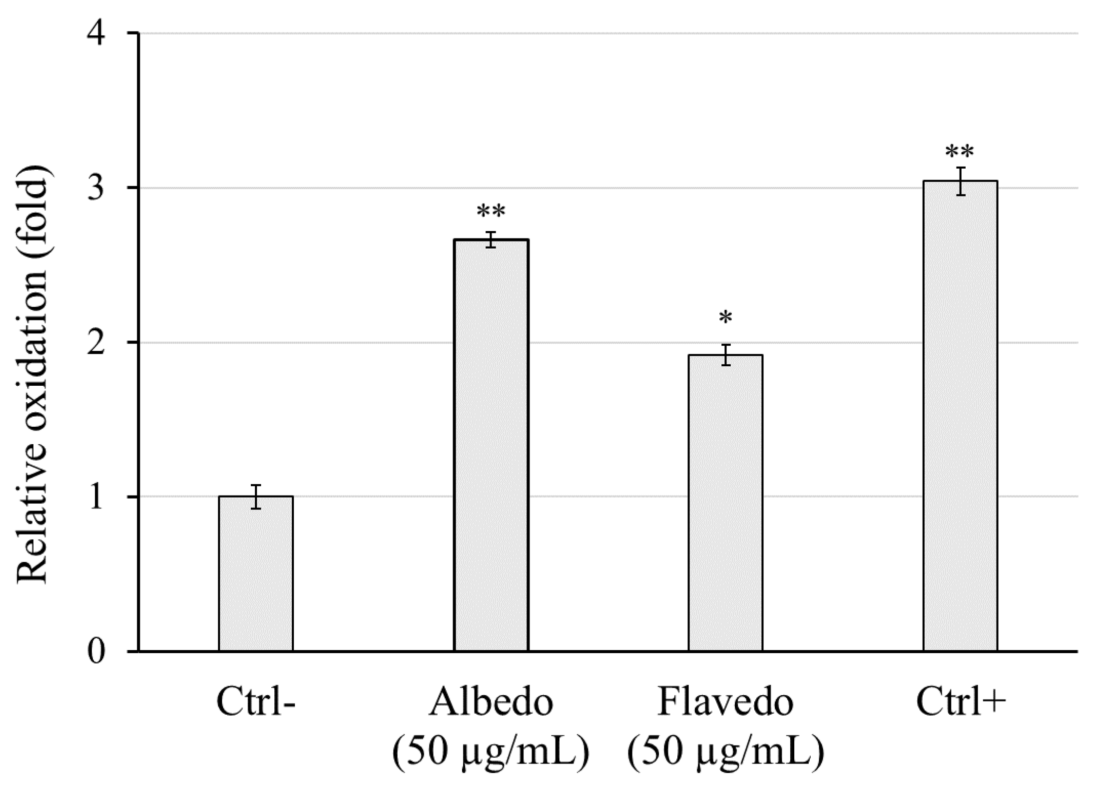
Limoncella preparations gave a highly significant reduction in H
2O
2-induced free radicals in flavedo extract and a less significant reduction in albedo extract, further supporting their action as a protective antioxidant against oxidative stress. The lipoxygenase enzymes contribute to the emergence of inflammation and allergic reactions by producing leukotrienes. Soybean lipoxygenase has substrate selectivity and inhibitory properties similar to humans, exhibits good stability, and was used in inhibition assay [
49]. Albedo and flavedo extracts significantly inhibited lipoxygenase action with an EC
50 of 0.54 ± 0.10 0.14 ± 0.06 for albedo and flavedo, respectively. In literature, it was reported that coumarin and hesperidin show considerable lipoxygenase inhibitory activity [
49,
50], and as reported above, these phenolic compounds are present in large amounts in
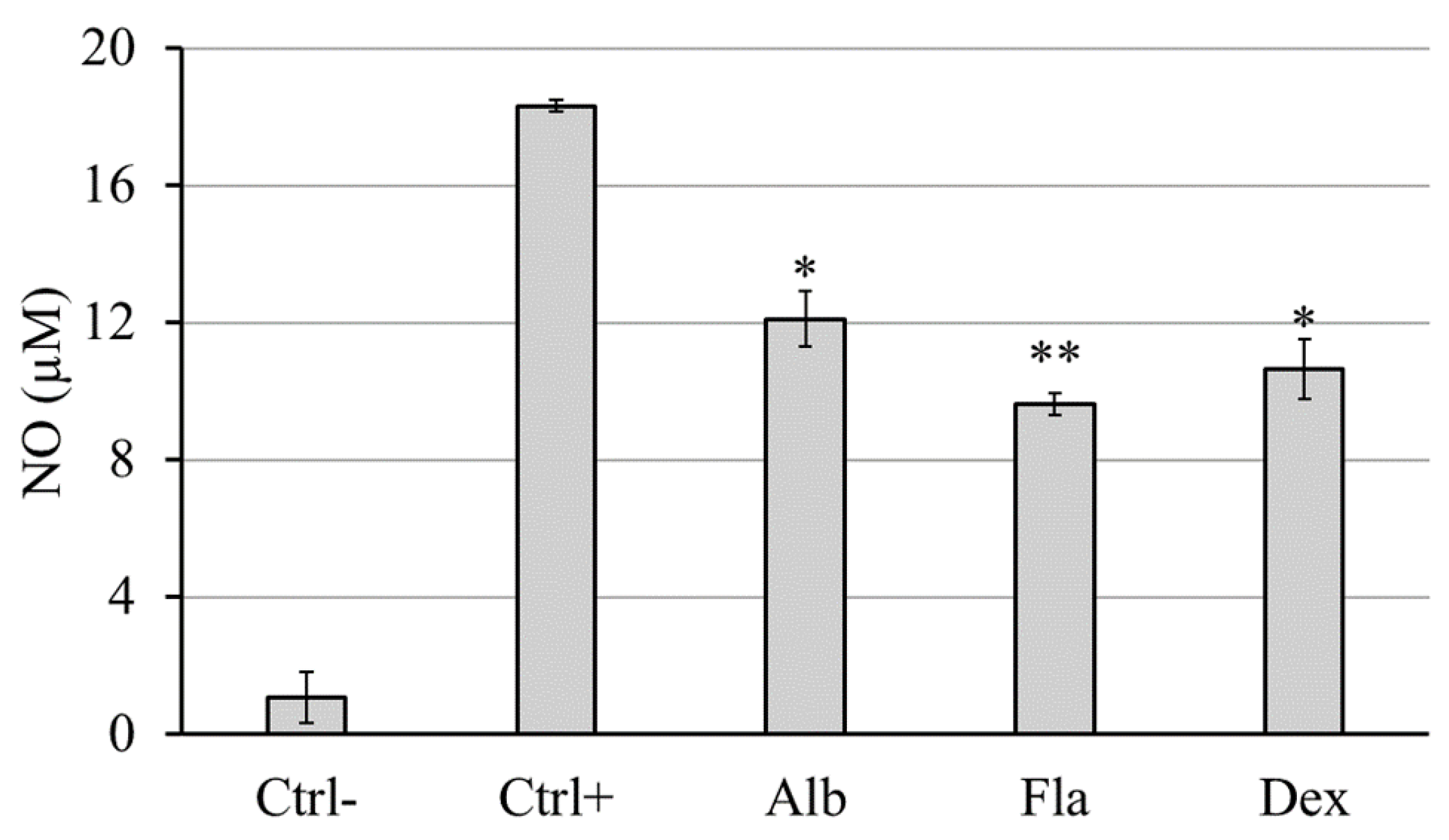
Limoncella extracts. The results regarding the oxidation of linoleic acid catalyzed by lipoxygenase suggested a possible anti-inflammatory activity by these extracts, which was investigated by choosing the Griess test, a cell-based model that uses the RAW264.7 cells, an appropriate macrophage model to study the inflammatory cell responses rapidly [
51]. Also, this assay showed that the flavedo exhibits more anti-inflammatory activity than the albedo, -0.54-fold and -0.67-fold versus Ctrl+, respectively. Both extracts show properties similar to the drug dexamethasone (-0.60-fold). On the other hand, anti-inflammatory activity is not surprising, as polyphenols can also have anti-inflammatory activity. The anti-inflammatory properties of lemon peel extract could be explained by the presence of
p-coumaric acid, a phenolic acid, ten times more represented in flavedo vs albedo extract, showing immunosuppressive effects by reducing cell-mediated immune responses and macrophage phagocytic activity in rats [
52]. It also shows that anti-inflammatory action decreased the expression of the inflammatory mediator tumor necrosis factor-alpha (TNF-α) and circulating immune complexes in adjuvant-induced arthritic rats. The rutin, a bioflavonoid, 40 times more present in flavedo, shows anti-inflammatory properties by inhibiting inflammatory mediators and enzymes, which help reduce inflammation in the body and can alleviate symptoms of inflammatory conditions like arthritis and may contribute to preventing chronic diseases [
53]. In addition, other compounds with anti-inflammatory properties that still need to be identified may be present in the two extracts. Fruits and vegetables are the main anticancer foods, rich in antioxidants, Vitamins C and E, beta-carrots, and lipothin. Citrus fruits, in particular, play an essential role as genoprotective agents. We evaluate the genoprotective effects against oxidative damage by DNA nicking assay. The results proved that albedo and flavedo provided significant antioxidant protection against oxidative damage to biomolecules, acting on pathogenic hydroxyl radicals generated
in vitro by Fe
2+ + O
2 [
54,
55,
56]. However, their cytotoxicity in HaCaT (IC
50 albedo, 250 g/ml; IC
50 flavedo, 161 µg/ml) is much higher than the EC
50 obtained in the nicking assay (albedo, 90 µg/mL; flavedo, 42 µg/mL), and both extracts are indeed safe when expressing protective capacity against hydroxyl radicals. Hydroxyl radicals are mainly produced in mitochondria during oxidative phosphorylation, causing oxidative damage to mtDNA, which is implicated in physiological senescence and age-related disorders [
57]. Our experimental model reveals that both phytoextracts protect plasmid DNA (pEMBL8), which resembles the structure of mtDNA, from oxidation by hydroxyl radicals. Thus, although flavedo was found superior, albedo protects against oxidative DNA damage and can counteract mutagenic and carcinogenic agents. Many studies have been performed on the genoprotective properties of lemon juice and peel. A study conducted on Citrus medica L. through the Ames test demonstrated the antimutagenic and anticarcinogenic effects of the juice [
58]. Koolaji et al. found that citrus peel contains numerous bioactive compounds, including carotenes, essential oils, pectins, and phenolic compounds, which may reduce cancer risk. The peel extract of the genus
Citrus has a higher antitumor activity than the single isolated compound.
In particular, it has been shown that the compounds contained in the peel extract of these citrus fruits have an antitumor activity, which is manifested through suppression of cell proliferation, the inhibition of the cell cycle, the induction of apoptosis, the reduction of the formation of metastases and the inhibition of angiogenesis. The polymethoxyflavonones (PMFs) and other compounds found in the citrus peel can increase apoptotic activity through the intrinsic pathway, eliminating cancer cells [
59].
Furthermore, the flavonoids are also antiangiogenic, inhibiting the expression of vascular endothelial growth factor (VEGF) and suppressing endothelial cell migration. Koolaji et al. also showed that the peel bioactive compounds of these citrus fruits reduce the formation of metastases, decreasing the expression and activity of MMP-2 and MMP-9, the main proteins involved in forming metastases [
60]. Therefore, the flavonoids present in citrus peels act as free radical scavengers and modulators of several fundamental molecular events implicated in cancer development [
36,
59,
60,
61]. The present study, for the first time, highlights the genoprotective property of albedo and suggests that every part of the fruit can be used as a source of compounds beneficial to human health.
Author Contributions
Conceptualization, L.P., L.D.P. and E.B.; methodology, R.S., F.P., L.D.P., S.B., and L.V.; validation, R.S., L.P., F.P., L.D.P., P.G., G.C., and A.S.; formal analysis, R.S., F.P., S.B., L.V.; investigation, R.S., L.P., F.P., L.D.P., G.A., S.B., L.V., G.L., and A.S.; resources, R.S., L.P., and E.B.; data curation, L.P., F.P., and P.G.; writing—original draft preparation, R.S., L.P., F.P., S.P., L.V., G.C., and A.S.; writing—review and editing, L.P., F.P., S.V., and E.B.; visualization, R.S., L.P., S.B., and G.C.; supervision, L.P., G.A., and S.V.; project administration, E.B.; funding acquisition, E.B. All authors have read and agreed to the published version of the manuscript
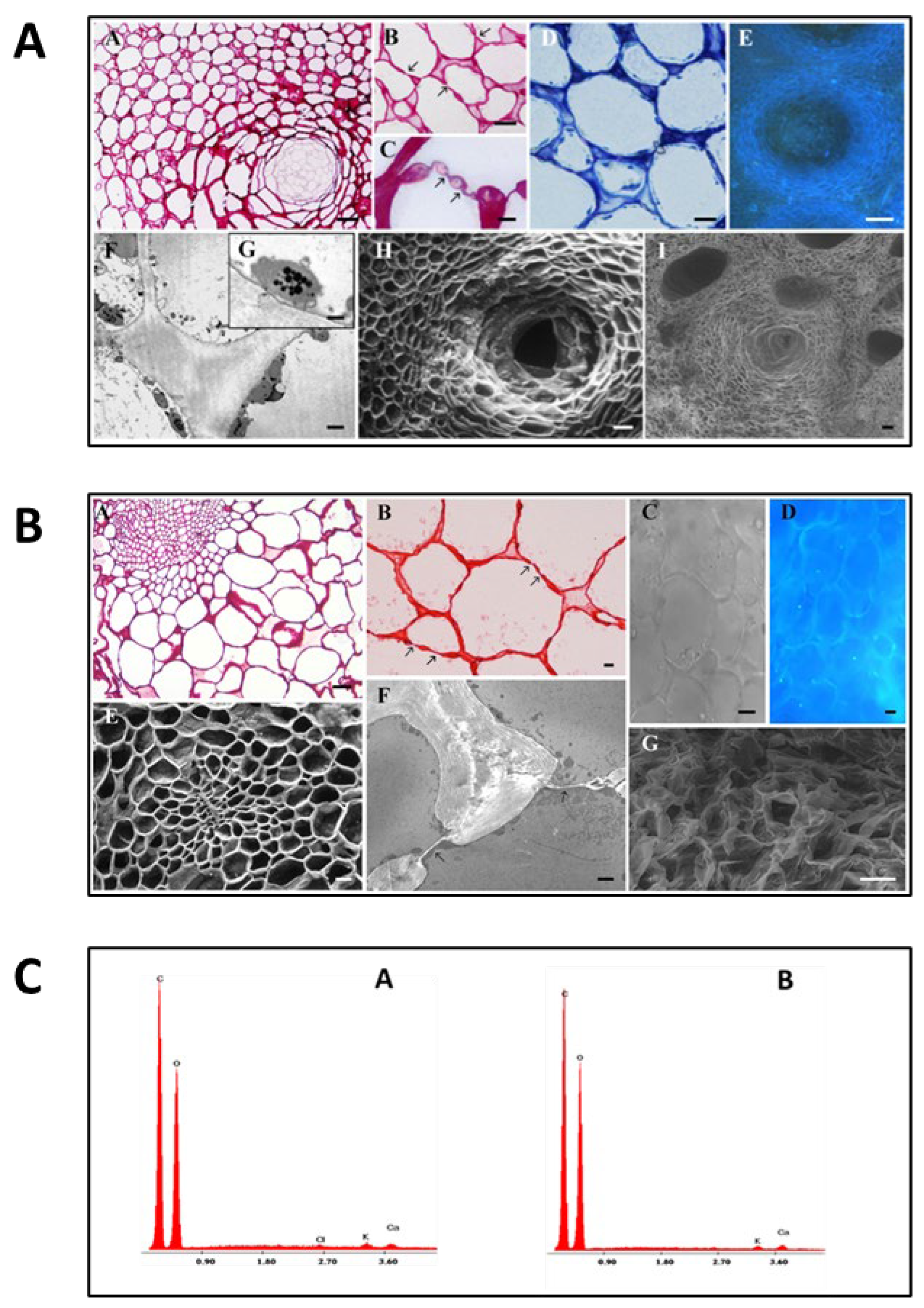
Figure 1.
Morphological analysis and semi-quantitative data of flavedo and albedo from Limoncella. A: Flavedo parenchymal cells were observed by LM (A-D), fluorescent (E), TEM (F, G), SEM (H) and ESEM (I) microscopy. Basic fuchsin (A-C) staining highlighted cellular wall and plasmodesmata Toluidine Blue (B) evidenced cytoplasmic organelles and central vacuoles. In F and G, some plastids were visible. A, H, I oil glands morphology. (A, E) Bar = 5 μm; (B, F) Bar = 2.5 μm; (D) Bar = 1 μm; (G) Bar = 0.5 mm; (H) Bar = 50 μm; (I) Bar = 100 μm.pp. B: Albedo parenchymal cells were observed by LM (A, B), differential interference contrast (C), fluorescent (D), SEM (E), TEM (F), and ESEM (G) microscopy. Basic fuchsin (A) and safranin (B) staining are used to identify cellular walls. In F, plasmodesmata structures were visible. (A, B, E, G) Bar = 20 μm; (C, D) Bar = 0.2 μm; (F) Bar = 2 μm. C: Semi-quantitative analysis performed on the albedo (A) and flavedo (B) surfaces.
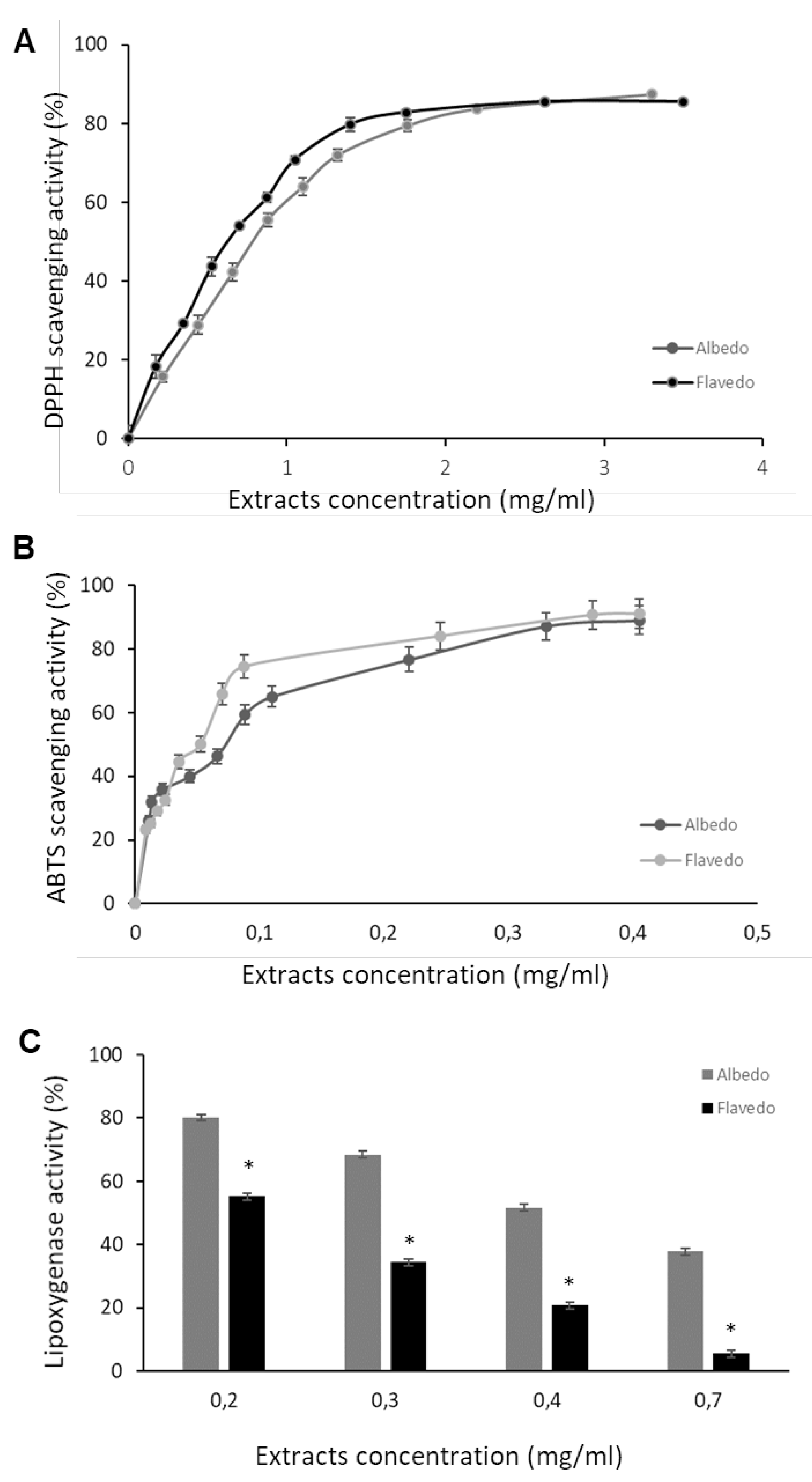
Figure 2.
Antioxidant capacity of albedo and flavedo Limoncella extracts. Scavenging effect on DPPH (A) and ABTS (B) tests. Effect on lipoxygenase activity in vitro (C). The data represent the inhibition percentage induced by increasing albedo and flavedo extracts. Data are expressed as mean ± SD (n = 3). * p < 0.05.
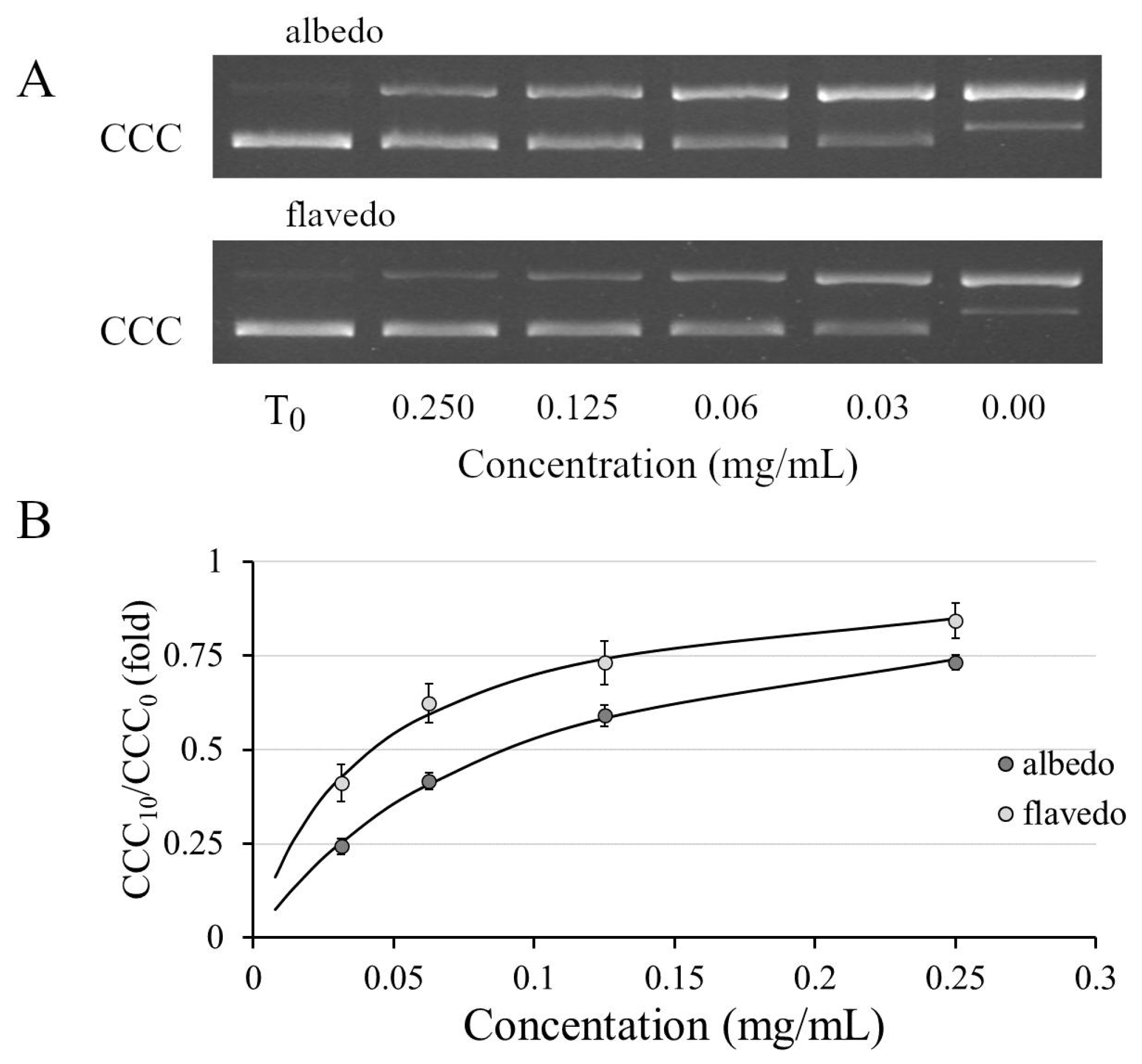
Figure 3.
Protective activity of albedo and flavedo extract versus oxidative DNA damage evaluated by DNA nicking assay. Square A reports a representative agarose gel electrophoresis of the pEMBL8 samples before (T0) and after treatment with different concentrations of hydroalcoholic extracts. CCC is a supercoiled form of a plasmid. Square B reports the quantification analyses expressed as the ratio between the CCC after treatment and CCC at time 0.
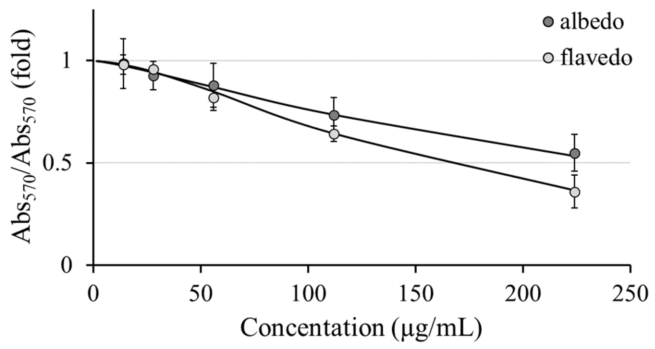
Figure 4.
Cell viability evaluation by SRB colorimetric assay upon Limoncella extracts administration (from 14 to 224 µg/ml) to HaCaT cell lines for 24 h. The cell viability was expressed as the ratio between absorbance at 570 nm of treated and untreated samples.
Figure 4.
Cell viability evaluation by SRB colorimetric assay upon Limoncella extracts administration (from 14 to 224 µg/ml) to HaCaT cell lines for 24 h. The cell viability was expressed as the ratio between absorbance at 570 nm of treated and untreated samples.
Figure 5.
Evaluation of the antioxidant properties of albedo and flavedo extract in HaCaT cells. The relative intracellular oxidation levels were obtained by incubating the cells with 50 µg/ml of each extract. The untreated and non-oxidized cells were reported as Ctrl- while the untreated and oxidized (H2O2) cells were Ctrl+. Data are expressed as mean ± SD (n = 3). * p < 0.05 vs. Ctrl-. ** p < 0.01 vs. Ctrl-.
Figure 6.
Extracellular NO release after RAW 264.7 stimulation by LPS for 24 h in the presence of albedo and flavedo extracts (50 µg/mL). Ctrl-: cell without stimulus or extract; Ctrl+: cell stimulated by 1 µg/mL LPS; Alb: 50 µg/mL albedo + 1 µg/mL LPS; Fla: 50 µg/mL flavedo + 1 µg/mL LPS; Dex: 5 µg/mL dexamethasone + 1 µg/mL LPS. Data are expressed as the mean ± SD (n = 3). * p < 0.05, ** p < 0.01 vs. Ctrl+ (Tukey’s post hoc test).
Table 1.
Concentration (mg kg-1) of bioactive compounds in Albedo and Flavedo extract (n = 3).
Table 1.
Concentration (mg kg-1) of bioactive compounds in Albedo and Flavedo extract (n = 3).
| No |
Compounds |
Albedo Concentration mg kg-1
|
Flavedo Concentration mg kg-1
|
| 1 |
Gallic acid |
5.57 |
41.20 |
| 2 |
Neochlorogenic acid |
n.d. a
|
n.d. |
| 3 |
Delphinidin-3-galactoside |
n.d. |
7.35 |
| 4 |
(+)-Catechin |
n.d. |
n.d. |
| 5 |
Procyanidin B2 |
n.d. |
n.d. |
| 6 |
Chlorogenic acid |
0.80 |
1.54 |
| 7 |
p-Hydroxybenzoic acid |
411.34 |
895.60 |
| 8 |
(-)-Epicatechin |
n.d. |
0.69 |
| 9 |
Cyanidin-3-glucoside |
n.d. |
n.d. |
| 10 |
Petunidin-3-glucoside |
0.45 |
1.82 |
| 11 |
3-Hydroxybenzoic acid |
n.d. |
n.d. |
| 12 |
Caffeic acid |
6.92 |
n.d. |
| 13 |
Vanillic acid |
134.39 |
n.d. |
| 14 |
Resveratrol |
n.d. |
n.d. |
| 15 |
Pelargonidin-3-glucoside |
n.d. |
0.50 |
| 16 |
Pelagonidin-3-rutinoside |
n.d. |
n.d. |
| 17 |
Malvidin-3-galactoside |
n.d. |
n.d. |
| 18 |
Syringic acid |
n.d. |
36.43 |
| 19 |
Procyanidin A2 |
n.d. |
n.d. |
| 20 |
p-Coumaric acid |
143.89 |
1566.65 |
| 21 |
Ferulic acid |
147.86 |
609.96 |
| 22 |
3,5-Dicaffeoylquinic acid |
n.d. |
n.d. |
| 23 |
Rutin |
137.04 |
5996.78 |
| 24 |
Hyperoside |
n.d. |
245.49 |
| 25 |
Isoquercitrin |
9.73 |
216.05 |
| 26 |
Delphindin-3,5-diglucoside |
68.98 |
212.24 |
| 27 |
Phloridzin |
0.82 |
n.d. |
| 28 |
Quercitrin |
1961.17 |
267.76 |
| 29 |
Myricetin |
0.41 |
n.d. |
| 30 |
Naringin |
3.86 |
1.65 |
| 31 |
Kaempferol-3-glucoside |
47.45 |
4.73 |
| 32 |
Hesperidin |
11625.45 |
778.88 |
| 33 |
Ellagic acid |
n.d. |
24.49 |
| 34 |
trans-cinnamic acid |
13.23 |
4.09 |
| 35 |
Quercetin |
12.92 |
73.08 |
| 36 |
Phloretin |
n.d. |
n.d. |
| 37 |
Kaempferol |
128.20 |
246.73 |
| 38 |
Isorhamnetin |
3.22 |
158.56 |
| Total content (mg kg-1) |
14863.69 |
11392.24 |
| Total content (%) |
1.48 |
1.14 |
| Total content (mg kg-1) Folin-Ciocalteu method |
18700 |
22040 |
Table 2.
Antioxidant activities of albedo and flavedo extracts.
Table 2.
Antioxidant activities of albedo and flavedo extracts.
| Part of fruit |
DPPH EC50 (mg/ml) |
ABTS EC50 (mg/ml) |
Lipoxygenase EC50 (mg/ml) |
Total Antioxidant Capacity (µg TE/mg) |
| Albedo |
0.996 ± 0.17 |
0.063 ± 0.029 |
0.54 ± 0.10 |
35.42 ± 3.31 |
| Flavedo |
0.971 ± 0.34 |
0.063 ± 0.012 |
0.14 ± 0.06 * |
40.41 ± 10.15 |

