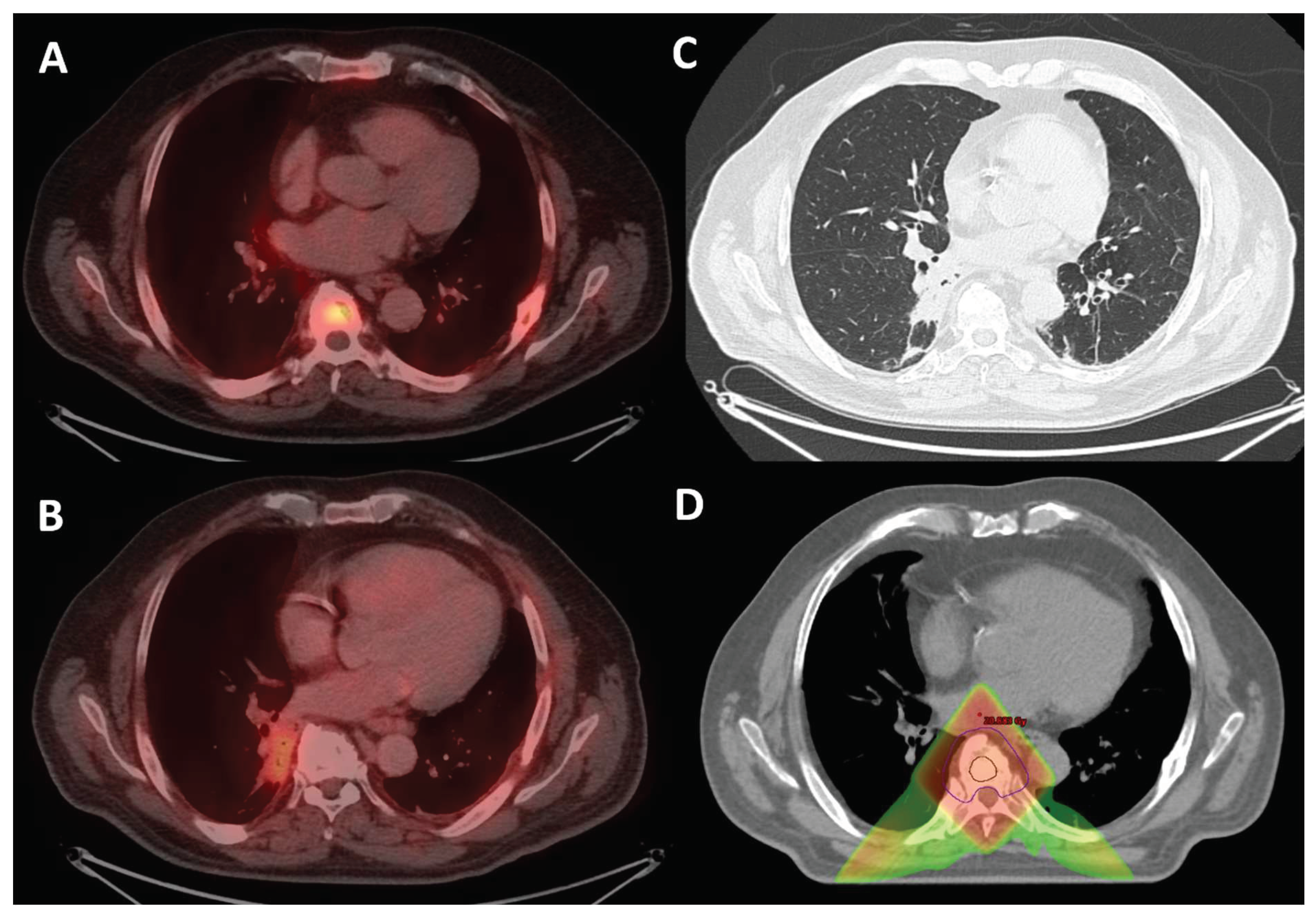
Figure 1.
A 68-year-old male with metastatic castration-resistant prostate cancer had a partial response to Carboplatin and Taxotere and received palliative radiation-therapy, 20 Gy in 5 fractions, to a dorsal vertebral (D7) metastasis positive on 18F-PSMA PET-CT (figure A). PSA serum levels dropped from 65 to 0.06 ng/mL. Four months later, on routine follow-up, a 18F-PSMA PET-CT scan showed increased mild uptake (figure B) in a new paravertebral lung consolidation/mass measuring 4x2 cm in the right lower lobe (figure C).
Due to the short time interval between the two 18F-PSMA PET-CT scans, a new primary malignant lung lesion was improbable, as would be a new metastatic prostate lesion, when the PSA serum level was almost indetectable. Hence, the most reasonable diagnosis for the new lesion remains infection or inflammation as a PSMA PET misleading finding1. Absence of pneumonia symptoms, normal inflammatory markers, directed us to check the radiation treatment planning (figure D) where the Inner circle represents Clinical Target Volume, outer circle the Planning Target Volume, the red rhombus corresponds the isodose lines 18.5-20.4 Gy, and finally the green triangle refers to absorbed 50% isodose lines, 10Gy, which overlap the new lung finding. Leading us to the most probable diagnosis of radiation pneumonitis2.
Radiation-induced lung injuries (RILI) are a significant complication of thoracic radiotherapy, broadly discussed in the literature mainly as a result of radiation treatment to lungs lesions3. Our review of the literature indicates that this is the first report to highlight the pitfall of PSMA PET-CT uptake in RILI resulting from irradiation of vertebral metastasis. Early and accurate diagnosis of RILI is crucial to avoid misdiagnosis, unnecessary additional testing, and potentially inappropriate and unnecessary treatment.
Author Contributions
Conceptualization, I.S. and S.M.; formal analysis, All authors; data curation, I.S. S.A.; writing—original draft preparation, S.M; writing—review and editing, I.S. and S.M.; supervision, I.S.
Funding
This research received no external funding.
Data Availability
The data presented in this article are available on request from the corresponding author.
Ethics approval
Not applicable.
Informed consent
Written informed consent has been obtained from the patient to publish this paper.
Consent to publish
All authors have read and agreed to the published version of the manuscript.
Competing interests
The authors declare no conflicts of interest.
References
- de Galiza Barbosa F, Queiroz MA, Nunes RF, et al. Nonprostatic diseases on PSMA PET imaging: a spectrum of benign and malignant findings. Cancer Imaging. 2020;20(1):23. Published 2020 Mar 14. [CrossRef]
- Rahi MS, Parekh J, Pednekar P, et al. Radiation-Induced Lung Injury-Current Perspectives and Management. Clin Pract. 2021;11(3):410-429. Published 2021 Jul 1. [CrossRef]
- Hanania AN, Mainwaring W, Ghebre YT, Hanania NA, Ludwig M. Radiation-Induced Lung Injury: Assessment and Management. Chest. 2019 Jul;156(1):150-162. [CrossRef] [PubMed]
|
Disclaimer/Publisher’s Note: The statements, opinions and data contained in all publications are solely those of the individual author(s) and contributor(s) and not of MDPI and/or the editor(s). MDPI and/or the editor(s) disclaim responsibility for any injury to people or property resulting from any ideas, methods, instructions or products referred to in the content. |
© 2024 by the authors. Licensee MDPI, Basel, Switzerland. This article is an open access article distributed under the terms and conditions of the Creative Commons Attribution (CC BY) license (http://creativecommons.org/licenses/by/4.0/).


