Submitted:
29 September 2024
Posted:
30 September 2024
You are already at the latest version
Abstract
Permanent pacemaker implantation is a low-risk procedure. However, complications may occur with a rate of around 3-8%. We present a case where initial implantation resulted in complications that could have been avoided by meticulous assessment of lead position in different projections and early post procedure X-ray that would have delineated other serious complications.
An eighty-seven-year-old female patient presented with syncope and documented high grade atrioventricular block. She had a dual chamber pacemaker implant, after which she had another episode of syncope with loss of pacing capture. This was rectified by ventricular lead repositioning that resulted in consistent ventricular pacing. A week later, she was re-admitted with worsening dyspnoea and fatigue. Chest X-ray (CXR) and CT revealed haemopericardium and haemothorax and she was treated with a chest drain and conservatively managed for pericardial effusion. She had a slow recovery and was discharged home after treatment with no further symptoms during follow up in pacing clinic. This case emphasises the importance of meticulous assessment during and after pacemaker implantation.
Keywords:
Complications
; Pacing
An eighty-seven-year-old female patient presented with syncope and documented high grade atrioventricular block. (Figure 1) She had a dual chamber pacemaker implant, after which she had another episode of syncope with loss of pacing capture in CCU. (Figure 2A–C)
The patient had repositing of the right ventricular (RV) lead in the right ventricular apex, and the ECG showed left bundle branch block (LBBB) pattern pacing. (Figure 3) Pacing checks were satisfactory after repositioning, and she was discharged home without a CXR.
A week later; she was re-admitted with worsening dyspnoea. CXR and CT revealed haemopericardium and haemothorax and she was treated with a chest drain and conservatively managed for pericardial effusion (Figure 4 and Figure 5). She had a slow recovery and was discharged home after treatment with no further symptoms during follow up in pacing clinic.
Learning Points
ECG evaluation post pacemaker implantation is essential, and pseudo RBBB with late V4 transition, should raise the suspicion of coronary sinus pacing of the right ventricular lead [2].
ST elevation with injury current is an essential step during implant, that alerts the operator to extracardiac placement, and post procedure [3].
LAO projection will allow the operator to identify right ventricular versus coronary sinus lead placement during procedure [4].
Chest Xray and pacing interrogation are essential after all pacemaker implantations [5].
Author Contributions
Conceptualization, NW; Data curation, NW; Project administration, NW-MI; Supervision, NW-MI; Validation, NW-MI-CB-AH-JP; Writing—original draft, NW-MI; Writing—review and editing, NW-MI. All authors have read and agreed to the published version of the manuscript.
Funding
The authors confirmed that no funding was received.
Informed Consent Statement
The patient had agreed in a written consent and informed consent for publication for this study because none of the data in the paper reveal the patient’s identity.
Data Availability Statement
The data are available on request from the authors.
Conflicts of Interest
The authors declare no conflicts of interest.
References
- Carrión-Camacho, M.R.; Marín-León, I.; Molina-Doñoro, J.M.; González-López, J.R. Safety of Permanent Pacemaker Implantation: A Prospective Study. J Clin Med. 2019, 8, 35. [Google Scholar] [CrossRef] [PubMed] [PubMed Central]
- Muthumala, A.; Herring, N.; Wong, K. A case of difficult RV lead placement. Heart 2013, 100, 434–435. [Google Scholar] [CrossRef] [PubMed]
- Sridharan, A.; Farmer, D.M.; Homoud, M. Pacemaker Interrogation and Programming. In Cardiology Procedures; Hendel, R.C., Kimmelstiel, C., Eds, *!!! REPLACE !!!*, Eds.; Springer: Cham, 2022. [Google Scholar] [CrossRef]
- Squara, F.; Scarlatti, D.; Riccini, P.; Garret, G.; Moceri, P.; Ferrari, E. Individualized Left Anterior Oblique Projection. A Highly Reliable Patient-Tailored Fluoroscopy Criterion for Right Ventricular Lead Positioning. Circulation: Arrhythmia and Electrophysiology. 2018, 11, e006107 ttps://doiorg/101161/CIRCEP117006107. [Google Scholar] [CrossRef]
- Mathew, R.P.; Alexander, T.; Patel, V.; Low, G. Chest radiographs of cardiac devices (Part 1): Cardiovascular implantable electronic devices, cardiac valve prostheses and Amplatzer occluder devices. SA J Radiol. 2019, 23, 1730. [Google Scholar] [CrossRef] [PubMed] [PubMed Central]
Figure 1.
demonstrates Mobitz type two, second degree atrioventricular block.
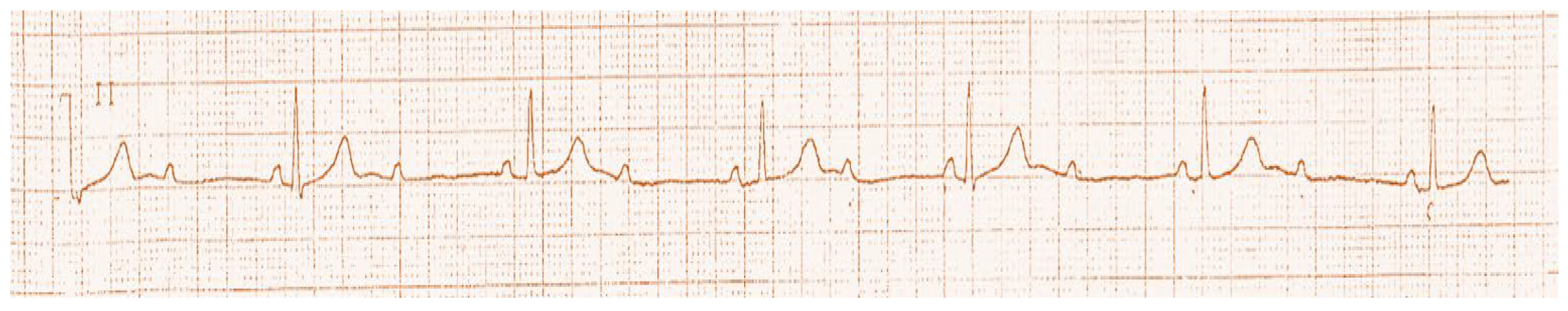
Figure 2.
(A): demonstrates ECG paced ventricular rhythm with right bundle branch block (RBBB) pattern. (B) demonstrates dual chamber final implant by fluoroscopy, with right ventricular lead in middle cardiac vein position rather than right ventricular apex. (C) Telemetry monitor revealing native sinus rhythm with loss of capture with long pause after pacemaker implant, followed by a syncope in CCU. This was confirmed by pacing checks with loss of capture at the highest output.
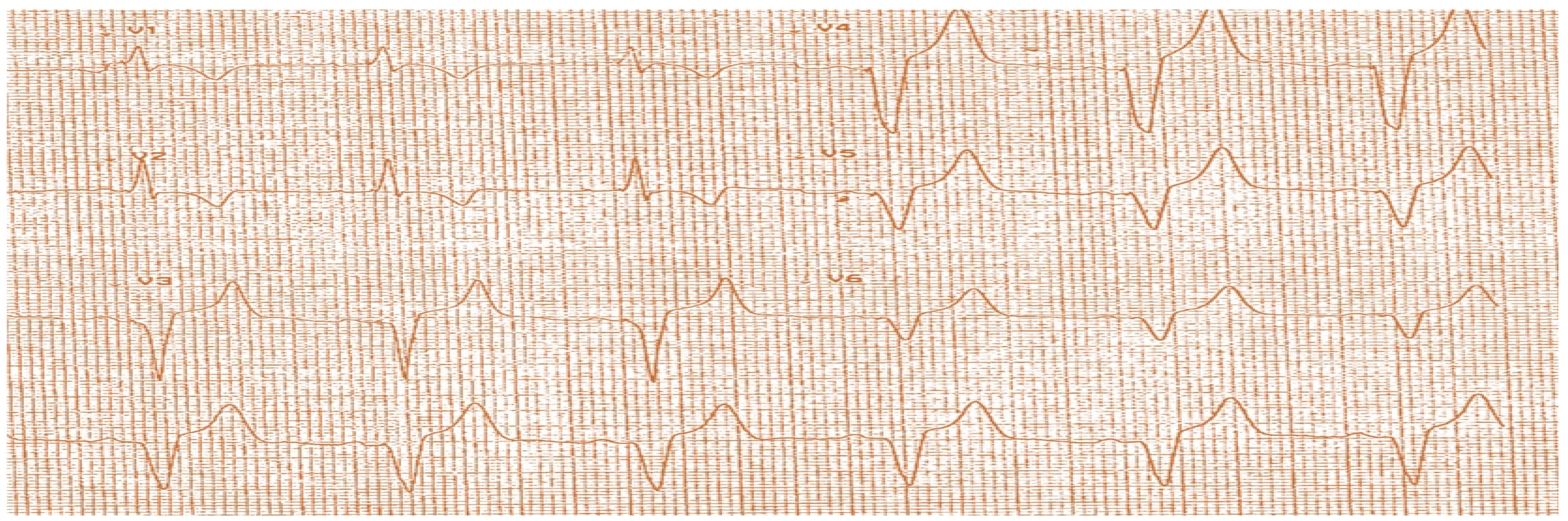
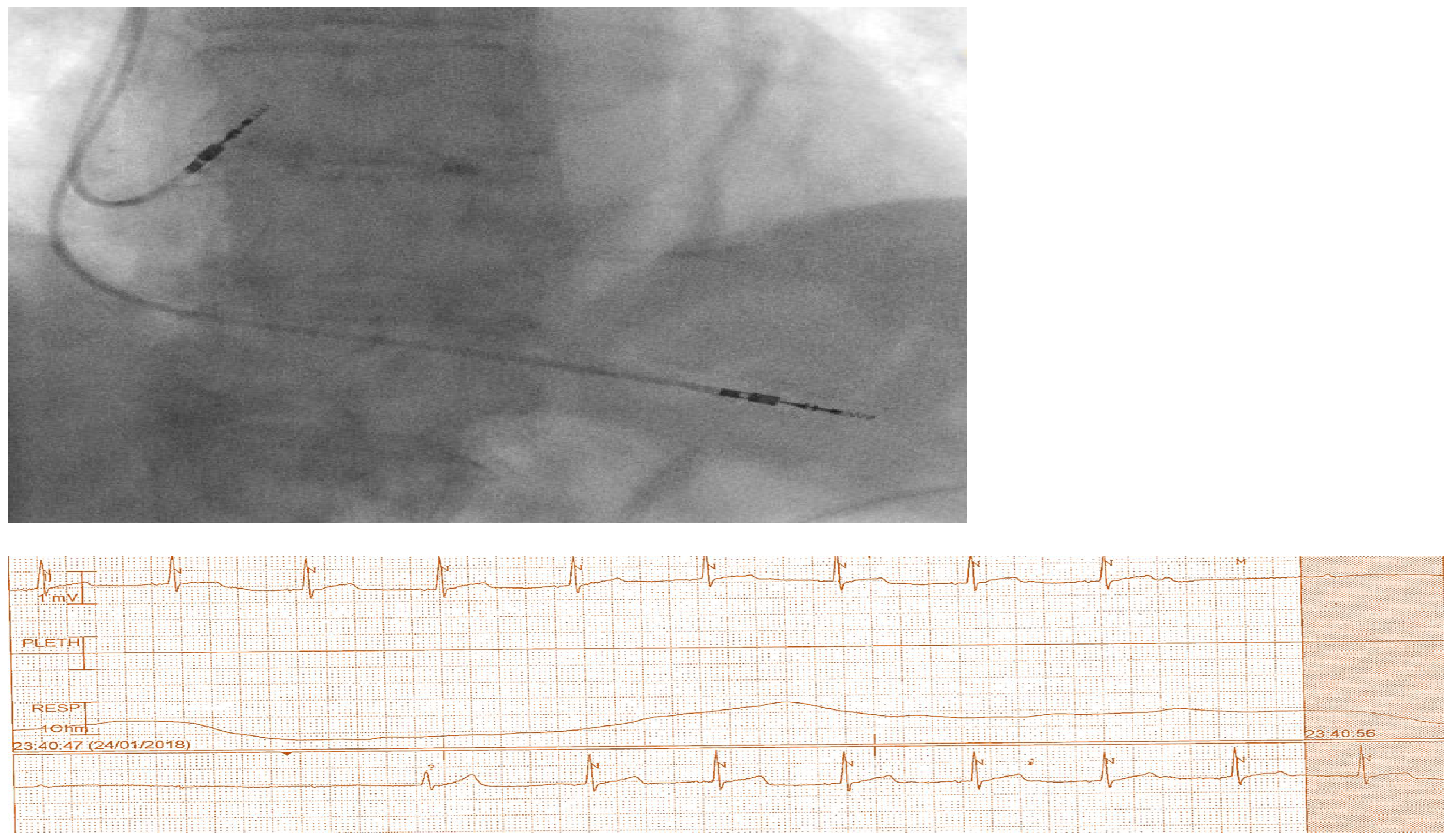
Figure 3.
ECG showed LBBB pattern pacing.
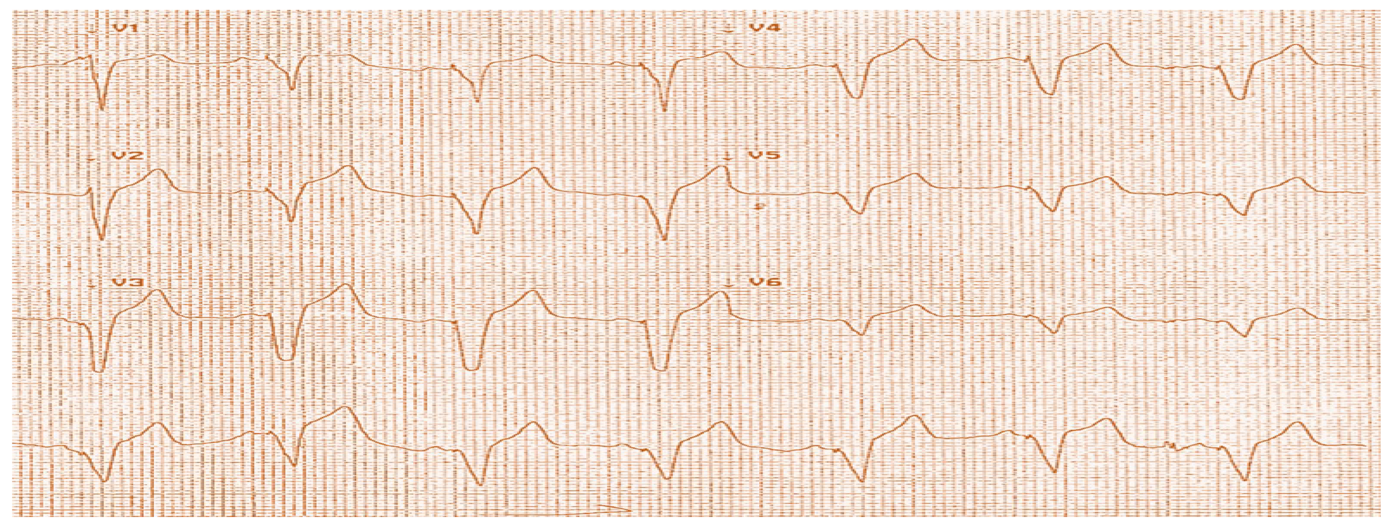
Figure 4.
(A) CXR demonstrates haemo-pneumothorax with dual chamber pacemaker leads in position. (B) CT showing bilateral pleural effusion and pericardial effusion. (C) Echocardiography from subcoastal and apical windows showing moderate pericardial effusion, with inferior vena cava (IVC) collapse more than 50%.
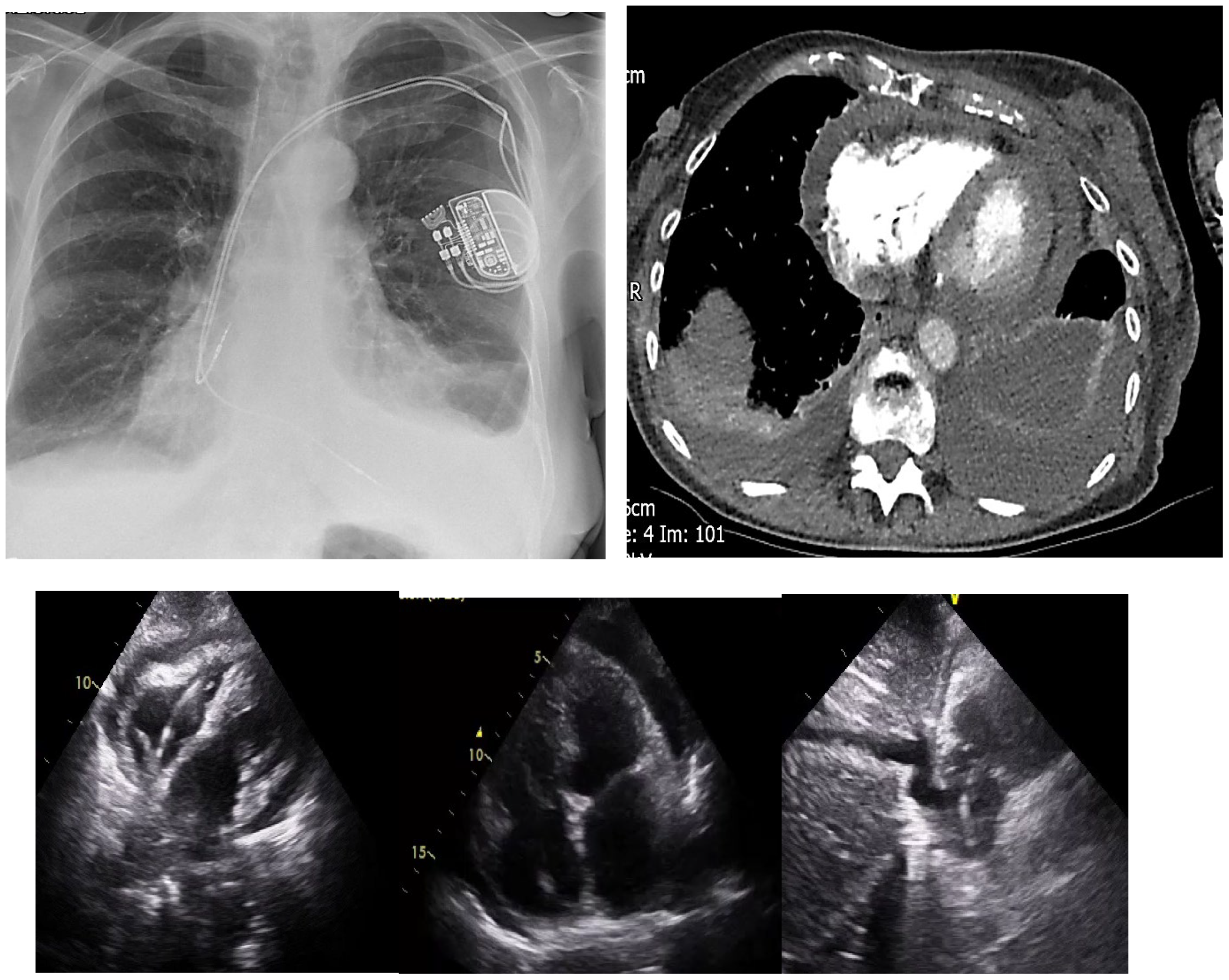
Figure 5.
(A) CXR demonstrates the left sided chest drain. (B) CXR demonstrates residual left sided haemo-pneumothorax after drain removal. (C) CT revealing residual loculated haemothorax.
Figure 5.
(A) CXR demonstrates the left sided chest drain. (B) CXR demonstrates residual left sided haemo-pneumothorax after drain removal. (C) CT revealing residual loculated haemothorax.
Disclaimer/Publisher’s Note: The statements, opinions and data contained in all publications are solely those of the individual author(s) and contributor(s) and not of MDPI and/or the editor(s). MDPI and/or the editor(s) disclaim responsibility for any injury to people or property resulting from any ideas, methods, instructions or products referred to in the content. |
© 2024 by the authors. Licensee MDPI, Basel, Switzerland. This article is an open access article distributed under the terms and conditions of the Creative Commons Attribution (CC BY) license (http://creativecommons.org/licenses/by/4.0/).
Copyright: This open access article is published under a Creative Commons CC BY 4.0 license, which permit the free download, distribution, and reuse, provided that the author and preprint are cited in any reuse.


