
Submitted:
10 October 2024
Posted:
10 October 2024
You are already at the latest version
Abstract
Introduction: To describe a case of unilateral exudative retinal detachment and severe uveitis with a large subretinal deposit in a 6-year-old girl presenting with left thigh lymphadenitis.
Case Presentation: A metagenome next-generation sequencing (mNGS) of the vitreous body was performed, revealing Bartonella henselae. The patient showed the resolution of retinal detachment and uveitis, as well as improvement of left thigh lymphadenitis, in response to systemic and ocular antibiotics and corticosteroids.
Conclusion: Bartonella henselae neuroretinitis may manifest as exudative retinal detachment with extensive subretinal lesions and uveitis. The treatment involving systemic and ocular antibiotics and corticosteroids proved to be effective.
Keywords:
Bartonella henselae
; Cat-scratch disease
Figure 1.
Fundus of a six-year-old Chinese girl with cat scratch disease. At the time of the patient’s visit for examination, the vision of the left eye was light perception, the vision of the right eye was 1.0, the intraocular pressure of the right eye was 17 mmHg, and the intraocular pressure of the left eye was 12 mmHg. There was corneal endothelial cell deposition on the left+, aqueous flare++, dilated pupil, clear lens, vitreous opacity++, total retinal shallow detachment. There is a huge yellow-white mass under the temporal superior retina adjacent to the optic disc. The mass involves the macular area. A month ago, a hard lump was found below the inguinal area on the inner side of her left thigh, accompanied by itching and tenderness. There was no rupture, swelling, fever or cough. Eighteen days ago, there was obvious conjunctival congestion in the left eye. The lump on the left thigh became enlarged and reddened. The skin temperature was high and there was tenderness but no rupture. The local hospital treated it as an infection (the medication is unclear). The parents unintentionally noticed a decrease in vision in the left eye, with only light perception. At the local hospital, ocular B-ultrasound showed enhanced band-like echo in the dark area of the vitreous body of the left eye. B-ultrasound of the lymph nodes at the root of the left thigh showed enhanced local tissue echo. The white blood cell count was 17.78×10^9/L. Considering infection, cephalosporin injection was given for 11 days without improvement. (The green arrow indicates the retinal folds caused by retinal detachment, the red arrow indicates the optic disc, and the white arrow indicates the huge yellow-white mass under the retina).
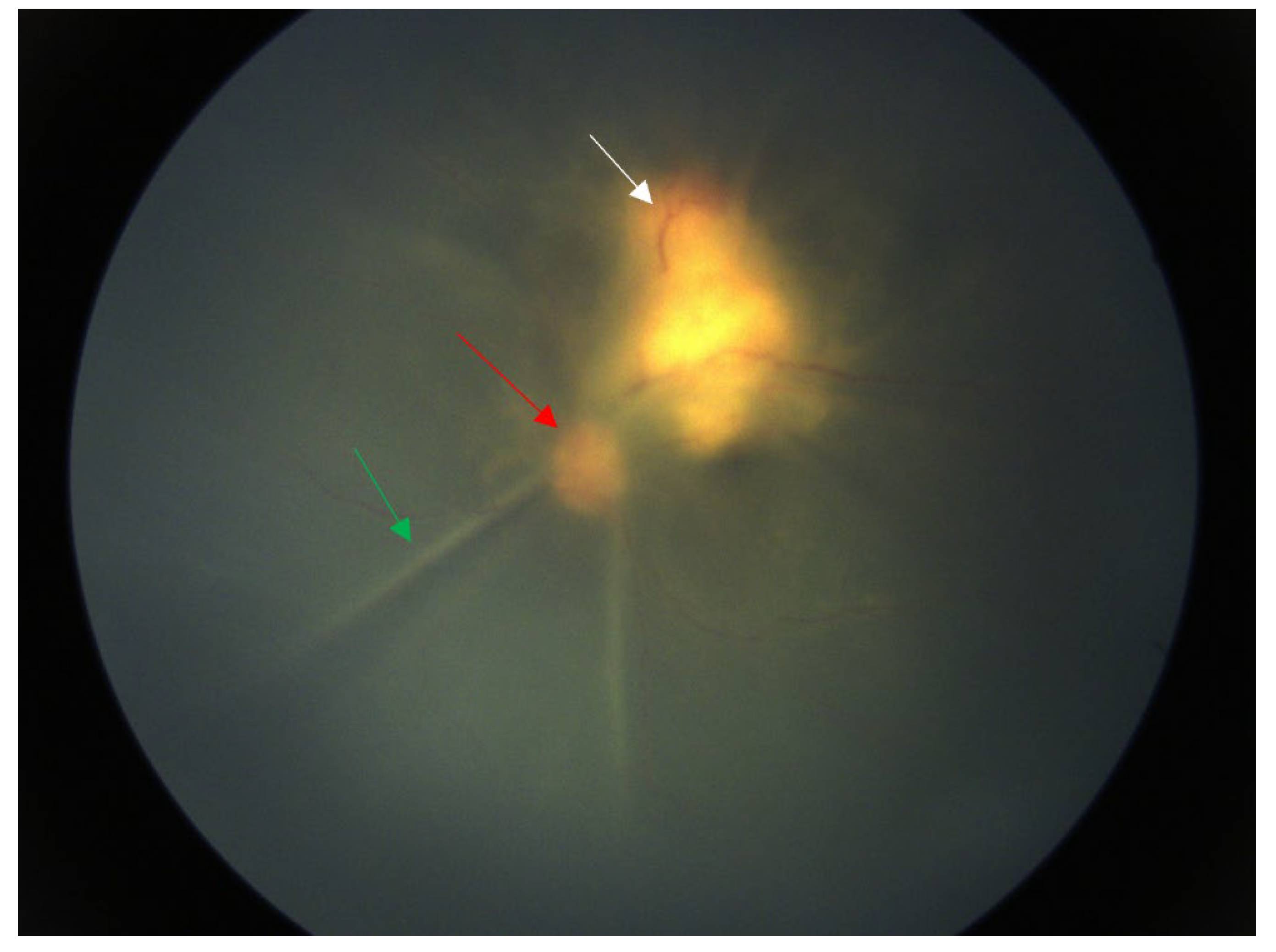
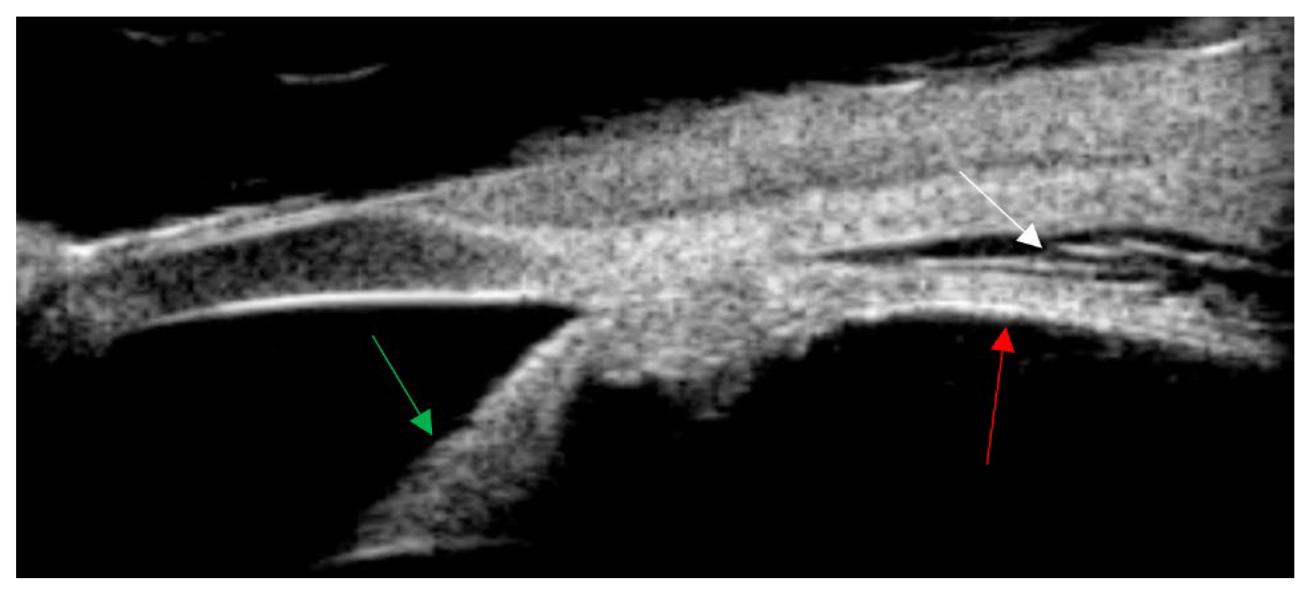
Figure 2.
Ultrasound biomicroscopy(UBM) showed leakage in the supraciliary space of the left eye. (The red arrow indicates the continuation of the ciliary body flat part and the choroid. The green arrow indicates the iris. The white arrow indicates the supraciliary space.).
Figure 2.
Ultrasound biomicroscopy(UBM) showed leakage in the supraciliary space of the left eye. (The red arrow indicates the continuation of the ciliary body flat part and the choroid. The green arrow indicates the iris. The white arrow indicates the supraciliary space.).
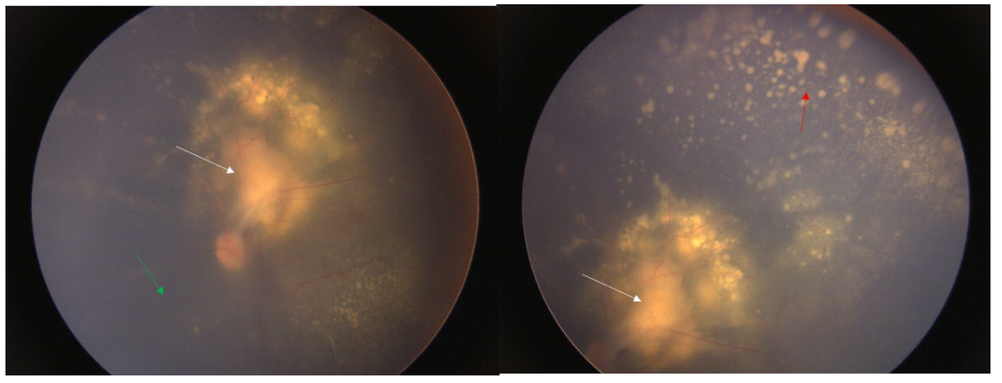
Figure 3.
After 25 days of medication treatment, the boundary of the optic disc was clearer than before, the retina was reattached. There was no significant change in the yellow-white exudate under the retina above the temporal side of the optic disc. Point-like exudates appeared under the peripheral retina, and the vitreous inflammation was reduced. Literature reports indicate that rifampicin, ciprofloxacin, gentamicin, azithromycin, and compound sulfamethoxazole are all effective against cat scratch disease8.9/4,5. It is recommended that gentamicin be used preferentially for severe cases. In clinical treatment, antibiotics are usually used in combination. It is recommended to use drugs such as aminoglycosides, azithromycin, and quinolones. Clinical treatment generally requires more than 2 weeks. If necessary, lymph node puncture, pus aspiration or resection in the affected area can be considered8/4. The effectiveness of steroids is uncertain. This patient was treated with rifampicin eye drops and erythromycin eye ointment topically in the eye, and rifampicin, azithromycin, and steroids systemically. The retinal detachment in the left eye was reattached, and the enlarged lymph nodes on the left thigh subsided. However, the huge exudate under the retina in the macular area could not be absorbed, resulting in a final visual acuity of only 0.03. (The white arrow is the yellow-white mass under the retina, the green arrow is the flattened retina, and the red arrow is the yellow-white point-like exudate.).
Cat scratch disease (CSD), also known as benign lymphohistiocytosis, is an infectious disease caused by cat or dog scratches or bites. The pathogen is Bartonella henselae. Systemic symptoms precede ocular manifestations. Visual symptoms appear approximately one month after vaccination, and CSD (2/3) is the most common cause of optic neuritis, although only 1-2% of infected patients experience it [6]
Cat scratch disease presents with various manifestations in the eyes, including Parinaud’s oculoglandic syndrome, optic disc edema and macular stellate neuroretinitis (LISN), multifocal retinitis, focal and multifocal choroiditis, vascular occlusion and bacterial angiomatosis, uveitis, vitritis, exudative retinal detachment, etc [7]. However, there are almost no reports of a large amount of subretinal exudate in the macular area, accompanied by retinal detachment and severe panuveitis, as described in this article [7]. Hao Hong et al. reported the first case of cat scratch disease with both uveitis and nodular lesions in the fundus, but without retinal detachment [4].
Informed Consent Statement
Informed consent was obtained from the parents of the patient in this study. Written informed consent has been obtained from the child’s parents to publish this paper.
Conflicts of Interest
The authors declare no conflict of interest.
References
- Kiu KH, Hanizasurana H, Zunaina E. Neuroretinitis with dual infections. Int Med Case Rep J. 2015;8:255-258. [CrossRef]
- Carithers, HA. Cat-scratch disease. An overview based on a study of 1,200 patients. Am J Dis Child 1960. 1985;139(11):1124-1133. [CrossRef]
- Fleissig E, Kim F, Sigford DK, Barr CC. Bilateral neuroretinits and exudative retinal detachment with multifocal subretinal deposits secondary to Bartonella henselae infection. Am J Ophthalmol Case Rep. 2021;24:101201. [CrossRef]
- Hong H, Li T, Ying Y, An Q, Liu H, Liang K. Cat-scratch disease manifesting as uveitis and binocular fundus nodular lesions: a case report. BMC Ophthalmol. 2023;23:345. [CrossRef]
- Acar A, Çakar Özdal P, Başarır B, Özdemir Yalçınsoy K, Altan Ç, Budakoğlu Ö. A Case Series of Cat-Scratch Disease with Ocular Manifestations: Clinical Findings and Treatment Approach. Turk J Ophthalmol. 2023;53(4):226-233. [CrossRef]
- Sykes DAW, Joseph SL, Williams SP, Das SU. A 13-Year-Old Girl With Unilateral Visual Changes. J Investig Med High Impact Case Rep. 2023;11:23247096221150635. [CrossRef]
- C Stephen Foster, Albert T Vitale. DIAGNOSIS AND TREATMENT OF UVEITIS. 2nd ed. Jaypee Brothers Medical Publishers (P) Ltd.; 2013.
Disclaimer/Publisher’s Note: The statements, opinions and data contained in all publications are solely those of the individual author(s) and contributor(s) and not of MDPI and/or the editor(s). MDPI and/or the editor(s) disclaim responsibility for any injury to people or property resulting from any ideas, methods, instructions or products referred to in the content. |
© 2024 by the authors. Licensee MDPI, Basel, Switzerland. This article is an open access article distributed under the terms and conditions of the Creative Commons Attribution (CC BY) license (http://creativecommons.org/licenses/by/4.0/).
Copyright: This open access article is published under a Creative Commons CC BY 4.0 license, which permit the free download, distribution, and reuse, provided that the author and preprint are cited in any reuse.


