
Submitted:
10 October 2024
Posted:
11 October 2024
You are already at the latest version
Abstract
Objectives: The purpose of this clinical study is to compare full-arch intraoral scans taken immediately after implant placement with those obtained after tissue healing in patients rehabilitated with implant-supported fixed prosthesis. Methods: Between September 2023 and March 2024, a total of 19 patients with compromised residual dentition (6 women, 13 men) were rehabilitated using 4 to 6 immediately loaded post-extraction implants. These implants supported fixed full-arch screw-retained prostheses in the lower jaw (9 patients) or upper jaw (10 patients). Intraoral scans were taken immediately after implant placement (termed ‘immediate scan’). After a healing period of four months, the provisional prosthesis was removed and a second intraoral scan was performed using the same scan bodies and scan pattern as the initial scan (termed ‘delayed scan’). The two scans were overlaid and discrepancies between them were measured. Results: The average discrepancy between the immediate and delayed scans was 0.1905 mm. Statistical analysis revealed larger discrepancies for implants placed in the posterior areas, with the implant in site 1.6 ( implant 1 ) showing a discrepancy of 0.2326 mm and the implant in site 2.6 (implant 4) showing 0.2124 mm (P = 0.05). No statistically significant difference was observed when comparing patients treated in the upper and lower jaws. Conclusions: Within the limitations of this study, overlapping digital scans of post-surgical and healed tissues revealed discrepancies that could be attributed to the more difficult conditions present in the post-surgical scenario or operator factors. Further studies are needed to assess the accuracy of post-surgical scans from a clinical point of view.
Keywords:
dental implants
; intraoral scan
; digital impression
; immediate loading
; full-arch rehabilitation
; split-mouth study
1. Introduction
The advent of computer-aided design systems (CAD-CAM) has had a huge impact on the daily practice of all dentists [1].
The digital workflow involves the acquisition of physical data, which is then transformed into digital data to enable computer-aided design and manufacturing (CAD-CAM) of a prosthesis [2]. The transformation of physical data into digital data can occur either by digitizing models obtained from traditional impressions in the laboratory using bench scanners operated by dental technicians or directly in the dental office using an intraoral scanner (IOS) [3].
Passivity is fundamental for any type of implant prosthesis, even more so for a screw-retained prosthesis, where tensile forces and overloads can also be generated during the screw-retained phase, causing tension at the implant/abutment-prosthesis interface. The impression is therefore the key moment for transferring the correct implant position to the master model and consequently for the accuracy and passivity of the prosthetic structure [4].
Until recently, conventional dental impressions were the most commonly used method for full-arch cases. However, even conventional workflows can introduce various errors. Factors such as the type of materials used, the number of implants, their positions, depth, and angulation can all impact precision and accuracy. The accumulation of these errors may ultimately compromise the passive fit of an implant prosthesis [5].
Intraoral scanning (that is so called “digital impressions”) have accelerated the data acquisition process and eliminated many inconveniences present in conventional impressions, thus reducing patient discomfort and improving the predictability of prosthesis design and production procedures over time [6]. To achieve high-precision prosthetic rehabilitation, it is crucial that the digitized physical data accurately replicate what is scanned and maintain this precision throughout the entire manufacturing process of the prosthetic device [7,8].
In partial and full-arch implant prosthetic rehabilitations, intraoral scans have achieved an accuracy comparable to that of traditional impressions [9,10]. Several in vitro and clinical studies have shown that the discrepancy between analogue and digital impressions on patients treated with full-arch rehabilitation have a reduced discrepancy, below the tolerability value reported in the literature (150 Microns) [11,12].
There are various stages within both the digital and analogue workflows where errors can be introduced, potentially compromising the passivity of the framework. In the analogue method, rigid materials were traditionally used during impression-taking to ensure the immobility of implants and the accuracy of the impression [13].
In the digital workflow, however, errors may occur during the scanning phase, as well as during the image design and matching phases [14].
Several factors influence the precision and accuracy of intraoral scans [15]. The type of IOS used plays a significant role in accuracy; in fact, some studies suggest that not all scanners are suitable for capturing full-arch implant-supported prostheses[16]. Scan-bodies can also impact accuracy, as their shape and position may affect the precision of the impression, even though they are generally easy to acquire with the scanner [17,18,19,20,21]. Another factor contributing to minor deviations in accuracy is the matching of the virtual scan body from the CAD software library to the virtual model.
If the original scan is not performed properly, this can lead to a mismatch of the scanbody and an error in the position of the analogue in the virtual model. The inter-implant distance significantly impacts the accuracy of impressions; in fact, an increase in distance tends to amplify errors, reducing both precision and accuracy [22].
The location of the scan also plays a role in digital impression accuracy. Scanning an edentulous jaw is often challenging due to the reduced presence of landmarks. Additionally, in cases involving osteoplasty or flap-less surgery, flap repositioning and bleeding can further obscure landmarks, complicating the scanning process. Numerous in vitro studies have shown that impressions obtained with intraoral and analogue scanners demonstrate similar and overlapping levels of accuracy. However, these studies do not account for the fact that impressions taken in the oral cavity are subject to various factors that can influence accuracy and precision, which cannot be fully replicated or studied in vitro [23].
Few studies have in fact analysed clinically whether digital impressions had the same predictability and accuracy of traditional impressions [23].
A study by Pera F. et al. [10] went to analyze the discrepancy between plaster impressions and impressions with full scans in full-arch rehabilitations on healed tissue and found that the discrepancies were not clinically relevant.
Capparé et al. [24] conducted a study to evaluate the accuracy and precision of prostheses obtained from traditional and digital impressions in patients undergoing full-arch rehabilitation. The study revealed that digital impressions offer clinically usable accuracy that is comparable to that of traditional impressions.
Few studies analyzed the accuracy of impressions taken just after implant surgery.
A study by De Angelis et al. [25] aimed to evaluate the accuracy of prostheses obtained through digital impressions immediately after surgery. This study found that digital impressions, compared to traditional impressions, offer similar accuracy and are clinically usable. However, in this study, no flap was performed, and no dislocation occurred, so the impact of soft tissue mobility could not be assessed.
To the authors' knowledge, there is currently no study evaluating the accuracy of post-surgical impressions in patients rehabilitated with immediately loaded full-arch prostheses. The authors hypothesize that the opening of a large flap, performing osteoplasty, and suturing the flap may increase flap mobility and reduce the reliability of reference stitches, potentially leading to greater errors and distortions in the impressions.
The aim of this clinical study is to compare the accuracy of digital impressions taken immediately after surgery with those obtained from healed tissue in patients rehabilitated with a full-arch implant-supported prosthesis. The null hypothesis tested is that there is no significant difference between post-surgical impressions and impressions taken on healed tissue.
2. Materials and Methods
Between September 2023 and March 2024, 8 patients were recruited and treated at the Division of Prosthodontics and Implantology (Department of Surgical Sciences, DISC) of the University of Genoa (centre 1) and 11 patients at the Division of Prosthodontics of the C.I.R. Dental School (Department of Surgical Sciences) of the University of Turin (centre 2).
All 19 patients had a compromised residual dentition (6 women, 13 men) and were treated with full-arch screw-retained fixed prostheses supported by 4-5 immediately loaded implants in the lower jaw (8 patients) or upper jaw (11 patients).
All subjects treated in this study, which was approved by the Scientific Ethics Committee of the University of Genoa (protocol approval number :2023/03) were carefully informed about the study design and aims and signed an informed consent before the start of the study. The study was conducted in accordance with the Declaration of Helsinki.
This research was reported in accordance with the CONSORT Statement for Quality Improvement in Reporting (www.consort-statement.org/).
All patients were recruited if they had complete impairment of residual teeth at least in one arch and were requiring an immediate loading rehabilitation of the upper or lower jaw and had adequate bone volume for the placement of 4 to 6 implants without the need for regenerative procedures.
All patients had to be in good general health, have no systemic conditions or be under pharmacological therapies that did not allow oral surgery, and not have a degree of bone atrophy requiring regenerative methods or the use of zygomatic implants [26,27].
The Columbus Bridge Protocol (CBP) was applied, which is a surgical and prosthetic protocol for full-arch immediate loading rehabilitation. This protocol allows for the restoration of aesthetics, phonetics, and function in patients with severely compromised teeth within 48-72 hours [28].
Following the extraction of compromised dentition, the protocol involves the insertion of 4-6 immediate implants with a minimum length of 10 mm into native bone, achieving high insertion torque. Implants are positioned vertically in the anterior regions of the jaws and, if necessary, inclined in the posterior regions to avoid anatomical structures and facilitate the distal placement of the implant head. Endosseous titanium implants with a tapered apex were used, Shard (Mech & Human, Grisignano di Zocco, Italy) and Syra (Sweden & Martina, Due Carrare PD, Italy). Multiunit abutments (MUA) were used when needed to correct implant inclination.
The fixed prosthesis was delivered and screwed onto the implants within 48 hours after surgery. It featured a rigid framework without distal extensions and was veneered with composite resin material. The classic protocol involves taking an impression immediately after the surgical phase using the plaster pick-up technique to achieve maximum precision for the metal framework [26].
In this study, the post-surgical impression was made using a scanner with a speed of 15 frames per second (Mach2 Intraoral Scanner, Shining 3D, distributed by Euromax Monaco) (Figure 1).
Scan bodies from the same manufacturer as the inserted implants were placed on the implants/MUAs, and an impression of the entire arch was taken immediately after suturing. No powder was used to opacify the elements to be scanned and the output file was in ‘open’ STL format.
In 71 implants the scanbodies were screwed onto the MUA, in 8 implants the scanbodies were screwed directly onto the implant heads.
On Shard metal implants Toothless® Scanbodies (Mech & Human, Grisignano di Zocco, Italy) were used. They present a rounded shape with a hexagon on the top useful to increase the reference points for the best-fitting operation and had two different types of connection depending on whether they had to be screwed directly onto the implant head or onto the MUA.
On eight implants in two patients, scanbodies were used that were screwed directly onto the implant head (Sweden & Martina, Due Carrare PD, Italy), which have a flag shape with two points on the surface.
Two of the authors with more than two years of experience with the IOS herein described (F.P. at the Centre 2 and F.B. at Centre 1) performed all the intraoral scans.
The two operators performed the scans using an ‘S’ scan path; the IOS tip followed the entire arch with a smooth movement starting from the most distal implant in the first quadrant to the contralateral implant, zig-zagging from the vestibular to the palatal side and vice versa. For each patient, an analogic post-surgical plaster impression was also acquired that served as a control. An aluminum test bar obtained from the digital impression was then initially tested on the analogic model obtained from the plaster impression. Based on the test bar fit, the scenarios were classified as following:
- -
- Clinically Acceptable. Good fit of the bar on the analogic model; the delivered full-arch prosthesis was completely realized from digital impression.
- -
- Not satisfying fit. The operators were able to screw the test bar on the model, but the fit was not evaluated as good; the final prosthesis delivered to the patients was produced on analogic model.
- -
- Not passive result. It was not possible to screw the test bar on analogic model; the final prosthesis delivered to the patients was produced on analogic model.
At four months, the provisional prosthesis was removed, and a new digital impression was taken with the same IOS and the same scanning pattern (Figure 2).
Before the intraoral scan, the stability of the implants/MUAs was carefully checked.
The two stl files obtained from the immediate and delayed intraoral scans were sent to a digital analysis laboratory experienced in digital rehabilitation to perform the superimposition and analysis.
In the upper arch, all implants in the scans were numbered from one to six (implant 1, 2, 3, etc.) starting with the most distal implant in the right hemiarch, numbering them in ascending order throughout the arch to the most distal contralateral implant, while in the lower hemiarch all implants were numbered in ascending order starting with the most distal implant on the right to the most distal implant in the contralateral hemiarch.
2.1. Outcome Measures
Deviations were assessed using the Hausdorff distance metric, which evaluates the extent of deviation between two sets by measuring the maximum distance between them. Minimum, maximum, and mean distances between surfaces, as well as RMS values (which measure the root mean square deviation between two sets of points), were determined.
The parametric software GOM Inspection Pro (GOM ZEISS) was used for this analysis. This software applies basic parametric concepts to the STL files obtained from the mouth scans. Points to be measured were isolated by determining subsets, eliminating mucosal flanges, and isolating scan bodies (Scanbody Toothless). For each Toothless subset, a point of origin was determined and centered as the zero point of the Cartesian axes. By superimposing the two scans using a best-fit logic, discrepancies between the Toothless subsets were measured, which were evidenced by differences in the position of the Cartesian axes' origin (Figure 3).
From the three Cartesian axes, the variations in coordinates (X, Y, Z) were expressed in millimeters, defining the most common direction of deviation between the different scans of the same edentulous arch.
2.2. Statistical Analysis
Confidence intervals at 5% have been computed for overall implants type and by group (Centre 2 and 1).
Then, centre 2 and 1 groups were compared to investigate possible statistical differences between the groups. The independent-samples t-test was calculated between the Centre 2 and 1 groups (Satterthwaite method for unequal variances was considered).
Only Facility 2 is significantly different between the Turin-Genoa groups (5% alpha was considered).
Furthermore, the mandible and maxilla groups were compared by means of the independent-sample t-test (the Satterthwaite method for unequal variances was considered) and no statistical differences were revealed.
A regression model was applied to each implant type, considering implant value as the dependent variable and time as the independent variable. The result of the model for implant 2 shows that time has a negative impact on implant value (as time increases, implant value decreases) at a significance level of 1%.
For all the analyses p < 0.05 was considered statistically significant and SAS 9.4 was used for the computation.
3. Results
Eleven patients were treated in the upper jaw and eight in the lower jaw. No patients dropped out and all of them received both the immediate and the delayed intraoral scan. No implants failed and all the implants were clinically stable and in function when the delayed scan was taken.
Seventeen dental arches were rehabilitated with four implants, one with five implants and one with six implants, for a total of 79 dental implants. All the patients treated at the University of Genoa were rehabilitated with 4 implants only. Following the test bar fit, 17 cases were classified as clinically acceptable and the prosthesis was manufactured following a full digital workflow. The other 5 cases, 4 were classified as not satisfying fit and 1 as not passive result and the prosthesis was manufactured following an analogic workflow.
The digital analysis showed an average discrepancy between the analyzed points of 0.1905 mm.
Evaluating the individual centres showed an average discrepancy of 0.1303 mm for the centre 2 and 0.279 mm fot centre 1.
From the analysis on the three Cartesian axes, the greatest discrepancy was 0.019 mm on the y-axis.
Analysing each RMS for each implant individually, a greater discrepancy was found for the implants positioned in the posterior areas, more precisely 0.2326 mm for implant 1 and 0.2124 for implant 4.
In the anterior areas, Implants 2 and 3 differed by 0.1777 and 0.1555 mm respectively.
The following table shows the average values of the discrepancies (distances) between the measured points, both as an average across all implants and each individual implant (Table 1).
The scans performed at the center 2 had a smaller variance with respect to the prosthesis of center 1 presenting a mean value of 130 μm and 279 μm respectively.
Independent Samples t-Test between center 2 and center 1 groups has been computed (Satterthwaite method for unequal variances has been considered). Only implant 2 is significantly different among the two centers (alpha at 5% has been considered).
No statistically significant difference was found when comparing treated patients in the upper and lower jaw. The accuracy of the scans increased over the study period. With increasing time and thus operator experience, the scanning error decreased.
This means that as time increases. the value of the discrepancy between the implants decreases (p-value at 6%) (Figure 4).
The implants placed in the posterior areas were those with the greatest discrepancy between the two scans.
Implant 5 and Implant 6 were only available for two and one dental arch. Respectively, all treated at the University of Turin.
Increasing the number of implants the discrepancy between the scans decreases.
The two clinicians who performed the intraoral scans reported anecdotally that scans on unhealed tissue were more difficult to perform as the presence of bleeding and non-keratinized tissue and mobility made scanning more complicated, especially in the distal mandibular areas.
4. Discussion
To the best of the authors' knowledge this is the first study to analyze the discrepancy between impression on surgical and healed sites in patients rehabilitated with full-arch immediate-loading rehabilitations.
The null hypothesis was partially rejected. The discrepancy between the two impressions was around the value of 200 μm, a discrepancy value defined as acceptable in the literature from a clinical point of view, more precisely with an average value of 190μm [29].
Furthermore, despite the fact that the same surgical and scanning protocol was applied, the scans performed in Centre 1 showed a greater discrepancy than the Centre 2 group (average: 0.279 mm versus 0.130 mm respectively).
In 100 % of the cases, the scans performed in Centre 2 showed a value of less than 150 μm in the anterior regions (implants 2 - 3), while in the posterior regions (implants 1 - 4) the values were greater than 150 μm but less than 200 μm in 50 % of the cases, while the scans performed in Centre 1 in the anterior regions (implants 2 - 3) showed a value of less than 150 μm in 12. 5 % and 25 % respectively and in the posterior regions (implants 1 - 4) less than 150 μm in 25 % and 50 % respectively.
In some impressions the discrepancy value between the two scans was greater than 200 microns, which is considered the maximum value for prosthetic misfit, but this value is theoretical [29,30].
In fact, the main limitation of this study is represented by the fact that the accuracy and precision of even the prosthetic artefacts obtained from the two scans was not evaluated, in order to evaluate the clinical reliability of the scans as well. The discrepancy between the two scans was evaluated only digitally, while the in vivo clinical evaluation of the prosthesis was only partially evaluated with the aluminum test bar, but no fit check was performed directly in the mouth.
Scans in patients undergoing full-arch rehabilitation are challenging due to the reduced number of reference points. This limitation can result in point clouds with deficiencies in the digital files, which may lead to improper stitching of the images [31,32,33].
Other factors such as saliva and blood can also lead to incongruous stitching of the stitch clouds, resulting in a reduction in the accuracy of the impression [34].
If reference points are missing. images are stitched with composition errors, including an inaccurate and noisy mesh or key parts of the scan may be identified as redundant points and cut by the post-processing algorithm [31,32,33,35].
The properties of the scanned area are also important factors that can influence the density of the point cloud. It is known that shiny, rough, undercut and sharp surfaces make scanning difficult, and that saliva creates reflective surfaces [32,33,36,37].
A study by Gimenez et al. evaluated scanning in edentulous areas. showing how the lack of reference points. the width and inhomogeneity of the tissues could lead to distortion and incorrect stitching of the images. with an increase in the difficulty of scanning as the inter-implant distance increases.
This is particularly evident in the posterior sectors of the lower jaws, where scanning is more difficult due to the small space, poorly keratinised mucosa, thus requiring more images to be stitched together and making scanning more error-prone [38,39,40].
In this study, as reported in the results, the greatest discrepancies were found in the posterior areas on patients treated in the lower jaws, maybe due to the reduced reference points and the reduced amount of keratinised gingiva.
It has been proposed that adding external landmarks could facilitate scanning and increase the accuracy of scans. A study by Campana et al. [41] showed that adding scan templates around scan bodies can increase scanning accuracy, especially in areas that are more difficult for the IOS to scan [42].
Maintaining a few stable teeth in strategic areas during implant placement could be another trick in order to increase the reference points and facilitate impression matching by the technician [43]. This could be valid if it is not necessary to perform extensive osteotomies to normalize bone ridges of post-extraction sites. In fact, in these cases, it is necessary to remove all the compromised residual elements before implant placement. To avoid the need for post-surgical scanning, an alternative approach could be to perform full-arch rehabilitations using guided surgery and then cement temporary prostheses in the mouth based on a pre-prepared digital design [35].
The use of a prosthesis glued directly into the mouth can be a valid alternative that allows the patient to be rehabilitated immediately after surgery. However. this can be performed with prostheses without metal frameworks when the prosthetic space is large enough to ensure rigidity of the prostheses and immobility of the newly inserted implants. During the luting phase space between the prosthetic cylinder and the prosthesis should be large enough to avoid possible inaccuracies and difficulties in inserting the prosthetic framework. This may result in reduced rigidity of the cemented prosthetic restoration if the amount of cement between the prosthetic cylinder and the prosthesis is excessive [44,45].
One of the limitations of this study was to perform a comparison of two digital scans without comparing them with a master model obtained from traditional impressions. Indeed the test bar was performed only in the post surgical scenario. It is not possible to know which of the two impressions was more similar to the clinical reality of the mouth, so it is not possible to determine which scans were more precise. However, the authors believe that scans performed on healed tissue were comparable in accuracy to those done using traditional methods and were likely more accurate than scans taken immediately after surgery. This is because post-surgical, sutured tissue is prone to bleeding and flap mobility, which can introduce errors and reduce the accuracy and precision of the scans.
In a previous study by the same team of authors [10], applying the same clinical protocol, the same IOS and the same scanning pattern, traditional and digital impressions taken on osseointegrated implants with healed tissues were compared and a statistically significant discrepancy was found between the digital scans and the digital models obtained from plaster impressions. In addition, a mean discrepancy of 110 μm was found between the cast derived from the digital impression and the analogic cast. However, the Sheffield test and radiographic examination showed an excellent passivity of the milled metal frameworks obtained through both the digital and the analogic work-flow, suggesting that both methods were clinically acceptable [10].
This, however, was not performed because of the possible discomfort that could have been caused to the patient especially during surgery.
These values are interesting because they show how the accuracy of a scan improves with time and scanner usage and is operator dependent..
Analysing the values individually, we can see that the scans obtained from the centre 2 have a small discrepancy of around 150 microns and most of them less than 200 microns.
From a clinical point of view these scans could be used to obtain a framework with sufficient and adequate passivity as reported in the literature. Frameworks obtained from scans with a discrepancy of more than 150 or 200 microns, as reported by the centre 1, could not be used from a clinical point of view as they would not allow for sufficiently passive frameworks to avoid biological and prosthetic complications.
It is therefore difficult to define that post-surgical intra-oral scanning can always be predictable in immediate load rehabilitations, as there are numerous factors that could influence the accuracy of the scan.
Increasing the reference points during post-surgical scanning would reduce possible scanning errors and possible errors due to a reduced point cloud and gaps caused by a lack of adequate reference points [24].
This is just one of the factors that can influence the results of a post-surgical scan on patients treated with full-Arch rehabilitations. In fact, scan results are influenced by so many other factors that can change the success or otherwise of the rehabilitation from the scanner to the number of implants to the intruding distance to the presence of keratinised tissue to the presence of saliva and the light and temperature of the environment.
So when clinicians decide to take a scan in a post-surgical full-arch rehabilitation they should take all these factors into account and assess the practicability of the scan on a case-by-case basis.
To conclude, implant supported rehabilitations have today achieved high levels of long-term term survival and succes rates [46]. Novel implant designs [47] as well as digital technologies [48] aim to further simplify the process and minimize the onset of complications [49]. However, as showed in the present study, despite the constant widespreading of digital tecnologies, research on the topic is still actual and further studies are required in order to validate the procedures for any clinical scenario.
5. Conclusions
Within the limits of this study, overlapping digital scans of post-surgical and healed tissues revealed discrepancies that could be attributed to the more challenging conditions present in the post-surgical scenario or to operator factors. Further studies are needed to evaluate the accuracy of post-surgical scans from a clinical perspective.
Author Contributions
Conceptualization, F.B., M.M., P.P., M.C. and F.P.; methodology, A.C., U.G. and F.D..; writing—original draft preparation, F.B., M.M., P.P., M.C., F.P.; writing—review and editing, F.B., M.M., P.P., M.C., F.P.; project administration, M.M, F.P.; funding acquisition, F.P. All authors have read and agreed to the published version of the manuscript.” Please turn to the CRediT taxonomy for the term explanation. Authorship must be limited to those who have contributed substantially to the work reported.
Funding
The authors declare that this study was partly funded by Mech and Human (Grisignano di Zocco, Italy).
Institutional Review Board Statement
The study was conducted in accordance with the Declaration of Helsinki, and approved by the Ethics Committee of the University of Genoa (protocol approval number:2023/03).
Informed Consent Statement
Informed consent was obtained from all subjects involved in the study.
Data Availability Statement
The datasets generated during and/or analyzed during the current study are available from the corresponding author on reasonable request.
Acknowledgments
Authors thank Luca Bagnoli and Mech&Human (Grisignano di Zocco. Italy) for their support in the present research.
Conflicts of Interest
The authors declare no conflicts of interest.
References
- Karl, M.; Holst, S. Strain development of screw-retained implant-supported fixed restorations: procera implant bridge versus conventionally cast restorations. Int J Prosthodont. 2012, 25, 166–169. [Google Scholar] [PubMed]
- Bernauer, S.A.; itzmann, N.U.; Joda, T. The Complete Digital Workflow in Fixed Prosthodontics Updated: A Systematic Review. Healthcare 2023, 11, 679. [Google Scholar] [CrossRef] [PubMed]
- Joda, T.; Zarone, F.; Ferrari, M. The complete digital workflow in fixed prosthodontics: a systematic review. BMC Oral Health 2017, 17, 124. [Google Scholar] [CrossRef] [PubMed]
- Wee, A.G.; Aquilino, S.A.; Schneider, R.L. Strategies to achieve fit in implant prosthodontics: a review of the literature, Int J Prosthodont, 1999, 12, 167-78.
- Donovan, T.E.; Chee, W.W. A review of contemporary impression materials and techniques. Dent Clin North Am, 2004, 48, 445-70. [CrossRef]
- Cicciù, M.; Fiorillo, L.; D’Amico, C.; Gambino, D.; Amantia, E.M.; Laino, L.; Crimi, S.; Campagna, P.; Bianchi, A.; Herford, A.S.; et al. 3D Digital Impression Systems Compared with Traditional Techniques in Dentistry: A Recent Data Systematic Review. Materials 2020, 13, 1982. [Google Scholar] [CrossRef] [PubMed]
- Zhang, Y.J.; Qian, S.J.; Lai, H.C.; Shi, J.Y. Accuracy of photogrammetric imaging versus conventional impressions for complete arch implant-supported fixed dental prostheses: A comparative clinical study. J Prosthet Dent, 2023, 130, 212-218. [CrossRef]
- Kong, L.; Li, Y.; Liu, Z. Digital versus conventional full-arch impressions in linear and 3D accuracy: a systematic review and meta-analysis of in vivo studies. Clin Oral Investig, 2022, 26, 5625-5642. [CrossRef]
- Papaspyridakos, P.; De Souza, A.; Finkelman, M.; Sicilia, E.; Gotsis, S.; Chen, Y.W.; Vazouras, K.; Chochlidakis, K. Digital vs Conventional Full-Arch Implant Impressions: A Retrospective Analysis of 36 Edentulous Jaws. J Prosthodont, 2023, 32, 325-330. [CrossRef]
- Pera, F.; Pesce, P.; Bagnasco, F.; Pancini, N.; Carossa, M.; Baldelli, L.; Annunziata, M.; Migliorati, M.; Baldi, D.; Menini, M. Comparison of Milled Full-Arch Implant-Supported Frameworks Realised with a Full Digital Workflow or from Conventional Impression: A Clinical Study. Materials 2023, 16, 833. [Google Scholar] [CrossRef] [PubMed]
- Mizumoto, R.M.; Yilmaz, B.; McGlumphy, E.A.; Seidt, J.; Johnston, W.M. Accuracy of different digital scanning techniques and scan bodies for complete-arch implant-supported prostheses. J Prosthet Dent, 2020, 123, 96-104. [CrossRef]
- Kim, K.R.; Seo, K.Y.; Kim, S. Conventional open-tray impression versus intraoral digital scan for implant-level complete-arch impression. J Prosthet Dent, 2019, 122, 543-549. [CrossRef]
- Menini, M.; Setti, P.; Pera, F.; Pera, P.; Pesce, P. Accuracy of multi-unit implant impression: traditional techniques versus a digital procedure. Clin Oral Investig, 2018, 22, 1253-1262. [CrossRef]
- Vandeweghe, S.; Vervack, V.; Dierens, M.; De Bruyn, H. Accuracy of digital impressions of multiple dental implants: an in vitro study. Clin Oral Implants Res, 2017, 28, 648-653. [CrossRef]
- Canullo, L.; Colombo, M.; Menini, M.; Sorge, P.; Pesce, P. Trueness of Intraoral Scanners Considering Operator Experience and Three Different Implant Scenarios: A Preliminary Report. Int J Prosthodont, 2021, 34, 250-253. [CrossRef]
- Carneiro Pereira, A.L.; Souza Curinga, M.R.; Melo Segundo, H.V.; da Fonte Porto Carreiro, A. Factors that influence the accuracy of intraoral scanning of total edentulous arches rehabilitated with multiple implants: A systematic review. J Prosthet Dent, 2023, 129, 855-862. [CrossRef]
- Meneghetti, P.C.; Li, J.; Borella, P.S.; Mendonca, G.; Burnett, L.H. Influence of scanbody design and intraoral scanner on the trueness of complete arch implant digital impressions: An in vitro study. PLoS One, 2023, 18, e0295790. [CrossRef]
- Pan, Y.; Tsoi, J.K.H.; Lam, W.Y.H.; Chen, Z.; Pow, E.H.N. Does the geometry of scan bodies affect the alignment accuracy of computer-aided design in implant digital workflow: An in vitro study? Clin Oral Implants Res, 2022, 33, 313-321. [CrossRef]
- Stimmelmayr, M.; Guth, J.F.; Erdelt, K.; Edelhoff, D.; Beuer, F. Digital evaluation of the reproducibility of implant scanbody fit--an in vitro study. Clin Oral Investig, 2012, 16, 851-6. [CrossRef]
- Mizumoto, R.M.; Yilmaz, B. Intraoral scan bodies in implant dentistry: A systematic review. J Prosthet Dent, 2018, 120, 343-352. [CrossRef]
- Zhang, Y.J.; Shi, J.Y.; Qian, S.J.; Qiao, S.C.; Lai, H.C. Accuracy of full-arch digital implant impressions taken using intraoral scanners and related variables: A systematic review. Int J Oral Implantol (Berl), 2021, 14, 157-179.
- Thanasrisuebwong, P.; Kulchotirat, T.; Anunmana, C. Effects of inter-implant distance on the accuracy of intraoral scanner: An in vitro study. J Adv Prosthodont, 2021, 13, 107-116. [CrossRef]
- Schmidt, A.; Wostmann, B.; Schlenz, M.A. Accuracy of digital implant impressions in clinical studies: A systematic review. Clin Oral Implants Res, 2022, 33,573-585. [CrossRef]
- Cappare, P.; Sannino, G.; Minoli, M.; Montemezzi, P.; Ferrini, F. Conventional versus Digital Impressions for Full Arch Screw-Retained Maxillary Rehabilitations: A Randomized Clinical Trial. Int. J. Environ. Res. Public Health 2019, 16, 829. [Google Scholar] [CrossRef] [PubMed]
- De Angelis, N.; Pesce, P.; De Lorenzi, M.; Menini, M. Evaluation of Prosthetic Marginal Fit and Implant Survival Rates for Conventional and Digital Workflows in Full-Arch Immediate Loading Rehabilitations: A Retrospective Clinical Study. J. Clin. Med. 2023, 12, 3452. [Google Scholar] [CrossRef] [PubMed]
- Pera, P.; Menini, M.; Pesce, P.; Bevilacqua, M.; Pera, F.; Tealdo, T. Immediate Versus Delayed Loading of Dental Implants Supporting Fixed Full-Arch Maxillary Prostheses: A 10-year Follow-up Report. Int J Prosthodont, 2019, 32, 27-31. [CrossRef]
- Tealdo, T. ; Menini, M.; Bevilacqua, M.; Pera, F.; Pesce, P.; Signori, A.; Pera, P. Immediate versus delayed loading of dental implants in edentulous patients' maxillae: a 6-year prospective study. Int J Prosthodont, 2014, 27, 207-14. [CrossRef]
- Tealdo, T.; Bevilacqua, M.; Menini, M.; Pera, F.; Ravera, G.; Drago, C.; Pera, P. Immediate versus delayed loading of dental implants in edentulous maxillae: a 36-month prospective study. Int J Prosthodont, 2011, 24, 294-302.
- Chochlidakis, K.; Papaspyridakos, P.; Tsigarida, A.; Romeo, D.; Chen, Y.W.; Natto, Z.; Ercoli, C. Digital Versus Conventional Full-Arch Implant Impressions: A Prospective Study on 16 Edentulous Maxillae. J Prosthodont, 2020, 29, 281-286. [CrossRef]
- Revilla-Leon, M.; Att, W.; Ozcan, M.; Rubenstein, J. Comparison of conventional, photogrammetry, and intraoral scanning accuracy of complete-arch implant impression procedures evaluated with a coordinate measuring machine. J Prosthet Dent, 2021, 125, 470-478. [CrossRef]
- Gimenez, B.; Pradies, G.; Martinez-Rus, F.; Ozcan, M. Accuracy of two digital implant impression systems based on confocal microscopy with variations in customized software and clinical parameters. Int J Oral Maxillofac Implants, 2015, 30, 56-64. [CrossRef]
- Goodacre, B.J.; Goodacre, C.J.; Baba, N.Z. Using Intraoral Scanning to Capture Complete Denture Impressions, Tooth Positions, and Centric Relation Records. Int J Prosthodont, 2018, 31, 377-381. [CrossRef]
- Rhee, Y.K.; Huh, Y.H.; Cho, L.R.; Park, C.J. Comparison of intraoral scanning and conventional impression techniques using 3-dimensional superimposition. J Adv Prosthodont, 2015, 7, 460-7. [CrossRef]
- Chen, Y.; Zhai, Z.; Li, H.; Yamada, S.; Matsuoka, T.; Ono, S.; Nakano, T. Influence of Liquid on the Tooth Surface on the Accuracy of Intraoral Scanners: An In Vitro Study. J Prosthodont, 2022, 31, 59-64. [CrossRef]
- Iturrate, M.; Eguiraun, H.; Etxaniz, O.; Solaberrieta, E. Accuracy analysis of complete-arch digital scans in edentulous arches when using an auxiliary geometric device. J Prosthet Dent, 2019, 121, 447–454. [Google Scholar] [CrossRef] [PubMed]
- Goodacre, B.J.; Goodacre, C.J. Using Intraoral Scanning to Fabricate Complete Dentures: First Experiences. Int J Prosthodont, 2018, 31, 166-170. [CrossRef]
- Gonzalez de Villaumbrosia, P.; Martinez-Rus, F.; Garcia-Orejas, A.; Salido, M.P.; Pradies, G. In vitro comparison of the accuracy (trueness and precision) of six extraoral dental scanners with different scanning technologies. J Prosthet Dent, 2016, 116, 543-550 e1. [CrossRef]
- Gimenez, B.; Ozcan, M.; Martinez-Rus, F.; Pradies, G. Accuracy of a digital impression system based on parallel confocal laser technology for implants with consideration of operator experience and implant angulation and depth. Int J Oral Maxillofac Implants, 2014, 29, 853-62.
- Gimenez-Gonzalez, B.; Hassan, B.; Ozcan, M.; Pradies, G. An In Vitro Study of Factors Influencing the Performance of Digital Intraoral Impressions Operating on Active Wavefront Sampling Technology with Multiple Implants in the Edentulous Maxilla. J Prosthodont, 2017, 26, 650-655. [CrossRef]
- Paratelli, A.; Vania, S.; Gomez-Polo, C.; Ortega, R.; Revilla-Leon, M.; Gomez-Polo, M. Techniques to improve the accuracy of complete arch implant intraoral digital scans: A systematic review. J Prosthet Dent, 2023, 129, 844-854. [CrossRef]
- Campana, V.; Papa, A.; Silvetti, M.A.; Del Fabbro, M.; Testori, T. Use of the universal scan template to achieve a predictable optical impression: Preliminary data of a case series study in complete edentulous patients. Clin Implant Dent Relat Res, 2024, 26, 237-244. [CrossRef]
- Canullo, L.; Pesce, P.; Caponio, V.C.A.; Iacono, R.; Luciani, F.S.; Raffone, C.; Menini, M. Effect of auxiliary geometric devices on the accuracy of intraoral scans in full-arch implant-supported rehabilitations: An in vitro study. J Dent, 2024, 145, 104979. [CrossRef]
- Bover-Ramos, F.; Vina-Almunia, J.; Cervera-Ballester, J.; Penarrocha-Diago, M.; Garcia-Mira, B. Accuracy of Implant Placement with Computer-Guided Surgery: A Systematic Review and Meta-Analysis Comparing Cadaver, Clinical, and In Vitro Studies. Int J Oral Maxillofac Implants, 2018, 33, 101-115. [CrossRef]
- Charette, J.R.; Goldberg, J.; Harris, B.T.; Morton, D.; Llop, D.R.; Lin, W.S. Cone beam computed tomography imaging as a primary diagnostic tool for computer-guided surgery and CAD-CAM interim removable and fixed dental prostheses. J Prosthet Dent, 2016, 116, 157-65. [CrossRef]
- Lewis, R.C.; Harris, B.T.; Sarno, R.; Morton, D.; Llop, D.R.; Lin, W.S. Maxillary and mandibular immediately loaded implant-supported interim complete fixed dental prostheses on immediately placed dental implants with a digital approach: A clinical report. J Prosthet Dent, 2015, 114, 315-22. [CrossRef]
- Carossa, M.; Pera, F.; Alovisi, M.; Ponzio, M.; Schierano, G.; Migliaretti, G.; Carossa, S.; Scotti, N. Implant survival rate and marginal bone loss of 174 implants with different variables associated over a minimum observational period of 20 years: A retrospective study. J Prosthodont, 2024, 33, 764–773. [Google Scholar] [CrossRef]
- Pera, F.; Carossa, M.; Bagnasco, F.; Crupi, A.; Ambrogio, G.; Isola, G.; Menini, M.; Pesce, P. Comparison between Bone-Level and Tissue-Level Implants in Immediate-Loading Full-Arch Rehabilitations: A Retrospective Multi-Center 1-Year Follow-Up Study. Prosthesis 2023, 5, 1301–1311. [Google Scholar] [CrossRef]
- Grande, F.; Tesini, F.; Pozzan, M.C.; Zamperoli, E.M.; Carossa, M.; Catapano, S. Comparison of the Accuracy between Denture Bases Produced by Subtractive and Additive Manufacturing Methods: A Pilot Study. Prosthesis 2022, 4, 151–159. [Google Scholar] [CrossRef]
- Carossa, M.; Scotti, N.; Alovisi, M.; Catapano, S.; Grande, F.; Corsalini, M.; Ruffino, S.; Pera, F. Management of a Malpractice Dental Implant Case in a Patient with History of Oral Bisphosphonates Intake: A Case Report and Narrative Review of Recent Findings. Prosthesis 2023, 5, 826–839. [Google Scholar] [CrossRef]
Figure 1.
Image of the post-surgical scan, performed immediately after surgery with scan-body mech and human (Grisignano di Zocco, Italy).
Figure 1.
Image of the post-surgical scan, performed immediately after surgery with scan-body mech and human (Grisignano di Zocco, Italy).
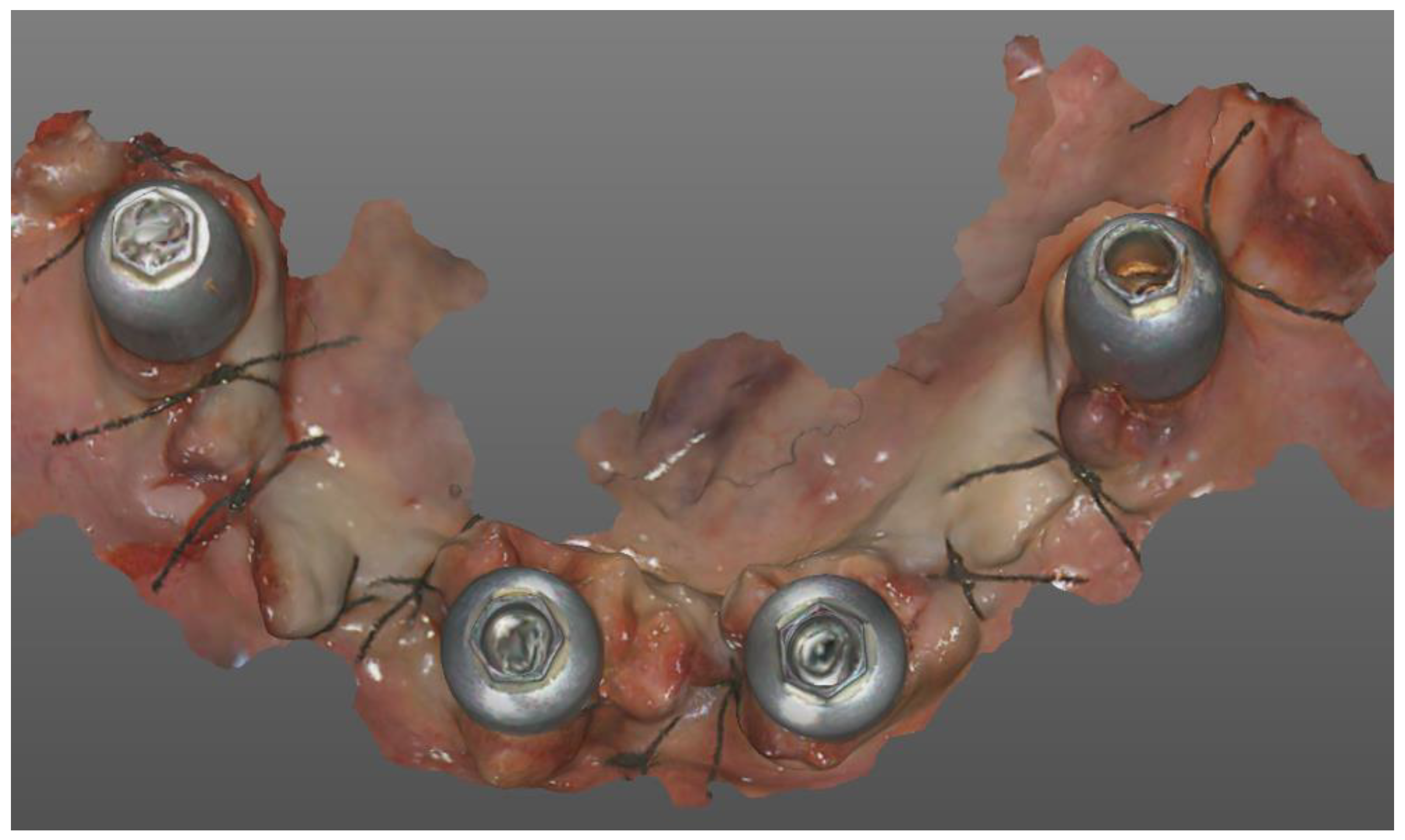
Figure 2.
Image of the post-surgical scan, performed on healed tissue with scan-body mech and human (Grisignano di Zocco, Italy).
Figure 2.
Image of the post-surgical scan, performed on healed tissue with scan-body mech and human (Grisignano di Zocco, Italy).
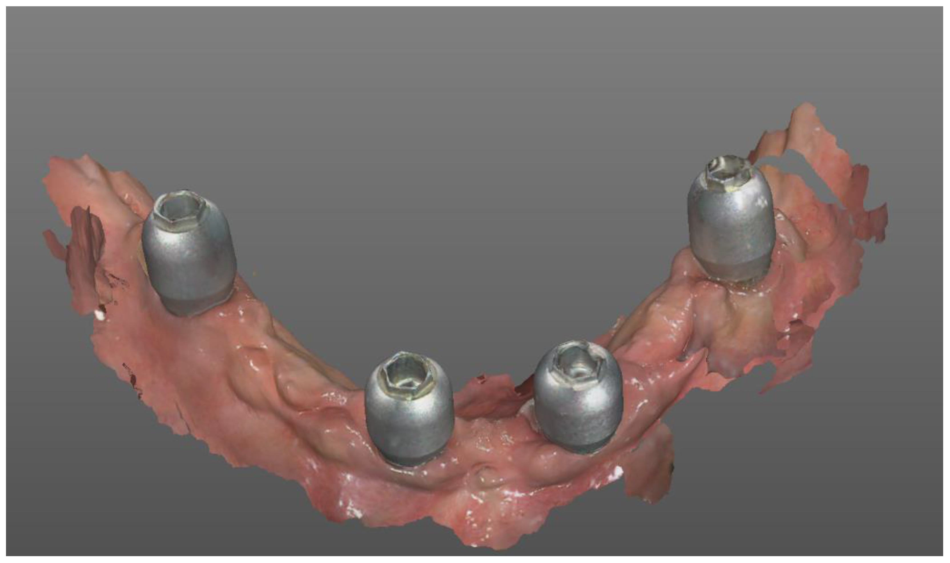
Figure 3.
Points identified in the analysis.
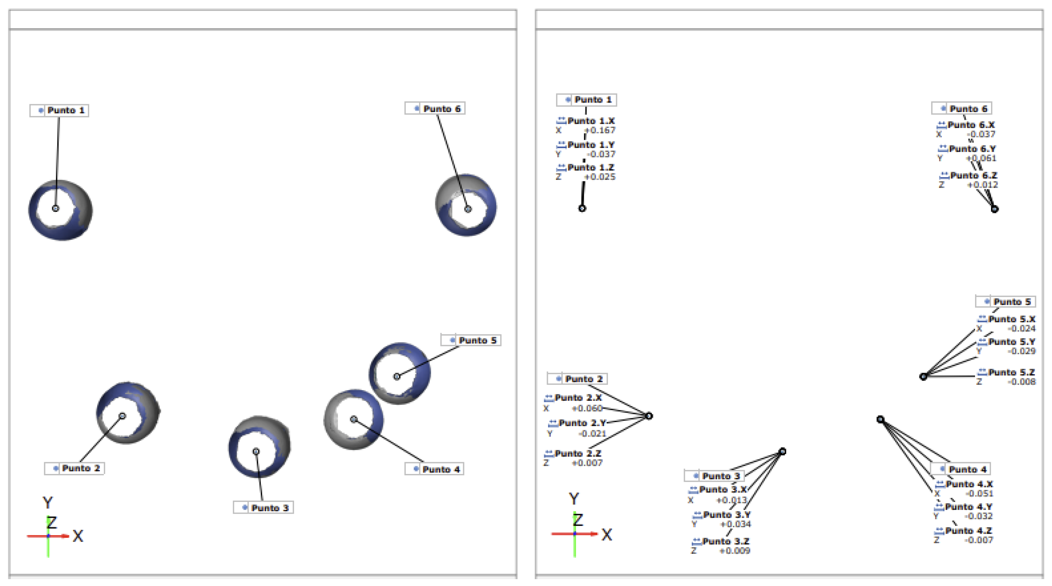
Figure 4.
Correlation between average discrepancy between scans and time in which they were performed. As time increases, the value of the discrepancy between the implants decreases. (p-value at 6%).
Figure 4.
Correlation between average discrepancy between scans and time in which they were performed. As time increases, the value of the discrepancy between the implants decreases. (p-value at 6%).
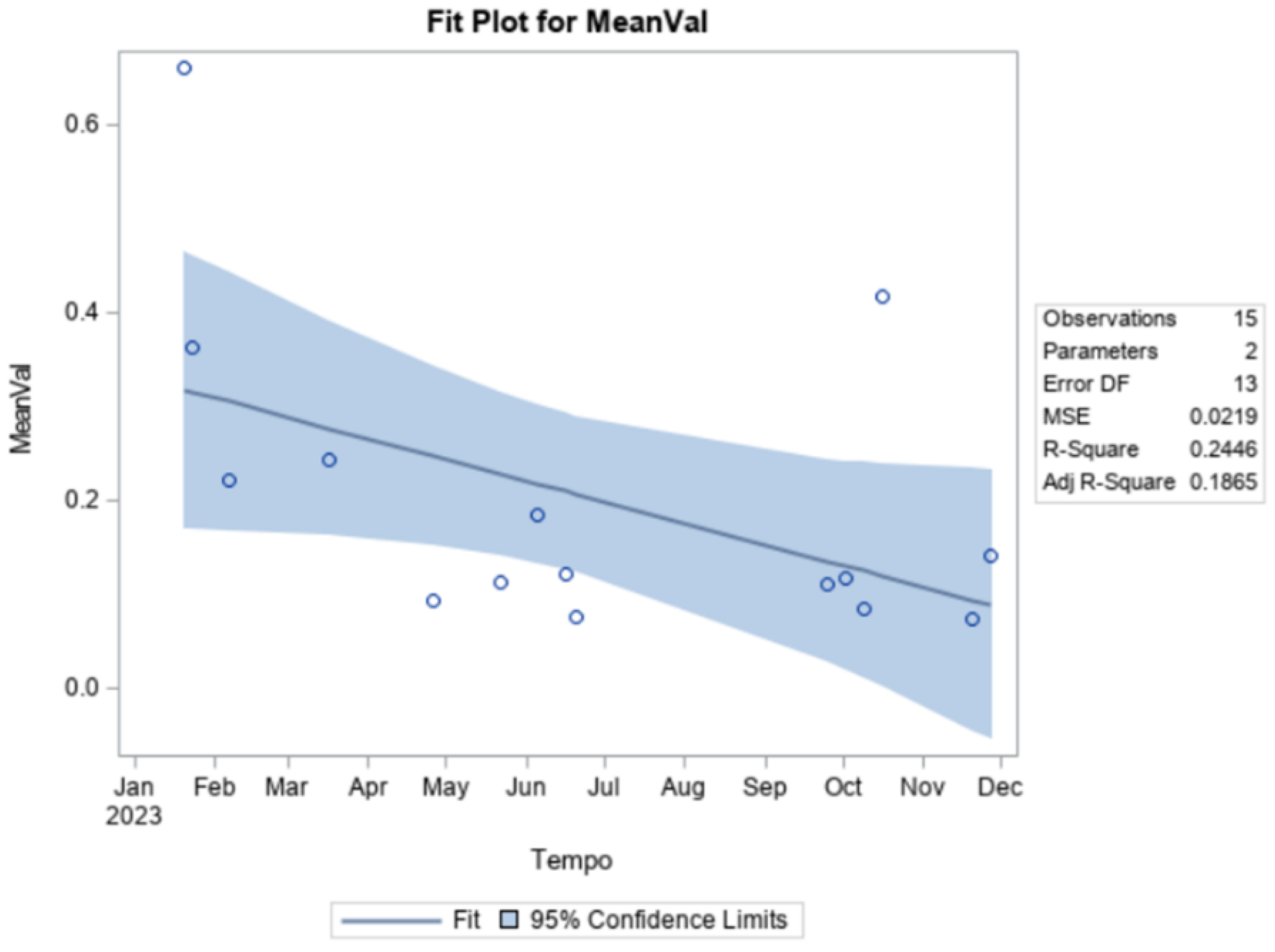

Table 1.
Average and mean discrepancy as a result of the superimposition of the digital scans for each individual implant.
Table 1.
Average and mean discrepancy as a result of the superimposition of the digital scans for each individual implant.
| CONFIDENCE INTERVALS IMPLANTS TYPE | |||
|---|---|---|---|
| Mean | Lower 95% | Upper 95% | |
| Implant 1 | 0.2362 | 0.1355 | 0.3368 |
| Implant 2 | 0.1777 | 0.1039 | 0.2515 |
| Implant 3 | 0.1555 | 0.0548 | 0.2562 |
| Implant 4 | 0.2124 | 0.114 | 0.3109 |
| Implant 5 | 0.0626 | -0.2436 | 0.3688 |
| Implant 6 | 0.0723 | NA | NA |
| All implants | 0.1905 | 0.1476 | 0.2335 |
Disclaimer/Publisher’s Note: The statements, opinions and data contained in all publications are solely those of the individual author(s) and contributor(s) and not of MDPI and/or the editor(s). MDPI and/or the editor(s) disclaim responsibility for any injury to people or property resulting from any ideas, methods, instructions or products referred to in the content. |
© 2024 by the authors. Licensee MDPI, Basel, Switzerland. This article is an open access article distributed under the terms and conditions of the Creative Commons Attribution (CC BY) license (http://creativecommons.org/licenses/by/4.0/).
Copyright: This open access article is published under a Creative Commons CC BY 4.0 license, which permit the free download, distribution, and reuse, provided that the author and preprint are cited in any reuse.


