
Submitted:
20 October 2024
Posted:
21 October 2024
You are already at the latest version
Abstract
Artificial intelligence AI is a sophisticated model or algorithm that has transformed a number of industries, including the medical field. In generative artificial intelligence, content is created using models or algorithms. Generative AI in healthcare revealed numerous new application domains for illness diagnosis. It has given models new outputs that they may use to create unique and fresh content. These can be used to learn new patterns that closely resemble the original data. The models covered in this review article serve as diagnostic tools in a variety of medical imaging applications. Medical specialists use generative AI in medical imaging analysis as a preliminary tool to generate large amounts of data that they can then correlate and extract useful information from. It is widely used in medical imaging, such as x-rays, magnetic resonance imaging, CTscan, COVID, cancer areas are utilized as AI diagnostic tools. Artificial intelligence (AI) has undergone a revolution because of to deep learning, which has made it possible for machines to learn from vast amounts of data and carry out complicated tasks based on that data. One of the most exciting uses of deep learning is in the creation of generative models, which are deep learning models that can produce realistic images, videos, and audio. These models have a variety of uses in the healthcare industry; including imaging. Medical imaging now has a plethora of options because to generative AI, a subfield of AI that focuses on producing original information. With improved patient outcomes, tailored treatment regimens, and greater diagnostic capabilities, it gives healthcare providers more control. Generative AI has transformed medical picture processing, interpretation, and application in clinical practice by harnessing the power of deep learning algorithms. Better diagnosis, New medicine discovery, Personalized medicine Improved medical imaging: The quality of medical images can be raised with the application of generative AI. With the aid of this technology, physicians may be able to diagnose and treat patients early by seeing more detail in images, More effective surgery: Virtual patient models can be produced using generative artificial intelligence, Surgeons can be trained and surgery plans can be made using these models, enhanced rehabilitation ,better mental health care: Chatbots that can provide patients therapy can be developed using generative AI. Medical imaging analysis is a field of healthcare that has revolutionized the way we approach patient care. The ability to examine medical images such as X-rays, CT scans, and MRI scans has allowed doctors and healthcare professionals to gain a better understanding of a patient's condition, monitor their illness, and plan their treatment accordingly. However, with the vast amounts of data produced by medical imaging technology, it is essential to have specialist analysis to retrieve useful information. One of the most significant advancements in medical imaging analysis has been the development of generative image AI models. These models, such as Generative Adversarial Networks (GANs), Variational Autoencoders (VAEs), and Auto encoders, have enabled healthcare professionals to analyze medical images more effectively and efficiently. The GAN model is one of the most popular generative picture AI models. It comprises two neural networks - a generator network and a discriminator network. The discriminator network's primary role is to discern between artificial and real visuals, while the generator network creates synthetic images. In an adversarial training process, the discriminator and generator work together to accurately categorize artificial and real images, with the generator trying to trick the discriminator. This technique has proved useful in radiology, where GANs are used for tasks such as image synthesis, image de-noising, and image segmentation. Image segmentation is a technique used in medical imaging analysis to split an image into several areas or segments. This has proved in identifying specific structures or regions in medical images, such as tumors, blood vessels, or organs. Generative deep models such as VAEs, GANs, and diffusion models have been applied to the augmentation of medical data, providing healthcare professionals with a more comprehensive understanding of a patient's condition. The development of generative image AI models has revolutionized medical imaging analysis, providing healthcare professionals with a more efficient and effective way to analyze medical images. Medical imaging has been a vital tool for healthcare professionals in diagnosing and treating various medical conditions. However, the interpretation of medical images is often challenging due to the complexity of the human body and the variations in anatomical structures. Therefore, image processing techniques are utilised to enhance the quality of medical images and extract relevant information. In this regard, fundamental image processing operations such as image segmentation, registration, and feature extraction are commonly utilised. However, the application of these operations might not always be appropriate due to the intricate structures of medical images, which include various anatomical variations and irregular tumour shapes. In the case of brain imaging, the presence of different tissues and structures, such as grey matter, white matter, and cerebrospinal fluid, can make it challenging to distinguish between healthy and diseased areas. More advanced image processing techniques such as machine learning algorithms are becoming increasingly popular for medical image analysis. The creation of irrelevant images that disturb the logical structure of the image can also occur due to the complexity of medical images. This adversely affect the accuracy of the interpretation of the image, leading to incorrect diagnoses and treatment plans. Hence, there is a need for sophisticated image processing techniques that can remove unwanted features and highlight the crucial information in the image. The performance of the model employed for image analysis might also be adversely affected by aberrant data and image deformations. Pre-processing techniques like noise reduction and picture normalization are essential for guaranteeing that the image is free of aberrations that could affect the interpretation's correctness. In conclusion, more sophisticated methods are needed to overcome the difficulties posed by the complexity of the images, fundamental image processing processes can be useful in improving the quality of medical images. In the end, these methods may improve patient outcomes by diagnosis.
Keywords:
Artificial intelligence
; medical imaging
; generative AI
; diagnosis
; diseases
1. Introduction
Artificial Intelligence
Artificial intelligence that requires training machine learning models to produce new data that is somewhat comparable to preexisting data is known as generative AI. Instead than concentrating on categorization tasks, this kind of AI focuses on creating new data, such as text, music, photos, or Videos [1].The practice of examining medical images to help with illness monitoring, diagnosis, and treatment planning is known as medical imaging analysis. Large amounts of data are produced by medical imaging technology, and in order to retrieve useful information, specialist analysis is required. In several medical specialties, such as radiology, cardiology, neurology, oncology, and more, medical imaging is essential [2,3].
One step in evidence-based practice is information searching, and access to information in health domains is crucial. Although the use of new technologies to enhance access is growing, information seekers frequently don’t know the fundamentals of the algorithms that rank data in information resources. The more readily disregarded algorithms are the ones that information workers are likely to come upon. Because of their seeming lesser influence than that of more well discussed algorithms, these algorithms, which have an impact on smaller, more routine jobs, could not be fully understood.
At the vanguard of technological progress, artificial intelligence (AI) is transforming economies, redefining human capacities, and upending entire sectors. Artificial Intelligence (AI) has become ubiquitous in modern life, ranging from complex neural networks operating driverless vehicles to intelligent algorithms powering virtual assistants. This essay highlights artificial intelligence’s transformative potential and significant impact on humanity by examining its evolution, uses, advantages, drawbacks, and ethical issues.
Artificial Intelligence Evolution
The idea of artificial intelligence has its roots in ancient philosophy and mythology, which mention artificial entities possessing intelligence like to that of humans. But the systematic investigation of AI as a scientific field started in the middle of the 20th century, thanks to ground-breaking work by luminaries like Alan Turing, John McCarthy, and Marvin Minsky [4,5]. Early artificial intelligence (AI) systems, such as rule-based algorithms and expert systems, set the groundwork for later developments in deep learning, machine learning, and neural networks.
Important Turning Points in the Development of AI
1. Symbolic AI: During the 1950s and 1960s, researchers concentrated on symbolic AI, which simulates human intelligence through the manipulation of symbols and logical reasoning [6]. This method resulted in the creation of expert systems that can handle challenging issues in particular fields, like finance analysis and medical diagnostics.
2. Machine Learning: The development of machine learning in the 1980s brought about a paradigm change in AI research by allowing computers to gain experience and learn from data. Pattern recognition, natural language processing, and predictive analytics have advanced thanks to methods like neural networks, decision trees, and support vector machines.
3 .Deep Learning: In the twenty-first century, artificial neural networks with numerous levels of abstraction served as the foundation for this subset of machine learning, which came to be known as deep learning. AI applications in speech recognition, picture classification, and autonomous systems have been revolutionized by advances in deep learning techniques, large-scale datasets, and powerful computer resources.
Applications of Artificial Intelligence: AI is used in a wide range of fields and sectors, spurring productivity, creativity, and revolutionary change. Several well-known uses of AI include:1. Natural Language Processing (NLP): Machines can now comprehend, interpret, and produce human language thanks to AI-powered NLP technology. NLP algorithms are used by virtual assistants such as Siri, Alexa, and Google Assistant to make tailored recommendations, respond to inquiries, and enable voice-activated interactions.
2. Computer Vision: Machines can now analyze and comprehend visual data from photos and videos thanks to computer vision algorithms. Applications include autonomous car navigation, medical image analysis, and facial recognition and object detection in security systems.
3. Healthcare: AI is revolutionizing healthcare by improving diagnostics, personalized treatment planning, and patient care. Machine learning models analyze medical data to assist in disease diagnosis, predict patient outcomes, and optimize treatment protocols. Additionally, AI-powered robotic systems assist surgeons in minimally invasive procedures, enhancing precision and patient safety.
4. Autonomous Vehicles: The development of autonomous vehicles represents a pinnacle achievement in AI and robotics. Advanced sensors, deep learning algorithms, and reinforcement learning techniques enable self-driving cars to perceive their environment, make real-time decisions, and navigate complex traffic scenarios with minimal human intervention.
5. Finance: AI is reshaping the finance industry by automating routine tasks, detecting fraudulent activities, and optimizing investment strategies.
Benefits of Artificial Intelligence: The proliferation of artificial intelligence offers numerous benefits to society, economy, and individuals, including:
1. Increased Efficiency and Productivity: AI automates repetitive tasks, streamlines workflows, and accelerates decision-making processes, thereby enhancing efficiency and productivity across industries. 2. Enhanced Accuracy and Precision: Machine learning algorithms excel at processing vast amounts of data and extracting actionable insights with unparalleled accuracy, leading to improved diagnostic accuracy, predictive modeling, and risk assessment[7].
3. Cost Savings: AI-driven automation reduces labor costs, minimizes errors, and optimizes resource allocation, resulting in significant cost savings for businesses and organizations. 4. Innovation and Creativity: AI fosters innovation by enabling the rapid development of new products, services, and solutions through data-driven insights, iterative experimentation, and adaptive learning.
4. Improved Quality of Life: From healthcare advancements to personalized recommendations and smart home automation, AI technologies enhance the quality of life by empowering individuals, improving accessibility, and addressing societal challenges.
5. Challenges and Ethical Considerations: Despite its transformative potential, artificial intelligence also presents several challenges and ethical considerations, including:
1. Algorithmic Bias: AI algorithms may exhibit biases inherent in the training data, leading to discriminatory outcomes, unfair treatment, and perpetuation of societal inequalities.
2. Privacy Concerns: The proliferation of AI-powered surveillance systems, data mining techniques, and predictive analytics raises concerns about privacy infringement, data breaches, and unauthorized access to personal information.
3. Job Displacement: Automation driven by AI technologies has the potential to disrupt labor markets, leading to job displacement, unemployment, and economic inequality, particularly for low-skilled workers.
4. Autonomous Weapons: The development of autonomous weapons systems raises ethical dilemmas regarding accountability, proportionality, and compliance with international humanitarian law, posing risks to global security and stability.
5. Existential Risks: Some experts warn about the existential risks associated with advanced AI systems surpassing human intelligence and autonomy, potentially leading to unforeseen consequences and existential threats to humanity.
Revolutionizing Medicine: The Importance of Medical Imaging with Generative Artificial Intelligence
Technological developments have played a key role in improving patient outcomes, treatment planning, and diagnostic accuracy in the field of modern medicine [8]. Among these developments, medical imaging is particularly noteworthy since it is essential to the diagnosis and treatment of a wide range of illnesses. The field of medical imaging has experienced a revolutionary shift with the advent of generative artificial intelligence (AI), which has unparalleled prospects for researchers and practitioners [9]. This essay explores the value of generative AI-enhanced medical imaging, explaining its relevance in the provision of healthcare.
Medical Imaging’s Development
Since its start, medical imaging has undergone tremendous progress. Imaging techniques have become essential tools in medical treatment, ranging from conventional X-rays to sophisticated modalities like computed tomography (CT), magnetic resonance imaging (MRI), and positron emission tomography (PET)[10,11].
The Benefits of Imaging in Medicine:
Almost every facet of healthcare delivery, including disease identification, treatment monitoring, and surgery planning, depends heavily on medical imaging. Here are some ways that medical imaging affects many aspects of medicine:
1. Prompt Identification and Assessment: Successful treatment outcomes are frequently dependent on a timely diagnosis. Physicians can identify anomalies early on with medical imaging, even before symptoms appear. For example, mammography increases patient survival rates by assisting in the early diagnosis of breast cancer [12,13,14].
2. Treatment Planning and Personalized Medicine: The anatomical and physiological traits of individual patients can be greatly understood through the use of medical imaging. Imaging helps with individualized treatment planning by clearly defining the position, size, and extent of lesions [15,16,17]. For instance, accurate imaging is beneficial in radiation therapy.
3. Interventional Procedures: Minimally invasive procedures have been transformed by imaging-guided interventions [18,19,20]. During biopsies, ablations, and catheter-based procedures, targets can be precisely localized to methods like fluoroscopy, ultrasonography, and MRI guidance. These processes are enhanced by generative AI, which also speeds up procedures and improves accuracy and real-time imagery.
4. Tracking Therapy Response Medical imaging is essential for tracking how well patients are responding to treatment [21,22]. Clinicians can analyze the effectiveness of treatment, track the development of the disease, and adjust treatment plans based on the results of serial imaging scans. In order to enable timely treatments, generative AI systems analyze longitudinal imaging data to discover small changes indicative of illness progression or therapy response.
The application of generative AI in medical imaging involves creating fresh data samples that bear similarities to the training set [23]. Generative AI is a subset of artificial intelligence. Generative AI algorithms in medical imaging use deep learning approaches to create new images, improve the quality of existing images, and extract information that is useful to clinical practice [24,25]. Numerous facets of diagnostic radiography and image-guided therapies have undergone revolutionary changes as a result of the use of generative AI into medical imaging.
1. Improving the image: Medical image quality can be improved by generative AI systems through noise reduction, resolution enhancement, and contrast optimisation. These algorithms produce higher diagnostic value high-fidelity images by extrapolating information from existing datasets. Generative adversarial networks (GANs) can, for instance, denoise MRI scans and fill in missing data to enhance image clarity and diagnostic precision [26].
2. Artefact Correction: Motion, metal implants, and flaws in the imaging apparatus can all result in artifacts in medical images. Images can be more easily interpreted and accurately diagnosed when these artifacts are correctly identified and corrected using generative AI algorithms [27]. For example, motion artefacts from MRI scans can be removed by deep learning-based algorithms, making anatomical components easier to see.
3. Image Reconstruction: By using generative AI, noisy or under sampled data can be used to reconstruct high-quality images, cutting down on scan times and radiation exposure. Imaging modalities like CT and MRI are made more efficient by methods like deep learning-based iterative reconstruction, which enables quick acquisition of diagnostic images without sacrificing image quality [28,29].
4. Synthetic Imaging: To help with data augmentation and algorithm training, generative AI may create synthetic medical images with anatomical features that are realistic. For a variety of imaging tasks, such as segmentation, classification, and anomaly detection, deep learning models can be robustly trained using synthetic datasets produced by GANs or variational auto encoders (VAEs)[30].
5. Clinical Decision Support: By using medical imaging data as a basis, generative AI models can produce predictive analytics that help physicians make decisions. These models can offer risk assessment, prognosis evaluations, and therapy recommendations based on the unique profiles of individual patients by examining patterns in imaging data.
Generative AI
In fact generative AI is taking the data of new origin but having similarity with already present data so that helps in medical imaging analysis. Medical pictures such as x aray utilized images , mri based images and ct scan related are diagnostic tools where artificial intelligence potentiates diagnosis of diseases. It is a rapid and precise diagnostic method. The interactive effect of the selected radiation on a particular tissue creates medical imaging. at about 600 million medical imaging procedures are available yearly in USA [31]. This is an expensive and time consuming if performed in manual mode. Many algorithms are developed by doctors in order to produce and convert images into a numerical format for diagnosis.In medical imaging 3D images are constructed of selected anatomical structures. These images are virtual device simulations so that further steps like surgery and treatment are planned.
Image Enhancement
According to Rizwan Khan et al. early stages of dental pathological conditions are performed using generative ai techniques. Image enhancement is one of such methods . in this ed-net , edification network place an important role[32]. New type of image enhancement strategy using denticle –edification network. Edification network was presented in Figure 1. Edification network consists of encoder and decoder. Generally poor resolution images pose problems. So the captured images are partitioned into 2 components ie. Reflection and illumination components. Unwanted artifacts are eliminated using clinical oriented enhancement method. Images are captured using radiance, finally image formation using x, y pixel coordinates depicts by global light component and light transmission component.
Image Segmentation
J Marie et al. worked on a rapid and robust, segmentation algorithm for MRI image enhancement. MRI images re fuzzy enhanced and healthy structures are separated from unhealthy structures. [33]. Lesion contour is then focused using initialization free level set evolution method. Image segmentation is a difficult task and challenge for high level recognition.
Slice by Slice Integration
In cardiac diseases estimation of diseases and their progression is ans important task. One important example for this situation is segmentation of epicardial fat. This is a risk factor for cardiovascular diseases. Utilizing semiautomatic approach epicardial fat present in ct medical images in 3d format was put forward by V. Zlokolica et al. . in this slice by slice integration of 2D processing finally helped in identification of undesired parts of target cluster.
Methods of Image Enhancement [34]
There are mainly 2 image enhancements methods are there
Spatial domain method
Frequency domain method
Spatial Domain Method
Direct manipulation of image pixels done by spatial domain method. Medicl image quality and appearance is improved by spatial domain method. A pixel value with coordinates in enhanced image is done by using some operation in neighborhood of the captured image. Some important aspects of spatial domain method include grey scale manipulation where operator works
Image appearance is enhanced by histogram equalization. Histogram is skewed to grey scale lower end and compressed into dark end of histogram.
Camera noise, spurious pixel values and other features are reduced by image smoothing. Fine details of image are highlighted and enhanced if they are blurring using image sharpening.
Frequency Domain Method
Captured image Fourier transform is modified using frequency domain processing method. Using Fourier transform signal is converted from time domain to frequency domain. It is indirect manipulation. Image enhancement methods involve filtering by law pass filtering, homomorphism filters.
2. Applications
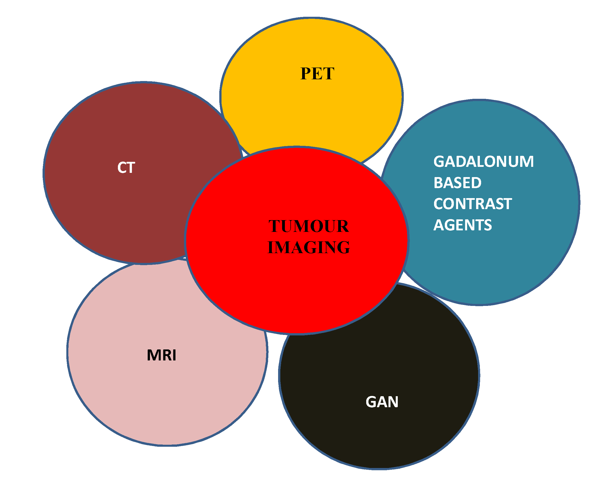
Tumour Imaging Using Generative AI [35]
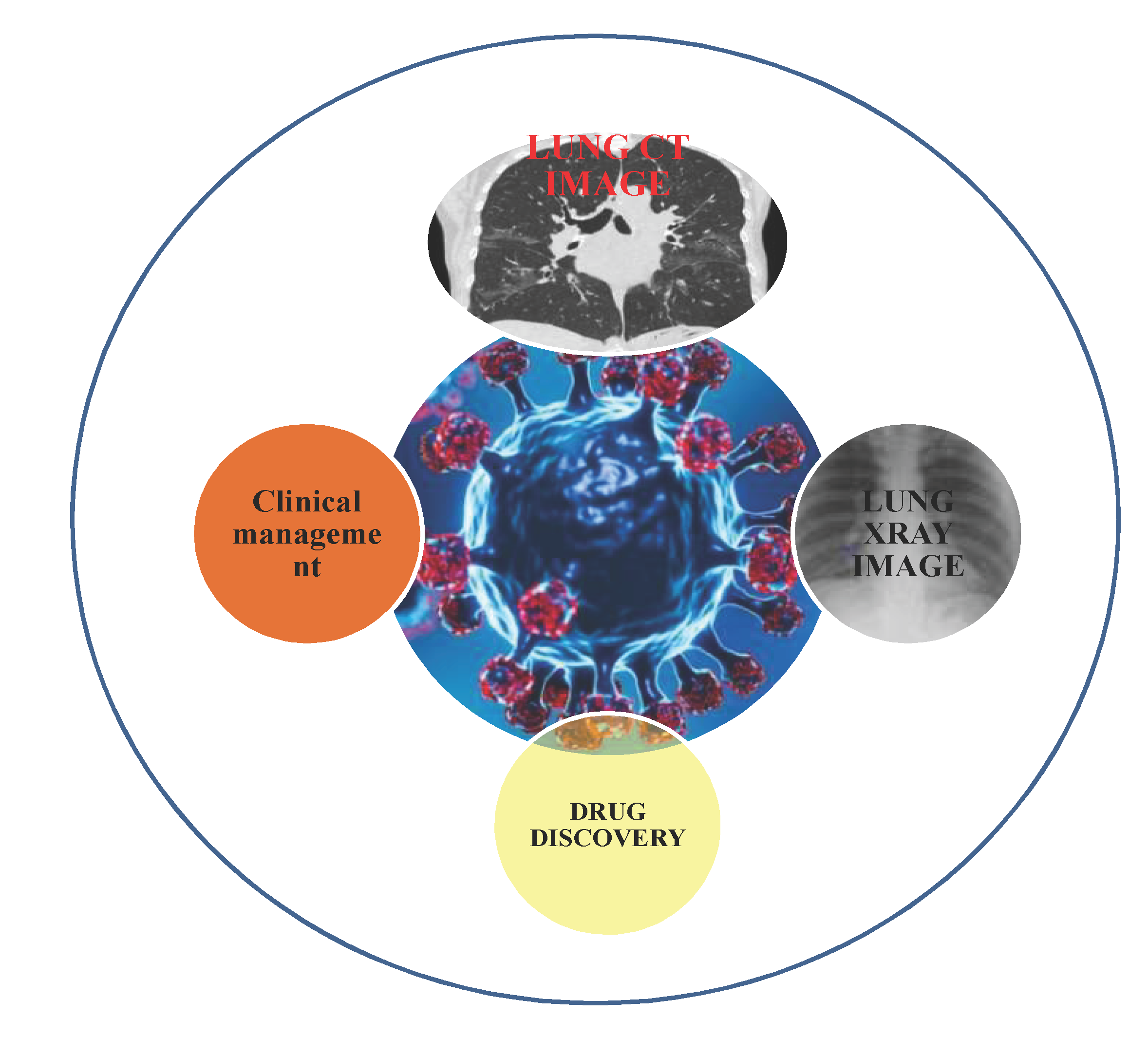
Generative adversial network helps in production of new images from the captured old images. This is called GAN, which is a deep learning technique. Because of his technique gadolinium based contrast agents usage is nullified. But gadolinium contrast agents are considered to be safe. Fig no. 2. Illustrates role of Generative AI in disease diagnosis- tumour imaging
Oncologic imaging is an important domain. It is one of the good non invasive too. This envisages the future advancements, further steps made easy. Digital revolution potentially transformed treatment options of cancer conditions. Captured image acquisition and optimization can be performed by deep neural networks. Radiation dose can be reduced using deep learning construction. Because of which image quality can be improved. In MRI imaging k-space base reconstruction methods especially robust artificial neural network for k-space interpolation is applied. Traditional or conventional imaging techniques have low efficiency in terms of performance parameters like speed which is 2-4 times more in ai method. Adversarial neural networks are having disadvantages like tendency to give image of poor quality real image appearance. among the contrast agents, gadolinium based derivatives are safer . But gadolinium administration causes deposition in brain parenchyma after subsequent doses. Sometimes contrast induced injury to nephritic organs is quiet common. In this aspect deep learning techniques can reduce dose of contrast agents. Further their associated costs, adverse events can be minimized. According to haubold et al. and gong et al. GAN linked with deep learning algorithm reduces ct imaging contrast media upto a level of 50 percentage but accuracy of detection and identification is not lost.
Cancer conditions early screening in case of breast, lung and colon is a challenging situation especially in asymptomatic individuals. Ai systems of commercial use may be beneficial in comparison to double reading consensus. 2 steps are involved in lung cancer screening i.e detection and risk assessment in malignancy [36]. In malignancy risk assessment ai algorithms shown more beneficial and efficient systems. But limitations are observed in inferior or equivalent performance in nodule detection.
Tumour related imaging helps in diagnosis and gives details in the extent of primary stage tumor disease and identification of local and distant metastases in order to go for best treatment plan. Surgeons well planned surgery is linked to anatomical imaging. AI helps to address variability and elevation of level of care is observed. 3d post processing software helps in precise evaluation of anatomy and volumes of lesion. Brain metastates can be diagnosed using radiation sources but takes lot of time.
After treatment tumour response assessment can be a difficult job and especially in tumor size measurement. Real extent of disease, changes in morphology is a time consuming phenomenon. Generative AI facilitated volumetric changes in tumour is a good approach. Assessment of responses in pleural tumors of lung uses deep learning AI algorithms . in case of glioma segmentation, multiple components related tumor enhancement is a not a simple phenomenon. It is a challenging event various deep learning algorithms are used. Lotal et al. .deep learning based clinical work flow is one of such applications where gliomas are segmented before and after surgery. Tumor distribution in sub regions are quantitatively well studied using MRI, PET CT related quantitative imaging procedures.
Clinical Setting
In health care generative ai disclosed many new areas of scope with disease diagnosis applications. AI given new outputs for models which can produce original, new content. These models are helpful for learning new patterns which can mimic the original data. These models have several applications in image synthesis in disease diagnosis. This review article focuses on medical imaging using generative ai as diagnostic tool. Medical imaging analysis is and important process to produce vast content of information which is a preliminary device used by experts to correlate and extract meaningful information. Artificial intelligence is one of the advanced model or algorithm and revolutionalised different industries including health care. Generative artificial intelligence involves algorithms or models to produce content. Medical images like x rays, magnetic resonance imaging, ct scan are utilized ai diagnostic tools.
In emergency health care services, handling tertiary care catering diagnostic technology. Generative AI interpretation on the produced radiographs is very important. Figure no. 3. Portrays role of generative AI in clinical setting . According to Jonathan Huang bs et al., chest radiograph interpretations were carried and data of 500 random samples selected. Radiographs anterior- posterior or posterior- anterior data information evaluated. During the process of clinical decision making quality parameters of AI report was taken [37]. AI applications is taken as a additional benefit of physician interpretation, therapeutic decision making. Radiograph acquit ion, review took less time. Doctor AI assisted clinical decision making is a synergistic process and this collaborative approach gives promising results.
In clinical setting, personalization/individualization of therapy by medical image analysis and patient data is done by generative AI. Empowerment of health care professionals is one of the key aspects in strengthening of health care system and improved quality of treatment. Because of AI algorithms generated simulations and predictions make a unique patient approach.
Figure 3.
generative AI in clinical setting.
Clinical Decision Support
Glass AI is LLM powered experimental tool for making clinical decision support.[38] For example the patients showing symptoms like tiredness, shortness of breath and paleness differential diagnosis can be provided by glass ai. Finally the conditions like anemia, myelodyspastic syndromes and leukemia can be suggested ultimately. So that clinical plan of actions can be taken for further guidance. DiseaseDiagNet- AI model leveraging vast medical data for accurate diagnosis, aiding clinicians in decision-making. It generates detailed diagnostic tables for comprehensive patient assessment and treatment planning.
Medical Imaging
Fast automated segmentation tool called ( FAST) (redbrios AI) offers applications in the field of medical imaging. SAAS program for medical image data annotation in case of radiology. FAST automates manual segmentation, increases diagnostic accuracy and speed n radiology. Image view, management and sharing of digital slides of tissue samples empowerment by HCP (generative AI) can be done. Complex tissue patterns are identified, analyzed and diagnosis of cases. Kahun’s AI inference engine delivers a ranked list of potential diagnoses speed up diagnostic processes. Grid space automates routine administrative tasks like appointment scheduling, patient reminders, insurance etc…
Synthetic mind uses generative AI to produce realistic synthetic patient records from real health care data like electronic health records fir patient privacy protection. Abridge is a digital tool that utilizes generative AI to document medical dialogues, saving physician from manual note taking. It plays a vital role in patient education..
Decision Support
Decision support in healthcare settings is important decision assistance is entirely new era. An algorithm created by a computer may be useful in much decision. In health care performance metrics are classification- accuracy, sensitivity, specificity etc.
Ephzibah developed a model for identifying diabetes. This model combines fuzzy logic with generative artificial intelligence.[39] This is used to improve classification accuracy. The fuzzy logic classifier uses 3 features which offer 87% accuracy compared to conventional biostatical approaches, machine learning approach is more beneficiak. Sen and dash discussed meta learning techniques for diabetes diagnosis.[40] When insulin deficiency conditions are observed, diseases develop. Parthiban and srivastsa attempted to diagnose cardiac problems in diabetic individuals by utilizing machine learning techniques. Chaurasia & pal recommended data mining techniques to identify heart diseases. For minining purposes, WEKA data mining tool is used which includes several machine learning techniques. Vembaandasamy et al. used naïve bayes algorithm to diagnose cardiac illness. This is based on Baye’ s theorem[41]. Sarwar and Sharma et al. recommended naïve bayes algorithm to differentiate 3 forms of diabetes like type-1 and type-2 diabetes and gestational diabetes [42].
COVID 19 and Drug Discovery
From the first reporting of COVID 19 cases in the year 2019, many people’s lives affected drastically various impacts of COVID 19 led to divesting and dreadful situations[43]. Figure no. 4. Illustrates applications of generative AI in COVID 19 and drug discovery. Clinical symptoms like flu, cold, cough , fever , tiredness , difficulty in breathing has happened. Because of the infected persons transmission to the contacted persons was observed. But false negative cases transmission was affected a lot. Ai enabled many public health diagnostic services, and there by decision making and treatment. Machine learning and deep learning methods with the assistance of electronic medical records and imaging technology like x ray, CT, and ultrasonic images in diagnosis of COVID 19. Some of the documented applications in COVID 19 gave information about data sources utilization. ‘According to Ardakani et al. real time data from university hospital analyzed 1020 number of images’. ‘Wu et al. documented 495 images from china medical university using deep learning screening framework.’ [44]. ‘Hasan et al. documented COVID 19 data set of SPIEAAPM- NCI lung nodule classification challenge data set of 321 patients of which 118 COVID 19 and 107 healthy.’[45] After studying of the literature, Res Net-101 exhibited 99.51 percentage of accuracy , 100 percentage of sensitivity of outstanding performance parameters. There is another variant of deep learning method which utilize multitasking deep learning to diagnose COVID 19 lesions in ct scans.
Figure 4.
Applications of generative AI in COVID 19 and drug discovery.
Machine learning (ML) has potential applications in imaging of lung infected by COVID 19. Pre trained model utilized , squeeze net , Google net , res-net -18,50,101 and xception techniques for ct image analysis of 1020 patients data (Ardakani et al.). Using customized model, QDE-DF technique 321 patients ct imaging was performed with an accuracy of 99.68 performances. Loey et al. documented COVID-19 x ray image analysis of 307 human information using GAN –generative adversarial net work utilizing deep learning gave excellent analyzing of chest x ray images [46]. Differential diagnosis of x ray images of COVID 19, pneumonia bacteria, pneumonia bacteria, pneumonia virus and normal were identified. Deep learning method utilized techniques helpful for using ultrasonogrpahy detection and management. Asymptomatic patients means COVID 19 infected but who doesn’t exhibit any symptoms were potential transmitters of disease. Recognition of such cases is important to reduce infection burden among public health. Another type of diagnosis is false medical image methods (laboratory diagnosis – RT-PCT). Medical image methods help in accurate diagnosis in conjugation with covid-19.
Some of the challenges in machine learning is reduction of false negative diagnosis, improvement of accuracy are main challenges taking into considerations of history, lab tests and imaging techniques like CT and x ray imaging . Differential diagnosis of other viral pneumonia challenge was solved by a 3 D CNN- based model using ct imaging with accuracy rate of 86.7 percentage. Influenza pneumonia and healthy patients was done. Radiological image manifestations should be observed in case of patients with many diseases. Viral and bacterial pneumonia, COVID pneumonia detection was proposed with an accuracy of 99.8. Combining the features of distinctive filter learning and integrating channel shuffled dual branched CNN model, in deep neural networks analyze COVID 19 radiographs segmentation into several small numbers of units for model training. This is one of the new technique introduced by oh et al. to overcome disadvantages like limited training data.
Generative AI utilized medical image analysis automates and improves accuracy and precision of medical image analysis. So that image segmentation, synthesis and patient outcomes. It uses models trained on large data sets so that new data is used in various areas. WGAN’s is a version of GAN’s, evaluates actual and produced data distribution differences. Image synthesis, style transfer and data creation tasks are tools. Auxiliary classifier GAN’s is the classifier produced samples are controlled in a better way. Veracity and class label are predicted using this. COVID-19 pandemic situation utilized some detection tools like COVID GAN, generating synthetic chest x ray images (ACGAN’S based model).[47,48] CNN’s based classification improved from 85-95% accuracy. Speed and robustness in radiology systems can be enhanced.
CT Imaging
CT imaging with assistance of ai especially in the area of diagnostic applications (CT imaging ) 19is a gold standard. Radiation at higher doses poses problems than traditional methods. Among all higher risk of patients like children, obese and cancer patients go for regular screening. ct imaging using new artificial intelligence using deep learning reconstruction and post processing techniques are available. During ct images analysis, image quality and reduction of dose exposure is complex. Radiologists generally faces high noise with poor quality images interpretations take more time for review of study data. Reading efficiency and report generation time makes a greater differences observing different methods of radiology based diagnostic procedures. Productivity is reduced with inefficient reading procedures. Iterative reconstruction called IR is one of the method was implemented in late 2000s. it reduce other disadvantages. IR algorithms are specific to scanner and vendor. Images captured and acquitted are procured using distinct artifacts and textures.
Challenges
New ai based machine learning techniques are used CT imaging in case of clinical and technical aspects . Image noise is observed in low dose CT procedures causes a huge impact on quality, efficiency and cost of imaging services.
CT and MR Imaging in Oncology
Computed tomography and magnetic resonance imaging was observed with newly emerged ai based tools. Tumor characterization uses deep learning image biomarker like histopathological, metabolic and functional status. Accurate tumor segmentation can be combined to overcome time limitation. Lung cancer, colonic polyps can be imaged using CT screening. MR guided radiotherapy images of head and neck are sufficient for segmentation to increase specificity compared to low dimensional machine learning method, uses computer aided detection software for lung cancer screening IDH1 mutations , IP /19 q co deletion , EGFR as well as VEGF and p53 are used to identify imaging biomarkers using machine learing and deep learning methods.
Advancements in Medical Imaging
Medical imaging is the practice of medicine help to disease diagnosis pathological specimen visualization. Biological tissues interact with electromagnetic energy waves so that images are formed.
- Tissue slide mechanism another variant is tissue microarray (TMA). TMA technology is a potential tool for diagnosis, gives information with respect to progression of disease and patient information related to treatment. From histological specimen smaller cylinders are taken and arranged in a matrix configuration on a paraffin block and simultaneously analyzed.
- Optical coherence tomography (OCT) generates a series of cross sectional optical images of 3D fashion. Echo delay time and back scattered light in linearity measurement of internal tissues are performed.
-
X-imaging is one of the less expensive methods and also takes less time for image acquisition. In order to enhance the resolution of image, visibility iodinated contrast agents are used and administered into the interested areas.
- Phase contrast x- ray imaging method enhances soft tissue contrasting using the principle of phase shift.
- X- ray projection imaging is generally used in pervasion of cardiovascular , mammography and abdominal imaging purpose.
-
Ultra sound imaging utilizes pulse in 1-100 MHz this procedure is a non- invasive and less expensive method. It is a rapid method. In this method non- ionizing radiation is used. Acoustic pulse interacts with internal structure and echo is measured in order to construct medical image.
- Contrast enhanced ultrasound is enabling more image accuracy and contrast using micro bubbles.
- Ultrasound elasticity image is another tool for analyzing and measurement of tissue softness.
By these methods 2D, 3D and 4D imaging methods are advanced and resolution decreased temporally.
- 5.
- X- Ray CT imaging method takes scans like that of MRI where as CT constructs 3D images of 2 D axial slices of body. Like MRI, 4D scans are also possible by gating to ECG and respiration. Spatial resolution improvement to 0.25 mm is possible by this technique and also diagnosis. In traditional method of tissue staining, where the haemotoxylin and eosin stains are most commonly used stains. Using staining tissue morphology can be studies so that pathologist can interpret in accurate manner.
Medical Image Analysis
Delineation of objects which is called segmentation and description called classification comes under medical image analysis. Machine learning strategies help in the understanding of image parameters and analysis. Deep learning algorithms are used for disease classification, screening. New fine tuning provided best results. Machine learning automated systems made the process easy by optimal networks hyper parameters.
Generative adversarial networks (GANs) are a form of deep learning algorithm utilized in unsupervised machine learning. Comprising two neural networks—a generator and a discriminator—GANs are trained on a dataset of medical images to generate new images resembling the training set. These generated images find application in tasks such as image segmentation, which entails partitioning an image into distinct regions and assigning labels to each region. The segmentation masks produced by GANs can be employed to train supervised segmentation algorithms.
Additionally, GANs are employed in medical image analysis for image-to-image translation, facilitating the conversion of one type of medical image into another, such as from CT to MRI scans. By generating synthetic images resembling the target modality, GANs serve as a valuable tool for data preprocessing, aiding in downstream analysis or segmentation tasks.
Wasserstein GANs (WGANs) belong to the category of GANs and aim to mitigate issues such as mode collapse and training instability. They offer a more accurate assessment of the dissimilarity between the distributions of real and generated data compared to traditional GANs. WGANs find applications in various fields including high-quality image generation, data augmentation, domain adaptation, and improving GAN training for specific tasks.
Auxiliary Classifier GANs (ACGANs) represent another variant of GANs. They introduce conditional generation, allowing for enhanced control over the characteristics of generated samples. By predicting both the authenticity of the data and its associated class label, ACGANs are adept at tasks like style transfer, image-to-image translation, and generating images with specified attributes. They are particularly useful in applications requiring precise and diversified data production, thanks to their ability to generate conditional outputs tailored to specific requirements.
- Generative AI health care market
The uses of generative AI in healthcare are numerous and extensive. Through the analysis of vast amounts of medical records, it can help with disease prediction, risk factor identification, and improved diagnostic accuracy. AI can help medical imaging by creating synthetic images that mimic various situations, which can help with image technique refinement and healthcare professional training. It can also be used to anonymize data, which removes personally identifiable information while maintaining statistical characteristics and protecting patient privacy. The global market for generative AI in healthcare is being pushed by a number of factors, such as increased R&D activity, technical improvements, and rising major player collaboration[48]. market size reached USD 1.45 billion in 2023, will reach USD 21.74 billion by 2032, and is predicted to grow at a CAGR of 35.14% between 2023 and 2032.
- b.
- Ethical issues
Due to generative AI algorithms vast medical image data is produced which is difficult to maintain so that data privacy at risk and patient data security may not be provided. Algorithm related interpretation of data is another feature to be considered[49]. So that clinical decision making will be easy. That means complexity in algorithm interpretation has to be minimized. Bias and error generation is to be avoided. Regulatory related standard like safety, efficacy and especially usage of ethical principles of technology has to be ensured.
Medical imaging generative artificial intelligence (AI) has great potential to improve healthcare by supporting physicians in research, diagnosis, and therapy planning. But in addition to its possible advantages, it also raises important ethical questions that need to be carefully considered. A number of ethical concerns, including patient privacy and consent, algorithmic bias, and professional decision-making, surface as AI systems get better at creating medical pictures.
The privacy and permission of patients are two of the main ethical issues. Sensitive information about a person’s health, including distinguishable characteristics, is contained in medical imaging data. There is a chance that patient identities will be unintentionally revealed when generative AI algorithms are used to make or modify medical images. Moreover, concerns concerning patient consent are raised by the secondary use of medical data for AI model training.
Ethical concerns around patient privacy and consent have gained prominence in the quickly changing field of medical imaging boosted by generative artificial intelligence (AI). Although there is great promise for new technology developments to improve healthcare outcomes, there are also serious issues about how sensitive patient data is protected and how medical data is used ethically. The unintentional release of patient identities via AI-generated photos and the subsequent use of patient data for AI model training without express authorization are the two main ethical concerns that arise in this situation.
The first ethical concern concerns the unintentional release of patient names during the creation or modification of medical images using generative AI algorithms. Sensitive information about a person’s health, including distinguishable traits like anatomical structures, distinct diseases, and biometric traits, is intrinsic to medical imaging data. There is a chance that patient names could be inadvertently disclosed through minute clues or identifiable patterns when generative AI algorithms are used to alter these pictures. For instance, minute differences in disease or anatomy might make it possible to identify particular people, especially in tiny or close-knit societies.
Furthermore, worries regarding data security and unauthorised access are raised by the widespread use of AI-generated medical pictures in clinical practice, education, and research. Due to the widespread distribution of these photos in academic, hospital, and commercial settings, there is an increased danger of data breaches, malicious use, or unintentional patient information exposure. Robust mechanisms for data encryption, access control, and anonymization are necessary to protect patient privacy and reduce the possibility of unauthorized disclosure. The secondary use of medical data for AI model training without express patient authorization is the subject of the second ethical dilemma. The basis for training generative AI systems to carry out operations like picture synthesis, artifact correction, and augmentation is provided by medical imaging datasets. Nonetheless, there are moral questions when using patient data for algorithmic advancement.
In addition, the commercialization of AI technology and the co modification of medical data provide new challenges in resolving consent and patient autonomy disputes. Technology businesses, research groups, and healthcare facilities frequently collaborate to gain access to huge medical datasets for the purpose of developing and validating algorithms. Transparent governance frameworks, moral standards, and patient involvement tools are necessary in these kinds of partnerships to guarantee that patients’ interests are taken seriously and safeguarded.
To tackle these moral quandaries, a multifaceted strategy is needed, one that combines institutional policies and technology safety measures with legal, regulatory, and ethical frameworks. In order to safeguard patient privacy and confidentiality, healthcare organizations and research institutions need to have strong data governance standards in place. These practices include data anonymization, de-identification, and encryption. Furthermore, laws and regulations should be passed by legislative and regulatory organizations.
In the context of generative AI in medical imaging, algorithmic bias poses yet another significant ethical dilemma. Biases included in algorithms or training data might provide skewed results and worsen already-existing health inequities. AI-generated images may, for example, produce incorrect diagnoses or insufficient treatment suggestions for underrepresented populations if they primarily feature members of a certain demographic group. In order to achieve justice and equity in healthcare results, addressing algorithmic bias necessitates the use of representative and diverse datasets in addition to ongoing monitoring and mitigation techniques.
Furthermore, there are ethical questions about clinical decision-making and responsibility raised by the interpretability and openness of AI-generated medical pictures. Artificial intelligence (AI)-generated images might not have explicit descriptions of the underlying processes, in contrast to traditional medical imaging methods where clinicians can interpret the images using accepted concepts.
Furthermore, ethical concerns about healthcare access and resource allocation are brought up by the economic consequences of generative AI in medical imaging. Although AI has the potential to improve diagnostic speed and accuracy, its application could also make the gap between those who have access to healthcare and those who do not increase. The exorbitant expenses linked to AI technology have the potential to worsen healthcare disparities by restricting access for underprivileged populations or healthcare establishments with restricted funds. In order to advance healthcare justice, ethical frameworks should give equal access to AI-driven medical imaging technology top priority and take into account the wider socioeconomic ramifications.
In summary, generative AI offers difficult ethical issues that need to be carefully considered even if it has great potential to transform medical imaging and enhance patient care. Ensuring transparency in healthcare decision-making, while also eliminating algorithmic bias and protecting patient privacy.
3. Challenges and Future Scope
Generative AI applications in medical imaging include accuracy in terms of image classification, segmentation, synthesis. Ai generated images are available in vast. Radio genomics is related with integrative analytical approach n disease in order to extract and analyze image data. Insilco modeling approaches medical image analysis with emphasis on disease etiology, pathogenesis and biomarkers should be developed at full potential. Fda regulatory approval for ai based medical imaging solutions must be obtained. Data storage / preservation, processing, privacy is one of the boundary for research. Reliability of data generated from multiple modalities and its interpretation is another challenge. Generative AI is a wing of machine learning and handles a variety of text, images, computer code in various fields.
Healthcare delivery, diagnosis, and treatment could all be completely transformed by the application of generative artificial intelligence (AI) in medical imaging. But even with all of its potential, generative AI in medical imaging raises a number of ethical questions that need to be carefully considered and dealt with early on. Navigating the ethical environment of generative AI in medical imaging necessitates a comprehensive strategy that strikes a balance between innovation, ethical principles, and patient welfare. This includes concerns about patient privacy and permission, algorithmic bias, and clinical decision-making.
Patient privacy and data protection are two of the main ethical issues with generative AI in medical imaging. Medical imaging data include sensitive information on the anatomy, health, and perhaps identifying traits of an individual. When using algorithms for generative artificial intelligence to build or modify medical imaging.
Another significant ethical issue with the application of generative AI in medical imaging is algorithmic bias. Biases in algorithmic design or training data might cause skewed outcomes, worsening already-existing health inequities and endangering patient care. AI-generated images may, for example, produce incorrect diagnoses or insufficient treatment suggestions for underrepresented populations if they primarily feature members of a certain demographic group. Diverse and representative datasets, ongoing monitoring, and mitigation techniques are needed to address algorithmic bias and guarantee accuracy, fairness, and equity in healthcare results. Furthermore, in order to successfully uncover and mitigate biases, AI development procedures must be transparent and accountable.
Clinical trust and decision-making are ethically challenged by the interpretability and openness of AI-generated medical pictures.
The possible abuse or hostile manipulation of AI-generated medical images is a further ethical consideration. With the increasing sophistication of AI technology, there exists a potential for adversaries to leverage vulnerabilities and produce misleading or counterfeit images. Malicious actions of this kind may have dire repercussions, such as incorrect diagnoses, ineffective therapies, or integrity in medical research compromised. Robust cyber security measures, regulatory monitoring, and ethical norms are necessary to safeguard the integrity and authenticity of AI-generated medical pictures, prevent misuse, and guarantee the dependability of AI-generated outputs.
Furthermore, ethical concerns about healthcare access and resource allocation are brought up by the economic consequences of generative AI in medical imaging. Although AI can improve the efficiency and accuracy of diagnosis, its application could also make healthcare disparities worse. The high expenses of AI technologies have the potential to increase the wealth gap in healthcare, making it more difficult for underprivileged populations or underfunded healthcare facilities to receive treatment. In order to advance healthcare justice, ethical frameworks should give equal access to AI-driven medical imaging technology top priority and take into account the wider socioeconomic ramifications.
In spite of these moral dilemmas, generative AI in medical imaging has enormous potential to advance medical research, improve patient outcomes, and streamline clinical procedures. We can fully utilize generative AI to help society while maintaining the principles of beneficence, autonomy, fairness, and non-malfeasance in healthcare by proactively addressing ethical considerations and incorporating ethical principles into AI development, deployment, and regulation. For generative AI in medical imaging to have a responsible and ethical future, stakeholders—including researchers, politicians, healthcare professionals, and patient advocacy groups—must work together. We can traverse the ethical complexities of generative AI in medical imaging and make sure that its deployment is in line with the values of patient-centered care and ethical healthcare delivery through interdisciplinary discourse, ethical reflection, and continuing review.
4. Conclusion
Generative AI applications are far more technologically advanced and useful in medical imaging filed for the analysis and interpretation of the acquired data from the image data acquired. Using various technology, models and algorithms image analysis is made easy. In the future time focused strategies in relation to ethical, data privacy and regulatory approval challenges their applications are more enjoyed by human society than at present.
References
- R. S. Michalski, J. G. Carbonell, and T. M. Mitchell, Eds., “Machine Learning: An Artificial Intelligence Approach,” Springer Science & Business Media, Apr. 17, 2013.
- R. Smith-Bindman, D. L. Miglioretti, and E. B. Larson, “Rising use of diagnostic medical imaging in a large integrated health system,” Health Affairs, vol. 27, no. 6, pp. 1491-1502, Nov. 2008.
- S. K. Zhou et al., “A review of deep learning in medical imaging: Imaging traits, technology trends, case studies with progress highlights, and future promises,” Proceedings of the IEEE, vol. 109, no. 5, pp. 820-838, Feb. 26, 2021.
- S. Sheikh, Ed., “Understanding the Role of Artificial Intelligence and Its Future Social Impact,” IGI Global, Jul. 17, 2020.
- P. R. Hiesinger, “The Self-Assembling Brain: How Neural Networks Grow Smarter,” Princeton University Press, May 4, 2021.
- M. Garnelo and M. Shanahan, “Reconciling deep learning with symbolic artificial intelligence: representing objects and relations,” Current Opinion in Behavioral Sciences, vol. 29, pp. 17-23, Oct. 1, 2019.
- S. Makridakis, “The forthcoming Artificial Intelligence (AI) revolution: Its impact on society and firms,” Futures, vol. 90, pp. 46-60, Jun. 1, 2017.
- C. Castaneda et al., “Clinical decision support systems for improving diagnostic accuracy and achieving precision medicine,” Journal of Clinical Bioinformatics, vol. 5, pp. 1-6, Dec. 2015.
- Y. Xu et al., “Artificial intelligence: A powerful paradigm for scientific research,” The Innovation, vol. 2, no. 4, Nov. 28, 2021.
- S. Hussain et al., “Modern diagnostic imaging technique applications and risk factors in the medical field: a review,” BioMed Research International, vol. 2022, Jun. 6, 2022.
- B. J. Pichler, H. F. Wehrl, A. Kolb, and M. S. Judenhofer, “Positron emission tomography/magnetic resonance imaging: the next generation of multimodality imaging?,” Seminars in Nuclear Medicine, vol. 38, no. 3, pp. 199-208, May 1, 2008.
- S. Pacilè, J. Lopez, P. Chone, T. Bertinotti, J.M. Grouin, and P. Fillard, “Improving breast cancer detection accuracy of mammography with the concurrent use of an artificial intelligence tool,” Radiology: Artificial Intelligence, vol. 2, no. 6, pp. e190208, Nov. 4, 2020.
- Sechopoulos, J. Teuwen, and R. Mann, “Artificial intelligence for breast cancer detection in mammography and digital breast tomosynthesis: State of the art,” in Seminars in Cancer Biology, vol. 72, pp. 214-225, Academic Press, Jul. 1, 2021.
- Rodríguez-Ruiz, E. Krupinski, J.J. Mordang, K. Schilling, S.H. Heywang-Köbrunner, I. Sechopoulos, and R.M. Mann, “Detection of breast cancer with mammography: effect of an artificial intelligence support system,” Radiology, vol. 290, no. 2, pp. 305-314, Feb. 2019.
- S.K. Zhou, H. Greenspan, C. Davatzikos, J.S. Duncan, B. Van Ginneken, A. Madabhushi, J.L. Prince, D. Rueckert, and R.M. Summers, “A review of deep learning in medical imaging: Imaging traits, technology trends, case studies with progress highlights, and future promises,” Proceedings of the IEEE, vol. 109, no. 5, pp. 820-838, Feb. 26, 2021.
- A.S. Panayides, A. Amini, N.D. Filipovic, A. Sharma, S.A. Tsaftaris, A. Young, D. Foran, N. Do, S. Golemati, T. Kurc, and K. Huang, “AI in medical imaging informatics: current challenges and future directions,” IEEE Journal of Biomedical and Health Informatics, vol. 24, no. 7, pp. 1837-1857, May 29, 2020.
- P. Savadjiev, J. Chong, A. Dohan, M. Vakalopoulou, C. Reinhold, N. Paragios, and B. Gallix, “Demystification of AI-driven medical image interpretation: past, present and future,” European Radiology, vol. 29, pp. 1616-1624, Mar. 2, 2019.
- N. Degrauwe, A. Digklia, and R. Duran, “Theranostics in interventional oncology: versatile carriers for diagnosis and targeted image-guided minimally invasive procedures,” Frontiers in Pharmacology, vol. 10, article 447271, , 2019. 9 May.
- H. Su, K.W. Kwok, K. Cleary, I. Iordachita, M.C. Cavusoglu, J.P. Desai, and G.S. Fischer, “State of the art and future opportunities in MRI-guided robot-assisted surgery and interventions,” Proceedings of the IEEE, vol. 110, no. 7, pp. 968-992, , 2022. 3 May.
- C.A. Linte, J. White, R. Eagleson, G.M. Guiraudon, and T.M. Peters, “Virtual and augmented medical imaging environments: Enabling technology for minimally invasive cardiac interventional guidance,” IEEE Reviews in Biomedical Engineering, vol. 3, pp. 25-47, Sep. 30, 2010.
- P. Lambin, R.T.H. Leijenaar, T.M. Deist, J. Peerlings, E.E. De Jong, J. Van Timmeren, S. Sanduleanu, R.T.H. Larue, A.J.G. Even, A. Jochems, and Y. Van Wijk, “Radiomics: the bridge between medical imaging and personalized medicine,” Nature Reviews Clinical Oncology, vol. 14, no. 12, pp. 749-762, Dec. 2017.
- C. Coolens, M.N. Gwilliam, P. Alcaide-Leon, I.M. de Freitas Faria, and F. Ynoe de Moraes, “Transformational role of medical imaging in (radiation) oncology,” Cancers, vol. 13, no. 11, p. 2557, May 23, 2021. 23 May.
- Castiglioni, L. Rundo, M. Codari, G. Di Leo, C. Salvatore, M. Interlenghi, F. Gallivanone, A. Cozzi, N.C. D’Amico, and F. Sardanelli, “AI applications to medical images: From machine learning to deep learning,” Physica Medica, vol. 83, pp. 9-24, Mar. 1, 2021.
- V. Sorin, Y. Barash, E. Konen, and E. Klang, “Creating artificial images for radiology applications using generative adversarial networks (GANs)–a systematic review,” Academic Radiology, vol. 27, no. 8, pp. 1175-1185, Aug. 1, 2020.
- Barragán-Montero, U. Javaid, G. Valdés, D. Nguyen, P. Desbordes, B. Macq, S. Willems, L. Vandewinckele, M. Holmström, F. Löfman, and S. Michiels, “Artificial intelligence and machine learning for medical imaging: A technology review,” Physica Medica, vol. 83, pp. 242-256, Mar. 1, 2021.
- K. Koshino, R.A. Werner, M.G. Pomper, R.A. Bundschuh, F. Toriumi, T. Higuchi, and S.P. Rowe, “Narrative review of generative adversarial networks in medical and molecular imaging,” Annals of Translational Medicine, vol. 9, no. 9, May 2021.
- L.K. Sundar, O. Muzik, I. Buvat, L. Bidaut, and T. Beyer, “Potentials and caveats of AI in hybrid imaging,” Methods, vol. 188, pp. 4-19, Apr. 1, 2021.
- G. Wang, J.C. Ye, and B. De Man, “Deep learning for tomographic image reconstruction,” Nature Machine Intelligence, vol. 2, no. 12, pp. 737-748, Dec. 2020.
- M. Yaqub, F. Jinchao, K. Arshid, S. Ahmed, W. Zhang, M.Z. Nawaz, and T. Mahmood, “Deep learning-based image reconstruction for different medical imaging modalities,” Computational and Mathematical Methods in Medicine, vol. 2022, 2022.
- D. Kornish, S. Ezekiel, and M. Cornacchia, “DCNN augmentation via synthetic data from variational autoencoders and generative adversarial networks,” in 2018 IEEE Applied Imagery Pattern Recognition Workshop (AIPR), Oct. 9, 2018, pp. 1-6.
- H. Hricak, D.J. Brenner, S.J. Adelstein, D.P. Frush, E.J. Hall, R.W. Howell, C.H. McCollough, F.A. Mettler, M.S. Pearce, O.H. Suleiman, and J.H. Thrall, “Managing radiation use in medical imaging: a multifaceted challenge,” Radiology, vol. 258, no. 3, pp. 889-905, Mar. 2011.
- R. Khan, S. Akbar, A. Khan, M. Marwan, Z.H. Qaisar, A. Mehmood, F. Shahid, K. Munir, and Z. Zheng, “Dental image enhancement network for early diagnosis of oral dental disease,” Scientific Reports, vol. 13, no. 1, p. 5312, Mar. 31, 2023.
- M. Eliezer, G. Poillon, C. Maquet, A. Gillibert, J. Horion, J.P. Marie, J.P. Guichard, N. Magne, and A. Attyé, “Sensorineural hearing loss in patients with vestibular schwannoma correlates with the presence of utricular hydrops as diagnosed on heavily T2-weighted MRI,” Diagnostic and Interventional Imaging, vol. 100, no. 5, pp. 259-268, May 1, 2019.
- A.M. Grigoryan and S.S. Agaian, “Transform-based image enhancement algorithms with performance measure,” Advances in Imaging and Electron Physics, vol. 130, pp. 165-242, May 1, 2004.
- J.E. Park, P. Vollmuth, N. Kim, and H.S. Kim, “Research highlight: use of generative images created with artificial intelligence for brain tumor imaging,” Korean Journal of Radiology, vol. 23, no. 5, p. 500, May 2022. 20 May.
- J. Li, Y. Tao, and T. Cai, “Predicting lung cancers using epidemiological data: A generative-discriminative framework,” IEEE/CAA Journal of Automatica Sinica, vol. 8, no. 5, pp. 1067-1078, Feb. 19, 2021.
- J. Rudolph, C. Huemmer, F.C. Ghesu, A. Mansoor, A. Preuhs, A. Fieselmann, N. Fink, J. Dinkel, V. Koliogiannis, V. Schwarze, and S. Goller, “Artificial intelligence in chest radiography reporting accuracy: added clinical value in the emergency unit setting without 24/7 radiology coverage,” Investigative Radiology, vol. 57, no. 2, pp. 90-98, Feb 1. 2022.
- P. Zhang and M.N. Kamel Boulos, “Generative AI in medicine and healthcare: Promises, opportunities and challenges,” Future Internet, vol. 15, no. 9, p. 286, Aug. 24, 2023.
- Kolli S, Patro P, Sharma R, Sharma A. Classification and Diagnosis of Heart Diseases Using Fuzzy Logic Based on IoT. Advances in Fuzzy-Based Internet of Medical Things (IoMT). 2024 Jun 28:149-62.
- S.K. Sen, S.K. Pani, A.C. Ojha, and S. Dash, “Meta-Learning in Data Classification: An Analysis,” IUP Journal of Computer Sciences, vol. 9, no. 1, p. 14, 2015.
- K. Vembandasamy, R. Sasipriya, and E. Deepa, “Heart diseases detection using Naive Bayes algorithm,” International Journal of Innovative Science, Engineering & Technology, vol. 2, no. 9, pp. 441-444, Sep. 2015.
- Sarwar and, V. Sharma, “Comparative analysis of machine learning techniques in prognosis of type II diabetes,” AI & Society, vol. 29, pp. 123-129, Feb. 2014.
- Top of Form.
- H. Liu, Z. Zhang, Y. Xu, N. Wang, Y. Huang, Z. Yang, R. Jiang, and H. Chen, “Use of BERT (bidirectional encoder representations from transformers)-based deep learning method for extracting evidences in Chinese radiology reports: development of a computer-aided liver cancer diagnosis framework,” Journal of Medical Internet Research, vol. 23, no. 1, p. e19689, Jan. 12, 2021.
- Ali AH, Al-Khalidi FQ. A review of covid-19 detection and diagnosis methods based on deep learning. InAIP Conference Proceedings 2022 Oct 25 (Vol. 2398, No. 1). AIP Publishing.
- N. A. Christakis, Apollo’s Arrow: The Profound and Enduring Impact of Coronavirus on the Way We Live. Hachette UK, Oct. 27, 2020. 2020; 27.
- Y. Karbhari, A. Basu, Z.W. Geem, G.T. Han, and R. Sarkar, “Generation of synthetic chest X-ray images and detection of COVID-19: A deep learning based approach,” Diagnostics, vol. 11, no. 5, p. 895, May 18, 2021.
- N.R. Mannuru, S. Shahriar, Z.A. Teel, T. Wang, B.D. Lund, S. Tijani, C.O. Pohboon, D. Agbaji, J. Alhassan, J. Galley, and R. Kousari, “Artificial intelligence in developing countries: The impact of generative artificial intelligence (AI) technologies for development,” Information Development, pp. 02666669231200628, Sep. 14, 2023.
- C. Gong, C. Jing, X. Chen, C.M. Pun, G. Huang, A. Saha, M. Nieuwoudt, H.X. Li, Y. Hu, and S. Wang, “Generative AI for brain image computing and brain network computing: a review,” Frontiers in Neuroscience, vol. 17, p. 1203104, Jun. 13, 2023.
Figure 1.
Denticle edification network processing method.
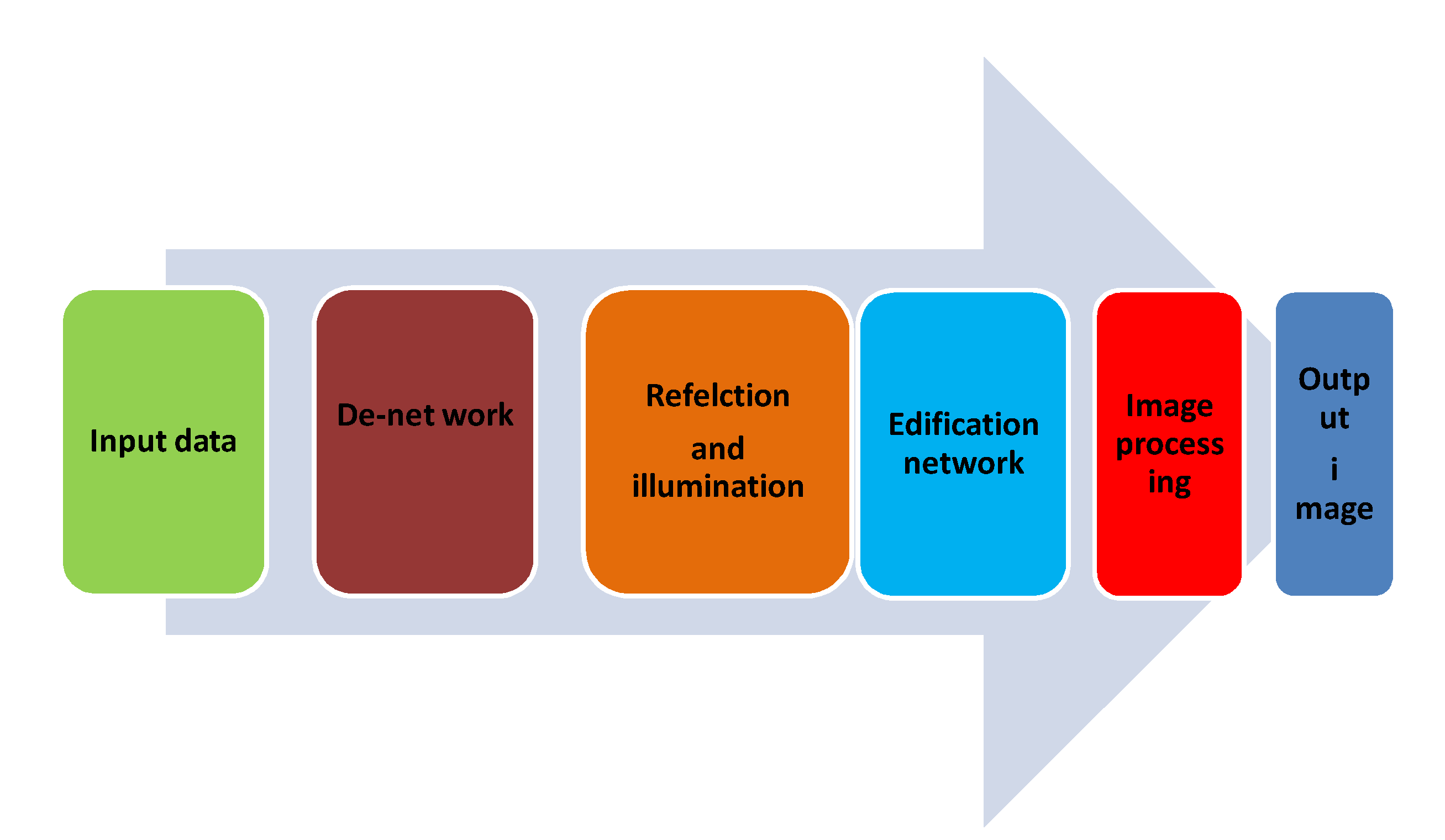
Figure 2.
Generative AI in disease diagnosis- tumour imaging.
Disclaimer/Publisher’s Note: The statements, opinions and data contained in all publications are solely those of the individual author(s) and contributor(s) and not of MDPI and/or the editor(s). MDPI and/or the editor(s) disclaim responsibility for any injury to people or property resulting from any ideas, methods, instructions or products referred to in the content. |
© 2024 by the authors. Licensee MDPI, Basel, Switzerland. This article is an open access article distributed under the terms and conditions of the Creative Commons Attribution (CC BY) license (http://creativecommons.org/licenses/by/4.0/).
Copyright: This open access article is published under a Creative Commons CC BY 4.0 license, which permit the free download, distribution, and reuse, provided that the author and preprint are cited in any reuse.


