Preprint
Article

Synthesis and Therapeutic Potential of Urolithin-C on Oxidative Stress Induced Tissue Damage

Submitted:

23 October 2024

Posted:

24 October 2024

You are already at the latest version

Abstract
Ellagitannins (ETs) are the group of tannins found in fruits, nuts and seeds; they undergo hydrolysis in the human digestive tract to form ellagic acid (EA). EA further converted into various forms of Urolithin derivatives (A, B, C, D, M-5, M-6 and M-7) by gut micro biota. Perhaps, the pharmacological properties of said Urolithins were extensively studied, while, little is known about the therapeutic role of synthetic Urolithin-C on oxidative stress induced eryptosis and tissue damage. Therefore, in the current study we investigated the ameliorative role of chemically syn-thesized Urolithin-C on sodium nitrite and Diclofenac (DFC) induced eryptosis and vital organ damage respectively. Urolithin-C was synthesized using chemical method and characterized by High-Performance Liquid Chromatography (HPLC), Nuclear Magnetic Resonance (NMR), Fourier Transform Infrared Spectroscopy (FTIR), and Liquid Chromatography Mass Spectroscopy (LC-MS). The synthesized Urolithin-C scavenged free radicals (DPPH, H2O2) and reduced ferric iron to ferrous iron, revealed its antioxidant activity. The observed antioxidant potential of Uro-lithin-C was investigated in sodium nitrite induced stress in RBC and Diclofenac induced tissue damage in rats. Interestingly, Urolithin-C normalized the sodium nitrate-induced oxidative stress in red blood cells by regulating stress markers, such as lipid peroxidation (LPO), total thiol (TT), and protein carbonyl content (PCC), and endogenous antioxidant enzymes superoxide dis-mutase (SOD) and catalase (CAT) in a dose dependent manner. Similar effect was also observed in the tissue homogenates of liver, kidney, heart and pancreas taken from the Diclofenac injected Sprague Dawley rats. The tissue sections (liver, kidney, heart and pancreas) obtained from the Diclofenac (DFC) received rats showed massive destruction of hepatocytes, nephrons, cardiocytes and pancreas. However, Urolithin-C received rats tissue sections showed normal histology compared to the positive (Silymarin) control treated groups. In addition, Urolithin-C regulated the key tissue specific biochemical markers in DFC injected Sprague Dawley rats serum strengthened its tissue protective role. In conclusion, chemically synthesized Urolithin-C regulated NaNO2 and diclofenac induced oxidative stress in RBC and vital organs through its antioxidant property.
Keywords: 
Subject: 
Biology and Life Sciences  -   Life Sciences

1. Introduction

Oxidative stress (OS) is a pathological condition of the body, characterized by an imbalance between Reactive Oxygen Species (ROS) and cellular antioxidant machineries. ROS, such as hydroxyl radicals (OH•), hydrogen peroxide (H2O2), superoxide anions (O2•−), and organic peroxides are the byproducts of aerobic metabolism formed in mitochondria [1]. In addition, Reactive Nitrogen Species (RNS), a family of nitrogen containing compounds, are also produced under oxygen deficient conditions and can further generate ROS [2,3]. Elevated levels of ROS/RNS are linked to lipid peroxidation, protein carbonylation, and DNA damage, potentially leading to cellular dysfunction or tissue damage [4]. Eventually, results in complex of life style diseases such as, hypertension, thrombosis, cancer, cardiovascular disorders, atherosclerosis, and diabetes mellitus [5,6]. Most importantly, when erythrocytes (RBCs) exposed to ROS/RNS undergo eryptosis a type of programmed cell death causes anemia, hypoxia, heart disease, tissue damage, and thrombosis as well [7]. The burden of lifestyle diseases has been tremendously increasing in the world population and they are the major cause for mortality and morbidity. Currently, chronic lifestyle diseases are treated by non-steroidal anti-inflammatory drugs by ignoring their life threatening side effects [8]. Hence, combitorial therapy is needed to support the exciting treatment strategies. Therefore, antioxidants from natural sources receive greater importance. For instance, isolated diarylheptanoids and isocoumarins from Alnus japonica steud and Papaelanthus have gained significant attention for their potent antioxidant and anti-inflammatory properties [9,10]. In addition, Rosmaric acid, Carnosol, and Carnosic acid were isolated from Rosemary extract, exhibited antioxidant property [11] Researchers also made an effort to synthesize diarylheptanoids and isocoumarins and proved their antioxidant and anti-inflammatory properties [12,13]. Despite, the controversial reputation, synthetic antioxidants such as tertiary butylhydroquinone (TBHQ), butylated hy-droxyanisole (BHA), butylated hydroxytoluene (BHT) and propyl gallate have been extensively used as food additives for the preservation of processed foods [14]. It is a truth that, several antioxidants isolated and synthesized are not fully utilized due to the issues associated with digestion, absorption, and bioavailability [15]. Therefore, identifying new antioxidants with multitude of therapeutic efficiency is the need of an hour. Pomegranates, with their rich jewel like seeds and vibrant juice, have long been celebrated for their remarkable health benefits. It is largely attributed to their high content of antioxidant polyphenols, especially ellagitannins like punicalagin [16]. This health-promoting potential extends to other fruits (strawberries, raspberries, blackberries and muscadine grapes) and nuts (walnuts and almonds) as they are rich in Ellagitannins (ETs). ETs are the class of hydrolysable tannins having diversified chemical complexity, in the stomach under acidic condition they are converted into ellagic acid (EA) [17]. The human gut microbiome metabolizes these EA into 6H-dibenzo[b,d]pyran-6-one, through the sequential removal of hydroxyl groups followed by decarboxylation of one of the lactone rings of EA to form the derivatives of Urolithins- A, B, C, D, M-5, M-6 and M-7 [17,18]. Researchers have synthesized various Urolithin derivatives, demonstrating biological activities such as neuroprotection, anticancer (colon breast and prostate), antiatherosclerosis, antimalarial, antioxidant and anti-inflammatory activity [19,20]. They are also patented for the prevention and treatment of neoplastic diseases [21]. It is noted that Urolithin-A and B were isolated and well characterized from fecal matters of experimental rats [22]. While, Urolithin-C was chemically synthesized using several methods, only antioxidant and anticancer activity was analyzed so far [23]. Recent reports suggested that the isolation of Urolithins from gut microbiota or from the fecal matter is the time consuming and tedious job [24]. Thus, chemical synthesis appears to be an alternative avenue for the synthesis of the natural product. Thus, in the current study, we made an attempt to synthesize Urolithin-C and its ameliorative role on stress induced RBC, liver, kidney, heart and pancreas damage through its antioxidant potential was examined and the results are presented.

2. Results

2.1. Synthesis of Urolithin-C:

Synthesis of Urolithin-C was a four step reaction. In the first step when 2-formylphenyl boronic acid was mixed with 2-bromo-1-iodo-4-methoxybenzene in presence of Bis-triphenylphosphine palladium (II) dichloride (PdCl2(PPh3)2) as a catalyst under basic condition in tetrahydrofuran (THF) at room temperature biaryl carboxy aldehyde was formed with a yield of about 70 %. In the second step, biaryl carboxy aldehyde undergone oxidation with potassium permanganate, pyridine and water. Further, it had undergone esterification with dimethyl formamide and methanol to form a biaryl carboxylic ester. In the third step, biaryl carboxylic ester was undergone a coupling reaction in presence of N,N,N’,N’ tetramethyl ethylene diamine and potassium carbonate resulted in methyl ether of Urolithin-C. Finally, methyl ether of Urolithin-C undergone demethylation with boron tribromide in the presence of hydrochloric acid and dichloromethane resulted in Urolithin-C with a yield of 10%. Figure 1 represents the process of Urolithin-C synthesis.

2.2. Purification and Characterization of Synthesized Urolithin-C:

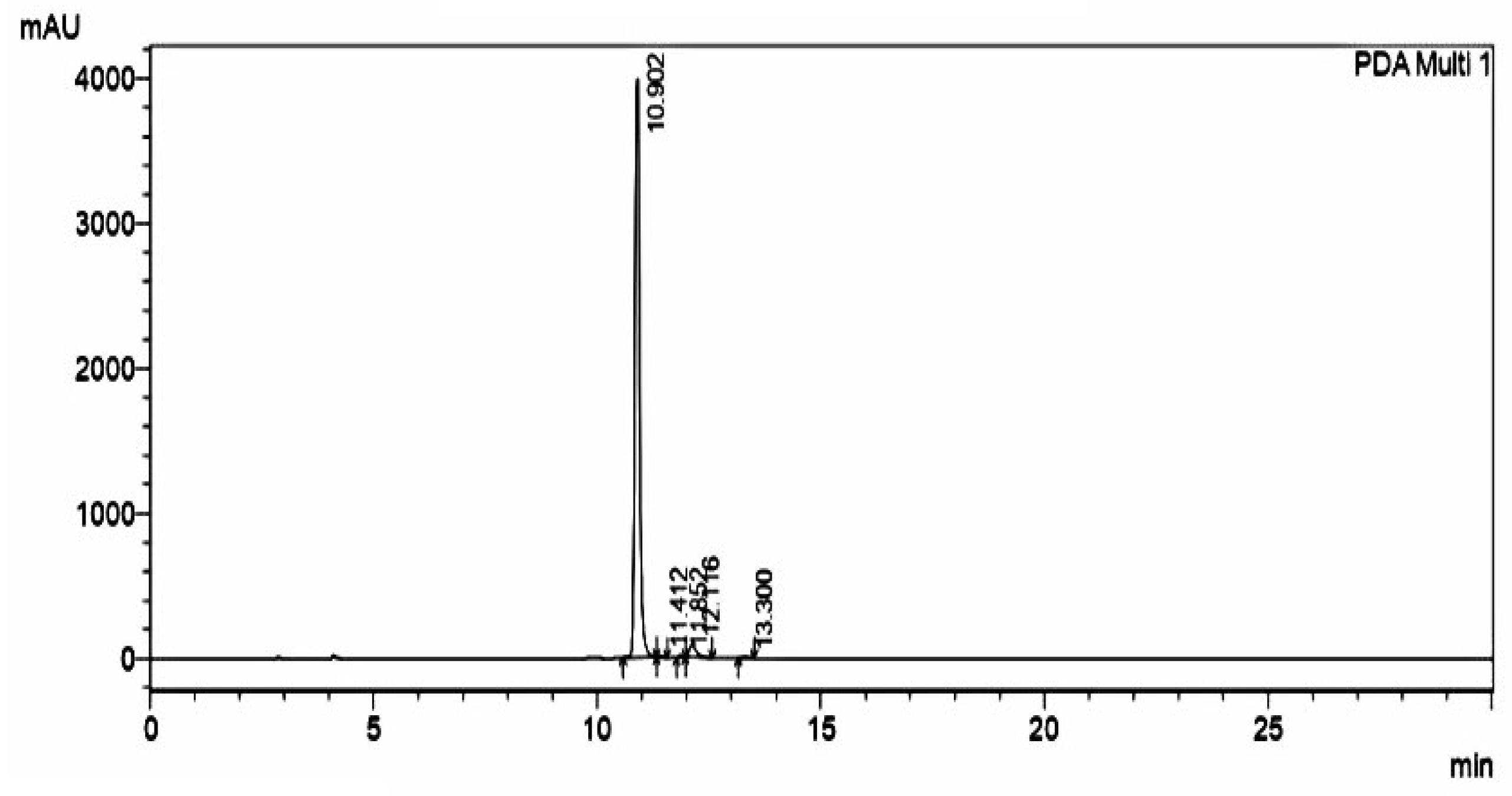
TLC was carried out to determine the purity of synthesized Urolithin-C, a single spot of Urolithin-C with a retardation factor of 0.3 cm for the first reaction intermittent and the retardation factor of 0.2 cm for the second reaction was noticed (bromination and acidification). Figures SA1 and SA2 indicates the TLC chromatogram of Urolithin-C. Further, the purity of Urolithin-C was examined using HPLC, interestingly synthesized Urolithin-C was eluted as a single sharp peak with a retention time of 10.902 minutes [ Figure 2].
Furthermore, the probable structure was analyzed using 1H and 13C NMR. The proton chemical shift (δ) was reported in parts per million (ppm) 1H NMR (400MHz, DMSO): δ 10.37 (brs 1H, ArOH), 10.11 (brs, 1H, ArOH), 10.01 (brs, 1H, ArOH), 7.85 (d,J = 8.4Hz, ArH, 1H), 7.49 (s, 1H, ArH), 7.44 (s, 1H, ArH), 6.79 (dd,J = 8.2,2.4Hz, 1H, ArH),6.69 (d,J = 2.4Hz, 1H, ArH) [Figure SA3-1]. 13C NMR (400MHz, DMSO): δ 160.814, 159.034, 153.911, 151.922, 146.594, 129.666, 124.196, 114.677, 113.341, 111.349, 110.259, 107.297, 103.255 [Figure SA3-2].
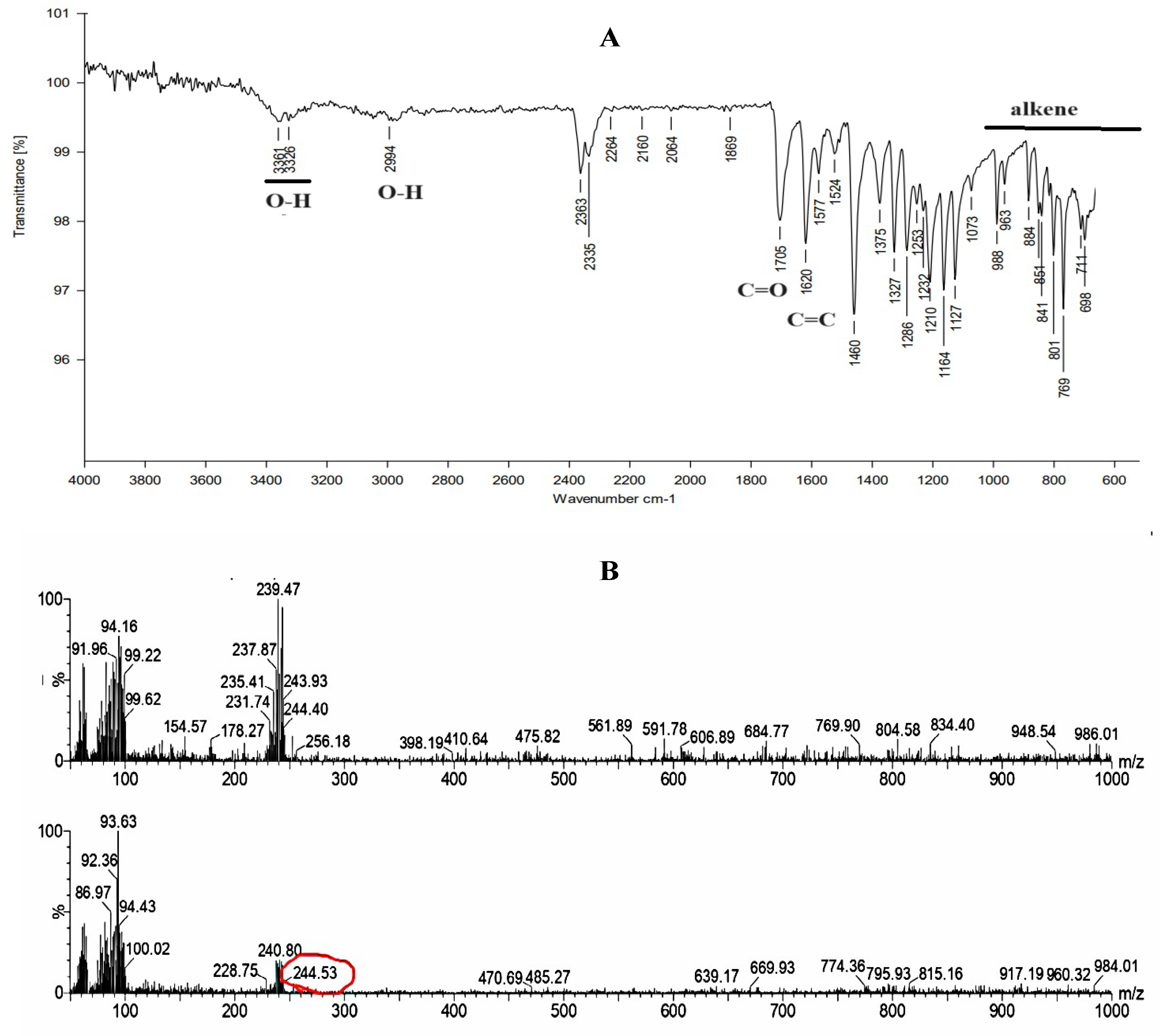
To identify the probable functional groups of synthesiszed Urolithin-C, Fourier transfer infrared (FTIR) spectroscopy was done. The wavelength 3361-3326 cm-1 repre-senting an overseas peak, indicating the stretching of a polymeric hydroxyl group (O-H stretching). The absorption peak at wave number 2994 cm-1 was caused by free phenolic functional groups (O-H stretching). Furthermore, C=O stretching at 1705 cm-1 and C=C stretching at 1620 cm-1 implies the presence of a ketone moiety. The area between 988-698 cm-1 reveals a C=C bending property of the alkene moiety, and this spectrum validated the structure of Urolithin-C [Figure 3A]. Further, the mass of the synthesized compound was found to be 244.53 g/mole concerning the negative mode of m/z ratio, which was con-firmed by the mass data, acquired using Liquid LCMS [Figure 3B].

2.3. Urolithin-C Exhibits Antioxidant Property by Scavenging DPPH, H2O2 and FRAP Radicals:

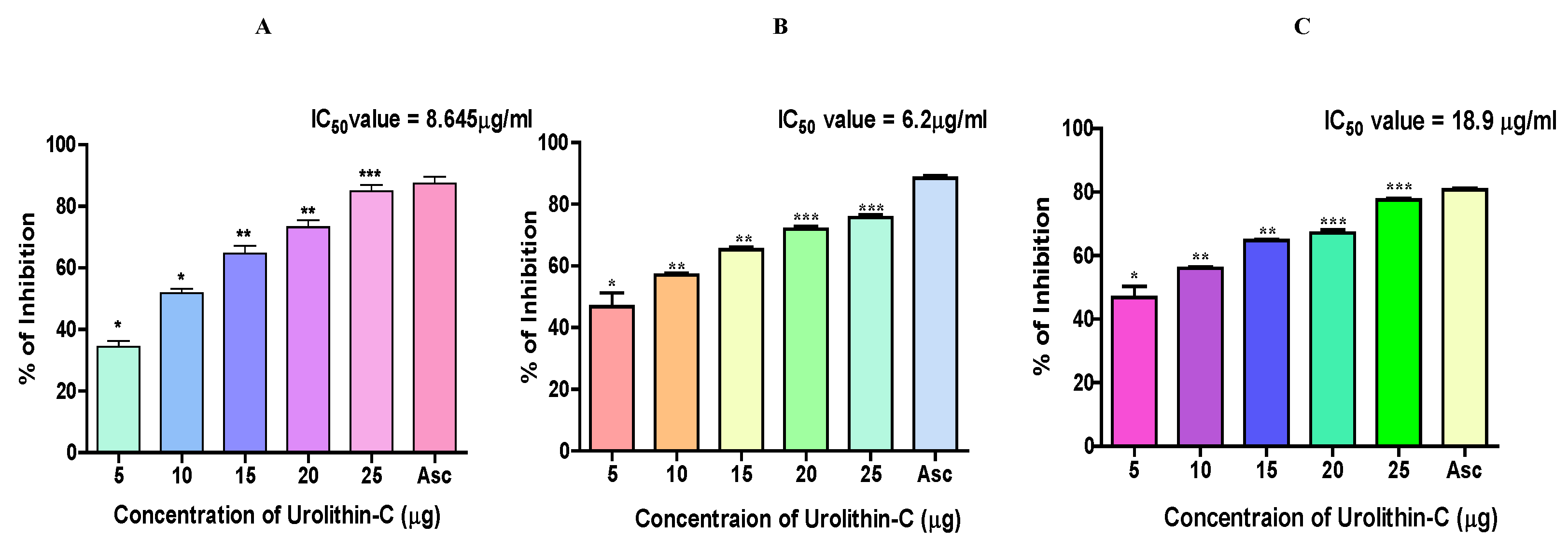
Urolithin-C scavenged the free radicals in a concentration dependent manner (0-25 µg) that was compared to ascorbic acid the positive control. The percentage of DPPH scavenging potential of Urolithin-C was found to be 79 % at the concentration of 25 µg, while ascorbic acid showed 85 % radical scavenging potential with an IC50 value of 8.645 µg/ml [Figure 4A]. Furthermore, Urolithin-C scavenged the H2O2 radicals (74 %) as well, that was compared to the positive control ascorbic acid (83 %). The calculated IC50 value of Urolithin-C was found to be 6.2 µg/ml [Figure 4B]. In addition, Urolithin-C also exhibited ferric-reducing properties with a reducing power of 81 %, somewhat higher than that of the typical FRAP value of ascorbic acid (78 %). The IC50 value of Urolithin-C was found to be 18.9 µg/ml [Figure 4C].

2.4. Urolithin-C Ameliorates the Oxidative Stress Parameters in the Sodium Nitrite (NaNO2) Induced Oxidative Stress in RBCs (In-Vitro):

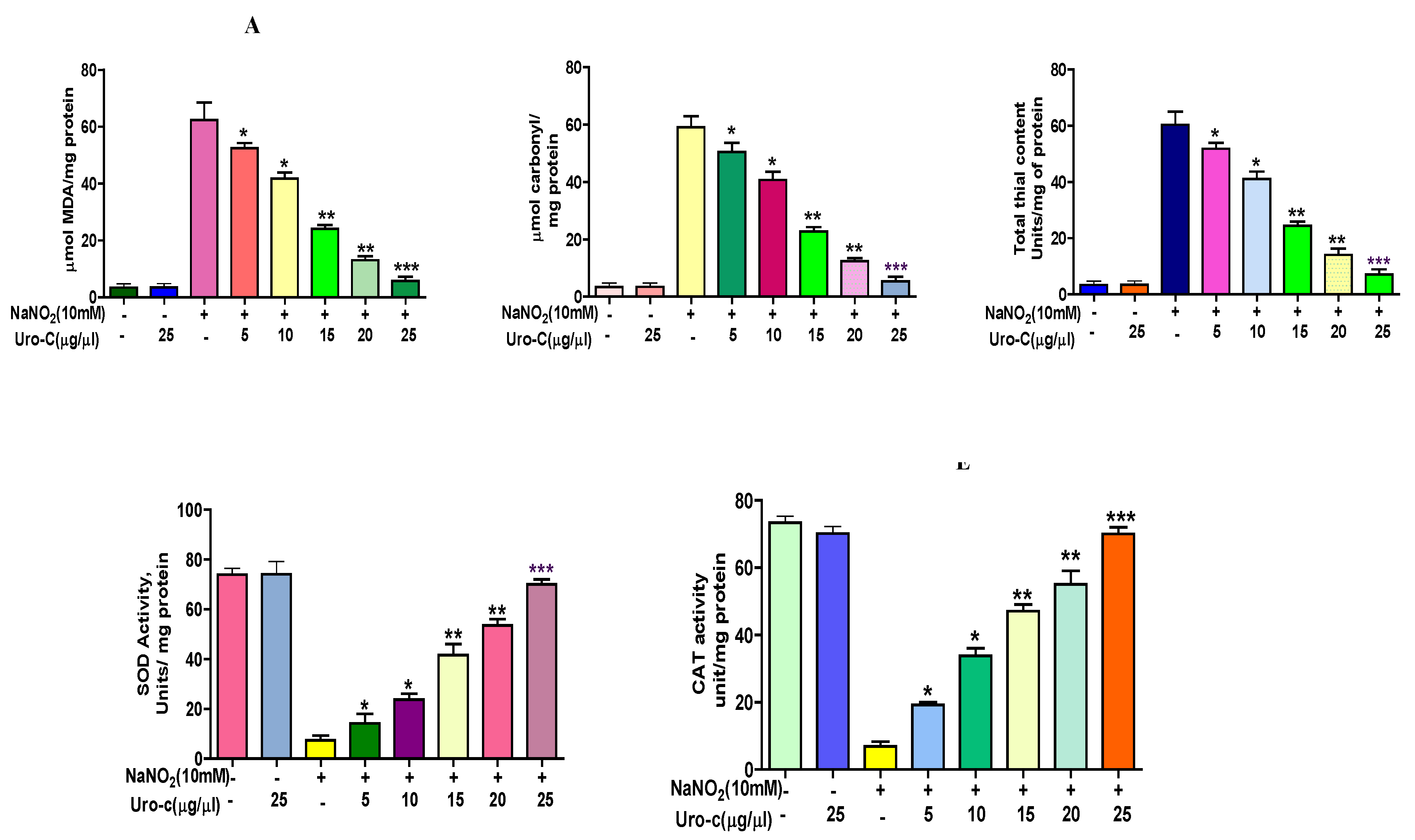
To substantiate the observed antioxidant property of Urolithin-C sodium nitrite was used as a stress inducer in human RBCs. The key stress markers such as lipid peroxidation (LPO), protein carbonyl content (PCC) and total thiol (TT) were assessed. The Malondialdehyde (MDA) level was tested to determine lipid peroxidation, and it was considerably raised in RBCs treated with NaNO2 (10 mmol/l). RBCs treated with Urolithin-C (0-25 µg) preincubated with NaNO2 exhibited a dose dependent reduction in MDA level. Interestingly, MDA level remained constant in the groups treated with Uro-lithin-C alone [Figure 5A]. In the case of NaNO2 treated RBCs lysate the protein carbonyl content (PCC) was increased compared to the control group. While, RBCs received various concentration of Urolithin-C (0-25 µg) preincubated with NaNO2, there was a significant restoration of PCC was identified compared to the control groups [Figure 5B]. Similarly, in case of NaNO2 alone treated RBCs there was an increased level of total thiol content was examined. However, RBCs treated with various concentrations of Urolithin-C (0-25 µg) preincubated with NaNO2 there was a significant restoration of total thiol content was also identified compared to the control groups [Figure 5C]. Notably, Urolithin-C alone did not affect any of these three markers compared to the control groups. Most importantly, RBCs exposed to NaNO2 showed a massive reduction in superoxide dismutase (SOD) and catalase (CAT) enzyme activities. While, RBCs treated with various doses of Urolithin-C (0-25 µg) preincubated with NaNO2, the activities of said enzymes were significantly improved. Perhaps, in case of Urolithin-C alone treated RBCs activity of SOD and CAT remains unaltered [Figure 5 D and E].

2.5. Urolithin-C Regulates the Diclofenac Induced Oxidative Stress Parameter in Animal Model (In-Vivo):

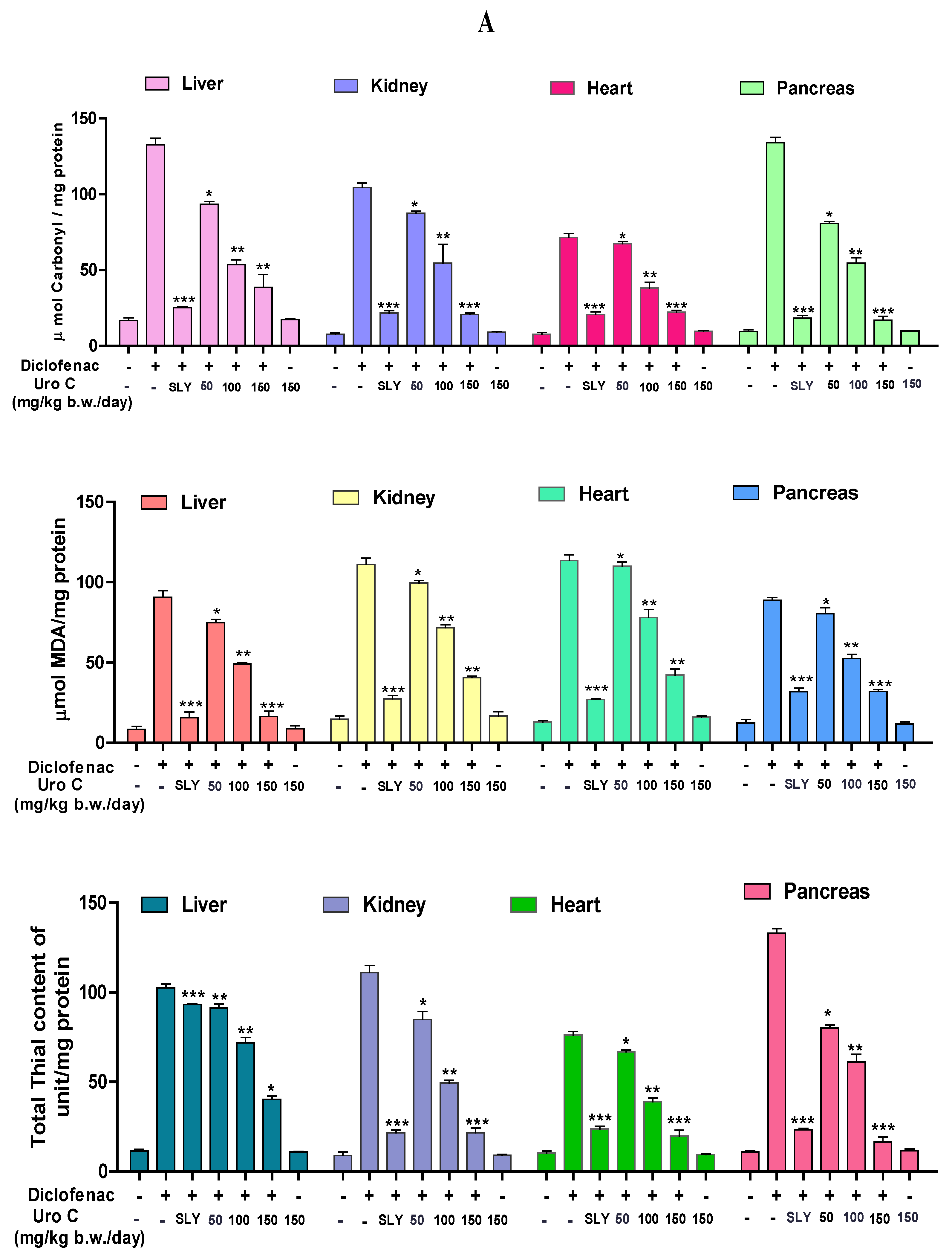
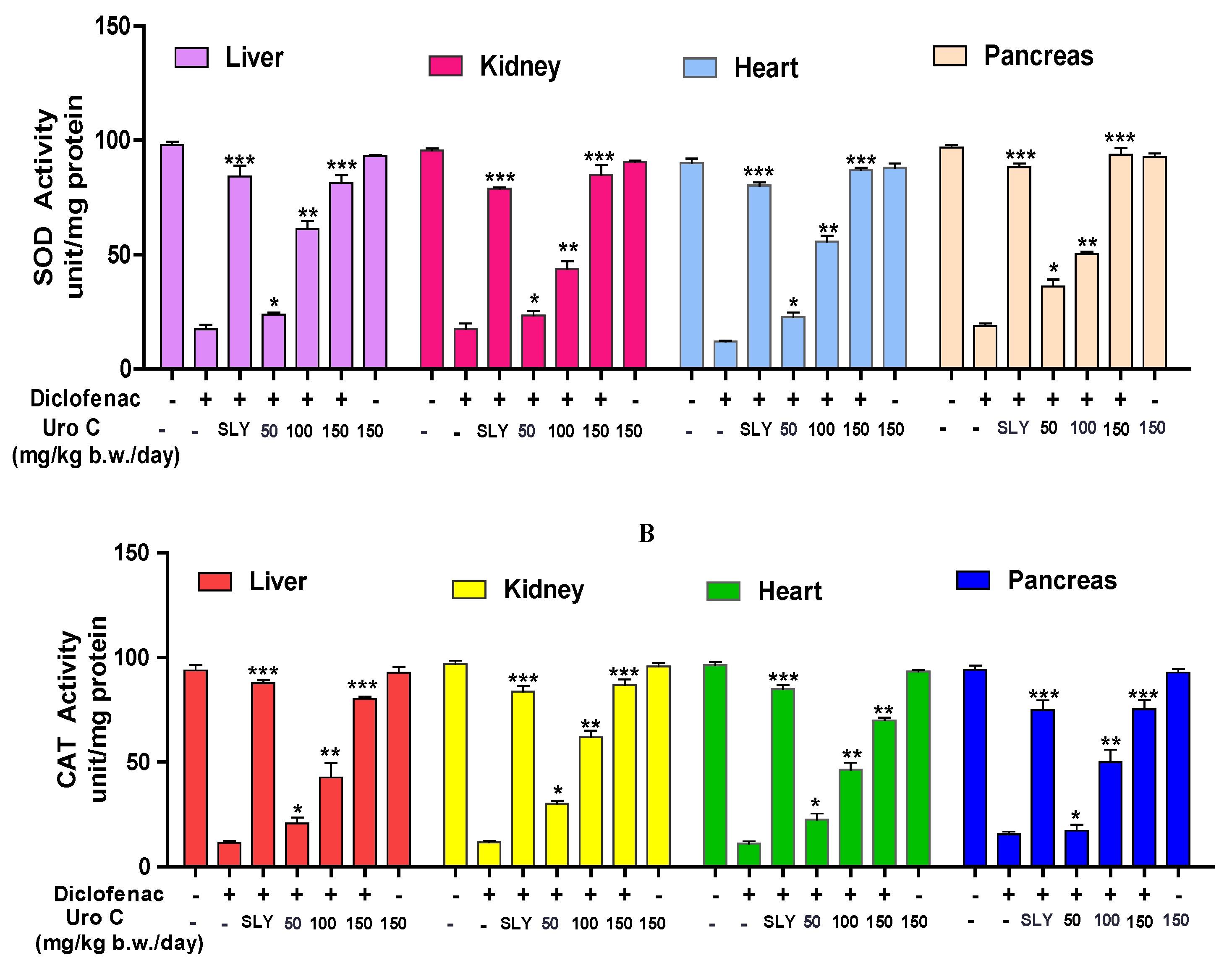
Liver, kidney, heart and pancreas, homogenates of Diclofenac (DFC) treated rats showed increased level of MDA, TT and PCC compared to rats injected with silymarin a positive control. On the other hand, rats administered 50-150 mg/kg of Urolithin-C that was preincubated with DFC all the said markers were significantly restored compared to the control group [Figure 6A, B & C]. Additionally, the activity of antioxidant enzymes such as, CAT and SOD was decreased in DFC injected tissue homogenates (liver, kidney, heart and pancreas). Whereas, various doses of Urolithin-C preincubated with DFC, the activity of said antioxidant enzymes was significantly restored. Notably, rats injected with Urolithin-C alone did not cause any change in the stress markers that was compared with the normal [Figure 7A & B].

2.6. Urolithin-C Restores the Biochemical Parameters in Diclofenac Injected Rat’s Serum:

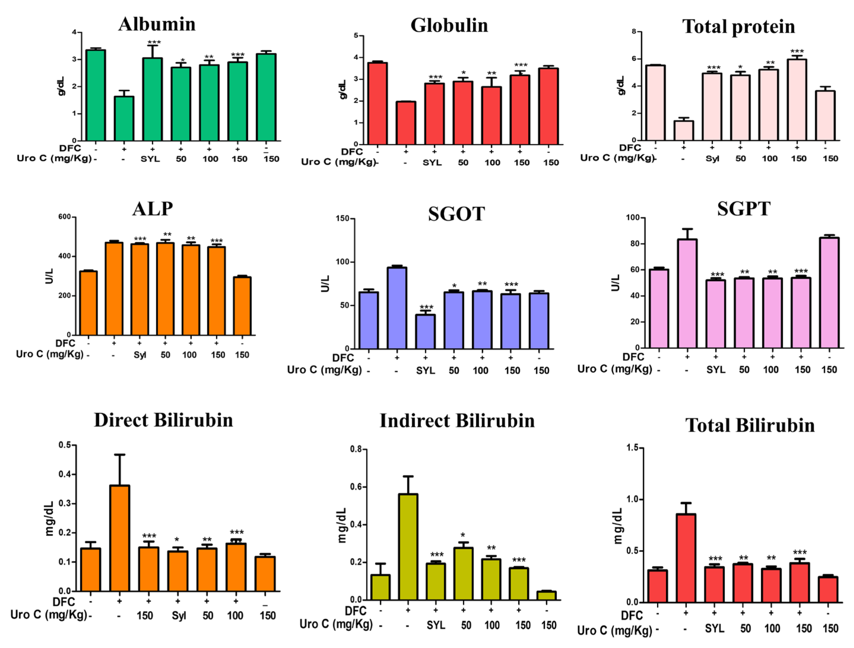
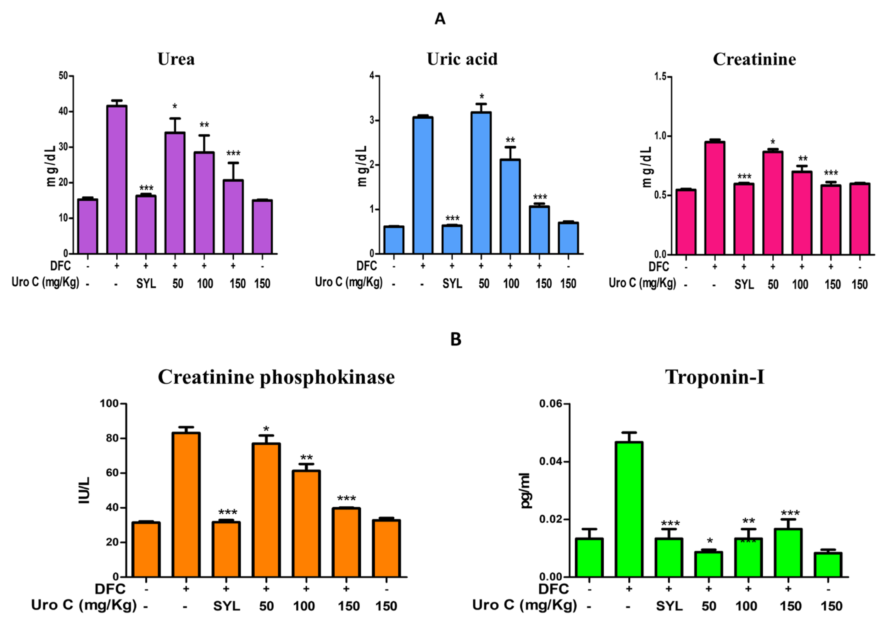
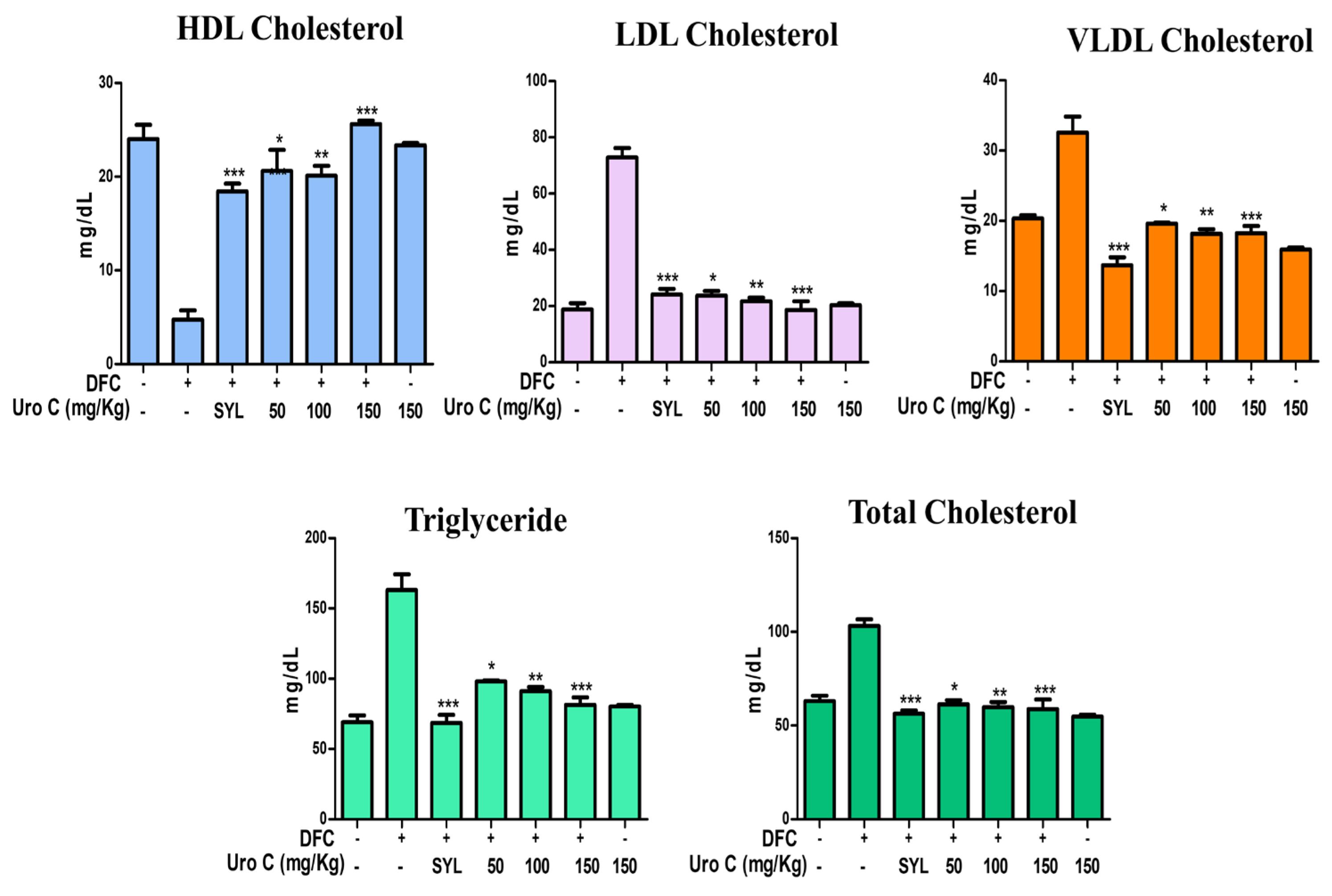
To test the protective effect of Urolithin-C on Diclofenac (DFC) induced liver, kidney, heart and pancreas damage the tissue specific biochemical markers were analyzed using rat’s serum sample. The level of serum albumin, globulin and total proteins decreased, whereas, ALP (Alkaline phosphatase), SGOT (serum glutamate oxaloacetate transaminase), SGPT (serum glutamate pyruvate transaminase), and total bilirubin (direct and indirect) were increased significantly suggesting the Diclofenac induced liver damage [Figure 8]. Renal function markers including urea, uric acid, and creatinine were raised suggesting DFC induced kidney damage [Figure 9A]. The rise in creatinine phosphokinase and Troponin-I revealed DFC induced heart damage [Figure 9B]. In addition, the elevated level of lipid profile such as triglyceride and cholesterol (HDL,LDL,VLDL and total cholesterol) was also identified [Figure 10]. Interestingly, Urolithin-C restored all the said biochemical markers of DFC treated rat’s serum, strengthened the protective role of Urolithin C on oxidative stress induced tissue damage.

2.7. Urolithin-C Normalizes the Liver, Kidney, Heart and Pancreas Morphology:

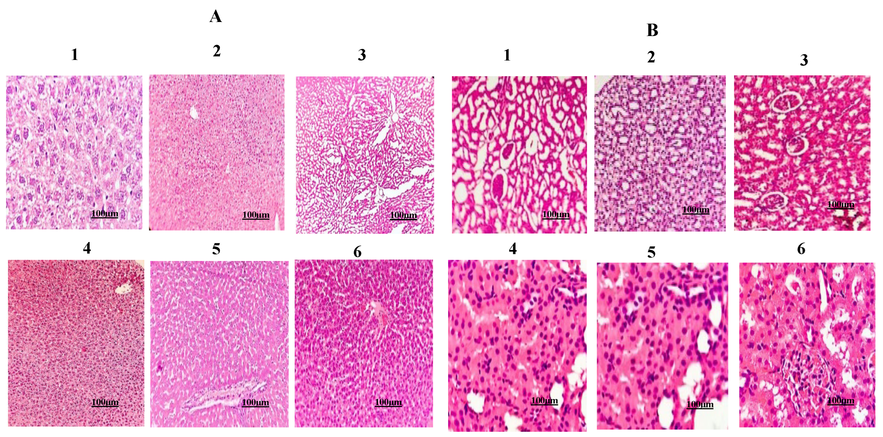
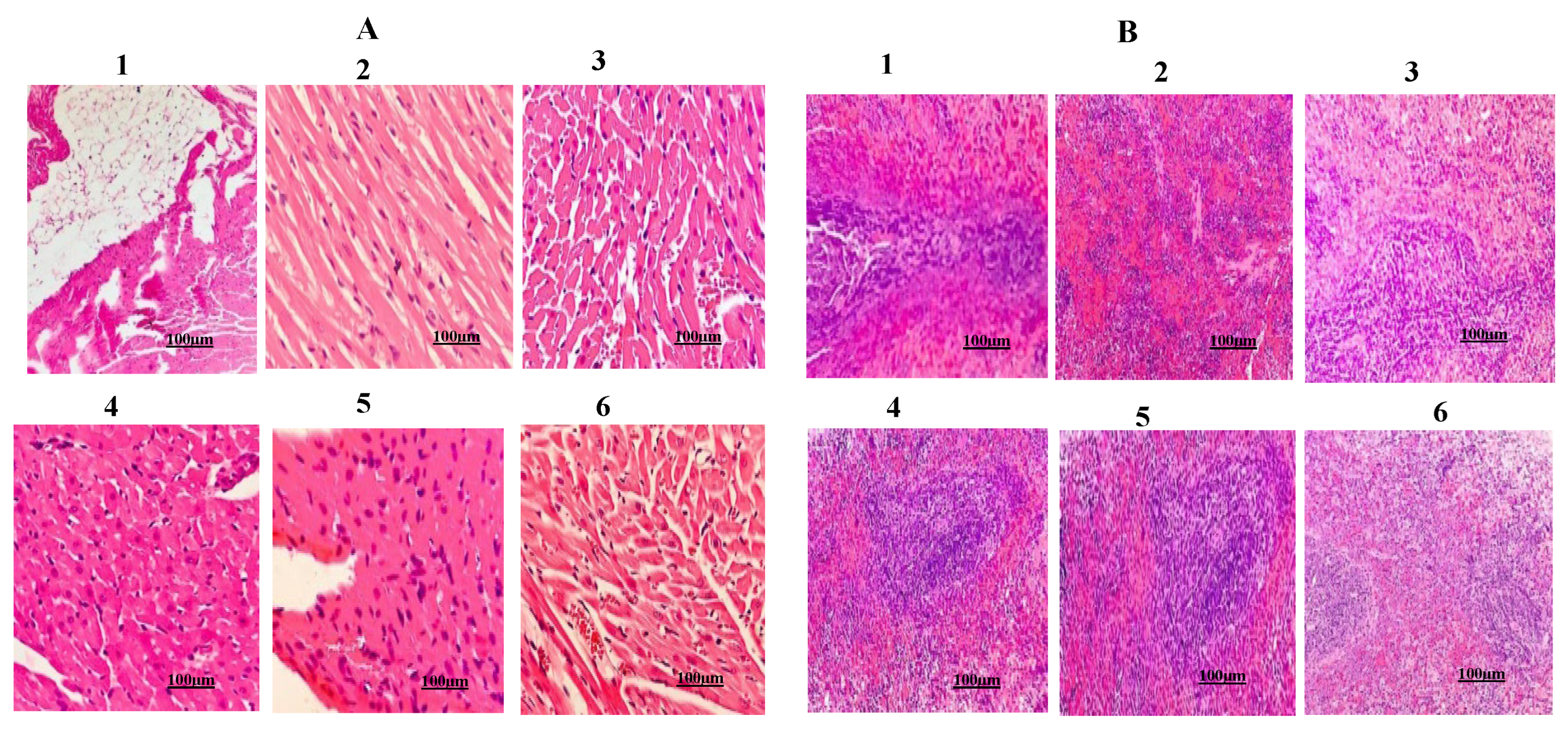
In order to evaluate the protective role of Urolithin-C on DFC induced tissue damage, microscopic examination of liver, kidney, heart and pancreas was done. DFC injected rat’s liver section revealed extensive damage of hepatocytes [Figure 11A1]. Saline injected liver tissue showed normal histology [Figure 11A2]. Whereas, Silymarin (SYL) a positive control drug preincubated with DFC treated rat’s liver section showed normal histology compared to the saline injected groups (Figure 11A3). While, Urolithin-C alone treated rat’s liver tissue section exhibited normal histology compared to the saline received rat’s liver section [Figure 11A4). Interestingly, Urolithin-C preincubated with DFC treated rat’s liver section there was a restoration of damaged hepatocytes were noticed [ Figure 11A5 and A6]. Similarly, DFC treated kidney, heart and pancreas there was a massive destruction of nephrons, cardiocytes and pancreatic cells were noticed respectively compared to the saline injected rat’s tissue sections (Figure 11B1, Figure 12A1,B1]. However, Urolithin-C preincubated with DFC treated rat’s kidney, heart and pancreas, there was a dose dependent restoration of damaged tissues were noticed as compared to the control and SYL received rat’s tissues sections [Figure 11B5-B6, Figure 12A5-A6,B5-B6]. Interestingly, Urolithin-C received rat’s kidney, heart and pancreas tissue sections revealed normal histology compared to the saline and SYL injected groups tissue sections [Figure 11B2-B3, Figure 12A2-A3,B2-B3].

3. Discussion

Although, smoking, physical inactivity, unhealthy diet and alcohol are considered as the key risk factors of lifestyle diseases such as hypertension, diabetes, heart failure, stroke, cancer and neurodegenerative complications. Perhaps, high levels of reactive oxygen species (ROS) and reactive nitrogen species (RNS) are the key modifiable agents that ini-tiate inflammation and tissue damage and make a way for the augmentation of said pathogenesis [25,26]. Eventhough several drugs have been entering into the market every day, their life threatening side effects limits their usage. For instance, diarylhep-tanoids and isocoumarins causes hormonal disturbances and reversible liver damage respectively [27]. While, synthetic antioxidants such as, tertiary butylhydroquinone, butylated hydroxyanisole, butylated hydroxytoluene and propyl gallate cause hemorrhage, liver damage, balder and stomach cancer [28]. Thus, identifying new antioxidant agent without side effect is of challenging task to the researchers. Most importantly, Oxidative stress modulators may directly reduce oxidative damage by scavenging free radicals, yet these in-vitro effects frequently have no clinical significance, it is crucial to understand how synthetic molecules function in-vivo. Hence, the current study aims to synthesis Urolithin-C a microbial gut metabolite and effort was made to understand its protective role on oxidative stress induced RBCs and vital organs damage. The synthesised Uro-lithin-C was characterized using techniques such as, Thin Layer Chromatography (TLC), High Performance Liquid Chromatography (HPLC), Nuclear Magnetic Resonance (NMR) and Liquid Chromatography-Mass Spectrometry (LCMS). Urolithin-C was found to be pure as it showed single band in TLC and single peak in HPLC. 1H and 13C NMR spectroscopy confirmed the chemical shifts of carbon and hydrogen in parts per million. LCMS results suggested that the molecular mass of the Urolitihin-C was found to be 244.53 m/z.
The compounds to be antioxidant they have the ability to donate an electron to the free radicals and transform them to be harmless to cellular biomolecules such as, lipids, proteins enzymes, carbohydrates and DNA [29]. They are either exogenous or endogenous but they can be capable of preventing the oxidation induced cellular changes followed by its damage [30]. Although, copious amounts of natural nutraceuticals, synthesised compounds and nanoparticles are marketed with a lot of claims and doctors often prescribed [31]. Most of them have not accomplished the required therapeutic utilities. Interestingly, Urolithin-C exhibited strong antioxidant activity by scavenging DPPH, H2O2 and reducing ferric to ferrous. The observed antioxidant potential of Urolithin-C was evaluated using sodium nitrite induced oxidative stress in RBCs. The circulating red blood cells (RBCs) have an average lifespan of 120 days. When they exposed to endoge-nous and exogenous oxidants undergo hemolysis elicits their suicidal death or eryptosis [32]. Eryptosis leads to loss of membrane integrity, release of hemoglobin and other proteins from RBCs [33]. Accumulation of free hemoglobin in cell and RBC derived ROS may further results in tissue damage, chronic anemeia, thrombosis and heart attack [34]. Elevated levels of LPO signify the alteration of lipid membranes, while high levels of PCC and TT suggests oxidation of proteins. The detoxification of ROS depends on catalase and SOD, if their level diminished in the cell an ROS accumulation eventually increased which marks the oxidative stress [35]. Urolithin-C through its free radical scavenging property normalized the high level of total thiol (TT) protein carbonyl content (PCC), lipid peroxidation (LPO) and antioxidant enzymes including catalase and superoxide dismutase (SOD) in sodium nitrite induced oxidative stress in RBC suggesting its RBC protecting role. In our study we also noticed that Urolithin-C did not cause hemolysis further strengthen its anti-eryptotic property without toxicity.
To substantiate observed in-vitro antioxidant and RBCs protective role of Urolithin-C, we have done in-vivo experiments using Sprague Dawley rats. The rationale for choosing rats over mice was that, Mice could be more sensitive for inducing oxidative stress, that may end in high variability of result. While, Rats are good model system as they are stable to obtain consistent data as for as oxidative stress is concerned. Interestingly, Urolithin-C normalized the altered level of LPO, PCC. TT, SOD and Cat in tissue homogenates of vital organs (liver, kidney, heart and pancreas) obtained from DFC induced rats. Most importantly, the protective role of Urolithin-C was also assessed using various biochemical markers. DFC toxicity caused a decrease in total blood proteins (albumin and globulin), while, increase in direct/indirect bilirubin, uric acid, urea and creatinine. In addition, elevated levels of hepatic enzymes like SGOT and SGPT in the blood stream suggested liver injury. The ALP enzyme, which is involved in metabolite transport across the cell membrane, was also increased, indicating liver dysfunction. Troponin, and creatinine kinase, increased in the heart that supports the severe heart damage. Surprisingly, Urolithin-C normalized the entire said biochemical serum markers linked to liver, kidney, heart and pancreas strengthened its tissue protecting role.
ROS/RNS induced inflammation is the key pathological event responsible for the progression of endothelial dysfunction and tissue injury [36]. Oxidative stress induced chronic damage of liver, kidney, pancreas and heart leads to numerous diseases including metabolic disorders, jaundice, hypertension, diabetes, cancer and heart attack [37]. It is to noted that, synthesized Urolithin-C successfully restored the structural integrity of DFC induced oxidative damage of liver, kidney, heart, and pancreas. Although, at this moment to predict the exact mechanism of action of Urolithin-C on mitigating oxi-dative stress induced tissue damage is premature. Perhaps, Urolithin-C exhibited anti-inflammatory activity (data not shown), thus, at this moment it is to speculate that the Urolithin-C through its anti-inflammatory and ROS scavenging potential normalized damaged tissues. In addition, considering its molecular structure dibenzopyron ring (3,8,9 trihdroxy-6H-dibenzo[b,d]pyran-6-one) with 3,8,9 hydroxyl group may have ability to donate both electron and proton (hydrogen atom) to numerous oxidants in particular cellular ROS/RNS. Most importantly Urolithin-C is even though it was chemically synthesized it was non-toxic in nature as it did not damage RBCs and vital organs. Thus, owing to the observed pharmacological properties of Urolithin-C, it could be a better candidate for the management of oxidative stress induced pathogenesis.

4. Materials and Methods

4.1. Chemicals and Reagents:

2-formyl phenyl boronic acid, 2-bromo-1-iodo-4- methoxybenzene, methanol, PdCl2(PPh3)2[dichlorobis (triphenylphosphine) palladium(II)], THF (Tetrahydrofuran), K2CO3 (Potassium Carbonate), EtOAc (Ethyl acetate), BBr3 (Boron tribromide), HCl (Hydrochloric acid), Methanol, DPPH (1,1-diphenyl-2-picrylhydrazyl), NaNO2 (Sodium Nitrate), EDTA (Ethylene diamine tetra acetic acid), TEMED (Tetra methyl ethylene diamine), DNPH (2,4-dinitro phenyl hydrazine), TCA(Tri-chloro acetic acid), DTNB (5,5′-dithiobis-2-nitrobenzoic acid), SDS (Sodium dodecyl sulphate), Acetic acid, Thiobarbituric acid, H2O2 (Hydrogen peroxide), DNS (3,5 dinitro salicylic acid), NaOH (Sodium hydroxide), Sodium potassium tartrate, Starch, PBS (Phosphate buffer saline), DFC. Potassium iodide, Potassium chloride, 20 mM Tris-HCl, NaCl and TFA (trifluoroacetic acid)

4.2. Synthesis of Urolithin-C:

4.2.1. Synthesis of Urolthin C Was Carried Out by Suzuki-Miyaura Coupling Method [38,39]

Step 1: Preparation of biarylcarboxy aldehyde:
In a reaction vessel, mixed 2-formylphenylboronic acid (1.0 equivalent), 2-bromo-1-iodo-4-methoxybenzene (2.0 equivalents), and 5 mol % Bis-triphenylphosphine palladium (II) dichloride (PdCl2(PPh3)2) in 5 ml of tetrahydrofuran (THF). Stirred this mixture under an inert atmosphere at room temperature. Gradually, introduced a solution of 2 N K2CO3 (Potassium carbonate) 25 ml into the reaction via a syringe that caused the mixture to turn into a brown red solution. Continued stirring the reaction mixture at room temperature overnight, monitoring the progress using thin layer chromatography (TLC). Quenched the reaction with water and extracted the mixture with ethyl acetate (EtOAc). Purified the crude product using preparative thin layer chromatography with a solvent system of EtOAc/hexane.
Step 2: Preparation of biaryl ester
To the solution of 2’-bromo-4-methoxy-(1,1’-biphenyl)-2-carbaldehyde (1.0 equiva-lent), added 5 ml of pyridine in 15 ml of water, to this mixture added KMnO4 (2.0 equivalent) at ambient atmosphere and refluxed the reaction mixture until the starting material was completely osxidised. Then cooled the reaction mixture to room temperature, acidified with 2 N HCl to obtain white precipitate and recrystallised the white solid with ethyl acetate (EtOAc)/Hexane (1:4) ratio to obtain biaryl carboxylic acid. The product was dissolved in 25 ml of dichloromethane (CH2Cl2) in a round bottom flask. To this precooled solution of carboxylic acid and 3 drops of dimethylformamide (DMF) was added dropwise oxalyl chloride (COCl)2 slowly. The reaction was stirred 0 °C for 2 hours, and concentrated under reduced pressure to obtain a yellow residue. The residue was esterified with 25 ml of methanol at room temperature. The reaction was quenched with water and extracted with ethyl acetate (EtOAc). The organic layer was removed by washing with brine, dried with anhydrous sodium sulfate (Na2SO4) and concentrated under reduced pressure to obtain the residue.
Step 3: Preparation of Urolithin-C:
Prepared a cooled solution of methyl ether of Urolithin-C (1.0 equivalent) at 0 °C under an aromatic atmosphere and was slowly added boron tribromide (BBr3) to the reaction mixture. Once the reaction was completed, acidified the mixture by adding a solution of 2 N HCl. Extracted the product with ethyl acetate (EtOAc) and purified the crude product by repeatedly washing it with water, methanol, and hot ethyl acetate. Filtered the final product and confirmed its purity using various techniques.

4.2.2. Thin Layer Chromatography (TLC):

The synthesised compound was mixed with 10 % methanol with DCM (Dichloro methane), by adding 1 drop of acetic acid, this mixture was spotted on silica-plated alu-minum sheets and the chromatogram was developed by using 1: 20 % ethyl acetate and n-hexane as a mobile phase. The developed chromatogram was detected by placing it in an iodine chamber to visualize the spot [40]. The first reaction intermediated was spotted with a retardation factor (RF) value 0.3 cm. The final product with acidification and bromination along with 2N HCl was spotted with an RF value 0.2 cm.

4.2.3. High Performance Liquid Chromatography (HPLC):

The synthesized Urolithin-C (20 µl) was subjected to HPLC using (Agilent Tech-nologies system in Santa Clara, CA) C-18 column with dimensions of 250X4.6 mm. The flow rate was maintained at 1 ml/min. Gradient elution was performed using 0.1 % tri-fluoroacetic acid (TFA), water and acetonitrile [41].

4.2.4. Nuclear Magnetic Resonance Spectroscopy (NMR):

1H NMR: 1H NMR was used to identified and confirmed the structure of organic compounds containing protons. The synthesized Urolithin-C was dissolved in dimethyl sulfoxide (DMSO) and subjected to an NMR spectrometer operating at a proton frequency of 400 MHz. 13C NMR: 13C NMR was used to elucidate the structure of molecules, provided spectra with peaks represented the diverse carbon atom environments within a molecule. As like 1H NMR, the sample was dissolved in DMSO and analyzed using an NMR spectrometer operated at a proton frequency of 400 MHz. NMR spectra was recorded using a Brüker 400 MHz instrument (Bruker, Bremen, Germany) [42].

4.2.5. Fourier Transform Infrared Spectroscopy (FTIR):

The FTIR analytical technique was used for the identification of active bio functional groups presented in the synthesized Urolithin-C. The FTIR spectra was determined at the range of wave number from 600–4000 cm-1 in a Shimadzu FTIR-8400 spectrometer [43].

4.2.6. Liquid Chromatography Mass Spectrometry (LCMS):

LCMS was used for the identification and quantification of analytes present in the synthesized Urolithin-C. The Mass data were acquired using Waters' Micro Mass Quattro Micro API (LCMS). The chromatographic separation was utilized a WATERS X Bridge C-18 column with dimensions of 50 X 4.6 mm and a particle size of 3.5 µ. The flow rate was maintained at 1.2 ml/min. Gradient elution was performed using a solvent system consisting of 0.1 % formic acid, water and acetonitrile [43].

4.3. Antioxidant Activity:

4.3.1. DPPH Method:

A previously described method by Okoh, S. et al., 2014 [44] was followed in order to evaluate the synthesized Urolithin-C' antioxidant capacity using the DPPH (2,2-diphenyl-1-picrylhydrazyl) test. Mixing thoroughly, 2.5 ml of 50 % methanol was added to Urolithin-C samples ranging from 0 to 25 μg. The mixture was next exposed to 30 minutes of dark incubation after 140 μl of 0.14 mM DPPH reagent was added. A reference with 50 % methanol was used to compare the optical density at 517 nm. Antioxidant activity of the Urolithin-C was calculated by using the following formula:
% 𝑠𝑐𝑎𝑣𝑒𝑛𝑔𝑖𝑛𝑔 = 𝐴0 − 𝐴1/ 𝐴0 × 100
where A0 was the absorbance of the control solution and A1 was the absorbance of the reaction mixture.

4.3.2. Hydrogen Peroxide (H2O2) Scavenging Assay:

Urolithin-C was tested for antioxidant activity using the H2O2 technique by following the method of Sahoo, S. et al., [45]. About 0.1 ml of sample were mixed with 3.4 ml of 0.1 M phosphate buffer and 0.6 ml of 40 mM H2O2. This mixture was incubated for 10 minutes at room temperature. After incubation, the absorbance at 230 nm was measured and compared with a blank solution and the proportion of H2O2 that was scavenged was calculated using the above equation.

4.3.3. Ferric Reducing Antioxidant Power (FRAP):

The Yen and Chen technique [46] was used to examine the FRAP activity of synthesized Urolithin-C. A mixture of 2.5 ml of phosphate buffer (200 mM, pH 6.6) and 2.5 ml of 1 % potassium ferricyanide was combined with different amounts of Urolithin-C, ranging from 0 to 25 μg. Following a 20 minute incubation period at 50 °C, the mixture was quickly chilled. The mixture was then centrifuged for 10 minutes at 3000 rpm after 2.5 ml of 10 % trichloroacetic acid (TCA) was added. It was blended with the same volume of distilled water after the top layer was removed. After adding 1 ml of 1 % ferric chloride (FeCl3) to the solution, the absorbance at 700 nm was determined. percentage of FRAP was calculated using the following formula:
% 𝑠𝑐𝑎𝑣𝑒𝑛𝑔𝑖𝑛𝑔 = 𝐴0 − 𝐴1/ 𝐴0 × 100
where A0 was the absorbance of the control solution and A1 was the absorbance of the reaction mixture.

4.4. Preparation of (2 %) Hematocrit:

Blood samples were collected from healthy people (between the ages of 20 - 25) who did not smoke, drink, or use drugs. The three ingredients of the acid citrate dextrose (ACD) anticoagulant were citric acid (71 mM), sodium citrate (85 mM), and dextrose, freshly obtained blood was mixed with an ACD mixture and centrifuged for 13 minutes at 800 rpm at 37 °C. Following three rounds of washing RBCs at pH 7.4 with 10 mM PBS, washed RBCs were utilised to create hematocrit (2 %) for use in further studies.

4.4.1. Oxidative Stress Induced by NaNO2:

The method of Manjula. et al., [47] was followed. After treating 1 ml of freshly syn-thesized hematocrit (2 %), with 20 µl of NaNO2, different concentrations of Urolithin-C (0–25 µg) was added. After that, the mixture was incubated at room temperature for 30 minutes. Following the incubation time, the reaction mixture was extracted from the pertinent tubes at a concentration of 2 mg/ml. RBC alone with NaNO2 served as a positive control, whereas RBC alone without NaNO2 served as a negative control.

4.4.2. Determination of Lipid Peroxidation (LPO):

The LPO was evaluated using the procedure of Ohkawa et al., [48]. About 2 mg of protein from 2 % hematocrit derived from RBC lysate was treated with NaNO2 and var-ious concentrations of Urolithin-C (0-25 µg) were mixed with 1.5 ml of acetic acid (pH 3.5), 0.2 ml of 8 % SDS, and 1.5 ml of TBA (0.8 %). The mixture was incubated for 45 minutes at 45 to 60 °C. After that, 3 ml of 1-butanol was added, and the resulting TBARS (TBA reactive substance) mixture was centrifuged for 15 minutes at 5000 rpm. The samples' intensity was measured photometrically at 532 nm.

4.4.3. Determination of Protein Carbonyl Content (PCC):

The PCC content of the samples was measured using DNPH according to Method of Lenz, A. et al., [49]. About 1.0 ml of RBC lysate (2 mg protein/ml) was treated with 10 mM NaNO2 and various concentrations of Urolithin-C (0–25 µg). Then to this mixture 5 µl of 10 mM DNPH in 2N HCl was added and shaken thrice before being incubated for 1 hour and HCl (2N) was used as a control. After the reaction mixtures were incubated, they were precipitated with 20 % TCA, centrifuged for 15 minutes at 5000 rpm, and the resulting precipitates were thoroughly cleaned with acetone. They were then resolubilized in 1.0 ml of 20 mM Tris buffer (pH 7.4), to which 2 % SDS and 0.14 M NaCl were added. Samples that had been solubilized were centrifuged for 15 minutes at 360 rpm.

4.4.4. Estimation of Total Thiol Content:

The procedure outlined in Zinellu, A. et al., [50] was used to assess the total thiol content. About 1.0 ml of RBC lysate (2 mg protein/ml) was mixed with 10 mM NaNO2 and various doses of Urolithin-C (0–25 µg). After adding 10 mM dithiol-bis-nitro benzoic acid (DTNB) and 1.975 ml of methanol, 0.375 ml of 0.2 M Tris-HCl buffer (pH 8.2) the mixture was vortexed and incubated for 30 minutes. The tubes were centrifuged at 5000 rpm for 10 minutes. The samples' clear supernatant was collected, and their photometric absorbance at 412 nm was recorded. The thiol concentration was expressed as nmol of oxidized DTNB per milligram of protein.

4.4.5. Determination of Activities of Superoxide Dismutase (SOD) and Catalase (CAT):

The SOD enzyme activity was measured according to the method of Sundaram et al., [51]. In brief, 2 mg of protein from 2 % hematocrit derived from RBC lysate was treated with NaNO2 and various concentration of Urolithin-C (0-25 µg) was mixed with 1.0 ml phosphate buffer (16 mmol/L, pH 7.8) and TEMED-EDTA (8 mmol/l / 0.08 mmol/l). The absorbance was measured at 406 nm. The findings were presented as U/mg of protein. Similarly, the CAT enzyme activity was measured using Shangari's technique [52]. About 2 mg of protein obtained from 2 % hematocrit derived from RBC lysate was treated with NaNO2 and various concentration of Urolithin-C (0-25 µg). 1 ml of the reaction mixture was taken in 100 mM sodium phosphate buffer pH 7.4 and 8.8 mM H2O2. The activity was measured as H2O2 decomposed/min/mg protein by measuring the reduction in absorbance at 240 nm for 3 minutes.

4.5. Animal Grouping:

The rats were housed in cages made of polypropylene, each holding six rats. The rats used for experiments were six to eight weeks old and weighed between 75 and 100 g. The standard laboratory meal was supplied to the experimental animals, and they were allowed unlimited access to water. Each cage had programmed light and dark cycles of 12 hours each, and a temperature of 25 ± 37 °C with a humidity of 55–65 %. The experiment procedure and study design were approved by the Institutional Animal Ethics Committee under reference CBPL-IAEC-027-01/2023. The male Sprague Dawley rats weighing between 75 and 100 g were divided into seven groups, with six rats in each group. The following methodology was adhered to:
Group I -Control (normal saline).
Group II -DFC alone.
Group III-Silymarin (25 mg/kg body weight/day) was injected intraperitoneally, and after 45 minutes of DFC (50 mg/kg body weight/day) was administered.
Group IV- Urolithin-C (50 mg/kg body weight/day) was injected intraperitoneally, and
after 45 minutes of DFC (50 mg/kg body weight/day) was administered.
Group V- Urolithin-C (100 mg/kg body weight/day) was injected intraperitoneally, and
after 45 minutes of DFC (50 mg/kg body weight/day) was administered.
Group VI- Urolithin-C (150 mg/kg body weight/day) was injected intraperitoneally, and
after 45 minutes of DFC (50 mg/kg body weight/day) was administered.
Group VII- Urolithin-C (150 mg/kg body weight/day) alone administered.
The rats were given medication for seven days, and after the last dose, they had to starve for 12 hours. On the seventh day, the animals were euthanized with diethyl ether and a cardiac puncture was made to obtain blood samples. Blood samples (2 ml) were taken from the aorta for biochemical parameter measurement using sterile voiles free of anticoagulant. The biochemical markers, which included ALP, SGOT, SGPT, albumin, globulin, total protein, total bilirubin (direct and indirect levels), and alkaline phosphatase, troponin-I, urea, uric acid, creatinine, triglycerides and cholesterol profile were analysed. The liver, kidney, pancreas and heart of the experimental animals were removed and preserved in phosphate buffer saline solution before homogenizing them for biochemical examination. The organs were also preserved in 10 % formalin to aid in the histological evaluation.

4.5.1. Histopathological Examination:

Each groups of tissues (liver, kidney, heart and pancreas) obtained from the ex-perimental rats were processed and paraffin embedded for histological examination. Hematoxylin and eosin stains were used to stain sections with thicknesses ranging from 3 to 5 μm.

4.6. Statistical Analysis:

The results were statistically evaluated and provided as mean ± SD. Individual pa-rameters were compared using one-way ANOVA in Graph Pad Prism 5.0 (Graph Pad Software, Inc., San Diego, CA, USA). The p-value (p < 0.05) was fair to consider as significant.

5. Conclusions

In the current study, Urolithin-C was effectively synthesized, characterized and showcasing its significant antioxidant qualities on oxidative stress induced cellular/tissue damage. Urolithin-C effectively normalized oxidative stress markers such as LPO, TT, PCC, SOD and CAT in sodium nitrate induced oxidative stress in RBC . In addition, similar result was noticed in case of DFC induced rat’s vital organs (liver, kidney, heart and pancreas ) homoginates. Urolihtin C was also normalized tissue specific biochemical markers of serum obatained from DFC injected rats. Moost importantly, DFC was ap-peared to be non-toxic and restored the morphology of damaged hepatocytes, nephrons, cardiocytes and pancreatic cells. It is to speculate that the mitigating efficiency of Uro-lithin-C on oxidative stress induced cellular/tissue damage could be due to its multiple hydroxyl groups responsible for donating electron/proton to the oxidants.

Supplementary Materials

The following supporting information can be downloaded at the website of this paper posted on Preprints.org.

Author Contributions

Conceptualization, methodology, V.K.M., M.M.V., K.M.E. and D.S.; soft-ware, validation, formal analysis, in vestigation, resources, data curation, V.K.M., M.M.V., K.M.E. and D.S.; writing—original draft preparation, V.K.M., M.M.V., K.M.E. and D.S; writing, review and editing, V.K.M., M.M.V., K.M.E. and D.S.; visualization, supervision, V.K.M., M.M.V., K.M.E. and D.S.; project administration, funding acquisition, V.K.M., M.M.V., K.M.E. and D.S. All authors have read and agreed to the published version of the manuscript.

Funding

“This research was funded by Researchers Supporting Project, grant number RSPD2024R952” “The APC was funded by RSPD2024R952”.

Institutional Review Board Statement

The animal experimental study was conducted in accord ance with the Declaration and was approved by the Institutional Review Board (Ethics Committee chromed bioscience labs, limited, Institutional Animal Ethical Committee Tumkur) protocol code CBPL-IAEC-027-01/2023. The ethical approval was not applicable as no human samples were used in this study.

Informed Consent Statement

Not applicable.

Data Availability Statement

Not applicable.

Acknowledgments

The authors extend their deep appreciation to Researchers Supporting Project; grant number RSPD2024R952, King Saud University, Riyadh, Saudi Arabia. Vani K Manjappa and Devaraja Sannaningaiah thank VGST for the financial support.

Conflicts of Interest

“The authors declare no conflicts of interest.

References

  1. Kim, C.H.; Leitch, H.A. Iron Overload-Induced Oxidative Stress in Myelodysplastic Syndromes and Its Cellular Sequelae. Critical Reviews in Oncology/Hematology 2021, 163, 103367. [Google Scholar] [CrossRef] [PubMed]
  2. Buonocore, G.; Perrone, S.; Tataranno, M.L. Oxygen Toxicity: Chemistry and Biology of Reactive Oxygen Species. Seminars in Fetal and Neonatal Medicine 2010, 15, 186–190. [Google Scholar] [CrossRef] [PubMed]
  3. Kovacic, P.; Pozos, R.S.; Somanathan, R.; Shangari, N.; O’Brien, P.J. Mechanism of Mitochondrial Uncouplers, Inhibitors, and Toxins: Focus on Electron Transfer, Free Radicals, and Structure -Activity Relationships. CMC 2005, 12, 2601–2623. [Google Scholar] [CrossRef] [PubMed]
  4. Boni, R.; Cecchini Gualandi, S. Relationship between Oxidative Stress and Endometritis: Exploiting Knowledge Gained in Mares and Cows. Animals 2022, 12, 2403. [Google Scholar] [CrossRef]
  5. Ďuračková, Z. Some Current Insights into Oxidative Stress. Physiol Res 2010, 459–469. [Google Scholar] [CrossRef]
  6. Cheng, Y.-C.; Sheen, J.-M.; Hu, W.L.; Hung, Y.-C. Polyphenols and Oxidative Stress in Atherosclerosis-Related Ischemic Heart Disease and Stroke. Oxidative Medicine and Cellular Longevity 2017, 2017, 1–16. [Google Scholar] [CrossRef]
  7. Furlanetto, V.; Zagotto, G.; Pasquale, R.; Moro, S.; Gatto, B. Ellagic Acid and Polyhydroxylated Urolithins Are Potent Cata-lytic Inhibitors of Human Topoisomerase II: An in Vitro Study. J. Agric. Food Chem. 2012, 60, 9162–9170. [Google Scholar] [CrossRef]
  8. Passi, S.J. Prevention of Non-Communicable Diseases by Balanced Nutrition: Population- Specific Effective Public Health Approaches in Developing Countries. CDR 2017, 13. [Google Scholar] [CrossRef]
  9. Sudarshan, K.; Yarlagadda, S.; Sengupta, S. Recent Advances in the Synthesis of Diarylheptanoids. Chemistry An Asian Journal 2024, 19, e202400380. [Google Scholar] [CrossRef]
  10. Ramanan, M.; Sinha, S.; Sudarshan, K.; Aidhen, I.S.; Doble, M. Inhibition of the Enzymes in the Leukotriene and Prostaglandin Pathways in Inflammation by 3-Aryl Isocoumarins. European Journal of Medicinal Chemistry 2016, 124, 428–434. [Google Scholar] [CrossRef]
  11. Zhong, X.-J.; Zhou, N.; Wang, X.; Li, J.-J.; Ma, H.; Jiao, Y.; Xu, J.-H.; Lin, P.-C.; Shang, X.-Y. Three New Ursane-Type Triterpenoids from Rosmarinus Officinalis and Their Biological Activities. Chinese Journal of Natural Medicines 2022, 20, 155–160. [Google Scholar] [CrossRef]
  12. Sun, Y.; Kurokawa, M.; Miura, M.; Kakegawa, T.; Motohashi, S.; Yasukawa, K. Bioactivity and Synthesis of Diarylheptanoids From Alpinia Officinarum. In Studies in Natural Products Chemistry; Elsevier, 2016; Vol. 49, pp. 157–187. ISBN 9780444636010. [Google Scholar]
  13. Aloia, A.; Casiello, M.; D’Accolti, L.; Fusco, C.; Nacci, A.; Monopoli, A. Direct Synthesis of 3-Aryl Substituted Isocoumarins and Phthalides through Palladium Acetate Catalyzed C( Sp 2 )−H Activation in Ionic Liquids. Chemistry A European J 2022, 28, e202202350. [Google Scholar] [CrossRef]
  14. Thbayh, D.K.; Fiser, B. Computational Study of Synthetic and Natural Polymer Additives — Antioxidant Potential of BHA, TBHQ, BHT, and Curcumin. Polymer Degradation and Stability 2022, 201, 109979. [Google Scholar] [CrossRef]
  15. Kandylis, P.; Kokkinomagoulos, E. Food Applications and Potential Health Benefits of Pomegranate and Its Deriva-tives. Foods 2020, 9, 122. [Google Scholar] [CrossRef]
  16. Chemistry and Biology of Ellagitannins: An Underestimated Class of Bioactive Plant Polyphenol; Quideau, S. (Ed.) World Scientific: Hackensack, NJ, USA, 2009; ISBN 9789812797407. [Google Scholar]
  17. Montes-Ávila, J.; López-Angulo, G.; Delgado-Vargas, F. Tannins in Fruits and Vegetables: Chemistry and Biological Func-tions. In Fruit and Vegetable Phytochemicals; Yahia, E.M., Ed.; Wiley, 2017; pp. 221–268. ISBN 9781119157946. [Google Scholar]
  18. Gulcan, H.O.; Unlu, S.; Esiringu, İ.; Ercetin, T.; Sahin, Y.; Oz, D.; Sahin, M.F. Design, Synthesis and Biological Evaluation of Novel 6H-Benzo[c]Chromen-6-One, and 7,8,9,10-Tetrahydro-Benzo[c]Chromen-6-One Derivatives as Potential Cholinesterase Inhibitors. Bioorganic & Medicinal Chemistry 2014, 22, 5141–5154. [Google Scholar] [CrossRef]
  19. González-Barrio, R.; Truchado, P.; Ito, H.; Espín, J.C.; Tomás-Barberán, F.A. UV and MS Identification of Urolithins and Nasutins, the Bioavailable Metabolites of Ellagitannins and Ellagic Acid in Different Mammals. J. Agric. Food Chem. 2011, 59, 1152–1162. [Google Scholar] [CrossRef]
  20. Vini, R.; Azeez, J.M.; Remadevi, V.; Susmi, T.R.; Ayswarya, R.S.; Sujatha, A.S.; Muraleedharan, P.; Lathika, L.M.; Sreeharshan, S. Urolithins: The Colon Microbiota Metabolites as Endocrine Modulators: Prospects and Perspectives. Front. Nutr. 2022, 8, 800990. [Google Scholar] [CrossRef]
  21. Espín, J.C.; Larrosa, M.; García-Conesa, M.T.; Tomás-Barberán, F. Biological Significance of Urolithins, the Gut Microbial Ellagic Acid-Derived Metabolites: The Evidence So Far. Evidence-Based Complementary and Alternative Medicine 2013, 2013, 1–15. [Google Scholar] [CrossRef]
  22. Yin, P.; Zhang, J.; Yan, L.; Yang, L.; Sun, L.; Shi, L.; Ma, C.; Liu, Y. Urolithin-C, a Gut Metabolite of Ellagic Acid, Induces Apoptosis in PC12 Cells through a Mitochondria-Mediated Pathway. RSC Adv. 2017, 7, 17254–17263. [Google Scholar] [CrossRef]
  23. Tomás-Barberán, F.A.; González-Sarrías, A.; García-Villalba, R.; Núñez-Sánchez, M.A.; Selma, M.V.; García-Conesa, M.T.; Espín, J.C. Urolithins, the Rescue of “Old” Metabolites to Understand a “New” Concept: Metabotypes as a Nexus among Phenolic Metabolism, Microbiota Dysbiosis, and Host Health Status. Molecular Nutrition Food Res 2017, 61, 1500901. [Google Scholar] [CrossRef]
  24. Tang, L.; Jiang, J.; Song, G.; Wang, Y.; Zhuang, Z.; Tan, Y.; Xia, Y.; Huang, X.; Feng, X. Design, Synthesis, and Biological Evaluation of Novel 6H-Benzo[c]Chromen-6-One Derivatives as Potential Phosphodiesterase II Inhibitors. IJMS 2021, 22, 5680. [Google Scholar] [CrossRef] [PubMed]
  25. Moco, S.; Barron, D. Bioactive Interactions in Food and Natural Extracts. In Nutrigenomics and Proteomics in Health and Disease; Kussmann, M., Stover, P.J., Eds.; Wiley, 2017; pp. 65–91. ISBN 9781119098836. [Google Scholar]
  26. Betteridge, D.J. What Is Oxidative Stress? Metabolism 2000, 49, 3–8. [Google Scholar] [CrossRef] [PubMed]
  27. Kirdeeva, Y.; Fedorova, O.; Daks, A.; Barlev, N.; Shuvalov, O. How Should the Worldwide Knowledge of Traditional Cancer Healing Be Integrated with Herbs and Mushrooms into Modern Molecular Pharmacology? Pharmaceuticals 2022, 15, 868. [Google Scholar] [CrossRef] [PubMed]
  28. Kahl, R. Synthetic Antioxidants: Biochemical Actions and Interference with Radiation, Toxic Compounds, Chemical Muta-gens and Chemical Carcinogens. Toxicology 1984, 33, 185–228. [Google Scholar] [CrossRef] [PubMed]
  29. Lobo, V.; Patil, A.; Phatak, A.; Chandra, N. Free Radicals, Antioxidants and Functional Foods: Impact on Human Health. Phcog Rev 2010, 4, 118. [Google Scholar] [CrossRef]
  30. Bouayed, J.; Bohn, T. Exogenous Antioxidants—Double-Edged Swords in Cellular Redox State: Health Beneficial Effects at Physiologic Doses versus Deleterious Effects at High Doses. Oxidative Medicine and Cellular Longevity 2010, 3, 228–237. [Google Scholar] [CrossRef]
  31. Institute of Pharmacy, Bundelkhand University, Jhansi-284128, Uttar Pradesh, India; Prajapati, R.N.; Kumar, S.; Institute of Pharmacy, Bundelkhand University, Jhansi-284128, Uttar Pradesh, India THE ROLE, SCOPE, HEALTH BENEFITS AND MARKET GROWTH OF NUTRACEUTICALS: AN OVERVIEW. Curr. Res. Pharm. Sci. 2020, 10, 30–42. [CrossRef]
  32. Kengaiah, J.; Nandish, S.K.M.; Ramachandraiah, C.; Chandramma; Shivaiah, A.; Vishalakshi, G.J.; Paul, M.; Santhosh, M.S.; Shankar, R.L.; Sannaningaiah, D. Protective Effect of Tamarind Seed Coat Ethanol Extract on Eryptosis Induced by Oxidative Stress. Biochemistry Moscow 2020, 85, 119–129. [CrossRef]
  33. Dreischer, P.; Duszenko, M.; Stein, J.; Wieder, T. Eryptosis: Programmed Death of Nucleus-Free, Iron-Filled Blood Cells. Cells 2022, 11, 503. [Google Scholar] [CrossRef]
  34. Gwozdzinski, K.; Pieniazek, A.; Gwozdzinski, L. Reactive Oxygen Species and Their Involvement in Red Blood Cell Damage in Chronic Kidney Disease. Oxidative Medicine and Cellular Longevity 2021, 2021, 6639199. [Google Scholar] [CrossRef]
  35. Battisti, U.M.; Monjas, L.; Akladios, F.; Matic, J.; Andresen, E.; Nagel, C.H.; Hagkvist, M.; Håversen, L.; Kim, W.; Uhlen, M.; et al. Exploration of Novel Urolithin-C Derivatives as Non-Competitive Inhibitors of Liver Pyruvate Ki-nase. Pharmaceuticals 2023, 16, 668. [Google Scholar] [CrossRef] [PubMed]
  36. Venkatappa, M.M.; Udagani, C.; Hanume Gowda, S.M.; Venkataramaiah, S.; Casini, R.; Moussa, I.M.; Achur, R.; San-naningaiah, D.; Elansary, H.O. Green Synthesised TiO2 Nanoparticles-Mediated Terenna Asiatica: Evaluation of Their Role in Reducing Oxidative Stress, Inflammation and Human Breast Cancer Proliferation. Molecules 2023, 28, 5126. [Google Scholar] [CrossRef] [PubMed]
  37. Rahman, T.; Hosen, I.; Islam, M.M.T.; Shekhar, H.U. Oxidative Stress and Human Health. Advances in Bioscience and Bio-technology 2012, 3, 997–1019. [Google Scholar] [CrossRef]
  38. Nealmongkol, P.; Tangdenpaisal, K.; Sitthimonchai, S.; Ruchirawat, S.; Thasana, N. Cu(I)-Mediated Lactone Formation in Subcritical Water: A Benign Synthesis of Benzopyranones and Urolithins A–C. Tetrahedron 2013, 69, 9277–9283. [Google Scholar] [CrossRef]
  39. Lin, I.-C.; Wu, J.-Y.; Fang, C.-Y.; Wang, S.-C.; Liu, Y.-W.; Ho, S.-T. Absorption and Metabolism of Urolithin A and Ellagic Acid in Mice and Their Cytotoxicity in Human Colorectal Cancer Cells. Evidence-Based Complementary and Alternative Medicine 2023, 2023, 1–11. [Google Scholar] [CrossRef] [PubMed]
  40. Battisti, U.M.; Monjas, L.; Akladios, F.; Matic, J.; Andresen, E.; Nagel, C.H.; Hagkvist, M.; Håversen, L.; Kim, W.; Uhlen, M.; et al. Exploration of Novel Urolithin-C Derivatives as Non-Competitive Inhibitors of Liver Pyruvate Kinase. Pharmaceuticals 2023, 16, 668. [Google Scholar] [CrossRef]
  41. Yin, P.; Zhang, J.; Yan, L.; Yang, L.; Sun, L.; Shi, L.; Ma, C.; Liu, Y. Urolithin-C, a Gut Metabolite of Ellagic Acid, Induces Apoptosis in PC12 Cells through a Mitochondria-Mediated Pathway. RSC Adv. 2017, 7, 17254–17263. [Google Scholar] [CrossRef]
  42. Wongsa, P.; Phatikulrungsun, P.; Prathumthong, S. FT-IR Characteristics, Phenolic Profiles and Inhibitory Potential against Digestive Enzymes of 25 Herbal Infusions. Sci Rep 2022, 12, 6631. [Google Scholar] [CrossRef]
  43. Tang, L.; Jiang, J.; Song, G.; Wang, Y.; Zhuang, Z.; Tan, Y.; Xia, Y.; Huang, X.; Feng, X. Design, Synthesis, and Biological Evaluation of Novel Urolithins Derivatives as Potential Phosphodiesterase II Inhibitors. Sci Rep 2021, 11, 23792. [Google Scholar] [CrossRef]
  44. Okoh, S.; Asekun, O.; Familoni, O.; Afolayan, A. Antioxidant and Free Radical Scavenging Capacity of Seed and Shell Es-sential Oils Extracted from Abrus Precatorius (L). Antioxidants 2014, 3, 278–287. [Google Scholar] [CrossRef]
  45. Sahoo, S.; Ghosh, G.; Das, D.; Nayak, S. Phytochemical Investigation and In Vitro Antioxidant Activity of an Indigenous Medicinal Plant Alpinia Nigra B.L. Burtt. Asian Pacific Journal of Tropical Biomedicine 2013, 3, 871–876. [Google Scholar] [CrossRef]
  46. Chen, T.-S.; Liou, S.-Y.; Wu, H.-C.; Tsai, F.-J.; Tsai, C.-H.; Huang, C.-Y.; Chang, Y.-L. New Analytical Method for Investigating the Antioxidant Power of Food Extracts on the Basis of Their Electron-Donating Ability: Comparison to the Ferric Reduc-ing/Antioxidant Power (FRAP) Assay. J. Agric. Food Chem. 2010, 58, 8477–8480. [Google Scholar] [CrossRef]
  47. Venkatappa, M.M.; Udagani, C.; Hanumegowda, S.M.; Pramod, S.N.; Venkataramaiah, S.; Rangappa, R.; Achur, R.; Alataway, A.; Dewidar, A.Z.; Al-Yafrsi, M.; et al. Effect of Biofunctional Green Synthesized MgO-Nanoparticles on Oxida-tive-Stress-Induced Tissue Damage and Thrombosis. Molecules 2022, 27, 5162. [Google Scholar] [CrossRef]
  48. Ohkawa, H.; Ohishi, N.; Yagi, K. Assay for Lipid Peroxides in Animal Tissues by Thiobarbituric Acid Reaction. Analytical Biochemistry 1979, 95, 351–358. [Google Scholar] [CrossRef]
  49. Lenz, A.-G.; Costabel, U.; Shaltiel, S.; Levine, R.L. Determination of Carbonyl Groups in Oxidatively Modified Proteins by Reduction with Tritiated Sodium Borohydride. Analytical Biochemistry 1989, 177, 419–425. [Google Scholar] [CrossRef]
  50. Zinellu, A.; Sotgia, S.; Usai, M.F.; Chessa, R.; Deiana, L.; Carru, C. Thiol Redox Status Evaluation in Red Blood Cells by Ca-pillary Electrophoresis-Laser Induced Fluorescence Detection. Electrophoresis 2005, 26, 1963–1968. [Google Scholar] [CrossRef]
  51. Sundaram, M.S.; Hemshekhar, M.; Santhosh, M.S.; Paul, M.; Sunitha, K.; Thushara, R.M.; NaveenKumar, S.K.; Naveen, S.; Devaraja, S.; Rangappa, K.S.; et al. Tamarind Seed (Tamarindus Indica) Extract Ameliorates Adjuvant-Induced Arthritis via Regulating the Mediators of Cartilage/Bone Degeneration, Inflammation and Oxidative Stress. Sci Rep 2015, 5, 11117. [Google Scholar] [CrossRef]
  52. Shangari, N.; O’Brien, P.J. Catalase Activity Assays. CP Toxicology 2006, 27. [Google Scholar] [CrossRef]
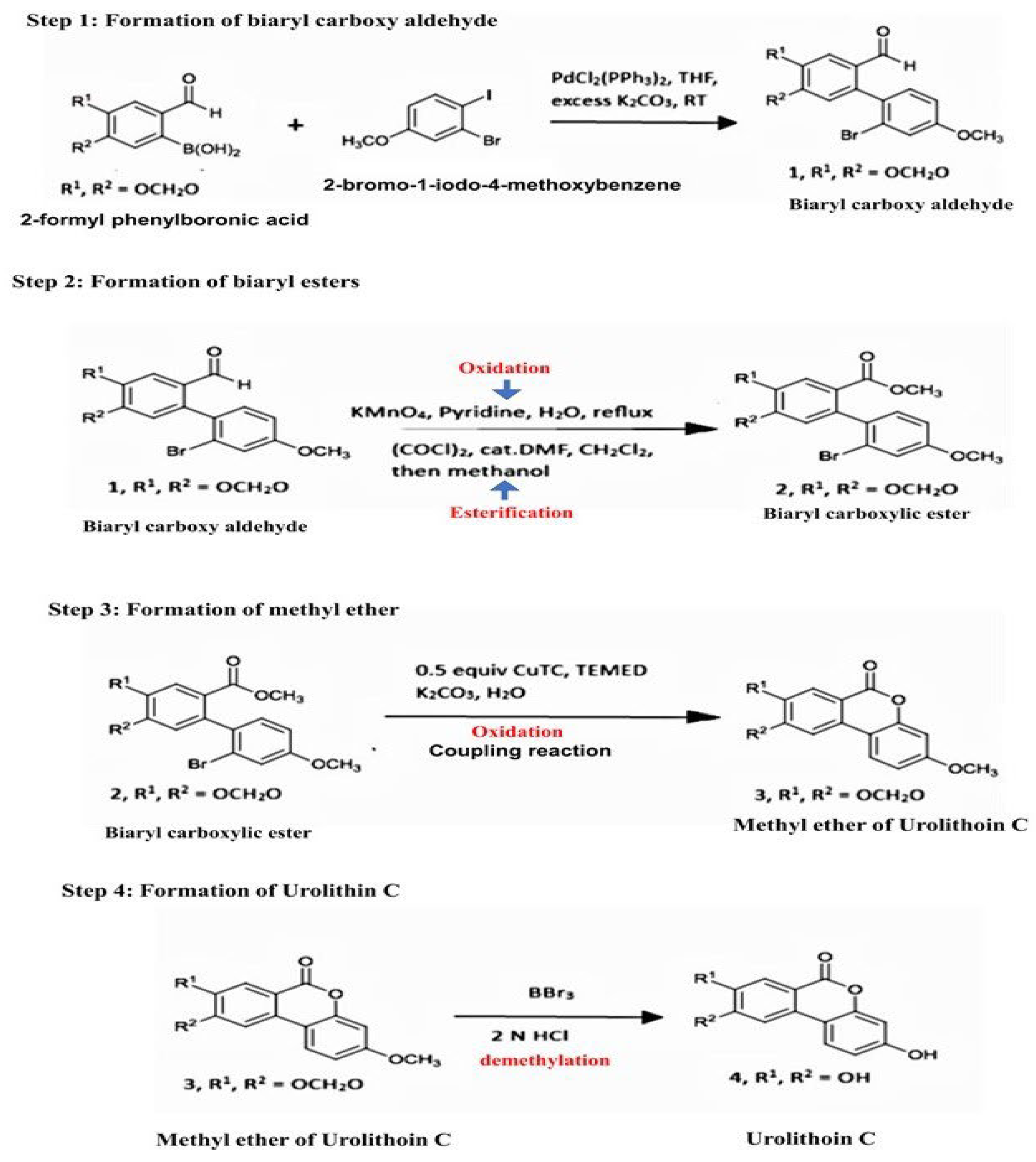
Figure 1. Process of Urolithin-C synthesis:.
Preprints 122120 g001
Figure 2. HPLC peak of Urolithin-C with 96.47 area % with a retention time of 10.902 minutes.
Preprints 122120 g002
Figure 3. FTIR and LCMS chromatogram of Urolithin-C: (A). FTIR spectra showing the presence of functional groups of Urolithin-C. (B) LCMS Chromatography of negative modes revealed with molecular weight of Urolithin-C.
Preprints 122120 g003
Figure 4. Antioxidant activity of Urolithin-C: (A) DPPH assay. (B) H2O2 assay. (C) FRAP assay. Each value is given as a mean ± SD. *significance at P ≤ 0.05 and *** at P ≤ 0.0001.
Preprints 122120 g004
Figure 5. Effect of Urolithin-C on NaNO2 induced oxidative stress in RBCs: (A) LPO.(B) PCC. (C) TT. (D) SOD. (E) CAT. To identify the protective effect of Urolithin-C against NaNO2 induced oxidative damage, red blood cells were preincubated for 10 min with various concentrations (0–25 µg) of Urolithin-C at 37 °C before treatment with NaNO2 (10 mM). The results are presented in average units/mg of protein and are expressed as mean ± SEM (n = 3). Significance * at p ≤ 0.005, ** at p ≤ 0.001 and *** at p ≤ 0.0001.
Preprints 122120 g005
Figure 6. Effect of Urolithin-C on Diclofenac induced oxidative stress in tissue homoginates of liver, kidney, heart and pancreas: (A) PCC. (B) LPO. (C) TT. DFC (50 mg/kg), SLY + DFC (25 mg/kg), Urolithin-C alone (150 mg/kg), Urolithin-C (50 mg/kg) + DFC, Urolithin-C (100 mg/kg) + DFC, and Urolithin-C (150 mg/kg) + DFC in comparison to the toxicity for control groups. All samples presented were administrated as mg/kg body weight/day. The data represented as mean (n = 3) ± SEM. *** Significance at p ≤ 0.0001 and ** at p ≤ 0.001.
Preprints 122120 g006
Figure 7. Effect of Urolithin-C on diclofenac induced oxidative stress in liver, kidney, heart and pancreas: (A) SOD. (B) CAT: DFC (50 mg/kg), SLY + DFC (25 mg/kg), Urolithin-C alone (150 mg/kg), Urolithin-C (50 mg/kg) + DFC, Urolithin-C (100 mg/kg) + DFC and Urolithin-C (150 mg/kg) + DFC in comparison to the toxicity for control groups. All samples presented were administrated as mg/kg body weight/day. The data represented as mean (n = 3) ± SEM. *** Significance at p ≤ 0.0001 and ** at p ≤ 0.001.
Preprints 122120 g007
Figure 8. Effect of Urolithin-C on Diclofenac induced oxidative stress in rat’s serum biochemical parameters as liver function test: The results presented are expressed as mean ± SEM (n = 3). Significance* at p ≤ 0.005, ** at p ≤ 0.001 and *** at p ≤ 0.0001.
Preprints 122120 g008
Figure 9. Effect of Urolithin-C on Diclofenac induced oxidative stress in rat’s serum biochemical parameters: (A) Renal function test. (B) Heart function test. The results are presented are expressed as mean ± SEM (n = 3). Significance* at p ≤ 0.005, ** at p ≤ 0.001 and *** at p ≤ 0.0001.
Preprints 122120 g009
Figure 10. Effect of Urolithin-C on DFC induced oxidative stress in rat’s serum lipid profile: The results presented are expressed as mean ± SEM (n = 3). Significance* at p ≤ 0.005, ** at p ≤ 0.001 and *** at p ≤ 0.0001.
Preprints 122120 g010
Figure 11. Effect of Urolithin-C on DFC induced histopathology of (A) liver and (B) kidney: (A1 and B1) DFC administered (50 mg/kg) group; showed liver hepatocellular degeneration and the kidneys' tubular degeneration, (A2 and B2) showed normal tissue morphology similar to the SYL treated positive control group (A3 and B3). (A4 and B4) Urolithin-C alone injected (100mg/kg) group showed normal tissue morphology; (A5 and B5) Urolithin-C (50 mg/kg) + DFC group) group showed almost normal architecture; (A6 and B6) Urolithin-C (150 mg/kg) + DFC group showed successfully restored their structural integrity.
Preprints 122120 g011
Figure 12. Effect of Urolithin-C on DFC induced histopathology of (A) heart and (B) pancreas: (A1 and B1) DFC administered (50 mg/kg) group; showed heart cell necrosis, and the pancreas' cellular degradation. (A2 and B2) showed normal tissue morphology similar to the SYL-treated positive control group (A3 and B3). (A4 and B4) Urolithin-C- alone injected (100mg/kg) group showed normal tissue morphology (A5 and B5) Urolithin-C (50 mg/kg) + DFC group) showed almost normal architecture; (A6 and B6) Urolithin-C (150 mg/kg) + DFC group showed successfully restored their structural integrity.
Preprints 122120 g012
Disclaimer/Publisher’s Note: The statements, opinions and data contained in all publications are solely those of the individual author(s) and contributor(s) and not of MDPI and/or the editor(s). MDPI and/or the editor(s) disclaim responsibility for any injury to people or property resulting from any ideas, methods, instructions or products referred to in the content.
Copyright: This open access article is published under a Creative Commons CC BY 4.0 license, which permit the free download, distribution, and reuse, provided that the author and preprint are cited in any reuse.
Alerts
Prerpints.org logo

Preprints.org is a free preprint server supported by MDPI in Basel, Switzerland.

Subscribe

© 2025 MDPI (Basel, Switzerland) unless otherwise stated