Preprint
Article

The Influence of Realism on the Sense of Presence in Virtual Reality: Neurophysiological Insights Using EEG

Submitted:

27 October 2024

Posted:

28 October 2024

You are already at the latest version

A peer-reviewed article of this preprint also exists.

Abstract
One of the most crucial aspects of the user experience in virtual reality (VR) is the sense of presence. To evaluate this, both subjective and objective methods can be employed. While subjective methods are easy to implement and interpret, they may not fully capture user feedback, and the results can sometimes lack consistency. In contrast, using objective methods, such as electroencephalography (EEG), can provide more reliable insights. To investigate the influence of realism on the sense of presence, we conducted an EEG study with 21 participants who experienced two VR environments—one realistic and one non-realistic. During the study, we continuously measured their brain activity using an EEG device. Our findings showed that alteration in the level of realism in an environment can be detected through changes in brain activity. Notably, we observed that users take longer to adapt to a non-realistic environment when transitioning from a realistic scene, compared to the reverse. Although our study has limitations, such as the total number of participants, we gained valuable initial insights into how realism may influence brain activity. These findings suggest that higher realism may lead to reduced cognitive load, increased attention, improved decision-making, and suppression of irrelevant information.
Keywords: 
Subject: 
Computer Science and Mathematics  -   Computer Science

1. Introduction

The rapid expansion of Virtual Reality (VR) can be attributed to the widespread adoption of consumer-based Head Mounted Displays (HMDs). VR has been increasingly utilized in various fields, including education, computer science, engineering, and healthcare. In the realm of education, VR has been employed as a pedagogical tool to enhance experiential learning across different subject areas to enhance student’s engagement, motivation, and academic performance [1,2,3]. In medical education and therapies, VR can be used as a visualization, training, and simulation medium to provide the conditions that are either difficult to demonstrate and imagine or critical to act against them [4,5]. In the engineering field, VR can improve design, decision-making, and collaboration for different stakeholders [6,7]. In order to ensure consistent quality across various VR applications, it is essential to establish guidelines for their design and development. One of the primary steps in creating these guidelines is to gain a comprehensive understanding of the factors that influence the design medium and user experience.
The measurement of influential factors on the user experience in VR is typically accomplished through subjective questionnaires or objective sensory measurements. Both methods are widely used in research and development. Questionnaires are essential tools for evaluating subjective responses and gaining insights into various aspects of user experiences in VR environments [8]. The employment of questionnaires has gained widespread acceptance as an evaluation tool to assess multiple facets of user experience, including but not limited to, immersion, presence, usability, and engagement. Its simplicity has made it a popular choice for varied applications within business and academic settings. These questionnaires are designed based on the required assessment factors. For example, measuring user experience with User Experience Questionnaire developed by Schrepp et al. [9], assessing the sense of presence using Igroup Presence Questionnaire (IPQ) by Schubert et al. [10], and user engagement using User Engagement Scale (UES) developed by O’Brient et al. [11]. Although questionnaires are a widely adopted tool for research studies and are often considered to be simple to implement and efficient in yielding interpretable results, they have been criticized for their inadequacy in capturing the complete range of experiences that subjects undergo in virtual environments [12,13]. This view is based on the argument that questionnaires may miss out on important experiential nuances that are difficult to capture through standardized responses [14].
In this case, utilizing objective measurements can be advantageous as they can directly and uninterrupted capture information based on real-time user behavior and experience. There are various physiological responses that can be measured using corresponding equipment, such as peripheral skin temperature, heart rate, heart rate variability, respiration, skin conductance, and brain activity [15,16,17]. Although objective measurements offer several benefits, running studies with these methods can be challenging, and interpreting the results is more complex than with questionnaires. Nevertheless, finding appropriate correlations between the results of these objective measurements and user experience factors can lead to more robust evaluations and findings in research. Furthermore, incorporating neurophysiological measurements into studies offers researchers a powerful tool for understanding human behavior, cognition, and emotion with greater precision and objectivity.
Among all factors of user experience, presence can play a crucial role in various aspects, such as satisfaction, motivation, performance, and engagement [18,19]. Although closely related, the notions of presence and immersion hold distinct meanings. Presence pertains to the personal sensation of being in a virtual space, whereas immersion is an objective quality of the setting that has the potential to enhance the feeling of presence [13,20,21]. Perceiving the virtual environment as real is possible due to a higher level of presence [22]. As presence is a subjective measure, assessing it using questionnaires can be challenging and in some cases lead to inconsistent results [12]. In addition, questionnaires cannot provide real-time and continuous measures of the sense of presence during gameplay.[18]. For this reason, finding a proper correlation between sensory measurements and the sense of presence in a VR environment can be beneficial.
As previous studies have shown, the sense of presence can be correlated to the level of realism in a VR environment [23]. By changing this factor, we can expect different levels of presence. On the other hand, neurophysiological insights of presence can be captured by utilizing objective measurements such as electroencephalography (EEG) devices [24,25]. EEG is a technique for capturing the brain’s spontaneous electrical activity and has recently been utilized in research exploring the sense of presence [18]. Drawing upon this concept, our study aims to address two research questions:
  • (RQ1) How does altering the visual realism of a virtual environment affect brain activity?
  • (RQ2) Are specific brain patterns associated with the subjective sense of presence in virtual environments?
Accordingly, we designed and developed a VR experience to investigate the influence of realism on the presence and measured neurophysiological data using a 32-channel active electrode EEG device. In previous studies, researchers primarily focused on analyzing data from one condition and comparing it with another. However, this study also aimed to explore the transition point between the two conditions, which can reveal important information and provide clearer comparisons. In our study, participants were randomly assigned to two groups: one experienced a non-realistic scene first, while the other experienced a realistic scene first. After completing the tasks in the initial environment, both groups immediately switched to the other condition. During the experience, we continuously measured different brain wave band powers in various brain lobes using an EEG device. In this way, we can observe whether altering the scene (virtual environment) between realistic and non-realistic conditions influences brain activity during the VR experience. Furthermore, since previous studies, such as [10], categorize presence into three main components—Involvement, Spatial Presence, and Realism—we want to investigate whether there is a relationship between brain activity in different frequency bands and lobes, and these factors.
Although the focus of our study is on evaluating the sense of presence using an EEG device, we also considered including the NASA TLX and IPQ questionnaires as subjective measurements at the end of the study. Since we need continuous measurement of brain activity, even during scene transitions, we could not use these questionnaires throughout the study. Consequently, the results of these questionnaires reflect the overall response of participants and might be influenced by the second environment they experienced, whether realistic or non-realistic.
The findings of this study can serve as valuable input for the development and design of user interactions, user interfaces, and gameplay elements in immersive VR games and applications. By leveraging the heightened sense of presence that can be achieved through these elements, game developers can enhance the overall user experience.

2. Background and Related Works

  • User experience and sense of presence: The evaluation of user experience in VR is a multifaceted area of research that spans various aspects, including ease of use, level of presence/immersion, engagement, and psychological impacts. Over the years, studies tried investigating and evaluating these areas to improve VR applications [26,27]. One of the important factors of user experience in an immersive environment is the sense of presence [28]. Users would expect the sense of presence as a basic functionality of these environments [23]. The stronger this sense of presence, the more likely participants are to behave similarly to how they would in a real-world setting.
  • Categorization and definition of presence: Researchers have delineated and categorized presence in various manners, contingent upon individuals’ perceptions of a virtual experience [29]. As a simple definition, presence can be defined as a subjective feeling of being in the virtual experience [30]. As a more detailed description, Skarbez et al. [31] categorize presence in "being there", "non-mediation", and "other". The dimension of "non-mediation" encompasses conceptualizations of Presence within technological contexts. Here, presence is delineated as the perceptual illusion of transcending mediation or as the suspension of disbelief, leading users to immerse themselves in an alternate reality detached from the actual world [32,33]. The "other" category regards presence as the perception of the virtual world as if it were real [34]. Within an alternative structure, users’ experience of presence can be categorized in two fields: spatial presence and social presence [32,35]. Social presence mainly focuses on experiencing interpersonal connections in a virtual world. Spatial presence can be considered as the "presence of place" and refers to the perceptual illusion of non-mediation, i.e., the illusion of being in a place while experiencing the virtual world. In this way, social presence requires a multi-user virtual experience, but spatial presence can be experienced whenever a virtual environment exists.
A postulation by Slater [36], considers the sense of presence as a result of two illusions: place illusion and plausibility illusion, based on neuroscience concepts. The place illusion can be defined as an illusion of "being there", even if the user knows it does not exist in reality. While the place illusion concerns perception, plausibility illusion involves the convincing belief that events in a virtual environment are real despite knowing otherwise. The plausibility illusion is reinforced by the virtual environment correlating external events with the participant’s sensations. For instance, direct eye contact from an avatar elicits physiological responses, unlike inanimate objects. This will lead users to behave like they act in a real situation of encountering that event.
  • Evaluation of sense of presence: In order to assess presence in VR, one can consider realism as an influencing factor on the level of presence, as it is also considered in IPQ and previous studies [37,38,39]. In this way, comparing two distinguishable conditions with clear differences in the level of realism can lead to an acceptable interpretation of the results. Earlier studies categorized realism based on the level of the perceived image into physical, photorealism, and functional realism categories [40]. In a different approach, a two-dimensional categorization approach proposed by Goncalves et al. [41] considers user perception and proximity to real-world counterparts. The first dimension is subjective and correlates with how users perceive the virtual world, regardless of whether it is designed to be realistic or imaginary. Here, the coherency of the experience plays an important role. Users may accept an environment as perceptually realistic if the sequence of events in a virtual environment has a reasonable coherency [42]. In the second dimension, realism is regarded as an objective measure that can be compared with the expectation of real-world conditions. Slater et al. [39] considered realism as the combination of two components: illumination and geometry. Illumination realism is influenced by the precision of lightning including shadows, global illumination, and reflection. Geometrical realism is related to the similarity of virtual objects to real ones, including polygonal shape, texture, and material. Most of these studies used questionnaires to assess the influence of differentiation in the level of realism on the sense of presence. Although these questionnaires can provide a general overview of the level of presence, an in-depth understanding can be achieved using physiological measures [39]. In this study, we will focus solely on visual realism, without considering other types such as auditory realism.
Given the diversity of approaches in defining and conceptualizing presence, corresponding variations exist in evaluation methodologies. These methods encompass objective measures through physiological assessments, subjective assessments via questionnaires, or a synthesis of both approaches[29].
  • Subjective measurement of presence: Subjective measures involve self-reported information provided by users following a VR experience, typically gathered through questionnaires. These measures are relatively simple to implement and straightforward to analyze due to their questionnaire-based structure. Throughout the years, researchers have developed various questionnaires, such as IPQ and Witmer-Singer [43], to assess the sense of presence in VR. However, evaluating presence factors is challenging due to the subjective nature of it. The initial problem that raise due to the change in the environment of experiencing VR and answering the questions. This change can cause a break in presence (BIP) and negatively influences the general user experience [28,44]. In recent years, several studies have been conducted to investigate the effect of implementing questionnaires in VR to avoid BIP [13,23,45]. Schwind et al. [23] assessed the presence of in-VR and out-VR conditions after VR intervention. In their study, participants played a first-person shooter game in two design conditions: abstract and realistic. The results indicate a significant influence of virtual realism on the sense of presence using IPQ. In a more comprehensive study, Alexandrovsky et al. [45] conducted an expert survey followed by two user studies to compare user responses to user experience questionnaires in both in-VR and out-VR conditions. According to their expert survey, most of the researchers see the necessity of embedding questionnaires in VR but their user study indicated lower usability and higher physical demand compared to out-VR questionnaires. Although in these studies the majority of users preferred in-VR questionnaires over the out-VR ones, they did not find significant differences in the results of questionnaires. In addition, some studies reported inconsistency of the responses with the experiment conditions [12]. As an example, they reported different responses to the realism item of IPQ questionnaires without changing the virtual environment and just by changing the questionnaire user interface, from out-VR to in-VR [12,13]. These inconsistencies can push studies towards using different methods such as different sensory measurements to improve the reliability of the results.
  • Objective measurement of presence: Utilizing physiological measurements as a tool for assessing presence in VR offers the potential for continuous, non-intrusive, and objective data capture [46,47]. Specifically, the integration of VR technology with objective physiological metrics, rather than solely relying on subjective assessments, is rapidly gaining traction as a valuable tool for guiding design choices in the initial stages of artificial environment projects [48,49] and for examining human/environment interactions. Additionally, there is a growing body of research exploring the utility and efficacy of VR-based therapy for psychiatric conditions [50,51]. Given the correlation between presence and certain brain activities, exploring the brain activity patterns associated with the sense of presence is crucial, which has been relatively understudied so far.
Some past studies have looked into various factors that contribute to enhancing the subjective feeling of presence with a focus on external, and more objective factors including technology-related aspects of VR [3,39,52]. Although there are studies that focus on subjective measures of presence recorded with questionnaires [29,53,54], they often lack causality. Hence, there is a burgeoning interest in delineating the neuronal correlates of presence, aiming to characterize the associated individual mental states of users through objective measurements. Among the prevalent methodologies employed for this purpose, measuring brain activity using EEG devices stands out as a common approach. However, finding such a correlation is very challenging and studies attempting to find physiological correlates of sense presence using EEG have reported mixed results [55,56,57].
  • EEG as an objective measurement tool: EEG waveforms are generally classified according to their frequency, amplitude, and shape, as well as the sites on the scalp at which they are recorded. The five most commonly used frequency bands and their corresponding frequency ranges are: delta (0.5-4 Hz), theta (4-8 Hz), alpha (8-13 Hz), beta (14-30 Hz), and gamma (> 30 Hz) [58,59]. Different mental states, like motivation, emotion, attention, and higher cognitive processes are strongly linked to brain waves in these different frequency bands [60,61,62,63,64]. Therefore, investigating frequency-specific changes in EEG data can lead to valuable insights in terms of how study participants process or respond to different stimuli.
In a recent study, Grassini et al. [18] used brain event-related potentials (ERPs) elicited by auditory stimuli to identify an objective physiological index of sense of presence during VR. They showed that late auditory ERP components recorded over the central brain may represent good electrophysiological correlates of the subjective sense of presence. However, there are some limitations of this study like the theoretical a priori selection of cognitive phenomena (and therefore ERP correlates) of interest. Thus, a data-driven approach [65] may be favorable to investigate the topography and latency of potential ERP correlates of the sense of presence.
  • Functional meaning of the different EEG bands: EEG bandpower analysis in different brain regions provides insight into various cognitive and emotional states [62]. The four primary frequency bands (alpha, beta, gamma, and theta) are associated with distinct neural activities and functions, and their significance can vary depending on the brain region in which they are observed.
-Alpha band: The general meaning of the alpha band (8–12 Hz) is on the one hand relaxation and idle state and on the other hand inhibition control. Higher alpha power is generally associated with relaxed, wakeful states, often when the brain is in an idle or resting state. But alpha activity can also reflect cortical inhibition, a mechanism that helps suppress irrelevant or distracting information [60,66,67,68]. Furthermore, the meaning of alpha activity depends on the different brain regions. For example, increased alpha power in the frontal lobes can indicate relaxation and decreased cognitive load. It may also be associated with positive emotional states when more pronounced in the left hemisphere. A higher alpha power in the parietal lobes is often linked to attentional processes and the allocation of attentional resources. In the occipital lobe, increased alpha power is associated with reduced visual processing, often seen when the eyes are closed or in a resting state. This common finding in cognitive neuroscience received counter-evidence by a recent study by Hohaia et al.[69]. Their data show that the power of occipital alpha-band brain waves can be increased by motion-sensitive visual processes that persist when the eyes are closed. Consequently, they suggested that this power of alpha-band oscillations might be a product of visual processes that can be modified by motion adaptation.
-Beta band: The meaning of the beta band (13–30 Hz) is more related to active thinking and concentration but also to anxiety and arousal. Increased beta power is associated with active cognitive processing, concentration, and alertness [70]. Elevated beta activity can also be linked to anxiety, arousal, and stress [71]. In the frontal lobes, higher beta activity is related to active thinking, decision-making, and executive functions. Left frontal beta activity is often associated with positive emotions and approach-related behaviors, while right frontal beta activity can be linked to anxiety and withdrawal-related behaviors. In central regions, like the sensorimotor cortex, an increased beta activity is related to motor planning and control. It is often observed during motor tasks and in states of focused attention. Whereas elevated beta activity in the temporal lobes can be associated with auditory processing and language tasks.
-Gamma band: The gamma band (30–100 Hz) is commonly associated with high-level cognitive functions such as perception, attention, and memory [72,73]. Gamma band activity is also linked to conscious awareness and the integration of sensory information [74,75]. Increased gamma activity in the frontal lobes is associated with executive functions, problem-solving, and complex cognitive tasks. On the other hand, gamma activity in the parietal lobes is linked to spatial awareness, sensory integration, and attentional control. In the temporal lobes, gamma activity is related to memory processes and auditory perception.
-Theta band: The general meaning of the theta band (4–7 Hz) activity is often associated with memory encoding and retrieval [76], as well as learning processes [77] and emotional processing [78]. Whereas frontal theta activity is associated with cognitive control, working memory, and decision-making, temporal theta oscillations are crucial for memory formation and navigation. In parietal regions, the theta activity is mainly linked to attentional processes and the integration of sensory information.
The measurement of the activity of these bands can be done either at specific time points when certain events occur or continuously throughout the entire experience. However, as the sense of presence is a continuous measure throughout the whole experience, recording EEG results at certain time points, such as ERP, may lead to losing the comprehensive understanding of the whole progress. Following that we investigated the sense of presence by recording brain activity continuously, revealing the ongoing EEG activity in specific frequency bands [79,80]. By manipulating the visual realism of the VR environment, we first investigated its influence on the sense of presence and second studied the exposure duration in each virtual environment by measuring the time that the brain needs to perceive and adapt to the new state. Based on the literature [55,81,82] we assume an increased parietal activation and decreased frontal activation with a stronger sense of presence and its dependency on realism. It’s essential to understand how perceiving a VR environment (or engagement in VR tasks) can impact the subject’s physiological parameters, especially EEG metrics, to accurately interpret behavioral data and psycho-physiological effects across these applications.

3. Methodology

In order to assess the impact of realism on user experience, we conducted a user study that incorporated sensory measures and questionnaires.

3.1. VR Application

We designed and developed a VR game for this study using Unreal Engine1 (UE) 5.2. The game features some of the most advanced tools and rendering features available in UE, including realistic lighting through the global illumination system (Lumen), highly detailed models using virtualized geometry (Nanite), and a deterministic procedural environment using procedural content generation (PCG). To ensure seamless support for various VR devices, we have utilized the OpenXR2 plugin for communication between the HMD and the game engine. Furthermore, we have incorporated The Lab Streaming Layer3 (LSL) plugin in UE to capture events on an EEG device during certain moments in the game, such as collecting coins or switching the environment. LSL is a reliable and efficient system for the unified collection of measurement time series in research experiments, handling networking, time-synchronization, real-time access, and even centralized collection, viewing, and disk recording of the data.
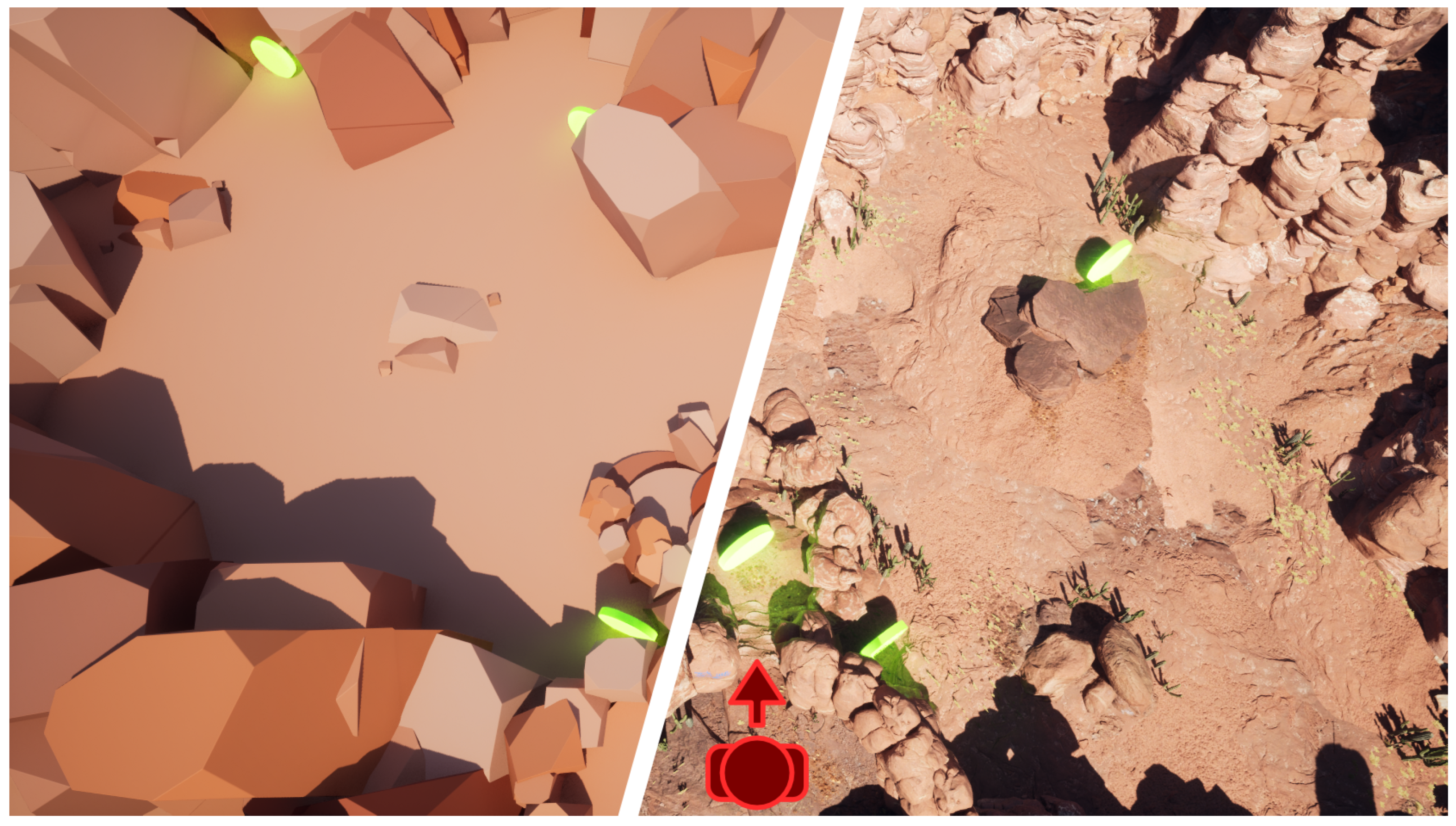
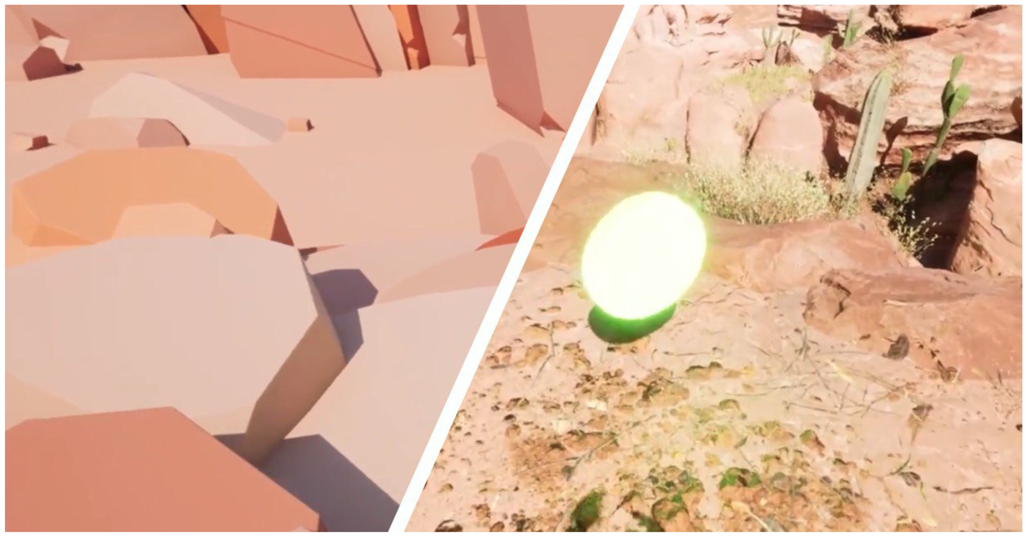
The VR game consists of two main environments (levels): realistic (HP) and non-realistic (LP)4, which are shown in Figure 1. The object positions and level designs for both environments are almost identical to prevent any influence on the study results. Both environments depict a desert scene, see Figure 2. The LP level uses low-detailed elements without textures and excessive geometry details. Since there is no texture at the LP level, we used random color assignments to the geometries to help the user distinguish between objects. On the other hand, the HP scene uses highly detailed geometries, realistic textures, high-quality shadows, and additional vegetation.
In both VR environments, the gameplay task is to locate and acquire six green coins. These coins are designed to be distinct from other objects in the scene, as they rotate along their axes, see Figure 3. The coins’ positions are deliberately varied at each level to avoid the impact of prior knowledge on the study results. Nevertheless, the difficulty level of finding the coins is maintained consistently at both levels. The interaction required to collect the coins is simplified as much as possible, with participants merely required to touch the coins using the virtual hand. This simplification is intended to alleviate excessive cognitive loads for participants with limited prior VR experience.
Participants are allowed to move freely in a dedicated area of the evaluation physical environment, but due to the limited physical space, we used a smooth locomotion system for navigation. Moreover, the incorporation of smooth locomotion mechanisms served to mitigate the requirement for extensive physical movement from participants. This approach was instrumental in minimizing potential muscle movement artifacts that could introduce noise into the EEG signal recordings, thus enhancing the overall data quality collected by the EEG device. To move around in the virtual world, participants can use a controller joystick. To make it less complicated for new VR users, participants can only move in the forward/backward direction of their facing direction. It is important to note that we did not include teleportation locomotion in this study to avoid disorientation that leads to a negative impact on participants’ sense of presence [83]. We chose not to use the sitting position for the experiment in order to reduce the influence of physical world sensations on the user’s experience, as used in the study by Grassini et al. [18]. Additionally, we used a simple natural scene, such as a desert, to avoid activating excessive emotional factors that could affect the measured EEG data, as in the study by Terkildsen and Makransky [57].

3.2. Material

We utilized a combination of a physiological measurement device and questionnaires. Although the primary focus was on the sensory measurements, questionnaires can provide comparative results for a quick overview. At the outset of the study, participants were requested to complete demographic questionnaires that included information about their gender, age, profession, and prior VR experiences. During the study, we employed an EEG to measure the electrical activity of the brain. In addition, at the end of the study, participants were asked to fill out post-questionnaires, including NASA TLX for task load and IPQ for the subjective sense of presence. As answering the questionnaire may lead to BIP [8], we did not include these questionnaires between the scene switches. This way, we can report the continuous measurements and clearly observe the influence of scene changes.

3.3. Setup

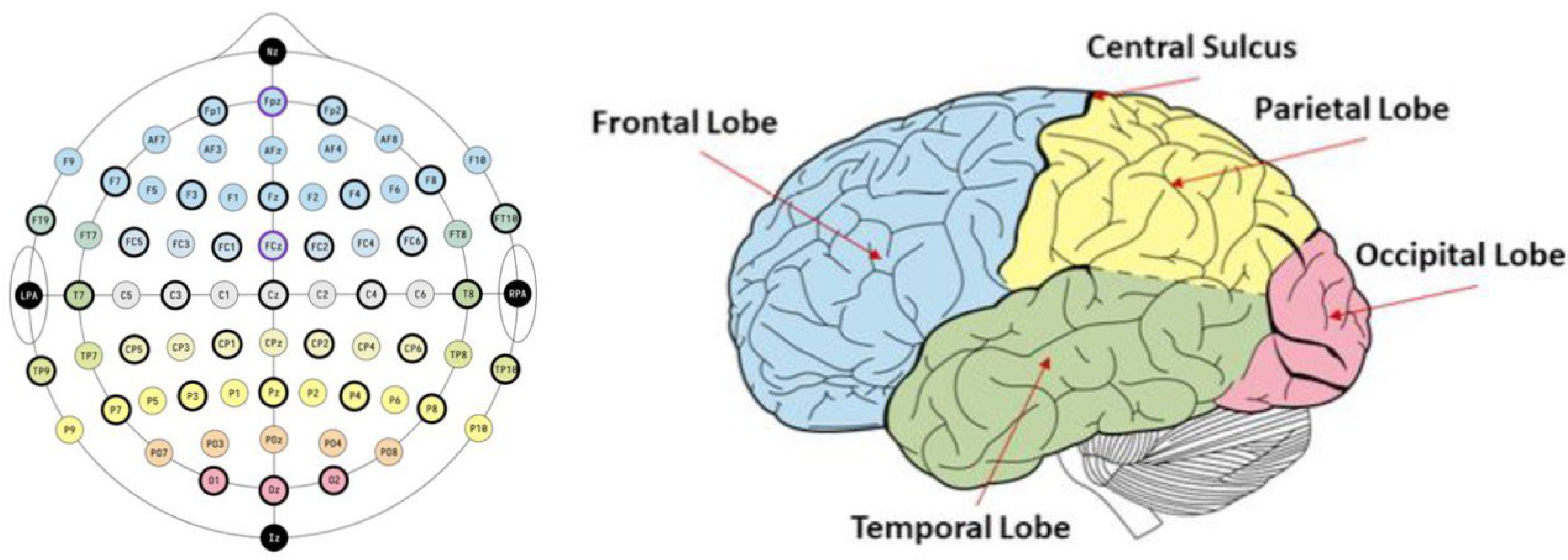
Multichannel EEG was measured using the BrainVision LiveAmp5 at a sampling rate of 500 Hz, see Figure 1. We used 32 channel active EEG electrodes with 28 electrodes placed according to the international 10-10 system, see Figure 4. The remaining four electrodes were specifically designated for recording the electrooculogram (EOG). The connected wireless amplifier was attached to the backside of a participant’s cloth. It sent the recorded EEG signals from the electrodes to the computer via Bluetooth, which were then visualized on the screen. We used the Meta Quest 2 headset as an HMD that provides both wired and wireless connection possibilities. It was mounted on top of the already mounted EEG cap, followed by another impedance check, to see if any impedance changed. If all impedances were still under 20 k Ω , the participant got a controller for their main hand and was asked to stand up to prepare for the game. Participants were provided with a designated area measuring 2m x 2m, allowing for unrestricted movement within the space during the VR experience. The experimenter arranged the participant in the correct position. For this study, we connected the VR headset to a PC with 16 GB of RAM, an Intel Core i7 CPU, and an NVIDIA RTX 2080 graphic card.

3.4. Participants

21 healthy participants, 14 males, and 7 females, took part in this study. The age range of the participants varied from 23 to 31 years, with an average age of 27.14 years and a standard deviation of 2.20. During the study, participants were asked to share their previous experience with VR applications and rate it on a scale of 1-5, where 1 indicated seldom use and 5 indicated everyday use. The findings revealed that the participants had relatively low experience with VR, with a mean value of 1.9 and a standard deviation of 1. The study was approved by the ethics committee of Graz University of Technology (Votum EK-37/2024) and is in accordance with the ethical standards of the Declaration of Helsinki.

3.5. Procedure

At the beginning of the study, we asked the participants to fill out a demographic questionnaire. We then explained the evaluation procedure to them, along with their rights, and provided brief information about the EEG device. Next, we measured the circumference of each participant’s head to determine the proper size for the EEG cap. The cap was then placed on their head and secured with fixed straps under the chin. We readjusted the cap to ensure that the Cz electrode was at the center of the intersection between the nasion-inion and left (LPA) and right pre-auricular (RPA) fiducials. The overall cap mounting process took approximately 30-40 minutes, during which the participants received more information about the game they were about to play. After mounting the EEG cap, they wore the VR headset in preparation for the study.
Participants were randomly assigned to two groups for an A/B study. One group began with LP level and switched to HP level, while the other group started with HP level and then switched to LP level. They played the game without any interruptions between levels. The duration of each level varied from 5 to 10 minutes, depending on how efficiently the participants were able to locate and collect coins. Once the game was completed and the headsets were removed, we requested the participants to complete post-questionnaires. These questionnaires encompassed the IPQ and NASA TLX.

3.6. Signal Processing

3.6.1. Preprocessing

The recorded EEG-Data was mainly pre-processed and analyzed using MATLAB2023b and EEGLab. The EEG-Data was bidirectionally filtered with a 4th-order Butterworth bandpass filter between 1 and 60Hz. A notch filter (Bandstop IIR with a filter order of 2) was used to filter out the 50Hz line noise. The EEG-Data was visually inspected for obvious artifacts and the Pz channel and in some subjects, the CP1 channel was removed. Afterward, an independent component analysis (ICA) was used to remove and filter out bad components (like blinks or muscle movements) with a probability of above 0.7. After conducting the ICA, the missing channels were interpolated. EEG data were epoched into HP and LP trials. Finally, several regions of interest (ROI) like frontal, parietal, temporal, and occipital regions were introduced by averaging all of the corresponding channels. Subsequently, the bigger ROIs were compartmentalized into right and left hemispheres like frontal left (FL), frontal right (FR), and so on. Further feature extraction was done using these averaged ROIs. The used cutoff frequencies of each frequency band were defined as follows: Theta: 3Hz to 7Hz, Alpha: from 8Hz to 12Hz, Beta: from 13Hz to 30Hz, and Gamma: from 30Hz to 100Hz.

3.6.2. Feature Extraction

  • Absolute Bandpower: The bandpower of the trials were calculated using the Matlab provided function bandpower() and cutoff frequencies of different frequency bands. This function gives the average bandpower of the entire trial of each ROI. The complete trial is determined by starting in one environment and collecting 6 coins there and is then terminated. The outliers of the calculated bandpower were removed when these outliers were located more than 3 scaled median absolute deviations from the median. The arithmetic means and the standard deviation of the absolute bandpower were displayed. For statistical analysis, the Wilcoxon rank-sum test with a significance level of 5% and 1% was used.
  • Relative Bandpower: The participant-specific relative bandpower was then derived by dividing the HP-bandpower with the LP-bandpower while using the LP as a baseline. This method demonstrates greater stability, as it reduces the occurrence of the outliers and is able to show, whether the bandpower was increased or decreased in the HP trial relative to the LP trial. The relative bandpower were presented within the group HP-LP and LP-HP.
  • Break Response: For displaying the bandpower activation over time and the neural responses due to the different environments the segmented HP and LP trials were concatenated accordingly (by the sequence of the polygon environments) and repositioned to ensure that the break was centered at the specific time point. Given the individual variations of the temporal parameters (recording time) concerning collecting 6 coins, the maximum duration of recording before and after the break was set at 3.3 minutes. The time-point of the break was defined as 0 seconds.
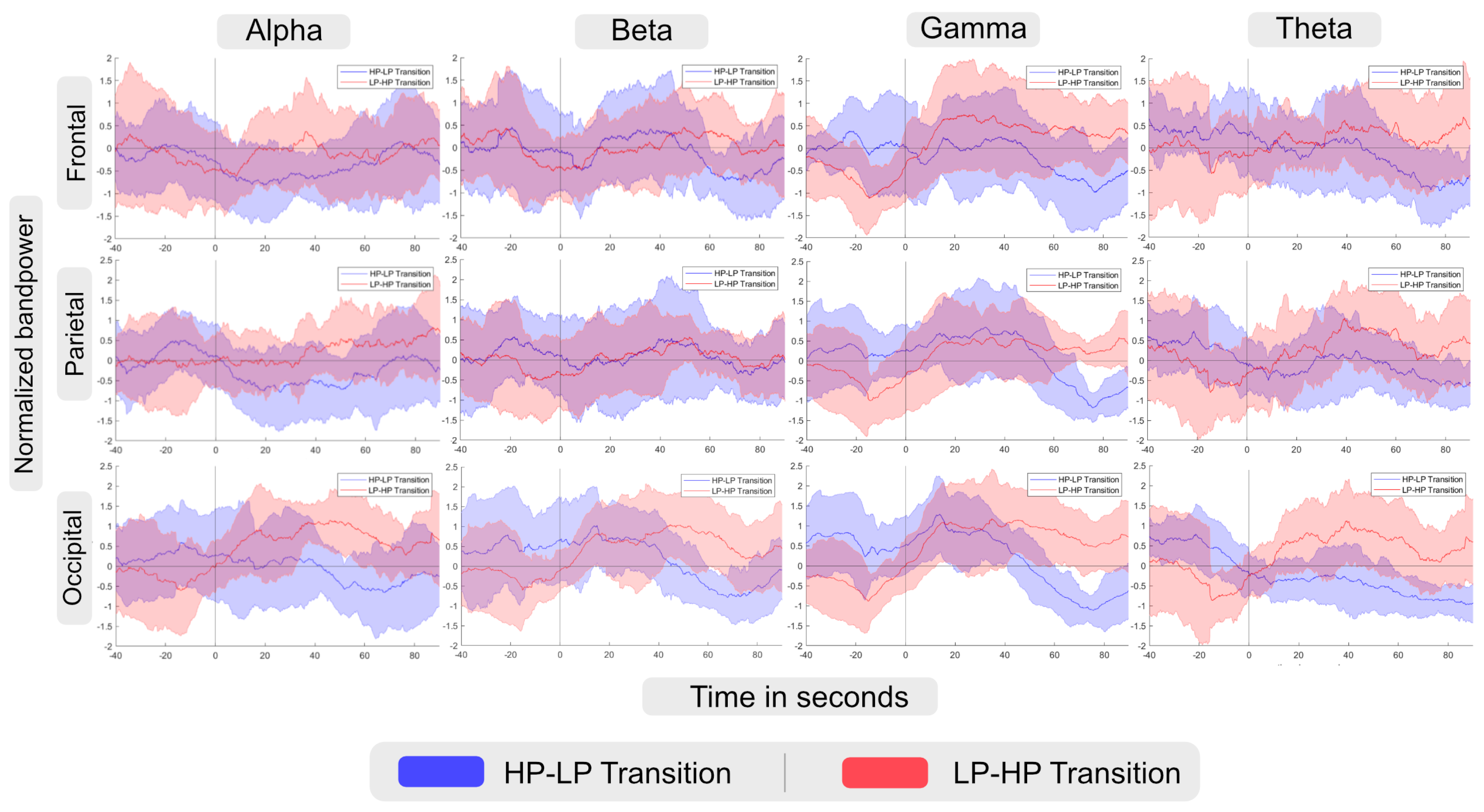
These realigned EEG data were then bidirectionally filtered with a 4th-order Butterworth bandpass filter with various cut-off frequencies of frequency-bands alpha, beta, gamma, and theta. The bandpass-filtered EEG data were then squared, averaged with a moving average window with a length of 30 seconds, and then normalized for each participant such that the mean is set to 0 and the standard deviation is set to 1. The normalizing step was required due to the individual variations of band power strength introduced by each participant. The break responses of different groups and lobes (F/Frontal, P/Parietal, T/Temporal, O/Occipital) were then displayed over time with the mean and standard deviations over participants, see Figure 5.
  • Adaptation time: In this study, we defined the adaptation time (AT) as the time required to achieve the maximum value of the normalized bandpower within 100 seconds. For extracting the AT for adapting to the new environment, the EEG data were exactly processed like in the previous part (filtering, squaring, moving averaging, and normalizing) with the difference that the length of the moving average window was set to 60 seconds. The upper boundary of the AT with 100 seconds was chosen as Baka et al. [84] showed that the time period needed for the adaptation of the brain was at approximately 40 seconds. The comparison was then conducted with the LP trials of the LP-HP in the beginning state and with the LP-trials of the HP-LP trials after the break. For the statistical analyses, the Wilcoxon rank-sum test was used. Statistical significant results were indicated with stars (single star for alpha=5% and double star for alpha=1%), see Figure 7.

4. Results

The main focus of this study is on understanding the influence of realism on brain activity using EEG results by switching the environment between realistic and simplified conditions. Accordingly, we first report the results of normalized band-power and then we discuss our findings regarding AT. In addition, we also asked participants to report their subjective experience using NASA TLX and IPQ.

4.1. EEG Measurement Results

Bandpower activity: According to the description of analysis in the methodology section, the normalized absolute band power over time is calculated and presented in Figure 5. At point zero, the environment changes from HP-LP (blue line), or LP-HP (red line). For each frequency band, the frontal, parietal, and occipital activation is displayed. The negative time range indicates the initial scene condition, and the positive time range corresponds to the measurements after changing the scene. Accordingly, for the LP-HP group, negative time indicates the LP scene, and positive time indicates the HP scene.
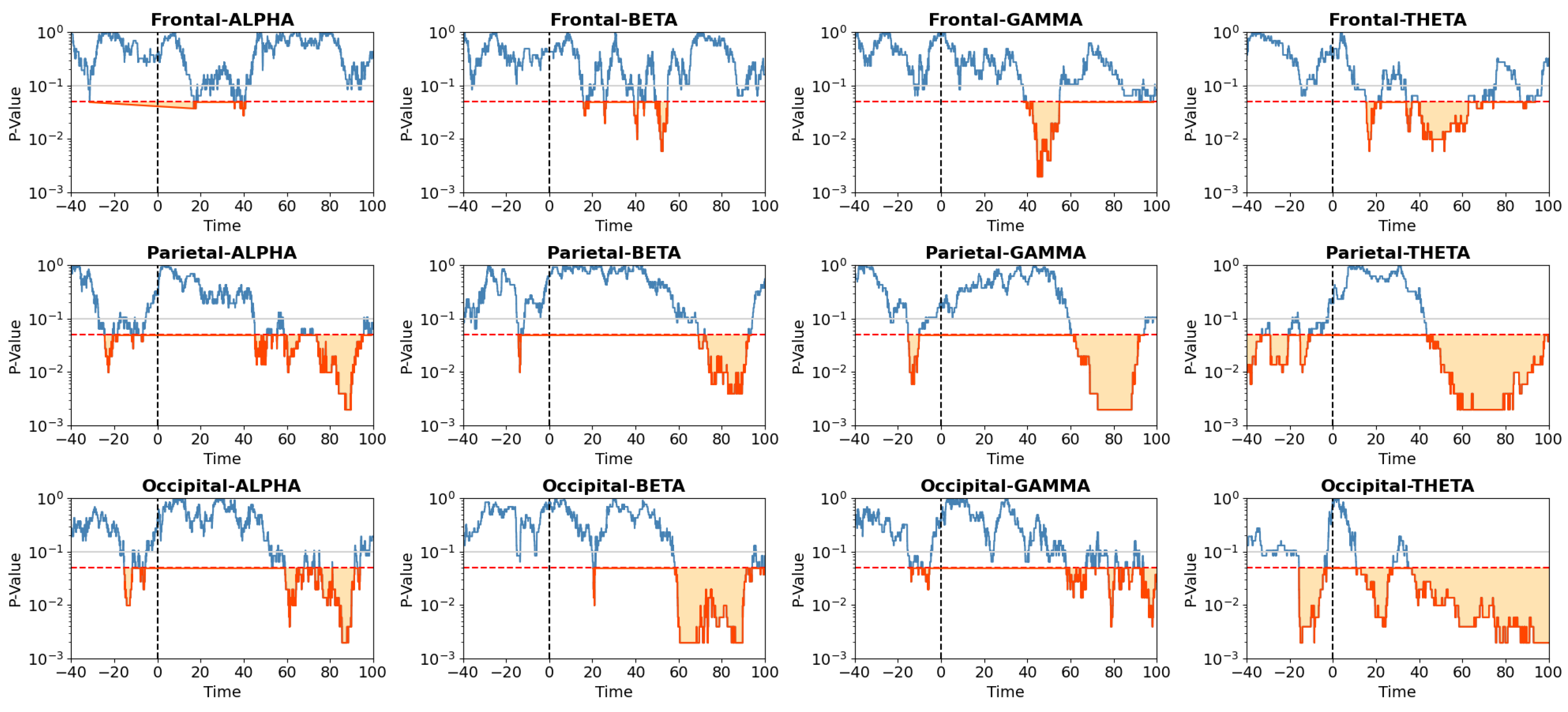
Considering the band power activity at each lobe, we run the Wilcoxon rank-sum test to find significant differences between groups at each time-point in continuous measurement and visualize it in Figure 6. According to this visualization, we can figure out if there is a significant difference in each time range that we observe differences between groups regarding band power activity in Figure 5. We used a logarithmic scale for the vertical axis of Figure 6 to help in reading p-values when they are close to zero. The red line in this figure represents a p-value of 0.05 (moderate evidence), and the gray line indicates a p-value of 0.1 (weak evidence or a trend). For the HP-LP group, negative time corresponds to the HP scene and positive time corresponds to the LP scene. Comparing these two figures in the frontal lobe shows significant differences in alpha band power between the two groups in the range of 10-50 seconds, with the HP condition having higher values. Similarly, we found significant differences between groups in gamma and theta band power after the scene change.
In contrast to the frontal lobe, where significant differences between the two groups are observed only after the scene change, in the parietal lobe there are significant differences both before and after the scene change for all wave bands. For the alpha and theta bands, significant differences after the scene change are observed after about 40 seconds, while for the beta and gamma bands, this occurs after about 60 seconds. In all cases, the HP condition shows higher band power both before and after the scene change.
Similar to the parietal lobe, significant differences are found both before and after the scene change for all wave bands in the occipital lobe. These differences are observable approximately 60 seconds after the scene change for the alpha, beta, and gamma bands. However, significant differences in the theta band are observed for most of the gameplay duration after the scene change. At all significant points, the HP condition shows higher mean band power than the LP condition.
  • Adaptation Time: The statistical analysis conducted on AT for adapting to the new environment utilized the Wilcoxon rank-sum test. In this part of the analysis, we compared AT between groups at the time of experiencing the same environment. In this way, we can compare RT for the participant’s experience within the HP environment either as the first scene or the second one, see Figure 7. We ran the same comparison also for the LP environment, see Figure 7. The detailed analysis for these two figures is presented in Table A1, Table A2, Table A3, Table A4, Table A5 and Table A6 of the Appendices section. In these figures, blue bars denote AT for the group experiencing the LP/HP scene before the break, and red bars represent AT for the group experiencing the LP/HP scene after the break. According to this analysis, no significant differences in AT were observed before and after the break in any regions between groups when they were experiencing the HP condition, i.e. the first environment for the HP-LP group and the second environment for the LP-HP group. However, significant differences in AT were discerned for various Regions of Interest (ROIs) in the LP condition:
  • (I) Alpha waves exhibited significant differences in the frontal (p=0.0067) and temporal lobes (p=0.038). (II) Beta waves displayed significant differences in the frontal-left (p=0.01), parietal (p=0.0378), and occipital lobes (p=0.007). (III) Gamma waves demonstrated significant differences in the occipital lobe (p=0.002). (IV) Theta waves revealed significant differences in the frontal (p=0.03), temporal (p=0.004), parietal-left (p=0.045), and occipital (p=0.001) lobes.
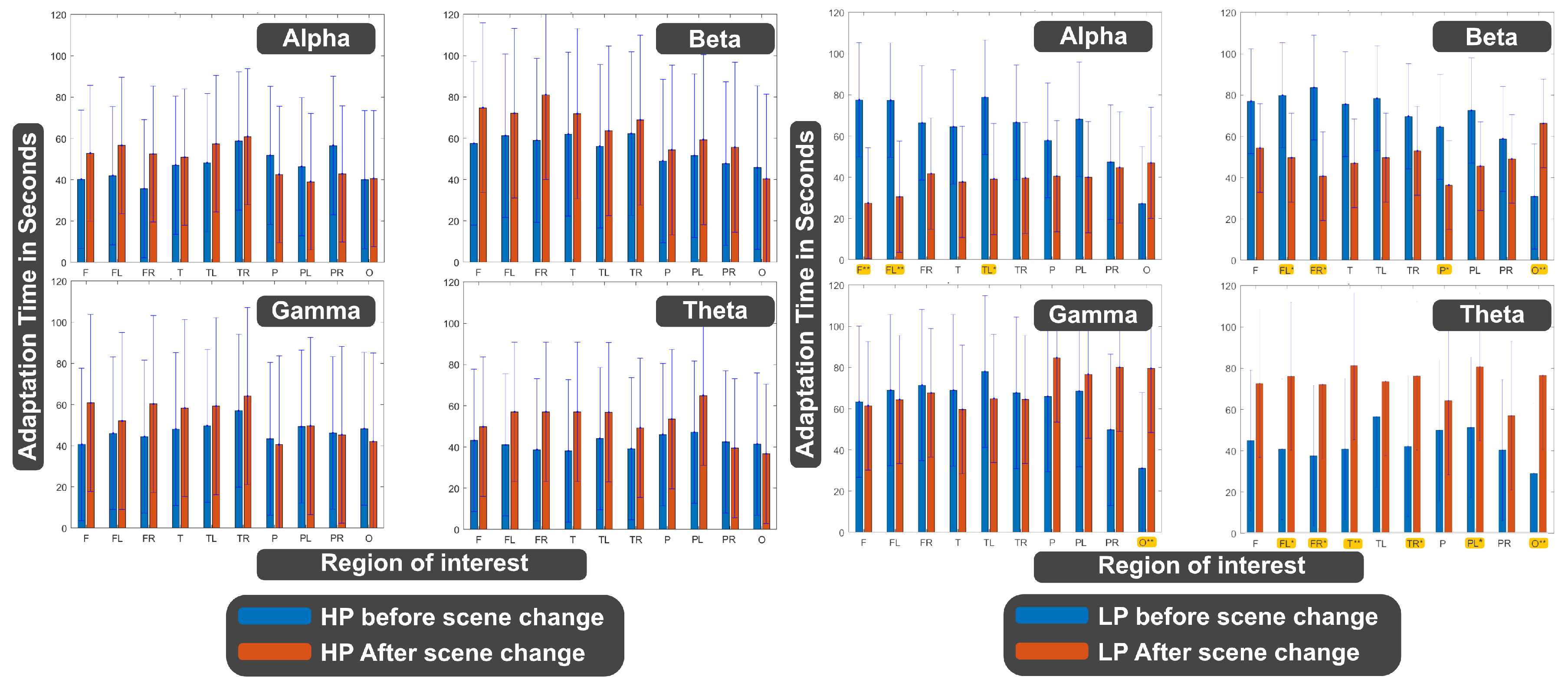
Figure 7. Adaptation time for adapting to the (Left) HP/(Right) LP environment in the same scene conditions. Blue bars indicate the adaptation time before switching the scene and red bars indicate after switching the scene, i.e. (Left) blue bars show the results for the HP-LP group and red bars are for the LP-HP group, (Right) blue bars show the results for the LP-HP group and red bars are for the HP-LP group. highlighted region of interest with yellow color indicates the areas with significant differences.
Preprints 137618 g007

4.2. Questionnaires

Although the focus of this study is on the analysis of EEG data, we evaluate task load and presence at the end of experience using NASA TLX and IPQ questionnaires. Afterward, we analyzed the results to figure out if there is any significant differences between groups. For the statistical analysis of post-questionnaires, first, we used Shapiro-Wilk test [85] to check the normality distribution of results. Accordingly, we used t-test [86] for normally distributed data and Wilcoxon rank-sum test [87] for not normally distributed ones.
  • IPQ: The evaluation of sense of presence using IPQ shows no significant difference between the two groups in all items. Table 1 shows the mean/standard-deviation as well as p-value for each item of this questionnaire.
  • NASA TLX: We found no significant difference between the two groups in all items except for a borderline condition of physical demand (p-value=0.056). According to these results, summarized in Table 2, participants experienced low-demanding tasks and found themselves successful in accomplishing the game quest.

5. Discussion

We investigated the impact of realism on brain activation and the related sense of presence in an EEG-based VR setting. Two types of environments were investigated: the HP scene with detailed geometry, realistic materials, and accurate lighting and the LP scene with reduced details, no textures, and non-realistic lighting.

5.1. The Influence of Realism on Neural Activity

  • Alpha band: According to the description provided in Section 2, an increase in alpha band power in the frontal lobe can be interpreted as a decrease in cognitive load. In the Alpha-Frontal subfigure of Figure 5, we can observe an increase in band power activity in both groups after changing the scene with a short delay. However, the LP-HP group experienced a faster increase in band power activity than the HP-LP group. In addition, considering Figure 6 we can also observe significant differences during the time period when LP-HP groups showed higher band power activity. As the second scene for LP-HP is the realistic scene and for HP-LP is the simplified one, we can conclude that a realistic scene may lead to a lower cognitive load compared to a simpler scene. It can be due to the point that the realistic scene is more compatible/similar to the environment that users are expected or used to see in real life.
As we discussed earlier in Section 2, the alpha bandpower in the parietal lobe can be associated with an attention process. In this way, an increase in alpha bandpower may indicate increasing attention and filtering out irrelevant information. Based on the Alpha-Parietal subfigure of Figure 5, we can see the HP scene leads to higher band power activity before (for HP-LP group) and after (for LP-HP) changing the scene. We also found significant differences between groups in different time periods for both conditions, see Figure 6. Accordingly, we can interpret that a more realistic scene may lead to higher attention and help in suppressing irrelevant information.
We discussed that an increase in alpha bandpower in the occipital lobe could be associated with visual processing. Although this may happen when users close their eyes, it is possible to see this effect by getting used to the environment or pre-adaptation to stimuli. In our case, as participants need to keep their eyes open to be able to interact with the environment, the increase in alpha bandpower may occur due to getting used to the environment. We can find this condition in the Alpha-Occipital subfigure of Figure 5. In this figure, band power activity of alpha waves in the occipital lobe is higher in HP condition for both before and after switching the scene (for the corresponding group). We also found significant differences between groups, i.e. between LP and HP conditions, for both conditions, see Figure 6.
  • Beta band: In Section 2, we discussed that beta bandpower in the frontal lobe is associated with decision-making and cognitive process load. As the Beta-Frontal subfigure of Figure 5 shows, after switching scenes, with a short delay (less than 20 seconds), participants in the LP scene (for the HP-LP group) experience higher beta bandpower in the frontal lobe and after about 50 seconds this behavior happens for the other group. However, we only found significant differences in the time range when EEG results for the HP-LP group show higher beta bandpower. In this case, we can interpret that participants in the LP scene experience a higher decision-making and cognitive process load after switching the scene with a short delay.
  • Gamma band: As a summary, gamma bandpower in the frontal lobe is known to be associated with enhanced executive functions, decision-making, and problem-solving, see Section 2. According to the Gamma-Frontal subfigure of Figure 5, participants who experienced the HP scene after the break, showed higher band power activity. Following that we assume the realistic condition might lead to improved executive function and problem-solving. Similarly, the gamma bandpower in parietal lobes shows a higher value after about 60 seconds, considering significant differences in Figure 6, in the HP scene after the break. Accordingly, we suppose a higher spatial awareness in this time period. In addition, since we observe higher gamma band power in the occipital lobes for the HP condition both before and after the break, we can interpret that the HP scene may lead to better visual information analysis and heightened visual attention.
  • Theta band: The Theta-Frontal subfigure of Figure 5, shows the higher theta bandpower in participants who experienced HP scene after the break (we just found significant differences between groups after the break, see Figure 6). This result suggests that this group may have better working memory capabilities and superior cognitive control, allowing them to manage and manipulate information effectively over short periods. In addition, we also observe higher theta bandpower for the HP scene after the break, in particular after 40 seconds based on Figure 6, in parietal lobes that can be associated with higher integration of sensory information for HP condition compared to LP.
Based on the results section and the above discussions, we can answer our research questions as follows.
  • (RQ1) How does altering the visual realism of a virtual environment affect the EEG-derived brain activity data?
The findings indicate that the impact of altering visual realism becomes apparent following the break, particularly evident in various lobes and frequency bands (FB). For instance, in the frontal lobe, there is an immediate reduction in normalized alpha bandpower post-break, followed by a subsequent increase over time until it reaches its maximum value. Conversely, a distinct pattern emerges in the occipital lobe, characterized by a reduction in alpha and theta bandpower for the transition from HP-LP, while observing an increase in the opposite scenario (LP-HP). This differential response underscores the nuanced influence of visual realism on neural activity across different brain regions and frequency bands.
According to the results of AT, that we defined in Section 3.6.2, there are significant differences in the AT between groups experiencing the LP condition as the first or second environment in the frontal and temporal lobes. For the beta and gamma bands, these significant differences can also be observed in the parietal and occipital lobes. It might be inferred that individuals adapt to the LP condition faster if they previously experienced HP condition. This may suggest that starting a VR experience in a non-realistic or simplified condition, which does not align with user expectations of reality, could lead to a longer AT. When they come to the LP environment (transition phase) the AT is higher since the brain tries to fill the gaps from real-world environmental details (they have already seen before and which are now missing). Such a phenomenon may have implications for the design and implementation of virtual environments that seek to replicate real-world scenarios, potentially enhancing the user’s experience and performance.
  • (RQ2) Are specific patterns of brain activation associated with the subjective sense of presence in virtual environments?
As we discussed in Section 2, presence can be defined and categorized in different ways. Using these factors, several questionnaires tried to measure the sense of presence in a subjective manner. One of these subjective questionnaires is IPQ which measures presence using 3+1 factors: Spatial Presence, Involvement, Realism, and General Presence. On the other hand, previous studies reported the influence of realism on the higher perceived sense of presence. Accordingly, we would expect realism leading to higher values for Spatial Presence, Involvement, and Realism factors. In our study, we have the results of EEG measurement that alters by changing the scene between realistic and non-realistic conditions. In other words, we can assume, the evolution of brain activation corresponding to a certain presence factor, might show an association between that EEG data and certain aspects of presence.
We found significant differences in brain activation related to the amount of realism of the virtual environment. In Section 5.1 we discussed that the results of gamma and theta bandpower in parietal lobes show higher spatial awareness and integration of sensory information in the more realistic condition. In addition, navigating through the more realistic environment leads to a stronger spatial presence experience, which was accompanied by a stronger Alpha power activity over parietal brain areas. This has also been shown by a study from Kober et al. [55], showing an increased sense of presence in the higher immersive VR environment which was accompanied by an increased parietal power (decrease in the Alpha band power). The lower presence experience in the low immersive VR environment elicited stronger functional connectivity between frontal and parietal brain regions, indicating an important role of both brain areas for the presence experience. Accordingly and based on our above assumption, we can expect that measuring alpha, gamma, and theta band powers in the parietal lobes might be used as indicators for spatial presence.
Furthermore, the “break in presence” which was defined as the point where the environment changes from HP to LP or vice versa elicited significant changes in brain activation specifically in the frontal and parietal regions. For the realism factor, we can assume to be related to visual analysis, recognition, and processing. A higher realism in the environment also evokes stronger alpha and theta band activation at occipital sites, indicating its role in visual attention and top-down processing. Here, as EEG band power in the occipital lobe is associated with visual processing and heightened visual attention, we assume an influence of realism factor and brain activation in the occipital lobe.
The alpha bandpower in different lobes can be related to user experience factors such as executive function, problem-solving, and cognitive control. Here, we assume these features can represent involvement factor. Accordingly, as HP condition shows higher alpha band power in different lobes, we can expect an association between alpha band power and involvement. Moreover, the increase in alpha power similar to levels of relaxation suggests its role in reflecting a state of isolation from the external world and an attentional shift toward internal aspects leading to a higher sense of presence. The association of a higher alpha activity with the inhibition of non-essential activity is not new. Klimesch et al. [88] already reported about such a relation and further suggest it as an index of top-down processing representing a mechanism for increasing the signal-to-noise ratio within the cortex by actively inhibiting non-essential or conflicting processes [89,90]. Others also reported that an increase in alpha power may either reflect active processing related to memory performance [91] or the inhibition of posterior brain sites not required for the task [64,88].
The observed higher theta and lower alpha power in frontal as well as parietal regions reflects the recruitment of oscillating networks processing focused attention, positive emotional experience, and engagement [92]. Specifically, the decreased alpha bandpower in frontal cortices is linked to top-down (high internal processing demands) modulation [88]. Furthermore, we observed high gamma activity in frontal and parietal regions while people experienced the HP environment. This is in line with previous studies including research on attention, working memory, or motor tasks, suggesting that activity in the gamma range reflects engagement/processing [93,94].
Overall, The outcome suggests that starting in non-realistic conditions requires higher AT for individuals as they are not visually consistent with their expectations of a real-life environment. However, once individuals are exposed to the HP environment, their adaptation process becomes faster, as most of the cues are already pre-activated and the brain is better equipped to fill the gaps. Specifically, this phenomenon has been observed in the alpha and beta bands at frontal sites. As both environments share the same interaction system, this finding shows the influence of visual realism (place illusion in [36]) on AT despite the interaction system (plausibility illusion). We can expect developers to use these results to adjust the AT in virtual environments based on visual realism, in order to achieve a desirable experience without altering gameplay mechanics.
It was observed that there were exceptions to the typical behavior in the Theta band when the adaptation phase was longer in the LP scene as the second environment. This deviation might be due to the fact that the Theta band plays a crucial role in memory processing and people try to interpret LP scenes from memory. As a result, such environments lead to stronger brain activation. Higher Theta bandpower also indicates lower working memory capacity. A similar pattern was noticed in the gamma-band at parieto-occipital sites. Since the gamma activity is also known to reflect high cognitive processing and memory, the AT in the transition mode is also higher in such cases.
While our study identified notable distinctions in EEG data, it is noteworthy that there were no significant differences observed in the outcomes of the NASA TLX and IPQ questionnaires. This result can be influenced by the limitation of the study design, which did not allow for the use of questionnaires during the study and only permitted their use at the end. However, this observation aligns with findings reported in previous research, e.g. [12,13], which suggest that traditional questionnaires may not fully capture the sense of presence within VR environments. This discrepancy underscores the necessity for further investigation to elucidate the underlying reasons for such disparities. Conversely, the continuous monitoring afforded by sensory devices presents a promising avenue for observing presence throughout the entirety of VR exposure.
  • Limitations: In our study, we discuss the influence of altering visual realism on brain activity using EEG results. However, previous research suggests that different types of realism can also have an impact on presence. To achieve these goals, additional features like animated objects or interactive characters can be added to the game. It should be considered, that these features need to be implemented with care, as any errors or inaccuracies in their design may negatively affect the sense of plausibility and presence.
In this work, we did not consider other physiological measurements in this study. Incorporating additional sensory measurements like electrocardiogram (ECG) or eye-tracking could be beneficial. ECG devices could help to investigate stress levels during different stages of the study, as well as workload. An eye-tracking system could be used to record specific eye movement patterns, like blinks or pupil dilation, during corresponding presence stages.
In general, conducting VR studies that involve sensitive sensory measurements can be a challenging task. Although we were able to obtain significant results in this study, it would be advantageous to increase the number of participants to gain better insights into the findings.
  • Future Works: We have shown how realism affects brain activity in different lobes of the brain. This information can be used to create research scenarios to explore the impact of specific activities in VR on the feeling of presence. For instance, researchers could examine how changes in presence occur during tasks such as solving puzzles, engaging in memory activities, or facing creative challenges in VR games.
Our findings provide insights into how altering realism may influence users’ adaptation time to a new environment. For example, if adapting to simplified environments is faster after encountering a complex one, a gradual or step-by-step reduction in complexity could be designed to improve user experience. This aspect can be further explored during the development of virtual environments to create more adaptable experiences. Since presence can influence user experience in games, extending this study to non-VR games (traditional desktop games) can offer additional insights into how variations in the level of realism affect presence and user experience. Furthermore, integrating brain activity in game development would enable the creation of personalized environments based on the users’ cognitive and emotional states.
There are many studies in the literature that investigate the effect of user interface and interaction on the user experience or evaluation outcomes, such as [13,45]. However, most of them rely on self-reported questionnaires which might not be sufficient to properly demonstrate comparison points. Our study’s findings can provide a basis for new research using EEG data to explore the impact of different design decisions on the user experience.

6. Conclusion

Virtual reality (VR) technology inherently possesses a notable characteristic known as high immersion, contributing to the subjective sensation termed "presence." This phenomenon is defined as the subjective feeling of being within a virtual environment. The degree of presence experienced is pivotal in shaping user interaction and satisfaction within virtual environments. Presence can be dissected into two interconnected components: the "place illusion," concerning the visual fidelity and portrayal of the virtual environment, and the "plausibility illusion," pertaining to the acceptance, interaction, and communication with virtual elements. Realism stands out as a fundamental determinant influencing the place illusion. Thus, alterations in the level of realism within virtual environments are expected to engender corresponding shifts in the sense of presence experienced by users.
Over the course of research endeavors, various methodologies have been devised to assess the sense of presence within virtual environments. These methodologies are typically classified based on the source of data collection from users, namely: subjective, objective, or a fusion of both approaches. Subjective evaluations predominantly rely on self-reported questionnaires. While questionnaires offer ease of implementation and straightforward analysis of results, they possess limitations in capturing the nuanced intricacies of user experiences, potentially yielding inconsistent outcomes. Moreover, questionnaire-based assessments lack the ability to provide continuous data representation over time, necessitating researchers to define discrete evaluation points. Furthermore, the completion of questionnaires entails interruptions in the user’s immersive VR experience, even when implemented within the virtual environment itself (i.e., in-VR questionnaires). An alternative avenue involves the utilization of objective methodologies employing sensory measurements. These techniques facilitate the continuous recording of data throughout the study duration without disrupting VR interactions. Nevertheless, the adoption of objective methods is comparatively less prevalent within the literature due to the inherent challenges associated with setup and the complexities involved in data analysis. Notably, establishing correlations between objectively measured sensory data and subjective questionnaire responses holds significant promise for elucidating insights beneficial to both the research community and developers alike.
In this study, electroencephalography (EEG) was employed as an objective measurement tool to capture and evaluate the influence of visual realism on brain activity while participants experienced two distinct VR scenes: one characterized by high visual realism (HP) and the other by low visual realism (LP).
Findings revealed discernible alterations in brain activity corresponding to changes in visual realism, indicative of potential implications for the sense of presence. Immediate transitions between VR scenes elicited observable shifts in the bandpowers of various brain waves across different regions. Furthermore, significant disparities in brain adaptation times were identified between participants who experienced LP as the first scene and those who experienced it as the second scene. This can provide initial insights into a differential response based on the level of visual realism. Notably, participants exhibited faster adaptation to non-realistic environments following exposure to realistic ones compared to the reverse scenario. Moreover, analyses unveiled preliminary findings of possible associations between specific aspects of brain activity and the different factors of presence. Highlighting parietal lobe activation with spatial presence, occipital lobe activity with perceptions of realism, and frontal lobe engagement with more focused attention to the task.
These findings contribute to a deeper understanding of how visual realism influences brain activity and presence within VR environments, thus offering a foundation for the development of guidelines for VR application design. Subsequent investigations exploring the impact of diverse activities and VR element designs within realistic and non-realistic contexts hold promise for yielding further insights valuable to the research and development community.

Author Contributions

Conceptualization, S.S., S.C.W. and J.P.; methodology, S.S., M.S. and S.C.W. ; software, S.S.; validation, V.G. and S.C.W.; formal analysis, S.S. and V.G.; investigation, S.S., S.C.W. and J.P.; resources, S.C.W. and J.P.; data curation, S.C.W.; writing—original draft preparation, S.S. and S.C.W.; writing—review and editing, S.S., S.C.W. and J.P.; visualization, S.S., V.G.,; supervision, S.C.W. and J.P.; project administration, S.C.W. and J.P.. All authors have read and agreed to the published version of the manuscript.

Funding

This research received no external funding

Institutional Review Board Statement

The study was conducted in accordance with the Declaration of Helsinki, and approved by the Ethics Committee Graz University of Technology (GZ: EK37/2024).

Informed Consent Statement

Informed consent was obtained from all subjects involved in the study.

Data Availability Statement

The data will be made available upon request.

Acknowledgments

Used AI tools: scite_ and ChatGPT

Conflicts of Interest

The authors declare no conflicts of interest.

Appendix A

Appendix A.1

In the section below we provide detailed results of analysis for adaptation time, considering mean, standard deviation, p-values, U, and z-value of the Wilcoxon rank-sum test.
Table A1. Mean and standard deviations of the adaptation times in seconds before and after break of the trials HP. aB is after break and bB is before break.
Frontal Frontal
left
Frontal
right
Temporal Temporal
left
Temporal
right
Parietal Parietal
left
Parietal
 right
Occipital
Alpha aB 40.2±33.5 42.0±33.0 35.7±33.4 47.1±35.0 48.2±33.8 58.7±23.9 51.8±33.0 46.4±33.7 56.6±31.1 40.0±28.7
Alpha bB 52.9±38.7 56.6±35.8 52.5±39.1 51.0±35.6 57.4±30.4 60.9±29.9 42.6±31.2 39.1±34.4 42.9±31.6 40.6±27.5
Beta aB 57.5±39.6 61.3±41.1 59.0±37.2 62.0±37.2 56.2±35.2 62.2±36.3 49.0±38.9 51.6±41.9 47.7±34.5 45.8±36.0
Beta bB 74.8±32.7 72.2±36.1 81.1±23.6 71.9±26.0 63.6±23.9 68.9±28.7 54.3±28.7 59.3±27.7 55.6±29.7 40.3±33.5
Gamma aB 40.7±37.1 46.0±43.0 44.4±38.0 48.1±40.3 49.7±43.5 57.0±32.2 43.3±43.0 49.3±46.7 46.2±41.4 48.3±33.2
Gamma bB 60.8±41.1 52.1±40.8 60.3±41.5 58.3±40.6 59.2±39.5 64.1±36.8 40.6±38.4 49.6±34.5 45.3±38.5 42.1±34.1
Theta aB 43.1±34.5 41.0±33.8 38.7±32.9 38.2±36.5 44.1±35.5 39.2±36.6 46.0±35.2 47.2±36.0 42.4±37.2 41.4±30.0
Theta bB 49.9±34.8 57.0±35.5 57.0±33.1 57.0±31.2 56.8±31.1 49.4±30.7 53.6±42.2 64.9±41.2 39.4±35.5 36.8±31.0
Table A2. P-Values of the Wilcoxon ranksum test with the adaptation time with the HP trials.
Frontal Frontal
left
Frontal
right
Temporal Temporal
left
Temporal
right
Parietal Parietal
left
Parietal
 right
Occipital
Alpha 0.5974 0.2597 0.4181 0.7513 0.3787 0.7513 0.5035 0.8053 0.2751 0.9159
Beta 0.2178 0.5035 0.0980 0.5974 0.7513 0.6985 0.8603 0.8053 0.7513 0.6985
Gamma 0.2907 1.0000 0.3072 0.4384 0.6220 0.5973 1.0000 1.0000 0.9159 0.5974
Theta 0.5974 0.3072 0.1697 0.1925 0.3416 0.4179 0.3418 0.1487 0.8603 0.7513
Table A3. U and z-values (z-statistics) of the Wilcoxon ranksum test with the adaptation time of HP trials.
Frontal Frontal
left
Frontal
right
Temporal Temporal
left
Temporal
right
Parietal Parietal
left
Parietal
 right
Occipital
Alpha z 0.5 1.1 0.8 0.3 0.9 0.3 -0.7 -0.2 -1.1 0.1
Alpha U 118 126.5 122 115 123 115 100 106 94 112
Beta z 1.2 0.7 1.7 0.5 0.3 0.4 0.2 0.2 0.3 -0.4
Beta U 128 120 134 118 115 116 113 114 115 104
Gamma z 1.1 0 1.0 0.8 0.5 0.5 0 0 -0.1 -0.5
Gamma U 125.5 110 125 121.5 117.5 118 109.5 109.5 108 102
Theta z 0.5 1.0 1.4 1.3 1.0 0.8 1.0 1.4 0.2 -0.3
Theta U 118 125 130 129 124 122 124 131 113 105
Table A4. Mean and standard deviations of the adaptation times in seconds before and after break of the trials LP. aB is after break and bB is before break.
Frontal Frontal
left
Frontal
right
Temporal Temporal
left
Temporal
right
Parietal Parietal
left
Parietal
 right
Occipital
Alpha bB 77.6±27.8 77.4±27.0 66.4±34.0 64.5±40.1 78.8±20.4 66.7±34.4 57.9±36.3 68.2±27.0 47.3±39.3 27.2±29.6
Alpha aB 27.5±34.9 30.5±34.3 41.8±43.6 37.8±39.7 39.2±38.2 39.7±41.0 40.6±35.9 40.0±34.8 44.8±38.1 47.0±30.6
Beta bB 77.0±25.4 79.9±21.5 83.6±21.8 75.6±21.0 78.4±19.2 69.7±25.5 64.6±32.2 72.5±28.5 58.8±36.4 30.8±32.0
Beta aB 54.3±38.1 49.7±36.3 40.7±38.7 47.0±39.9 49.7±36.5 53.0±41.1 36.4±34.6 45.6±34.6 49.1±37.9 66.3±20.5
Gamma bB 63.4±36.8 69.0±31.2 71.5±25.6 69.0±29.2 78.0±23.7 67.7±34.0 66.0±30.5 68.6±33.1 49.8±36.7 31.1±30.8
Gamma aB 61.5±31.9 64.4±33.3 67.7±34.8 59.7±39.9 65.0±33.5 64.5±37.6 84.7±14.0 76.7±29.7 80.1±23.4 79.6±15.2
Theta bB 42.7±32.7 38.7±34.4 35.6±32.2 38.8±27.2 53.8±28.6 40.1±35.4 47.4±34.9 48.8±34.1 38.3±36.1 27.5±24.6
Theta aB 69.2±26.5 72.6±26.9 68.7±26.2 77.5±29.0 70.2±27.7 72.7±27.3 61.3±39.9 77.0±28.9 54.3±38.4 72.9±23.0
Table A5. P-Values of the Wilcoxon ranksum test with the break adaptation time with the LP trials.
Frontal Frontal
left
Frontal
right
Temporal Temporal
left
Temporal
right
Parietal Parietal
left
Parietal
 right
Occipital
Alpha 0.0067 0.0092 0.1130 0.2450 0.0378 0.1589 0.3786 0.0844 0.9719 0.0725
Beta 0.1927 0.0317 0.0102 0.0980 0.0980 0.3787 0.0378 0.0725 0.5035 0.0067
Gamma 0.8053 0.5035 0.8603 0.5035 0.3787 0.5490 0.3418 0.9159 0.1489 0.0017
Theta 0.0620 0.0317 0.0151 0.0043 0.0980 0.0448 0.2453 0.0221 0.2751 0.0014
Table A6. U and z-values (z-statistics) of the Wilcoxon ranksum test with the adaptation time of LP trials.
Frontal Frontal
left
Frontal
right
Temporal Temporal
left
Temporal
right
Parietal Parietal
left
Parietal
 right
Occipital
Alpha z -2.711 -2.606 -1.585 -1.163 -2.077 -1.409 -0.881 -1.726 0.035 1.796
Alpha U 71 72.5 87 93 80 89.5 97 85 111 136
Beta z -1.303 -2.148 -2.57 -1.655 -1.655 -0.88 -2.077 -1.796 -0.669 2.712
Beta U 91 79 73 86 86 97 80 84 100 149
Gamma z -0.246 -0.669 -0.176 -0.669 -0.88 -0.599 0.951 0.106 1.444 3.135
Gamma U 106 100 107 100 97 101 124 112 131 155
Theta z 1.866 2.148 2.429 2.852 1.655 2.007 1.162 2.289 1.091 3.204
Theta U 137 141 145 151 134 139 127 143 126 156

Notes

1
2
3
4
"HP" denotes "High Polygons," while "LP" signifies "Low Polygons"
5
Brain Products GmbH, Gilching, Germany

References

  1. Asad, M.M.; Naz, A.; Churi, P.; Tahanzadeh, M.M. Virtual reality as pedagogical tool to enhance experiential learning: a systematic literature review. Education Research International 2021, 2021, 1–17. [Google Scholar] [CrossRef]
  2. Lampropoulos, G.; Keramopoulos, E. Virtual Reality in Education: A Comparative Social Media Data and Sentiment Analysis Study. Int. J. Recent Contributions Eng. Sci. IT 2022, 10, 19–32. [Google Scholar] [CrossRef]
  3. Kim, D.; Im, T.; others. A systematic review of virtual reality-based education research using latent dirichlet allocation: Focus on topic modeling technique. Mobile Information Systems 2022, 2022. [Google Scholar] [CrossRef]
  4. Safikhani, S.; Pirker, J.; Wriessnegger, S.C. Virtual Reality Applications for the Treatment of Anxiety and Mental Disorders. 2021 7th International Conference of the Immersive Learning Research Network (iLRN). IEEE, 2021, pp. 1–8.
  5. Banerjee, S.; Pham, T.; Eastaway, A.; Auffermann, W.F.; Quigley III, E.P. The use of virtual reality in teaching three-dimensional anatomy and pathology on CT. Journal of Digital Imaging 2023, 36, 1279–1284. [Google Scholar] [CrossRef] [PubMed]
  6. Safikhani, S.; Keller, S.; Schweiger, G.; Pirker, J. Immersive virtual reality for extending the potential of building information modeling in architecture, engineering, and construction sector: Systematic review. International Journal of Digital Earth 2022, 15, 503–526. [Google Scholar] [CrossRef]
  7. Catbas, F.N.; Luleci, F.; Zakaria, M.; Bagci, U.; LaViola Jr, J.J.; Cruz-Neira, C.; Reiners, D. Extended reality (XR) for condition assessment of civil engineering structures: A literature review. Sensors 2022, 22, 9560. [Google Scholar] [CrossRef]
  8. Putze, S.; Alexandrovsky, D.; Putze, F.; Höffner, S.; Smeddinck, J.D.; Malaka, R. Breaking the experience: effects of questionnaires in vr user studies. Proceedings of the 2020 CHI Conference on Human Factors in Computing Systems 2020. [Google Scholar]
  9. Schrepp, M.; Hinderks, A.; Thomaschewski, J. Design and evaluation of a short version of the user experience questionnaire (ueq-s). International Journal of Interactive Multimedia and Artificial Intelligence 2017, 4, 103. [Google Scholar] [CrossRef]
  10. Schubert, T.; Friedmann, F.; Regenbrecht, H. The experience of presence: Factor analytic insights. Presence: Teleoperators & Virtual Environments 2001, 10, 266–281. [Google Scholar]
  11. O’Brien, H.L.; Cairns, P.; Hall, M. A practical approach to measuring user engagement with the refined user engagement scale (UES) and new UES short form. International Journal of Human-Computer Studies 2018, 112, 28–39. [Google Scholar] [CrossRef]
  12. Graf, S.; Schwind, V. Inconsistencies of presence questionnaires in virtual reality. Proceedings of the 26th ACM Symposium on Virtual Reality Software and Technology, 2020, pp. 1–3.
  13. Safikhani, S.; Holly, M.; Kainz, A.; Pirker, J. The influence of In-VR questionnaire design on the user experience. Proceedings of the 27th ACM Symposium on Virtual Reality Software and Technology, 2021, pp. 1–8.
  14. Slater, M. How colorful was your day? Why questionnaires cannot assess presence in virtual environments. Presence 2004, 13, 484–493. [Google Scholar] [CrossRef]
  15. Moore, K.; Wiederhold, B.K.; Wiederhold, M.D.; Riva, G. Panic and agoraphobia in a virtual world. Cyberpsychology & Behavior 2002, 5, 197–202. [Google Scholar]
  16. Murphy, D.; Higgins, C. Secondary inputs for measuring user engagement in immersive VR education environments. arXiv 2019, arXiv:1910.01586 2019. [Google Scholar]
  17. Tremmel, C.; Herff, C.; Sato, T.; Rechowicz, K.; Yamani, Y.; Krusienski, D.J. Estimating cognitive workload in an interactive virtual reality environment using EEG. Frontiers in human neuroscience 2019, 13, 401. [Google Scholar] [CrossRef]
  18. Grassini, S.; Laumann, K.; de Martin Topranin, V.; Thorp, S. Evaluating the effect of multi-sensory stimulations on simulator sickness and sense of presence during HMD-mediated VR experience. Ergonomics 2021, 64, 1532–1542. [Google Scholar] [CrossRef]
  19. Baus, O.; Bouchard, S. Exposure to an unpleasant odour increases the sense of presence in virtual reality. Virtual Reality 2017, 21, 59–74. [Google Scholar] [CrossRef]
  20. Witmer, B.G.; Singer, M.J. Measuring presence in virtual environments: A presence questionnaire. Presence 1998, 7, 225–240. [Google Scholar] [CrossRef]
  21. Riva, G.; Davide, F.; IJsselsteijn, W.A. Being there: The experience of presence in mediated environments. Being there: Concepts, effects and measurement of user presence in synthetic environments 2003, 5. [Google Scholar]
  22. Barfield, W.; Zeltzer, D.; Sheridan, T.; Slater, M. Presence and performance within virtual environments. In Virtual environments and advanced interface design; 1995; pp. 473–513.
  23. Schwind, V.; Knierim, P.; Haas, N.; Henze, N. Using presence questionnaires in virtual reality. Proceedings of the 2019 CHI conference on human factors in computing systems, 2019, pp. 1–12.
  24. Grassini, S.; Laumann, K.; Rasmussen Skogstad, M. The use of virtual reality alone does not promote training performance (but sense of presence does). Frontiers in psychology 2020, 11, 553693. [Google Scholar] [CrossRef]
  25. Hoffman, H.G.; Richards, T.; Coda, B.; Richards, A.; Sharar, S.R. The illusion of presence in immersive virtual reality during an fMRI brain scan. Cyberpsychology & behavior 2003, 6, 127–131. [Google Scholar]
  26. Mubarrat, S.T.; Opafunso, O.; Chowdhury, S.K. The Evaluation of User Experience and Functional Workload of a Physically Inter-active Virtual Reality System. Proceedings of the Human Factors and Ergonomics Society Annual Meeting. SAGE Publications Sage CA: Los Angeles, CA, 2020, Vol. 64, pp. 2084–2086.
  27. Somrak, A.; Pogačnik, M.; Guna, J. Suitability and comparison of questionnaires assessing virtual reality-induced symptoms and effects and user experience in virtual environments. Sensors 2021, 21, 1185. [Google Scholar] [CrossRef] [PubMed]
  28. Lorenz, M.; Brade, J.; Klimant, P.; Heyde, C.E.; Hammer, N. Age and gender effects on presence, user experience and usability in virtual environments–first insights. PloS one 2023, 18, e0283565. [Google Scholar] [CrossRef]
  29. Melo, M.; Gonçalves, G.; Bessa, M.; others. How much presence is enough? qualitative scales for interpreting the igroup presence questionnaire score. IEEE Access 2023, 11, 24675–24685. [Google Scholar] [CrossRef]
  30. Skarbez, R.; Brooks, Jr, F.P.; Whitton, M.C. A survey of presence and related concepts. ACM computing surveys (CSUR) 2017, 50, 1–39. [CrossRef]
  31. Skarbez, R.; Neyret, S.; Brooks, F.P.; Slater, M.; Whitton, M.C. A psychophysical experiment regarding components of the plausibility illusion. IEEE transactions on visualization and computer graphics 2017, 23, 1369–1378. [Google Scholar] [CrossRef]
  32. Lombard, M.; Ditton, T. At the heart of it all: The concept of presence. Journal of computer-mediated communication 1997, 3, JCMC321. [Google Scholar] [CrossRef]
  33. Slater, M.; Usoh, M. Representations systems, perceptual position, and presence in immersive virtual environments. Presence: Teleoperators & Virtual Environments 1993, 2, 221–233. [Google Scholar]
  34. Parola, M.; Johnson, S.; West, R. Turning presence inside-out: MetaNarratives. Electronic Imaging 2016, 28, 1–9. [Google Scholar] [CrossRef]
  35. IJsselsteijn, W.A.; De Ridder, H.; Freeman, J.; Avons, S.E. Presence: concept, determinants, and measurement. Human vision and electronic imaging V. SPIE, 2000, Vol. 3959, pp. 520–529.
  36. Slater, M. Place illusion and plausibility can lead to realistic behaviour in immersive virtual environments. Philosophical Transactions of the Royal Society B: Biological Sciences 2009, 364, 3549–3557. [Google Scholar] [CrossRef]
  37. Newman, M.; Gatersleben, B.; Wyles, K.; Ratcliffe, E. The use of virtual reality in environment experiences and the importance of realism. Journal of environmental psychology 2022, 79, 101733. [Google Scholar] [CrossRef]
  38. Hvass, J.; Larsen, O.; Vendelbo, K.; Nilsson, N.; Nordahl, R.; Serafin, S. Visual realism and presence in a virtual reality game. 2017 3DTV conference: The true vision-capture, Transmission and Display of 3D video (3DTV-CON). IEEE, 2017, pp. 1–4.
  39. Slater, M.; Khanna, P.; Mortensen, J.; Yu, I. Visual realism enhances realistic response in an immersive virtual environment. IEEE computer graphics and applications 2009, 29, 76–84. [Google Scholar] [CrossRef] [PubMed]
  40. Ferwerda, J.A. Three varieties of realism in computer graphics. Human vision and electronic imaging viii. SPIE, 2003, Vol. 5007, pp. 290–297.
  41. Gonçalves, G.; Coelho, H.; Monteiro, P.; Melo, M.; Bessa, M. Systematic review of comparative studies of the impact of realism in immersive virtual experiences. ACM Computing Surveys 2022, 55, 1–36. [Google Scholar] [CrossRef]
  42. Skarbez, R.; Brooks, F.P.; Whitton, M.C. Immersion and coherence: Research agenda and early results. IEEE transactions on visualization and computer graphics 2020, 27, 3839–3850. [Google Scholar] [CrossRef] [PubMed]
  43. Witmer, B.G.; Jerome, C.J.; Singer, M.J. The factor structure of the presence questionnaire. Presence: Teleoperators & Virtual Environments 2005, 14, 298–312. [Google Scholar]
  44. Slater, M.; Brogni, A.; Steed, A. Physiological responses to breaks in presence: A pilot study. Presence 2003: The 6th annual international workshop on presence. Citeseer, 2003, Vol. 157.
  45. Alexandrovsky, D.; Putze, S.; Bonfert, M.; Höffner, S.; Michelmann, P.; Wenig, D.; Malaka, R.; Smeddinck, J.D. Examining design choices of questionnaires in VR user studies. Proceedings of the 2020 CHI Conference on Human Factors in Computing Systems, 2020, pp. 1–21.
  46. Wriessnegger, S.C.; Autengruber, L.M.; Chacón, L.A.B.; Pirker, J.; Safikhani, S. The influence of visual representation factors on bio signals and its relation to Presence in Virtual Reality Environments. 2022 IEEE International Conference on Metrology for Extended Reality, Artificial Intelligence and Neural Engineering (MetroXRAINE). IEEE, 2022, pp. 199–204.
  47. Athif, M.; Rathnayake, B.L.K.; Nagahapitiya, S.D.B.S.; Samarasinghe, S.A.K.; Samaratunga, P.S.; Peiris, R.L.; De Silva, A.C. Using biosignals for objective measurement of presence in virtual reality environments. 2020 42nd Annual International Conference of the IEEE Engineering in Medicine & Biology Society (EMBC). IEEE, 2020, pp. 3035–3039.
  48. Aromaa, S.; Väänänen, K. Suitability of virtual prototypes to support human factors/ergonomics evaluation during the design. Applied ergonomics 2016, 56, 11–18. [Google Scholar] [CrossRef]
  49. Vecchiato, G.; Tieri, G.; Jelic, A.; De Matteis, F.; Maglione, A.G.; Babiloni, F. Electroencephalographic correlates of sensorimotor integration and embodiment during the appreciation of virtual architectural environments. Frontiers in psychology 2015, 6, 160641. [Google Scholar] [CrossRef]
  50. Bohil, C.J.; Alicea, B.; Biocca, F.A. Virtual reality in neuroscience research and therapy. Nature reviews neuroscience 2011, 12, 752–762. [Google Scholar] [CrossRef]
  51. Rus-Calafell, M.; Garety, P.; Sason, E.; Craig, T.J.; Valmaggia, L.R. Virtual reality in the assessment and treatment of psychosis: a systematic review of its utility, acceptability and effectiveness. Psychological medicine 2018, 48, 362–391. [Google Scholar] [CrossRef]
  52. Freeman, J.; Avons, S.E.; Meddis, R.; Pearson, D.E.; IJsselsteijn, W. Using behavioral realism to estimate presence: A study of the utility of postural responses to motion stimuli. Presence: Teleoperators & Virtual Environments 2000, 9, 149–164. [Google Scholar]
  53. Thorp, S.O.; Rimol, L.M.; Lervik, S.; Evensmoen, H.R.; Grassini, S. Comparative analysis of spatial ability in immersive and non-immersive virtual reality: the role of sense of presence, simulation sickness and cognitive load. Frontiers in Virtual Reality 2024, 5, 1343872. [Google Scholar] [CrossRef]
  54. Caroux, L. Presence in video games: A systematic review and meta-analysis of the effects of game design choices. Applied Ergonomics 2023, 107, 103936. [Google Scholar] [CrossRef] [PubMed]
  55. Kober, S.E.; Kurzmann, J.; Neuper, C. Cortical correlate of spatial presence in 2D and 3D interactive virtual reality: an EEG study. International Journal of Psychophysiology 2012, 83, 365–374. [Google Scholar] [CrossRef] [PubMed]
  56. Burns, C.G.; Fairclough, S.H. Use of auditory event-related potentials to measure immersion during a computer game. International Journal of Human-Computer Studies 2015, 73, 107–114. [Google Scholar] [CrossRef]
  57. Terkildsen, T.; Makransky, G. Measuring presence in video games: An investigation of the potential use of physiological measures as indicators of presence. International journal of human-computer studies 2019, 126, 64–80. [Google Scholar] [CrossRef]
  58. Teplan, M.; others. Fundamentals of EEG measurement. Measurement science review 2002, 2, 1–11. [Google Scholar]
  59. Niedermeyer, E.; da Silva, F.L. Electroencephalography: basic principles, clinical applications, and related fields; Lippincott Williams & Wilkins, 2005.
  60. Klimesch, W. EEG alpha and cognitive processes. In Time and the Brain; CRC Press, 2000; pp. 252–277.
  61. Klimesch, W. EEG alpha and theta oscillations reflect cognitive and memory performance: a review and analysis. Brain research reviews 1999, 29, 169–195. [Google Scholar] [CrossRef]
  62. Cohen, M.X. Where does EEG come from and what does it mean? Trends in neurosciences 2017, 40, 208–218. [Google Scholar] [CrossRef]
  63. Colgin, L.L. Mechanisms and functions of theta rhythms. Annual review of neuroscience 2013, 36, 295–312. [Google Scholar] [CrossRef] [PubMed]
  64. Başar, E.; Güntekin, B. A short review of alpha activity in cognitive processes and in cognitive impairment. International Journal of Psychophysiology 2012, 86, 25–38. [Google Scholar] [CrossRef]
  65. Grassini, S.; Railo, H.; Valli, K.; Revonsuo, A.; Koivisto, M. Visual features and perceptual context modulate attention towards evolutionarily relevant threatening stimuli: Electrophysiological evidence. Emotion 2019, 19, 348. [Google Scholar] [CrossRef]
  66. Bazanova, O.; Vernon, D. Interpreting EEG alpha activity. Neuroscience & Biobehavioral Reviews 2014, 44, 94–110. [Google Scholar]
  67. Pfurtscheller, G. Induced oscillations in the alpha band: functional meaning. Epilepsia 2003, 44, 2–8. [Google Scholar] [CrossRef]
  68. Petsche, H.; Kaplan, S.; Von Stein, A.; Filz, O. The possible meaning of the upper and lower alpha frequency ranges for cognitive and creative tasks. International journal of psychophysiology 1997, 26, 77–97. [Google Scholar] [CrossRef]
  69. Hohaia, W.; Saurels, B.W.; Johnston, A.; Yarrow, K.; Arnold, D.H. Occipital alpha-band brain waves when the eyes are closed are shaped by ongoing visual processes. Scientific reports 2022, 12, 1194. [Google Scholar] [CrossRef]
  70. Herrmann, C.S.; Strüber, D.; Helfrich, R.F.; Engel, A.K. EEG oscillations: from correlation to causality. International Journal of Psychophysiology 2016, 103, 12–21. [Google Scholar] [CrossRef]
  71. Hayashi, T.; Okamoto, E.; Nishimura, H.; Mizuno-Matsumoto, Y.; Ishii, R.; Ukai, S. Beta activities in EEG associated with emotional stress. International Journal of Intelligent Computing in Medical Sciences & Image Processing 2009, 3, 57–68. [Google Scholar]
  72. Kaiser, J.; Lutzenberger, W. Cortical oscillatory activity and the dynamics of auditory memory processing. Reviews in the Neurosciences 2005, 16, 239–254. [Google Scholar] [CrossRef] [PubMed]
  73. Herrmann, C.S.; Fründ, I.; Lenz, D. Human gamma-band activity: a review on cognitive and behavioral correlates and network models. Neuroscience & Biobehavioral Reviews 2010, 34, 981–992. [Google Scholar]
  74. Fell, J.; Axmacher, N.; Haupt, S. From alpha to gamma: electrophysiological correlates of meditation-related states of consciousness. Medical hypotheses 2010, 75, 218–224. [Google Scholar] [CrossRef]
  75. Balconi, M.; Lucchiari, C. Consciousness and arousal effects on emotional face processing as revealed by brain oscillations. A gamma band analysis. International Journal of Psychophysiology 2008, 67, 41–46. [Google Scholar] [CrossRef]
  76. Sauseng, P.; Griesmayr, B.; Freunberger, R.; Klimesch, W. Control mechanisms in working memory: a possible function of EEG theta oscillations. Neuroscience & Biobehavioral Reviews 2010, 34, 1015–1022. [Google Scholar]
  77. Begus, K.; Bonawitz, E. The rhythm of learning: Theta oscillations as an index of active learning in infancy. Developmental Cognitive Neuroscience 2020, 45, 100810. [Google Scholar] [CrossRef]
  78. Bekkedal, M.Y.; Rossi III, J.; Panksepp, J. Human brain EEG indices of emotions: delineating responses to affective vocalizations by measuring frontal theta event-related synchronization. Neuroscience & Biobehavioral Reviews 2011, 35, 1959–1970. [Google Scholar]
  79. Ninaus, M.; Kober, S.E.; Friedrich, E.V.; Dunwell, I.; De Freitas, S.; Arnab, S.; Ott, M.; Kravcik, M.; Lim, T.; Louchart, S.; others. Neurophysiological methods for monitoring brain activity in serious games and virtual environments: a review. International Journal of Technology Enhanced Learning 2014, 6, 78–103. [Google Scholar] [CrossRef]
  80. Salminen, M.; Ravaja, N. Increased oscillatory theta activation evoked by violent digital game events. Neuroscience letters 2008, 435, 69–72. [Google Scholar] [CrossRef] [PubMed]
  81. Baumgartner, T.; Valko, L.; Esslen, M.; Jäncke, L. Neural correlate of spatial presence in an arousing and noninteractive virtual reality: an EEG and psychophysiology study. CyberPsychology & Behavior 2006, 9, 30–45. [Google Scholar]
  82. Baumgartner, T.; Speck, D.; Wettstein, D.; Masnari, O.; Beeli, G.; Jäncke, L. Feeling present in arousing virtual reality worlds: prefrontal brain regions differentially orchestrate presence experience in adults and children. Frontiers in human neuroscience 2008, 2, 279. [Google Scholar] [CrossRef]
  83. Sayyad, E.; Sra, M.; Höllerer, T. Walking and teleportation in wide-area virtual reality experiences. 2020 IEEE international symposium on mixed and augmented reality (ISMAR). IEEE, 2020, pp. 608–617.
  84. Baka, E.; Stavroulia, K.E.; Magnenat-Thalmann, N.; Lanitis, A. An EEG-based evaluation for comparing the sense of presence between virtual and physical environments. In Proceedings of Computer Graphics International 2018; 2018; pp. 107–116.
  85. Shapiro, S.S.; Wilk, M.B. An analysis of variance test for normality (complete samples). Biometrika 1965, 52, 591–611. [Google Scholar] [CrossRef]
  86. Student. The probable error of a mean. Biometrika 1908, pp. 1–25.
  87. Woolson, R.F. Wilcoxon signed-rank test. Wiley encyclopedia of clinical trials 2007, pp. 1–3.
  88. Klimesch, W.; Sauseng, P.; Hanslmayr, S. EEG alpha oscillations: the inhibition–timing hypothesis. Brain research reviews 2007, 53, 63–88. [Google Scholar] [CrossRef]
  89. Cooper, N.R.; Croft, R.J.; Dominey, S.J.; Burgess, A.P.; Gruzelier, J.H. Paradox lost? Exploring the role of alpha oscillations during externally vs. internally directed attention and the implications for idling and inhibition hypotheses. International journal of psychophysiology 2003, 47, 65–74. [Google Scholar] [CrossRef]
  90. Von Stein, A.; Chiang, C.; König, P. Top-down processing mediated by interareal synchronization. Proceedings of the National Academy of Sciences 2000, 97, 14748–14753. [Google Scholar] [CrossRef] [PubMed]
  91. Palva, S.; Palva, J.M. New vistas for α-frequency band oscillations. Trends in neurosciences 2007, 30, 150–158. [Google Scholar] [CrossRef] [PubMed]
  92. Aftanas, L.I.; Golocheikine, S.A. Human anterior and frontal midline theta and lower alpha reflect emotionally positive state and internalized attention: high-resolution EEG investigation of meditation. Neuroscience letters 2001, 310, 57–60. [Google Scholar] [CrossRef] [PubMed]
  93. Jensen, O.; Kaiser, J.; Lachaux, J.P. Human gamma-frequency oscillations associated with attention and memory. Trends in neurosciences 2007, 30, 317–324. [Google Scholar] [CrossRef]
  94. Crone, N.E.; Sinai, A.; Korzeniewska, A. High-frequency gamma oscillations and human brain mapping with electrocorticography. Progress in brain research 2006, 159, 275–295. [Google Scholar]
Figure 1. Virtual environments: (Left) Simplified/non-realistic environment/LP, (Center) The study setup: a user wearing 32 channel active electrodes EEG device and VR headset, (Right) Complex/Realistic environment/HP.
Preprints 137618 g001
Figure 2. The map (top-view) of the virtual scene in the non-realistic (LP)/ realistic (HP) setting. The red symbol indicates the player’s initial location.
Preprints 137618 g002
Figure 3. Collectable coin in non-realistic (LP)/ realistic (HP) level as a main task in VR game.
Preprints 137618 g003
Figure 4. EEG cap electrode layout with 32 electrodes and brain sectioned into different brain areas with corresponding color coding
Preprints 137618 g004
Figure 5. Normalized absolute bandpower activation over time. At point zero the environment changes from HP to LP (blue line) or LP to HP (red line). For each frequency band, the frontal, parietal, and occipital activation is displayed.
Preprints 137618 g005
Figure 6. Visualization of the p-value of Wilcoxon rank-sum test over time comparing the band power activity between HP-LP and LP-HP groups.
Preprints 137618 g006
Table 1. The statistical results of IPQ post-questionnaire based on questionnaire items: General Presence (GP), Spatial Presence (SP), Involvement (INV), Realism (REAL).
GP SP INV REAL
Mean/SD
HP-LP 5.00/0.94 4.70/0.61 4.65/0.73 2.83/0.61
LP-HP 5.00/1.7 4.44/0.82 4.23/0.76 3.05/0.74
p-value
0.56 0.3 0.45 0.48
Table 2. The statistical results of NASA TLX post-questionnaire. MD: Mental Demand, PD: Physical Demand, TD: Temporal Demand, Perf: Performance, Eff: Effort, Fru: Frustration.
MD PD TD Perf. Eff. Fru.
Mean/SD
HP-LP 19/11 10/5 14/16 11/16 27/26 21/20
LP-HP 22/21 22/22 9/6 11/14 21/21 12/11
p-value
0.5 0.06 0.14 0.27 0.25 0.12
Disclaimer/Publisher’s Note: The statements, opinions and data contained in all publications are solely those of the individual author(s) and contributor(s) and not of MDPI and/or the editor(s). MDPI and/or the editor(s) disclaim responsibility for any injury to people or property resulting from any ideas, methods, instructions or products referred to in the content.
Copyright: This open access article is published under a Creative Commons CC BY 4.0 license, which permit the free download, distribution, and reuse, provided that the author and preprint are cited in any reuse.
Alerts
Prerpints.org logo

Preprints.org is a free preprint server supported by MDPI in Basel, Switzerland.

Subscribe

© 2025 MDPI (Basel, Switzerland) unless otherwise stated