Preprint
Article

Synthesis of Antibacterial Norlabdane Compounds with Rearranged Cycle B and Molecular Docking Studies

Submitted:

31 October 2024

Posted:

01 November 2024

You are already at the latest version

A peer-reviewed article of this preprint also exists.

Abstract

The synthesis of tetra- and pentanorlabdane compounds with rearranged cycle B based on commercially available (+)-sclareolide is reported. Desired compounds were prepared from intermediate ketones via Baeyer-Villiger oxidation. The structures of synthesized compounds were confirmed by spectral IR, 1D (1H, 13C and 15N, DEPT) and 2D (H-COSY, H,C-HSQC, H,C-HMBC, H,N-HMBC, NOESY) NMR analyses, mass-spectrometry and single crystal X-rays diffraction. Two of the four obtained compounds showed high antifungal and antimicrobial activity, comparable and exceeding that of standards Caspofungin and Kanamycin. DFT calculations revealed the stability of compounds in gas, DCM, water and methanol. Molecular docking to four targets (1KZN, 1U1Z, 3QLS, 2Q85) showed good agreement with the results of in vitro evaluation and confirmed the biological activity of compounds 3 and 4, with binding affinities comparable and for some targets exceeding that of Caspofungin and Kanamycin.

Keywords: 
Subject: 
Chemistry and Materials Science  -   Applied Chemistry

1. Introduction

It is well known that natural pentanorlabdane lactones and some of the tetranorlabdane compounds possess a wide range of biological activity [1,2]. A large number of synthetic polyfunctional tetra- and pentanorlabdane biologically active compounds, including nitrogen containing compounds were prepared starting from (+)-sclareolide via related intermediate methyl 7-oxo-13,14,15,16-tetranorlabd-8-en-12-oate (1) and drim-8-en-7-one (2) (drimenone) [3,4,5,6,7,8,9,10,11]. These series can be completed by recently reported hybrid tetra- and pentanorlabdane heterocyclic compounds [12,13,14]. All these structures were obtained with the involvement of functional groups from the side chain or from cycle B of the initial molecules, without modifying their carbon skeleton. Currently, only one synthesis of some lactams with the modified cycle B obtained by the Beckmann rearrangement of the oximes corresponding to compounds 1 and 2 was reported [15].
However, methyl 7-oxo-13,14,15,16-tetranorlabd-8-en-12-oate (1) and drim-8-en-7-one (2) offer new synthetic opportunities and this research confirmed hypothesis by the synthesis of new tetra- and pentanorlabdane compounds with rearranged cycle B.

2. Results and Discussion

2.1. Synthesis

The starting materials, methyl 7-oxo-13,14,15,16-tetranorlabd-8-en-12-oate (1) and drim-8-en-7-one (2) (drimenone), traditionally, are prepared synthetically because only trace amounts of the last were detected in natural sources [26]. They can be prepared by earlier known procedures in two and three steps, respectively, from commercially available (-)-sclareolide, in 60% and 57% yields [3,27], or by recently improved methods from the same starting material in comparable yields [28].
Baeyer-Villiger oxidation (BVO) is widely and efficiently used more than one century for the conversion of ketones into lactones or esters [29], some of these compounds being reported as biologically active. We tried to apply a modified procedure of BVO for terpenic ketones 1 and 2, using 3-chloroperbenzoic acid (m-CPBA) combined with trifluoroacetic acid (TFA). Authors proved its efficiency for a series of cyclic, including α,β-unsaturated, and acyclic ketones which were converted into corresponding esters and lactones in shorter time and high yields [30].
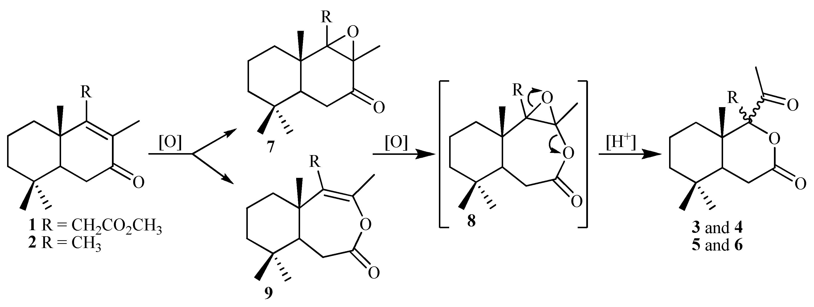
According to TLC data the reaction mixture derived from ketoester 1 contains one major product and that derived from drimenone 2 two chromatographycally separable products. The NMR analysis showed that unseparable mixture of compounds 3/4 and individual compounds 5 and 6 are not expected seven-membered enol lactones, as normal products of BVO. Surprisingly, in both cases after BVO reaction epimeric on C9 six-membered ring lactones 3/4 and 5/6 were obtained in 57% and 88% overall yields, respectively (Scheme 1).
The literature doesn’t report such cases of rearrangement for compounds from norlabdane series during BVO, but an explanation of compounds 3-6 formation can be found. One of the ways of the ketones 1 and 2 conversion, probably, starts from epoxidation of the C8-9 double bond with epoxyketone 7 formation, followed by BVO which leads to epoxylactone 8 (Scheme 2).
Another way for the formation of compound 8, starts with BVO of ketones 1 and 2 which gives enol lactone 9 followed by the epoxidation of the double bond. Such types of transformations have been described for cholestane-type compounds, when both processes occur as a result of treating α,β-unsaturated ketones with peroxyacid [31].
However, in our case, as a result of both oxidative processes and due to acidic medium, a rearrangement of intermediate seven-membered ring epoxilactone 8 into a mixture of enantiomeric more stable six-membered lactones 3/4 and 5/6 occurs (Scheme 2).

2.2. Single-Crystal X-Ray Diffraction Study

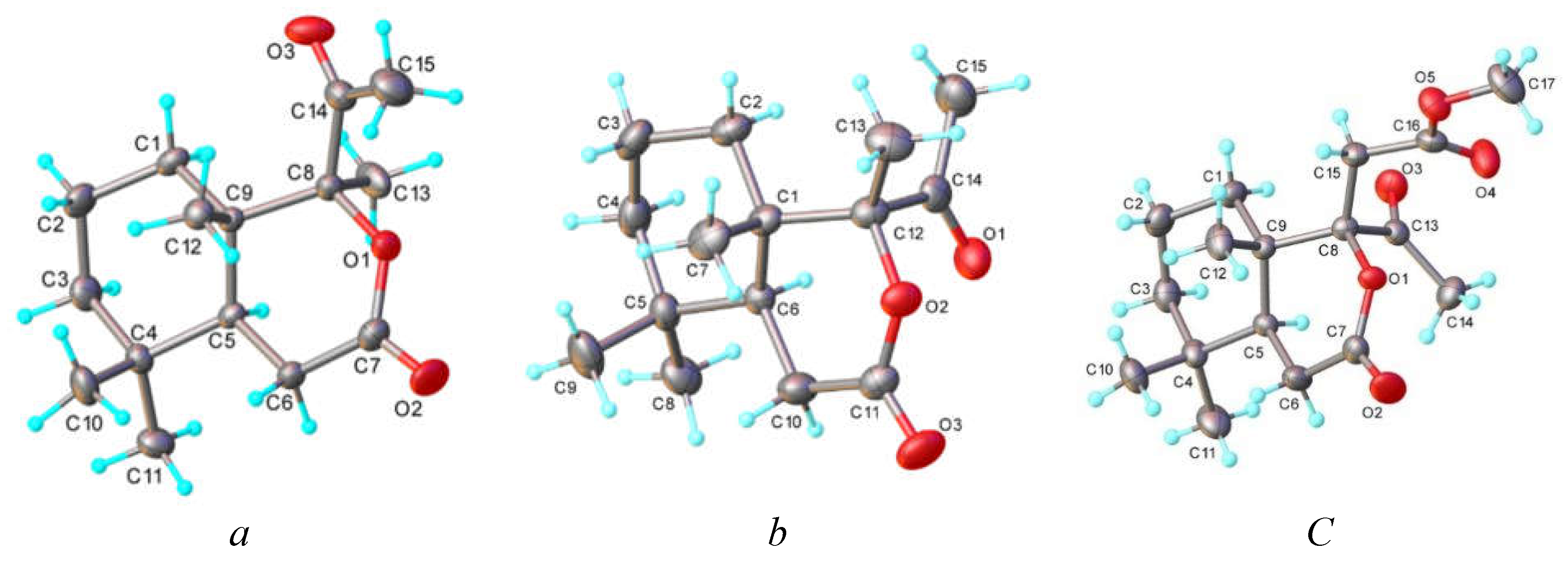
The structures of compounds 4, 5 and 6 were definitely confirmed by single crystal X-rays diffraction. According to X-ray crystallography, they crystallize in the Sohnke P212121, P21 and P212121 space groups, respectively and exhibit a molecular crystal structure, where the asymmetric part is consisting from one neutral entity, as shown in Figure 1A–C, respectively. There are no co-crystallized solvate molecules in both crystals. The bond distances and angles are summarized in Table 1.
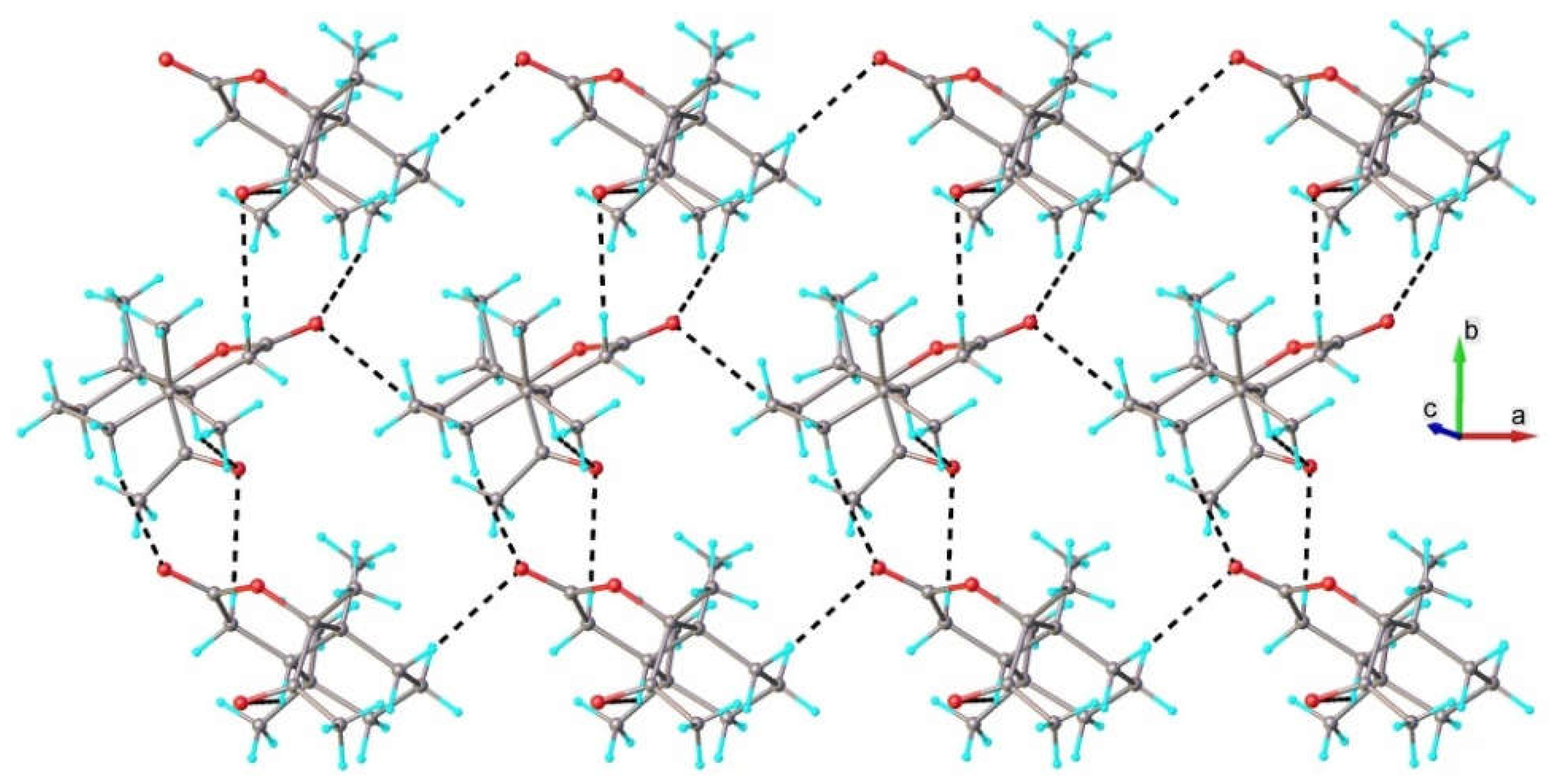
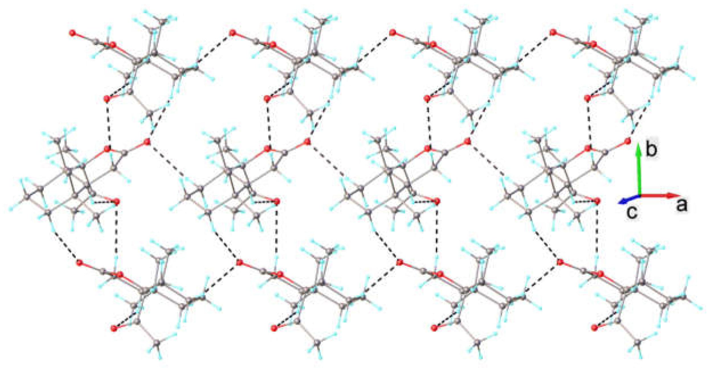
Further analysis has revealed that crystal structure of 4, 5 and 6 is driven by C-H---O hydrogen bonding. The aggregation of the molecules in the crystal 4, which occurs along hydrogen bonding, determines the formation of the discrete one-dimensional array having the form of zigzag chain running along a axis, as shown in Figure 2. In the crystals 5 and 6 C-H---O hydrogen bonds, being more numerous but weaker, dispose the crystal structure formed by the parallel packing of two-dimensional network. A fragment of 2D supramolecular layer is shown in Figure 3 and Figure 4, respectively.
H-bond parameters: C15-H---O2 [C15-H 0.97 Å, H···O2(x − 0.5, 0.5 − y, −z) 2.565 Å, C15···O2 3.525(4) Å, ∠C15HO2 169.8°.
H-bond parameters: C6-H---O3 [C6-H 0.99 Å, H···O3(1 − x, 0.5 + y, 1 − z) 2.70 Å, C6···O3 3.553(5) Å, ∠C6HO3 144.1°; C15-H---O2 [C15-H 0.98 Å, H···O2(1 − x, y − 0.5, 1 − z) 2.83 Å, C15···O2 3.667(5) Å, ∠C15HO2 143.5°.
H-bond parameters: C2-H---O3 [C2-H 0.90 Å, H···O3(x − 1, y, z) 2.633 Å, C2···O3 3.572(4) Å, ∠C2HO3 158.4°; C4-H---O3 [C4-H 0.99 Å, H···O3(1 − x, y − 0.5, 1 − z) 2.654 Å, C4···O3 3.584(4) Å, ∠C4HO3 156.5°; C10-H---O1 [C10-H 0.99 Å, H···O1(1 − x, 0.5 + y, 1 − z) 2.704 Å, C10···O1 3.553(5) Å, ∠C10HO1 144.1°.

2.3. Antimicrobial Activity

The novel tetra- and pentanorlabdane compounds with rearranged cycle B 3-6 were preliminary screened for their in vitro antifungal and antibacterial activities against three pure cultures of fungi and two gram-positive and gram-negative bacteria strains (Table 1). Compounds 3 and 4 were inseparable from the racemic mixture, therefore were screened together. Compounds 5 and 6 did not show a significant biological activity in the test.
According to the antimicrobial activity evaluation data, compounds 3 and 4 showed higher antifungal activity than the reference compound Caspofungin at MIC= 6.4·10-2 µg/mL, and comparable to the reference compound Kanamycin antibacterial activity at MIC= 2.0 µg/mL. The activity of compounds 3 and 4 is due to the co-existence of the lactone, >C=O and ester groups.

2.4. DFT Study of the Structure of Compounds 3 and 4

Considering that compounds 3 and 4 showed good antifungal and antimicrobial activity, and that epimerizable product distribution in a reaction is controlled by their thermodynamic stability, the study continued with computational methods, in order to reveal details regarding the stability of these epimers in various media.
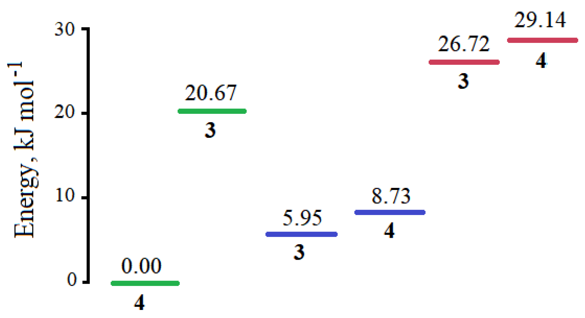
The structures of the two epimers 3 and 4 were optimized to global minima, in gas, water, DCM and methanol, and the coordinates are presented in supplementary Tables SI–VIII. In gas, compound 4 is more stable than 3 with a difference in the Gibbs free energy of 23.3 kJ/mol. The relative energies of compounds 3 and 4 global minima in the studied solvents is shown in Figure 5.
As seen in Figure 5, compound 4 is more stable than compound 3 in DCM, with a difference in the Gibbs free energy of 20.7 kJ/mol. In water and methanol, compound 3 is slightly more stable, by 2.4 kJ/mol and 2.78 kJ/mol, respectively.

2.5. Molecular Docking of Compounds 3 and 4

In order to explain the mechanisms of inhibition of microorganisms through the intermolecular ligand-receptor interaction that define the antimicrobial activity, a comparative molecular docking study of epimers 3 and 4 was carried out on four protein models: DNA gyrase from E. coli (1KZN), Fabz from P. aeruginosa (1U1Z), dihydrofolate reductase from C. albicans (3QLS) and MurB from E. coli (2Q85).
The binding energies of the researched compounds (Table 2) are similar to those of the standards used, and in many cases they even exceed them, demonstrating the fact that they effectively bind to the target enzymes contributing to their inhibition. The results of computational calculations demonstrate the fact that most of the compounds have a higher binding affinity to 1U1Z, suggesting the idea that the interruption of fatty acid biosynthesis would be the most likely way of action of the investigated compounds on pathogens.
In the case of E. coli ADN girase (1KZN), Kanamycin has the highest binding affinity -6.8 kcal/mol, closely followed by compound 3, (-6.6 kcal/mol), while compound 4 has a binding affinity very similar and a bit higher than Caspofungin, -6.2 kcal/mol and -6.1 kcal/mol, respectively. Both epimers bind into the same pocket as the standards, Caspofungin and Kanamycin (supplementary Figure S1).
In binding to P. aeruginosa FabZ (1U1Z), compounds 3 and 4 have an activity of -7.7 kcal/mol, higher than Caspofungin (-7.3 kcal/mol), and close to that of Kanamycin (-8.0 kcal/mol). Both epimers bind into the same pocket as the standard Kanamycin (supplementary Figure S2).
In the case of C. albicans dihidrofolat reductase (3QLS), Caspofungin and Kanamycin have very similar values of binding affinity, of -7.7 kcal/mol and -7.6 kcal/mol, respectively, while compounds 3 and 4 have lower affinities, of -6.7 kcal/mol and -6.4 kcal/mol, respectively. Both epimers bind into the same pocket as the standard Caspofungin (supplementary Figure S3).
Lastly, in the case of binding to E. coli MurB (2Q85), Kanamycin has an affinity of -8.1 kcal/mol, followed by compound 3 with a binding affinity of -7.1 kcal/mol, and then Caspofungin and compound 4 with binding affinities of -6.8 kcal/mol and 6.4 kcal/mol, respectively. Both epimers bind into one pocket, different from that of Caspofungin and Kanamycin (supplementary Figure S4).

3. Materials and Methodst

The following reagents and solvents were used in the research: dichloromethane (DCM), trifluoroacetic acid (TFA), meta-chloroperbenzoic acid (m-CPBA), petroleum ether (PE), ethyl acetate (EtOAc) and deuterated chloroform (CDCl3). Reagents and solvents were purchased from Sigma-Aldrich.
Melting points (mp) were determined on a Boetius hot-stage apparatus. Optical rotations were measured on a Perkin-Elmer 241 polarimeter with a 1-dm microcell, in CHCl3. Infrared spectra (IR) were obtained on Bio-Rad-Win-IR and Spectrum-100FT-IR (Perkin-Elmer) spectrometers with ATR technique. 1H, and 13C NMR spectra were recorded in CDCl3 on Bruker Avance DRX 400 and Bruker Biospin Avance 500 spectrometers. Chemical shifts are given in parts per million values (ppm) in δ scale and referred to CHCl3 (δH at 7.26 ppm) and to CDCl3 (δC 77.00 ppm), respectively. Coupling constants (J) are reported in Hertz (Hz). Correlation spectroscopy (H,H-COSY), heteronuclear single quantum correlations (H,C-HSQC), and heteronuclear multiple-bond correlations (H,C-HMBC) experiments were recorded using standard pulse sequences, in the version with z-gradients, as delivered by Bruker Corporation. Carbon substitution degrees were established by distortionless enhancement by polarization transfer (DEPT) pulse sequence. GS-MS analysis were run on an Agilent-5975C model provided a DB-17 fused silica capillary column, 30 × 0.25 mm i.d., with helium as carrier gas, coupled with a Agilent-5975C spectrometer (EI, 70 eV). High-resolution mass spectra (MS) were run on Kratos MS80 spectrometer (EI, 70 eV) with home-built data system. The ration m/z and relative intensity (%) are indicated for the significant peaks. For analytical thin-layer chromatography (TLC), Sorbfil and Merck silica-gel plates 60 G in 0.25 mm layers were used. The chromatograms were sprayed with concentrated H2SO4 and heated at 80°C for 5 min. Column chromatography was carried out on Асross (60-200 mesh) and Merck silica gel 60 (70-230 mesh) using mixture of petroleum ether (bp 40-60 °C) with EtOAc in gradient. All solvents were purified and dried by standard techniques before use. Organic extracts were dried over anhydrous Na2SO4, then filtered and evaporated under reduced pressure.

3.1. General Procedure of Baeyer-Villiger Oxidation of Ketones 1 and 2

To a solution of α,β-unsaturated ketone (1 mmol) in DCM (4 mL) m-CPBA (2.2 mmol) was added. After cooling to 0 °C, freshly distilled TFA (0.076 mL) was added dropwise. In continuation, the reaction mixture was warmed to room temperature and stirred in the dark for 20 hours, then diluted with DCM (20 mL) and washed consecutively with aqueous Na2SO3 (10%) (15 mL), saturated K2CO3 solutions, water (10 mL), and dried over anhydrous Na2SO4. After solvent removal under reduced pressure, the crude reaction products were purified by column chromatography on silica gel (eluent PE/EtOAc gradient 90/10→70/30) to yield:
Mixture of methyl 2-((1S,8aS)-1-acetyl-5,5,8a-trimethyl-3-oxooctahydro-1H-isochromen-1-yl)acetate (3) and methyl 2-((1R,8aS)-1-acetyl-5,5,8a-trimethyl-3-oxooctahydro-1H-isochromen-1-yl)acetate (4). White crystals (177 mg, 57%, in ~2:1 ratio). IR (ν, CCl4, cm-1): 2954, 1742, 1715, 1438, 1353, 1180, 1046, 816.
Compound 3. 1H NMR (400 MHz, CDCl3, δ): 3.65 (s, 3H, CH3COO), 3.15 (d, 1H, J = 16.5, H-11), 2.82 (d, 1H, J = 16.5, H-11), 2.76 (dd, 1H, J1 = 18.7, J2 = 7.0, H-6), 2.58 (dd, 1H, J1 = 19.3, J2 = 12.6, H-6), 2.40 (s, 3H, H17), 1.09 (s, 3H, H-20), 0.93 (s, 3H, H-19), 0.83 (s, 3H, H-18). 13C NMR (100 MHz, CDCl3, δ): 209.5 (C=O), 170.3 (C7=O), 169.7 (C12), 93.9 (C9), 52.1 (CH3COO), 44.1 (C5), 40.8 (C10), 40.5 (C3), 38.7 (C11), 33.0 (C4), 32.3 (C18), 32.1 (C1), 30.6 (C17), 29.6 (C6), 21.3 (C19), 17.9 (C2), 15.7 (C20).
Compound 4. 1H NMR (400 MHz, CDCl3, δ): 3.67 (s, 3H, CH3COO), 3.33 (d, 1H, J = 15.2, H-11), 2.82 (d, 1H, J = 15.8, H-11), 2.66 (dd, 1H, J1 = 18.8, J2= 5.8, H-6), 2.45 (dd, 1H, J1 = 18.4, J2= 13.1, H-6), 2.40 (s, 3H, H-17), 1.04 (s, 3H, H-20), 0.92 (s, 3H, H-19), 0.92 (s, 3H, H-18). 13C NMR (100 MHz, CDCl3, δ): 209.3 (C8), 169.4 (C12), 169.3 (C7=O), 93.8 (C9), 52.2 (CH3COO), 41.8 (C5), 40.9 (C3), 40.8 (C10), 40.8 (C11), 33.0 (C4), 32.8 (C18), 32.8 (C1), 30.6 (C17), 28.2 (C6), 21.4 (C19), 17.6 (C2), 15.6 (C20).
HR-EI-MS: Found 310.27802.C17H26O5 Calcd. 310.3843. m/z 310 (M+, 1), 279 (29), 249 (36), 218 (4), 193 (19), 165 (10), 145 (9), 133 (11), 121 (6), 109 24), 101 (8), 81 (15), 68 (4).
(1S,8aS)-1-acetyl-1,5,5,8a-tetramethylhexahydro-1H-isochromen-3(4H)-one (5). White crystals (83 mg, 33%), mp 133-134 °С (PE), [ α ] D 20 -102.6 (c 2.6, CHCl3). IR (ν, CCl4, cm-1): 2940, 1729, 1705, 1459, 1381, 1260, 1090, 825. 1H NMR (400 MHz, CDCl3, δ): 2.69 (dd, 1H, J1 = 18.8, J2 = 6.7, H-6), 2.47 (dd, 1H, J1 = 18.9, J2 = 12.4, H-6), 2.28 (s, 3H, H-17), 1.40 (s, 3H, H-20), 1.08 (s, 3H, H-11), 0.93 (s, 3H, H-19), 0.83 (s, 3H, H-18). 13C NMR (100 MHz, CDCl3, δ): 209.7 (C=O), 171.3 (C7=O), 94.6 (C9), 44.1 (C5), 40.9 (C3), 40.0 (C10), 33.0 (C1), 32.9 (C4), 32.3 (C18), 29.6 (C17), 29.3 (C6), 21.2 (C19), 18.7 (C20), 18.1 (C2), 14.7 (C11). HR-EI-MS: Found 209.15444. C15H24O3 Calcd. 253.2513. m/z 209 (M+, -44, 44), 181 (6), 123 (100), 69 (20), 65 (2), 54 (1), 42 (3).
(1R,8aS)-1-acetyl-1,5,5,8a-tetramethylhexahydro-1H-isochromen-3(4H)-one (6). White crystals (137 mg, 55%), mp 90-91 °С (PE), [ α ] D 20 -73.6 (c 2.3, CHCl3). IR (ν, CCl4, cm-1): 2937, 1733, 1714, 1455, 1354, 1279, 1101, 827. 1H NMR (400 MHz, CDCl3, δ): 2.61 (dd, 1H, J1 = 18.8, J2 = 6.2, H-6), 2.41 (dd, 1H, J1 = 18.8, J2= 13.2, H-6), 2.31 (s, 3H, H-17), 1.44 (s, 3H, H-11), 1.02 (s, 3H, H-20), 0.91 (s, 3H, H-19), 0.89 (s, 3H, H-18). 13C NMR (100 MHz, CDCl3, δ): 210.5 (C=O), 170.2 (C7=O), 95.0 (C9), 41.2 (C5), 41.0 (C3), 38.7 (C10), 33.1 (C1), 32.9 (C4), 32.7 (C18), 28.9 (C17), 28.4 (C6), 21.2 (C20), 21.2 (C11), 17.8 (C2), 15.7 (C19). HR-EI-MS: Found 209.15413. C15H24O3 Calcd. 253.2513. m/z 209 (M+, -44, 47), 181 (6), 110 (3), 107 (7), 95 (13), 91 (2), 83 (8), 79 (4), 67 (11), 59 (4), 55 (15), 51 (1).

3.2. Crystallographic Studies

Single-crystal X-ray diffraction data were collected on an Oxford-Diffraction XCALIBUR Eos CCD diffractometer with graphite-monochromated MoKα radiation. The unit cell determination and data integration were carried out using the CrysAlisPro package from Oxford Diffraction [16]. A multi-scan correction for absorption was applied. The structures were solved with the SHELXT using the intrinsic phasing method and refined by the full-matrix least-squares method on F2 with SHELXL [17,18]. Olex2 was used as an interface to the SHELX programs [19]. Non-hydrogen atoms were refined anisotropically. Hydrogen atoms were added in idealized positions and refined using a riding model. In the absence of a significant anomalous scattering, the absolute configuration of the structures could not be reliably determined, and therefore, Friedel pairs were merged and any references to the Flack parameter were removed. The molecular plots were obtained using the Olex2 program. Selected crystallographic data and structure refinement details for compounds 4, 5 and 6 are provided in supplementary Table S9.

3.3. Antifungal and Antibacterial Activity Assay

The synthesized compounds were tested in vitro for antimicrobial activity on three species of fungi: Aspergillus niger (ATCC 53346), Penicillium frequentans (ATCC 10110) and Alternaria alternate (ATCC 8741), and both Gram-positive Bacillus polymyxa (ATCC 15970) and Gram-negative Pseudomona aeruginosa (ATCC 27813) bacteria strains, provided by the American Type Culture Collection (ATCC, USA). Compounds Caspofungin and Kanamycin, both from Liofilchem (Roseto degli Abruzzi, Italy), were used as standards for antifungal and antibacterial activity testing at (MIC= 0.38 µg/mL) and (MIC= 2.0 µg/mL), respectively. Microorganism’s suspensions were prepared using the method of successive agar dilutions according to the standard MIC [20], and their cultivation was carried out according to standard procedures [21].

3.4. DFT Calculations

The ORCA program was used to optimize and properly characterize the structures [22]. All the calculations were performed using the wB97X functional with def2-SVP basis set, and the empirical dispersion correction of Grimme’s D3 in the DFT method [23]. Frequency analyses were done at the same level of theory. All the calculations in solvents (water, dichloromethane, methanol) were performed by employing the Solvation Model Density calculation model (SMD).

3.5. Molecular Docking

Four protein models were used in the study: DNA gyrase from Escherihia coli (PDB code: 1KZN), FabZ from P. aeruginosa (PDB code: 1U1Z), dihydrofolate reductase from Candida albicans (PDB code: 3QLS) and MurB from E. coli (PDB code: 2Q85).
The structures were prepared according to the protocol, using the AutoDockTools 1.5.7 program: water molecules and native ligands were removed from the protein structures, polar hydrogens and Gasteiger charges were added [24].
In the docking protocol, the ligands were treated as flexible molecules and the docking software was allowed to rotate all rotatable bonds of the ligands to obtain the ligand conformer with the optimal position in the active site of the enzyme. In each case, the grid was constructed and properly centered to cover the entire enzyme. Docking was performed using the routine procedure and default parameters of the molecular docking program Vina [25].
From the set of ligand conformations obtained by the docking procedure, the conformation with the lowest binding energy was selected for further analysis. The Discovery Studio Visualizer program was used to investigate the hydrophobic interactions and hydrogen bonds between the ligands and the investigated enzymes. The 3D structures of the Kanamycin and Caspofungin standards were retrieved from The Human Metabolome Database (hmdb.ca).

4. Conclusions

A series of novel tetra- and pentanorlabdane six-membered lactones with rearranged cycle B were synthesized in good yields by the Baeyer-Villiger oxidation with m-CPBA and the mechanism of their formation was proposed.
Computational calculations revealed that epimer 4 is more stable in gas and DCM, by 23.3 kJ/mol and 20.7 kJ/mol, respectively, while epimer 3 is more stable in water and methanol, by 2.4 kJ/mol and 2.78 kJ/mol, respectively.
Molecular docking results are in good agreement with experimental findings, showing that compounds 3 and 4 have a biological activity comparable, and in some cases exceeding that of the standards used, Casponfungin and Kanamycin. According to the values of calculated binding affinities, epimer 3 has a higher biological activity than epimer 4.
Due to their original structure and biological activity reported compounds are of real scientific interest.

Supplementary Materials

The following supporting information can be downloaded at: Preprints.org.

Author Contributions

Conceptualization, A.C. and A.A.; methodology, L.L. and N.V.; software, S.S and N.B.; writing—original draft preparation, A.C.; writing—review and editing, A.A.; visualization, A.A.; supervision, A.A.; project administration, V.K.; funding acquisition, V.K. All authors have read and agreed to the published version of the manuscript.

Funding

This research was supported by the Institutional Research Program of the State University of Moldova (MSU), subprogram code 010601.

Institutional Review Board Statement

Not applicable.

Informed Consent Statement

Not applicable.

Data Availability Statement

Data presented in this study are available on request from the corresponding author.

Conflicts of Interest

The authors declare no conflicts of interest.

References

  1. Fraga, B.M. Natural sesquiperpenpids. Nat. Prod. Rep. 2013, 30(9), 1226–1264. [Google Scholar] [CrossRef] [PubMed]
  2. Barrero, A.F., Manzaneda, E.A., Altarejos, J., Salido, S., Ramos, J.M., Simmonds, M.S.J., Blaney, W.M. Synthesis of biologically active drimanes and homodrimanes from (−)-sclareol. Tetrahedron 1995, 51(27), 7435-7450. [CrossRef]
  3. Koltsa, M.N., Mironov, G.N., Malinovskii, S.T., Vlad, P.F. Synthesis of drim-8(9)-en-7-one, drima-5,8(9)-dien-7-one and their 11,12-dibromo derivatives from norambreinolide. Russ. Chem. Bull. 1996, 45, 208-214. [CrossRef]
  4. D’Ambrosio, M., Guerriero, A., Deharo, E., Debitus, C., Munoz, V., Pietra, F. New Types of Potentially Antimalarial Agents: Epidioxy-substituted norditerpene and norsesterpenes from the marine sponge Diacarnus levii. Helv. Chim. Acta 1998, 81(5-8), 1285-1292. [CrossRef]
  5. Vlad, P.F., Popa, D.P., Gorincioi, E.K., Coltsa, M.N., Mironov, G.N. Synthesis of 11-hydroxydrim-8(9)-en-7-one and 11,12-dihydroxydrim-8(9)-en-7-one from drim-8(9)-en-7-one. Russ. Chem. Bull. 2000, 49(1), 98-101. [CrossRef]
  6. Vlad, P.F., Gorincioi, E.K., Coltsa, M.N. Synthesis of 5,6-dehydro-7-oxoisodrimenin from drim-8-en-7-one. Russ. Chem. Bull. 2003, 52(2), 502-504. [CrossRef]
  7. Vlad, P.F., Aryku, A.N., Ciocarlan, A.G. Synthesis of (+)-drim-9(11)-en-8-ol from sclareol. Russ. Chem. Bull. 2004, 53, 443–446. [CrossRef]
  8. Vlad, P.F., Coltsa, M.N., Aricu, A.N., Ciocarlan, A.G., Gorincioi, E.C., Edu, C.G., Deleanu, C. Photooxidative dehydrogenation of Δ8-drimen-and Δ8-11-homodrimen-7-ones into α,α′-dienones. Russ. Chem. Bull. 2006, 55, 703–707. [CrossRef]
  9. Vlad, P.F., Ciocarlan, A.G., Coltsa, M.N., Baranovsky, A.V., Khripach, N.B. Synthesis of drim-7,9(11)-diene and its hydroxylated derivatives. Synth. Commun. 2008, 38(22), 3960-3972. [CrossRef]
  10. Vlad, P.F., Edu, K., Koltsa, M.N., Chocyrlan, A., Nikolescu, A., Delyanu, K. Enantioselective synthesis of 11-homodrim-7-en-9α,12,13-triol. Chem. Nat. Compd. 2011, 47(4), 574-578. [CrossRef]
  11. Ciocarlan, A., Aricu, A., Lungu, L., Edu, C., Barba, A., Shova, S., Mangalagiu, I.I., D’Ambrosio, M., Nicolescu, A., Deleanu, C., Vornicu, N. Synthesis of novel tetranorlabdane derivatives with unprecedented carbon skeleton. Synlett 2017, 28(5), 565–571. [CrossRef]
  12. Aricu, A., Ciocarlan, A., Lungu, L., Shova, S., Zbancioc, Gh., Mangalagiu, I.I., D’Ambrosio, M., Vornicu, N. Synthesis, antibacterial, and antifungal activities of new drimane sesquiterpenoids with azaheterocyclic units. Med. Chem. Res. 2016, 25(10), 2316-2323. [CrossRef]
  13. Lungu, L., Cucicova, C., Blaja, S., Ciocarlan, A., Dragalin, I., Barba, A., Vornicu, N., Geana. E.-I., Mangalagiu, I.I., Aricu, A. Synthesis of homodrimane sesquiterpenoids earing 1,3-benzothiazole unit and their antimicrobial activity evaluation. Molecules 2022, 27(16), 5082-5094. [CrossRef]
  14. Lungu, L., Blaja, S., Cucicova, C., Ciocarlan, A., Barba, A., Kulcițki, V., Shova, S., Vornicu, N., Geana, E.-I., Mangalagiu, I.I., Aricu, A. Synthesis and antimicrobial activity evaluation of homodrimane sesquiterpenoids with a benzimidazole Unit. Molecules 2023, 28(3), 933–946. [CrossRef]
  15. Secara, E. Synthesis of new drimane and homodrimane lactams by beckmann rearrangement of some ketoximes. Chem. J. Mold. 2016, 11(1), 50–54. [Google Scholar] [CrossRef] [PubMed]
  16. Rigaku Oxford Diffraction. CrysAlisPro Software System, Version 1.171.41.64; Rigaku Corporation: Oxford, UK (2015).
  17. Sheldrick, G.M. SHELXT – Integrated space-group and crystal-structure determination. Acta Cryst. Section A 2015, 71(1), 3–8. [Google Scholar] [CrossRef] [PubMed]
  18. Sheldrick, G.M. Crystal structure refinement with SHELXL. Acta Cryst. Section C 2015, 71(1), 3–8. [Google Scholar] [CrossRef]
  19. Dolomanov, O.V., Bourhis, L.J., Gildea, R.J., Howard, J.A.K., Puschmann, H. OLEX2: a complete structure solution, refinement and analysis program. J. Appl. Crystallogr. 2009, 42(2), 339-341. [CrossRef]
  20. Russell, A.D. Antibiotic and biocide resistance in bacteria: Introduction. J. Appl. Microbiol. 2002, 92(s1), 1S–3S. [Google Scholar] [CrossRef]
  21. NCCLS guidelines (National Committee on Clinical Laboratory Standards (NCCLS) Antimicrobial Susceptibility Standards (ATS) 2003, for M7 (CMI) and M100).
  22. Neese, F., Wennmohs, F., Becker, U., Riplinger, C. The ORCA quantum chemistry program package. J. Chem. Phys. 2020, 152(22), 224108. [CrossRef] [PubMed]
  23. Grimme, S., Hansen, A., Brandenburg, J.G., Bannwarth, C. Dispersion-Corrected Mean-Field Electronic Structure Methods. Chem. Rev. 2016, 116(9), 5105-5154. [CrossRef]
  24. Forli, S., Huey, R., Pique, M.E., Sanner, M.F., Goodsell, D.S., Olson, A. Computational protein-ligand docking and virtual drug screening with the AutoDock suite. J. Nat. Protoc. 2016, 11(5), 905-919. [CrossRef]
  25. Trott, O., Olson, A.J. AutoDock Vina: improving the speed and accuracy of docking with a new scoring function, efficient optimization, and multithreading. J. Comput. Chem., 2010, 31(2), 455-461. [CrossRef]
  26. Aasen, A.J., Vogt, C.H.G., Enzel, C.R. Tobaco Chemistry. 28. Structure and synthesis of drim-8-en-7-one, a new tobacco constituent. Acta Chem. Scand. 1975, B29, 51–55.
  27. Vlad, P.F., Vorob’eva, E.A. Synthesis of drim-8-en-7-one. Chem. Nat. Compd. 1983, 19, 139–141. [CrossRef]
  28. Ciocarlan, A., Lungu, L., Blaja, S., Dragalin, I., Aricu, A. The use of some non-conventional methods in chemistry of bicyclohomofarnesenic methyl esters. Chem. J. Mold. 2020, 15(20), 69-77. [CrossRef]
  29. Renz, M., Meunier, B. 100 Years of Baeyer–Villiger Oxidations. Euro. J. Org. Chem. 1999, 4, 737–750.
  30. Koch, S.S.C., Chamberlin, A.R. Modified conditions for efficient Baeyer-Villiger oxidation with m-CPBA. Synth. Commun. 1989, 19(5-6), 829-833. [CrossRef]
  31. Mendelovich, M., Glotter, E. Epoxidation and Baeyer–Villiger oxidation of γ-hydroxy-α,β-unsaturated ketones on exposure to m-chloroperbenzoic acid. J. Chem. Soc., Perkin Trans. 1. 1992, 1, 1735–1740. [CrossRef]
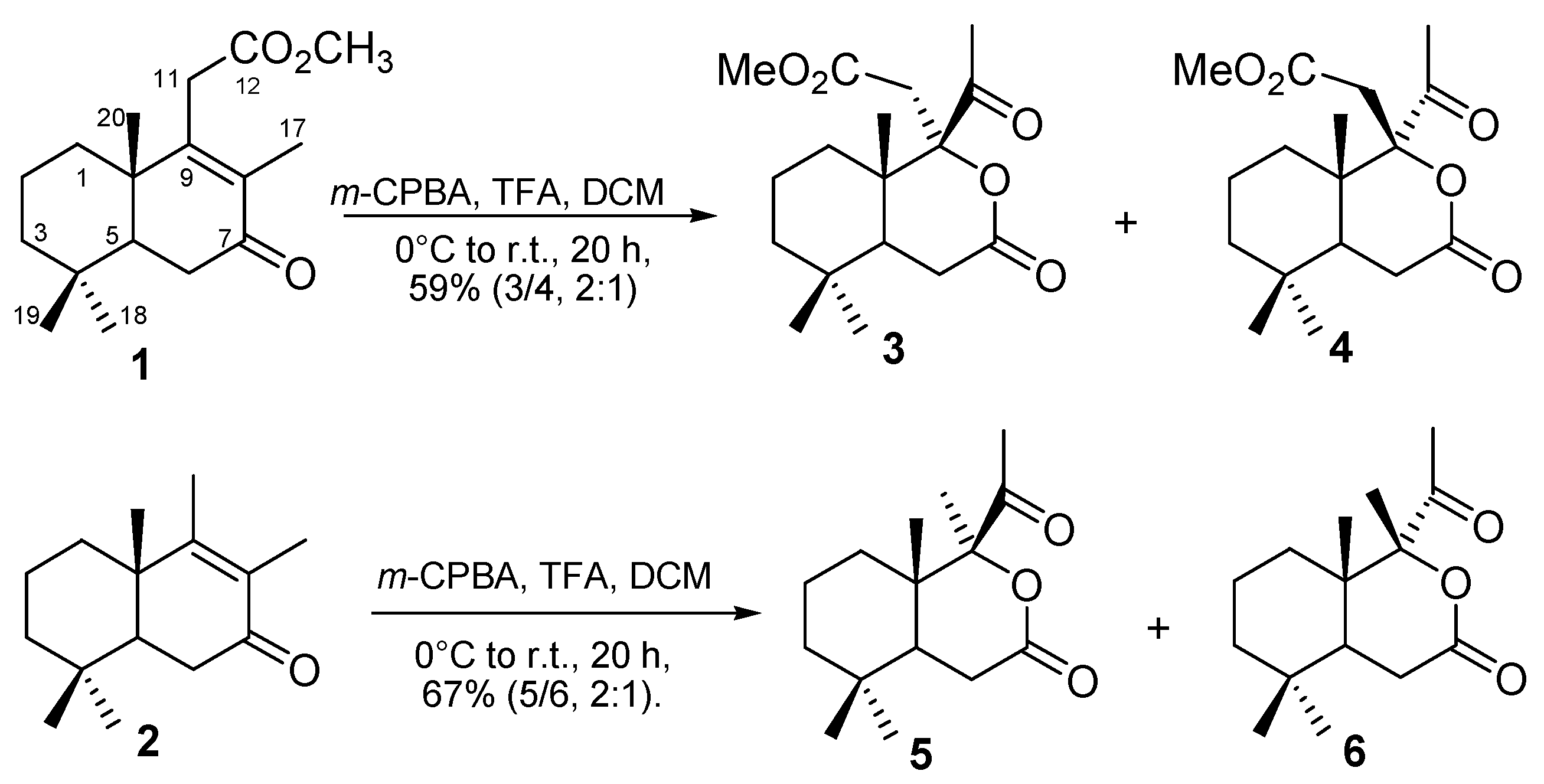
Scheme 1. Baeyer-Villiger oxidation of norlabdane lactones 1 and 2.
Preprints 138135 sch001
Scheme 2. The proposed mechanism of the Baeyer-Villiger oxidation of norlabdane ketones 1 and 2.
Preprints 138135 sch002
Figure 1. X-ray molecular structure of compounds 4 (a), 5 (b) and 6 (c) with atom labelling and thermal ellipsoids at 40% probability.
Preprints 138135 g001
Figure 2. 1D supramolecular chain in the crystal structure 4.
Preprints 138135 g002
Figure 3. 1D supramolecular chain in the crystal structure 5.
Preprints 138135 g003
Figure 4. 2D supramolecular layer in the crystal structure 6.
Preprints 138135 g004
Figure 5. Relative energies of epimers 3 and 4 in DCM (green), methanol (blue) and water (red), calculated at the wB97X/def2-SVP level.
Preprints 138135 g005
Table 1. Results of "in vitro" antimicrobial and antifungal activity of tested compounds.
Sample MIC (µg/mL)
Aspergillus niger Penicillium frequentans Alternaria alternata Bacillus polymyxa Pseudomonas aeruginosa
1/2 >128 >128 >128 >128 >128
3/4 0.064
(±0.035)
0.064
(±0.035)
0.064
(±0.035)
2.0
(±0.060)
2.0
(±0.060)
Caspofungin* 0.38 0.38 0.38 - -
Kanamycin* - - - 2.0 2.0
*Standard compound.
Table 2. The binding energy of the studied compounds to the target enzymes.
Compound Calculated binding energy (kcal/mol)
E. coli
DNA girase,
1KZN
P. aeruginosa FabZ,
1U1Z
C. albicans dihidrofolate reductase,
3QLS
E. coli MurB,
2Q85
3 -6.6 -7.7 -6.7 -7.1
4 -6.2 -7.7 -6.4 -6.4
Caspofungin -6.1 -7.3 -7.7 -6.8
Kanamycin -6.8 -8.0 -7.6 -8.1
Disclaimer/Publisher’s Note: The statements, opinions and data contained in all publications are solely those of the individual author(s) and contributor(s) and not of MDPI and/or the editor(s). MDPI and/or the editor(s) disclaim responsibility for any injury to people or property resulting from any ideas, methods, instructions or products referred to in the content.
Copyright: This open access article is published under a Creative Commons CC BY 4.0 license, which permit the free download, distribution, and reuse, provided that the author and preprint are cited in any reuse.
Alerts
Prerpints.org logo

Preprints.org is a free preprint server supported by MDPI in Basel, Switzerland.

Subscribe

© 2025 MDPI (Basel, Switzerland) unless otherwise stated