Preprint
Article

Association of BMI with Hepatic Steatosis, Fibrosis, and Inflammation: Insights from a Deceased Donor Liver Cohort Study Based on the UNOS Database

This version is not peer-reviewed.

Submitted:

01 November 2024

Posted:

05 November 2024

You are already at the latest version

Abstract

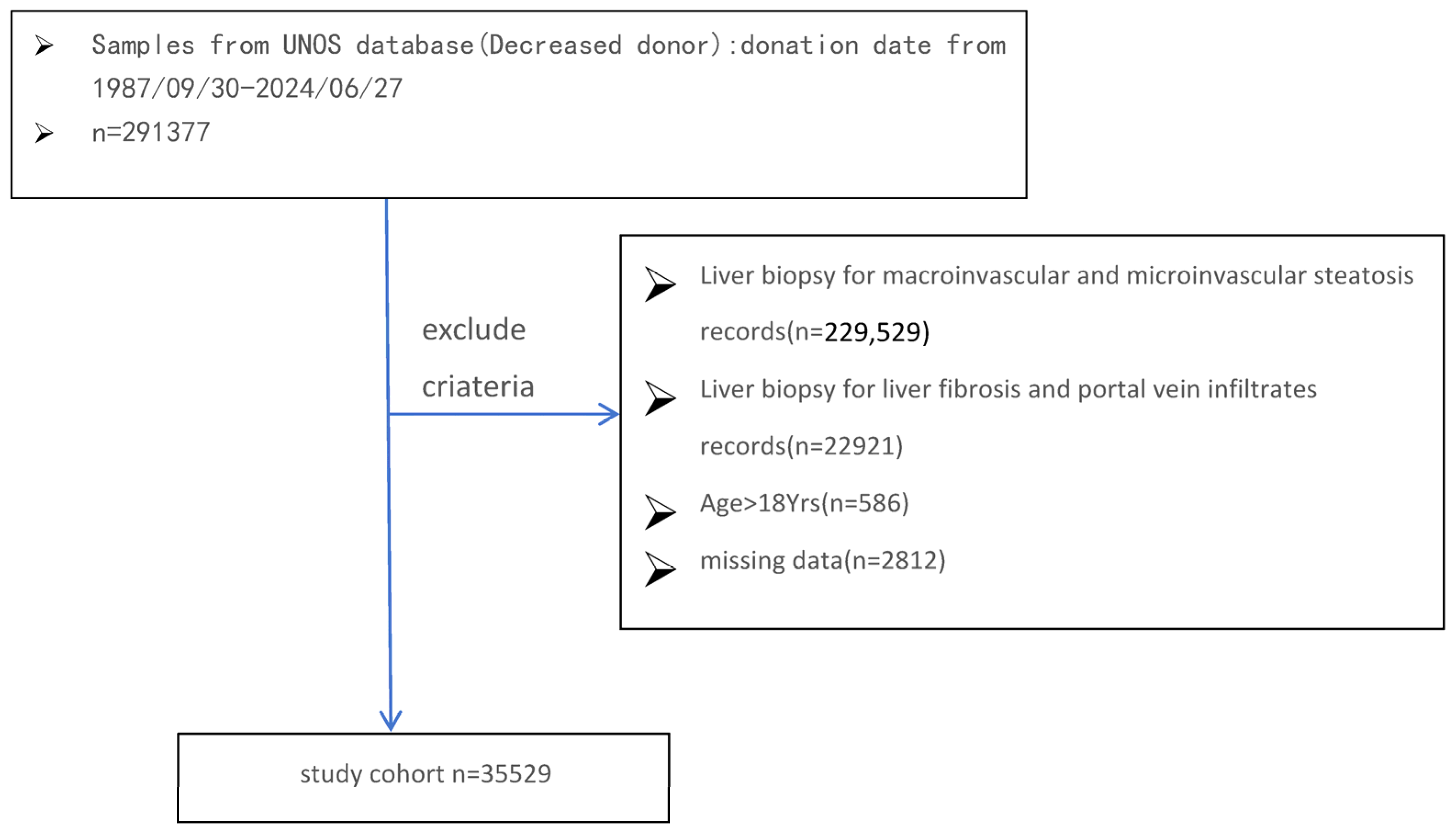
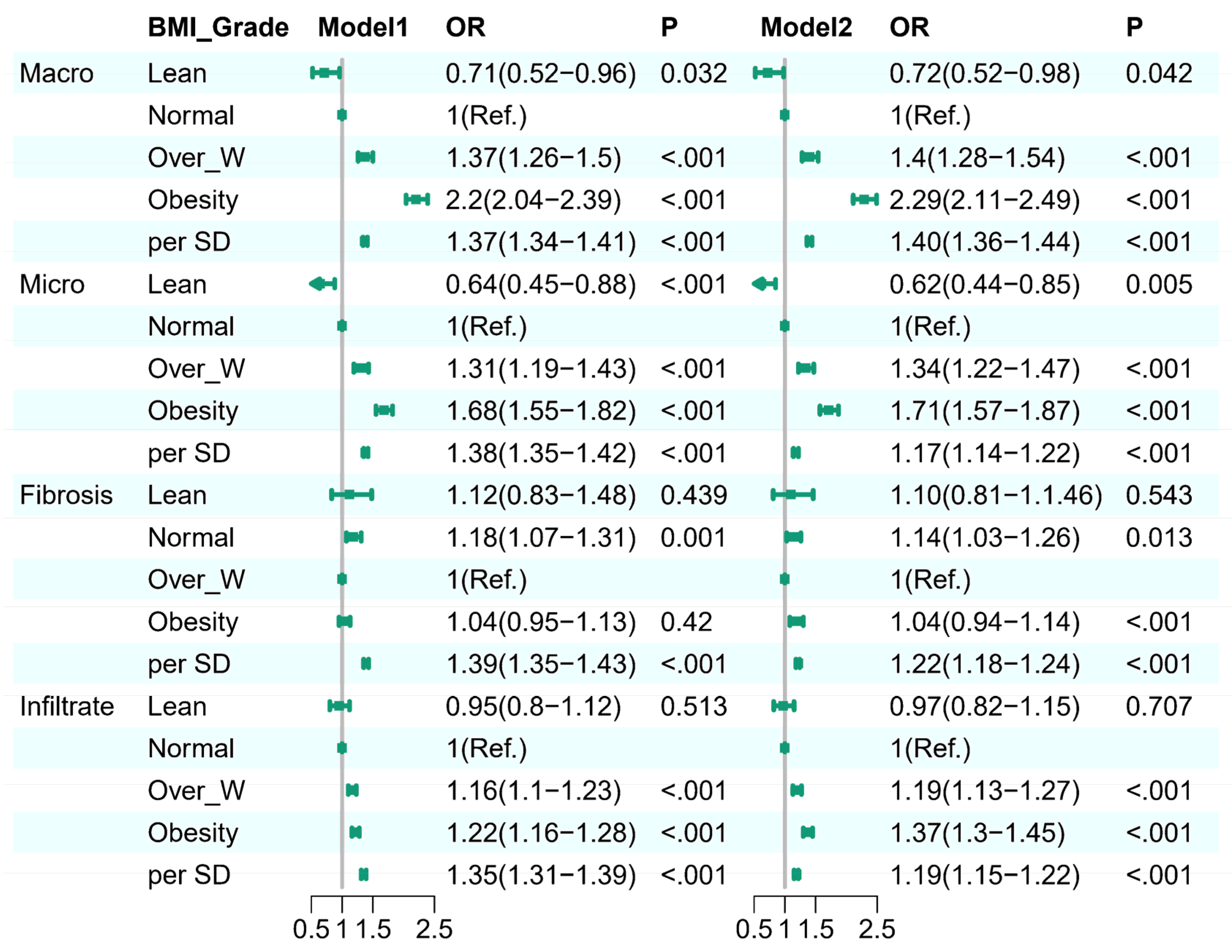
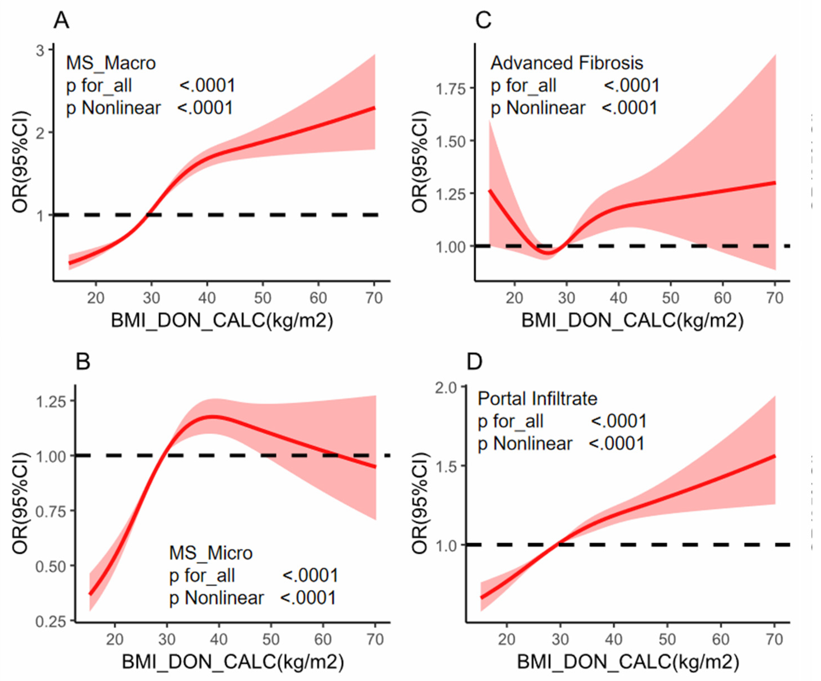
Background: While the prevalence of obesity and its negative effects on graft steatosis in liver transplantation are well recognized, the impact of obesity in the absence of hepatic steatosis, fibrosis, and inflammation in deceased donor livers remains unclear. This knowledge gap may affect the decision to adopt a transplant strategy and influence the following outcomes. Methods: A donor population-based cohort study was performed on 35,529 donors who received liver biopsy from 1987 to 2024. Donor BMI was categorized and assessed for its association with liver conditions, including macrovesicular steatosis, microvesicular steatosis, liver fibrosis, and portal infiltrates. Multivariable logistic regression and restricted cubic spline (RCS) regression models were employed to explore both linear and nonlinear relationships between BMI and the specified liver conditions. Results: In a cohort of 35,529 donors, donor livers from higher BMI groups exhibited higher percentages of macro-steatosis, micro-steatosis,advanced fibrosis and portal infiltrate.Logistic regression indicated obesity as an independent predictor of liver histology assessment: for higher risk with moderate-servere macro-steatosis (OR 2.29, 95% CI 2.11–2.49, p < 0.001),moderate-servere micro-steatosis (OR 1.71, 95% CI 1.57–1.87, p < 0.001), portal infiltrate (OR 1.37, 95% CI 1.3–1.45, p < 0.001) and advanced fibrosis (OR 1.04, 95% CI 0.94–1.14, p < 0.001). Restricted cubic spline regression depicted J-shaped with moderate-servere macro-steatosis and portal infiltrate,a U-shaped advanced fibrosis, and a upside down U-shaped with moderate-servere micro-steatosis,P for for nonlinearity <0.0001),respectively. Subgroup analyses identified interactions with factors such as gender, hypertension, and hepatitis C, highlighting BMI’s complex influence on liver histology and reinforcing its role in liver donor evaluation. Conclusion: This donor population-based cohort study found a different association pattern between donor BMI and liver biopsy outcomes,integrating BMI into donor liver assessments could enhance decision-making during transplantation, potentially reducing organ discard rates and improving transplant success by identifying high-risk organs earlier.

Keywords: 
;  ;  ;  ;  

Introduction

Liver transplantation is a life-saving procedure for patients with end-stage liver disease and certain acute liver conditions [1]. However, the success of liver transplantation is highly dependent on the quality of the donor liver [2]. Various factors, including donor health and lifestyle, can significantly impact the condition of the donor’s liver and consequently affect transplant outcomes [3]. One such factor is the donor’s Body Mass Index (BMI), which has been increasingly recognized as a crucial determinant of liver quality [4].
Higher BMI has been associated with an increased risk of liver steatosis, also known as fatty liver disease [5]. Liver steatosis can range from mild to more severe grade based on the percentage of lipid drop accumulation in the liver cell, with two main phenotypes, named macrovesicular and microvesicular steatosis which were distinguished by the lipid drop size with widespread differences in molecular characterization in metabolic pathways [6,7]. Liver fibrosis, which is often accompanied by steatosis, is the excessive accumulation of extracellular matrix proteins that can lead to liver cirrhosis [8]. Additionally, portal infiltrates, which involve the infiltration of inflammatory cells into the portal tracts of the liver, referring to the immune-activated status of the donor’s liver, can further the complexity of LT [9].
Understanding the relationship between donor BMI and liver status is crucial for improving liver transplantation outcomes. Previous studies have suggested that higher BMI is associated with an increased risk of liver steatosis and fibrosis [10]. However, the role of deceased donor BMI and donor-related factors in modifying these associations remains unclear, especially for different features of steatosis and portal inflammation. This study aims to fill this gap by investigating the association of donor BMI with the risk of liver steatosis related outcomes before transplantation. Additionally, the study explores the potential modifying effects of other donor-related factors on these associations.
To achieve these aims, we analyzed data from the United Network for Organ Sharing (UNOS) and Scientific Registry of Transplant Recipients (SRTR) registry, which includes comprehensive information on liver transplant recipients and donors. We utilized stepwise multi-model logistic regression analysis to investigate the association between different BMI categories and the presence of macrovesicular and microvesicular steatosis, liver fibrosis, and portal infiltrates. Furthermore, we employed a restricted cubic spline (RCS) regression model to explore potential nonlinear associations of cumulative BMI with these liver conditions. Subgroup analyses were conducted to identify potential modifying effects and interactions among the covariates.
In this study, we aim to provide insights into the impact of donor BMI on liver quality, which may help in developing better assessment criteria for donor liver selection and precisely matching recipients, ultimately improving the success rates of liver transplantation procedures.

Methods

Database and Study Cohort

This study analyzed data from the UNOS-OPTN database, covering the period from 1987 to April 1, 2024. All data were anonymized and handled by stringent data use agreements and security protocols. The perspectives and conclusions presented in this manuscript reflect the authors’’ views and do not represent the official stance of any government.

Donor Study Population and Exclusion Criteria

The data for this analysis were released on April 1, 2024. A total of 291,377 deceased donors were procured between 1987 and 2024. Exclusion criteria included the absence of data on hepatic micro- and macro-steatosis, liver fibrosis, and portal infiltrates (n=229,529), other missing variables (n=2,812), and donors under 18 years of age (n=586). This resulted in a final cohort of 35,529 donors, stratified by donor BMI as recorded in UNOS. The flow chart illustrates the donor selection process for the final cohort(Figure 1). The study protocol was exempted by the Ethics Review Committee of The First Affiliated Hospital of Kunming Medical University for the OPTN database is deidentified and publicly available.

BMI Classification

We utilized the Criteria of Weight for Adults for BMI classification [11]. Given the large sample size, we could investigate the dose-response relationship between BMI and donor liver quality in detail. The BMI categories were defined as follows: <18 kg/m² (Lean), 18-25 kg/m² (Normal), 25-30 kg/m² (Overweight), and >30 kg/m² (Obesity).

Donor Liver Quality Assessment and Outcomes

Donor liver quality was assessed by biopsy pre-transplantation after procurement, including evaluations for macro- and micro-steatosis, liver fibrosis, and portal infiltrates. UNOS mandates that participating institutions stain histopathology slides using standardized protocols with hematoxylin and eosin, as well as fat-specific methods (Red-Oil-O or Sudan Red) to identify and quantify fatty deposits in the parenchyma [12]. Steatosis grading was based on cross-sectional hepatic histology with varying threshold percentages: mild (0-30%) and moderate-to-severe (≥30%) for both macro and micro steatosis. Advanced fibrosis was indicated by Fibrosis stages F2-6, and portal infiltrate was classified as positive with stages F1-4. We identified ≥30% for both macro and micro steatosis, advanced fibrosis and portal infiltrate were a risk factor for LT and as the primary outcome for this study.

Covariates

Potential confounders included baseline age (continuous), sex (male or female), ethnicity (white,non-Hispanic or Hispanic/Latino, others), history of hypertension (yes or no), history of coronary artery disease (yes or no), history of myocardial infarction, history of diabetes (yes or no), status of hepatitis B virus (HBV) and hepatitis C virus (HCV) infection, alcohol heavy consumption (yes or no), smoking status (never, past, or current within 6 months and 20 packs/year), and liver function tests at procurement (SGOT, SGPT, and TBILI; continuous).

Statistical Analysis

Baseline characteristics were presented as means (standard deviation) or medians (interquartile range [IQR]) for continuous variables and as frequencies (percentages) for categorical variables, stratified by BMI category. Differences in these characteristics were assessed using ANOVA or Kruskal–Wallis H tests for continuous variables and chi-squared tests for categorical variables. All statistical tests were two-tailed, with a significance level set at P<0.05. Statistical analyses were performed using Zstats [13] and R Studio (version 4.3)

Results

Baseline characteristics of donors in each group based on BMI categories were presented in Table 1. Liver quality varied significantly across BMI categories. Mean macro-steatosis percentages increased with BMI: lean (7.24% ± 13.96%), normal (8.96% ± 14.91%), overweight (11.32% ± 16.19%), and obesity (15.09% ± 17.65%) (P < 0.001). Micro-steatosis showed a similar trend: lean (6.67% ± 12.86%), normal (8.63% ± 15.17%), overweight (10.37% ± 16.62%), and obesity (12.21% ± 17.77%) (P < 0.001). Advanced fibrosis percentages were slightly different across BMI categories, with advanced fibrosis (F2-6) percentages being: lean (9.25%), normal (9.68%), overweight (8.62%), and obesity (8.33%) (P = 0.008). Portal infiltrate also increased with BMI: lean (0.60 ± 0.66), normal (0.64 ± 0.70), overweight (0.68 ± 0.70), and obesity (0.69 ± 0.68) (P < 0.001).
In stepwise logistic regression analysis based on BMI grade, types of covariates were sequentially enrolled to explore confounding factors. The association between BMI and histology outcomes (macro-steatosis, micro-steatosis, fibrosis, infiltrate)remained significant after adjusting for different covariates. Model 1, a crude model, obesity showed an OR of 2.2 (95%CI: 2.04-2.39, p<0.001), 1.68 (95%CI: 1.55-1.82, p<0.001), and 1.22(95%CI: 1.16-1.28, p<0.001) for macro-steatosis, micro-steatosis and portal infiltrate, but BMI was a risk factor for fibrosis with no statistical significance. In Model 2, we added gender, age, metabolic diseases (e.g., hypertension, coronary heart disease, diabetes), viral hepatitis (HBV, HCV), and lifestyle (e.g., alcohol abuse and smoking). After adjustment, obesity was still associated with macro-steatosis (OR: 2.29, 95%CI: 2.11-2.49, p<0.001),micro-steatosis(OR: 1.71, 95%CI: 1.57-1.87, p<0.001) and portal infiltrate (OR: 1.37, 95%CI: 1.3-1.45, p<0.001), showing an independent predictor of mild-severe macro-steatosis micro-steatosis, and hepatic inflammation. The BMI showed a weakened effect on fibrosis both in the crude model, but after adjusted several covariates,obesity was observed a strong impact on advanced fibrosis with an OR of 1.04 (95%CI: 0.94-1.14, p<0.001), compared with the overweight group. Additionally, for each per SD increase in continuous BMI, the ORs of macro-steatosis, micro-steatosis, fibrosis, portal infiltrate were 1.40(95% CI:1.36-1.44,p<0.001), 1.17 (95% CI: 1.14-1.22,p<0.001), 1.22 (95% CI: 1.18-1.24, p<0.001) and 1.19 (95% CI: 1.15-1.22,p<0.001), respectively(Figure 2).
We also apply a restricted cubic spline (RCS) regression model to explore the potential nonlinear association of continuous BMI with liver steatosis and other outcomes. There are distinct associations between BMI and primary outcomes depicted in Figure 1. For macro-steatosis (A) and portal infiltrate (D), an S-shaped relationship is observed, with reference point at the BMI equal to 29.6kg/m²and a p-value for nonlinearity < 0.0001, respectively. For micro-steatosis (B), an upside-down U-shaped relationship is observed, with the high OR in the 30-60kg/m²and with a p-value for nonlinearity < 0.0001. Fibrosis follows a U-shaped relationship (C), with OR peaking at both low and high BMI levels and a p-value for nonlinearity <0.0001.
In the subgroup and sensitivity analyses, continuous BMI generally showed a stable effect across most subgroups for macro-steatosis, micro-steatosis, and portal infiltrates, with significant interactions observed between BMI and certain covariates. Specifically, for macro-steatosis, notable interactions were identified with gender (p for interaction = 0.002), hypertension (p = 0.028), heavy alcohol consumption (p for interaction= 0.025), and HCV (p < 0.001). Similarly, for micro-steatosis, significant interactions with gender (p for interaction= 0.003), coronary artery disease (CAD) (p for interaction = 0.011), and HCV (p for interaction = 0.004) were observed. In the context of portal infiltrates, gender (p for interaction = 0.013) and CAD (p for interaction = 0.039) showed significant interactions with BMI. While assessing liver fibrosis, no significant association was found with continuous BMI. However, lower odds ratios were observed in females (OR = 0.99, 95% CI: 0.99–1.00, p = 0.046), age <60 (OR = 0.99, 95% CI: 0.99–1.00, p = 0.008), individuals without hypertension (OR = 0.99, 95% CI: 0.98–1.00, p = 0.012), without CAD (OR = 0.99, 95% CI: 0.99–1.00, p = 0.017), without myocardial infarction (OR = 0.99, 95% CI: 0.99–1.00, p = 0.017), and without diabetes (OR = 0.99, 95% CI: 0.98–1.00, p < 0.001). Significant interactions with age (p = 0.022), CAD (p = 0.045), and diabetes (p = 0.01) further underscore the importance of these covariates, suggesting that these factors modify the association between BMI and liver fibrosis.

Discussion

In this study, we examined the association between donor BMI and liver steatosis, fibrosis, and portal infiltrates in the largest cohort of liver transplant donors to date, with high accuracy through histological assessment. Our findings highlight a significant association between BMI and hepatic steatosis, fibrosis, and inflammation, aligning with existing literature on the adverse impact of obesity on donor liver health [14,15,16,17]. Our study provides more detailed insights into specific liver conditions, offering implications not only for deceased donor liver transplantation(DDLT) but also for living donor liver transplantation(LDLT) and public health strategies.
Our results are consistent with previous studies indicating that elevated BMI is a risk factor for moderate to severe liver steatosis. In addition to confirming this relationship, we examined different types of liver steatosis and observed distinct patterns between macrovesicular and microvesicular steatosis. Restricted cubic spline (RCS) analysis (Figure 3) shows that macrovesicular steatosis increases with BMI, while microvesicular steatosis remains stable once BMI exceeds a certain threshold. Non-alcoholic fatty liver disease (NAFLD), often found in obese individuals, was associated with both macrovesicular and microvesicular steatosis [18,19,20]. A 2019 meta-analysis demonstrated that steatosis greater than 30% significantly increases the risk of graft dysfunction, primary nonfunction, and reduced survival outcomes [21], supporting our finding that elevated BMI is a key factor in determining liver graft quality and transplant outcomes. Specifically, increased BMI has been linked to higher donor organ discard rates due to moderate to severe macrovesicular steatosis, a primary factor in organ non-utilization [22].
The relationship between obesity and liver fibrosis in our cohort differed from findings by Liu et al., who reported a J-shaped correlation between BMI and increased liver stiffness, an alternate measure of fibrosis [23]. Evidence from our restricted cubic spline analysis indicated a U-shaped relationship, with both very low and very high BMI levels associated with advanced fibrosis. This finding contrasts with Chan et al., who observed a linear relationship between BMI and fibrosis in metabolic-associated fatty liver disease (MAFLD). Differences in donor populations may explain this discrepancy, as a substantial portion of Chan et al.’s cohort was affected by alcoholic liver disease and viral hepatitis [24]. Prior studies suggest that grafts with portal fibrosis beyond stage 3 increasing the risk of post-transplant complications [25], and our findings further emphasize that both undernutrition and overnutrition detrimentally affect liver health, underscoring the importance of BMI in donor selection criteria for liver transplantation.
Our study’s analysis of BMI and portal infiltrates adds a novel dimension to the literature, as few studies have examined this specific feature in donor livers. Portal infiltrates, characterized by inflammatory cells in the liver’s portal tracts, are increasingly recognized as markers of immune activation and liver injury. Common in alcoholic and metabolic-associated liver diseases, portal infiltrates are associated with advanced disease stages [24]. This is particularly relevant to transplantation, where inflammation in the donor liver could amplify post-transplant immune responses, increasing the risk of graft rejection or dysfunction and negatively impacting graft survival [27]. Our findings underscore the importance of considering BMI as a marker for immune-related alterations affecting liver quality.
A recent study reported that 68.3% of potential living donor liver transplantation (LDLT) candidates were deemed unsuitable due to metabolic-associated fatty liver disease (MAFLD), illustrating the increasing impact of fatty liver disease on LDLT [26]. As MAFLD prevalence rises in the general population, the challenge of identifying suitable donors grows. In agreement with these findings, our analysis of organ discard rates reveals that livers from obese donors are more likely to be rejected due to concerns over steatosis, fibrosis, and portal infiltrates. Supporting evidence from Orman et al. further suggests that donor obesity is a key predictor of organ discard, especially in cases involving severe steatosis [29]. The higher discard rates with increasing BMI observed in our study indicate that transplant centers may hesitate to use livers from obese donors due to a higher risk of graft failure, consistent with prior research [30,31].
This study highlights the complex, multifaceted relationship between BMI and donor liver quality, influenced by several confounding factors. We included various donor-related factors, such as age, sex, and metabolic comorbidities, to better understand interactions between BMI and liver outcomes. Our findings suggest that male donors, older age groups, and individuals with histories of hypertension or diabetes are particularly vulnerable to the negative impact of elevated BMI on liver quality. These interactions align with literature showing that components of metabolic syndrome, such as hypertension and diabetes, exacerbate liver steatosis and fibrosis [32,33,34]. Such donor characteristics should inform liver transplantation decision-making.
Given these challenges, our study delved deeper into the relationship between BMI and liver conditions, specifically steatosis, fibrosis, and inflammation. By identifying risk factors associated with these conditions, we aim to improve donor liver selection criteria and enhance overall donor organ quality. Our findings may also guide living donor candidates in managing modifiable risk factors, such as BMI, before donor evaluation.

Strengths and Limitations

This study’s strengths include its large sample size and use of the well-established UNOS-SRTR national database, which provides comprehensive data on donor characteristics and liver biopsy results. The application of advanced statistical techniques, such as restricted cubic spline regression, allowed us to uncover nonlinear relationships between BMI and liver outcomes, offering a nuanced understanding of BMI’s impact on liver health.
However, our study also has limitations. First, its observational design limits our ability to establish causation between BMI and liver steatosis, fibrosis, and portal infiltrate. Second, despite adjustments for multiple confounders, residual confounding by unmeasured variables (e.g., diet and physical activity) may still be present. Third, variability in liver biopsy interpretation between pathologists and centers could introduce bias. Lastly, our findings may be limited in generalizability due to the demographic composition of donors in the UNOS database, which may not fully represent donors’ characteristics.

Conclusion

Our study provides valuable insights into the association between donor BMI and hepatic macrosteatosis, microsteatosis, fibrosis, and inflammation, with significant implications for liver transplantation practices. These findings strongly support the use of BMI as a noninvasive screening tool for assessing liver histology and identifying high-risk donors. Future research should focus on refining donor selection criteria to optimize transplantation outcomes and explore interventions that improve liver health among obese individuals.

Author Contributions

All authors have made substantial contributions to all of the following: (1) the conception and design of the study, or acquisition of data, or analysis and interpretation of data, (2) drafting the article or revising it critically for important intellectual content, (3) final approval of the version to be submitted. No writing assistance was obtained in the preparation of the manuscript. The manuscript, including related data, figures, and tables has not been previously published, and the manuscript is not under consideration elsewhere. All authors approve the final version of the manuscript, including the authorship list, and agree to be accountable for all aspects of the work in ensuring that questions related to the accuracy or integrity of any part of the work are appropriately investigated and resolved. Data collection and analysis –Congwen Bian, Supervision—Hanfei Huang, Zhong Zeng. Original draft—Congwen Bian, Review, and editing—Zhong Zeng, Hanfei Huang. All authors have read and approved the final version of the manuscript for submission.

Funding

This work was supported by Major Science and Technology Projects in Yunnan Province (No.202302AA310025), The funding source had no role in the design, conduct, or reporting of this study.

Institutional Review Board Statement

The study protocol was exempted by the Ethics Review Committee of The First Affiliated Hospital of Kunming Medical University for the OPTN database is deidentified and publicly available.

Data Availability Statement

Data for this analysis were obtained from the UNOS-SRTR database. The availability of these data is restricted and was permitted under a specific license for this study. Requests for UNOS data access can be directed to the National Health Insurance Sharing Service website (http://unos.org). Access requirements include a completed application form, a detailed research proposal, and an Institutional Review Board approval, all subject to review by the UNOS research support inquiry committee.

Conflicts of Interest

The authors declare that the research was conducted in the absence of any commercial or financial relationships that could be construed as a potential conflict of interest.

Publisher’s Note

All claims expressed in this article are solely those of the authors and do not necessarily represent those of their affiliated organizations, or those of the publisher, the editors and the reviewers. Any product that may be evaluated in this article, or claim that may be made by its manufacturer, is not guaranteed or endorsed by the publisher.

References

  1. Starzl TE, Fung JJ. Themes of liver transplantation. Hepatology. 2010 Nov;52(5):1383-94. [CrossRef]
  2. Bruinsma BG, Avruch JH, Sridharan GV, Weeder PD, Jacobs ML, Crisalli K, Amundsen B, Porte RJ, Markmann JF, Uygun K, Yeh H. Peritransplant Energy Changes and Their Correlation to Outcome After Human Liver Transplantation. Transplantation. 2017 Jul;101(7):1637-1644. [CrossRef]
  3. Moore DE, Feurer ID, Speroff T, et al. Impact of Donor, Technical, and Recipient Risk Factors on Survival and Quality of Life After Liver Transplantation. Arch Surg. 2005;140(3):273–277. [CrossRef]
  4. Lin JS, Muhammad H, Lin T, Kamel I, Baghdadi A, Rizkalla N, Ottmann SE, Wesson R, Philosophe B, Gurakar A. Donor BMI and Post-living Donor Liver Transplantation Outcomes: A Preliminary Report. Transplant Direct. 2023 Jan 12;9(2):e1431. [CrossRef]
  5. Loomis AK, Kabadi S, Preiss D, Hyde C, Bonato V, St Louis M, Desai J, Gill JM, Welsh P, Waterworth D, Sattar N. Body Mass Index and Risk of Nonalcoholic Fatty Liver Disease: Two Electronic Health Record Prospective Studies. J Clin Endocrinol Metab. 2016 Mar;101(3):945-52. [CrossRef]
  6. Leow WQ, Chan AW, Mendoza PGL, Lo R, Yap K, Kim H. Non-alcoholic fatty liver disease: the pathologist’s perspective. Clin Mol Hepatol. 2023 Feb;29(Suppl):S302-S318. [CrossRef]
  7. Kristiansen MNB, Veidal SS, Christoffersen C, Jelsing J, Rigbolt KTG. Molecular Characterization of Microvesicular and Macrovesicular Steatosis Shows Widespread Differences in Metabolic Pathways. Lipids. 2019 Jan;54(1):109-115. [CrossRef]
  8. 8. Bataller R, Brenner DA. Liver fibrosis. J Clin Invest. 2005 Feb;115(2):209-18. doi: 10.1172/JCI24282. Erratum in: J Clin Invest. 2005 Apr;115(4):1100.
  9. Gadd VL, Skoien R, Powell EE, Fagan KJ, Winterford C, Horsfall L, Irvine 8.K, Clouston AD. The portal inflammatory infiltrate and ductular reaction in human nonalcoholic fatty liver disease. Hepatology. 2014 Apr;59(4):1393-405. [CrossRef]
  10. Fabbrini E, Sullivan S, Klein S. Obesity and nonalcoholic fatty liver disease: biochemical, metabolic, and clinical implications. Hepatology. 2010 Feb;51(2):679-89. [CrossRef]
  11. Weir CB, Jan A. BMI Classification Percentile And Cut Off Points. 2023 Jun 26. In: StatPearls [Internet]. Treasure Island (FL): StatPearls Publishing; 2024 Jan.
  12. Lee DU, Yoo A, Kolachana S, Lee J, Ponder R, Fan GH, Lee KJ, Lee K, Schuster K, Chou H, Chou H, Sun C, Chang M, Pu A, Urrunaga NH. The impact of macro- and micro-steatosis on the outcomes of patients who undergo liver transplant: Analysis of the UNOS-STAR database. Liver Int. 2024 Aug;44(8):2011-2037. [CrossRef]
  13. Vass JK, Wilson RH. ‘Z’STATS’’--a statistical analysis for potential Z-DNA sequences. Nucleic Acids Res. 1984 Jan 11;12(1 Pt 2):825-32. [CrossRef]
  14. Lee YS, Hwang LC, Hsu HY, Tsou MT. The Association Between Different Obesity Phenotypes and Liver Fibrosis Scores in Elderly Individuals with Fatty Liver in Taiwan. Diabetes Metab Syndr Obes. 2021 Mar 30;14:1473-1483. [CrossRef]
  15. Tutunchi H, Naeini F, Ebrahimi-Mameghani M, Najafipour F, Mobasseri M, Ostadrahimi A. Metabolically healthy and unhealthy obesity and the progression of liver fibrosis: A cross-sectional study. Clin Res Hepatol Gastroenterol. 2021 Nov;45(6):101754. [CrossRef]
  16. Zarrinpar A, et al. Body mass index as a predictor of outcomes in liver transplantation: does increased body mass index adversely affect outcome? Liver Transplantation. 2013;19(11):1334-42. [CrossRef]
  17. Fisher RA, et al. Donor Body Mass Index and Outcomes in Liver Transplantation. Transplantation. 2019;103(1):86-94. [CrossRef]
  18. Burra P, Becchetti C, Germani G. NAFLD and liver transplantation: Disease burden, current management and future challenges. JHEP Rep. 2020 Oct 9;2(6):100192. [CrossRef]
  19. Reichman TW, Therapondos G, Serrano MS, Seal J, Evers-Meltzer R, Bohorquez H, Cohen A, Carmody I, Ahmed E, Bruce D, Loss GE. “Weighing the risk”: Obesity and outcomes following liver transplantation. World J Hepatol. 2015 Jun 18;7(11):1484-93. [CrossRef]
  20. Soma, Daiki MD, PhD1; Park, Yujin MD1; Mihaylov, Plamen MD1; Ekser, Burcin MD, PhD1; Ghabril, Marwan MD2; Lacerda, Marco MD2; Chalasani, Naga MD2; Mangus, Richard S. MD1; Kubal, Chandrashekhar A. MD, PhD1. Liver Transplantation in Recipients With Class III Obesity: Posttransplant Outcomes and Weight Gain. Transplantation Direct 8(2):p e1242, February 2022. [CrossRef]
  21. Brunt EM, et al. The effect of donor liver steatosis on post-transplant outcomes: A meta-analysis. Transplantation. 2019;103(4):511-522. [CrossRef]
  22. IKwong AJ, Kim WR, Lake J, Stock PG,et.al. Impact of Donor Liver Macrovesicular Steatosis on Deceased Donor Yield and Posttransplant Outcome. Transplantation. 2023 Feb 1;107(2):405-409. [CrossRef]
  23. Nonalcoholic Fatty Liver Disease: Two Electronic Health Record Prospective Studies. J Clin Endocrinol Metab. 2016 Mar;101(3):945-52. [CrossRef]
  24. Chan AW, et al. Underweight and severely obese individuals have higher risks of liver graft failure. Liver Transplantation. 2019;25(5):579-589. [CrossRef]
  25. Rabindranath M, Zaya R, Prayitno K, Orchanian-Cheff A, Patel K, Jaeckel E, Bhat M. A Comprehensive Review of Liver Allograft Fibrosis and Steatosis: From Cause to Diagnosis. Transplant Direct. 2023 Oct 16;9(11):e1547. [CrossRef]
  26. Rakha EA, Adamson L, Bell E, Neal K, Ryder SD, Kaye PV, Aithal GP. Portal inflammation is associated with advanced histological changes in alcoholic and non-alcoholic fatty liver disease. J Clin Pathol. 2010 Sep;63(9):790-5. [CrossRef]
  27. Vasuri F, Riefolo M, Ravaioli M, Cescon M, Pasquinelli G, Germinario G, D’‘E’rrico A. Predictive value of portal fibrosis and inflammation in transplanted liver grafts treated with hypothermic oxygenated perfusion. Pathol Res Pract. 2023 Mar;243:154361. [CrossRef]
  28. Rajaram RB, Jayaraman T, Yoong BK, Koh PS, Loh PS, Koong JK, et al. Non-alcoholic fatty liver disease and obesity among adult donors are major challenges to living-donor liver transplantation: a single-center experience. Asian J Surg. 2022;45(1):441–7.
  29. Orman ES, et al. Donor obesity and organ discard in liver transplantation: A registry-based analysis. American Journal of Transplantation. 2018;18(5):1160-1171. [CrossRef]
  30. Chan AW, et al. Obesity‘s’ Impact on Liver Transplant Outcomes: Evaluating both ends of the spectrum. Liver Transplantation. 2019;25(5):789-796. [CrossRef]
  31. Trotter JF, et al. The impact of obesity on liver transplantation outcomes. Current Opinion in Organ Transplantation. 2021;26(2):203-210. [CrossRef]
  32. Long MT, Zhang X, Xu H, Liu CT, Corey KE, Chung RT, Loomba R, Benjamin EJ. Hepatic Fibrosis Associates With Multiple Cardiometabolic Disease Risk Factors: The Framingham Heart Study. Hepatology. 2021 Feb;73(2):548-559. [CrossRef]
  33. Tuong TTK, Tran DK, Phu PQT, Hong TND, Chu Dinh T, Chu DT. Non-Alcoholic Fatty Liver Disease in Patients with Type 2 Diabetes: Evaluation of Hepatic Fibrosis and Steatosis Using Fibroscan. Diagnostics. 2020; 10(3):159. [CrossRef]
  34. 34. Ampuero J, Aller R, Gallego-Durán R, Crespo J,. Significant fibrosis predicts new-onset diabetes mellitus and arterial hypertension in patients with NASH. J Hepatol. 2020 Jul;73(1):17-25. doi: 10.1016/j.jhep.2020.02.028. Epub 2020 Mar 6. Erratum in: J Hepatol. 2020 Sep;73(3):740-741. doi: 10.1016/j.jhep.2020.06.018.
Figure 1. Flow chart of donor selection and exclude process.
Preprints 138324 g001
Figure 2. Associations of donor BMI with liver quality and donor liver discard probability. OR: Odds Ratio, CI: Confidence Interval. Model1: Crude, Model2: Adjust: GENDER, AGE, HYPERTENSION, CAD, MI, DIABETES, HBV, HVC, ALCOHOL, SMOKING.
Preprints 138324 g002
Figure 3. Association of early adulthood BMI with liver steatosis, fibrosis, infiltrate and discard probability. Graphs show associations of BMI with macro steatosis generally (A), with micro steatosis (B), with liver fibrosis (C), with portal infiltrate (D). Estimates were adjusted for donor gender, age, history of hypertension, CAD, MI, DM, status of HBV and HCV ,Alcohol heavy consumption and status of smoking. Solid lines are multi-variable adjusted ORs, with the area showing 95% CIs derived from restricted cubic spline regressions with four knots. The dashed line indicates a reference for no association at a hazard ratio of 1.0. The reference point is the median BMI (29·6 kg/m²). OR=odds ratio.
Preprints 138324 g003
Table 1. baseline characteristic of donor.
Variables Total (n = 35529) Lean (n = 584) Normal (n = 8426) Over_W (n = 9996) Obesity (n = 16523) P
AGE,Mean(SD),y 48.31 ± 14.13 48.08 ± 15.42 47.30 ± 15.36 48.87 ± 14.39 48.50 ± 13.20 <.001
<60y,n,(%) 27133 (76.37%) 423 (72.43%) 6394 (75.88%) 7405 (74.08%) 12911 (78.14%) <.001
≥60y,n,(%) 8396 (23.63%) 161 (27.57%) 2032 (24.12%) 2591 (25.92%) 3612 (21.86%)
GENDER(M),n,(%) 20027 (56.37%) 261 (44.69%) 4894 (58.08%) 6285 (62.88%) 8587 (51.97%) <.001
ETHNICITY,n,(%) <.001
White,Non-Hispanic 23237 (65.40%) 399 (68.32%) 5728 (67.98%) 6553 (65.56%) 10557 (63.89%)
Hispanic/Latino 5406 (15.22%) 59 (10.10%) 1057 (12.54%) 1680 (16.81%) 2610 (15.80%)
BMI,Mean(SD),kg/m2 30.55 ± 7.77 17.36 ± 0.90 22.47 ± 1.71 27.46 ± 1.43 37.00 ± 6.35 <.001
SGOT,Mean(SD),U/L 98.38 ± 249.05 78.14 ± 135.66 109.16 ± 308.32 98.67 ± 226.28 93.43 ± 230.61 <.001
SGPT,Mean(SD),U/L 106.10 ± 261.36 74.73 ± 135.56 117.35 ± 310.45 111.51 ± 264.21 98.19 ± 233.84 <.001
TBIL, Mean(SD),mg/L 0.89 ± 1.07 0.81 ± 0.78 0.92 ± 1.23 0.92 ± 1.03 0.86 ± 1.01 <.001
HYPERTENSION,n,(%) 17536 (49.36%) 219 (37.50%) 3129 (37.14%) 4518 (45.20%) 9670 (58.52%) <.001
CAD,n,(%) 3675 (10.34%) 49 (8.39%) 677 (8.03%) 984 (9.84%) 1965 (11.89%) <.001
MI,n,(%) 2288 (6.44%) 36 (6.16%) 432 (5.13%) 597 (5.97%) 1223 (7.40%) <.001
DIABETES,n,(%) 7038 (19.81%) 61 (10.45%) 1059 (12.57%) 1674 (16.75%) 4244 (25.69%) <.001
HBV,n,(%) 2704 (7.61%) 64 (10.96%) 840 (9.97%) 874 (8.74%) 926 (5.60%) <.001
HCV,n,(%) 4864 (13.69%) 110 (18.84%) 1689 (20.05%) 1647 (16.48%) 1418 (8.58%) <.001
ALCOHOL,n,(%) 8503 (23.93%) 172 (29.45%) 2601 (30.87%) 2725 (27.26%) 3005 (18.19%) <.001
SMOKING,n,(%) <.001
Y 1683 (4.74%) 25 (4.28%) 275 (3.26%) 494 (4.94%) 889 (5.38%)
current in 6M/20 packs/yr 8246 (23.21%) 188 (32.19%) 2324 (27.58%) 2375 (23.76%) 3359 (20.33%)
MACRO,Mean(SD),% 12.45 ± 16.78 7.24 ± 13.96 8.96 ± 14.91 11.32 ± 16.19 15.09 ± 17.65 <.001
Mild,n(%) 29715 (83.64%) 538 (92.12%) 7524 (89.30%) 8584 (85.87%) 13069 (79.10%) <.001
Moderate-Severe,n(%) 5814 (16.36%) 46 (7.88%) 902 (10.70%) 1412 (14.13%) 3454 (20.90%)
MICRO,Mean(SD),% 10.76 ± 16.86 6.67 ± 12.86 8.63 ± 15.17 10.37 ± 16.62 12.21 ± 17.77 <.001
Mild,n(%) 30738 (86.52%) 545 (93.32%) 7575 (89.90%) 8715 (87.18%) 13903 (84.14%) <.001
Moderate-Severe,n(%) 4791 (13.48%) 39 (6.68%) 851 (10.10%) 1281 (12.82%) 2620 (15.86%)
FIBROSIS, Mean(SD),score 0.45 ± 0.93 0.47 ± 0.91 0.47 ± 0.99 0.43 ± 0.91 0.44 ± 0.91 0.01
F0-1,n(%) 32402 (91.20) 530 (90.75) 7610 (90.32) 15099 (91.38) 9163 (91.67) 0.008
F2-6,n(%) 3127 (8.80) 54 (9.25) 816 (9.68) 1424 (8.62) 833 (8.33)
PORTAL INFILTRATE, Mean(SD),score 0.67±0.69 0.60 ±0.66 0.64 ±0.70 0.68 ±0.70 0.69 ±0.68 <.001
F0,n(%) 15485 (43.58%) 282 (48.29%) 3951 (46.89%) 4310 (43.12%) 6942 (42.01%) <.001
F1-4,n(%) 20044 (56.42%) 302 (51.71%) 4475 (53.11%) 5686 (56.88%) 9581 (57.99%)
SD:standard_deviation,BMI: Body Mass Index, SGOT: Serum Glutamic-Oxaloacetic Transaminase, TBIL: Total Bilirubin, CAD: Coronary Artery Disease, MI: Myocardial Infarction, HBV: Hepatitis B Virus, HCV: Hepatitis C Virus, ECD: Extended Criteria Donor.Macro:moderate-severe-macro -steatosis,Micro::moderate-severe-micro-steatosis.
Table 2. subgroup analysis of BMI in covariates.
Macro-steatosis Micro-steatisis Fibrosis Portal infiltrate
Variables n (%) OR (95%CI) P Pi OR (95%CI) P Pi OR (95%CI) P Pi OR (95%CI) P Pi
All patients 35529 (100.00) 1.04 (1.04-1.05) <.001 1.02 (1.02-1.03) <.001 1.00 (0.99-1.00) 0.076 1.01 (1.01-1.01) <.001
GENDER 0.002 0.003 0.119 0.476
F 15502 (43.63) 1.04 (1.03-1.04) <.001 1.02 (1.01-1.02) <.001 0.99 (0.99-1.00) 0.046 1.01 (1.01-1.01) <.001
M 20027 (56.37) 1.05 (1.04-1.05) <.001 1.03 (1.02-1.03) <.001 1.00 (0.99-1.01) 0.811 1.01 (1.01-1.02) <.001
AGE 0.4 0.293 0.022 0.039
<60 27133 (76.37) 1.04 (1.04-1.05) <.001 1.02 (1.02-1.03) <.001 0.99 (0.99-1.00) 0.008 1.01 (1.01-1.01) <.001
≥60 8396 (23.63) 1.04 (1.03-1.05) <.001 1.03 (1.02-1.04) <.001 1.01 (1.00-1.02) 0.21 1.02 (1.01-1.02) <.001
HYPERTENSION 0.028 0.127 0.254 0.161
N 17993 (50.64) 1.05 (1.04-1.05) <.001 1.03 (1.02-1.03) <.001 0.99 (0.98-1.00) 0.012 1.01 (1.00-1.01) <.001
Y 17536 (49.36) 1.04 (1.03-1.04) <.001 1.02 (1.02-1.03) <.001 1.00 (0.99-1.00) 0.206 1.01 (1.01-1.02) <.001
CAD 0.242 0.011 0.045 0.013
N 31854 (89.66) 1.04 (1.04-1.05) <.001 1.02 (1.02-1.03) <.001 0.99 (0.99-1.00) 0.017 1.01 (1.01-1.01) <.001
Y 3675 (10.34) 1.05 (1.04-1.06) <.001 1.04 (1.03-1.05) <.001 1.01 (1.00-1.02) 0.192 1.02 (1.01-1.03) <.001
MI 0.781 0.273 0.207 0.348
N 33241 (93.56) 1.04 (1.04-1.05) <.001 1.02 (1.02-1.03) <.001 0.99 (0.99-1.00) 0.043 1.01 (1.01-1.01) <.001
Y 2288 (6.44) 1.04 (1.02-1.06) <.001 1.01 (1.00-1.03) 0.121 1.01 (0.99-1.03) 0.431 1.02 (1.00-1.03) 0.006
HBV 0.909 0.119 0.967 0.277
N 32825 (92.39) 1.04 (1.04-1.04) <.001 1.02 (1.02-1.03) <.001 1.00 (0.99-1.00) 0.226 1.01 (1.01-1.01) <.001
P 2704 (7.61) 1.04 (1.02-1.06) <.001 1.03 (1.02-1.05) <.001 1.00 (0.98-1.01) 0.752 1.00 (0.99-1.02) 0.405
HCV 0.025 0.004 0.937 0.445
N 30665 (86.31) 1.04 (1.03-1.04) <.001 1.02 (1.02-1.02) <.001 1.00 (1.00-1.01) 0.213 1.02 (1.01-1.02) <.001
P 4864 (13.69) 1.05 (1.04-1.07) <.001 1.04 (1.03-1.05) <.001 1.00 (0.99-1.01) 0.644 1.01 (1.00-1.02) 0.023
ALCOHOL <.001 0.277 0.047 0.124
N 27026 (76.07) 1.05 (1.05-1.06) <.001 1.02 (1.02-1.03) <.001 1.00 (1.00-1.01) 0.245 1.01 (1.01-1.01) <.001
Y 8503 (23.93) 1.02 (1.01-1.03) <.001 1.03 (1.02-1.04) <.001 0.99 (0.98-1.00) 0.106 1.02 (1.01-1.02) <.001
DIABETES 0.606 0.469 0.01 0.699
N 28491 (80.19) 1.04 (1.04-1.05) <.001 1.02 (1.02-1.03) <.001 0.99 (0.98-1.00) <.001 1.01 (1.01-1.01) <.001
Y 7038 (19.81) 1.04 (1.04-1.05) <.001 1.03 (1.02-1.04) <.001 1.00 (1.00-1.01) 0.366 1.01 (1.00-1.02) 0.001
SMOKING 0.456 0.243 0.398 0.912
N 25600 (72.05) 1.04 (1.04-1.04) <.001 1.02 (1.02-1.02) <.001 1.00 (0.99-1.00) 0.17 1.01 (1.01-1.01) <.001
Y 1683 (4.74) 1.04 (1.02-1.06) <.001 1.02 (1.00-1.04) 0.052 1.01 (0.99-1.03) 0.339 1.01 (1.00-1.02) 0.074
current in 6M 8246 (23.21) 1.05 (1.04-1.05) <.001 1.03 (1.02-1.04) <.001 1.00 (0.99-1.01) 0.917 1.01 (1.01-1.02) <.001
/20 packs/yr
Disclaimer/Publisher’s Note: The statements, opinions and data contained in all publications are solely those of the individual author(s) and contributor(s) and not of MDPI and/or the editor(s). MDPI and/or the editor(s) disclaim responsibility for any injury to people or property resulting from any ideas, methods, instructions or products referred to in the content.
Copyright: This open access article is published under a Creative Commons CC BY 4.0 license, which permit the free download, distribution, and reuse, provided that the author and preprint are cited in any reuse.

Downloads

62

Views

40

Comments

0

Subscription

Notify me about updates to this article or when a peer-reviewed version is published.

Email

Prerpints.org logo

Preprints.org is a free preprint server supported by MDPI in Basel, Switzerland.

Subscribe

© 2025 MDPI (Basel, Switzerland) unless otherwise stated