Preprint
Article

Risk Factors for Malignancy of Thyroid Nodules in Patients Undergoing Thyroid Resection

Submitted:

05 November 2024

Posted:

06 November 2024

You are already at the latest version

A peer-reviewed article of this preprint also exists.

Abstract

Background: An accurate diagnosis of thyroid nodules is crucial for avoiding unnecessary surgical procedures and making timely treatment possible. The objective of the present study was to evaluate the diagnostic accuracy of FNAB (fine-needle aspiration biopsy) using histopathological findings as the reference standards. Patients with the diagnostic categories (DCs) III, IV, V were subjected to a special analysis. In addition, the authors assessed whether other factors, i.e. age, gender, BMI (body mass index), obesity and histopathologically confirmed lymphocytic thyroiditis had an impact on the occurrence of malignant tumors. Methods: A retrospective analysis was conducted on 535 patients who underwent thyroid surgery between October 2022 and September 2023. To assess the FNAB reliability, the result obtained using the Bethesda classification was compared with the histopathological result. Results: The ROM (risk of malignancy) values for DCs I-VI were 38.1%, 15.6%, 29.8%, 18.6%, 91.0%, 93.2%, respectively. DC V (OR 62.34, p< 0.0001) and age≤50 (OR=2.31, p<0.006) had a statistically significant effect on the risk of thyroid cancer. DCs III and IV were not statistically significantly associated with the risk of malignancy (OR=1.68,p=0.16; OR=1.51,p=0.3,respectively). There were no statistically significant differences in sex, BMI and obesity between the patients with benign and malignant lesions. Conclusion: DC V is associated with a high likelihood of malignancy, especially in patients under 50 years of age, and therefore surgery is indicated in this category of subjects. In the DCs III and IV categories, the risk of malignancy is lower and conservative management with active clinical and ultrasound surveillance can be considered. In patients < 50 years of age, with the Bethesda categories III and IV, surgical treatment should be considered.

Keywords: 
Subject: 
Medicine and Pharmacology  -   Endocrinology and Metabolism

1. Introduction

Thyroid nodules are common and may be present in 50% of people. The majority of these nodules are benign; nevertheless, approximately 8% of them are malignant [1]. Recently, the worldwide incidence of thyroid cancer has remarkably increased [2]. The correct diagnosis of thyroid nodules is therefore essential in order to avoid unnecessary surgery and to allow for prompt treatment of malignant tumours. Ultrasound (US) is the primary imaging test for the diagnosis of thyroid cancer and other thyroid diseases [3,4]. According to the 5-stage EUTIRADS classification, the risk of malignancy depends on the characteristics of the focal lesion and is close to 0% for the categories I and II, 2-4% for the category III, 6-17% for the category IV, and more than 26-87% for the category V [5]. Fine-needle aspiration biopsy (FNAB) is a good diagnostic tool for thyroid nodules; however, the high false-negative rate of results obtained using the method renders it controversial.
In 2007, the Bethesda System for the Reporting of Thyroid Cytopathology (TBSRTC) was introduced and the risk of malignancy for the six diagnostic categories (DCs) was defined. For the DCs I, II, III, IV, V and VI, the percentages were 1-4%, 0-3%, 5-15%, 15-30%, 60-75% and 97-99%, respectively [6].
Subsequently, the second edition of the TBSRTC was published, which showed a higher risk of malignancy for the lower Bethesda diagnostic categories. As it is apparent, the biopsy result will affect the extent of the surgical procedure [7,8,9].
The Bethesda III and IV cytological diagnoses account for approximately 30% of FNAB results [10] and the management of this group of patients is widely varied from the clinical and ultrasound follow-up, repeated FNAB, molecular testing to thyroid surgery. In case of the Bethesda V category, patients are generally referred for surgery.
The objective of the present study was to assess the risk of malignancy in patients referred for surgery for thyroid nodules, focusing on DCs III, IV and V. In addition, an attempt was made to assess whether other factors, i.e. age, sex, BMI (body mass index), obesity and coexisting lymphocytic thyroiditis, had an impact on increasing the risk of malignancy in thyroid nodules.

2. Materials and Methods

2.1. Patients and Methods

The authors performed a retrospective data analysis of 535 patients who underwent surgery at the Department of Endocrine Surgery of the University Hospital in Krakow, between October 2022 and September 2023. Data were collected for all the patients referred for surgery due to a nodular goitre, suspected malignancy, or a finding of malignancy on FNA. Patients who were reoperated for metastatic or recurrent cancer in the lymph nodes were excluded from the study (14 cases). The entire group of patients (n-521) was evaluated first, followed by the the assessment of the patients divided into groups with the DCs III, IV and V according to Bethesda (n=221). The results of particular patients were anonymised and collected in the international EUROCRINE database [11]. To assess the reliability of FNAB, the result obtained according to the Bethesda classification was compared with the histopathological result. In addition, such parameters as age, sex, BMI, obesity and the presence of lymphocytic thyroiditis in histopathological examination were analyzed.

2.2. Diagnosis and Evaluation

Each patient underwent preoperative thyroid ultrasonography (US) and preoperative FNAB of the suspected focal lesions. The patients were referred from various outpatient clinics and thus not in every case the EUTIRADS thyroid lesion assessment was performed. Therefore, this parameter was not included in the calculation. The thyroid US-guided FNAB procedure and the evaluation of FNAB cytology slides were performed prior to admission to hospital. The FNAB results were classified based on the criteria according to TBSRTC. The patients underwent total thyroidectomies or unilateral thyroid lobectomies. The extent of lymphadenectomy depended on the result of the preoperative FNAB and preoperative staging of the disease. The postoperative material was examined histopathologically. The histopathological diagnoses of thyroid nodules were established according to the World Health Organization 2022 classification guidelines for thyroid nodules [12]. In addition, the histopathological examination assessed the presence of other pathological changes, i.e. lymphocytic thyroiditis and others.
The histopathological findings were divided into two groups: benign and malignant. The authors assessed in which Bethesda categories there were benign nodules and in which malignant nodules. The histopathological results of the Bethesda III, IV and V patients were compared with the histopathological results of the Bethesda II patients using the multivariate regression analysis.

2.3. Statistical Analysis

The continuous variables were presented as mean, standard deviation (SD), median and quartiles (lower and upper). The Shapiro -Wilk test was used to verify the assumption of the normal distribution of the continuous variables. The Mann–Whitney U-test was performed to compare two groups because of the lack of normality for continuous variables. Due to the departures from normality, the Kruskal-Wallis test was applied for the comparison of the continuous variables between more than two groups. The post-hoc analysis was also performed. The qualitative variables were presented as numbers (percentages) and the Chi-square test was used for comparing them between two or more groups. The Receiver Operating Characteristic (ROC) curve was created to point out the Bethesda category manifesting malignancy. The multivariable logistic regression was used for determining the risk factors of malignancy. The results were showed as odds ratios (OR) with 95% confidence intervals (CI). The Hosmer-Lemehsow test was used as the fit of measure for the logistic regression model. The McFadden coefficient was also calculated and its value equalling to 0.3 showed good fitting. To check the prediction power of the logistic regression model, the c-statistics (i.e. the area under the curve, AUC) was applied. The value of AUC equal to 0.82 showed also a quite good prediction power of the presented model. The outlier points were also verified for this model. The p-values below 0.05 were considered statistically significant for the two-sided tests. Statistical analyses were conducted to calculate the sensitivity, specificity, positive predictive value (PPV), negative predictive value (NPV), false positive rate (FP), false negative rate (FN), true positive rate (TP) and true negative rate (TN). In addition, the multivariate logistic regression was performed to assess the risk of malignancy (ROM).
The package R v. 4.1.0 [13] and Statistica 13 software (StatSoft Inc., Tulsa, Oklahoma, United States) were used to perform the analyses.

3. Results

3.1. Results Obtained from the Entire Study Group of Patients (n=521)

The group of 521 patients included 96 men (18.4%) and 425 women (81.5%) (the ratio of 1:4.4), with the mean age of 52.3 ± 15.2 years. The reasons for referral to surgery were as follows: exclusions of malignant neoplasms (32.6%), presence of malignancies (22.2%), compression symptoms (19.5%), thyrotoxicosis (8.6%), and other indications (18.2%). The demographic and pathologic characteristics of the entire group is shown in Table 1.
In the study population, 37.8% (197 cases) of thyroid tumours were malignant. The classification of the types of malignant thyroid neoplasms (n=197) by sex was as follows: PTC - 147 (74.65%) females and 25 (12.6%) males; FTC - 9 (4.5%) females and 7 (3.5%) males, MTC - 2 (1.0%) females and 3 (1.5%) males; ATC - 1 (0.5%) male; lymphoma - 1 (0.5%) female.
Among the 172 cases of PTC, the following subtypes were identified: classic PTC in 121 cases (70.4%), the follicular variant of PTC in 44 cases (25.5%), the encapsulated variant of PTC in 3 cases (1.8%), other unusual variants of PTC in 4 cases (2.3%).
In 16 cases of FTC, the following subtypes were found: the widely invasive FTC - 3 (18.7%), the minimally invasive FTC - 8 (50.0%), the encapsulated angioinvasive FTC - 4 (25.0%), no data - 1 (6.2%).
The following low-risk neoplasms were noted: follicular tumours with uncertain malignant potential (FT-UMP) - 8 cases (1.5%), non-invasive follicular thyroid neoplasms with papillary-like nuclear features (NIFTP) – 10 cases (1.8%), well-differentiated tumours of uncertain malignant potential (WDT-UMP) – 4 cases (0.7%).
There was a statistically significant association between the cytological and histopathological results (p<0.0001), between the age (p<0.0001) and the presence of thyroid malignancy. The patients with confirmed thyroid cancer were on average 8 years younger than the patients without malignancies. The mean age in the patients with malignant thyroid cancer was 47.4±14.9 years (median - 45.0), while the mean age of the patients with a benign diagnosis was 55.3±14.7 years (median – 57.0). Among the patients with DC V, malignancy was diagnosed in more than 90% of the cases. In contrast, in the DC III group, benign lesions were found in 59 cases (70.24%) and malignant lesions in 25 cases (29.76%). In DC IV, a benign lesion was found in 57 cases (81.4%) and a malignant lesion in 13 cases (18.6%). There was no statistically significant association between gender (p = 0.76), BMI (p = 0.52) and obesity (p = 0.76) and the presence of thyroid malignancy.
In addition, the presence of lymphocytic thyroiditis (LT) was assessed histopathologically in the operated patients. LT was found in 79 patients (14.8%), i.e. in 47 patients (59.5%) with the diagnosis of malignant thyroid cancer and in 32 patients (40.5%) diagnosed with benign thyroid lesions. The following types of thyroid cancer were found in people with LT in the background: FTC - 2 (4.2%) in females only; OTC -1 (2.1%) in a female; PTC - 44 (93. 6%) in 40 females (91.0%) and 4 males (9.0%).
The histopathological findings of the patients in the DCs I, II, III, IV, V, VI of the Bethesda system are shown in Table 2.
The risk of malignancy (ROM) was calculated for each Bethesda category (Table 3).
Explanation: Criterion 6 means a positive diagnosis only within the DC VI group relative to all other categories. Criterion 5 means a positive diagnosis within the DCs VI and V relative to all other categories.
The highest probability of confirming malignancy (PPV) by histopathological examination was characteristic for the criterion 6 - 0.93, and for the criterion 5 - 0.92, while for the subsequent criteria, the PPV was, respectively: criterion 4 - 0.66; criterion 3 - 0.55; criterion 2 - 0.40.
A ROC curve was then determined, from which it was concluded that patients with DCs V and VI (criterion 5) would have thyroid malignancy with the sensitivity of 0.61 and specificity of 0.97 (Figure 1).

3.2. Results of Patients in DCs III, IV and V According to Bethesda (n=221)

Due to the inconclusive results found in the literature, the patients with DCs III, IV and V were separated out and an analysis of the incidence of malignancy in this subgroup was performed. Among the 521 patients, a group of 221 patients was identified who were referred for surgery due to DCs III, IV and V according to the Bethesda system. On the basis of the histopathological examination, two groups were distinguished: malignant and benign, in order to present the results according to the Bethesda category. DC II is included in the table because the logistic regressions for DCs III, IV and V were calculated for DC II (Table 5).
It was found that patients with DC V were statistically significantly younger than patients with DCs II, III, IV (p< 0,0001). The patients with DC V were on the average 10 years younger as compared to the patients with DCs II, III, and IV.
As there was a statistically significant correlation between the HP result and the cytology result in the patients with DCs II, III, IV, V, the authors assessed in which groups the malignant lesions were more frequent. And thus, malignant lesions were statistically significantly more frequent in DC V compared to DC II (p<0.0001), compared to DC III (p<0.0001), and compared to DC IV (p<0.0001) - Table 6.
Of the 61 patients with histopathologically confirmed background lymphocytic thyroiditis (LT), 42.6% were category V, 13.1% category IV, 16.4% category III and 21.9% category II. The most commonly observed type of cancer was PTC – 44 cases (93.0%). In the group of patients with background-confirmed LT, category V was statistically significantly more frequent than category III (p=0.0008), category IV (p=0.0009) and category II (p<0.0001) – Table 7.
The multivariate logistic regression analysis was used to determine the risk of thyroid malignancy in patients with DCs III, IV and V. The above cytology groups were compared with DC II. The effect of age, sex, cytology score, obesity (BMI ≥30.0) and the presence of lymphocytic thyroiditis on the development of thyroid cancer was assessed (Table 8).
Based on the multivariate logistic regression, DC V (p<0.0001) and age (p=0.006) were found to have a statistically significant effect on the occurrence of thyroid cancer. In contrast, no statistically significant effect was found to be exerted by DCs III and IV on the presence of a malignant lesion.
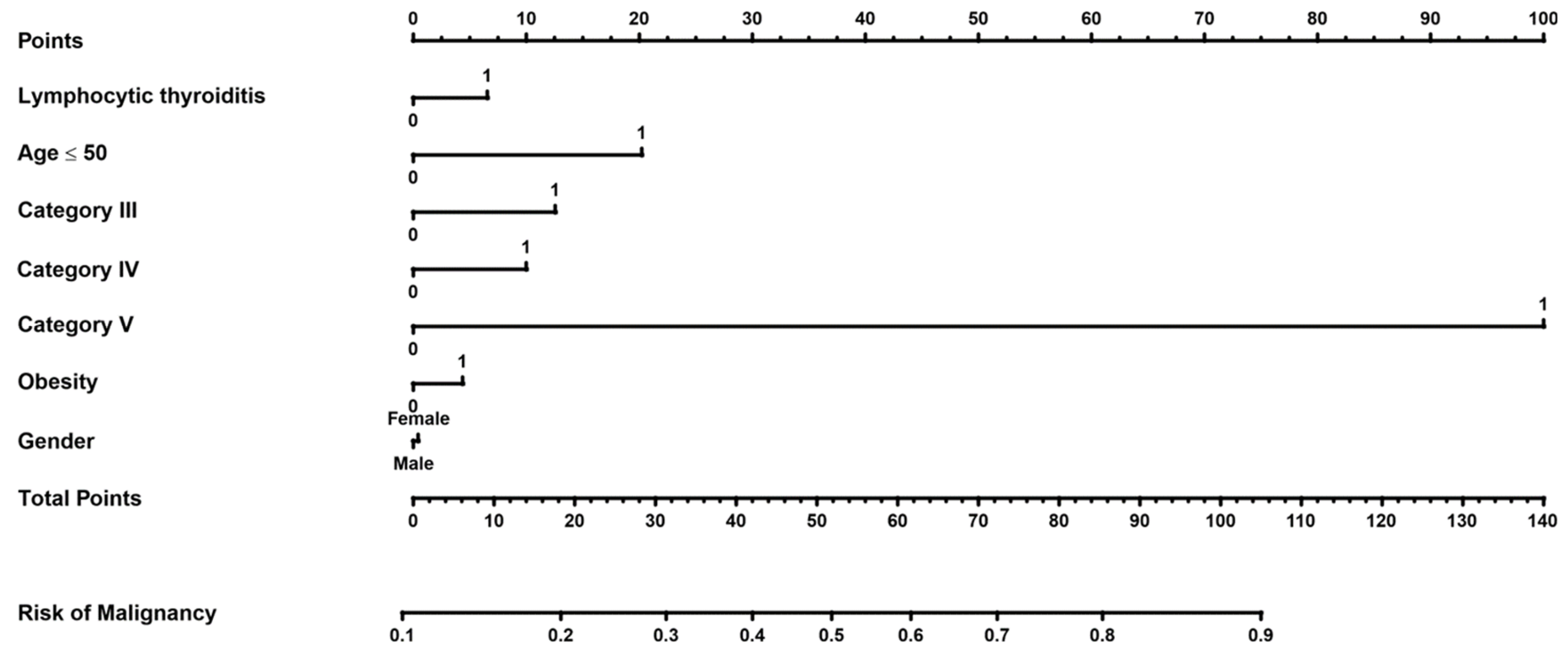
To visualise the results of the multivariate analysis, a nomogram was created to assess the risk of thyroid cancer when several factors were present at the same time. The scores for each parameter were marked on the scale of 'points'. The scores were then summed up and displayed on the "total score" scale, which was related to the cancer risk read on the "risk of cancer" scale (Figure 2).
To demonstrate how to interpret the nomogram please note that we marked a point on the scales for the parameters in the regression: gender, LT=1, age <50. etc. and then placed the relevant points on the "Points" scale. We added up the resulting points and marked them on the "Total points" scale, which is related to the risk of cancer on the "Risk of Cancer" scale. For example:
Age<50 (20 points), Sex=male (0 points), DC III=1 (13 points), DC IV=0 (0 points), DC V =0 (0 points), No obesity, i.e. Obesity=0 (0 points), No lymphocytic Hashimoto's, i.e. (0 points). We then added these points, i.e.: 20+0+13+0+0+0+0+0 = 33, which gave the Total points value = 33. When placed on the "Risk of Cancer" scale, the added value gave an approximate 30% risk of cancer.

4. Discussion

In the study group of 521 patients with thyroid nodules, malignant lesions were found in 197 cases (37.85%), similarly as was noted in other studies [14]. The vast majority of the said lesions were well-differentiated carcinomas (95.4%), which is also consistent with the literature [15]. US-guided FNAB is commonly used as a preoperative diagnostic tool because of its high accuracy [16,17,18]. However, false-positive results are common, where the cytology suggests malignancy, but the histopathology does not confirm it. A number of studies have evaluated the diagnostic reliability of biopsies on the basis of histopathological findings [4,19,20,21,22,23].
The risk of malignancy (ROM) is important in the assessment of thyroid nodules. The following risk values have been reported for each Bethesda DCs I-VI: 1-4%, 0-3%, 5-15%, 15-30%, 60-75%, 97-99% (6). Many investigators undertook an attempt to assess ROM with variable outcomes (24,25,26). In our study, among 521 patients, the presence of a malignant lesion (ROM) was found for different Bethesda DCs, respectively: I - 38.1%, II - 15.6%, III - 29.8%, IV - 18.6%, V - 91.0%, VI - 93.2% (Table 9).
It should be emphasised that non-diagnostic FNAB (DC I) can involve any category. In our study, there were false-negative category I cytology results in 8 cases where malignancy was confirmed by histopathology.
In 180 patients with DC II, 28 malignant lesions (15.5%) were found. Similar results have been reported in the literature [27]. In our study, the false-negative results for DC II may have been due to the fact that 11 cases were papillary microcarcinomas, which may not have been detected due to their small size. The most common reason for referral to surgery was tracheal compression due to multinodular goitre. In the case of the histopathological confirmation of medullary carcinoma despite the Bethesda cytology category II, the patients were referred to a surgery department because of elevated calcitonin levels.
For DC III, it is important to add the subcategories AUS, FLUS, FLUS/AUS [9,28] as AUS is associated with a higher risk of malignancy than the FLUS subcategory and refers mainly to smears with cellular features suggestive of papillary carcinoma. Several studies have assessed the risk of malignancy in the Bethesda subcategories III [29,30,31]. In our study, of the DC III patients, there were 25 thyroid carcinomas (29.7%) and benign lesions were detected in 59 cases (70.2%). In the cytological biopsy results for DC III in our study, we did not have a specific subcategory for AUS or FLUS type, which was undoubtedly a drawback of our research.
In our study, out of 70 cases with the DC IV, only 13 (18.5%) thyroid carcinomas were found, and benign lesions were present in 57 cases (81.5%). The incidence of malignant lesions in this category was estimated to be approximately 37-39% [32,33]. Based on the multivariate regression analysis, the present authors found no statistically significant effect of DCs III and IV on the risk of malignancy. Therefore, with this diagnosis, the risk of malignancy should be assessed together with clinical and ultrasound risk before deciding whether to refer the patient for surgery.
In our group of patients with DC V, a malignant lesion was found in 91.0 % of the subjects and other lesions accounted for 9%. On the basis of the multivariate regression analysis, this category was found to be statistically significantly associated with the risk of malignancy (p< 0.0001, OR- 62.34); the ROC curve showed as well that from this category onwards, the risk of malignancy increased with the specificity at the level 0.97 and sensitivity of 0.61. Thus, in the case of category V, surgical treatment is indicated.
It should also be noted that 7 of the 40 patients who did not undergo FNAB prior to surgery were also found to have a malignant tumour (PTC) on HP examination. The lack of FNAB was due to the fact that the indication for surgery was compression symptoms and thyroid dysfunction, mainly hyperthyroidism.
Based on our study, it can be concluded that several factors affected the diagnostic performance of thyroid biopsy. The sensitivity was affected by the data sets assigned to the numerator or denominator. The sensitivity increased when more Bethesda categories were included in the numerator. In our study, the sensitivities for categories 2,3,4,5,6 were; 0.96; 0.81; 0.68; 0.61; 0.29, respectively. A similar trend was found by Ha et al, who showed in a study of 4,822 thyroid nodules that sensitivity increased when more Bethesda categories were included in the numerator and when non-diagnostic results were excluded [34].
The PPVs for the criterion 5 (category VI, VI), criterion 4 (VI, V, IV) and criterion 3 (category VI, V, IV, III) were 0.92, 0.66 and 0.55, respectively. Based on the ROC curve, the criterion 5 (i.e. the Bethesda category V and VI tumours) was associated with the presence of thyroid cancer with a specificity of 0.97 and a sensitivity of 0.61. Criterion 5 represents the cut-off point associated with a higher risk of malignancy.
Our study also analysed whether demographics, age, gender and BMI had an effect on the risk of thyroid malignancy. We found that age statistically significantly differed (p<0.0001) between the patients with and without malignancy. The patients with a malignant neoplasm were on the average eight years younger (47.4 years) than those diagnosed with a benign lesion (55.3 years). However, there were no statistically significant differences in gender, BMI, or obesity. Velsen et al. found that the risk that a nodule was cancerous decreased with age [35]. In contrast, Raparia et al. showed that there were no statistically significant differences in age between the benign and malignant lesion groups [36].
The study also sought to determine whether lymphocytic thyroiditis (LT) was a risk factor for malignancy. Of our 61 patients with the background of confirmed lymphocytic thyroiditis, 42.6% were in the category V, 13.1% - category IV, 16.4% - category III and 21.9%in the category II, which was statistically significant. There are studies that confirm that differentiated thyroid cancer can coexist with lymphocytic thyroiditis [37], mainly papillary carcinoma [38].
To assess whether cumulative factors affected the risk of malignancy, the logistic regression was performed including 5 parameters, i.e. age, sex, BMI, the presence or absence of LT, and the Bethesda DCs III, IV, V. Based on the multivariate regression analysis, DC V and age ≤50 years were statistically significantly associated with the risk of malignancy (p < 0.0001 and p < 0.006, respectively).
It should be noted that a limitation of our study was the retrospective nature, the single institution design, the lack of EUTIRADS assessment in a significant number of patients prior to surgery and the lack of fine needle biopsy in a proportion of patients referred for surgery due to multinodular goitre and compression symptoms. The combined EUTIRADS and Bethesda assessment of the focal lesion would have significantly affected the decision to operate and the extent of surgery.
The large size of the study group was undoubtedly an advantage of our study.

5. Conclusions

Based on the study, it can be concluded that in DCs III and IV, consideration may be given to keeping the patient under close clinical, ultrasound and cytological surveillance without a premature referral for surgery. In the case of DC V, it is advisable to refer the patient for surgery. In our study, we found that patients under 50 years of age had a higher risk of malignancy than older patients.
In addition, if lymphocytic thyroiditis and a focal lesion coexist, a biopsy should be taken into consideration.
Further research and the development of more reliable diagnostic methods are needed to optimise the management of thyroid nodules and avoid unnecessary surgery.

Supplementary Materials

The following supporting information can be downloaded at: https://eurocrine.eu; last accessed at April 28, 2024.

Funding

This research was funded by Ministry of Science and Higher Education grant number WSUB/2024/02/00001 and the APC was funded by Ministry of Science and Higher Education in this section.

Institutional Review Board Statement

Research involving human subjects has been reviewed and approved by the Bioethics Committee of the Andrzej Frycz Modrzewski Krakow University (KBKA/59/O/2023).

Informed Consent Statement

Written Informed Consent was not required for participation in this study according to the national legislation and institutional requirements. Patient data has been anonymized.

Data Availability Statement

The original contributions presented in the study are included in the article. Further inquiries can be directed to the corresponding authors.

Acknowledgments

The authors wish to thank the staff of the Department of Endocrine Surgery for their kind care of the patients and their help in collecting patient data.

Conflicts of Interest

The authors declare no conflicts of interest.

References

  1. Siegel, R.L.; Miller, K.D.; Wagle, N.S.; Jemal, A. Cancer statistics, 2023. CA Cancer J Clin. 2023, 73, 17–48. [Google Scholar] [CrossRef] [PubMed]
  2. Li, M.; Maso, L.D.; Vaccarella, S. Global trends in thyroid cancer incidence and the impact of overdiagnosis. Lancet Diabetes Endo 2020, 8, 468–70. [Google Scholar] [CrossRef] [PubMed]
  3. Lewis, C.M.; Chang, K.P.; Pitman, M.; Faquin, W.C.; Randolph, G.W. Thyroid fine-needle aspiration biopsy: variability in reporting. Thyroid. 2009, 19, 717–23. [Google Scholar] [CrossRef] [PubMed]
  4. Gupta, M.; Gupta, S.; Gupta, V.B. Correlation of fine needle aspiration cytology with histopathology in the diagnosis of solitary thyroid nodule. J Thyroid Res. 2010, 2010, 379051. [Google Scholar] [CrossRef]
  5. Russ, G.; Bonnema, S.J.; Erdogan, M.F.; Durante, C.; Ngu, R.; Leenhardt, L. European Thyroid Association Guidelines for Ultrasound Malignancy Risk Stratification of Thyroid Nodules in Adults: The EU-TIRADS. Eur Thyroid J. 2017, 6, 225–237. [Google Scholar] [CrossRef]
  6. Cibas, E.S.; Ali, S.Z. The 2017 Bethesda System for Reporting Thyroid Cytopathology. J Am Soc Cytopathol. 2017, 6, 217–222. [Google Scholar] [CrossRef]
  7. Pusztaszeri, M.; Rossi, E.D.; Auger, M.; Baloch, Z.; Bishop, J.; Bongiovanni, M.; Chandra, A.; Cochand-Priollet, B.; Fadda, G.; Hirokawa, M.; Hong, S.; Kakudo, K.; Krane, J.F.; Nayar, R.; Parangi, S.; Schmitt, F.; Faquin, W.C. The Bethesda System for Reporting Thyroid Cytopathology: Proposed Modifications and Updates for the Second Edition from an International Panel. Acta Cytol. 2016, 60, 399–405. [Google Scholar] [CrossRef]
  8. Baloch ZW, Cooper DS, Gharib H, Alexander EK (2018) Overview of diagnostic terminology and reporting. In: Syed Z,Ali ESC (eds) The Bethesda system for reporting cytopathology: definitions, criteria and explanatory notes. Springer, Cham, pp 1–6.
  9. Krajewska, J.; Chmielik, E.; Dedecjus, M.; Jarząb, B.; Hubalewska-Dydejczyk, A.; Karbownik-Lewińska, M.; Kos-Kudła, B.; Lewiński, A.; Ruchała, M. Diagnosis and treatment of thyroid cancer in adult patients - Recommendations of Polish Scientific Societies and the National Oncological Strategy. Update of the 2022. Endokrynol Pol. 2022, 73, 799–802. [Google Scholar] [CrossRef]
  10. Kobaly, K.; Kim, C.S.; Mandel, S.J. Contemporary Management of Thyroid Nodules. Annu Rev Med. 2022, 73, 517–528. [Google Scholar] [CrossRef]
  11. EUROCRINE: https://eurocrine.eu; last accessed at April 28, 2024.
  12. Jung, C.K.; Bychkov, A.; Kakudo, K. Update from the 2022 World Health Organization Classification of Thyroid Tumors: A Standardized Diagnostic Approach. Endocrinol Metab (Seoul). 2022, 37, 703–718. [Google Scholar] [CrossRef]
  13. R Core Team (2021). R: A language and environment for statistical computing. R Foundation for Statistical Computing, Vienna, Austria. URL https://www.R-project.org/.
  14. Osseis, M.; Jammal, G.; Kazan, D.; Noun, R. Comparison between Fine Needle Aspiration Cytology with Histopathology in the Diagnosis of Thyroid Nodules. J Pers Med. 2023, 13, 1197. [Google Scholar] [CrossRef] [PubMed]
  15. Sherman, S.I. Thyroid carcinoma. Lancet. 2003, 361, 501–11. [Google Scholar] [CrossRef] [PubMed]
  16. McCoy KL, Jabbour N, Ogilvie JB, Ohori NP, Carty SE, Yim JH, et al. The incidence of cancer and rate off alse-negative cytology in thyroid nodules greater than or equal to 4 cm in size. Surgery 2007, 142, 837–844. [Google Scholar] [CrossRef] [PubMed]
  17. Haugen BR, Alexander EK, Bible KC, Doherty GM, Mandel SJ, Nikiforov YE, et al. American Thyroid Association management guidelines for adult patients with thyroid nodules and differentiated thyroid cancer: the American Thyroid Association guidelines task force on thyroid nodules and differentiated thyroid cancer. Thyroid 2016, 26, 1–133. [Google Scholar] [CrossRef] [PubMed]
  18. Hegedüs, L. The thyroid nodule. New Engl J Med 2004, 351, 1764–71. [Google Scholar] [CrossRef]
  19. Rai, K.; Park, J.; Gokhale, S.; Irshaidat, F.; Singh, G. Diagnostic Accuracy of the Bethesda System for Reporting Thyroid Cytopathology (TBSRTC): An Institution Experience. Int J Endocrinol. 2023, 2023, 9615294. [Google Scholar] [CrossRef]
  20. Garg, S.; Desai, N.J.; Mehta, D.; Vaishnav, M. To Establish Bethesda System for Diagnosis of Thyroid Nodules on the Basis of Fnac with Histopathological Correlation. J Clin Diagn Res. 2015, 9, EC17–EC21. [Google Scholar] [CrossRef]
  21. Nandedkar, S.S.; Dixit, M.; Malukani, K.; Varma, A.V.; Gambhir, S. Evaluation of Thyroid Lesions by Fine-needle Aspiration Cytology According to Bethesda System and its Histopathological Correlation. Int J Appl Basic Med Res. 2018, 8, 76–82. [Google Scholar] [CrossRef]
  22. Patel, K.A.; Anandani, G.; Sharma, B.S.; Parmar, R.A. Study of Fine Needle Aspiration Cytology (FNAC) of Thyroid Gland According to the Bethesda System. Cureus. 2023, 15, e37371. [Google Scholar] [CrossRef]
  23. Mondal, S.K.; Sinha, S.; Basak, B.; Roy, D.N.; Sinha, S.K. The Bethesda system for reporting thyroid fine needle aspirates: A cytologic study with histologic follow-up. J Cytol. 2013, 30, 94–9. [Google Scholar] [CrossRef]
  24. Inabnet WB 3rd, Palazzo F, Sosa JA, Kriger J, Aspinall S, Barczynski M, Doherty G, Iacobone M, Nordenstrom E, Scott-Coombes D, Wallin G, Williams L, Bray R, Bergenfelz A. Correlating the Bethesda System for Reporting Thyroid Cytopathology with Histology and Extent of Surgery: A Review of 21,746 Patients from Four Endocrine Surgery Registries Across Two Continents. World J Surg. 2020, 44, 426–435. [Google Scholar] [CrossRef] [PubMed]
  25. Anand, B.; Ramdas, A.; Ambroise, M.M.; Kumar, N.P. The Bethesda System for Reporting Thyroid Cytopathology: A Cytohistological Study. J Thyroid Res. 2020, 2020, 8095378. [Google Scholar] [CrossRef] [PubMed]
  26. Zarif, H.A.; Ghandurah, S.E.; Al-Garni, M.A.; Binmahfooz, S.K.; Alsaywid, B.S.; Satti, M.B. Thyroid Nodules Cytopathology Applying the Bethesda System with Histopathological Correlation. Saudi J Med Med Sci. 2018, 6, 143–148. [Google Scholar] [CrossRef] [PubMed]
  27. Alshathry, A.H.; Almeshari, N.Z.; Alarifi, A.S.; Aleidy, A.M.; Aldhahri, S. The Prevalence of Thyroid Papillary Microcarcinoma in Patients With Benign Thyroid Fine Needle Aspiration. Cureus. 2020, 12, e11820. [Google Scholar] [CrossRef]
  28. Gan TRX, Nga ME, Lum JHY, Wong WM, Tan WB, Parameswaran R, et al. Thyroid cytology—nuclear versus architectural atypia within the “Atypia of undetermined significance/follicular lesion of undetermined significance” Bethesda category have significantly different rates of Malignancy. Cancer Cytopathology 2017, 125, 245–56. [Google Scholar] [CrossRef]
  29. Mosca, L.; Silva, L.F.F.D.; Carneiro, P.C.; Chacon, D.A.; Araujo-Neto, V.J.F.; Araujo-Filho, V.J.F.; Cernea, C.R. Malignancy rates for Bethesda III subcategories in thyroid fine needle aspiration biopsy (FNAB). Clinics (Sao Paulo). 2018, 73, e370. [Google Scholar] [CrossRef]
  30. Kim, S.J.; Roh, J.; Baek, J.H.; Hong, S.J.; Shong, Y.K.; Kim, W.B.; Song, D.E. Risk of malignancy according to sub-classification of the atypia of undetermined significance or follicular lesion of undetermined significance (AUS/FLUS) category in the Bethesda system for reporting thyroid cytopathology. Cytopathology. 2017, 28, 65–73. [Google Scholar] [CrossRef]
  31. Erdogan-Durmus, S.; Balta, H.; Demirtas, R.; Kurt, A. Malignancy Rates of Atypia of Undetermined Significance/Follicular Lesion of Undetermined Significance (AUS/FLUS) Cases: A Tertiary Center Study. Acta Endocrinol (Buchar). 2021, 17, 77–82. [Google Scholar] [CrossRef]
  32. Yang, W.; Fananapazir, G.; LaRoy, J.; Wilson, M.; Campbell, M.J. Can the American Thyroid Association, K-Tirads, and Acr-Tirads Ultrasound Classification Systems Be Used to Predict Malignancy in Bethesda Category IV Nodules? Endocr Pract. 2020, 26, 945–952. [Google Scholar] [CrossRef]
  33. Marina, M.; Zatelli, M.C.; Goldoni, M.; Del Rio, P.; Corcione, L.; Martorana, D.; Percesepe, A.; Bonatti, F.; Mozzoni, P.; Crociara, A.; Ceresini, G. Combination of ultrasound and molecular testing in malignancy risk estimate of Bethesda category IV thyroid nodules: results from a single-institution prospective study. J Endocrinol Invest. 2021, 44, 2635–2643. [Google Scholar] [CrossRef]
  34. Ha, S.M.; Baek, J.H.; Na, D.G.; Jung, C.K.; Suh, C.H.; Shong, Y.K.; Sung, T.Y.; Song, D.E.; Lee, J.H. Assessing the diagnostic performance of thyroid biopsy with recommendations for appropriate interpretation. Ultrasonography. 2021, 40, 228–236. [Google Scholar] [CrossRef] [PubMed]
  35. Velsen, E. , Peeters R., Stegenga M, van Kemenade F., van Ginhoven T, Verburg F., Visser E, The influence of age on disease outcome in 2015 ATA high-risk differentiated thyroid cancer patients. European Journal of Endocrinology 2021, 185, 421–429. [Google Scholar] [CrossRef] [PubMed]
  36. Raparia, K.; Min, S.K.; Mody, D.R.; Anton, R.; Amrikachi, M. Clinical outcomes for “suspicious” category in thyroid fine-needle aspiration biopsy: patient's sex and nodule size are possible predictors of Malignancy. Arch Pathol Lab Med 2009, 133, 787–90. [Google Scholar] [CrossRef]
  37. Konturek, A.; Barczyński, M.; Wierzchowski, W.; Stopa, M.; Nowak, W. Coexistence of papillary thyroid cancer with Hashimoto thyroiditis. Langenbecks Arch Surg. 2013, 398, 389–94. [Google Scholar] [CrossRef] [PubMed]
  38. Zosin, I.; Balaş, M. Clinical, ultrasonographical and histopathological aspects in Hashimoto's thyroiditis associated with malignant and benign thyroid nodules. Endokrynol Pol. 2013, 64, 255–62. [Google Scholar] [CrossRef] [PubMed]
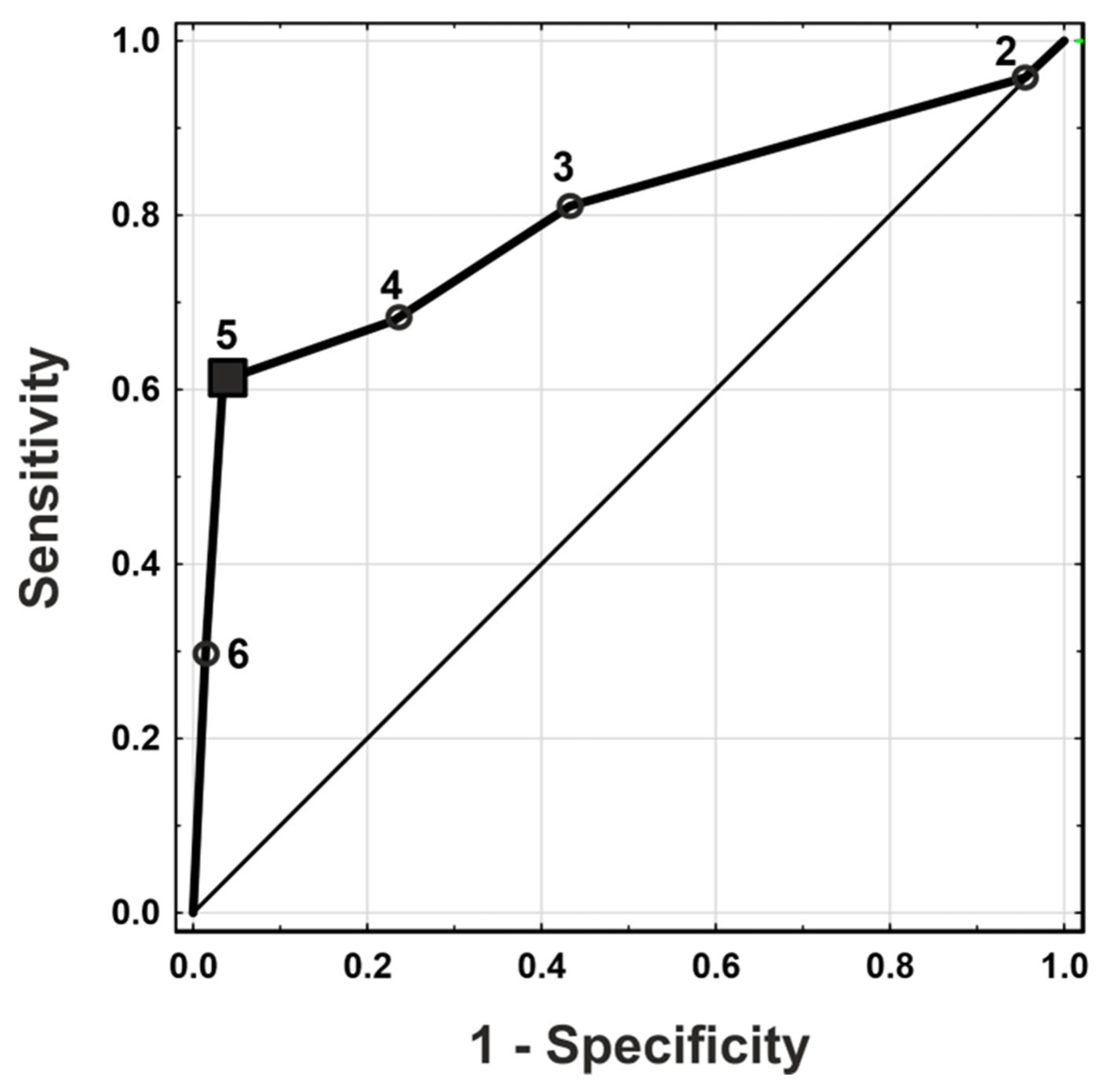
Figure 1. ROC curve of probability of malignancy for criteria 2-6.
Preprints 138658 g001
Figure 2. A nomogram to predict the risk of malignancy in the group with categories Bethesda II, III, IV, V.
Preprints 138658 g002
Table 1. Demographic and pathologic features of 521 patients who underwent surgery due to thyroid nodules.
Total
(N=521)
benign
(N=324)
cancer
(N=197)
p-value
Sex 0.94
Female 425 (81.6%) 264 (81.5%) 161 (81.7%)
Male 96 (18.4%) 60 (18.5%) 36 (18.3%)
BMI 0.52
Mean (SD) 27.3 (4.87) 27.4 (4.76) 27.1 (5.08)
Median [Q1-Q3] 26.6 [23.5-30.5] 26.7 [23.8-30.5] 26.4 [23.2-30.8]
Missing 98 (18.8%) 57 (17.6%) 41 (20.8%)
Obesity (BMI ≥ 30) 0.76
no 302 (58.0%) 192 (59.3%) 110 (55.8%)
yes 121 (23.2%) 75 (23.1%) 46 (23.4%)
Missing 98 (18.8%) 57 (17.6%) 41 (20.8%)
Age < 0.0001
Mean (SD) 52.3 (15.2) 55.3 (14.7) 47.4 (14.9)
Median [Q1-Q3] 53.0 [40.0-65.0] 57.0 [43.8-67.0] 45.0 [36.0-59.0]
Cytology < 0.0001
I 21 (4.0%) 13 (4.0%) 8 (4.1%)
II 180 (34.5%) 152 (46.9%) 28 (14.2%)
III 84 (16.1%) 59 (18.2%) 25 (12.7%)
IV 70 (13.4%) 57 (17.6%) 13 (6.6%)
V 67 (12.9%) 6 (1.9%) 61 (31.0%)
VI 59 (11.3%) 4 (1.2%) 55 (27.9%)
Not performed 40 (7.7%) 33 (10.2%) 7 (3.6%)
Lymphocytic thyroiditis < 0.0001
no 442 (84.8%) 292 (90.1%) 150 (76.1%)
yes 79 (15.2%) 32 (9.9%) 47 (23.9%)
Malignancy
PTC 172 (33.0%) 172 (87.3%)
FTC 16 (3.1%) 16 (8.1%)
MTC 5 (1.0%) 5 (2.5%)
ATC 1 (0.2%) 1 (0.5%)
OTC 2 (0.4%) 2 (1.0%)
Lymphoma 1 (0.2%) 1 (0.5%)
Thyroid operation
Total thyroidectomy 480 (92.1%) 295 (91%) 185 (94%)
Unilateral lobectomy 30 (5.7%) 22 (6.8%) 8 (4.1%)
Other operation on thyroid gland 11 (2.1%) 7 (2.2%) 4 (2.0%)
Lymph nodes
Bilateral central lymph node dissection 191 (36.7%) 89 (27.5%) 102 (51.8%)
Central lymph node dissection AND one-sided lat. Lymph node dissection 18 (3.5%) 2 (0.6%) 16 (8.1%)
Unilateral central lymph node dissection 80 (15.4%) 40 (12.3%) 40 (20.3%)
One-sided lateral lymph node dissection 7 (1.3%) 2 (0.6%) 5 (2.5%)
Bilateral lateral lymph node dissection 2 (0.4%) 0 (0%) 2 (1%)
None 223 (42.8%) 191 (59.0%) 32 (16.2%)
Legend: Cytology: I- Non-diagnostic or unsatisfactory; II – Benign; III - Atypia of undetermined significance (AUS) or follicular lesion of undetermined significance(FLUS); IV-Follicular neoplasm or suspicious follicular neoplasm; V - Suspicious for malignancy; VI – Malignant; PTC-papillary thyroid carcinoma; FTC- follicular thyroid carcinoma; MTC- medullary thyroid carcinoma; OTC – oncocytic thyroid carcinoma; ATC -anaplastic thyroid carcinoma.
Table 2. Comparison of the results of cytology with the results of histopathology for DCs I-VI.
Cytology Total, n (%) Histopathology
Benign, n (%) Cancer, n (%)
I 21 (4.0%) FA 2 (15.4%) FTC 2 (25%)
GD 1 (7.7%) PTC 6 (75%)
NG 9 (69.2%)
Other diagnosis 1 (7.7%)
II 180 (34.5%) FA 15 (9.9%) FTC 4 (14.3%)
FT-UMP 1 (0.7%) MTC 3 (10.7%)
GD 1 (0.7%) PTC 21 (75%)
OA 1 (0.7%)
LT 1 (0.7%)
NG 130 (85.5%)
NIFTP 2 (1.3%)
III 84 (16.1%) FA 14 (23.7%) FTC 7 (28.0%)
FT-UMP 2 (3.4%) OTC 1 (4.0%)
OA 1 (1.7%) PTC 17 (68.0%)
LT 3 (5.1%)
NG 35 (59.3%)
NIFTP 2 (3.4%)
WDT-UMP 2 (3.4%)
IV 70 (13.4%) FA 15 (26.3%) OTC 1 (7.7%)
FT-UMP 5 (8.8%) PTC 12 (92.3%)
OA 13 (22.8%)
LT 2 (3.5%)
NG 18 (31.6%)
NIFTP 2 (3.5%)
WDT-UMP 2 (3.5%)
V 67 (12.9%) FA 1 (16.7%) ATC 1 (1.6%)
NG 3 (50.0%) FTC 2 (3.3%)
NIFTP 1 (16.7%) PTC 58 (95.1%)
Other diagnosis 1 (16.7%)
VI 59 (11.3%) NG 2 (50.0%) FTC 1 (1.8%)
Other diagnosis 2 (50.0%) Lymphoma 1 (1.8%)
MTC 2 (3.6%)
PTC 51 (92.7%)
Not
performed
40 (7.7%) FA 4 (12.1%) PTC 7 (100%)
GD 3 (9.1%)
OA 2 (6.1%)
LT 1 (3.0%)
NG 19 (57.6%)
NIFTP 3 (9.1%)
Legend: FA, Follicular thyroid adenoma; GD, Graves´ disease; NG, Nodular goitre; FTUMP, Follicular tumour with uncertain malignant potential; OA, Oncocytic adenoma of the thyroid; LT, Lymphocytic thyroiditis Hashimoto; WDT-UMP, Well differentiated tumour of uncertain malignant potential; NIFTP, Non-invasive follicular thyroid neoplasm with papillary-like nuclear features; FT-UMP, Follicular tumour with uncertain malignant potential; PTC - Papillary thyroid cancer; FTC- Follicular thyroid cancer; MTC -Medullary thyroid cancer; ATC, Anaplastic thyroid cancer; OTC, oncocytic thyroid carcinoma.
Table 3. Risk of malignancy (ROM).
Cytology No. of malignant cases (all) ROM (%)
I 8 (21) 38.1
II 28 (180) 15.6
III 25 (84) 29.8
IV 13 (70) 18.6
V 61 (67) 91
VI 55 (59) 93.2
In order to assess the relevance of FNAB, the authors determined five groups of subjects (criteria 2-6) for which the following diagnostic values were analysed: TP, TN, FP, FN, PPV, NPV. We calculated the biopsy sensitivity according to the inclusion of different Bethesda categories in the numerator (Table 4).
Table 4. Changes in the sensitivity for malignancy according to the criteria for a positive diagnosis (with exclusion patients without cytology performed).
Criterion TP FP FN TN Sensitivity Specificity PPV NPV
6 55 4 135 287 0.29 (0.23, 0.36) 0.99 (0.97, 1.00) 0.93 (0.84, 0.98) 0.68 (0.63, 0.72)
5 116 10 74 281 0.61 (0.54, 0.68) 0.97 (0.94, 0.98) 0.92 (0.86, 0.96) 0.79 (0.75, 0.83)
4 129 67 61 224 0.68 (0.61, 0.74) 0.77 (0.72, 0.82) 0.66 (0.59, 0.72) 0.79 (0.73, 0.83)
3 154 126 36 165 0.81 (0.75, 0.86) 0.57 (0.51, 0.62) 0.55 (0.49, 0.61) 0.82 (0.76, 0.87)
2 182 278 8 13 0.96 (0.92, 0.98) 0.04 (0.02, 0.08) 0.40 (0.35, 0.44) 0.62 (0.38, 0.82)
Legend: The diagnostic categories (DC) assigned to the numerator and denominator for each criterion were as follows: Criterion 6: DCs VI/ V, IV, III, II, I; Criterion 5: DCs VI, V/ IV, III, II, I; Criterion 4: DCs VI, V, IV/III, II, I; Criterion 3: DCs VI, V, IV, III/ II, I; Criterion 2: DCs VI, V, IV, III, II/I. TP, true positive; TN, true negative; FP, false positive; FN, false negative; PPV, positive predictive value; NPV, negative predictive value.
Table 5. Characteristics of patients with DCs II, III, IV, V included in multivariate logistic regression.
Total
(N=401)
II
(N=180)
III
(N=84)
IV
(N=70)
V
(N=67)
p-value
Sex 0.39
Female 326 (81.3%) 140 (77.8%) 69 (82.1%) 60 (85.7%) 57 (85.1%)
Male 75 (18.7%) 40 (22.2%) 15 (17.9%) 10 (14.3%) 10 (14.9%)
Age 0.00001
Mean (SD) 53.6 (14.7) 55.2 (14.4) 54.6 (12.0) 55.8 (16.3) 45.8 (14.3)
Median [Q1-Q3] 54.0 [42.0-66.0] 56.0 [44.0-67.0] 57.0 [44.0-65.3] 58.0 [42.0-68.0] 42.0 [35.5-56.0]
Histological main diagnosis < 0.0001
Benign 274 (68.3%) 152 (84.4%) 59 (70.2%) 57 (81.4%) 6 (9.0%)
Cancer 127 (31.7%) 28 (15.6%) 25 (29.8%) 13 (18.6%) 61 (91.0%)
Lymphocytic thyroiditis < 0.0001
no 340 (84.8%) 163 (90.6%) 74 (88.1%) 62 (88.6%) 41 (61.2%)
yes 61 (15.2%) 17 (9.4%) 10 (11.9%) 8 (11.4%) 26 (38.8%)
Table 6. Comparison of the presence of malignant lesions among patients with DCs II, III, IV, V.
Bethesda categories Malignancy
n(%)/ n(%) p value
III vs II 25 (29.8%)/ 28 (15.6%) 0.019
III vs IV 25 (29.8%)/ 13 (18.6%) 0.161
III vs V 25 (29.8%)/ 61 (91.0%) <0.0001
II vs IV 28 (15.6%)/ 13 (18.6%) 0.572
II vs V 28 (15.6%)/ 61 (91.0%) <0.0001
IV vs V 13 (18.6%)/ 61 (91.0%) <0.0001
Table 7. Frequency of Bethesda categories II, III, IV, V among patients with the background of confirmed lymphocytic thyroiditis.
Bethesda categories Lymphocytic thyroiditis
n(% )/ n(%) p value
III vs II 10 (11.9%)/ 17 (9.4%) 0.98
III vs IV 10 (11.9%)/ 8 (11.4%) 0.99
III vs V 10 (11.9%)/ 26 (38.8%) 0.0008
II vs IV 17 (9.4%)/ 8 (11.4%) 0.98
II vs V 17 (9.4%)/ 26 (38.8%) < 0.0001
IV vs V 8 (11.4%)/ 26 (38.8%) 0.0009
Table 8. Multivariate logistic regression analysis for Bethesda categories II, III, IV, V.
Variable OR per Univariate Multivariate*
OR (95% CI) p value OR (95% CI) p value
Sex Female/male 0.87 (0.51-1.51) 0.63 1.02 (0.47-2.18) 0.96
Lymphocytic thyroiditis yes/no 2.85 (1.63-4.96) 0.0002 1.31 (0.55-3.11) 0.54
Bethesda III yes/no 0.89 (0.53-1.51) 0.67 1.68 (0.81-3.47) 0.16
Bethesda IV yes/no 0.43 (0.23-0.83) 0.011 1.51 (0.69-3.32) 0.30
Bethesda V Bethesda V yes/no 41.28 (17.11-99.61) < 0.0001 62.34 (20.16-192.8) < 0.0001
Bethesda V
Age<=50 yes/no 2.67 (1.74-4.12) < 0.0001 2.31 (1.27-4.19) 0.006
Obesity (BMI>=30) yes/no 0.90 (0.53-1.51) 0.68 1.2 (0.62-2.31) 0.59
Table 9. Comparison of the risk of a neoplasm detected in our study with results of other studies [6,8,24,25,26].
Bethesda
category
TBSRTC [6]
first
edition
TBSRTC [8]
second
edition
ROM
of our study
n=521
ROM
Inabnet et al. [24]
n= 1,746
ROM
Anand et al. [25]
n=646
ROM
Zarif at al. [26]
n=373
I – nondiagnostic
II – benign
III – AUS/FLUS
IV - FN/SFN
V – SFM
VI -malignant
1-4%
0-3%
5-15%
15-30%
60-75%
97-99%
5-10%
0-3%
10-30%
25-40%
50-75%
97-99%
38.8%
15.6%
29.8%
18.6%
91.0%
93.2%
19.2%
12.7%
31.9%
31.4%
77.8%
96.0%

8.5%
66.7%
63.6%
100%
100%
34.6%
15.6%
50%
52%
95.7%
100%
Disclaimer/Publisher’s Note: The statements, opinions and data contained in all publications are solely those of the individual author(s) and contributor(s) and not of MDPI and/or the editor(s). MDPI and/or the editor(s) disclaim responsibility for any injury to people or property resulting from any ideas, methods, instructions or products referred to in the content.
Copyright: This open access article is published under a Creative Commons CC BY 4.0 license, which permit the free download, distribution, and reuse, provided that the author and preprint are cited in any reuse.
Alerts
Prerpints.org logo

Preprints.org is a free preprint server supported by MDPI in Basel, Switzerland.

Subscribe

© 2025 MDPI (Basel, Switzerland) unless otherwise stated