Submitted:
07 November 2024
Posted:
08 November 2024
You are already at the latest version
Abstract
Catamenial pneumothorax (CP) is an uncommon condition, usually associated with thoracic endometriosis syndrome (TES). TES is characterized by the presence of endometriotic lesions in pleura and lung parenchyma and presents with various clinical signs and symptoms, including catamenial pneumothorax. Their diagnosis is often delayed. Pulmonary endometric lesions, however, often detected in patients with hemothorax and hemoptysis, may be absent in a pro-portion of cases of pneumothorax. The typical presentation of CP includes signs and symptoms of pneumothorax, which occur along with menstruation, most commonly around 24 hours before and 48–72 hours after its onset. However, they may not occur during every menstrual cycle. Suggestive CP lesions on conventional radiography (RTG) include pneumoperitoneum accompanying right-sided pneumothorax, lung opacities, pleural effusion, and nodular infiltrates. Chest and abdomen computed tomography (CT), particularly contrast-enhanced, may additionally show pneumoperitoneum and diaphragmatic lesions. The management of CP includes supportive treatment of acute symptoms and causal treatment to prevent recurrent disease. The article pre-sents the pathophysiology of CP, an overview of diagnostic methods and current therapeutic approaches.
Keywords:
pneumothorax
; catamenial pneumothorax
; endometriosis
1. Introduction
Catamenial pneumothorax (CP) is an uncommon condition, usually associated with thoracic endometriosis syndrome (TES) [1]. TES is characterized by the presence of endometriotic lesions in pleura and lung parenchyma and presents with various clinical signs and symptoms, including catamenial pneumothorax (30-73%), catamenial hemothorax (14%), catamenial hemoptysis (7%), and lung nodules (6%) [2]. Although endometriosis is known to affect around 3-10% of women of childbearing age and approximately 2-5% of women after menopause, TES and CP remain a rare condition. Their diagnosis is often delayed [3]. Pulmonary endometric lesions, however, often detected in patients with hemothorax and hemoptysis, may be absent in a proportion of cases of pneumothorax. This phenomenon may indicate a different pathophysiological mechanism of CP. Usually, surgery is the first intervention; however, it has limited efficacy, and most patients will require additional treatment.
This article reviews the diagnostic and therapeutic options available in CP.
2. Pathogenesis
Maurer et al.'s first disease report comes from 1958 [4]. The name "catamenial" derives from the Greek word "katamenios" and translates into "monthly occurrence" [5]. It is widely accepted that endometriotic lesions in pleura and lung parenchyma are responsible for CP in the proportion of patients. However, the exact mechanism of their occurrence is not well recognized. Further, for the cases in which endometriotic lesions cannot be found, other pathophysiology has been proposed to explain CP (Table 1).
2.1. Theories for the Development of Endometrial Foci
Various theories attempted to explain the mechanism of extra-pelvic endometrial tissue development. According to the oldest “retrograde menstruation”, dating back to 1927, menstrual blood flows from the uterus through fallopian tubes into the pelvis and further to the abdomen. Endometrial cells' repetitive proliferation and necrosis around the diaphragm area leads to diaphragmatic fenestration formation [6]. As a result, endometrial cells pass into the chest and visceral pleura, causing alveolar injury and pneumothorax [1]. The second, metastatic theory, suggests that endometrial cells spread from the uterus via lymph or blood vessels — their proliferation and necrosis in the pleural space results in pneumothorax [7]. In turn, the vascular embolization hypothesis postulates that fragments of endometrial tissue travel in the venous system from the uterus into the right heart and eventually deposit in the lung parenchyma and pleura [8]. Another assumption states that the microvascular endothelium of ectopic endometrial tissue derives from endothelial progenitor cells, which develop de novo in vasculogenesis, as opposed to traditional angiogenesis [9].
Several studies demonstrated that autoimmune diseases are more common in women with endometriosis, which suggests that systemic immune alternations may contribute to the disease pathogenesis [10]. Patients with endometriosis present with elevated concentrations of activated macrophages and decreased cytotoxic T-cell and natural killer cell activity [11,12]. A possible explanation for the development of thoracic foci could be the regurgitation of endometrial debris into the lung parenchyma (retrograde menstruation), followed by a defective "immune surveillance" reaction [11]. Alternatively, it may result from defective cytotoxic natural killer cell activity and the inability to eliminate ectopic endometrial cells [13]. Additionally, cytokines and growth factors secreted by ectopic endometrial cells may be responsible for the activation of cell proliferation and angiogenesis, consequently producing thoracic lesions [11].
2.2. Theories for the Development of CP Without Endometrial Foci
In some cases, the lack of intrathoracic endometriotic lesions may be caused by air passing from the uterus into fallopian tubes, which results in increased permeability of menstruation blood to the peritoneal cavity and, via diaphragmatic fenestrations into the pleural space. It is also believed that high F2 prostaglandin levels during menstruation induce vasoconstriction and bronchospasm, resulting in alveolar rupture and pneumothorax [14].
3. Clinical Presentation
The typical presentation of CP includes signs and symptoms of pneumothorax, which occur along with menstruation, most commonly around 24 hours before and 48–72 hours after its onset. However, they may not occur during every menstrual cycle [15,16]. The disease typically affects women of reproductive age, less frequently young girls and post-climacteric women [17]. CP most commonly develops on the right side (85-95%) due to the clockwise circulation of peritoneal fluid [1,15].
The clinical manifestations of CP may include chest pain, shortness of breath, increased heart rate, rapid breathing, cough, fatigue, and pleural effusion. A history of infertility, chronic pelvic pain, and dysmenorrhea or dyspareunia may be suggestive of TES. In some cases, endometriosis-related diaphragmatic hernia may be present [18,19]. Other symptoms can vary depending on the foci location. Those located more centrally usually cause hemoptysis [20]. Occasional symptoms include also hemothorax and chest or scapular pain [17,21]. There was also a case of pulmonary endometriosis mimicking an acute abdomen [22,23]. Symptoms’ intensity exacerbates around the time of menstruation and may vary from very mild to severe. Significantly, pneumothorax may be associated with decreased lung function and should be treated promptly. The development of tension pneumothorax could potentially be fatal; therefore, in some cases, a chest-tube insertion may be necessary to allow re-expansion of the lung tissue.
A comprehensive clinical examination and medical interview, including family gynecological history, are necessary to facilitate proper diagnosis [1,2]. The reoccurrence of characteristic symptoms during menstruation in reproductive-age women should hint at the possibility of CP. An elevated level of CA-125 may accompany the disease. Although this biomarker is not specific, it can support early diagnosis of CP related to thoracic endometriosis [24]. Some studies suggest that elevated CA-125 could help implement prompt therapy to prevent reoccurrence of CP [25]. However, this approach warrants prospective evaluation in a large-scale study. Some putative biomarkers related to nerve fibre growth or cell cycle control are promising candidates for allowing a non-invasive CP diagnosis [26].
4. Diagnostic Methods
Suggestive CP lesions on conventional radiography (RTG) include pneumoperitoneum accompanying right-sided pneumothorax, lung opacities, pleural effusion, and nodular infiltrates [27,28]. In some cases, extensive diaphragmatic features and multiple “air-filled bubbles” – small diaphragmatic perforations may also be present [29]. Chest and abdomen computed tomography (CT), particularly contrast-enhanced, may additionally show pneumoperitoneum and diaphragmatic lesions [30,31]. The CT should preferably be performed during menstruation [32]. High-resolution CT is preferred, as it best determines the anatomical localization of ectopic plaque before thoracic surgery. It is also important to carefully screen the posterosuperior diaphragm, a relatively common disease location [32].
Although commonly used, both RTG and CT have poorer specificity in CP than magnetic resonance imaging (MRI) in detecting endometriosis-related hemorrhage. Like RTG and CT, the optimal time to perform MRI is during menstruation. MRI has lower spatial resolution than CT but provides better contrast resolution and more precise characterization of hemorrhagic lesions [33]. CP lesions have various features, including nodules, opacities, bullous formation, and ground glass infiltrates [34]. A study by Rousset et al. showed that MRI allows for identifying diaphragmatic nodules with up to 83% sensitivity [34]. Fat-suppressed T1-weighted sequences are optimal for detailed examination. Most lesions appear right-sided and are hyperintense.
Bronchoscopy is not routinely recommended since most lesions occur in the peripheral lung parenchyma [35,36]. However, bronchoscopy may be helpful in selected cases if performed during the first two days of menstruation [37]. On the contrary, video-assisted thoracoscopy (VAT) exploration of the thoracic cavity around the menstruation time allows optimal visualization of ectopic lesions [6,38]. Extensive lung, visceral, parietal pleurae, and pericardium examinations seem compulsory. Any bullae, air leaks, or blebs must be identified. Care should be taken when inspecting the diaphragm, especially the posterosuperior surface, as thoracic lesions are commonly found in this area [33]. The classic thoracotomy procedure is generally considered not sufficient for examination of the whole chest cavity and the diaphragm and should be restricted for patients with disease relapse. [17]. A video-assisted mini-thoracotomy is recommended instead of VATs when multiple diaphragmatic lesions are present within the diaphragm or in individual cases where endoscopy is unsafe [17]. Otherwise, VATs is considered superior to classical thoracotomy though due to better magnification, allowing the detection of the smallest endometrial foci. Histopathological tissue examination is essential to confirm recurrent disease.
There are no specific pathological diagnostic criteria for CP. The final confirmation is established by histopathological examination of the tissue material acquired via VATs or open thoracotomy. The typical features of CP include the presence of endometrial stroma, glans, and hemosiderin-filled macrophages [15]. Immunostaining for estrogen and progesterone receptors was postulated to support the diagnosis [39,40]. Further, circulating endometrial cells (CECs) were demonstrated in the blood of CP patients [41]. They may present four phonotypes: epithelial, stem cell-like, stroma-like, and glandular. It was postulated that CECs can be used to distinguish between catamenial and spontaneous pneumothorax [42].
Up to 80% of women with thoracic endometriosis have co-existing abdominopelvic endometriosis [43]. Ultrasonography may be used to detect abdominal or pelvic endometriotic lesions, which are usually hypoechoic with internal vascularity and cysts [44]. It can also detect endometriosis-related peritoneal fluid and monitor CP.
5. Treatment
The management of CP includes supportive treatment of acute symptoms and causal treatment to prevent recurrent disease (Figure 2). There is no convincing evidence regarding the optimal supportive care. A meta-analysis including 6344 and 5578 patients treated with tube drainage or conservative management, respectively showed no significant differences in recurrence rates between these two approaches [46].
Casual treatment includes thoracic surgery and hormonal therapy, combining of which is considered most effective [47,48]. However, in the study of Kim et al., of 27 patients treated with hormonal therapy, eight experienced recurrence within one year, and the diaphragm resection was the only independent protective factor [49]. Thoracic surgery for CP is relatively safe and has almost no mortality, however, a four-year recurrence rate is in the range of 8-40%. In the study of Joseph et al., the recurrence rate for patients who did and did not receive postoperative hormonal treatment was 60% and 30%, respectively [1].
5.1. Surgery
Most frequent surgical procedures include bullectomy, pleurectomy, or pleurodesis. In a review study including 195 CP patients, 154 (79%) were treated surgically; of those, 33% underwent pleurodesis, 39% diaphragmatic repair, and 20% lung wedge resection [6]. The mean relapse-free time after diaphragm excision (with or without pleurodesis) or pleurodesis alone was 24 months and 61 months, respectively [43]. However, these results are likely to be associated with diaphragmatic defects rather than with the extent of surgery. The lining of the diaphragmatic surface with a polyglactin mesh, polypropylene polytetrafluoroethylene mesh, or bovine pericardial patch (after cessation of hormonal treatment) provides additional support for the diaphragm, close diaphragmatic perforations, and induce fibrotic adhesion to the surrounding lung tissue [50,51]. These methods allow for durable freedom from recurrence and have been proposed for all patients, as endometriosis and diaphragmatic defects may not be evident during surgery [52]. Interestingly, endometriotic pleural nodules were more frequently associated with inferior outcomes (prolonged air leak and early recurrence) than diaphragmic defects [53].
Generally, pleurodesis is a procedure considered safe in short- and long-term observation [54,55]. Some authors state that pleural abrasion alone is sufficient to control CP symptoms, whereas others suggest removing all visible lesions to prevent further intrathoracic dissemination. According to the lesion location, their recommendations include resection of endometriotic lesions or bullae, partial diaphragm resection of parietal pleura, or limited lung wedge resection [56,57]. Due to high disease recurrence rates after surgical treatment, they also agree on placing polyglactin mesh over the diaphragmatic surface [58].
5.2. Hormonal Therapy
Pharmacotherapy aims to suppress ovarian estrogen production and atrophy of functional endometrium (including ectopic endometrium within the chest cavity), resulting in a lack of menstruation. Commonly used pharmaceutics include danazol, oral contraceptives, progesterone agents, and gonadotropin-releasing hormone (GnRH) agonists [1,59].
There is a general agreement that hormonal treatment supports the prevention of recurrent disease and is particularly useful in high-risk patients [1]. The use of GnRH analogs has been correlated with a lower incidence of CP recurrence than surgery alone or postoperative estrogen-progesterone therapy. Several authors demonstrated the high efficacy of GnRH in preventing CP recurrence [59,60]. Hormonal treatment should be commenced immediately after the surgery to suppress ectopic endometrial tissue activity and allow enough time for the pleural adhesions to mature. [58,61]. Cyclic hormonal changes could disrupt this process, reduce adhesion formation, and increase the risk of disease reoccurrence. Another option is a short treatment with GnRH agonist before and shortly after the surgical treatment to allow for maturation of the pleurodesis [58].
It is necessary to consider the patient’s plans regarding pregnancy before treatment induction. Various side effects, such as tiredness, digestive system problems, weight gain, hair thinning, and depression, may develop during GnRH therapy; therefore, it must be time-limited [49]. Treatment tolerance and patient preference are essential aspects that have to be considered to obtain satisfactory results.
6. Conclusions
Due to an increased awareness, the reported incidence of CP is increasing. However, diagnosis and treatment of this disease remain challenging. The awareness of cyclic reoccurrence of severe symptoms around the time of menstruation may significantly reduce the quality of life and prevent life-threatening complications. The guidelines on diagnosing and treating CP are still lacking. Hence, a medical pathway of each patient should be developed by a multi-disciplinary board involving a pulmonologist, gynecologist, surgeon, and radiologist (Figure 3).
Future Directions
Recently the improvement of understanding of the endometriosis angiogenic network regulation system has brought new hope for its diagnosis and treatment. Currently, the primary anti-angiogenic drugs used in the targeted therapy include anti-VEGF antibodies (bevacizumab, ranibizumab), VEGFR tyrosine kinase inhibitor (sorafenib, sunitinib), COX-2 inhibitor (celecoxib, parecoxib) and dopamine receptor agonists (cabergoline), and others [62,63,64,65,66,67,68]. Angiotensin II receptor blocker - telmisartan effectively inhibits vascularization and growth of murine endometriosis-like lesions [69]. The most promising of these potential therapies are currently investigated for further testing in both rodent and nonhuman primate trials.
Future research should focus on standardizing surgical management according to the type of pleural lesion. Multicenter studies, including larger series of patients and new techniques, such as confocal microscopy or AI, may refine treatment recommendations. The same holds true for hormonal treatment – larger studies are necessary to define its optimal duration and most effective drugs.
Funding
This research received no external funding.
Conflicts of Interest
The authors declare no conflicts of interest.
References
- Joseph, J.; Sahn, SA. Thoracic endometriosis syndrome: New observations from an analysis of 110 cases. Am. J. Med. 1996, 100, 164–170. [Google Scholar] [CrossRef] [PubMed]
- Alifano, M.; Trisolini, R.; Cancellieri, A.; Regnard, J.F. Thoracic endometriosis: Current knowledge. Ann. Thorac. Surg. 2006, 81, 761–769. [Google Scholar] [CrossRef] [PubMed]
- Rometti, M.; Patti, L. Catamenial Pneumothorax in a Patient with Endometriosis: A Case Report. Cureus. 2023, 15, e42193. [Google Scholar] [CrossRef] [PubMed]
- Maurer, E.R.; Schaal, J.A.; Mendez, F.L. Chronic recurring spontaneous pneumothorax due to endometriosis of the diaphragm. JAMA 1958, 168, 2013–2014. [Google Scholar] [CrossRef]
- Visouli AN; et al. Catamenial pneumothorax. J Thorac Dis 2014, 6, 448.
- Korom, S.; Canyurt, H.; Missbach, A.; Schneitner, D.; Kurrer, MO.; Haller, U.; Keller, PJ.; Furrer, M.; Weder, W. Catamenial pneumothorax revisited: Clinical approach and systematic review of the literature. J. Thorac. Cardiovasc. Surg. 2004, 128, 502–508. [Google Scholar] [CrossRef]
- Hey-Cinningham, AJ.; Peters, KM. ; Zevallos HB-V. ; Berbic M.; Markham R.; Fraser IS. Angiogenesis, lymphangiogenesis and neurogenesis in endometriosis. Front Biosci 2013, 5, 1033–1056. [Google Scholar]
- Molinar, L. ; Romero; Padilla, M. A. Endometriosis parenquimatosa pulmonar multifocal. Patol Rev Latinoam 2011, 49, 262–266. [Google Scholar]
- Laschke, M.W.; Giebels, C.; Menger, M.D. Vasculogenesis: A new piece of the endometriosis puzzle. Hum. Reprod. Update 2011, 17, 628–636. [Google Scholar] [CrossRef]
- Augoulea, A.; Alexandrou, A.; Creatsa, M.; Vrachnis, N.; Lambrinoudaki, I. Pathogenesis of endometriosis: The role of genetics, inflammation and oxidative stress. Arch. Gynecol. Obstet. 2012, 286, 99–103. [Google Scholar] [CrossRef]
- Sourial, S.; Tempest, N.; Hapangama, D.K. Theories on the Pathogenesis of Endometriosis. Int J Reprod Med 2014, e179515. [Google Scholar] [CrossRef] [PubMed]
- Sikora, J.; Mielczarek-Palacz, A.; Kondera-Anasz, Z. Role of natural killer cell activity in the pathogenesis of endometriosis. Curr. Med. Chem 2011, 18, 200–208. [Google Scholar] [CrossRef] [PubMed]
- Fukui, A.; Mai, C.; Saeki, S.; Yamamoto, M.; Takeyama, R.; Kato, T.; Ukita, Y.; Wakimoto, Y.; Shibahara, H. Pelvic endometriosis and natural killer cell immunity. AM J Reprod Immunol 2021, 85, e13342. [Google Scholar] [CrossRef] [PubMed]
- Kolos, A.; Dzhieshev, Z.; Dikolaev, V.; Amangaliev, A. Catamenial Pneumothorax. Exp Clin Transplant 2015, 13 (suppl 3), 144–145. [Google Scholar] [PubMed]
- Channabasavaiah, AD.; Joseph, JV. Thoracic endometriosis: Revisiting the association between clinical presentation and thoracic pathology based on thoracoscopic findings in 110 patients. Medicine 2010, 89, 183–188. [Google Scholar] [CrossRef]
- Gil, Y.; Tulandi, T. Diagnosis and treatment of catamenial pneumothorax: A systematic review. J Minim Invasive Gynecol 2020, 27, 48–53. [Google Scholar] [CrossRef]
- Visouli, AN.; Zaragoulidis, K.; Kougioumtzi, I.; Huang, H.; Li, Q.; Dryllis, G.; Kioumis, I.; Pitsiou, G.; Machairiotis, N.; Katsikogiannis, N.; Papiwannou, A.; Lampaki, S.; Zaric, B.; Branislav, P.; Porpodis, K.; Zarogoulidis, P. Catamenial pneumothorax. J Thorac Dis 2014, 6 (suppl 4), 448–460. [Google Scholar]
- Larraín, D.; Suárez, F.; Braun, H.; Chapochnick, J.; Diaz, L.; Rojas, I. Thoracic and diaphragmatic endometriosis: Single-institution experience using novel, broadened diagnostic criteria. J Turk Ger Gynecol Assoc 2018, 19, 116–121. [Google Scholar] [CrossRef]
- Bobbio, A.; Gherzi, L.; Tormen, F.; Sion, A.; Prieto, M.; Daffre, E.; Fournel, L.; Alifano, M. A surgical series on endometriosis-related diaphragmatic hernia. Gen Thorac Cardiovasc Surg 2014, 72, 668–673. [Google Scholar] [CrossRef]
- Sharma, N.; Todhe, P.; Ochieng, P.; Ramakrishna, S. Refractory thoracic endometriosis. BMJ Case Rep 2020, 13, e235965. [Google Scholar] [CrossRef]
- Rometti, M.; Patti, L. Catamenial pneumonthorax in patient with endometriosis: A case report. Cureus 2023, 15, e42193. [Google Scholar] [PubMed]
- Guo S-W. ; Wang, Y. The prevalence of endometriosis in women with chronic pelvic pain. Gynecol Obstet Invest 2006, 62, 121–130.
- Grunewald, RA.; Wiggins, J. Pulmonary endometriosis mimicking an acute abdomen. Postgrad Med J 1988, 64, 865–866. [Google Scholar] [CrossRef] [PubMed]
- Bagan, P.; Berna, P.; Assouad, J.; Hupertan, V.; Le Pimpec Barthers, F.; Riquet, M. Value of cancer antigen 125 for diagnosis of pleural endometriosis in females with recurrent pneumothorax. Eur Respir J 2008, 31, 140–142. [Google Scholar] [CrossRef]
- Azizad-Pinto, P.; Clarke, D. Thoracic endometriosis syndrome: Case report and review of the literature. Perm J 2014, 18, 61–65. [Google Scholar] [CrossRef]
- May, KE.; Villar, J.; Kirtley, S.; Kennedy, SH.; Becker, CM. Endometrial alterations in endometriosis: A systematic review of putative biomarkers. Hum Reprod Update 2011, 17, 637–653. [Google Scholar] [CrossRef]
- Ciudad, MJ.; Santamaria, N.; Bustos, A.; Cabeza, B.; Gomez, A. Imaging findings in catamenial pneumothorax. Radiologia 2007, 49, 263–267. [Google Scholar] [CrossRef]
- Roth, T.; Alifano, M.; Schussler, O.; Magdaleinat, P.; Regnard, J.-F. Catamenial pneumothorax: Chest X-ray sign and thoracoscopic treatment. Ann. Thorac. Surg 2002, 74, 563–565. [Google Scholar] [CrossRef]
- Suwatanapongched, T.; Boonsarngsuk, V.; Amornputtisathaporn, N.; Leelachaikul, P. Thoracic endometriosis with catamenial haemoptysis and pneumothorax: Computed tomography findings and long-term follow-up after danazol treatment. Singapore Med J 2015, 56, 120–e123. [Google Scholar] [CrossRef]
- Baoquan, L.; Liangjian, Z.; Wiang, W.; Hai, J.; Hezhong, C.; Zhiyun, X. Catamenial pneumothorax assocated with multiple diaphragmatic perforations and pneumoperitoneum in a reproductive woman. J Formos Med Assoc 2014, 113, 385–387. [Google Scholar] [CrossRef]
- Jablonski, C.; Alifano, M.; Regnard, JF.; Gompel, A. Pneumopeeritoneum associated with catamenial pneumothorax in women with thoracic endometriosis. Fertil Steril 2009, 91, 19–22. [Google Scholar] [CrossRef] [PubMed]
- Kalapura, T.; Okadigwe, C.; Fuchs, Y.; Veloudios, A.; Lombardo, G. Spiral computerised tomography and video thoracoscopy in catamenial pneumothorax. Am J Med Sci 2000, 319, 186–188. [Google Scholar] [CrossRef] [PubMed]
- Rousset, P.; Rousset-Jablonski, C.; Alifano, M.; Mansuet-Lupo, A. ; Buy J-N. ; Revel MP. Thoracic endometriosis syndrome: CT and MRI features. Clin Radiol 2014, 69, 323–330. [Google Scholar] [PubMed]
- Rousset, P.; Gregory, J. ; Rousset-Jablonsky C, Hugon-Rodin J. ; Regnard JF.; Chapron C.; Coste J.; Golfier F.; Revel MP. MR diagnosis of diaphragmatic endometrisosis. Eur Radiol 2016, 26, 3968–3977. [Google Scholar]
- Azizad-Pinto, P.; Clarke, D. Thoracic endometriosis syndrome: Case report and review of the literature. Perm J 2014, 18, 61–65. [Google Scholar] [CrossRef]
- Leonardo-Pinto, JP.; Benetti-Pinto, CL.; Quagliato, I.; Yela, DA. Hemoptysis and Endometriosis: An Unusual Association—Case Report and Review of the Literature. Rev Bras Ginecol Obstet 2018, 40, 300–303. [Google Scholar] [CrossRef]
- Wang H-C. ; Kuo PH.; Kuo SH.; Luh KT. Catamenial hemoptysis from tracheobronchial endometriosis: Reappraisal of diagnostic value of bronchoscopy and bronchial brush cytology. Chest 2000, 118, 1205–1208.
- Shrestha, B.; Shrestha, S.; Peters, P.; Ura, M.; Windsor, M. Catamenial pneumothorax, a commonly misdiagnosed thoracic condition: Mutlticentre experience and usit of a small case series with review of the literature. Heart Lung Circ 2019, 28, 850–857. [Google Scholar] [CrossRef]
- Kawaguchi, Y.; Fujita, T.; Hanaoka, J. Catamenial Pneumothorax With Bullae. Ann Thorac Surg 2015, 99, 1075–1078. [Google Scholar] [CrossRef]
- Janowiak, P.; Czyż, A. ; Wojtylak Sz; Jelitto M. ; Turek-Muczyńska I.; Jassem, E. Catamenial pneumothorax: Estrogen receptor staining may prompt a diagnosis. Pol Arch Intern Med 2022, 132, e16164. [Google Scholar]
- Kiss, I.; Pospisilova, E. ; Kolostova k; Maly V. ; Stanek I.; Liscke R.; Schtzener J.; Pawlak I.; Bobek, V. Circulating endometrial cells in women with spontaneous pneumothorax. Chest 2020, 157, 342–355. [Google Scholar]
- Manker, S. Circulating endometrial cells: A diagnostic test for distinguishing catamenial from spontaneous pneumothorax. Chest 2020, 157, 245–246. [Google Scholar] [CrossRef] [PubMed]
- Nezhat C;, Main J. ; Paka C, Nezhat, A., Beygui RE. Multidisciplinary Treatment for Thoracic and Abdominopelvic Endometriosis. JSLS 2014, 18, e2014.00312.
- Machairiotis, N.; Stylianaki, A.; Dryllis, G.; Zarogoulidis, P.; Kouroutou, P.; Tsiamis, N.; Katsikogiannis, N.; Sarika, E.; Courcoutsakis, N.; Tsiouda, T.; Gschwendtner, A.; Zarogoulidis, K.; Sakkas, L.; Baliaka, A.; Machairiotis, C. Extrapelvic endometriosis: A rare entity or an under diagnosed condition? Diagn Pathol 2013, 8, 194. [Google Scholar] [CrossRef]
- Hirsch, M.; Berg, L.; Gamaleldin, J.; Vyas, S.; Vashisht, A. The management of women with thoracic endometriosis: A national syrvey of Brithish gynecological endoscopists. Facts Views Vis Obgyn 2021, 12, 291–298. [Google Scholar]
- Lee, JH.; Kim, R. ; Park ChM. Chest Tube Drainage Versus Conservative Management as the Initial Treatment of Primary Spontaneous Pneumothorax: A Systematic Review and Meta-Analysis. J Clin Med 2020, 27, 3456. [Google Scholar]
- Mikroulis, D.; Didilis, V.; Konstantinou, F.; Vretzakis, G.; Bougioukas, G. Catamenial Pneumothorax. Thorac Cardiovasc Surg 2008, 56, 374–375. [Google Scholar] [CrossRef]
- Quercia, R.; De Palma, A. ; De Blasi F; et al. Catamenial pneumothorax: Not only VATS diagnosis. Front Surg 2023, 10, 1156465. [Google Scholar]
- Kim, JH.; Woo, WG. ; Jung Y-H; et al. Recurrence-free survival after postoperative hormone therapy for catamenial pneumothorax. J Chest Surg 2024, 57, 484–489. [Google Scholar]
- Leong, AC.; Coonar, AS.; Lang-Lazdunski, L. Catamenial Pneumothorax: Surgical Repair of the Diaphragm and Hormone Treatment. Ann R Coll Surg Engl 2006, 88, 547–549. [Google Scholar] [CrossRef]
- Attaran, S.; Bille, A.; Karenovics, W.; Lang-Lazdunski, L. Videothoracoscopic repair of diaphragm and pleurectomy/abrasion in patients with catamenial pneumothorax: A 9-year experience. Chest 2013, 143, 1066–1069. [Google Scholar] [CrossRef] [PubMed]
- Bagan, P.; Le Pimpec Barthes, F. ; Assouad J, Souilamas R, Riquet, M. Catamenial pneumothorax: Retrospective study of surgical treatment. Ann Thorac Surg 2003, 75, 378–381. [Google Scholar] [PubMed]
- Issard, J.; Vaudelin, C. ; Imberton C; et al. In endometriosis-related pneumothorax surgery, presence of endometriotic nodules increases postoperative air leaks and long-term relapse. Eur J Obstet Gynecol Reprod Biol 2024, 296, 1–5. [Google Scholar] [PubMed]
- Bridevaux, PO.; Tschopp, JM.; Cardillo, G.; Marquette, CH.; Noppen, M.; Asoul, P.; Driesen, P.; Bollinger, CT.; Froudarakis, ME.; Janssen, JP. Short-term safety of thoracoscopic talc pleurodesis for recurrent primary spontaneous pneumothorax: A prospective European multicentre study. Eur Respi J 2011, 38, 770–773. [Google Scholar] [CrossRef]
- Hunt, I.; Barber, B.; Southon, R.; Treasure, T. Is talc pelurodesis safe for young patients following primaty spontaneous pneumothorax? Interacr Caardiovasc Thorac Surg 2007, 6, 117–120. [Google Scholar] [CrossRef]
- Alifano, M. Catamenial pneumothorax. Curr Opin Pulm Med 2010, 16, 381–386. [Google Scholar] [CrossRef]
- Koike, S.; Kobayashi, N.; Miyazawa, M. Positive outcome of diaphragm covering and total pleural covering techniques for catamenial pneumothorax. J Surg Case Rep 2023, 2023, 421. [Google Scholar] [CrossRef]
- Marshall, M.B.; Ahmed, Z.; Kucharczuk, J.C.; Kaiser, L.R.; Shrager, J.B. Catamenial pneumothorax: Optimal hormonal and surgical management. Eur J Cardiothorac Surg 2005, 27, 662–666. [Google Scholar] [CrossRef]
- Leong, AC.; Coonar, AS.; Lang-Lazdunski, L. Catamenial Pneumothorax: Surgical Repair of the Diaphragm and Hormone Treatment. Ann R Coll Surg Engl 2006, 88, 547–549. [Google Scholar] [CrossRef]
- Garner M;, Ahmed E. ; Gatiss S.; West, D. Hormonal manipulation after surgery for catamenial pneumothorax. Interact Cardiovasc Thorac Surg 2018, 26, 319–322.
- Visouli, A.N.; et al. Catamenial pneumothorax: A rare entity? J Thorac Dis 2012, 4 (Suppl 1), 17–31. [Google Scholar] [PubMed]
- Zani, A.C.T.; Valerio, F.P.; Meola, J.; da Silva, A.R.; Nogueira, A.A.; Candido-Dos-Reis, F.J.; Poli-Neto, O.B.; Rosa-E-Silva, J.C. Impact of Bevacizumab on Experimentally Induced Endometriotic Lesions: Angiogenesis, Invasion, Apoptosis, and Cell Proliferation. Reprod Sci. 2020, 27, 1943–1950. [Google Scholar] [CrossRef] [PubMed]
- Ureyen Ozdemir, E.; Adali, E.; Islimye Taskin, M.; Yavasoglu, A.; Aktug, H.; Oltulu, F.; Inceboz, U. Effects of ranibizumab and zoledronic acid on endometriosis in a rat model. Arch Gynecol Obstet. 2022, 305, 267–274. [Google Scholar] [CrossRef] [PubMed]
- Yildiz, C.; Kacan, T.; Akkar, O.B.; Karakus, S.; Kacan, S.B.; Ozer, H.; Cetin, A. Effects of Pazopanib, Sunitinib, and Sorafenib, Anti-VEGF Agents, on the Growth of Experimental Endometriosis in Rats. Reprod Sci. 2015, 22, 1445–51. [Google Scholar] [CrossRef]
- He, Y.; Hung, S.W.; Liang, B.; Zhang, R.; Gao, Y.; Chu, C.Y.; Zhang, T.; Xu, H.; Chung, J.P.W.; Wang, C.C. Receptor Tyrosine Kinase Inhibitor Sunitinib as Novel Immunotherapy to Inhibit Myeloid-Derived Suppressor Cells for Treatment of Endometriosis. Front Immunol. 2021, 12, 641206. [Google Scholar] [CrossRef]
- Olivares, C.; Ricci, A.; Bilotas, M.; Barañao, R.I.; Meresman, G. The inhibitory effect of celecoxib and rosiglitazone on experimental endometriosis. Fertil Steril. 2011, 96, 428–33. [Google Scholar] [CrossRef]
- Nenicu, A.; Gu, Y.; Körbel, C.; Menger, M.D.; Laschke, M.W. Combination therapy with telmisartan and parecoxib induces regression of endometriotic lesions. Br J Pharmacol. 2017, 174, 2623–2635. [Google Scholar] [CrossRef]
- Hamid, A.M.; Madkour, W.A.; Moawad, A.; Elzaher, M.A.; Roberts, M.P. Does cabergoline help in decreasing endometrioma size compared to LHRH agonist? A prospective randomized study. Arch Gynecol Obstet. 2014, 290, 677–82. [Google Scholar] [CrossRef]
- Nenicu, A.; Körbel, C.; Gu, Y.; Menger, M.D.; Laschke, M.W. Combined blockade of angiotensin II type 1 receptor and activation of peroxisome proliferator-activated receptor-γ by telmisartan effectively inhibits vascularization and growth of murine endometriosis-like lesions. Hum Reprod. 2014, 29, 1011–24. [Google Scholar] [CrossRef]
Figure 1.
Diagnostic schema.
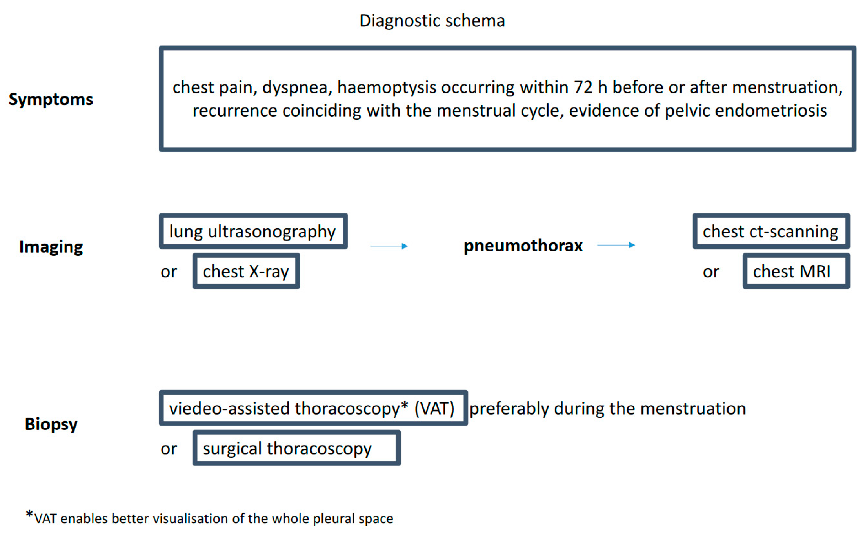
Figure 2.
Treatment strategy.
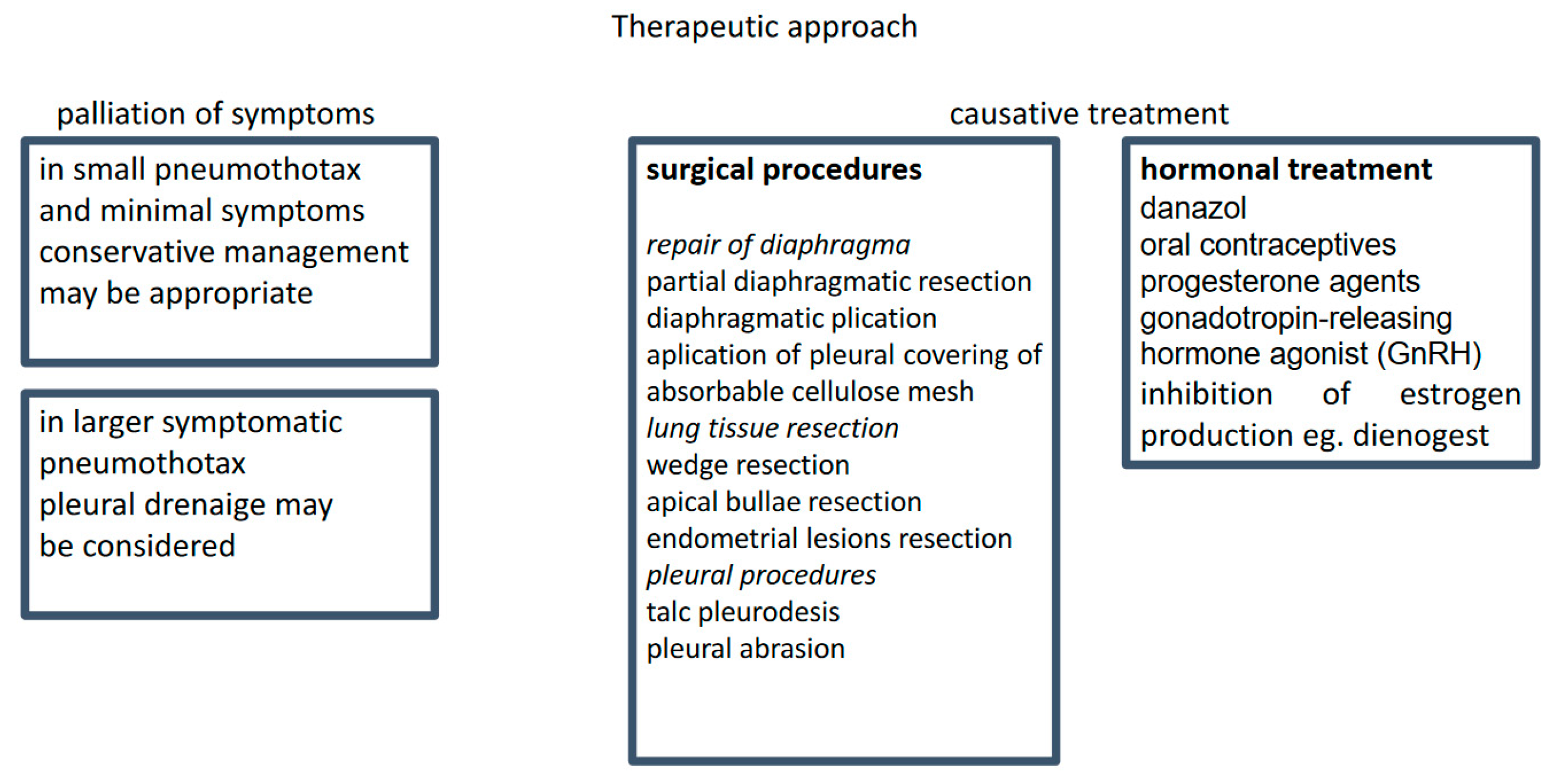
Figure 3.
Diagnostic and therapeutic pathway.
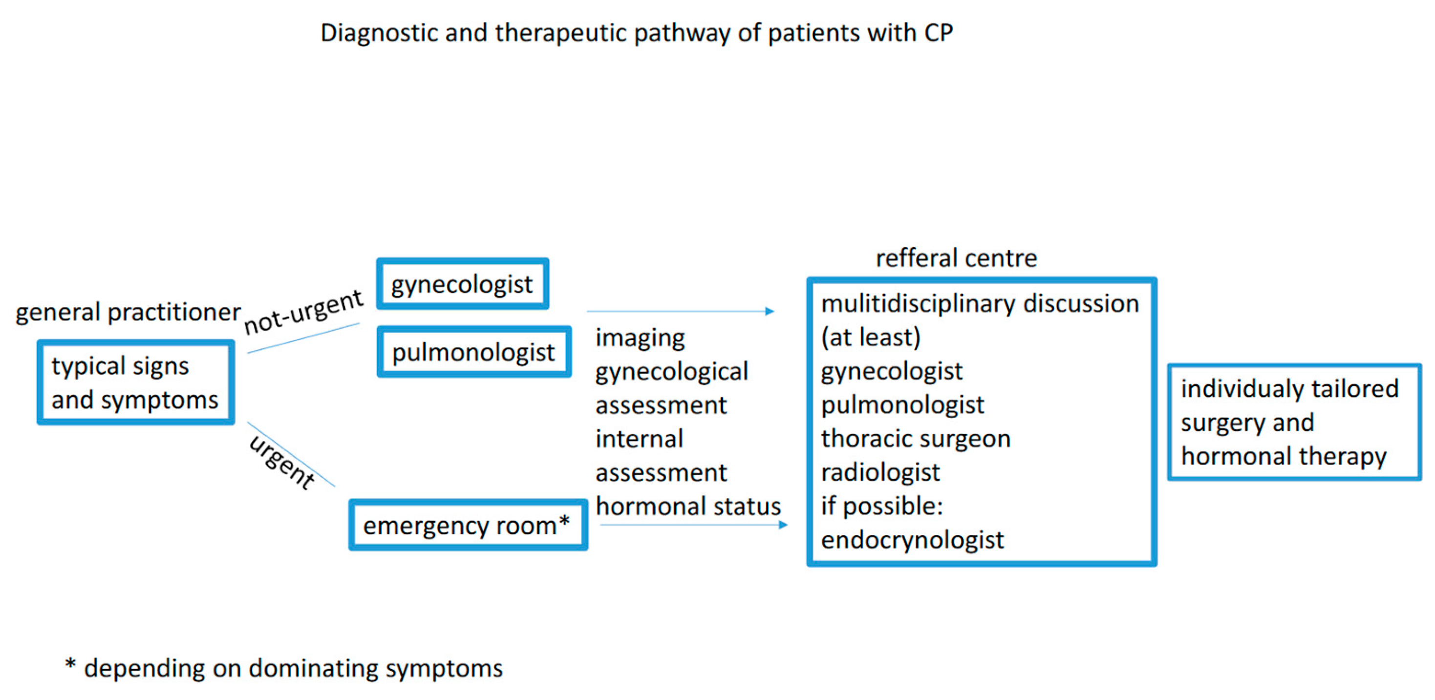

Table 1.
Theories for development of endometrial foci.
| Retrograde menstruation | Migration of endometrial debris from the uterus through the fallopian tubes into the lesser pelvis and further. Repetitive proliferation and necrosis of endometrial cells around the diaphragm area leads to the formation of diaphragmatic fenestrations. Passage of endometrial cells through the fenestrations into the chest and visceral pleura leads to alveolar damage and pneumothorax. |
| Transdiaphragmatic air passage | The passage of air from the uterus via the diaphragmatic fenestrations into the pleural cavity results in the development of pneumothorax. |
| Physiological | Increased prostaglandin F2 production during menses leads to vasoconstriction and bronchospasm, resulting in alveolar rupture and pneumothorax. |
| Metastatic | Lymphatic or hematogenous spread of endometrial cells from the uterus into extrapelvic localisations. |
| Coelomic metaplasia | Metaplasia of mesothelial cells that line the thoracic cavity. |
| Vascular embolisation | The endometrial debris travels in the venous system from the uterus to the right heart and into the pulmonary circulation, where it is deposited in the lung pleura or parenchyma. |
| Vasculogenesis | De novo development of microvascular endothelium of ectopic endometrial tissue from endothelial progenitor cells. |
| Immune dysfunction | Defective “immune-surveillance reaction induced by the inflammatory response. Defective cytotoxic natural killer cell activity and inability to eliminate ectopic endometrial cells. Secretion of cytokines and growth factors by ectopic endometrial cells, which induce the development of thoracic lesions by activating cell proliferation and angiogenesis. |
Disclaimer/Publisher’s Note: The statements, opinions and data contained in all publications are solely those of the individual author(s) and contributor(s) and not of MDPI and/or the editor(s). MDPI and/or the editor(s) disclaim responsibility for any injury to people or property resulting from any ideas, methods, instructions or products referred to in the content. |
© 2024 by the authors. Licensee MDPI, Basel, Switzerland. This article is an open access article distributed under the terms and conditions of the Creative Commons Attribution (CC BY) license (http://creativecommons.org/licenses/by/4.0/).
Copyright: This open access article is published under a Creative Commons CC BY 4.0 license, which permit the free download, distribution, and reuse, provided that the author and preprint are cited in any reuse.


