Preprint
Article

Asbestos Evidence in Roman Buildings from Micia Archaeological Site (Romania)

Altmetrics

Downloads

52

Views

52

Comments

0

Submitted:

17 October 2024

Posted:

20 November 2024

You are already at the latest version

Alerts
Abstract
The Micia site, is recognized as an archaeological civil settlement that was inhabited, and soldiers from several troops, were stationed in the Roman camp. From the end of the 2nd century AD, the civil settlement was rebuilt, with residential areas, industrial areas, port, public baths (civilian and military), amphitheater, religious areas (temples) and enjoying the facilities of a city. In this regard, the present work will first address the composition of the samples taken from the Roman monuments identified in Micia area and will highlight for the first time for this Roman site the presence of a form of tremolite-asbestos. This paper analyzes for the first time the presence of traces of tremolite-asbestos in stone samples collected from Roman monument buildings extracted from quarries near the city of Deva and used in civil, military and funerary structures from Micia. Highly performant and sensitive analytical techniques have been used to put into evidence the tremolite-asbestos species, to identify the structure, composition and morphology of these minerals inside of the building materials from Roman monuments, as follows: optical, stereo and scanning electron microscopy (SEM), X-ray diffraction (XRD), X-ray fluorescence with wavelength dispersion (EDXRF), FTIR and Raman spectroscopy, thermal analysis (TGA/DTA). It is presumed that tremolite-asbestos species has been included in the material layers used as mortars at Micia settlement in order to protect these monuments.
Keywords: 
Subject: Arts and Humanities  -   Archaeology

1. Introduction

Historical-archaeological studies of different Roman edifices have been completed in recent years with new structural, compositional, and morphological investigations of stone, mortars, and different pigments, trying to identify the execution techniques, recipes, and resource location [1,2,3,4]. The huge interest in the characterization of the Roman building materials has raised the necessity for establishing the sampling criteria and defining effective archaeometric techniques for this. This approach not only enhances our understanding of the materials and methods used in Roman construction but also aids in the preservation and restoration efforts of archaeological sites, offering the ancient engineering practices and the cultural significance of these structure [5,6,7,8,9].
The Roman Empire remains in history one of the greatest military and economic power of Antiquity. The period of the empire coincides with a considerable expansion of the borders (the Mediterranean Sea becomes an inland sea for the Romans) and with an unmeasurable accumulation of wealth from the province-territories. The Roman constructions are of enormous dimensions, producing an imposing impression [10] .
Knowledge of building materials was partly facilitated by the expansion of the Roman Empire. For economic reasons, the ancient Romans tended to use locally available building materials (and cheap labor) whenever possible. The import and transport of building materials was limited to the bare necessities or high-value luxury items such as marble. The most used building materials by the Romans were wood, unburned brick, stone, and concrete, called concretum. Roman concrete was made from a mixture of sand, water, and pozzolanic ash. Walls with a core filled with a general mixture of mortar and rubble which include pieces of broken pottery were also used [11] .
When the Romans started to build farms on the Mureș Valley, around the year 120, they were very convinced that they would remain masters in Dacia forever. Although the military presence was massive in the area, to ensure that their wealth was safe, in case of a barbarian invasion, the Romans of Dacia surrounded their properties with strong defense walls [12,13,14,15,16,17,18,19,20] .
As the Roman dominions grew, so did their access to new local materials. The advancement of building material technologies was determined by events such as the great fire of Rome in July 64 AD. This is how fireproof stone for public buildings appeared. "Lapis Gabinus" was a particularly popular type of quarry volcanic rock, used as fire protection in many public structures. It came from quarries around the town of Gabii, which was near a nearby volcano area. This type of volcanic rock was full of intrusions, including basalt, which made it particularly resistant to fire. This is the point when asbestos has been accepted by the Romans [21,22] .
Micia, adorned with numerous monuments made of andesite stone mined in nearby quarries (Deva city area and Măgura Uroiului), was a flourishing settlement, defended by a strong fort, spread over seven hectares. The civil settlement (pagus) was inhabited by about 5,000 people, and over 1500 soldiers from several troops, were stationed in the Roman camp.
A series of inscriptions discovered especially on the bricks made by the soldiers give us clues that detachments of several troops would have been stationed in the castrum of Micia, including the vexilla of the XIIIth Gemina Legion, but the basic troops of this important fortification were: Cohors II Flavia Commagenorum, Ala I Hispanorum Campagonum and Numerus Maurorum Miciensium. [23] .
In its first phase, the castrum was fortified with a ditch and a wave of beaten earth, coming from the excavation of the ditch, was trapped in a reinforcement of beams that ended in the front with a palisade (Holzerdmauer). Behind the ground wave was the usual road, via sagularis, and inside the troops' barracks, stables, and command buildings (principia). The troops' barracks and stables were built with 0.30-0.40 m thick walls of wooden beams deepened in the ground up to 0.70-0.80 m. The walls of these buildings were covered with adobe, in which the traces of the twigs can still be seen [24] .
After the middle of the second century, this part of the Roman Empire went through a long and difficult war that led to the destruction and burning of many civilian settlements and military camps, including those at Micia.
After the restoration of the military situation in the years 170-175, the destroyed fortifications were rebuilt, including Micia. In this H-th phase of existence, the castrum was fortified with a 12 m wide and 5 m deep ditch and a 1.80 m thick and 6 m high, which plastered the wall technically called agger to the outside. The enclosure wall had the elevation built of large quadrilateral blocks of Uroiu augustite-andesite (opus quadratum), and the foundation, up to 0.80 m deep, of quarry stone (micasist) bound with mortar (opus incertum). The 10.50 m wide agger, including the ascent slope, was paved with a layer of gravel fastened with mortar. From place to place, the enclosure was provided with interior curtain towers, arranged in agger. The rounded cultes, with thickened masonry, also presenting a 0.50 m wide projection, had trapezoidal towers inside. The entrance was made through 4 gates, of which the one on the south side was dug [19].
Inside the Micia castrum there are some constructive interventions, made during the long period of use of the buildings, which do not seem to modify the general plan [20] .
Also towards the end of the 2nd century AD, the civil settlement that accedes to the status of pagus is also rebuilt, having a well-organized building structure in residential areas, industrial areas, port, public baths (civilian and military), amphitheater, religious areas (temples) and enjoying the facilities of a city. In the extremities of Micia lay two necropolises with graves marked with spectacular funerary monuments made mostly of andesite.
Considering that only studies of geographical location for this monument are reported so far in the literature [25], without sufficient details related to the complete composition, structure and weathering status, such a study is required. In this regard, the present work will first address the composition of the samples taken from the Roman monuments identified in Micia fort and civil settlement area and will highlight for the first time for this Roman site the presence of a form of tremolite-asbestos.
The Romans extensively mined and utilized asbestos throughout Europe and the Mediterranean for its remarkable properties, including strength, insulation, and resistance to fire and corrosion. Asbestos found its way into various products, with the ancient Greeks weaving it into cloth, while Romans incorporated it into their building materials. Despite awareness of its detrimental health effects, as noted by historical figures like Strabo and Pliny, who documented the asbestos exposure, its applications persisted, highlighting a historical conflict between utility and safety that resonates even in contemporary discussions about the material [26,27,28].
Asbestos is a naturally occurring mineral that was used for everything from building tiles to gloves. The Romans loved asbestos, using it in several different ways. The mineral fibers, whilst providing a material to weave into non-flammable cloth, are so small that they can easily become inhaled and trapped in the body. These trapped fibers can cause scarring, inflammation, and cancer. At the beginning, the Romans knew absolutely nothing about this. Asbestos, to the Romans, was simply a wonder material that could do wonderful things. The only downside was the price [28].
At that time, asbestos was brought from Orșova area, Ponicova (Cazanelor reservation) where on Ciucaru Mare hill, there is a recognized deposit of asbestos. In fact, the Mining Entreprise Orșova is famous for Feldspar, Mica, Asbestos, Talc, Dolomite and Quarts. Considering the composition detected by analytical techniques, asbestos and mica could be sourced from Orșova.

2. Description of the Archaeological Site

2.1. The Fort and the Ancient Settlement of Micia

Micia, the western most Roman settlement on the Mureş Valley, was an important center of the province of Dacia. The strategic location of the castrum here, about 2.5 km east of the Mureş Gorge, in the area where the river meadow widens generously, allowed the development of a large civil settlement in the vicinity of the fortification. The numerous road and industrial development works carried out in the last two centuries have affected the archaeological site to a great extent [29,30] .
Micia, one of the largest castra of the auxiliary troops of Roman Dacia, along with those of Porolissum and Tibiscum, was built on the south bank of the Mureş River, about 3 km east of the Brănisca gorge, the narrowest place in the valley of this river. Being at an obligatory crossing point of the communication route that connects the Transylvanian Plateau to the Tisa Plain, it played a first-hand strategic role in Dacia defense system. But it is also because of its position that it has suffered a series of destructions in modern times. Thus, its center is intersected by the Arad-Deva double railway and by the national road between the same localities, and one of the short sides of the enclosure, the SE one, was destroyed by the construction of a railway of the Mintia thermal power plant (Figure 1). However, the remaining parts: the northern half, a strip of the central area and a part of the southern half, still allow large-scale research of this archaeological objective [31] .
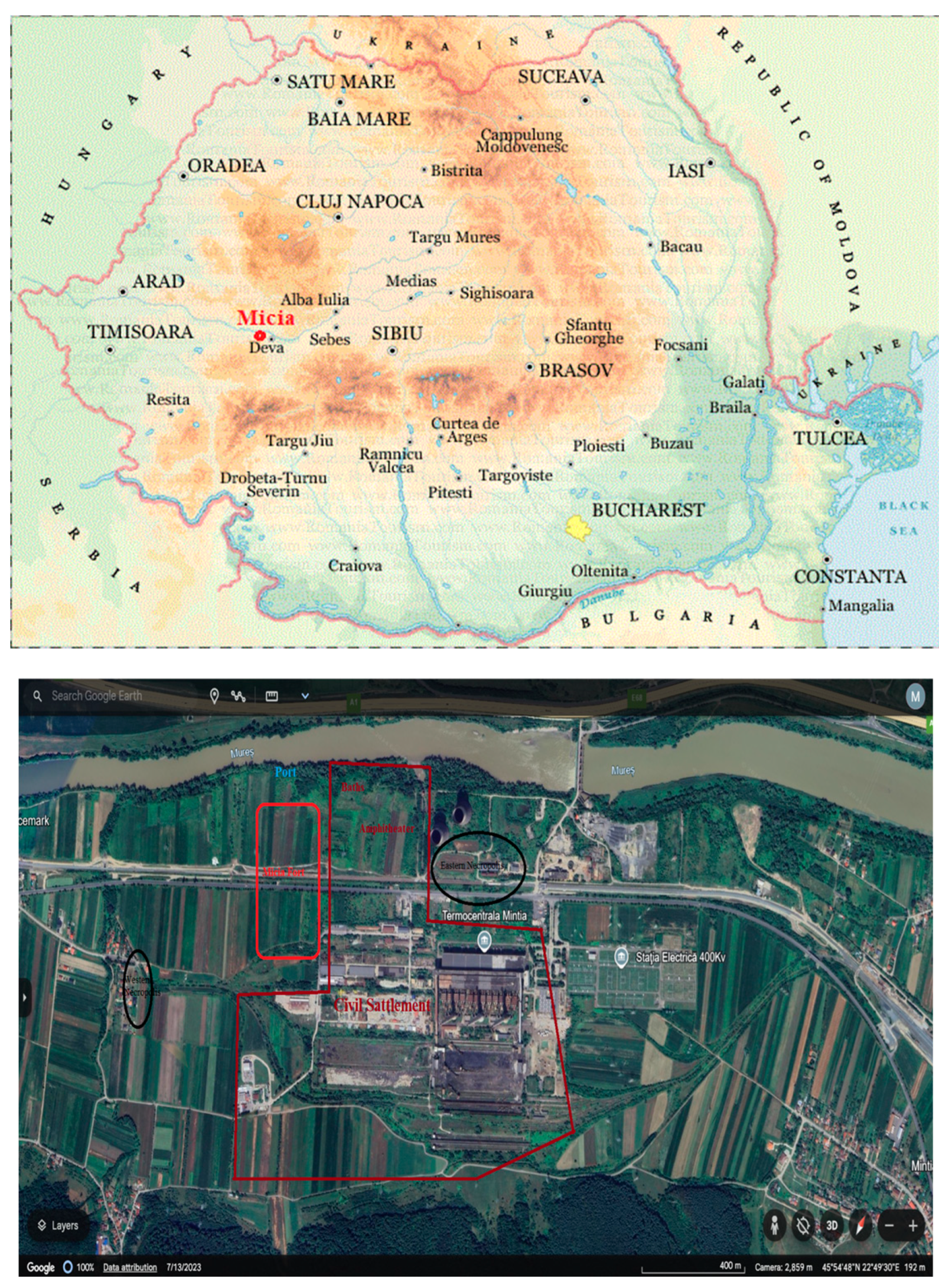
Micia is an archaeological site recently designated as a UNESCO Heritage site, located near Deva along the left side of the Mureș River. This site encompasses one of the largest Roman fors in Dacia, which played a crucial role in controlling access to the Mureș Valley (Figure 2). Established as a quasi-urban settlement, Micia features significant structures, including the fort, an amphitheater, a thermal complex, craftsmanship areas, sacred zones, and two known necropolises, all spanning approximately 25 hectares. The site is geographically framed by the Mureș River to the north and the foothills of the Poiana Ruscă Mountains to the south, extending into the administrative territories of the nearby villages of Vețel, Mintia, Herepeia, and Vulcez [32] .
The commune was connected to one of the main Roman roads that crossed the territory of Transylvania, but its most important role was that of defending the western borders of Roman Dacia, whose capital, the ancient city of Ulpia Traiana Sarmizegetusa, was approximately at 50 kilometers, and the city of Apulum, at 80 kilometers away. Another strategic mission of Micia was to protect the gold and silver mines in the Apuseni Mountains.
Micia was inhabited between the 2nd and 3th centuries, the settlement being mostly destroyed since Antiquity, following the wars. Micia was not completely abandoned after the Aurelian retreat. Even if the Roman army and administration were taken from the area, the population remained in Micia, at least until the invasions of the Huns (5th century), when all the Roman settlements were abandoned [33,34] .
The destruction of the Micia ruins continued in the 19th century, when stone blocks from its walls were extracted by the locals to repair the Brănişca road, devastated by the waters of Mureș. In 1869, due to the construction of the Deva - Arad railway, the ancient town continued to degrade. In the same manner, the Mintia thermal power plant, built in the 1960s, negatively contributed to this deterioration process, while the Mureş River, whose course was diverted in 1967, submerged part of the ruins of Micia. The andesitic tuffs of Padurea Bejanului, Almaşul Sec and those south of the town of Deva (on the way to Almaşul Sec) have been identified in this area [35,36] . Also, over time, many pieces of andesite, the red colored rock, were extracted and used as construction materials.
The proximity of the Mureş River also played a crucial role in the settlement's development, providing resources and a means for trade and communication. Today, efforts continue to preserve these historical sites while balancing modern developments around them, ensuring that the rich history of this area is not forgotten in the face of industrialization and urban expansion.
This paper analyzes for the first time the presence of traces of tremolite-asbestos in the stone samples collected from Roman monument buildings extracted from quarries near the city of Deva and used in civil, military and funerary structures from Micia. Highly performant and sensitive analytical techniques have been used to put into evidence the tremolite-asbestos species, to identify the structure, composition and morphology of these minerals.

3. Materials and Methods

3.1. Materials

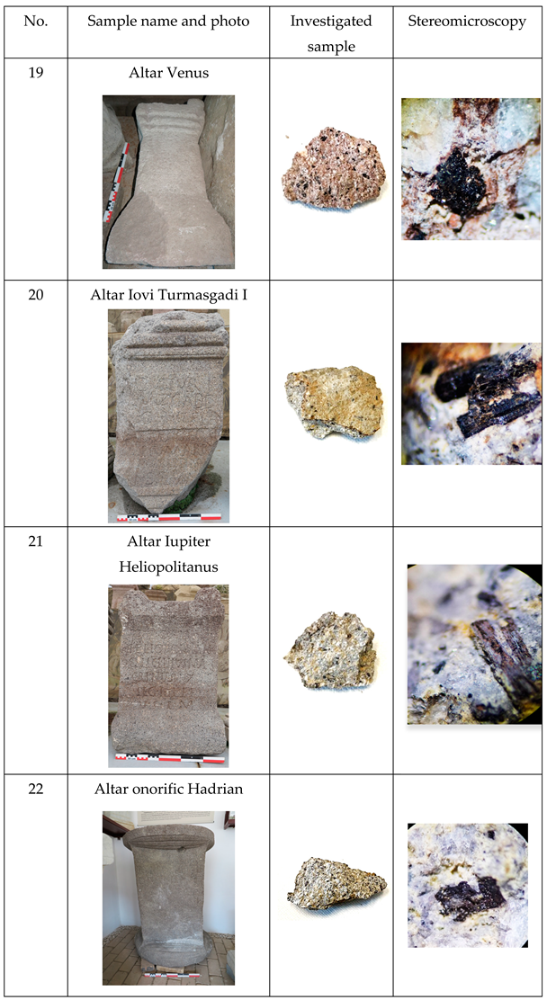
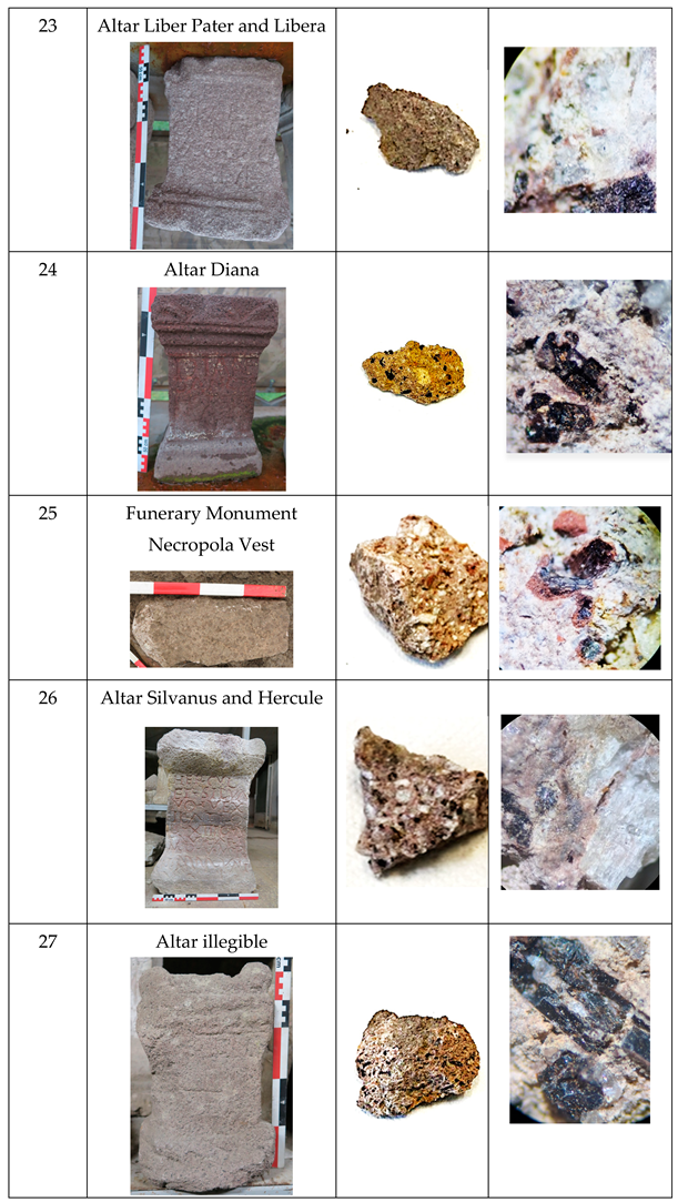
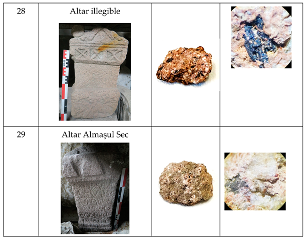
For this study, 11 samples have been collected from the buildings belonging to the Micia archaeological site in Romania (Table 1).

3.2. Analytical Techniques

Wavelength dispersion X-ray fluorescence spectroscopy (WDXRF) have been recorded by using the Rigaku ZSX Primus II spectrometer (Rigaku, The Woodlands, TX, USA), which includes a Rh anode X-ray tube and a 4.0 kW power source, including a 30 µm front Be window, to conduct detailed analyses. The utilization of an EZ scan method with Rigaku's SQX software helped for data processing, contributing to accurate and thorough spectroscopic results.
X-ray diffraction (XRD) analysis has been achieved by using a Rigaku Ultima IV diffractometer (Rigaku Corporation, Tokyo, Japan), which used the following parameters: Cu Kα radiation (λ = 1.5406 Å), 40 kV and 200 mA. For the diffractograms, a 2θ range from 5° to 80°, and a scanning rate of 4°/min, have been followed.
Optical microscopy (OM) has been achieved with a Novex Microscope BBS trinocular microscope (Euromex Microscopen B.V., Arnhem, The Netherlands) at various magnifications, a digital video camera (Axiocam 105, Zeiss, Göttingen, Germany), ZenPro software for a real-time data acquisition, and with analysis conducted using ImageJ 1.50.
Also, for concise and detailed images, a Video-microscope Edmund Optics C-Mount, has been used, equipped with AmScope 3.2MP MT9T001 CMOS C-Mount camera, an extension videotub, ZOOM ring, focus ring, a Zabber T-NA08A25 XYZ Micro linear actuators with a Medium Intensity Spot/Coaxial Light, an Edmund Optics 2X EO M Plan Apo Long Working Distance Infinity Corrected objective and Edmund Optics XYZ Stage.
Additionally, stereomicroscopy was carried out with a Stereo trinocular stereomicroscope from EUROMEX Microscopen B.V., model 1903 EUROMEX Microscopen B.V., BD Arnhem, Netherlands), offering magnification capabilities from 5× to 40×.
Environmental scanning electron microscopy (ESEM-FEI Quanta 200, Eindhoven, The Netherlands) was utilized to evaluate sample morphology, employing a high vacuum setup with a magnification ranging from 50× to 100,000×. For sample preparation, each specimen was coated with a 5 nm layer of gold using a Q150R-ES sputter coater (Quorum Technologies Ltd., West Sussex, UK), to reduce charging effects and enhance conductivity during SEM imaging.
Fourier Transform Infrared Spectroscopy (FTIR) with attenuated total reflection (ATR) mode (Perkin Elmer, Waltham, MA, USA) was achieved with a resolution of 4 cm−1 and accumulating 32 spectra, with measurements spanning the range of 4000–400 cm−1 for a comprehensive analysis. The FTIR analysis was conducted in transmission mode using KBr pellets, which were prepared by thoroughly mixing the sample with potassium bromide (KBr) powder and then compressing the mixture into transparent discs.
Raman spectra were obtained with a Rigaku portable analyzer (Xantus-2, Rigaku, The Woodlands, TX, USA) with 785 and 1064 nm stabilized laser, a resolution of 4 cm−1 using a laser power of 252 mW, and an Opus 7.0 software from Bruker Optics GmbH has been used for data processing.
The textural properties of the samples were investigated using the Brunauer-Emmett-Teller (BET) method, which involved analyzing nitrogen adsorption and desorption isotherms at a temperature of 77 K across a relative pressure range of 0.005 to 1.0. A NOVA2200e gas absorption analyzer from Quantachrome Instruments in Boynton Beach, FL, USA, was used. The samples were degassed for 4 hours at 180 °C under vacuum. Also, a NovaWin version 11.03 software was utilized for data processing.
Thermogravimetric analyses were performed using a Pyris 1 TGA/DTG analyzer from Perkin Elmer (TGA-7) in Waltham, MA, USA, on a temperature range of 50–800 °C heating rate of 10 °C/min and nitrogen flow at 50 mL/min.
The color of the mortars was analyzed Konica Minolta CR-410 spectrophotometer following the CieLab system (L*, a*, b*) [37] and the EN 15886 (2011) standard [38]. Measurements were performed under a standard D65 illuminant and a 2° observer angle. The data were processed using the Color Data Spectramagic NX CM-S100W software.

4. Results

4.1. Microscopic, Mineralogical and Compositional Analysis

All samples taken from the archaeological site are white to black in color, with a red area due to iron oxide samples. The black color may be due to andesite, as it is known that this area is a large deposit of andesite. The samples exhibit a sub rounded morphology and are predominantly siliceous, comprising monocrystalline quartz grains alongside fragments of volcanic rocks characterized by trachytic and vacuolar textures. Additionally, they contain trace amounts of heavy minerals of volcanic origin, such as augite and other pyroxenes, as well as black particles likely derived from andesite. The grain selection in these mortars is adequate, falling within the fine to medium sand size range of 0.125 to 0.5 mm. Their irregular, vacuolar appearance is attributed to fluids trapped during formation, with notable occurrences of tremolite-actinolite present. [39,40].
By analyzing the optical microscopy images, for the samples collected from the exterior layers, some asbestos species have been identified. Asbestos refers to a group of six naturally occurring mineral fibers, which are categorized into two main groups: the serpentine group, primarily represented by chrysotile, and the amphibole super-group, which includes asbestiform varieties of riebeckite, grunerite, anthophyllite, tremolite, and actinolite. These fibers are known for their heat resistance and fibrous nature, making them useful in various industrial applications, although their health risks are well-documented due to their potential to cause serious respiratory diseases when inhaled. [41]. Actinolite is an intermediate member of a series between magnesium-rich tremolite, Ca2(Mg5.0-4.5Fe2+0.0-0.5)Si8O22(OH)2, and iron-rich ferro-actinolite, Ca2(Mg2.5-0.0Fe2+2.5-5.0)Si8O22(OH)2. Mg and Fe ions can be freely exchanged in the crystal structure.
Tremolite and actinolite are closely related minerals that form a continuous series characterized by their chemical compositions, where the tremolite is richer in magnesium and actinolite in iron. Both minerals exhibit similar physical properties, including various recognized varieties that can be fibrous and leathery, featuring a silky luster and interlocking fibers that are dense and often indistinguishable. While both minerals contain asbestos forms with movable and elastic fibers, tremolite asbestos is far more prevalent than the less common actinolite asbestos.
Asbestos was widely utilized by ancient civilizations, particularly the Romans and Greeks, for its remarkable properties, such as strength, insulation, and resistance to fire and corrosion. The Romans mined asbestos throughout Europe and the Mediterranean, incorporating it into various products, including building materials. Meanwhile, the Greeks notably used asbestos fibers in textiles, showcasing its versatility and widespread appeal during that period.
One of the first mention about the asbestos dated from the 1st century AD historian Pliny the Elder, when he noted "it is quite indestructible by fire" and "affords protection against all spells, especially those of the Magi." In our experiments has not identified only linear crystals, but also, flexible one, are represented, too. Some asbestoid structures have been identified in Diana and Venus altars, most probably due to the reconstruction after the fire of these religious settlements. The religious cult is another supposition.
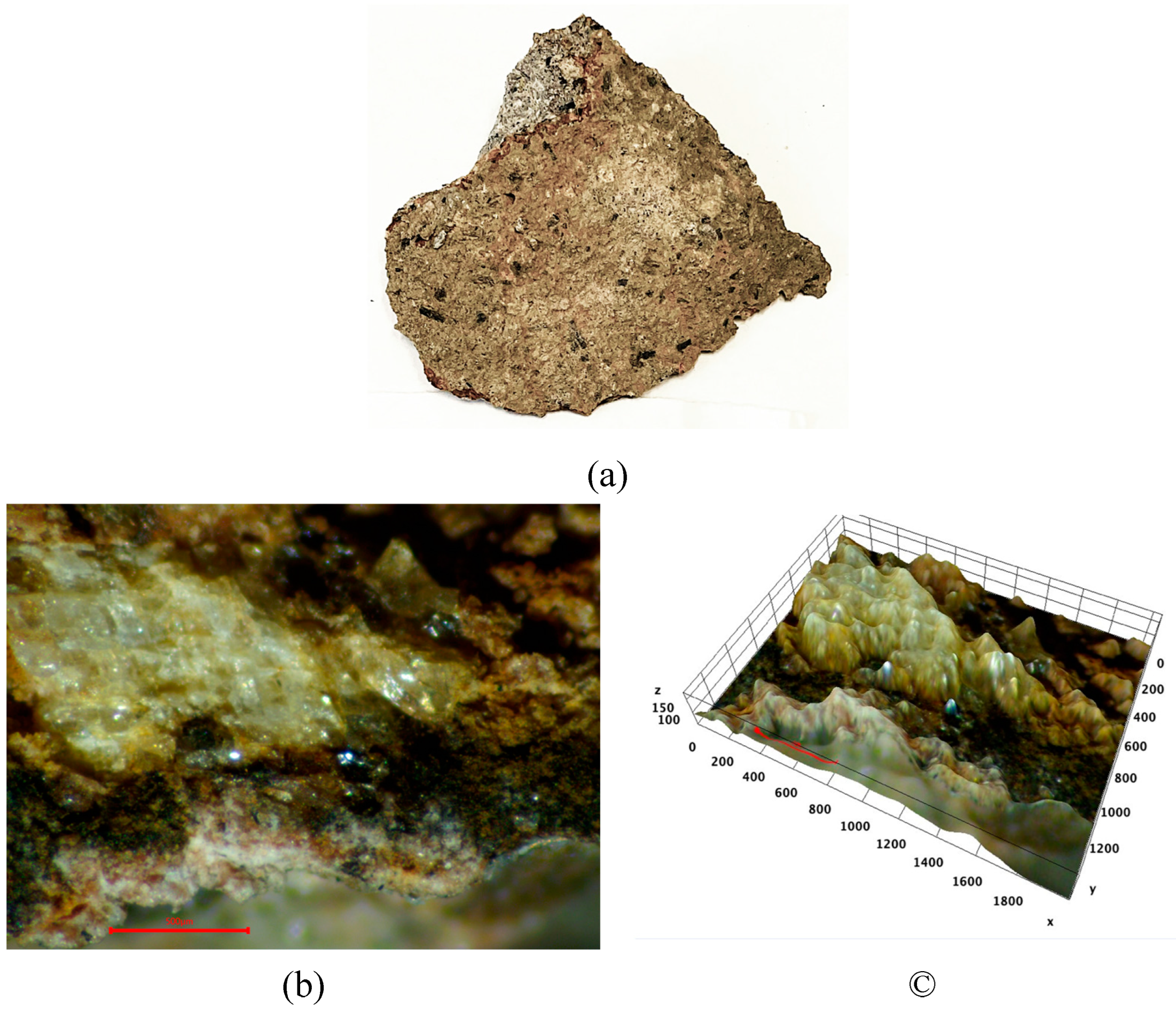
Also, for these samples, three material layers have been identified: inferior(white-black), middle (red, black), exterior layers (whitish), as could be visualize by optical microscopy, Figure 3.
The separation of the three layers can be much more clearly highlighted by using and processing the optical microscopy image (Figure 3(b), by the Image J soft (Figure 3(c)).
In our opinion, and based on the literature data, the middle layer could be a mixture of andesite with asbestos, while the exterior layer could be assigned to tremolite-asbestos presence in the reconstruction mortars. All the subsequent investigation results will convince on the existence of this layer of asbestos applied by the Romanian population for the fire protection of the respective constructions.
Andesite is a type of dark color volcanic with a fine-grained texture, primarily composed of zoned sodic plagioclase along with additional minerals such as biotite, hornblende, and pyroxene in minor proportions. Its ground mass typically contains a mix of sodic plagioclase and small amounts of quartz, and it is mainly formed from the lava that cools relatively quickly upon eruption, resulting in its distinct characteristics.
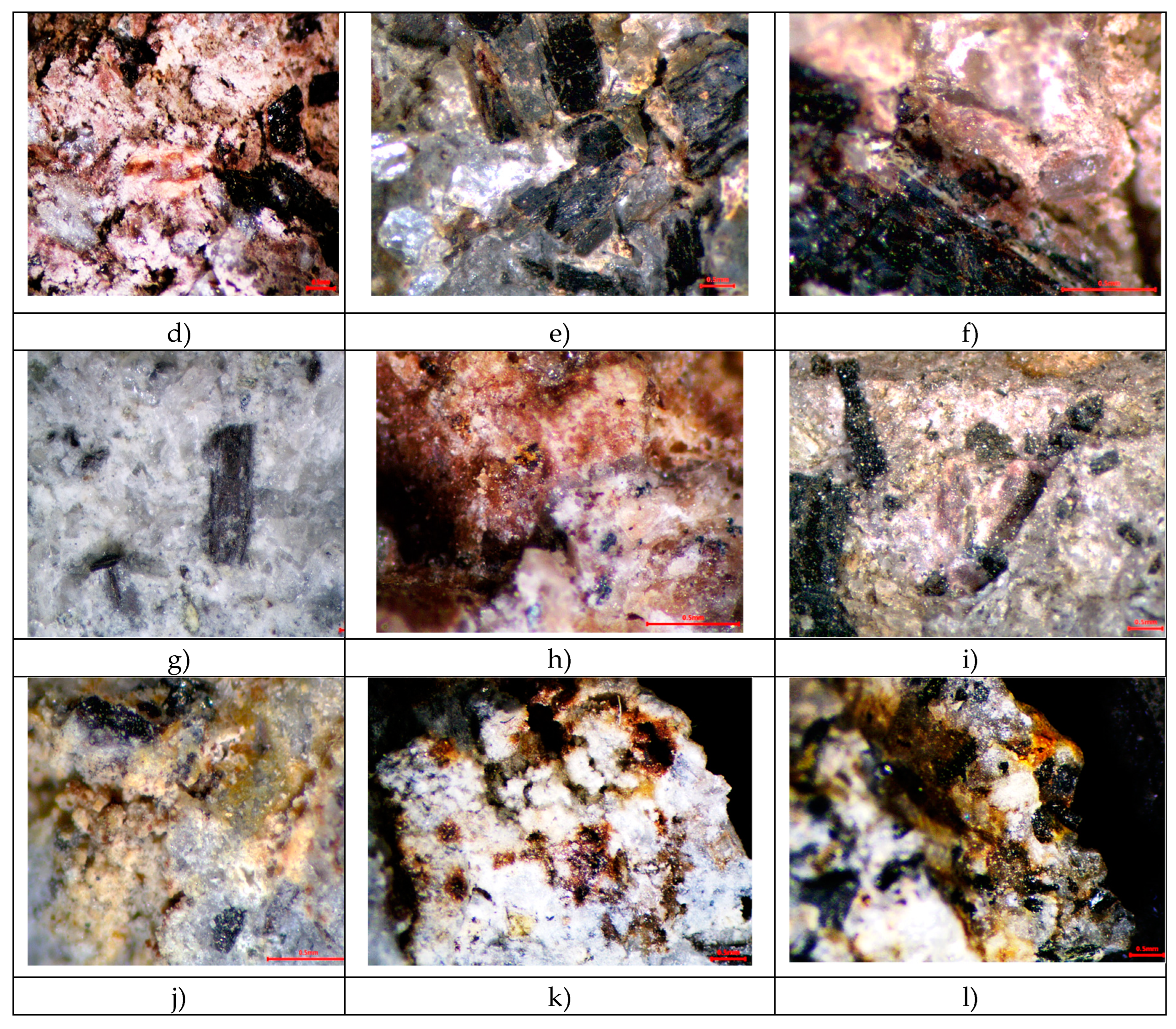
In order to be sure about the identity of these asbestos forms in Roman Monuments, a comparison between the roman monuments building materials with those from the Carriers’ fronts samples, as could be seen in optical microscopy images, Figure 4.
If in the samples from the Carriers’ fronts an andesite and various minerals based on iron oxides together with quartz predominates, in the samples from the Roman monuments, all these minerals are found in much smaller quantities, the tremolite-type asbestoid forms being present on large scale. Some zones contain some aggregates (up to 2 mm), and ferruginous and fine sand-sized quartz grains (0.125–0.25 mm) (Figure. 4). Also, some volcanic rock fragments with vacuolar texture and some Na-Ca and K-Na plagioclase, with black particles, are identified (Figure. 4) [42].

4.2. Microscopy Evidence of Tremolite-Asbestos Fibers

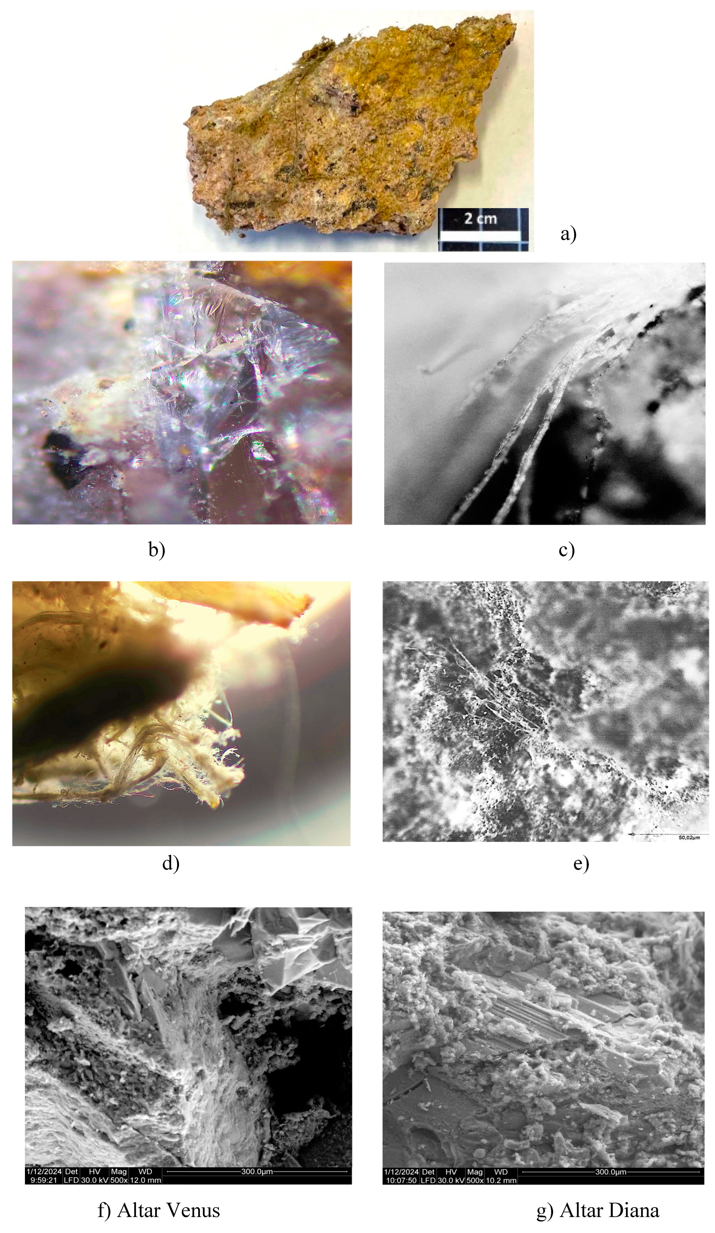
Visual and optical microscopic examination of the investigated samples, can easily put into evidence the presence of long fibers, translucide and flexible, as could be observed in Figure 5b-e. Also, the identification of these fibers is more feasible from the SEM images (Figure 5f,g), which confirm the heterogeneity of these samples, as previously with OM (Figure 5b-e). The asbestos fibers are randomly oriented both as individual threads and as fiber bundles, which denotes the heterogeneous mixing of these fibers in the prepared mortar. Asbestos fibers are extremely fine, typically ranging in diameter from 100 nanometers to 1 micrometer, with lengths that can extend to several centimeters.
These samples predominantly consist of tremolite, characterized by elongated prismatic and fibrous crystal, as revealed through electron and optical microscopy. The tremolite asbestos could displays a table structure, which indicates subsequent kinking and folding that occurred after its initial crystallization, highlighting a complex history of polyphasic deformation. Similar images have been reported in Rianudo paper [43,44] .
XRD/WDXRF
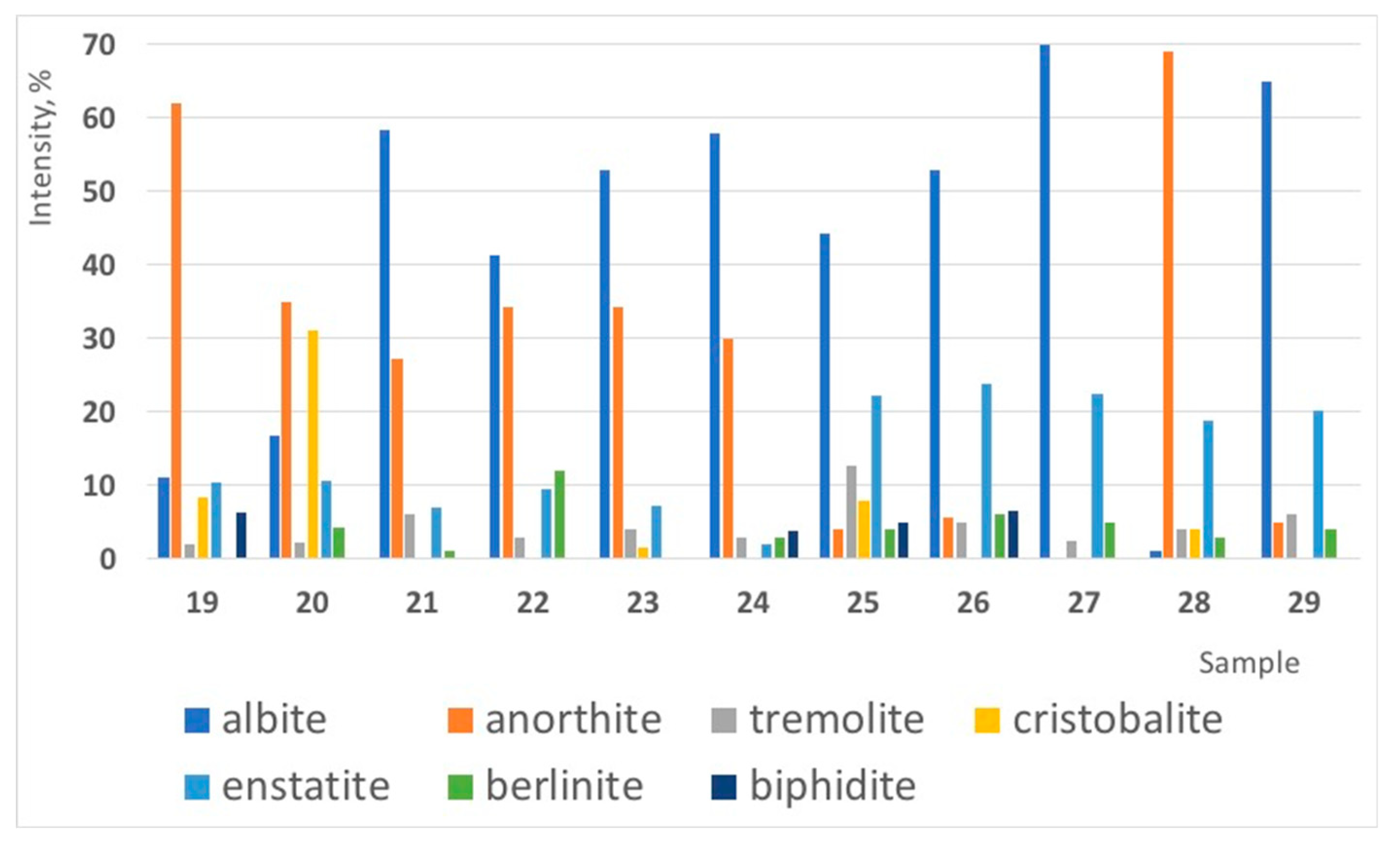
In the same manner, tremolite mineral has been identified by XRD techniques, Figure 6. In this figure, the collected samples have been analyzed, and tremolite, anorthite, cristobalite, quartz and berlinite were put into evidence.
XRD analysis indicates that the mineral paragenesis in the matrix of these samples consists primarily of tremolite, albite, and anorthite, suggesting a complex interplay of these minerals within the geological context.
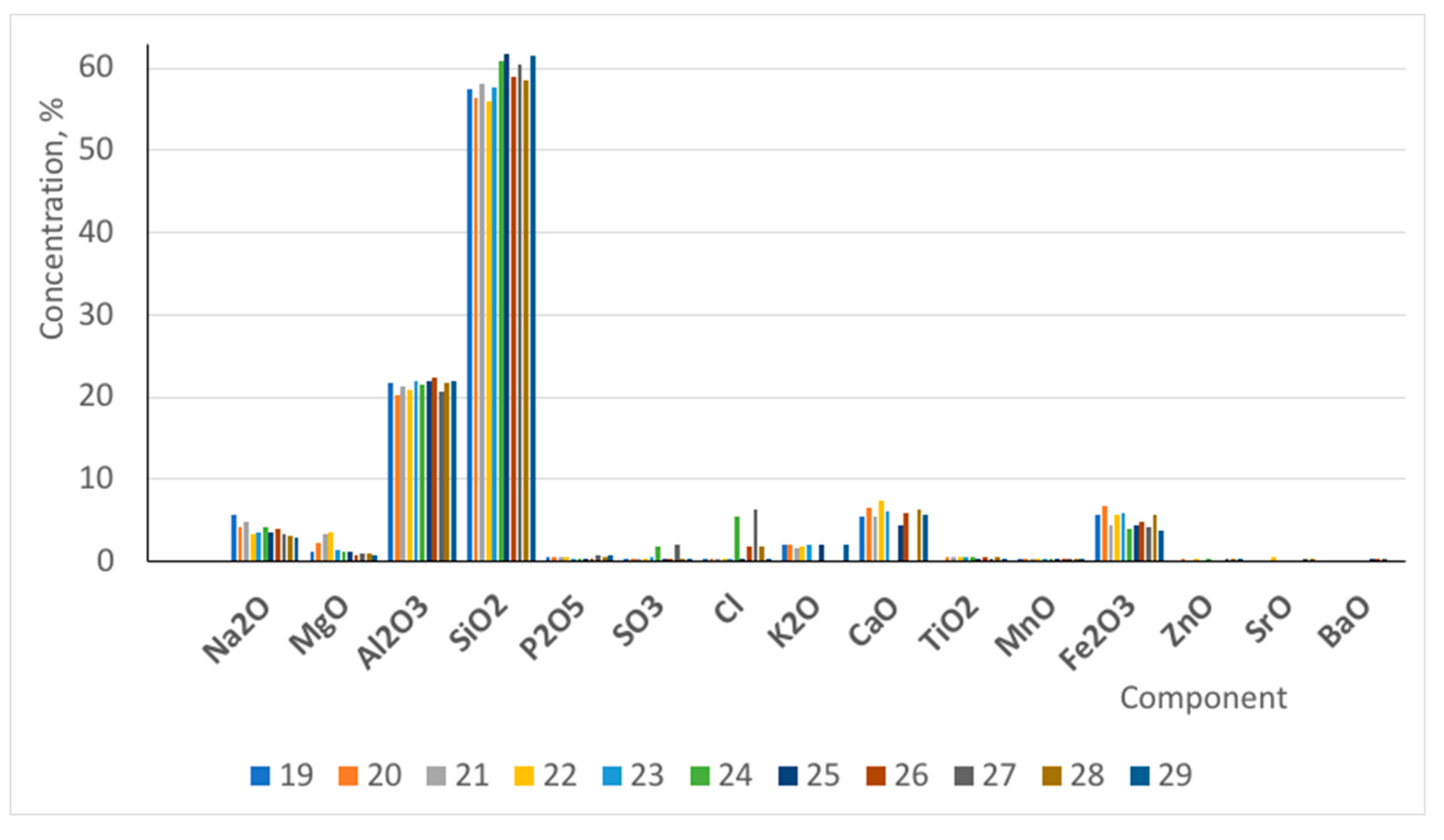
In the case of the fragments with volcanic source, the chemical composition of the matrix identified by EDXRF (Figure 7) reveals variable amounts of CaO (7-9 %), SiO2 (56-62 %), Al2O3 (20–23 %), FeO (4–7 %), K2O (1–4 %), MgO (1–2 %), TiO2 (1–1.5 %) and Na2O (4–7 %). These results could be observed to be quite similar for the all the analyzed samples. Major element analyses allowed classifying the amphibole from sample as tremolite according to Hawthorne et al [41]. Tremolite as a member of the amphibole group, is characterized by its specific chemical composition and structure, primarily consisting of calcium, magnesium, and iron in a silicate framework.
FTIR
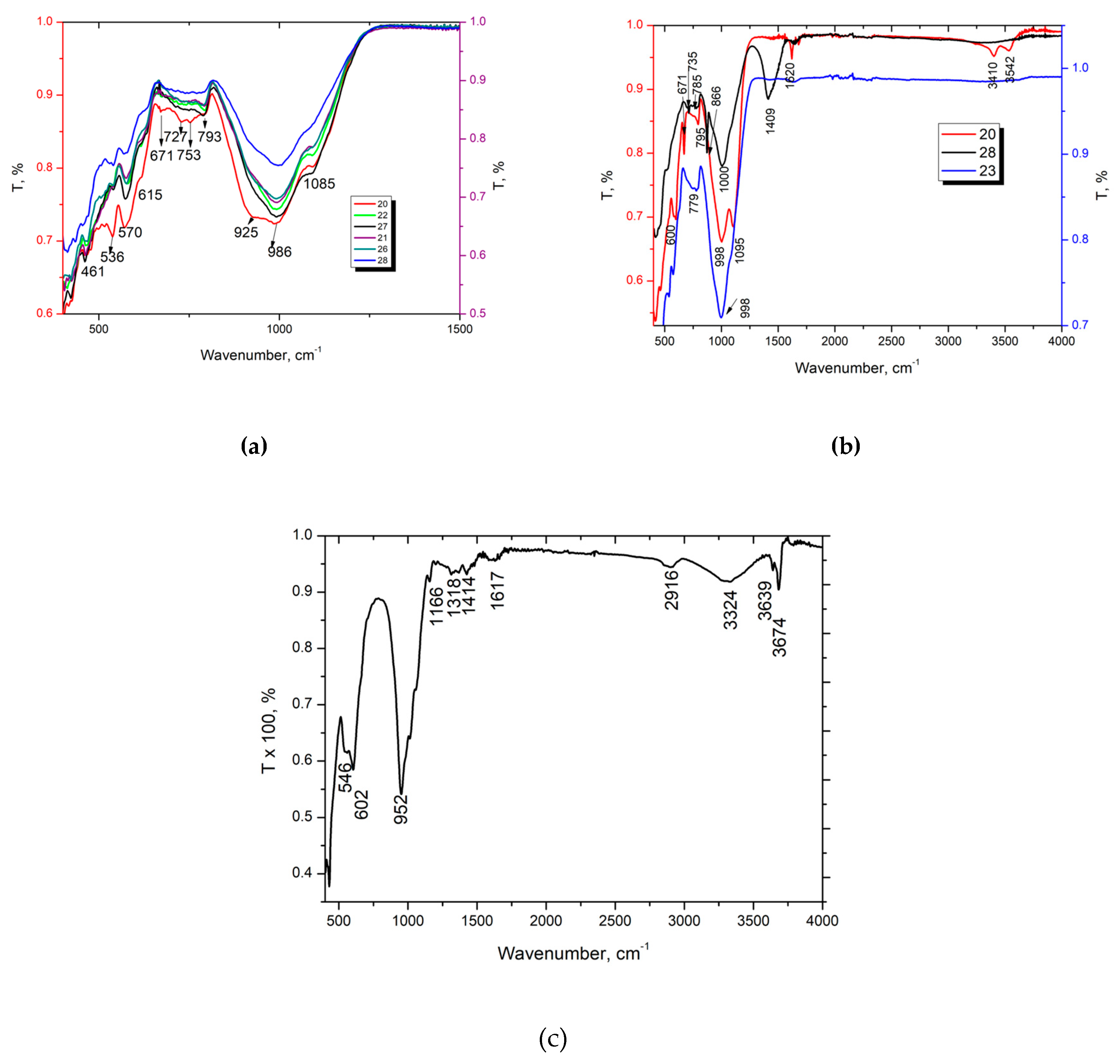
In order to certify the presence of asbestos in this area, the carriers’front samples have beenanalysed by XRD, and no specific bands for asbestos have been found (Figure 8a). However, the samples collected from Roman buildings (20,23,28) show the specific bands of asbestos (Figure 8b). As literature indicated, asbestos forms as asbestos-tremolite has a specific band around 1400 cm-1, Figure 8c.
Figure 8 show representative FTIR ATR spectra collected on powdered fragments of the investigated samples. For some samples (ex. P20, P23 or P28) was possible to observe three layers: exterior, middle and interior distinctly layers. The specific bands, as Si-O stretching and bending modes at ~960, 1015, 1080 and 1400 cm-1) and 620 cm-1 (Figure 8), are characteristic of asbestos and similar amphiboles and pyroxenes compounds [45,46,47,48,49,50,51,52] .
Tremolite is a mineral that primarily consists of calcium, magnesium, and silicon, closely aligning with its ideal end-member composition. However, it can incorporate varying amounts of magnesium through substitutions for calcium, and it typically contains trace elements such as sodium, potassium, iron, aluminum, and fluorine. This variability in composition reflects its natural occurrence and the ability of minerals to accommodate different ions in their crystal structures [51]. This variability in composition arises from the inherent flexibility of the structural framework formed by silica ribbons in the fibers, which can incorporate various ions from the surrounding environment, including those contributed by the diverse characteristics of the host rocks. This ability to accommodate different ions leads to diverse compositional outcomes, reflecting the interplay between the structural capacity of the material and the geochemical context in which it forms [52,53,54].
Raman spectra
Raman spectroscopy effectively identifies and differentiates minerals within the amphibole group, but the peak assignment can be challenging due to the complex structural variations among them, leading to inconsistencies in peak/vibration assignments across different studies.
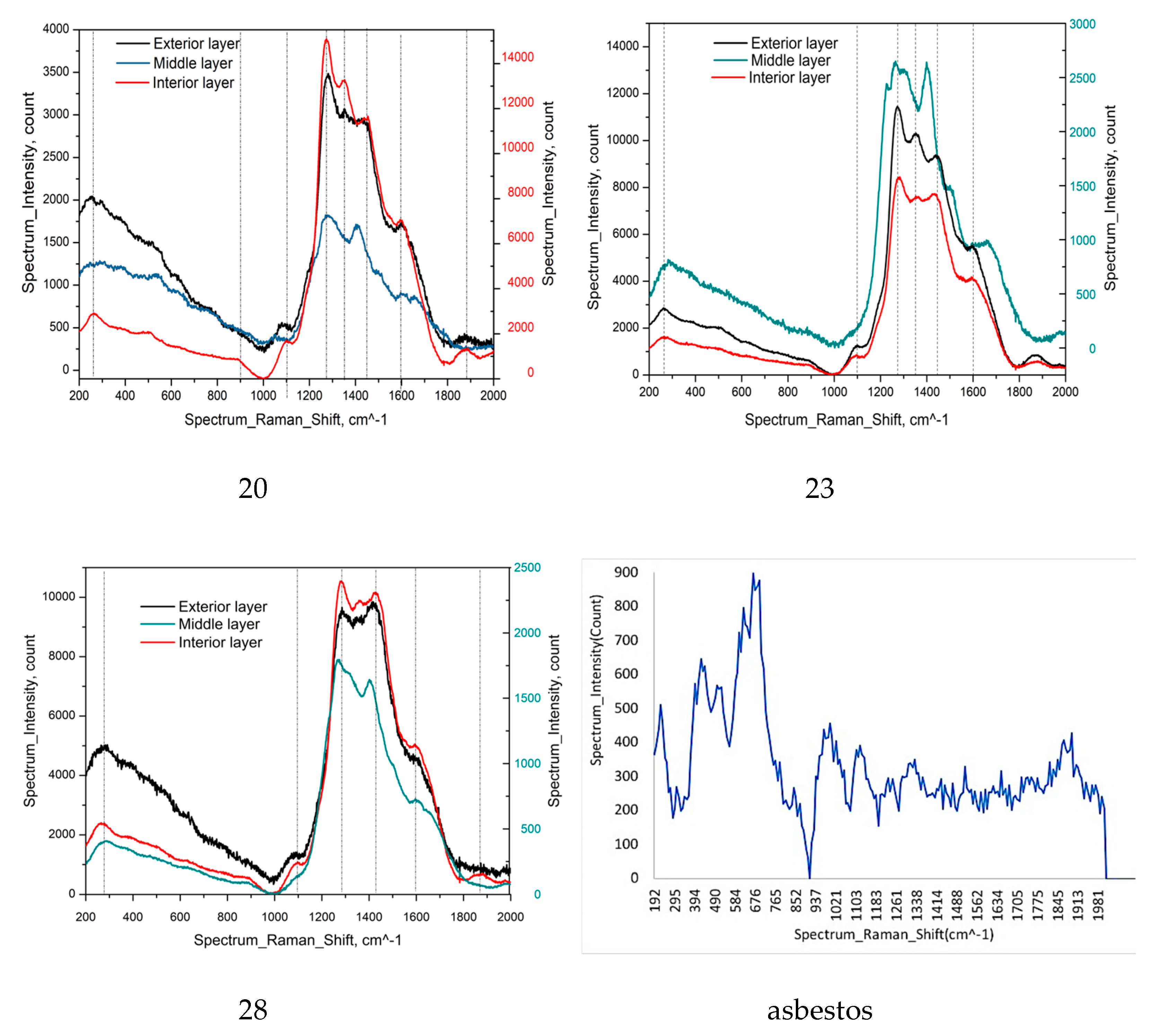
By using 782 nm laser for Raman equipment, it was possible to put into evidence the main bands of asbestos (1000-2000 cm-1). The band from 1400 cm-1 is predominant in all the investigated selected samples (20,23,28), especially for middle and exterior layers. The most distinct Raman peak, Figure 9, is between 1200 - 1400 cm-1, which is assigned to the symmetric stretching vibrations of the Si-O-Si bridges [55,56].
The obtained Raman spectrum of tremolite - actinolite (Figure 9) is characterized by the typical features of the spectra of amphibole minerals [57,58]: between 300 and 600 cm−1 (Mg–OH and Fe–OH vibrations, Si–O–Si bending motions and OH– vibrations); between 650 and 750 cm−1 (Si–O–Si symmetric stretching); over 750 cm−1 (O–Si–O symmetric stretching and the O–Si–O and Si–O–Si asymmetric stretching bands). The main feature of the Raman spectra in the low-wavenumber region, at nearly 675 cm−1, is the Si–O–Si symmetrical stretching with Ag symmetry. This mode, when substituting Mg2+ with the heavier Fe2+, downshifts from 675 cm−1 in pure tremolite to 667 cm−1 in Fe-rich actinolite [59].
In the investigated representative samples (P20, P23 and P28), the bands from 1290, 1350 and 1400 cm-1 are specific for middle layer of each of them. Well deconvoluted, the band from 1400 cm-1, specific for asbestos, could be clearly identified, this band belonging to tremolite. The other bands (1290 and 1350 cm-1) could be assigned to the other minerals, amphiboles and pyroxenes compounds, confirming the FTIR results.
4.3. Thermogravimetric Analysis
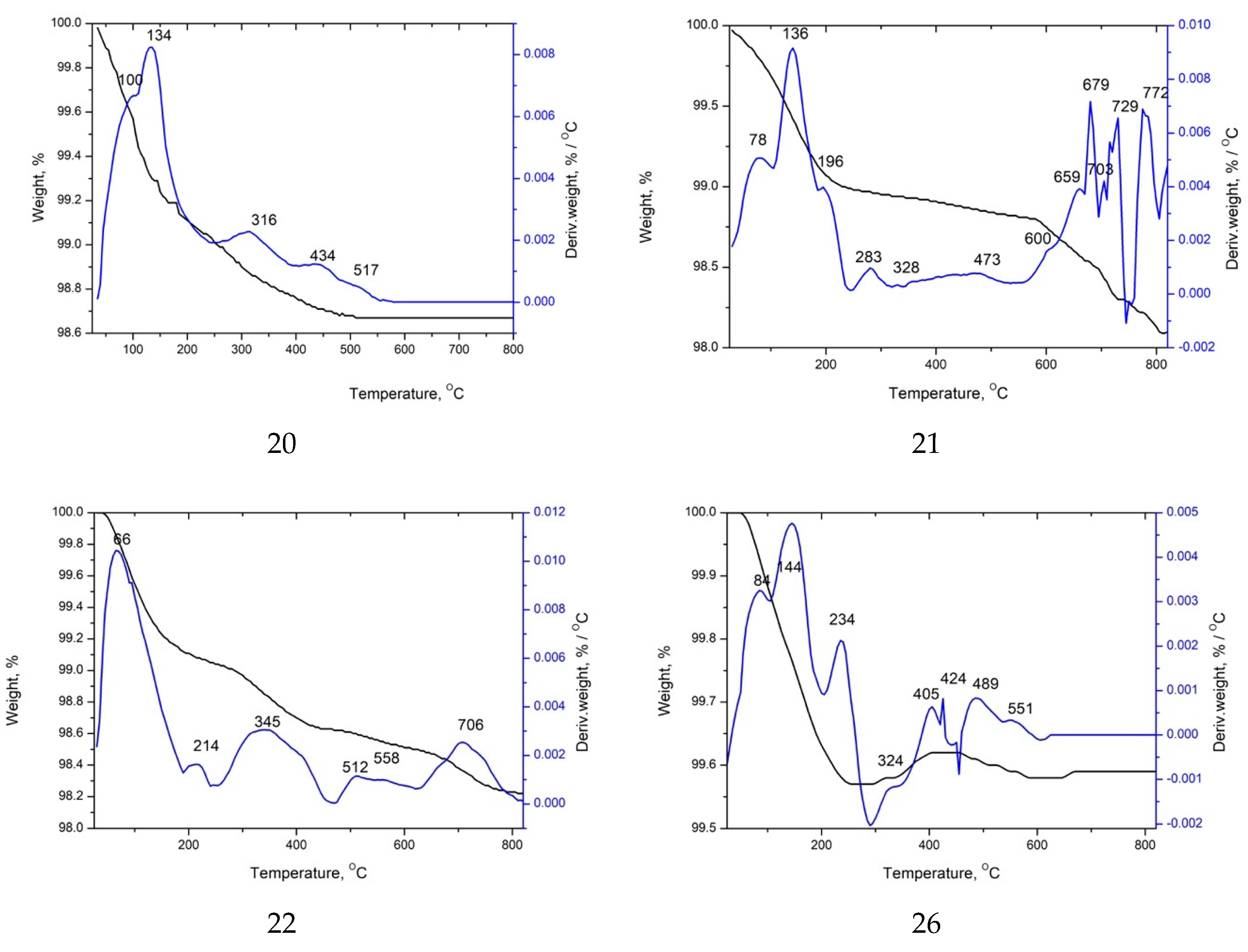
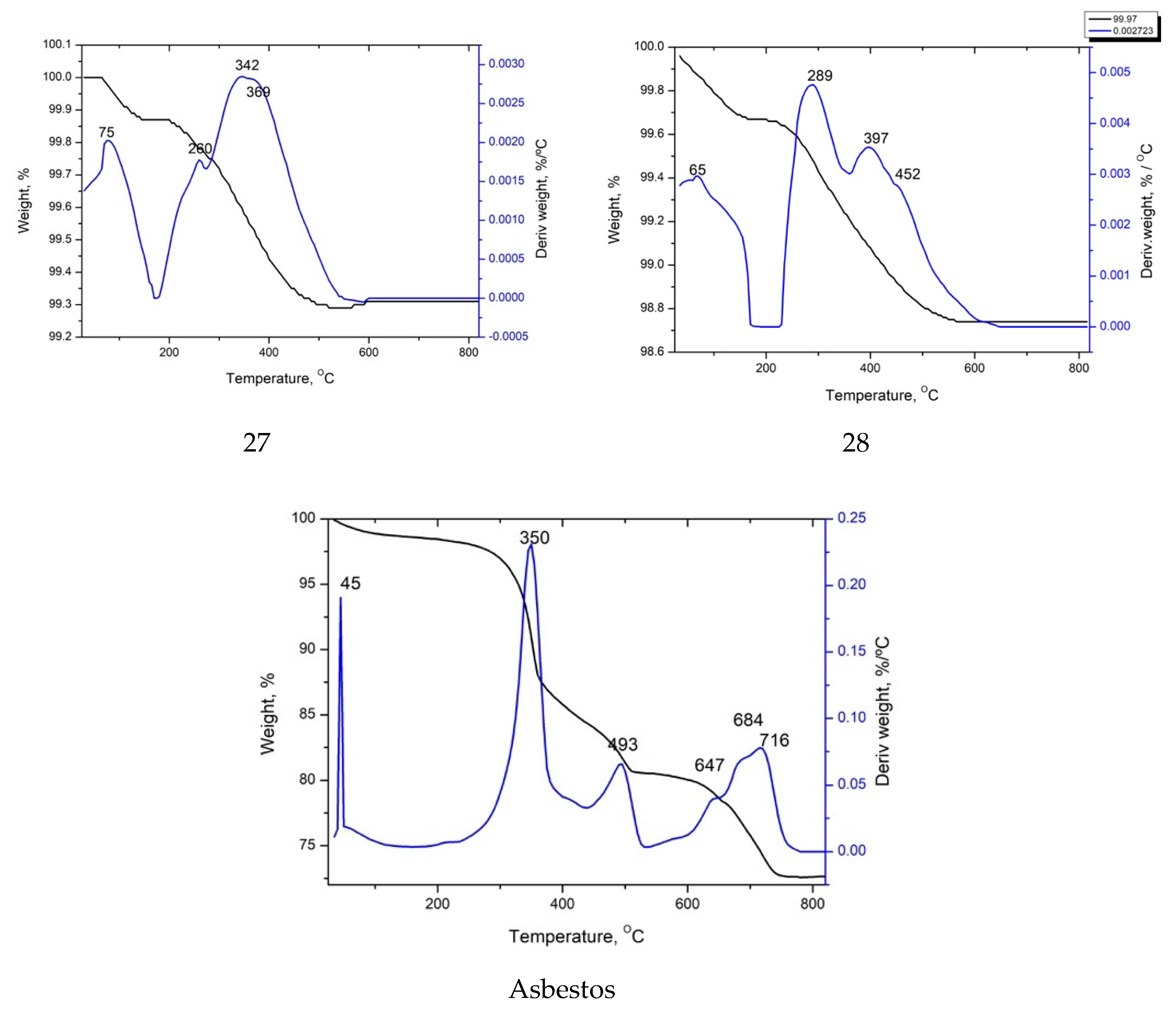
Temperature ranges (Table 2) were selected as a function of the major thermal reactions suffered by mortars during the heating: loss of adsorbed water (<120°C), dehydration of salts as well as loss of zeolitic water and/or other hygroscopic compounds (120-200°C), loss of structural water from hydraulic compounds like phyllosilicates, C-S-H and/or C-A-H (200-600°C), release of CO2 by decomposition of calcium carbonate (600-850°C), and other phenomena (>850°C) as decomposition of sulphates and/or loss of residual water and carbon dioxide [60]. These thermal reactions were clearly recorded in TGA/DTA curves and confirmed by FTIR. However, in these mortars, the dehydratation and decomposition of hydrated sulphates also occurred, as showed in figure 10. A double peak of CO2 emission likely due to the presence of Mg-bearing carbonates is also reported [61,62,63]. Results of the thermogravimetric and thermodifferential analyses (TGA-DTA) of the investigated samples showed six main thermal process: the first, at less than 100 °C, is due to the loss of weakly adsorbed water [64]. The bands around 550, 700 and 770 °C denote the release of bound water and are related to the dehydroxylation of minerals from serpentine subgroup species [65,66].
All the studied samples from this site are a sort of mortars and have hydraulic properties, with a CO2/H2O ratio quite low [67], indicating an slightly hydraulicity in majority. A possible cause could be the low concentration of lime, responsible for CO2 releases.
The most important thermal effects for a correct hydraulic classification are the weight loss of Structural Bound Water (200–600 OC), and decomposition of calcite and other carbonates (600–850 OC) with a consequent release of CO2.
In the DTA curves (Figure 10), these samples exhibits peaks attributed to decomposition of the carbonates, also, due to the presence of organic substances, indicating pozzolanic activity [68]. All the analyzed amphiboles could be assigned to calcic or sodic-calcic amphiboles with a tremolitic composition, reflecting medium-low pressure and low to medium temperature metamorphic conditions [69].

4.4. Chemical Analysis

The chemical composition of the the samples analyzed by X-ray fluorescence techniques also shows the details of the types of mortars (middle layers) and their uses at the other materials present in this archaeological site [70]. The samples have a low CaO content, which shows a close relationship between the CaO content and SiO2 + Fe2O3 + Al2O3, content, usually due both to their aggregates (volcanic aggregates). These samples also contain a higher percentage of K2O (4.78 ± 0.66 %), this content being slightly lower in the coating mortar – external layer (1.83 ± 0.05 %).
The carbonation process influences the microstructure of mortar by leading to alterations in its pore structure and density, which in turn affects the setting and hardening of the render. As carbon dioxide from the environment reacts with calcium hydroxide in the mortar, it forms calcium carbonate, impacting the overall strength and durability of the render through changes in moisture permeability and bond strength. [71].
Also, K2O and Na2O have an important role in Roman mortars. Alvarez et al. [72] reported that the addition of alkali as Na2O and K2O, led to a higher durability and turning them into suitable repair materials for the built heritage. The highest K2O concentration, the lowest Surface area value and the highest Na2O concentration, the highest Surface area value.
The reduced concentrations of K2O and Na2O in ancient Roman mortars lead to lower water absorption through capillarity, which enhances their durability against freezing and thawing cycles. These materials used in the middle layer could be considered as mortars and they exhibit textural properties characterized by the presence of small-diameter mesopores (2–5 nm), significantly smaller than those found in modern hydraulic mortars, which may contribute to their unique water adsorption characteristics and overall mechanical properties, resulting in higher durability and resistance.[73].

5. Discussion

Roman cities strategically used various mortars based on their construction needs, prioritizing durability and effectiveness in alignment with Vitruvius's principles. While selecting the most appropriate aggregates for each scope, there was a strong emphasis on sourcing materials locally to enhance efficiency; only when local options were insufficient would alternative materials, particularly pozzolans, be transported from other regions [2,74,75].
The 1st-century AD houses and industrial buildings at the Micia Roman site utilized mortars made from local inert siliceous aggregates, primarily andesite, characterized by a distinctive mineral composition. These aggregates mainly consisted of fine to medium-grained monocrystalline quartz sands, with a small admixture of less than 10% of carbonate and volcanic materials, reflecting the region's geological resources. The quartz sands utilized in the construction of these houses likely originate from sedimentary deposits near the Mureș River, located close to the settlement. Additionally, andesite, a volcanic rock commonly used in Roman mortars, may have been sourced as aggregate to enhance the hardness and strength of these middle material, underscoring the resourcefulness and regional material usage of ancient Roman builders. [76]. On the other hand, TGA analyses indicate a CO2/H2O ratio lower than 3 specific for slightly hydraulic one. Hydraulicity could be provided by the small fractions of volcanic aggregates detected in microscopic analysis in the analysed samples.
The Roman plastering technique effectively rendered walls waterproof, significantly enhancing their durability. A key technological advancement in the production of Roman mortars was the incorporation of both natural and artificial pozzolanic aggregates, which not only contributed to the material's hydraulic properties but also improved its strength and longevity in various environmental conditions. [77]. In addition, this type of mortar was probably applied to improve the waterproofing.
Asbestoid formations of the tremolite type were identified in the wall layers, considering that this mineral was accessible in the area, and was known for its magnificent properties. Or, during the reconstruction period, it was necessary to use a fireproof layer for the projection of the walls and especially of the religious constructions. This is how, in our expertise, the construction materials from these Roman monuments showed the presence of this asbestos mineral.
This type of aggregate may be transported from the Drobeta area where there was a strong trade with this raw material which was then distributed throughout the Mediterranean basin due to a significant increase in construction activities during the Imperial period, particularly related to civil works, and later spread across the Roman Empire, highlighting the interconnectivity and resource utilization characteristic of Roman engineering .[78].
The characteristics of the mortars studied at Micia are similar to those found at other Roman sites in the Mediterranean region, attesting to the continued use of the mortar manufacturing techniques stipulated by Vitruvio, and not only in Italy, but also in Slovenia, Tunisia, Turkey, Malta, and Romania [2,62,63,79,80,81].
The mortars applied in Micia were made by the most suitable aggregates and fibrillar tremolite-asbestos depending on the available raw materials and the intended use of these materials.
The chemical composition of the middle layer materials reveals the presence of volcanic aggregates with high SiO2 + Al2O3 + Fe2O3 values (higher than 80%), accompanied by the corresponding decrease in CaO values (5-8%).
These are black-grey materials that contains several ions, where the alkali ions, potassium ions (K+) and sodium ions (Na+) as predominant cations, while hydroxyl ions (OH−) play a crucial role in balancing the charge within the solution. The high abundance of K+ and Na+ contributes to the overall ionic strength and chemical properties of the pore fluid, with hydroxyl ions primarily serving to maintain charge neutrality.
Also, the alkalis present in the aggregates could increase the hydroxyl ion concentrations [82], lead to high alkalinity the solution, which are responsible for the dissolution of silicate anions.

5. Conclusions

Recognized as an important archaeological civil settlement, the Dacian Roman Micia site, is very reach in residential areas, industrial areas, port, public baths (civilian and military), amphitheater, religious areas (temples) and enjoying the facilities of a city. Even inside the Micia location, there were some constructive interventions, made during the long period of use of the buildings, it is important to identify for the first time in this part of Roman Dacia the presence of asbestos species, and the presence of a form of tremolite-asbestos, especially. Highly performant and sensitive analytical techniques have been used to put into evidence the tremolite-asbestos species, to identify the structure, composition and morphology of these minerals inside of the building materials from Roman monuments, as follows: optical, stereo and scanning electron microscopy (SEM), X-ray diffraction (XRD), X-ray fluorescence with wavelength dispersion (EDXRF), FTIR and Raman spectroscopy, thermal analysis (TGA/DTA). It is presumed that asbestos species has been used after the fire from Micia settlement in order to protect these monuments.
Visual, optical, stereo and scanning electron microscopic examination of the investigated samples put into evidence the presence of long fibers, translucid and flexible, elongated prismatic and fibrous crystal, a wavy structure, belonging to tremolite-asbestos. This species has been identified by XRD, XRF, FTIR (by the bands from 1400 cm-1), Raman (1000-2000 cm-1).
Asbestoid formations of the tremolite type were identified during the reconstruction period, it was necessary to use a fireproof layer for the projection of the walls and especially of the religious constructions. With a significant concentration of K2O and Na2O, the slightly hydraulic mortars identified with an increased durability. The highest K2O concentration, the lowest Surface area value and the highest Na2O concentration, the highest Surface area value.
The materials (repairing mortars) analyzed at Micia exhibit characteristics consistent with those observed at various Roman sites throughout the Mediterranean, indicating a sustained adherence to the mortar production methods outlined by Vitruvius. This continuity in technique underscores the influence of classical architectural principles on Roman construction practices across the region.

Author Contributions

Conceptualization, R.M.I., and M.G.B.; methodology, R.M.I., R.M.G., L.I.; software, V.I.G.; validation, R.M.I. and and M.G.B.; formal analysis, R.M.G.; investigation, V.I.G., S.S.T, A.I.G, G.V., L.I., R.M.G., E.A.; resources, R.M.I; data curation, R.M.I.; writing—original draft preparation, R.M.I., R.M.G. and M.G.B. writing—review and editing, R.M.I.; visualization, R.M.I.; supervision, R.M.I.; project administration, R.M.G., R.M.I..; funding acquisition, R.M.I. All authors have read and agreed to the published version of the manuscript.

Funding

This work received financial support from the project PN 23.06 NUCLEU Program - ChemNewDeal within the National Plan for Research, Development and Innovation 2022-2027, developed with the support of Ministry of Research, Innovation, and Digitization, project no. PN 23.06.02.01 (InteGral).

Conflicts of Interest

The authors declare no conflicts of interest.

References

  1. Adam, J. Roman building: materials and techniques; Taylor & Francis Group: London, 1994. [Google Scholar]
  2. Ion, R.-M.; Barbu, M.G.; Gonciar, A.; Vasilievici, G.; Gheboianu, A.I.; Slamnoiu-Teodorescu, S.; David, M.E.; Iancu, L.; Grigorescu, R.M. A Multi-Analytical Investigation of Roman Frescoes from Rapoltu Mare (Romania). Coatings 2022, 12, 530. [Google Scholar] [CrossRef]
  3. Ion, R.M.; Bakirov, B.A.; Kichanov, S.E.; Kozlenko, D.P.; Belushkin, A.V.; Radulescu, C.; Dulama, I.D.; Bucurica, I.A.; Gheboianu, A.I.; Stirbescu, R.M. Non-Destructive and micro-invasive techniques for characterizing the ancient Roman Mosaic fragments. Appl. Sci. 2020, 10, 3781. [Google Scholar] [CrossRef]
  4. Borsoi, G.; Silva, A.S.; Menezes, P.; Candeias, A.; Mirão, J. Analytical characterization of ancient mortars from the archaeological roman site of Pisões (Beja, Portugal). Constr. Build. Mater. 2019, 204, 597–608. [Google Scholar] [CrossRef]
  5. Bakolas, A.; Biscontin, G.; Contardi, V.; Franceschi, E.; Moropoulou, A.; Palazzi, D.; Zendri, E. Thermoanalytical research on traditional mortars in Venice. Thermochim. Acta 1995, 269, 817–828. [Google Scholar] [CrossRef]
  6. Belfiore, C.M.; Fichera, G.V.; La Russa, M.F.; Pezzino, A.; Ruffolo, S.; Galli, G.; Barca, D. A Multidisciplinary Approach for the Archaeometric Study of Pozzolanic Aggregate in R oman Mortars: The Case of V illa dei Q uintili (R ome, I taly). Archaeometry 2015, 57, 269–296. [Google Scholar] [CrossRef]
  7. Belfiore, C.M.; Fichera, G.V.; Ortolano, G.; Pezzino, A.; Visalli, R.; Zappalà, L. Image processing of the pozzolanic reactions in Roman mortars via X-Ray Map Analyser. Microchem. J. 2016, 125, 242–253. [Google Scholar] [CrossRef]
  8. Böke, H.; Çizer, Ö.; İpekoğlu, B.; Uğurlu, E.; Şerifaki, K.; Toprak, G. Characteristics of lime produced from limestone containing diatoms. Constr. Build. Mater. 2008, 22, 866–874. [Google Scholar] [CrossRef]
  9. Bruno, P.; Calabrese, D.; Di Pierro, M.; Genga, A.; Laganara, C.; Manigrassi, D.; Traini, A.; Ubbrıaco, P. Chemical–physical and mineralogical investigation on ancient mortars from the archaeological site of Monte Sannace (Bari—Southern Italy). Thermochim. Acta 2004, 418, 131–141. [Google Scholar] [CrossRef]
  10. DeLaine, J. Theory and practice of Roman architecture: studies offered to Pierre Gros. 2006.
  11. DeLaine, J. Production, transport and on-site organisation of Roman mortars and plasters. Archaeol. Anthropol. Sci. 2021, 13, 1–17. [Google Scholar] [CrossRef]
  12. Alicu, D. Turnuri de porti la castrele romane din Dacia. Acta Musei Napoc. 1973, 107–122. [Google Scholar]
  13. Alicu, D. Micia: Studii monografice. 1. Monumentele de spectacol şi de cult. 2004.
  14. Petculescu, L. Sapaturile arheologice din castrul Micia. Cercet. Arheol. 1984, VII, 117–119. [Google Scholar]
  15. Andritoiu, I. Istoricul cercetarilor privitoare la asezarea si fortificatia romana de la Micia. Sargetia 2003, XXXI, 181–202. [Google Scholar]
  16. Andrițoiu, I. Necropolele Miciei; Excelsior Art: Timisoara, 2006. [Google Scholar]
  17. Barbu, M.G. Consideratii privind planimetria castrului Micia. In Proceedings of the Simpozionul Stiintific National "Micia Rediviva", Deva, 2018., October 24-26.
  18. Czajlik, Z.; Berencki, S.; Rupnik, L. Aerial geoarchaeological survey in the valleys of the Mureş and Arieş rivers (2009-2013). Diss. Archaeol. 2014, 459–483. [Google Scholar] [CrossRef]
  19. Daicoviciu, C. Micia, I. Cercetări asupra castrului. Analele Comisiei Monumentelor Istorice; 1931; pp. 1–43.
  20. Floca, O.; Marghitan, L. Noi consideratii privitoare la castrul roman de la Micia. Sargetia 1970, VII, 43–57. [Google Scholar]
  21. Barbu, M.G.; Barbat, I.A. New Archaeological Information Regarding the Exploitation of Andesite in Măgura Uroiului (Hunedoara County). Sargetia 2017, VIII, 71–121. [Google Scholar]
  22. Barbu, M.G.; Barbat, I.A.; Tutuianu, C.-D. New Data about the Exploitation of Andesite in Prehistory and Antiquity in the Proximity of Deva City. Terra Sebus 2021, 13, 57–91. [Google Scholar]
  23. Petculescu, L. Cercetari arheologice in castrul roman de la Micia. Cercet. Arheol. 1979, III, 111–114. [Google Scholar]
  24. Neigebaur, J.D.F. Dacien: aus den Ueberresten des klassischen Alterthums, mit besonderer Rücksicht auf Siebenbürgen; Gött: 1851.
  25. Negula, D.I.; Moise, C.; Lazăr, A.M.; Rișcuța, N.C.; Cristescu, C.; Dedulescu, A.L.; Mihalache, C.E.; Badea, A. Satellite remote sensing for the analysis of the Micia and Germisara archaeological sites. Remote Sens. 2020, 12, 2003. [Google Scholar] [CrossRef]
  26. Bostock, J.; Riley, H.T. The Natural History. Pliny the Elder.. 1885. [Google Scholar]
  27. King, D.; History of Asbestos. Asbestos.com. Available online: https://www.asbestos.com/asbestos/history/. (accessed on October 1).
  28. Bianchi, C.; Bianchi, T. Asbestos between science and myth. A 6,000-year story. La Med. Del Lav. 2015, 106, 83–90. [Google Scholar]
  29. Oltean, I.; Hanson, W. Military vici in Roman Dacia: an aerial perspective. Acta Musei Napoc. 2001, 38, 123–134. [Google Scholar]
  30. Oltean, I.A.; Hanson, W.S. Defining the Roman limes in Romania: the contribution of aerial and satellite remote sensing. In Proceedings of the Limes XXII: Proceedings of the 22nd International Congress of Roman Frontier Studies, Ruse, Bulgaria, 2012; pp. 889–900.
  31. Marinescu, L.; Sion, A.; Petculescu, L.; Andritoiu, I.; Ionescu, E.; Braileanu, M. Santierul arheologic Micia (Vetel, jud. Hunedoara). Cercet. Arheol 1979, 3, 105–126. [Google Scholar]
  32. Petculescu, L.; Nemoianu, A.T.; Anastasiu, R. Raport preliminar asupra săpăturilor arheologice din 1978– 1979 în castrul Micia, co-muna Vetel, jud. Hunedoara. Cercet. Arheol. 1981, IV, 70–75. [Google Scholar]
  33. Petculescu, L. Castrul de la Micia (Vețel, jud. Hunedoara) în cadrul sistemului de apărare al Daciei romane. MUZEUL NAȚIONAL 1981, 109–114. [Google Scholar]
  34. Petculescu, L. Raport asupra sapaturilor arheologice din castrul Micia. Cercet. Arheol. 1983, VI, 45–50. [Google Scholar]
  35. Petculescu, L.; Filipescu, B.; Craciun, C. Raport asupra sapaturilor arheologice din castrul Micia (cpm. Vetel, Jud. Hunedoara), in campania 1983. Cercet. Arheol. 1986, VIII, 59–62. [Google Scholar]
  36. Allaby, M. A dictionary of earth sciences 3th Edition. 2008.
  37. D-16, A. Standard practice for calculation of color tolerances and color differences from instrumentally measured color coordinates. 2016.
  38. 15886, U.-E. Conservation of Cultural Property—Test Methods—Colour Measurement of Surfaces. 2011.
  39. Gheorghiu, C.; Mares, I. Erupţiunile neogene din Valea Mureşului (regiunea Deva-Zam) şi particularităţile petrografice ale rocilor; 1961.
  40. Leake, B.E.; Woolley, A.R.; Arps, C.E.; Birch, W.D.; Gilbert, M.C.; Grice, J.D.; Hawthorne, F.C.; Kato, A.; Kisch, H.J.; Krivovichev, V.G. Nomenclature of amphiboles; report of the Subcommittee on Amphiboles of the International Mineralogical Association Commission on new minerals and mineral names. Mineral. Mag. 1997, 61, 295–310. [Google Scholar] [CrossRef]
  41. Hawthorne, F.C.; Oberti, R.; Harlow, G.E.; Maresch, W.V.; Martin, R.F.; Schumacher, J.C.; Welch, M.D. Nomenclature of the amphibole supergroup. Am. Mineral. 2012, 97, 2031–2048. [Google Scholar] [CrossRef]
  42. Ballirano, P.; Bloise, A.; Gualtieri, A.F.; Lezzerini, M.; Pacella, A.; Perchiazzi, N.; Dogan, M.; Dogan, A.U. The crystal structure of mineral fibres. In Mineral Fibres: Crystal Chemistry, Chemical-Physical Properties, Biological Interaction and Toxicity, Gualtieri, A.F., Ed.; European Mineralogical Union: 2017; Volume 18, p. 0.
  43. Altaner, S.P.; Grim, R.E. Mineralogy, chemistry, and diagenesis of tuffs in the Sucker Creek Formation (Miocene), eastern Oregon. Clays Clay Miner. 1990, 38, 561–572. [Google Scholar] [CrossRef]
  44. Andreozzi, G.B.; Ballirano, P.; Gianfagna, A.; Mazziotti-Tagliani, S.; Pacella, A. Structural and spectroscopic characterization of a suite of fibrous amphiboles with high environmental and health relevance from Biancavilla (Sicily, Italy). Am. Mineral. 2009, 94, 1333–1340. [Google Scholar] [CrossRef]
  45. Bozhilov, K.N.; Jenkins, D.M. Analytical electron microscopy of tremolite. Mod. Res. Educ. Top. Microsc. 2007, 2, 616–625. [Google Scholar]
  46. Elsen, J. Microscopy of historic mortars—a review. Cem. Concr. Res. 2006, 36, 1416–1424. [Google Scholar] [CrossRef]
  47. Yang, H.; Evans, B.W. X-ray structure refinements of tremolite at 140 and 295 K; crystal chemistry and petrologic implications. Am. Mineral. 1996, 81, 1117–1125. [Google Scholar] [CrossRef]
  48. Zussman, J. Minerals and the electron microscope. Mineral. Mag. 1987, 51, 129–138. [Google Scholar] [CrossRef]
  49. Hawthorne, F.C.; Ventura, G.D.; Robert, J.-L.; Welch, M.D.; Raudsepp, M.; Jenkins, D.M. A Rietveld and infrared study of synthetic amphiboles along the potassium-richterite–tremolite join. Am. Mineral. 1997, 82, 708–716. [Google Scholar] [CrossRef]
  50. Petry, R.; Mastalerz, R.; Zahn, S.; Mayerhöfer, T.G.; Völksch, G.; Viereck-Götte, L.; Kreher-Hartmann, B.; Holz, L.; Lankers, M.; Popp, J. Asbestos Mineral Analysis by UV Raman and Energy-Dispersive X-ray Spectroscopy. Chemphyschem: A Eur. J. Chem. Phys. Phys. Chem. 2006, 7, 414–420. [Google Scholar] [CrossRef]
  51. Della Ventura, G.; Vigliaturo, R.; Gieré, R.; Pollastri, S.; Gualtieri, A.F.; Iezzi, G. Infra Red spectroscopy of the regulated asbestos amphiboles. Minerals 2018, 8, 413. [Google Scholar] [CrossRef]
  52. Millette, J.R.; Bandli, B.R. Asbestos identification using available standard methods. MICROSCOPE-LONDON THEN CHICAGO- 2005, 53, 179. [Google Scholar]
  53. Campopiano, A.; Olori, A.; Cannizzaro, A.; Iannò, A.; Capone, P.P. Quantification of tremolite in friable material coming from Calabrian ophiolitic deposits by infrared spectroscopy. J. Spectrosc. 2015, 2015, 974902. [Google Scholar] [CrossRef]
  54. Speil, S.; Leineweber, J. Asbestos Minerals in. Environ. Res. 1969, 2. [Google Scholar] [CrossRef]
  55. Apopei, A.I.; Buzgar, N. The Raman study of amphiboles. Analele Stiintifice De Univ. AI Cuza Din Iasi. Sect. 2 Geol. 2010, 56, 57. [Google Scholar]
  56. Rinaudo, C.; Belluso, E.; Gastaldi, D. Assessment of the use of Raman spectroscopy for the determination of amphibole asbestos. Mineral. Mag. 2004, 68, 455–465. [Google Scholar] [CrossRef]
  57. Bersani, D.; Andò, S.; Scrocco, L.; Gentile, P.; Salvioli-Mariani, E.; Fornasini, L.; Lottici, P.P. Composition of amphiboles in the tremolite–ferro–actinolite series by Raman Spectroscopy. Minerals 2019, 9, 491. [Google Scholar] [CrossRef]
  58. Leissner, L. Crystal chemistry of amphiboles studied by Raman spectroscopy. Mineral. -Petrogr. Inst. -Univ. Hambg. Hambg. Ger. 2014. [Google Scholar]
  59. Leissner, L.; Schlüter, J.; Horn, I.; Mihailova, B. Exploring the potential of Raman spectroscopy for crystallochemical analyses of complex hydrous silicates: I. Amphiboles. Am. Mineral. 2015, 100, 2682–2694. [Google Scholar] [CrossRef]
  60. Hawthorne, F.C.; Oberti, R. Amphiboles: crystal chemistry. Rev. Mineral. Geochem. 2007, 67, 1. [Google Scholar] [CrossRef]
  61. Rispoli, C.; De Bonis, A.; Esposito, R.; Graziano, S.F.; Langella, A.; Mercurio, M.; Morra, V.; Cappelletti, P. Unveiling the secrets of Roman craftsmanship: mortars from Piscina mirabilis (Campi Flegrei, Italy). Archaeol. Anthropol. Sci. 2020, 12, 1–18. [Google Scholar] [CrossRef]
  62. Kramar, S.; Zalar, V.; Urosevic, M.; Körner, W.; Mauko, A.; Mirtič, B.; Lux, J.; Mladenović, A. Mineralogical and microstructural studies of mortars from the bath complex of the Roman villa rustica near Mošnje (Slovenia). Mater. Charact. 2011, 62, 1042–1057. [Google Scholar] [CrossRef]
  63. Ergenç, D.; Fort, R.; Varas− Muriel, M.J.; Alvarez de Buergo, M. Mortars and plasters—How to characterize aerial mortars and plasters. Archaeol. Anthropol. Sci. 2021, 13, 197. [Google Scholar] [CrossRef]
  64. Kostova, B.; Dumanov, B.; Stoyanov, V.; Shivachev, B. Thermal and phase analysis of Roman and Late Antiquity mortars from Bulgarian archaeological sites. J. Therm. Anal. Calorim. 2023, 148, 1543–1555. [Google Scholar] [CrossRef]
  65. Bloise, A.; Catalano, M.; Barrese, E.; Gualtieri, A.F.; Bursi Gandolfi, N.; Capella, S.; Belluso, E. TG/DSC study of the thermal behaviour of hazardous mineral fibres. J. Therm. Anal. Calorim. 2016, 123, 2225–2239. [Google Scholar] [CrossRef]
  66. Viti, C. Serpentine minerals discrimination by thermal analysis. Am. Mineral. 2010, 95, 631–638. [Google Scholar] [CrossRef]
  67. Izzo, F.; Grifa, C.; Germinario, C.; Mercurio, M.; De Bonis, A.; Tomay, L.; Langella, A. Production technology of mortar-based building materials from the Arch of Trajan and the Roman Theatre in Benevento, Italy. Eur. Phys. J. Plus 2018, 133, 1–12. [Google Scholar] [CrossRef]
  68. Moropoulou, A.; Bakolas, A.; Anagnostopoulou, S. Composite materials in ancient structures. Cem. Concr. Compos. 2005, 27, 295–300. [Google Scholar] [CrossRef]
  69. Gambino, F.; Glarey, A.; Cossio, R.; Appolonia, L.; d’Atri, A.; Borghi, A. SEM-EDS characterization of historic mortar as a tool in archaeometric study: an updated analytical protocol tested on the Roman theatre of Aosta (NW Italy). Archaeol. Anthropol. Sci. 2022, 14, 179. [Google Scholar] [CrossRef]
  70. De Luca, R.; Cau Ontiveros, M.A.; Miriello, D.; Pecci, A.; Le Pera, E.; Bloise, A.; Crisci, G.M. Archaeometric study of mortars and plasters from the Roman City of Pollentia (Mallorca-Balearic Islands). Period. Di Mineral. 2013, 82, 353–379. [Google Scholar]
  71. Santos, A.R.; do Rosário Veiga, M.; Silva, A.S.; de Brito, J.; Álvarez, J.I. Evolution of the microstructure of lime based mortars and influence on the mechanical behaviour: The role of the aggregates. Constr. Build. Mater. 2018, 187, 907–922. [Google Scholar] [CrossRef]
  72. Alvarez, J.I.; Martı́n, A.; Casado, P.G.a.; Navarro, I.; Zornoza, A. Methodology and validation of a hot hydrochloric acid attack for the characterization of ancient mortars. Cem. Concr. Res. 1999, 29, 1061–1065. [Google Scholar] [CrossRef]
  73. Stade, H. On the reaction of CSH (di, poly) with alkali hydroxides. Cem. Concr. Res. 1989, 19, 802–810. [Google Scholar] [CrossRef]
  74. Pagano, S.; Germinario, C.; Alberghina, M.F.; Covolan, M.; Mercurio, M.; Musmeci, D.; Piovesan, R.; Santoriello, A.; Schiavone, S.; Grifa, C. Multilayer technology of decorated plasters from the domus of Marcus Vipsanus Primigenius at Abellinum (Campania Region, Southern Italy): an analytical approach. Minerals 2022, 12, 1487. [Google Scholar] [CrossRef]
  75. Ward-Perkins, J.B. Roman imperial architecture; Yale University Press: 1994.
  76. Özkaya, Ö.A.; Böke, H. Properties of Roman bricks and mortars used in Serapis temple in the city of Pergamon. Mater. Charact. 2009, 60, 995–1000. [Google Scholar] [CrossRef]
  77. Fort, R.; Varas-Muriel, M.; Zoghlami, K.; Ergenç, D.; Zaddem, A. Analytical characterisation of 1st-and 2nd-century Roman mortars at the Utica archaeological site (Tunisia): Construction phases and provenance of the raw materials. J. Archaeol. Sci. : Rep. 2024, 54, 104404. [Google Scholar] [CrossRef]
  78. Marra, F.; Danti, A.; Gaeta, M. The volcanic aggregate of ancient Roman mortars from the Capitoline Hill: Petrographic criteria for identification of Rome's “pozzolans” and historical implications. J. Volcanol. Geotherm. Res. 2015, 308, 113–126. [Google Scholar] [CrossRef]
  79. Farci, A.; Floris, D.; Meloni, P. Water permeability vs. porosity in samples of Roman mortars. J. Cult. Herit. 2005, 6, 55–59. [Google Scholar] [CrossRef]
  80. Jackson, M.D.; Logan, J.M.; Scheetz, B.E.; Deocampo, D.M.; Cawood, C.G.; Marra, F.; Vitti, M.; Ungaro, L. Assessment of material characteristics of ancient concretes, Grande Aula, Markets of Trajan, Rome. J. Archaeol. Sci. 2009, 36, 2481–2492. [Google Scholar] [CrossRef]
  81. Pleuger, E.; Goiran, J.-P.; Mazzini, I.; Delile, H.; Abichou, A.; Gadhoum, A.; Djerbi, H.; Piotrowska, N.; Wilson, A.; Fentress, E. Palaeogeographical and palaeoenvironmental reconstruction of the Medjerda delta (Tunisia) during the Holocene. Quat. Sci. Rev. 2019, 220, 263–278. [Google Scholar] [CrossRef]
  82. Kawabata, Y.; Yamada, K.; Igarashi, G.; Sagawa, Y. Effects of Soak Solution Type on Alkali Release from Volcanic Aggre-gates− Is Alkali Release Really Responsible for Accelerating ASR Expansion?−. J. Adv. Concr. Technol. 2018, 16, 61–74. [Google Scholar] [CrossRef]
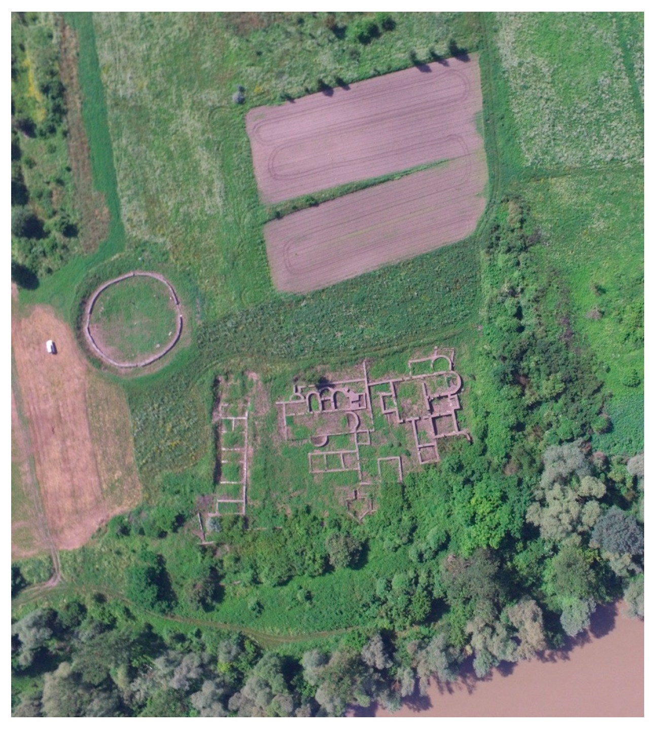
Figure 1. Location of the Micia site (Personal photo).
Preprints 121559 g001
Figure 2. Map of Romania (up) and Micia location (down) (Google Earth).
Preprints 121559 g002
Figure 3. Photo (a), optical microscopy image (b) and Image J processed images of the investigated sample (P26 example).
Preprints 121559 g003
Figure 4. The optical microscopy images of the samples from Carriers’ fronts.
Preprints 121559 g004aPreprints 121559 g004b
Figure 5. Optical (a-e) and SEM (f, g) images of Venus and Diana altars.
Preprints 121559 g005
Figure 6. XRD for Micia samples.
Preprints 121559 g006
Figure 7. WDXRF analysis for all the samples collected from Micia Roman monuments.
Preprints 121559 g007
Figure 8. FTIR spectra of the main samples collected from Roman monuments: (a) all the investigated samples; (b) samples 20, 23 and 28; (c) asbestos.
Preprints 121559 g008
Figure 9. Raman spectra obtained with 782 nm, for 20,23,28 and asbestos samples.
Preprints 121559 g009
Figure 10. TGA/DTA graphs of the investigated samples.
Preprints 121559 g010aPreprints 121559 g010b
Table 1. Photo of the samples studied.
Preprints 121559 i001a
Preprints 121559 i001b
Preprints 121559 i001c
Table 2. Weight losses (%) at certain temperature ranges, calculated from TGA data.
Samples <120 120–200 200–600 >600 CO2/H2O
20 0.61 0.28 0.44 0 0
21 0.44 0.49 0.32 0.65 2.03
22 0.61 0.28 0.6 0.29 0.48
26 0.17 0.2 0.05 0.01 0.2
27 0.1 0.03 0.56 0 0
28 0.26 0.07 0.87 0 0
asbestos 3.59 1.2 4.23 10.15 2.4
Table 3. BET specific surface area (S.A.), total pore volume (Vtot) and maximum values of pore diameter (D) of Micia samples.
Sample Surface Area (m2/g) Pore Volume (cc/g) Pore Diameter (nm) K2O
(%)
Na2O
(%)
20 1.215 0.001 3.083 1.97 4.23
21 1.751 0.002 2.945 1.61 4.82
22 1.531 0.002 4.786 1.79 3.22
26 0.893 0.001 4.807 1.87 4.01
27 0.112 0.000 4.759 2.08 3.29
28 0.522 0.001 3.668 1.81 3.08
Asbestos 0.375 0.001 2.949
Disclaimer/Publisher’s Note: The statements, opinions and data contained in all publications are solely those of the individual author(s) and contributor(s) and not of MDPI and/or the editor(s). MDPI and/or the editor(s) disclaim responsibility for any injury to people or property resulting from any ideas, methods, instructions or products referred to in the content.
Copyright: This open access article is published under a Creative Commons CC BY 4.0 license, which permit the free download, distribution, and reuse, provided that the author and preprint are cited in any reuse.
Prerpints.org logo

Preprints.org is a free preprint server supported by MDPI in Basel, Switzerland.

Subscribe

© 2024 MDPI (Basel, Switzerland) unless otherwise stated