Preprint
Article

Evaluation of Osseointegration Levels of Different Surface Feature Titanium Implants Installed Simultaneously with Graft: An Animal Study

Submitted:

21 November 2024

Posted:

21 November 2024

You are already at the latest version

Abstract

Background and Objectives: This study aims to assess the bone implant osseointegration after the healing process of machined, reabsorbable blast material (RBM) and sandblasted and large acid grid (SLA) surface titanium implants placed simultaneously using graft material in rat tibias. Materials and Methods: The study involved 30 Sprague Dawley rats that were divided into three groups: machined surfaced (MS) (n = 10), resorbable blast material (RBM) (n = 10), and sandblasted and large acid grid (SLA) surfaced (n = 10). The titanium implants were inserted into the bone sockets along with the graft, and the rats were euthanized after a 4-week experimental period. The implants and the bone tissue surrounding them were extracted for reverse torque analysis (Newton’s). Results: The biomechanical bone implant contact rate was greater in SLA-surfaced implants than in RBM and machined surface implants (P<0.05). Conclusions: The application of graft material may be more effective in implants with SLA and RBM surfaces when used locally, as per the biomechanical parameters.

Keywords: 
;  ;  ;  ;  

1. Introduction

Dental implant supported prostheses, which are connected without connective tissue between the bone surface, has been a scientifically accepted and frequently used treatment option in complete and partial edentulism [1,2]. However, jawbone defects cause problems in the placement and function of dental implants in the jawbone. In order to overcome these problems, various biomaterials and methods are used in the jaw bone defects treatment [3]. In this age, with the increase in aesthetic perception and the widespread use of chairside technology, patients desire the radip resolution of their existing bone defects. The succesful and desirable early clinical outcome of dental implants depend on bone implant osseointegration. Dental implant topography is one of the most important parameters affecting bone implant osseointegration [4]. The categorization of dental implant topography can be done on a scale basis into macro, micro, and nano subgroups [5,6]. The visible shape of an implant defines its macrotopography [7]. The microtopography is determined by mikro roughness (1-100 μm) [8]. Changes in microtopography influence the growth, metabolism, movement, and the production of cytokines and growth factors in osteogenic cells by expanding the surface area [9]. It is thought that the nanotopography (1-100nm) of implants have an effect on a cellular and protein level [10]. Implant topography modifications fall into three interrelated categories; physical, mechanical and chemical [11]. Machine, SLA and RBM are micro level modifications in the physical category. These modifications can enhance the fibrin matrix formation as an osteoconductive scaffold by increasing the surface area of the implants on a micrometer scale [12]. In clinical studies in which implant surfaces were evaluated in terms of bone implant osteintegration, it has been observed that implant surface systems do not have a complete superiority to each other [13,14]. It can be thought that the bone surface may have an effect on the implant osteointegration in implants placed simultaneously with graft material. This research intended to examine the osteointegration of titanium implants with turned, sandblasted, etched and roughened surface, which was implanted in rat tibias along with graft material, post-healing. The hypothesis of this study is that different implant surface treatments may affect the osteointegration of implants applied simultaneously with the graft material.

2. Materials and Methods

2.1. Animals and Study Design

All experimental and surgical process was conducted at the Experimental Research Center of Firat University, in Elazıg,Turkey. Fırat University’s Animal Experiments Ethics Committee granted ethical approval for the research and supplied the study animals (XXX, XXX). The study adhered to all recommendations of the Declaration of Helsinki regarding the protection of laboratory research animals of the World Medical Association. Thirty healthy adult female Sprague-Dawley rats, aged 1 to 1.5 years, were included in the study. The rats’ average body weight ranged from 250 to 300 g at the start of the experimental protocol. The rats were housed in a temperature-controlled environment using plastic cages. The rats were allowed to eat and drink freely and were exposed to a 12-hour light-dark cycle throughout the study. The rats were randomly allocated into three groups, each with ten rats, ensuring similar mean weights across all groups. Experimental groups; The implant groups were formed from turned (n=10), roughened with fusible material (n=10) and sandblasted etched surface (n=10) implant groups.

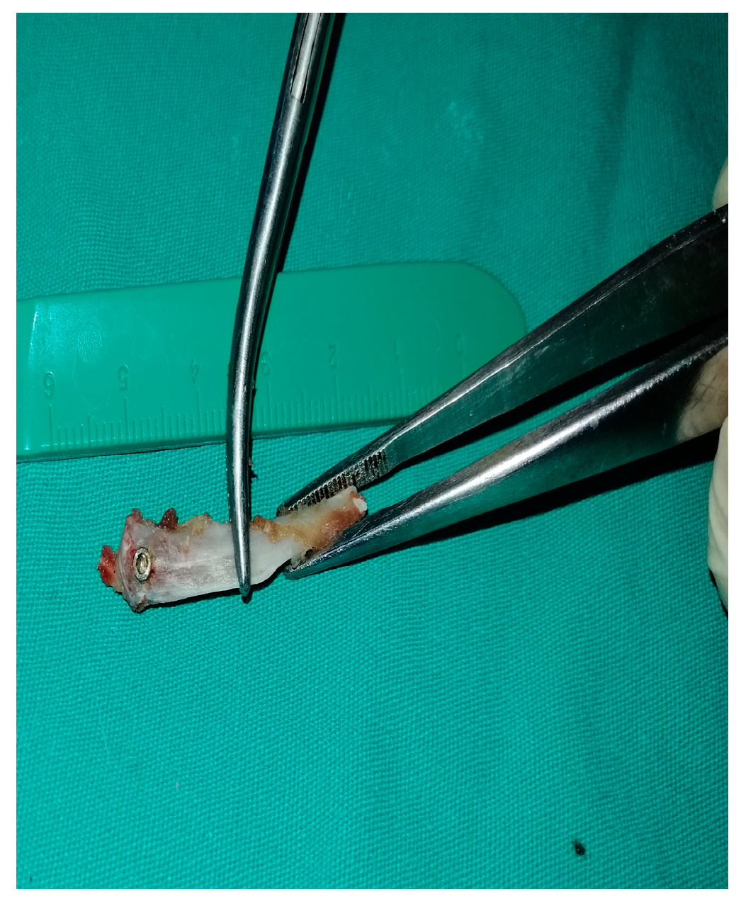
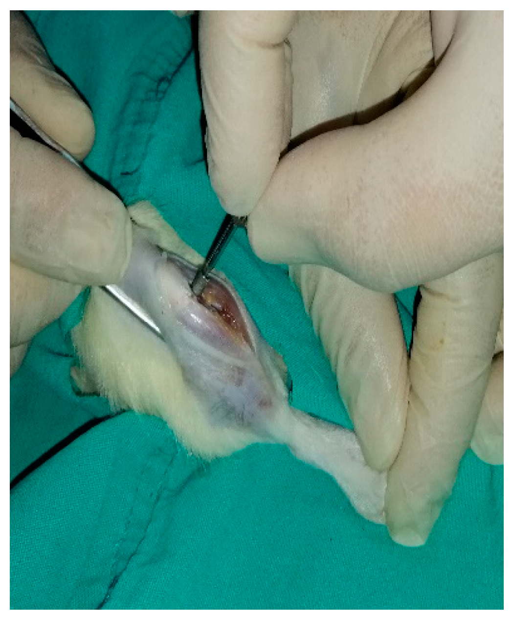
2.2. Surgical Procedures

No additional nutritional supplements were administered before, during or after the experiment and no state of deprivation was applied. All surgical procedures were carried out in compliance with the principles of maintaining a sterile environment and preventing the growth of microorganisms. All subjects were administered 35 mg/kg of ketamine hydrochloride and 5 mg/kg of xylazine intramuscularly for anesthesia. After general anesthesia, the surgical area was washed with povidone-iodine and shaved. After the surgical area was cleaned with povidone iodine, it was covered with sterile drapes that exposed the surgical area. First, the cavities opened to the corticocancellous bone in the metaphyseal part of the right tibia bones of all rats were filled with bovine bone grafts. Then titanium implants with turned surface (n=10), roughened surface with soluble material (n=10) and sandblasted acidified surface (n=10) were placed inside the graft (Figure 1). The crest was incised to access the metaphyseal part of the right tibia. The skin flap was lifted and the periosteum was dissected. Each tibia was made the bone sockets that measured 4 mm in diameter and 5 mm in length. Bone grafts were inserted into the sockets, and 2.5 mm diameter and 4 mm length titanium implants (TiAl6V4, Implance Dental Implant Systems, AGS Medikal Corporation, Istanbul, Turkey) were placed into the grafts simultaneously. After these procedures, the subjects’ skin and soft tissues were returned to their initial positions and closed using 4-0 prolene sutures.At the end of the 12-week recovery period, the subjects were sacrificed (Figure 2). Bone-implant fusion analyzes of the obtained samples were performed using a biomechanical reverse torque device (Figure 3). The data was obtained as force. Improperly placed implants were excluded from the study.

2.3. Statistical Analysis

IBM SPSS Statistics 22 software was utilized to perform statistical analysis on the data in the study. The normality of the parameters was assessed using the Kolmogorov-Smirnov test. The Kruskal Wallis test was used to compare the non-normally distributed parameters between groups, and identified the group that caused the difference using the Mann Whitney U test. Significance was evaluated at the P<0.05 level.

3. Results

When compared, the machined surface (4.04 N/cm) showed statistically significantly lower biomechanical bone implant osseointegration than both RBM (6.88 N/cm) and SLA (11.11 N/cm) surfaces. When the RBM and SLA surfaces were compared, the SLA surface showed statistically significantly higher biomechanical bone implant osseointegration than the RBM surface (Table 1).

4. Discussion

In dental implant treatment, there may not always be enough bone tissue to allow the surgical placement of the implant due to reasons such as; vertical and horizontal bone deficiencies, sinus droop, and non-ideal conditions such as low bone mineral density and osteoporosis. Graft materials and implants with the ideal surface properties that can be simultaneously applied with them in order to have successful and early clinical results in patients with bone defects have attracted the attention of researchers [2,4]. In this study, a statistically significant difference was found in the osteintegration values between the MS, SLA and RBM surface dental implants placed simultaneously with the graft. The biomechanical bone-implant osseointegration values of the SLA and RBM surface implants were found to be higher than the machined surface implants. In addition, when the RBM and SLA surfaced implants were compared, it was observed that the osseointegration values of the SLA surfaced implants were statistically higher than the RBM surfaced implants. These results suggest that implant surface preparation methods are not only effective in the interaction of healthy bone tissue with the implant surface, but also in the interaction of graft materials and the implant surface [2,4]. It has been reported in many studies that rough surface implants are more advantageous in bone-implant osseointegration than machine surface implants. Machined surfaces are used as a control group in studies examining implant surface properties [2,4]. Demetoğlu et al. clinically compared RBM and SLA surface implants in their study involving 2005 patients and reported that a significant difference in terms of osseointegration success between the two groups of implants was not detected [15]. In a study evaluating the osseointegration levels of implants with five different surfaces, Dundar et al. placed implants in the femurs of rats and evaluated bone-implant osseointegration with non-decalcified histological examination. In the study, the levels of osteointegration in the SLA and RBM implants and the percentage of contact with bone tissue was examined, and it was stated that the two different surfaces did not have superiority to each other [16]. In their study examining the osteintegration and surface properties of nano, SLA and RBM surface-like target-ion-induced plasma-sputtered surface (TIPS) implants produced with 3D printed, Lee et al. discovered that treating the surface of 3D printed implants can help with the initial deposition of organic matrix and mineralized bone, however the surface comparison of features did not produce any significant results [17]. On the other hand, Özcan et al. compared the effects of locally applied ankaferd hemorrhage stopper (ABS) on peri-implant bone healing using implants with three different surfaces; RBM, SLA, and turned surface. It was reported that implants with SLA and RBM surfaces showed higher bone implant osseointegration than implants with turned surfaces and that the surface of SLA implants may be more effective in ABS applications [18]. In parallel with the study by Özcan et al., the osteointegration success of implants with SLA surfaces were statistically higher in the present study. It has been observed in the current study that osseointegration was higher in grafts placed simultaneously with SLA_surfaced implants. In an experimental histomorphometric study on rabbits by Le Guehennec et al., it has been repoted that implants with an RBM surface did not make a statistically significant difference in osseointegration levels compared to the SLA surface implants. In addition, the osseointegration levels of the SLA surface implants were higher on a numerical level. It can be stated that the data found in this present study may be similar to the study by Le Guehennec et al [4]. The researchers reported that the histomorphometric and scanning electron microscobic samples obtained at both the 2nd and 8th weeks of the experimental period, showed that the osseointegration levels of the SLA-surfaced implants were numerically higher than the RBM_surfaced implants . Im et al. compared the osseointegration values of implants with SLA and RBM surfaces in a study using the canine maxilla and could not detect a statistically significant difference between the two implant surfaces [19]. In addition, researchers reported that SLA-surfaced implants showed greater osseointegration on the numerical level. This study has some limitations. In this study, the rat tibia model was considered suitable for this study due to its ease of application and use. Animal studies have been used successfully in implant research. However, the tibial bone cannot fully model the jawbone. In addition, the structure of the tibia bone differs from that of the jaw bone. The tibia is encircled by well-vascularized and bulky muscles. There are differences between experimental rat studies and human studies in skeletal morphology, bone tissue maturation, bone turnover, and healing behavior. The response of the tibia-femur long bones to graft-implant application may differ from that of the jawbones (mandible-maxilla) due to differences in their osteogenic potential. The method used in this study did not allow for a comprehensive investigation of the relationship between graft_implant application and peri-implant bone tissue, including molecular mechanisms. While experimental animal studies are necessary to clarify the relationship between bone and implant surfaces, the data obtained can only be used to predict pathways in humans. The study did not evaluate the long-term success or survival rate of the implants, which is an important criterion for determining implant success [20,21].

5. Conclusions

In this study, the effect of implants with different surface structures and graft material placed simultaneously on the level of osteointegration was evaluated. The analyzes showed higher osseointegration at the bone-implant junction with SLA surface implants in the comparrison of the turned surface, SLA surface and RBM surface implant groups. In addition, it has been observed that the RBM surface implants with surface roughening applied by the spraying of fusible material also show better osseointegration than the turned surface. It is suggest that these results may be due to the increase in the surface area of the implant with sandblasting/acid-etching and calcium phosphate spraying, therefore increasing retention with the graft material. The SLA implant surface may be more effective in graft material applications. Further research is required to elucidate the connection between osseointegration, implant surface, and graft material.

Author Contributions

Conceptualization, I.A. and S.D.; methodology, I.A. and S.D.; software, I.A. and S.D.; validation, I.A. and S.D.; formal analysis,I.A. and S.D.; investigation, I.A. and S.D.; resources, I.A. and S.D.; data curation, I.A. and S.D.; writing—original draft preparation, I.A. ; writing—review and editing, I.A. and S.D.; visualization, I.A. and S.D.; supervision, I.A. and S.D.; project administration, I.A. and S.D.; funding acquisition, A.S. and M.B.B.. All authors have read and agreed to the published version of the manuscript.

Acknowledgments

The authors wish to thanks Implance Dental Implant System, AGS Medical Corporation, Istanbul, Turkey for providing the titanium implants.

Conflicts of Interest

The authors have no competing interests to disclose with respect to the authorship or publication of this article.

References

  1. Topkaya T, Solmaz M, Dündar S, et al. Numerical analysis of the effect of implant geometry to stress distributions of dental implant system. Cumhuriyet Dent J. 2014;18(1):17-24.
  2. Buser D, Mericske-stern R, Pierre Bernard JP, et al. Long-term evaluation of non-submerged ITI implants. Part 1: 8-year life table analysis of a prospective multi-center study with 2359 implants. Clin Oral Implants Res. 1997;8(3):161-72.
  3. Ezirganli Ş, Polat S, Barış E, et al. Comparative investigation of the effects of different materials used with a titanium barrier on new bone formation. Clin Oral Implants Res. 2013;24(3):312-9.
  4. Le Guehennec L, Goyenvalle E, Lopez-Heredia MA, et al. Histomorphometric analysis of the osseointegration of four different implant surfaces in the femoral epiphyses of rabbits. Clin Oral Implants Res. 2008;19(11):1103-10.
  5. Junker R, Dimakis A, Thoneick M, et al. Effects of implant surface coatings and composition on bone integration: a systematic review. Clin Oral Implants Res. 2009;20:185-206.
  6. Le Guéhennec L, Soueidan A, Layrolle P, et al. Surface treatments of titanium dental implants for rapid osseointegration. Dent Mater J. 2007;23(7):844-54.
  7. Coelho PG, Jimbo R, Tovar N, et al.. Osseointegration: hierarchical designing encompassing the macrometer, micrometer, and nanometer length scales. Dent Mater J. 2015;31(1):37-52.
  8. Ehrenfest DMD, Coelho PG, Kang B-S, et al. Classification of osseointegrated implant surfaces: materials, chemistry and topography. Trends Biotechnol. 2010;28(4):198-206.
  9. Shibata Y, Tanimoto Y. A review of improved fixation methods for dental implants. Part I: Surface optimization for rapid osseointegration. J Prosthodont Res. 2015;59(1):20-33.
  10. Mendonça G, Mendonça DB, Aragao FJ, et al. Advancing dental implant surface technology–from micron-to nanotopography. Biomaterials. 2008;29(28):3822-35.
  11. Anil S, Anand P, Alghamdi H, et al. Dental implant surface enhancement and osseointegration. Implant Dent—a rapidly evolving practice. 2011:83-108.
  12. Zhu L, Luo D, Liu Y. Effect of the nano/microscale structure of biomaterial scaffolds on bone regeneration. Int J Oral Sci. 2020;12(1):1-15.
  13. Jokstad A, Braegger U, Brunski JB, et al. Quality of dental implants. Int Dent J. 2003;53(S6P2):409-43.
  14. Sanz A, Oyarzún A, Farias D, et al. Experimental study of bone response to a new surface treatment of endosseous titanium implants. Implant Dent. 2001;10(2):126-31.
  15. Demetoglu U, Ocak H, Songur T, et al. Which dental implant surface is more effective in osteointegration: RBM surface versus SLA surface. Ann Clin Anal Med. 2021;12:736–739.
  16. Dundar S, Yaman F, Bozoglan A, et al. Comparison of osseointegration of five different surfaced titanium implants. J Craniofac Surg. 2018;29(7):1991-5.
  17. Lee J, Lee J-B, Yun J, et al. The impact of surface treatment in 3-dimensional printed implants for early osseointegration: a comparison study of three different surfaces. Sci Rep. 2021;11(1):1-10.
  18. Ozcan EC, Gul M, Dundar S, et al. Effects of local application of the ankaferd blood stopper on osseointegration in three different surface titanium implants. J Oral Biol Craniofac Res. 2021;11(4):524-8.
  19. El Chaar E, Zhang L, Zhou Y, et al. Osseointegration of Superhydrophilic Implants Placed in Defect Grafted Bones. Int J Oral Maxillofac Implants. 2019;34(2).
  20. Hao C-P, Cao N-J, Zhu Y-H, et al. The osseointegration and stability of dental implants with different surface treatments in animal models: a network meta-analysis. Sci Rep. 2021;11(1):1-12.
  21. Zhang T, Zhang T, Cai X. The application of a newly designed L-shaped titanium mesh for GBR with simultaneous implant placement in the esthetic zone: a retrospective case series study. Clin Implant Dent Relat Res. 2019;21(5):862-72.
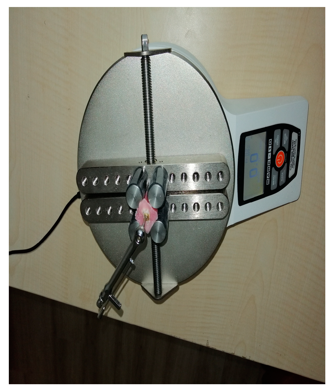
Figure 1. Surgical integration of the titanium implants in the metaphysial part of the corticocancellous bone of the tibia after the skin incision and elevation of the soft tissues.
Preprints 140426 g001
Figure 2. Titanium implants and surrounded bone tissues after the samples collection.
Preprints 140426 g002
Figure 3. Biomechanical reverse torque analysis.
Preprints 140426 g003
Table 1. Bone-implant fusion analysis.
Groups N Mean (N) Minimum (N) Maximum P* Value
Machined 9 4.04 3.2 5.2 <0.005
RBMa1 8 6.88 4.3 11.2 <0.005
SLAa2, b 9 11.11 5.9 19.2 <0.005
*Kruskal-Wallis Test. a: Statistically different compared with Machined group, according to a Mann-Whitney U test. a1: 0,001, a2:0.000. b: bStatistically different compared with RBM group, MannWhitney U. b: 0,036. RBM: Reabsorbable Blast Material, SLA: Sandblasted and Large Acid grid.
Disclaimer/Publisher’s Note: The statements, opinions and data contained in all publications are solely those of the individual author(s) and contributor(s) and not of MDPI and/or the editor(s). MDPI and/or the editor(s) disclaim responsibility for any injury to people or property resulting from any ideas, methods, instructions or products referred to in the content.
Copyright: This open access article is published under a Creative Commons CC BY 4.0 license, which permit the free download, distribution, and reuse, provided that the author and preprint are cited in any reuse.

Downloads

58

Views

42

Comments

0

Subscription

Notify me about updates to this article or when a peer-reviewed version is published.

Email

Prerpints.org logo

Preprints.org is a free preprint server supported by MDPI in Basel, Switzerland.

Subscribe

© 2025 MDPI (Basel, Switzerland) unless otherwise stated