
Submitted:
20 November 2024
Posted:
22 November 2024
You are already at the latest version
Abstract
Nowadays PSMA ligands are widely used for radiotheragnostic purposes of prostate cancer. The goal of this study was to synthesize and estimate stability in vitro of copper complex with diacetate bispidine ligand (Bisp1), its bifunctional derivative (Bisp-alkyne) and its conjugate – bispidine-PSMA ligand (PSMA-Bisp) to evaluate the possibility of applying one as radiotheragnostic agent. The synthesis for PSMA-Bisp conjugate was developed and realized with good yields. All newly synthesized compounds were characterized by a set of physicochemical methods: 1H and 13C NMR, HRMS and LCMS (for final molecules). Subsequently, Bisp1 and Bisp-alkyne and its derivative PSMA-Bisp were labelled by 64Cu at mild conditions. In vitro studies of the labelled conjugate [64Cu]Cu-PSMA-Bisp have shown the great stability in model solutions. Finally, [64Cu]Cu-PSMA-Bisp was compared to well-known PSMA-617 conjugate labelled with 64Cu and they have showed similar stability in excess of bovine serum (BVS), and at the same time labelling PSMA-Bisp with 64Cu is characterized by extremely high kinetics at mild conditions, while labeling PSMA-617 with 64Cu requires heating (90 °C) [PMCID: PMC5435610]. Thus, the conjugate PSMA-Bisp could be promising candidate for further investigations expanded in vitro and in vivo investigations.
Keywords:
PSMA
; Bispidine bifunctional chelator
; copper-64
; complexation
; delivery
1. Introduction
Prostate cancer is one of the most common cancers among the male population. [1] Diagnosis of the disease is possible by using biomarkers such as prostate-specific membrane antigen (PSMA). PSMA is normally found in the prostate epithelium’s secretory cells and is weakly expressed by cells of other organs, such as the kidneys, salivary glands, and small intestine. At the same time, in prostate cancer, overexpression of PSMA is observed in prostate cells. This antigen can be detected not only affected prostate cells, but also the smallest metastases, which made it possible to use PSMA as a target for visualization of carcinomas on SPECT or PET/CT. [2,3,4,5,6] Thus, study of the new prostate specific radiopharmaceuticals is relevant and important task.
Among copper isotopes several ones have potential to be used in nuclear medicine. 61Cu (t1/2=3.3 h), 62Cu (t1/2=9.7 min) and 64Cu (t1/2=12.7 h) are positron-emitting radionuclides with suitable nuclear properties. The copper isotope 67Cu (t1/2=61.8 h) is the longest-lived copper radionuclide, which can also be used as a theragnostic agent: 67Cu emits β--particles with an energy of 0.4 to 0.6 MeV, which is ideal for radionuclide therapy of tumors. In addition, 67Cu emits 2 γ-quants with photon energies of 92 and 184 keV, that can be used for scintigraphy. [7,8]
Among a number of known ligands for the formation of stable complexes with cations of theragnostic radionuclides, used in nuclear medicine, bispidine ligands have proven themselves to be readily available, easy to synthesize and to obtain modified derivatives. [9,10,11,12,13] Both high stability and inertness of complexes of bispidine ligands with transition metal ions, especially copper cations, are noted. [14,15]
2. Results and Discussion
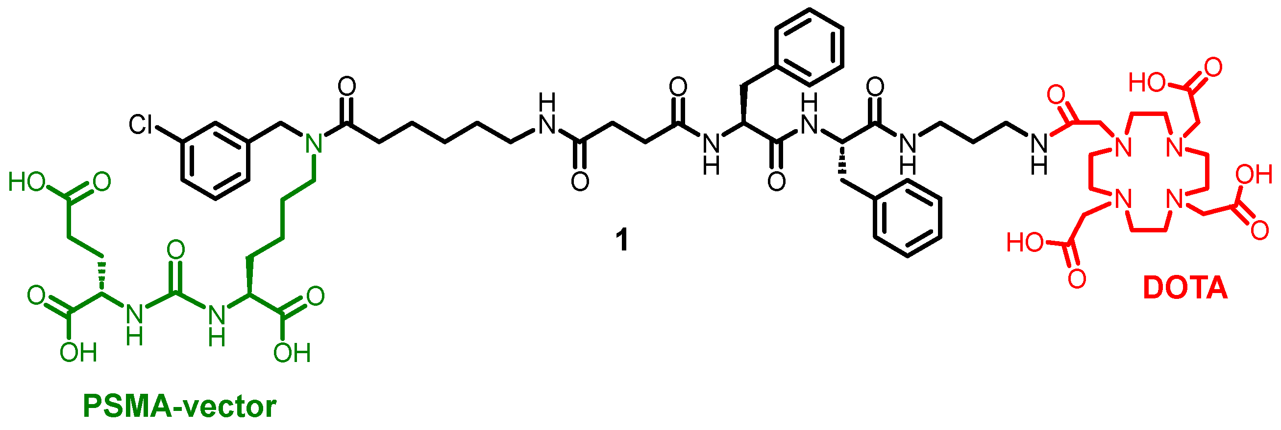
One of the standard chelating agents for metal ions is DOTA. It allows binding of such metal ions such as Ga3+, Gd3+, In3+, Fe2+, Co2+, Mn2+, Ni2+ as well as copper ions Cu2+. [16,17,18,19,20] For example recently, a conjugate 1 with this chelating agent DOTA was synthesized for Lu3+ complexation. [21]
Scheme 1.
Structure of recent DOTA-based conjugate. [21.]
Subsequently, an original synthetic scheme was proposed to produce a conjugate with bispidine fragment, suitable for binding copper radioisotopes. In view of the availability of tert-butyl-protected bispidine derivative Bisp-Alkyne-t-Bu2 containing an alkyne moiety, it was decided to use the reaction to produce 1,2,3-triazole as a method of coupling these fragments together (Scheme 2).
Scheme 2.
Synthesis of bispidine-triazole-based conjugate.
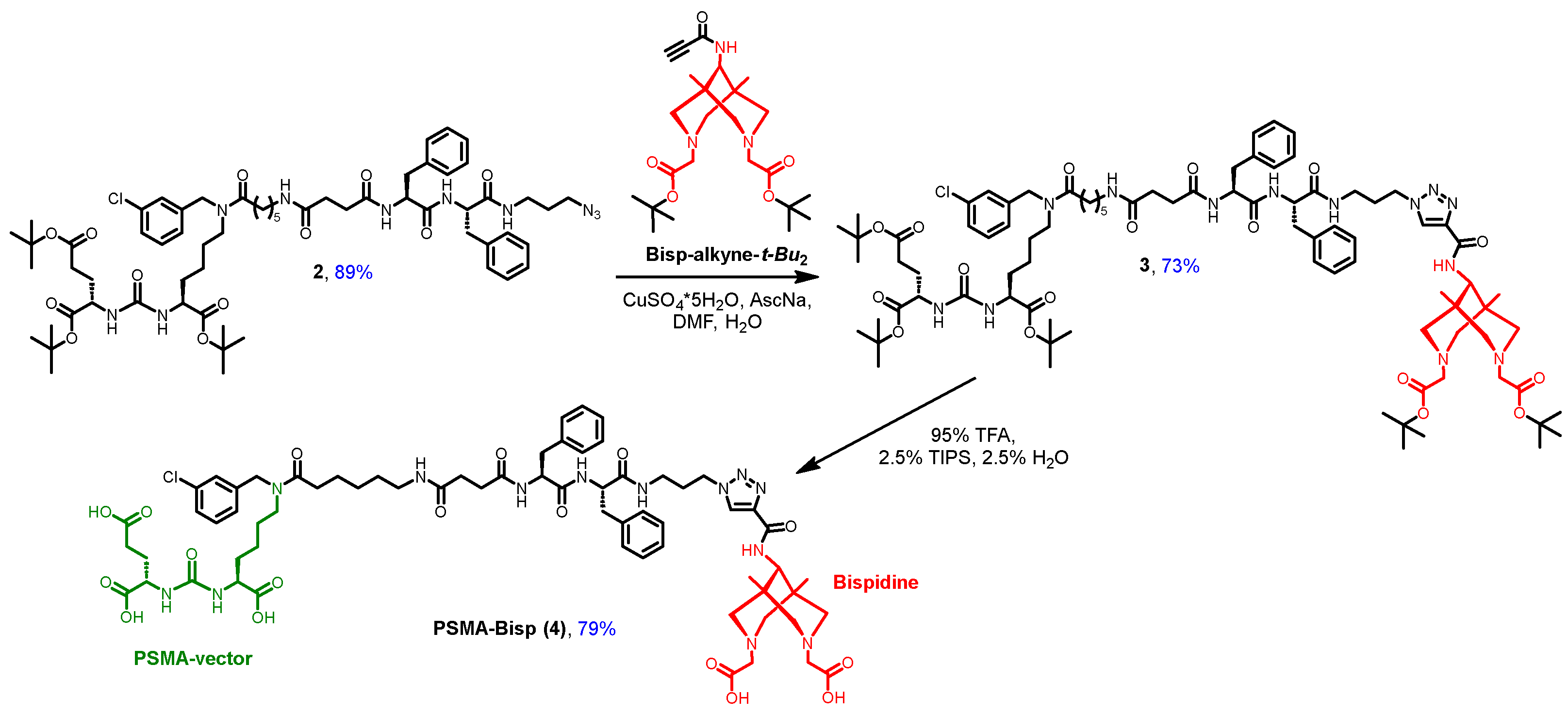
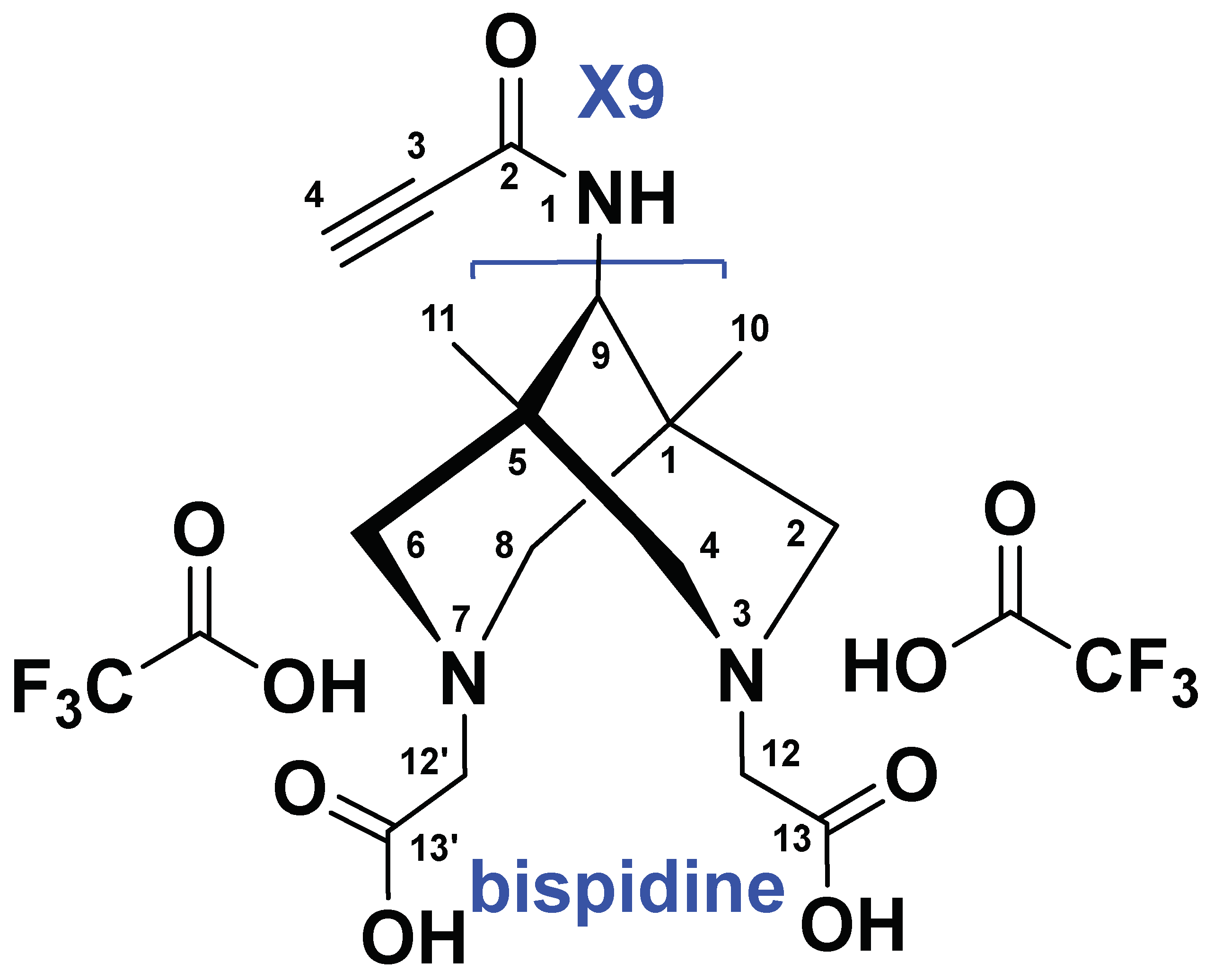
Since the full exploitation of bispidine chelating ability requires both nitrogen atoms, the linking through the position number 9 of the bicycle was chosen. And in order to have orthogonal functionalities at nitrogens and at carbon 9, the introduction of the alkyne moiety at that atom was suggested.
A copper (I)-catalyzed 1,3-dipolar cycloaddition reaction was chosen to prepare conjugate PSMA-Bisp (4) from corresponding tert-butyl protected azide residue 2 and alkyne-containing bispidine Bisp-Alkyne-t-Bu2 (Scheme 2). As the corresponding azide-vector m-chloro- benzyl residue with L-Phe-L-Phe linker was used. Recently the wide library of such azides was synthesized and evaluated in vitro. Particularly, azido-residue, applied in this paper demonstrated IC50= 38 nM (by PSMA inhibition assay) evaluated in previous papers. [22] Azide-alkyne cycloaddition reaction is widely used in the synthesis of bioactive organic compounds and is an alternative bioconjugation method for the introduction of a variety of functional fragments. Synthesis of Bisp1 and 9-benzylamino-1,5-dimethylbispidine trihydrochloride (S1) was performed as it was described in previous publications. [14,23] Detailed synthesis of Bisp-Alkyne-t-Bu2is described in Supporting materials (Figure S1). The bispidine fragment was selected as a chelator capable of efficient chelation of copper ions, including the 64Cu isotope widely used in PET/CT diagnostics. [24] A di-tert-butyl-protected bispidine derivative containing a terminal alkyne group was used as the reagent. After addition of the chelator, removal of the protecting tert-butyl groups was carried out with the TFA/TIPS/H2O mixture. As a result, conjugate PSMA-Bisp (4) was obtained with 79% yield.
As a comparative molecule, additionally, a modified derivative of a bispidine chelating agent with an alkyne moiety was obtained. For this purpose, the previously used compound Bisp-Alkyne-t-Bu2 was introduced into the reaction for the removal of tert-butyl protecting groups, resulting in compound Bisp-Alkyne with 82% yield (Scheme 3). All newly synthesized conjugates and intermediate compounds were characterized by a set of physicochemical methods: 1H and 13C NMR, HRMS. The purity of final molecules such as PSMA-Bisp and Bisp alkyne were confirmed by LCMS analysis.
Labeling conditions
Labeling with 64Cu was carried out in the presence of 0.1 M sodium acetate pH 5 at room temperature (25°C). Bisp1, Bisp-alkyne and PSMA-Bisp were labeled with high efficiency at concentrations ˃0,1 mM within less than 5 minutes. According to HPLC and TLC analysis labeled and stable complexes are chemically identical (Figures S3-S4). Compared to PSMA-617 labeling, a higher ligand’s concentrations are required, [25] however labeling Bisp1, Bisp-alkyne and PSMA-Bisp with 64Cu is characterized by higher rate in mild conditions (Table 1) while PSMA-617 can be quantitatively labeled (˃99%) after heating for 5-30 min at 90°C. [25] The ligand Bisp1 and its complex with copper cation stability was studied previously.[14]
Stability
It is important to evaluate the stability of all labelled compounds against transchelation and transmetalation in the presence of relevant metal cations such as Ca2+, Mg2+, Zn2+, Cu2+ and Fe3+ and serum proteins. These processes may lead to release of 64Cu in solution causing the irradiation of non-target tissues and incorrect diagnostics in the case of 64Cu-based PET It was shown that 90-95% were bound by proteins within first minutes of mixing. All labelled complexes were highly stable (80-100%) both in excess of microelements and in the medium of serum proteins (Table 2, Figure 1).
ROS level and cytotoxicity evaluation
ROS measurement for the conjugate was performed to analyze the possible intracellular effects of the conjugate, also Cu2+ ion electrochemical assay techniques were performed to potentially evaluate the intracellular delivery of copper ions. We used well-established methods for electrochemical intracellular detection of ROS and Cu2+. [26,27] Before the incubation with cells, PSMA-Bisp was stoichiometrically mixed with CuCl2 in a 1:1 ratio to obtain a copper complex. According to the data of electrochemical intracellular studies, it was found that after PSMA-Bisp - Cu2+ complex exposure the intracellular ROS level increased by 26% in the PC-3 cell line and by 89% in the 22Rv1 cell line compared to the control. At the same time, the ROS level following incubation with PSMA-Bisp after 1 h showed a significant increase in comparison with both the control cell lines (Figure 3A, B). Since the PC-3 cell line lacks PSMA on its membrane surface, the effect of the conjugates is not as significant as for the 22Rv1 cell line (Figure 3A, B). Since this cell line has PSMA on its surface, the efficacy of the conjugate is greater than for PC-3 cell line. PSMA-Bisp - Cu2+ targets PSMA receptors, which are overexpressed on the surface of 22Rv1. When PSMA-Bisp - Cu2+ binds to these receptors, it is taken up by the cells more efficiently. This enhanced cellular uptake likely increases the intracellular concentration of PSMA-Bisp - Cu2+, facilitating greater ROS generation through its interaction with cellular components.
We also note that after incubation time, significantly more Cu2+ accumulated in the 22Rv1 cell line than in PC-3. This difference in Cu2+ accumulation further underscores the varying cellular responses to PSMA-Bisp - Cu2+ exposure between the two prostate cancer cell lines, potentially pointing to differences in their transport mechanisms related to copper ions (Figure 3C).
Similar correlations were observed in cytotoxicity experiments. Cytotoxicity probably correlates with the level of copper ions that can enter the cell (Figure 4). Coincubation of the conjugate with copper ions may lead to the formation of a complex that, through binding to PSMA and subsequent endocytosis, can penetrate through the membrane of PSMA+ cell cells, thus providing an increased content of copper ions inside the cells, which accounts for the main toxic effect on the cells, particularly by the generation of intracellular ROS level. In the case of PSMA+ cells, this conjugate is dragged into the cells, and thus we see toxicity at 22Rv1 indistinguishable from the copper salts themselves. Whereas with PC-3 cell culture the complex that bound copper remains outside the cell and cannot be transported inside the cell due to the lack of a receptor, thus reducing the total amount of copper that enters the cell as its direct ions.
Also, it may be noted that at concentrations of PSMA-Bisp lower than 200 µM used in this experiment almost complete cells survival is observed, only coincubation of PSMA-Bisp/CuCl2 (200 µM /1000 µM) results in significant toxicity on PC-3 cells and moderate toxicity on 22Rv1 cells.
3. Materials and Methods
3.1. General
More detailed experimental data can be found in the electronic supplementary material.
3.2. Synthesis
Synthesis and spectral description of compounds 2 and 3 was performed according to the previously published data.[21] Bisp1 [14] and 9-benzylamino-1,5-dimethylbispidine trihydrochloride (S1) [23] were prepared as described previously.
- Synthesis of compound 3.
Compound 2 (1 eq .; 82 mg; 0.068 mmol) and Bisp-Alkyne-tBu2 (1 eq; 31.5 mg; 0.068 mmol), CuSO4×5H2O (0.4 eq.; 7 mg; 27.2 μmol) were dissolved in DMF/H2O (6 mL/1 mL). The system was purged with argon. Sodium ascorbate (1.2 eq .; 16 mg; 81.6 μmol) in H2O (1 mL) was added to the mixture using a syringe. The resulting solution was stirred for 24 hours in an inert atmosphere, after which EDTA (0.8 eq; 16 mg; 54.4 μmol) was added. The mixture was stirred for 3 hours. After filtration the reaction mixture from the precipitate and removing the solvent under reduced pressure, the residue was dissolved in DCM (15 mL), and washed with 1) H2O (1×15 mL), 2) NaHCO3 (2×15 mL). 2) with saturated NaCl solution (1×15 mL). The organic fraction was dried over Na2SO4, the solvent was removed under reduced pressure. After purification, the residue was purified by column chromatography (Puriflash on a PF-15C18HP-F0012 column (15 μm 20 g), eluent: H2O-TFA(0.1%) (90%)/MeCN (10%) => H2O-TFA(0.1%) (0%)/MeCN (100%). for 20 min after MeCN (100%) for 5 min. Compound 3 was obtained as a salt of ×2 TFA as a white powder (94 mg, 73% yield).
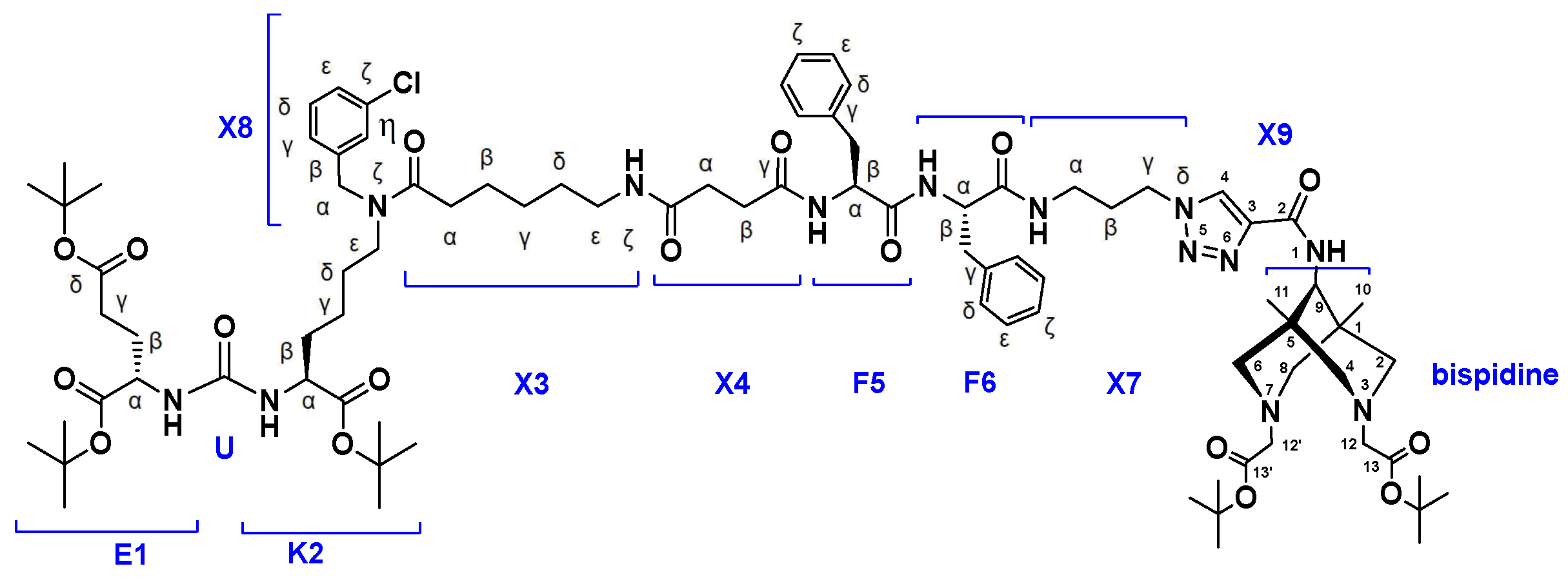
1H NMR (400 MHz, DMSO-d6, δ): 8.65 (s, 1H, X9(4)), 8.35 (d, J=10.3 Hz, 1Н, X9NH), 8.33 (d, J=7.3 Hz, 1Н, F5NH), 8.25-8.16 (br.d, 1Н, F6NHmn), 7.94 (t, J=5.4 Hz, m) & 7.91 (t, J=5.4 Hz, n) (1H, X3NHk, m+n), 7.72 (t, J=5.4 Hz, m) & 7.70 (t, J=5.4 Hz, n) (1H, X7NH, m+n), 7.42-7.09 (m, 14H, X8Hdn+X8Hen+X8Hdm+X8Hem+F6He+F6Hd +X8Htmn+F5He+F6Hk+F5Hk+F5Hd+X8Hgmn), 6.37-6.22 (m, 2Н, K2NH+E1NH, m+n), 4.55 (s, n) & 4.47 (s, m) (2H, X8Ha, m+n), 4.43-4.26 (m, 4H, F6Ha+X7Hg+F5Ha), 4.14 (d, J=10.3 Hz, 9), 4.06-3.88 (m, 2H, E1Ha+K2Ham+K2Han), 3.81 (s, 2H, 12), 3.47 (s, 2H, 12’), 3.39-3.30 (m, 2H, bispidine(cyclic)), 3.25-3.11 (m, 6H, bispidine(cyclic)+K2Hemn), 3.11-2.84 (m, 9Н, F6Hb(a)+X7Ha+X3He(mn)+F6Hb(b)+bispidine(cyclic)+F5Hb(a)), 2.70-2.61 (m, 1Н, F5Hb(b)), 2.40-2.10 (m, 8H, X3Ham+X4Hbmn+E1Hg+X4Hamn +X3Han), 2.00-1.90 (X7Hb), 1.90-1.80 (m, 1Н, E1Hb(a)), 1.72-1.61 (m, 1Н, E1Hb(b)), 1.62-1.54 (m, 1Н, K2Hb(a)), 1.54-1.10 (m, 11Н, X3Hb+K2Hb(b)+X3Hd +K2Hd+K2Hg+X3Hg, m+n), 1.47 (s, 9H, tBu), 1.44 (s, 9H, tBu), 1.40–1.34 (m, 27H, tBu), 0.76 (s, 6H, 10+11)
13C NMR (100 MHz, DMSO-d6, δ): 173.00 (X4Cg(n)), 172.96 (X4Cg(m)), 172.23 (K2C(n)), 172.19 (K2C(m)), 172.10 (X3C(nm)), 171.91 (E1C), 171.61 (X4C(mn)), 171.44 (E1Cd), 171.11 (F5C), 170.72 (F6C), 169.65 (br. peak, 16+24), 165.98 (br. peak, 20), 164.88 (br. peak, 1), 158.89 (C(O)TFA), 158.53 (C(O)TFA), 158.17 (C(O)TFA), 157.81 (C(O)TFA),157.15 (U(m)), 157.13 (U(n)), 141.17 (X8Cb(m)), 140.76 (X8Cb(n)), 138.14 (F6Cg), 138.04 (F5Cg), 133.42 (X8Ck(n)), 133.07 (X8Ck(m)), 130.58 (X8Cd(n)), 130.22 (X8Cd(m)), 128.98 (F6Cd+F5Cd), 128.16 (F6Ce), 128.08 (F5Ce), 127.18 (X8Ct(m)), 127.14 (X8Ce(n)), 126.85 (X8Ce(m)), 126.28 (F6Ck+X8Ct(n)), 126.26 (F5Ck), 126.05 (X8Cg(m)), 124.96 (X8Cg(n)), 120.08 (CF3), 117.18 (CF3), 114.27 (CF3), 111.37 (CF3), 83.44 (21), 81.27 (17+21), 80.55 (E1tBu), 80.38 (K2tBu(m)), 80.30 (K2tBu(n)), 79.75 (E1dtBu), 55.16 (F5Ca), 54.58 (br. peak, 2), 54.38 (F6Ca+19), 53.29 (br. peak, 15+23), 53.01 (K2Ca(n)), 52.87 (K2Ca(m)), 52.19 (E1Ca), 50.79 (series of br. peaks, DOTAcyclic), 49.60 (X8Ca(n)), 48.30 (series of br. peaks, DOTAcyclic), 47.83 (series of br. peaks, DOTAcyclic), 47.10 (X8Ca(m)), 46.80 (K2Ce(m)), 45.21 (K2Ce(n)), 38.65 (X3Ce(m)), 38.58 (X3Ce(n)), 37.02 (F5Cb), 36.71 (F6Cb+X7Cg), 36.52 (X7Ca), 32.31 (X3Ca(n)), 31.93 (X3Ca(m)), 31.79 (K2Cb), 30.91 (E1Cg), 30.71 (X4Ca), 30.54 (X4Cb), 29.05 (X3Cd(m)), 28.96 (X3Cd(n)), 28.68 (X7Cb), 27.72 (tBuE1+18+22+26), 27.63 (tBuK2+K2Cd(m)), 27.61 (tBuE1d), 27.57 (E1Cb), 26.69 (K2Cd(n)), 26.31 (X3Cg(m)), 26.22 (X3Cg(n)), 24.72 (X3Cb(m)), 24.57 (X3Cb(n)), 22.44 (K2Cg(n)) 22.25 (K2Cg(m)).
LCMS: 100% in positive ions detection mode, 100% in negative ions detection mode.
ESI-MS C86H12835ClN13O17: m/z calculated for [M+2H+]2+: 825.97, found: 826.65.
HRMS (m/z, ESI): calculated for C86H12835ClN13O17 - [M+H+]+ 1650.9312, found: 1650.9319.
- Synthesis of compound 4 (PSMA-Bisp).
Compound 3 (1 eq; 86 mg; 45.42 μmol) was dissolved in a mixture consisting of TFA/TIPS/H2O (95%/2.5%/2.5%, V = 4 mL). The mixture was stirred for 4 hours. Next, the solvent was removed. The product was precipitated with DEK and washed twice with Et2O (2 mL). The residue was purified by reversed-phase chromatography (Puriflash PF-15C18AQ-F0012 (15μ 20g), eluent: H2O×TFA(0.1%) (90%)/MeCN(10%) => H2O×TFA(0.1%) (0%)/MeCN(100%) for 30 min after MeCN (100%) for 5 min. Compound 4 (PSMA-Bisp) was obtained as salt×2 TFA as a white powder (58 mg, 79% yield).
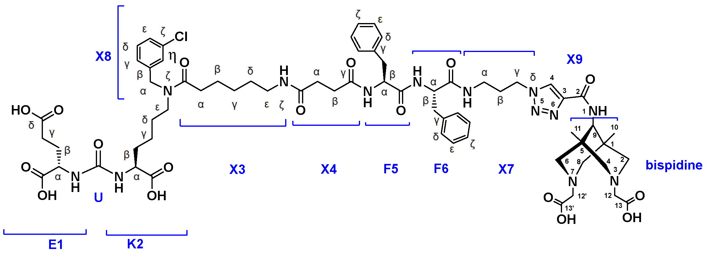
1H NMR (400 MHz, DMSO-d6, δ): 12.28 (br.s, 5H, COOH), 8.64 (s, 1H, X9(4)), 8.38 (d, J=10.3 Hz, 1Н, X9NH), 8.32 (d, J=7.3 Hz, 1Н, F5NH), 8.25-8.16 (br.d, 1Н, F6NHmn), 7.93 (t, J=5.4 Hz, m) & 7.91 (t, J=5.4 Hz, n) (1H, X3NHk, m+n), 7.76-7.66 (m, 1H, X7NHmn), 7.42-7.09 (m, 14H, X8Hdn+X8Hen+X8Hdm+X8Hem+F6He+F6Hd+X8Htmn+F5He +F6Hk+F5Hk+F5Hd+X8Hgmn), 6.40-6.25 (m, 2Н, K2NH+E1NH, m+n), 4.55 (s, n) & 4.46 (s, m) (2H, X8Ha, m+n), 4.43-4.26 (m, 4H, F6Ha+X7Hg+F5Ha), 4.14-3.97 (m, 3H, E1Ha+9+K2Ham+K2Han), 3.67 (s, 2H, 12), 3.43 (s, 2H, 12’), 3.40-3.30 (m, 2H, bispidine(cyclic)), 3.25-3.11 (m, 6H, bispidine(cyclic)+K2Hemn), 3.11-2.82 (m, 9Н, F6Hb(a)+X7Ha+X3He(mn)+F6Hb(b)+bispidine(cyclic)+F5Hb(a)), 2.70-2.60 (m, 1Н, F5Hb(b)), 2.40-2.10 (m, 8H, X3Ham+X4Hbmn+E1Hg+X4Hamn +X3Han), 1.99-1.84 (m, 3Н, X7Hb+E1Hb(a)), 1.77-1.66 (m, 1Н, E1Hb(b)), 1.66-1.56 (m, 1Н, K2Hb(a)), 1.56-1.10 (m, 11Н, X3Hb+K2Hb(b)+X3Hd +K2Hd+K2Hg+X3Hg, m+n), 0.76 (s, 6H, 10+11)
13C NMR (100 MHz, DMSO-d6, δ): 174.57 (K2C(n)), 174.53 (K2C(m)), 174.24 (E1C(mn)), 173.80 (E1Cd), 172.89 (X4Cg(n)), 172.84 (X4Cg(m)), 172.18 (X3C(nm)), 171.58 (X4C(mn)), 171.27 (F5C), 170.97 (F6C), 170.53 (C13), 168.16 (C13’), 160.98 (X9C2), 157.33 (U), 142.04 (C3), 141.25 (X8Cb(m)), 140.84 (X8Cb(n)), 138.06 (F6Cg), 137.99 (F5Cg), 133.42 (X8Ck(n)), 133.07 (X8Ck(m)), 130.63 (X8Cd(n)), 130.26 (X8Cd(m)), 129.07 (F6Cd+F5Cd), 128.23 (F6Ce), 128.08 (F5Ce), 127.46 (C4), 127.21 (X8Ct(m)), 127.16 (X8Ce(n)), 126.86 (X8Ce(m)), 126.37 (F6Ck), 126.31 (X8Ct(n)) 126.27 (F5Ck), 126.10 (X8Cg(m)), 124.99 (X8Cg(n)), 62.81 (9), 56.32 (12), 55.99 (12’+bispidine(cyclic)), 54.96 (F5Ca), 54.56 (bispidine(cyclic)), 54.52 (F6Ca), 52.29 (K2Ca(n)), 52.18 (K2Ca(m)), 51.71 (E1Ca), 49.66 (X8Ca(n)), 47.40 (X7Cg), 47.19 (X8Ca(m)), 46.90 (K2Ce(m)), 45.36 (K2Ce(n)), 38.68 (X3Ce(m)), 38.60 (X3Ce(n)), 37.00 (F5Cb), 36.85 (F6Cb), 35.70 (X7Ca), 35.07 (1+5), 32.31 (X3Ca(n)), 31.88 (X3Ca(m)), 31.82 (K2Cb), 30.68 (X4Ca), 30.55 (X4Cb), 29.96 (E1Cg), 29.52 (X7Cb), 29.08 (X3Cd(m)), 28.97 (X3Cd(n)), 27.82 (K2Cd(m)), 27.57 (E1Cb), 26.79 (K2Cd(n)), 26.29 (X3Cg(m)), 26.22 (X3Cg(n)), 24.73 (X3Cb(m)), 24.59 (X3Cb(n)), 22.53 (K2Cg(n)) 22.35 (K2Cg(m)), 19.63 (10+11).
LCMS: 97.4% in positive ions detection mode, 100% in negative ions detection mode.
ESI-MS C66H8835ClN13O17: m/z calculated for [M+2H+]2+: 685.81, found: 686.4.
HRMS (m/z, ESI): calculated for C66H8835ClN13O17 - [M-2H+]2- 683.7982, found: 683.7986.
- Synthesis of compound Bisp-alkyne.
Bisp-alkyne-t-Bu2 (1 eq.; 20 mg; 43.14 μmol) was dissolved in a mixture consisting of TFA/TIPS/H2O (95%/2.5%/2.5%, V = 2 mL). The mixture was stirred for 4 hours. Next, the solvent was removed. The product was precipitated with DEK and washed twice with Et2O (1 mL). The residue was purified by reversed-phase chromatography (Puriflash PF-15C18AQ-F0012 (15μ 20g), eluent: H2O×TFA(0.1%) (100%)/MeCN(0%) => H2O×TFA(0.1%) (0%)/MeCN(100%) for 30 min after MeCN (100%) for 5 min. Bisp-alkyne was obtained as salt×2 TFA as a white powder (20 mg, 82% yield).
1H NMR (400 MHz, DMSO-d6, δ): 11.38 (br.s, 2H, COOH), 8.83 (d, J=10.3 Hz, 1Н, X9NH), 4.40 (s, 1H, X9(4)), 3.88 (d, J=10.3 Hz, 1Н, 9), 3.67 (s, 2H, 12) 3.47 (s, 2H, 12’), 3.23-3.13 (m, 4H, bispidine(cyclic)), 3.13-3.03 (m, 2H, bispidine(cyclic)), 2.96-2.86 (m, 2Н, bispidine(cyclic)), 0.73 (s, 6H, 10+11)
13C NMR (101 MHz, DMSO-d6, δ): 170.31 (13), 168.38 (C3’), 152.61 (X9(2)), 77.71 (X9(3)), 77.53 (X9(4)), 62.50 (9), 56.18 (bispidine(cyclic)), 55.68 (12), 54.88 (12’), 35.01 (1+5), 19.45 (10+11).
LCMS: 100% in positive ions detection mode, 100% in negative ions detection mode.
ESI-MS C16H23N3O5: m/z calculated for [M+H+]+: 338.17, found: 338.25.
HRMS (m/z, ESI): calculated for C16H23N3O5 - [M+H+]+ 338.1710, found: 338.1710.
3.3. Isolation of 64Cu
A nickel target of natural isotopic composition was irradiated with a proton flow with an energy of 7-8 MeV at 1-2 μA for 6 hours. The target was dissolved in concentrated HCl upon heating with the addition of H2O2 to accelerate the dissolution of the metal. The solution was evaporated to dryness and dissolved in 0.01 M HCl. On a Cu-resin sorbent saturated with 0.01 M HCl, 61,64Cu was separated from natNi and 55Co. 0.01 M HCl was passed through the chromatographic column until the release of nickel and cobalt ended, then the copper was washed off with 8 M HCl. The process was monitored using gamma spectrometry using peaks at 931 keV (55Co), 283 keV (61Cu) and 1346 keV (64Cu). The resulting solution was evaporated to dryness and dissolved in 1 ml of 0.1 M HCl.
3.4. Measurement of radioactivity
Gamma spectrometry was performed using gamma spectrometers GR3818 (Canberra Packard Ind., USA) and ORTEC DSPec50 (16013585) with coaxial HPGe-detector GEM-C5060P4-B (56-TP23840B).
3.5. Labeling experiments
Labeling efficiency experiments were performed in sodium acetate buffer solution (0.15 M) with final pH of 4.5-5.0. The incubation time for each radiolabeling experiment was less than 5 minutes. To study the labelling efficiency, solutions (300 μL) containing 1 kBq of 64Cu, stable Cu(ClO4) (to reach c(Cu2+)=1×10⁻⁸ M) and Bisp1/Bisp-alkyne/PSMA-Bisp with concentrations varying from 1×10⁻⁵ М to 1×10⁻³ М were equilibrated at room temperature in a plastic Eppendorf tubes. The degree of radiochemical conversion (RCС) was measured by thin-layer chromatography (TLC) analysis and gamma-spectrometry.
3.6. Thin-layer chromatography
For Thin-layer chromatography (TLC) solvent system 10% AcONH4/CH3OH 1/1 as a mobile phase and silica gel on aluminum plates (Sigma) as solid phase were used. The solution of free [64Cu]-Cu2+ (blank) or labeled complex [64Cu]Cu-Bisp1 or labelled complex with bifunctional chelator [64Cu]Cu-Bisp-alkyne or labeled conjugate [64Cu]Cu-PSMA-Bisp solution containing 100 Bq of 64Cu (5-30 μL) was deposited on TLC sheet. The sample distribution on TLC plates after elution was preliminarily visualized by autoradiography using a Perkin Elmer Cyclone Plus Storage Phosphor System and associated software. To quantify the TLC results, plates were cut according to the radiography images and measured by gamma spectrometry.
3.7. In vitro evaluation
In order to estimate stability of [64Cu]Cu-PSMA-Bisp in presence of biologically relevant cations [64Cu]Cu-PSMA-Bisp was prepared in sodium acetate buffer solution (0.15 M) to a final pH of 4.5–5.0 and ligand concentration of 5×10⁻⁴ M. It was added to solutions of cations (5 mM Ca2+ and Mg2+, 0.1 mM Fe3+, Zn2+, and Cu2+). After 1 hour of incubation at 37°C, the bound fraction of radionuclide determined using TLC and measured via gamma spectrometry: the mixture of complex and salt’s solution was spotted on TLC plate and chromatography was performed with 10% AcONH4/CH3OH 1/1 as a mobile phase. The sample distribution on TLC plates after elution was preliminarily visualized by autoradiography (Fig S3) and cut according to the radiography images.Radioactivity distribution on the plates was measured by gamma spectrometry.
For serum tests, 64Cu blank and [64Cu]Cu-Bisp1 and [64Cu]Cu-Bisp-alkyne⸱and [64Cu]Cu-PSMA-Bisp (300 μL) were prepared in sodium acetate buffer solution (0.15 M) to a final pH of 4.5–5.0 and ligand concentration of 1×10⁻³ M. Initial solutions (100 μL) were added to fetal bovine serum (900 μL; HyClone), mixed, and incubated at 37°C. Aliquot of the labeled complex-serum mixture of 100 μL was taken at time points of 1, 15, 30, 60, 180 min and an aliquot of the labeled conjugate-serum mixture of 100 μL was taken at time points of 5, 10, 35, 60, 300 min and treated with ethanol (300 μL) for protein precipitation. Samples were cooled to 2–4°C and centrifuged at 4,000 g for 5 min. Aliquots (300 μL) of the supernatant were separated and measured using gamma spectrometry. As a reference, initial samples (75 μL) were taken, diluted to 300 μL, and measured. For complexes in each case the supernatant was analyzed by TLC and autoradiography: in every case the Rf of radioactive compound in the supernatant had the same Rf that had original appropriate complex.
3.8. Cell Lines
22Rv1 and PC-3 cells were received from the MISIS collection of cell lines (less than ten passages from ATCC stock). All cells were cultured in humidified 37 °C incubators with 5% CO2. All cell lines were tested for the absence of mycoplasma. 22Rv1, androgen-responsive PSMA-positive human prostate carcinoma cells, were cultured in RPMI 1640 media (Gibco) with 10% FBS (Gibco), 1× GlutaMax (Gibco), and 1× Penicillin-Streptomycin (10,000 U/mL, Gibco). PC-3, PSMA-negative human prostate cancer cells, were cultured in DMEM/F12 media (Gibco) with 10% FBS (Gibco), 1× GlutaMax (Gibco), and 1× Penicillin-Streptomycin (10,000 U/mL, Gibco).
3.9. Single cell ROS and Cu2+ Measurement by Using nanoelectrodes
A detailed description of the fabrication processes of nanoelectrodes for ROS and Cu2+ intracellular measurement has been described elsewhere. [26,27]
PC-3 (2.0 × 10⁵)/22Rv1 (3.5 × 10⁵) cells were seeded in 35 mm Petri dishes and cultivated under the normal conditions for 24 h. Previously, PSMA-Bisp was stoichiometrically mixed with CuCl2 in a 1:1 molar ratio to obtain a copper complex (PSMA-Bisp - Cu2+). Then, the copper complexes (PSMA-Bisp - Cu2+) were dissolved in DMSO, diluted in fresh culture medium with <1% FBS 2 mL, and added to Petri dishes. The final concentration of the compounds in the culture medium was IC50 with 1 h of incubation time. Untreated cells were used as a control, which was evaluated at the beginning and at the end of the experiment. Attached cells in Petri dishes were washed three times using Hanks’ Balanced Salt solution to remove the media and traces of complexes. On average, about 10 cells were measured by 2–3 nanoelectrodes in 2 independent Petri dishes for each complex in one independent biological replicate. The intracellular ROS/Cu2+ level was determined based on the calibration curve. Results are shown as means ± SEM, where n = 3 (three independent biological replicates). Statistical analyses were conducted using one-way ANOVA test in Origin 2021.
3.10. Cytotoxicity
Cells were seeded in the 96-well plates (Corning) at concentration of 4 000 cells per well for PC-3 culture and 6 500 cells per well for 22Rv1 line. Automated cell counter MOXI was used to calculate the cells. After 1 day, serial dilutions of PSMA-Bisp or its combination with CuCl2 whith correspinding molar ratios: 1/1 or 1/5 in DMSO and CuCl2 itself were added to cells. Culture medium with DMSO at concentrations corresponding to that added with the experimental samples was used as control. The cell medium was used as a negative control, while 30% DMSO diluted in the medium was used as a positive control. Cells were incubated then for 72 h at 37°C and 5% CO2. Later, the culture medium from each well was removed and 20 μl of MTS reagent (CellTiter 96 AQueous Non-Radioactive Cell Proliferation Assay, Promega) were added to each well with 100 μl of new culture medium. After 4 h incubation at 37°C in darkness, the absorbance of the solution was measured at 490 nm wavelength using Thermo Scientific Multiskan GO spectrometer. Cell viability was calculated as percent compared to cells incubated in the culture medium. The absorbance of MTS reagent in culture medium without cells was taken as zero. MTS assay revealed 100% cell death after incubation with 30% DMSO (data not shown). DMSO at concentrations corresponding to that added with the samples did not cause cell viability decrease (data not shown). Experiments were performed in triplicates. Data were analyzed using the t-test in GraphPad Prism 9 software. P values <0.05 were considered significant.
4. Conclusions
In this work, we developed and synthesized the first PSMA-targeted bispidine based conjugate, suitable for Cu2+ chelation. All newly synthesized conjugates and intermediate compounds were characterized by a set of physicochemical methods: 1H and 13C NMR, HRMS. The purity of final molecules such as PSMA-Bisp and Bisp-alkyne were confirmed by LCMS analysis. Initial physicochemical and in vitro evaluations were carried out. The conjugate PSMA-Bisp demonstrated good labeling ability with [64Cu]-Cu2+ ions and found to be stable against transchelation and transmetalation in the presence of endogenous metal cations such as Ca2+, Mg2+, Zn2+, Cu2+ and Fe3+ and serum proteins. The use of the conjugate in in vitro prostate cancer cell models demonstrates high copper ion accumulation on 22Rv1 (PSMA+) cell cultures, which probably leads to excessive ROS formation, as well as increased cytotoxicity when excessive amounts of copper ions are used in the incubation medium, compared to the results on PC-3 (PSMA-) cell culture. Subsequently, analysis of PSMA-targeted conjugate biodistribution would be performed in the further investigations.
ASSOCIATED CONTENT
Supporting Information
The Supporting Information is available at
NMR, HR-MS, LC-MS spectra; methods of bispidine residues synthesis; radiography results (PDF).
Author Contributions.
All authors have given approval to the final version of the manuscript.
Conceptualization, S.Z.V., E.K.B. and A.E.M.; methodology, S.A.P. and A.E.M.; validation, A.S.G., R.V.T. and A.N.V.; formal analysis, B.V.E., A.S.E., A.V.M., M.A.K. A.N.V. and S.A.P; investigation, A.S.G., M.D.K., R.V.T., A.B.P., A.V.M., M.A.K., A.N.V., and S.A.P.; resources, E.K.B. ; data curation, A.S.E. and B.V.E.; writing—original draft preparation A.E.M. and B.V.E.; writing—review and editing, A.S.E., S.Z.V., E.K.B., A.V.M., S.N.K., B.V.E. and A.E.M.; visualization, A.E.M., A.N.V. and B.V.E.; supervision, S.Z.V. and E.K.B.; project administration, E.K.B. and A.E.M.; funding acquisition, A.E.M. and A.S.E. All authors have read and agreed to the published version of the manuscript.
ACKNOWLEDGMENT
This work was financially supported by Russian Science Foundation, Grant № 22-15-00098, https://rscf.ru/project/22-15-00098/ (PSMA-ligands and conjugates design, synthesis and physico-chemical characterization). ROS measurements were carried out within the framework of the Implementation Program Priority 2030 (NUST MISIS).
ABBREVIATIONS
DCM, dichloromethane; DMF, N,N-Dimethylformamide; DMSO, dimethyl sulfoxide; DOTA, 2,2′,2′′,2′′′-(1,4,7,10-Tetraazacyclododecane-1,4,7,10-tetrayl)tetraacetic acid; EDTA, Ethylenediaminetetraacetic acid; ESI-MS, electrospray ionization - mass spectrometry; Et2O, diethyl ether; HRMS, high-resolution mass spectrometry; LCMS, liquid chromatography mass spectrometry; NMR, nuclear magnetic resonance; PET/CT, positron emission tomography/computed tomography; PSMA, prostate-specific membrane antigen; ROS, reactive oxygen species, SPECT, Single-photon emission computed tomography; TIPS, triisopropylsilane; TFA, trifluoroacetic acid.
Conflicts of Interest
The authors declare no conflict of interest.
References
- Bray, F.; Laversanne, M.; Sung, H.; Ferlay, J.; Siegel, R.L.; Soerjomataram, I.; Jemal, A. Global Cancer Statistics 2022: GLOBOCAN Estimates of Incidence and Mortality Worldwide for 36 Cancers in 185 Countries. CA Cancer J Clin 2024, 74, 229–263. [Google Scholar] [CrossRef]
- Petrov, S.A.; Zyk, N.Y.; Machulkin, A.E.; Beloglazkina, E.K.; Majouga, A.G. PSMA-Targeted Low-Molecular Double Conjugates for Diagnostics and Therapy. Eur J Med Chem 2021, 225, 113752. [Google Scholar] [CrossRef]
- Sharifi, M.; Yousefnia, H.; Zolghadri, S.; Bahrami-Samani, A.; Naderi, M.; Jalilian, A.R.; Geramifar, P.; Beiki, D. Preparation and Biodistribution Assessment of 68Ga-DKFZ-PSMA-617 for PET Prostate Cancer Imaging. Nuclear Science and Techniques 2016, 27. [Google Scholar] [CrossRef]
- Giesel, F.L.; Cardinale, J.; Schäfer, M.; Neels, O.; Benešová, M.; Mier, W.; Haberkorn, U.; Kopka, K.; Kratochwil, C. 18F-Labelled PSMA-1007 Shows Similarity in Structure, Biodistribution and Tumour Uptake to the Theragnostic Compound PSMA-617. Eur J Nucl Med Mol Imaging 2016, 43, 1929–1930. [Google Scholar] [CrossRef]
- Afshar-Oromieh, A.; Hetzheim, H.; Kratochwil, C.; Benesova, M.; Eder, M.; Neels, O.C.; Eisenhut, M.; Kübler, W.; Holland-Letz, T.; Giesel, F.L.; et al. The Theranostic PSMA Ligand PSMA-617 in the Diagnosis of Prostate Cancer by PET/CT: Biodistribution in Humans, Radiation Dosimetry, and First Evaluation of Tumor Lesions. Journal of Nuclear Medicine 2015, 56, 1697–1705. [Google Scholar] [CrossRef]
- Hasnowo, L.A.; Larkina, M.S.; Plotnikov, E.; Bodenko, V.; Yuldasheva, F.; Stasyuk, E.; Petrov, S.A.; Zyk, N.Y.; Machulkin, A.E.; Vorozhtsov, N.I.; et al. Synthesis, 123I-Radiolabeling Optimization, and Initial Preclinical Evaluation of Novel Urea-Based PSMA Inhibitors with a Tributylstannyl Prosthetic Group in Their Structures. Int J Mol Sci 2023, 24. [Google Scholar] [CrossRef] [PubMed]
- Krasnovskaya, O.O.; Abramchuck, D.; Erofeev, A.; Gorelkin, P.; Kuznetsov, A.; Shemukhin, A.; Beloglazkina, E.K. Recent Advances in 64Cu/67Cu-Based Radiopharmaceuticals. Int J Mol Sci 2023, 24. [Google Scholar] [CrossRef]
- Hussain, M.; Qaim, S.M.; Spahn, I.; Aslam, M.N.; Neumaier, B. Copper Radionuclides for Theranostic Applications: Towards Standardisation of Their Nuclear Data. A Mini-Review. Front Chem 2023, 11. [Google Scholar] [CrossRef]
- Kubeil, M.; Neuber, C.; Starke, M.; Arndt, C.; Rodrigues Loureiro, L.; Hoffmann, L.; Feldmann, A.; Bachmann, M.; Pietzsch, J.; Comba, P.; et al. 64Cu Tumor Labeling with Hexadentate Picolinic Acid-Based Bispidine Immunoconjugates. Chemistry – A European Journal 2024, 30, e202400366. [Google Scholar] [CrossRef] [PubMed]
- Kopp, I.; Cieslik, P.; Anger, K.; Josephy, T.; Neupert, L.; Velmurugan, G.; Gast, M.; Wadepohl, H.; Brühlmann, S.A.; Walther, M.; et al. Bispidine Chelators for Radiopharmaceutical Applications with Lanthanide, Actinide, and Main Group Metal Ions. Inorg Chem 2023, 62, 20754–20768. [Google Scholar] [CrossRef] [PubMed]
- Kovács, A. Metal-Ligand Bonding in Bispidine Chelate Complexes for Radiopharmaceutical Applications. Struct Chem 2023, 34, 5–15. [Google Scholar] [CrossRef]
- Cieslik, P.; Kubeil, M.; Zarschler, K.; Ullrich, M.; Brandt, F.; Anger, K.; Wadepohl, H.; Kopka, K.; Bachmann, M.; Pietzsch, J.; et al. Toward Personalized Medicine: One Chelator for Imaging and Therapy with Lutetium-177 and Actinium-225. J Am Chem Soc 2022, 144, 21555–21567. [Google Scholar] [CrossRef] [PubMed]
- Comba, P.; Starke, M.; Wadepohl, H. Optimization of Hexadentate Bispidine Ligands as Chelators for 64CuII PET Imaging. Chempluschem 2018, 83, 597–604. [Google Scholar] [CrossRef] [PubMed]
- Medved’ko, A. V; Egorova, B. V; Komarova, A.A.; Rakhimov, R.D.; Krut’ko, D.P.; Kalmykov, S.N.; Vatsadze, S.Z. Copper–Bispidine Complexes: Synthesis and Complex Stability Study. ACS Omega 2016, 1, 854–867. [Google Scholar] [CrossRef]
- Roux, A.; Gillet, R.; Huclier-Markai, S.; Ehret-Sabatier, L.; Charbonnière, L.J.; Nonat, A.M. Bifunctional Bispidine Derivatives for Copper-64 Labelling and Positron Emission Tomography. Org. Biomol. Chem. 2017, 15, 1475–1483. [Google Scholar] [CrossRef]
- Voráčová, I.; Vaněk, J.; Pasulka, J.; Střelcová, Z.; Lubal, P.; Hermann, P. Dissociation Kinetics Study of Copper(II) Complexes of DO3A, DOTA and Its Monosubstituted Derivatives. Polyhedron 2013, 61, 99–104. [Google Scholar] [CrossRef]
- Joyner, J.C.; Cowan, J.A. Target-Directed Catalytic Metallodrugs. Brazilian Journal of Medical and Biological Research 2013, 46, 465–485. [Google Scholar] [CrossRef]
- Lilly Thankamony, A.S.; Wittmann, J.J.; Kaushik, M.; Corzilius, B. Dynamic Nuclear Polarization for Sensitivity Enhancement in Modern Solid-State NMR. Prog Nucl Magn Reson Spectrosc 2017, 102–103, 120–195. [Google Scholar] [CrossRef]
- Rinne, S.S.; Leitao, C.D.; Mitran, B.; Bass, T.Z.; Andersson, K.G.; Tolmachev, V.; Ståhl, S.; Löfblom, J.; Orlova, A. Optimization of HER3 Expression Imaging Using Affibody Molecules: Influence of Chelator for Labeling with Indium-111. Sci Rep 2019, 9, 655. [Google Scholar] [CrossRef]
- Rinne, S.S.; Dahlsson Leitao, C.; Gentry, J.; Mitran, B.; Abouzayed, A.; Tolmachev, V.; Ståhl, S.; Löfblom, J.; Orlova, A. Increase in Negative Charge of 68Ga/Chelator Complex Reduces Unspecific Hepatic Uptake but Does Not Improve Imaging Properties of HER3-Targeting Affibody Molecules. Sci Rep 2019, 9, 17710. [Google Scholar] [CrossRef]
- Machulkin, A.E.; Petrov, S.A.; Bodenko, V.; Larkina, M.S.; Plotnikov, E.; Yuldasheva, F.; Tretyakova, M.; Bezverkhniaia, E.; Zyk, N.Yu.; Stasyuk, E.; et al. Synthesis and Preclinical Evaluation of Urea-Based Prostate-Specific Membrane Antigen-Targeted Conjugates Labeled with 177Lu. ACS Pharmacol Transl Sci 2024, 7, 1457–1473. [Google Scholar] [CrossRef] [PubMed]
- Machulkin, A.E.; Shafikov, R.R.; Uspenskaya, A.A.; Petrov, S.A.; Ber, A.P.; Skvortsov, D.A.; Nimenko, E.A.; Zyk, N.U.; Smirnova, G.B.; Pokrovsky, V.S.; et al. Synthesis and Biological Evaluation of PSMA Ligands with Aromatic Residues and Fluorescent Conjugates Based on Them. J Med Chem 2021, 64, 4532–4552. [Google Scholar] [CrossRef] [PubMed]
- Kalinin, M.A.; Medved’ko, A. V; Minyaev, M.E.; Vatsadze, S.Z. Synthesis of N,N′-Unsymmetrical 9-Amino-5,7-Dimethyl-Bispidines. J Org Chem 2023, 88, 7272–7280. [Google Scholar] [CrossRef] [PubMed]
- Mozhaitsev, E.S.; Ponomarev, K.Y.; Patrusheva, O.S.; Medvedko, A. V; Dalinger, A.I.; Rogachev, A.D.; Komarova, N.I.; Korchagina, D. V; Suslov, E. V; Volcho, K.P.; et al. Conjugates of Bispidine and Monoterpenoids as Ligands of Metal Complex Catalysts for the Henry Reaction. Russian Journal of Organic Chemistry 2020, 56, 1969–1981. [Google Scholar] [CrossRef]
- Cui, C.; Hanyu, M.; Hatori, A.; Zhang, Y.; Xie, L.; Ohya, T.; Fukada, M.; Suzuki, H.; Nagatsu, K.; Jiang, C.; et al. Synthesis and Evaluation of [ 64 Cu]PSMA-617 Targeted for Prostate-Specific Membrane Antigen in Prostate Cancer; 2017; Vol. 7.
- Vaneev, A.N.; Gorelkin, P. V.; Garanina, A.S.; Lopatukhina, H. V.; Vodopyanov, S.S.; Alova, A. V.; Ryabaya, O.O.; Akasov, R.A.; Zhang, Y.; Novak, P.; et al. In Vitro and In Vivo Electrochemical Measurement of Reactive Oxygen Species After Treatment with Anticancer Drugs. Anal Chem 2020, 92, 8010–8014. [Google Scholar] [CrossRef]
- Timoshenko, R. V.; Gorelkin, P. V.; Vaneev, A.N.; Krasnovskaya, O.O.; Akasov, R.A.; Garanina, A.S.; Khochenkov, D.A.; Iakimova, T.M.; Klyachko, N.L.; Abakumova, T.O.; et al. Electrochemical Nanopipette Sensor for In Vitro/In Vivo Detection of Cu 2+ Ions. Anal Chem 2024, 96, 127–136. [Google Scholar] [CrossRef]
- Tietze L.F.; Eicher T.; Diederichsen U.; Speicher A.; Schützenmeister N. Reactions and Syntheses: In the Organic Chemistry Laboratory; Wiley-VCH, 2015; ISBN 978-3-527-33814-6.
- Zyk, N.Y.; Ber, A.P.; Nimenko, E.A.; Shafikov, R.R.; Evteev, S.A.; Petrov, S.A.; Uspenskaya, A.A.; Dashkova, N.S.; Ivanenkov, Y.A.; Skvortsov, D.A.; et al. Synthesis and Initial in Vitro Evaluation of PSMA-Targeting Ligands with a Modified Aromatic Moiety at the Lysine ε-Nitrogen Atom. Bioorg Med Chem Lett 2022, 71, 128840. [Google Scholar] [CrossRef]
- Machulkin, A.; Shafikov, R.; Uspenskaya, A.; Petrov, S.; Ber, A.; Skvortsov, D.; Nimenko, E.; Zyk, N.; Smirnova, G.; Pokrovsky, V.; et al. Synthesis and Biological Evaluation of PSMA Ligands with Aromatic Residues and Fluorescent Conjugates Based on Them. J Med Chem 64, 4532–4552. [CrossRef]
Scheme 3.
Structure of bispidine residues Bisp-alkyne-t-Bu2, Bisp-alkyne, Bisp1.
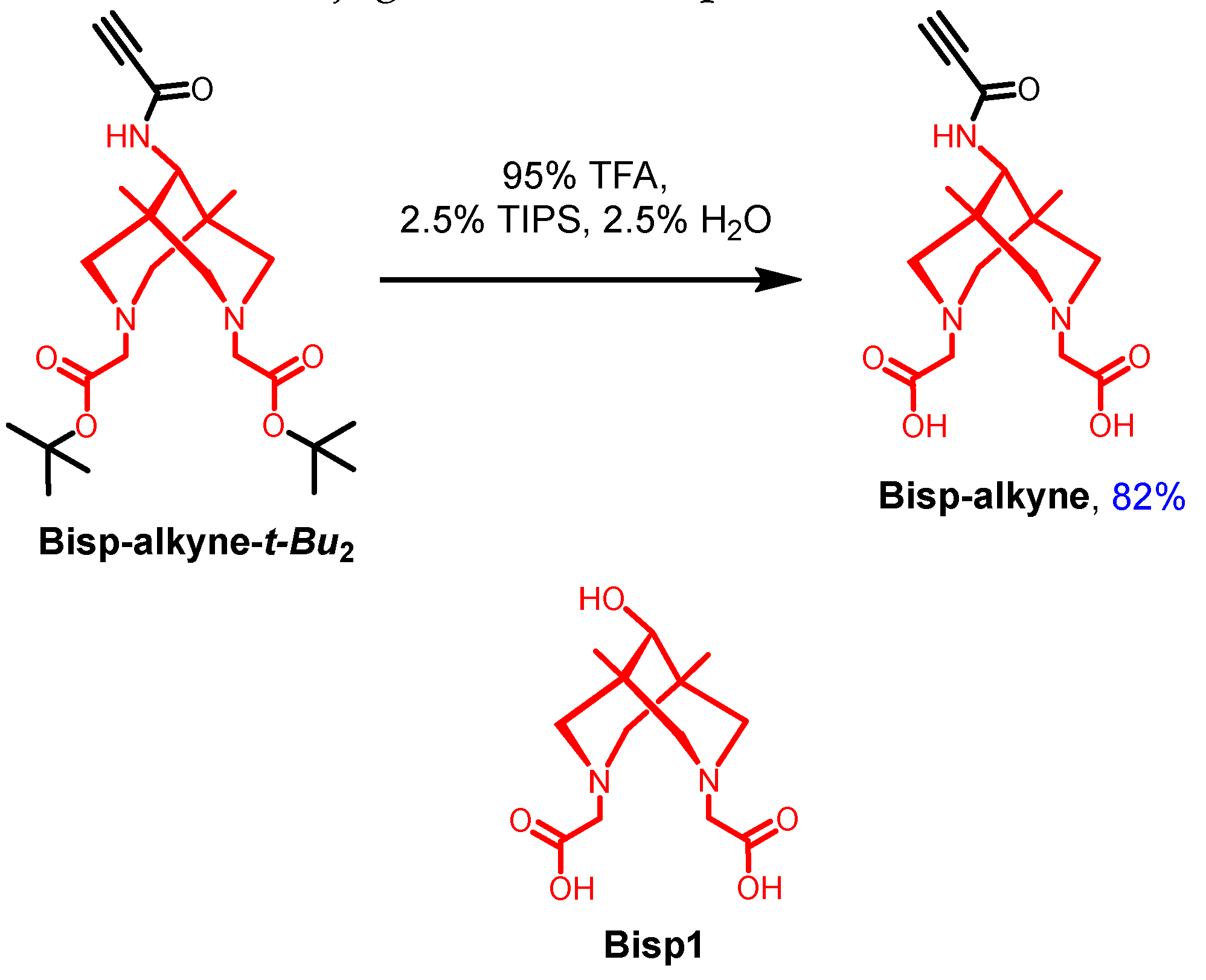
Figure 1.
Comparison of Stability of [64Cu]Cu-PSMA-Bisp and [64Cu]Cu-PSMA-617 in serum at 37 °C.
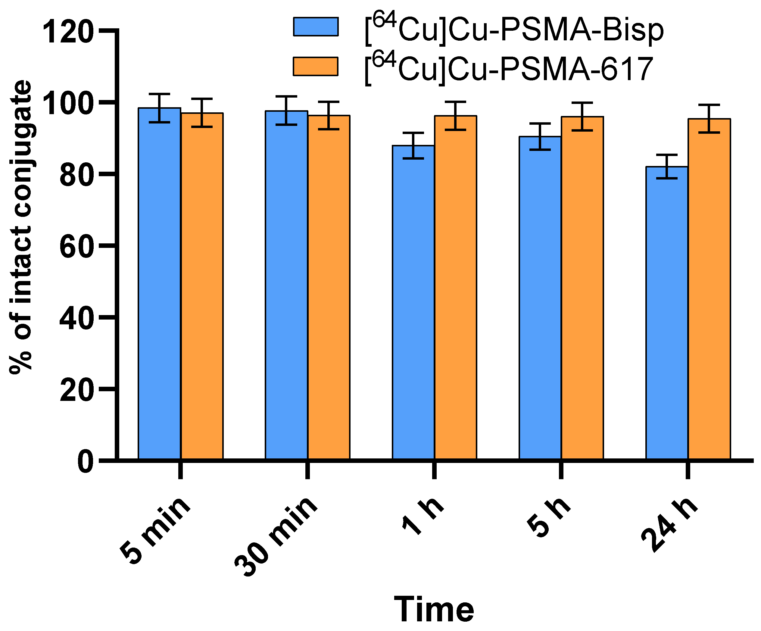
Figure 3.
Comparison of intracellular levels of ROS (A, B) and Cu2+ (C) after treatment with PSMA-Bisp - Cu2+ (50 µM, 1h). Control (untreated) cells have a copper concentration lower than the sensor's linear detection range. Results are shown as means ± SE, * p < 0.05 (one-way ANOVA).
Figure 3.
Comparison of intracellular levels of ROS (A, B) and Cu2+ (C) after treatment with PSMA-Bisp - Cu2+ (50 µM, 1h). Control (untreated) cells have a copper concentration lower than the sensor's linear detection range. Results are shown as means ± SE, * p < 0.05 (one-way ANOVA).
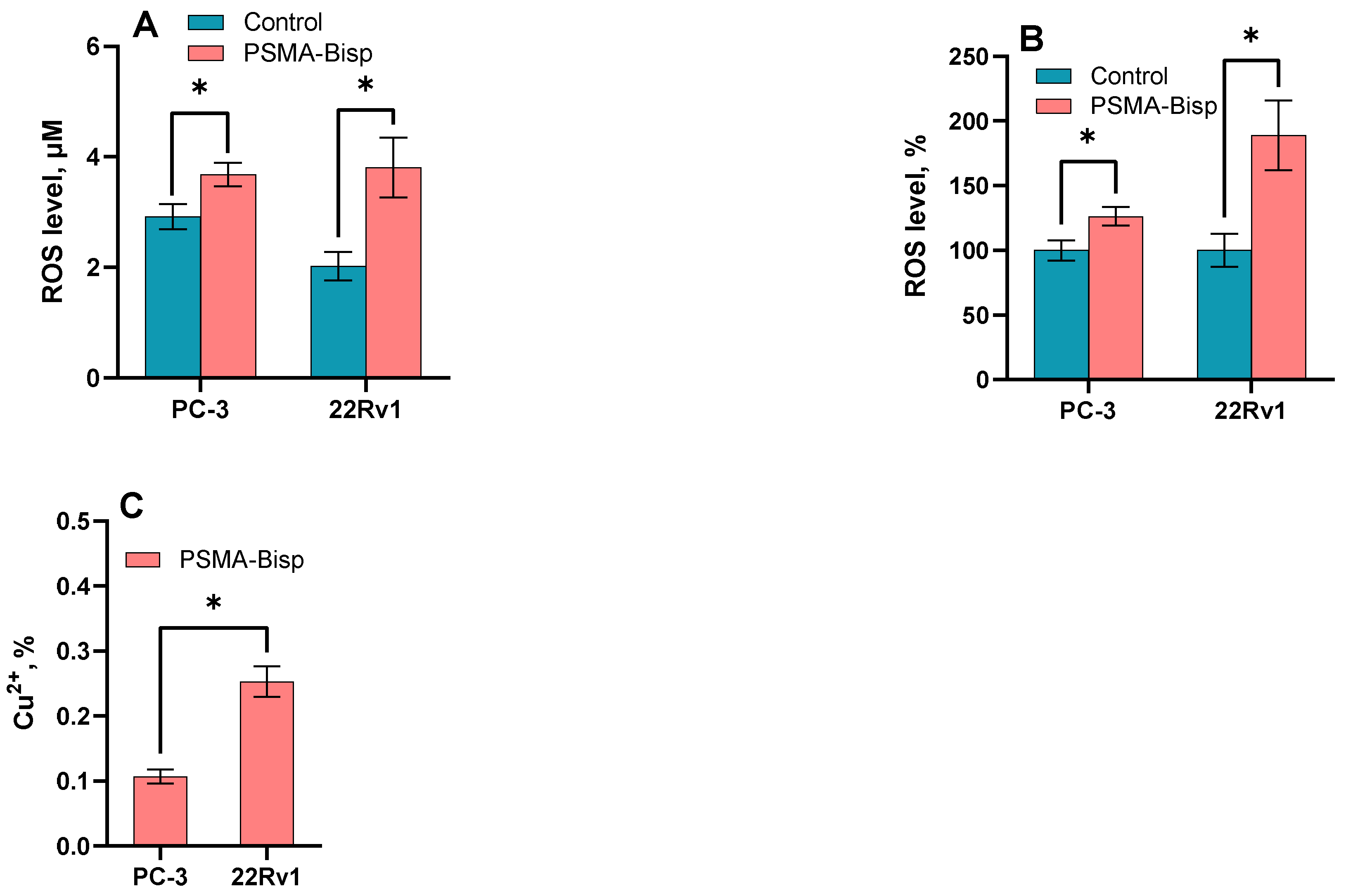
Figure 4.
Cytotoxicity of PSMA-Bisp and PSMA-Bisp + Cu2+ coincubation on PC-3 and 22Rv1 cells. MTS assay 48h* p<0.05, t-test.
Figure 4.
Cytotoxicity of PSMA-Bisp and PSMA-Bisp + Cu2+ coincubation on PC-3 and 22Rv1 cells. MTS assay 48h* p<0.05, t-test.
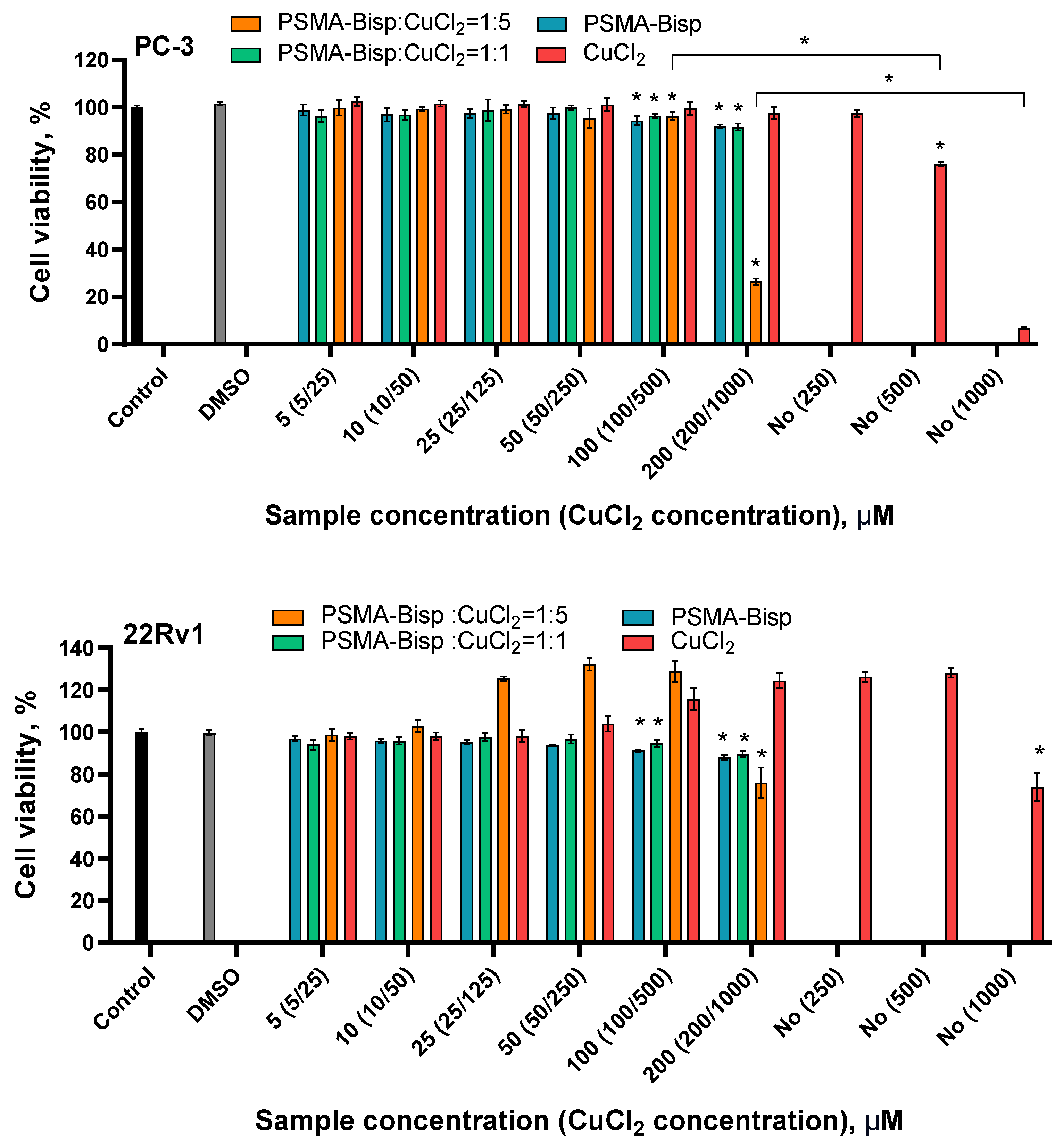

Table 1.
Labeling yields for [64Cu]Cu-Bisp1, [64Cu]Cu-Bisp-alkyne and conjugate [64Cu]Cu-PSMA-Bisp at varied ligand’s and conjugate’s concentration.
Table 1.
Labeling yields for [64Cu]Cu-Bisp1, [64Cu]Cu-Bisp-alkyne and conjugate [64Cu]Cu-PSMA-Bisp at varied ligand’s and conjugate’s concentration.
| c(L), µM | 10 | 20 | 50 | 100 | 200 | 500 | 1000 |
|---|---|---|---|---|---|---|---|
| [64Cu]Cu-Bisp1, % | - | 19 | 38 | 76 | 96 | 97 | 98 |
| [64Cu]Cu-Bisp-alkyne, % | 17 | 31 | 71 | 100 | 100 | 100 | - |
| [64Cu]Cu-PSMA-Bisp, % | - | - | 23 | 42 | - | 99 | 99 |
Table 2.
The content of intact [64Cu]Cu-PSMA-Bisp at after 1 hour of incubation in the solution with an excess of microelements (%).
Table 2.
The content of intact [64Cu]Cu-PSMA-Bisp at after 1 hour of incubation in the solution with an excess of microelements (%).
| % [64Cu]Cu-PSMA-Bisp | Mg2+ 10 mM |
Ca2+ 50 mM |
Zn2+ 1 mM |
Cu2+ 1 mM |
Fe3+ 1mM |
| γ-spectrometry | 98 ± 1 | 98 ± 1 | 93 ± 1 | 100 ± 1 | 100 ± 1 |
Disclaimer/Publisher’s Note: The statements, opinions and data contained in all publications are solely those of the individual author(s) and contributor(s) and not of MDPI and/or the editor(s). MDPI and/or the editor(s) disclaim responsibility for any injury to people or property resulting from any ideas, methods, instructions or products referred to in the content. |
© 2024 by the authors. Licensee MDPI, Basel, Switzerland. This article is an open access article distributed under the terms and conditions of the Creative Commons Attribution (CC BY) license (http://creativecommons.org/licenses/by/4.0/).
Copyright: This open access article is published under a Creative Commons CC BY 4.0 license, which permit the free download, distribution, and reuse, provided that the author and preprint are cited in any reuse.


