Preprint
Review

This version is not peer-reviewed.

Systematic Review of the Status of Microbiota in Cirrhosis. A Change Towards a More Pathogenic Predisposition

A peer-reviewed article of this preprint also exists.

Submitted:

01 December 2024

Posted:

02 December 2024

You are already at the latest version

Abstract

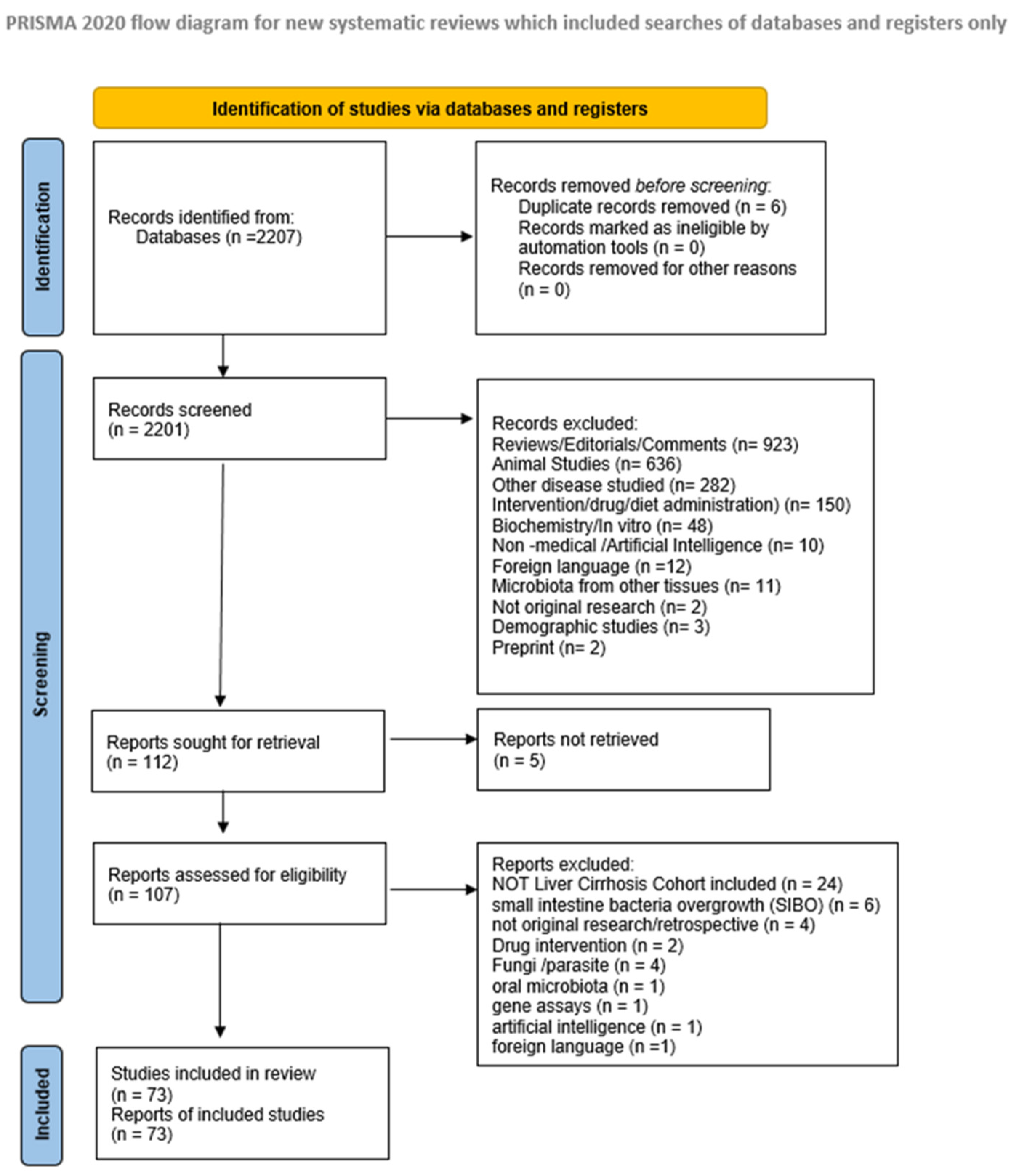
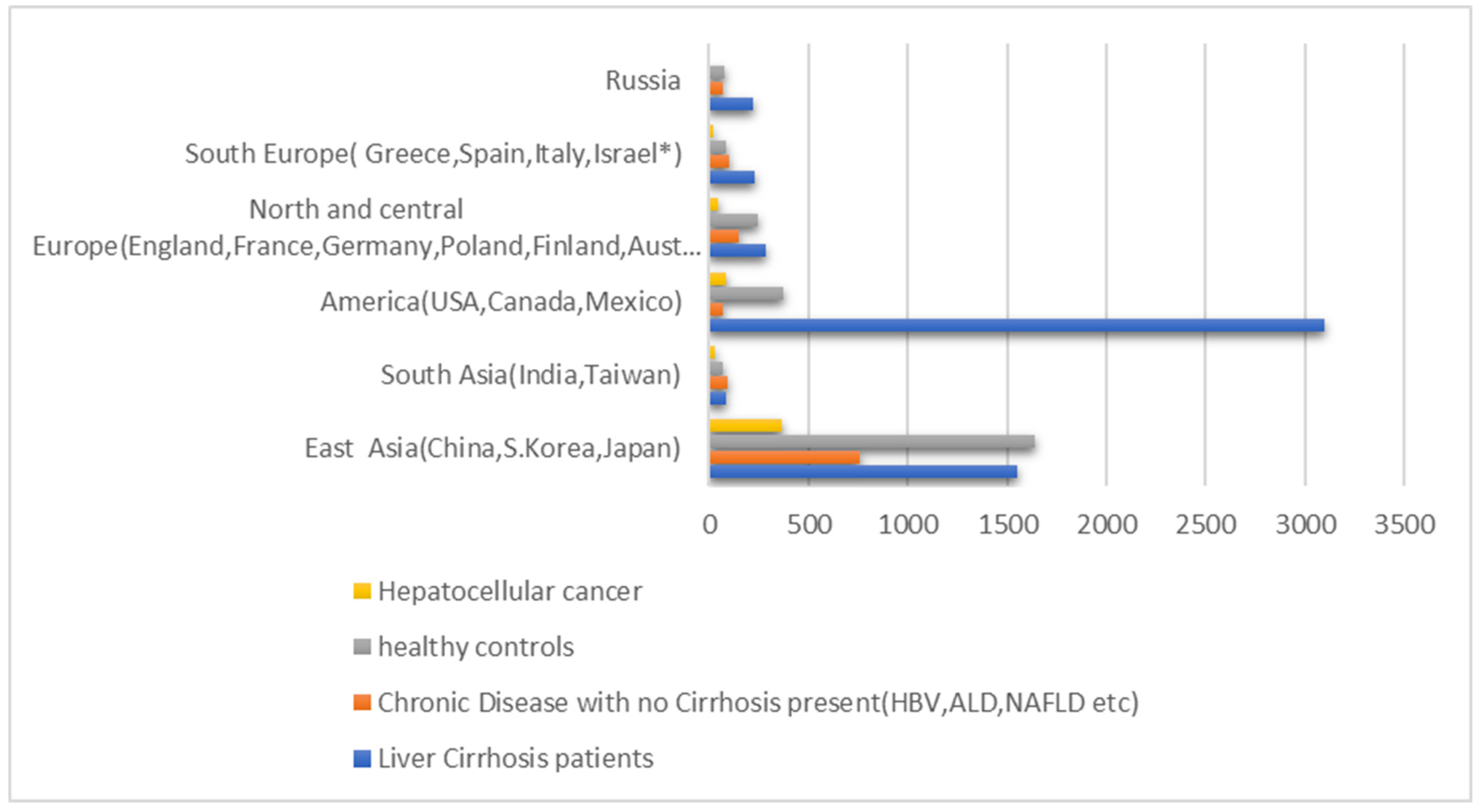
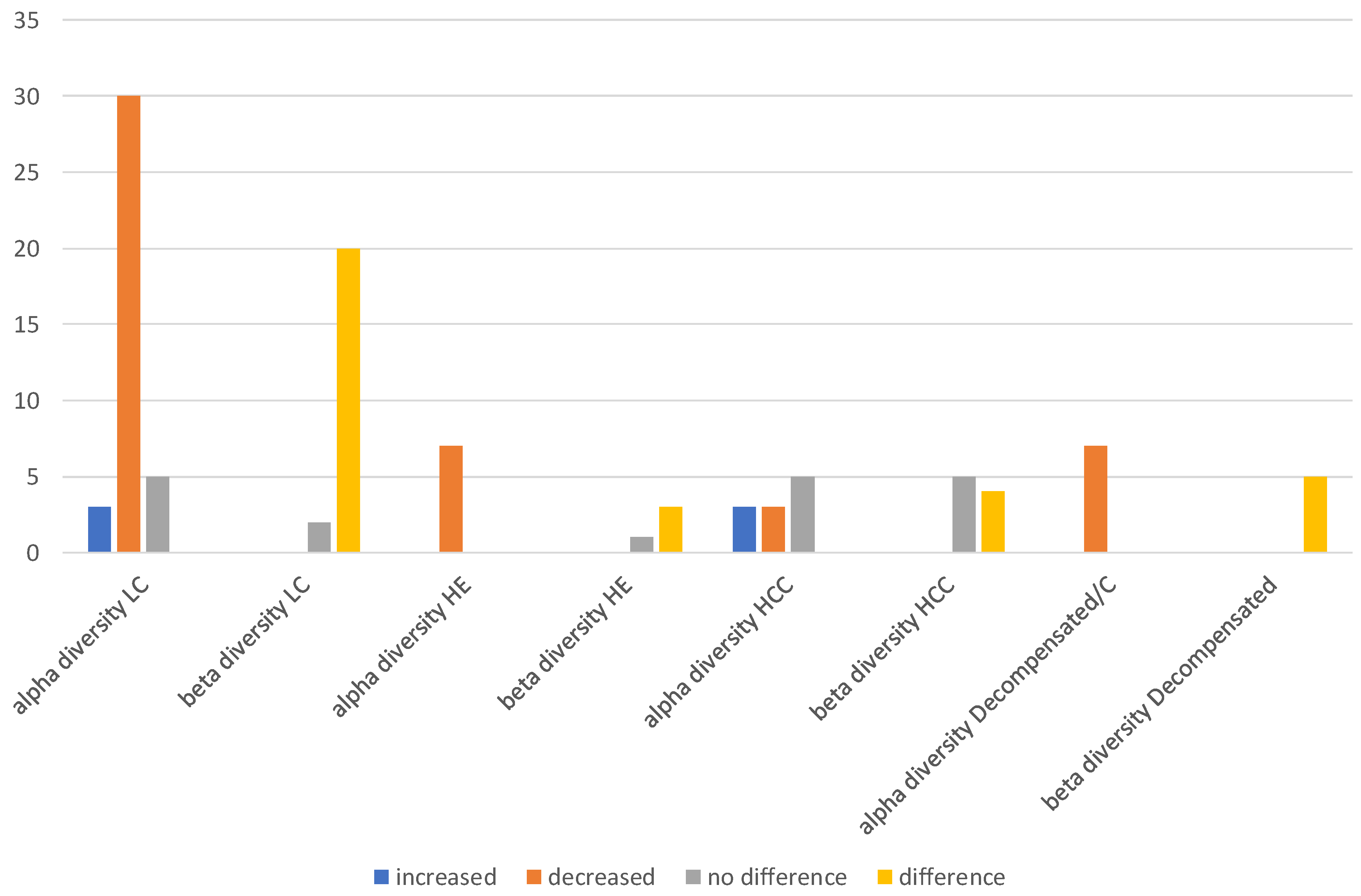
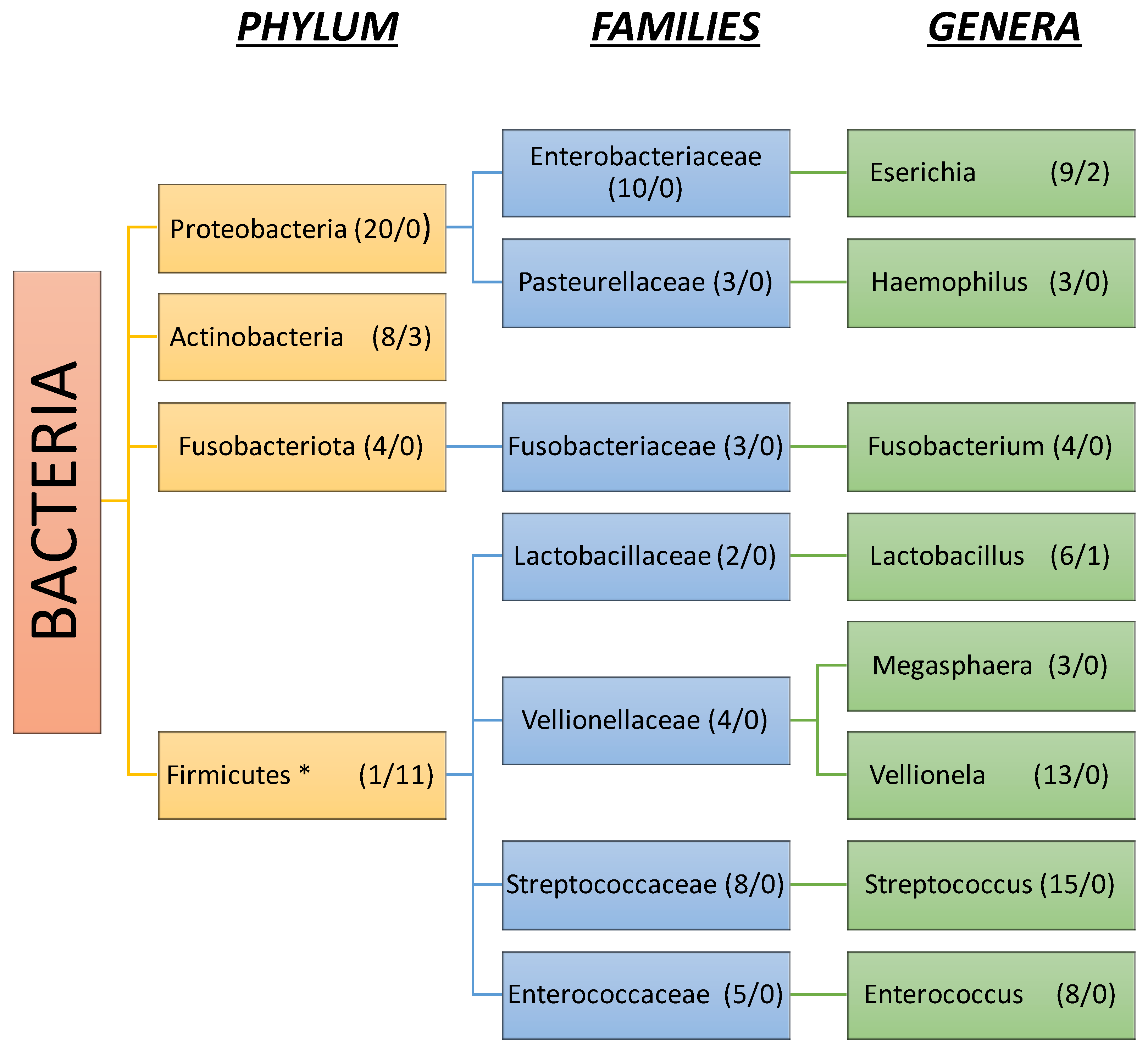
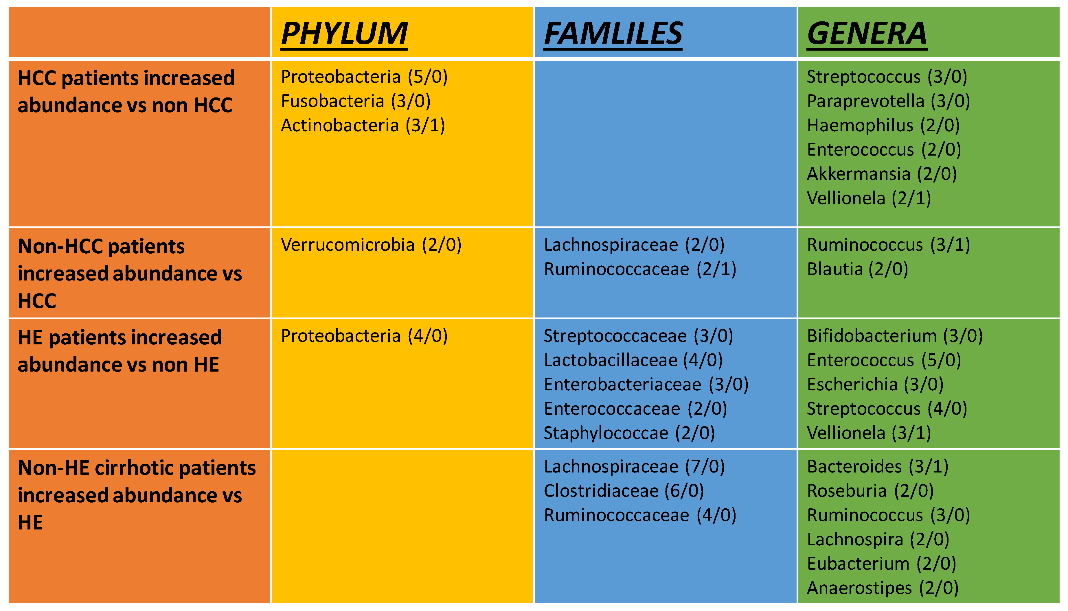
Background: The microbiome of the human intestine is a regulator of health by modulating immune response and playing important role in metabolism. Dysbiosis, diversity and abundance of microbiota communities in the gut has shown changes in cirrhosis and its complications. Aim: To review current knowledge regarding microbiota alterations in cirrhosis, its potential differences according to etiology and its role in the development of cirrhosis complications. Methods: A systematic search of online bibliographic database up to July 2024 was performed. Randomized controlled trials, observational and cohort studies that included in total or at least a cohort of cirrhotic adult patients were enlisted for data extraction and analysis. Results: A total of 73 publications were included for data extraction. Alpha diversity was found decreased in cirrhotic patients in 30/38 (78%) of the studies, while beta diversity 20/22 (90%) presented significant difference between healthy and cirrhotic groups. Proteobacteria were significantly increased in 20/27 (74%) studies followed by Actinobacteria and Fusobacteria while 22/25 (88%) studies found either reduction in cirrhotic patients or increased abundance in healthy controls for Firmicutes and Bacteroidetes. The most abundant genera in hepatic encephalopathy groups were pathobionts such as Enterococcus and Streptococcus followed by Vellionella and Escherichia. Heterogeneity was found among studies regarding Alpha-diversity in Hepatocellular Carcinoma (HCC) as it was decreased in 3 studies, indifferent in 5 and increased in 3 in comparison to cirrhotic non-HCC patients. Conclusion: Dysbiosis of the gut microbiota is associated with cirrhosis and development of complications such as hepatic encephalopathy and hepatocellular carcinoma.

Keywords: 
;  ;  ;  ;  ;  

1. Introduction

The liver as an organ has multiple metabolic functions but by staying in between the gut and the rest of human organism exerts also important immune mediated – defensive activities. It has been described to be part of the gut – liver axis due to significant first pass effect over microbes, medical molecules, toxic substances such as alcohol and food [1,2]. Many chronic liver diseases have been developing once this important protective mechanism has been damaged or saturated [1,3]. Additionally, several complications that appear in liver cirrhosis depend on these altered pathophysiological mechanisms [1].
The gut microbiome which grows and resides in all parts of the gut, especially in the small intestine exerts many effects that help or sometimes damage liver function, such as, the metabolism of bile acids [4], alcohol [5], and immune tolerance [3,6]. Even though liver has a high capacity of immune protection and metabolic reactions, these are not unlimited and therefore an intact barrier that can control input is necessary [7]. This barrier is represented by the gut, which forms a complicate system built on different layers [1]. The main layers of this barrier are: the mucus layer, the enteric cellular layer including enterocytes, goblet cells, tuft cells and enterochromaffin cells, the rich immune system layer in between and under the enteric cells and finally the vascular system layer which delivers all contents to the portal vein. In liver diseases significant changes have been described in the production of mucins and short chain fatty acids both controlled by the presence of “healthy” commensal bacteria [1,6]. In addition, a reduction in the population of bacteria containing 7a glucuronidase change the composition of secondary bile acids and induces reactions through FXR receptor [4]. Finally, a leaky gut status is caused by microbiota changes which produce an immune mediated reaction that increase turnover of enterocytes and alters the formation of proteins of the tight junctions [5,8]. These are probably the most important pathophysiologic mechanisms that have been described to date.
Dysbiosis, is called every stable change or imbalance in number, diversity and abundance of microbial populations in the gut as from the previous healthy state [9]. Dysbiosis is found in early states of many chronic liver diseases before and after the development of cirrhosis [10,11]. All microbiota changes represent an imbalance between protective bacteria like the ones included in the firmicutes taxa and potentially pathogenic bacteria like Proteobacteria (E.Coli, Klebsiella) [10]. In addition, in several instants during chronic liver diseases certain microbial populations reach a number that we conventionally describe as bacterial overgrowth [10,11,12,13,14]. In cirrhosis a significant factor for the development of bacterial overgrowth is small intestinal dysmotility which seems to be reduced by portal hypertension [15,16].
Therefore, in the cirrhosis stage especially when portal hypertension has been developed the presence of dysbiosis with or without bacterial overgrowth and leaky gut can cause bacterial translocation and consequently septic episodes of bacterial peritonitis, variceal bleeding and hepatic encephalopathy [1,8,10,12,16]. The purpose of this review is to collect current knowledge regarding microbiota changes in cirrhosis, its differences in comparison to etiology and its role on the development of cirrhosis complications.

2. Methods

2.1. Data Identification

We searched PUBMED database from 1970 to 14 of July 2024 for English and non-English publications using text words Gut Microbiota/Gut Flora/Gut Microbioma AND Cirrhosis. We manually searched references from review articles and original studies.

2.2. Inclusion and Exclusion Criteria

Studies included for analysis needed to fulfil the following criteria: (i) published as full paper; (ii) retrospective, prospective or randomized controlled design; (iii) including in total or at least a cohort of cirrhotic patients (iv) no animal studies, (v) involving population of age more or equal to 18 years;
Studies were excluded if: (i) were analyzing or comparing results of a specific treatment on microbiota (ii) groups referring on transplanted patients (iii) had no taxonomic analysis (iv) reporting microbiota other than intestinal (v) focused on fungal or viral species.

2.3. Data Extraction and Methodological Assessment

Data were extracted independently tabulated using a predefined review form. This work was conducted with the PRISMA statement of Preferred Reporting Items for Systematic Reviews and Meta-analyses [17]. The flowchart is licensed under CC BY 4.0. Thereafter two authors were assigned for quality assessment. The AMSTAR 2 tool was used to assess the quality of the studies. AMSTAR 2 doesn’t involve a scoring system but rather a rating of the overall confidence of the results of the review ranging from high to critically low [18].
The decision to include or exclude a study was made before the review analysis was carried out and agreed by consensus amongst the authors. In the present review agreement between reviewers for selection of articles for all analyses was 100%.

2.4. End Points

Our endpoints for the present review were to assess changes in microbiota in patients with cirrhosis and additionally make subgroups: 1) according to cirrhosis aetiology (ALD, MASLD, HBV etc.) 2) according to cirrhosis stage or complications (Compensated vs Decompensated, Hepatocellular Carcinoma (HCC), Hepatic Encephalopathy (HE), Spontaneous Bacterial Peritonitis (SBP). Infections, Acute on Chronic Liver Failure (ACLF) 3) According to the sample used (fecal samples, duodenal fluid, rectal swabs or colon biopsies), 4) According to the test used (16S PCR, deep sequencing metagenomics, other) 5) According to geographical distribution. In addition, we checked for papers that report microbiota in cirrhotics vs non cirrhotics of the same aetiology.

3. Results and Discussion

The search strategy found 2207 references that were imported for screening (Figure 1).
Six duplicates were found and removed. Studies screened by title and abstract were 2201 and 2095 of them were excluded according to the aforementioned criteria. Of the remaining 122 articles, 5 were excluded due to no full text retrieval. Of the 117 full text articles screened for eligibility 44 were excluded as 24 did not include a liver cirrhosis group, 4 were not original research, 10 did not report results on microbiota taxonomic differences (4 were about fungi or parasites and 6 reported only the presence of small intestinal bacterial overgrowth) and 6 on not meeting the inclusion criteria (2 included drug intervention, 1 was about oral microbiota, 1 was on artificial intelligence practice, 1 was conducted as a gene assay and 1 was in non-English language).
Finally, we identified 73 studies that met inclusion criteria [12,15,19,20,21,22,23,24,25,26,27,28,29,30,31,32,33,34,35,36,37,38,39,40,41,42,43,44,45,46,47,48,49,50,51,52,53,54,55,56,57,58,59,60,61,62,63,64,65,66,67,68,69,70,71,72,73,74,75,76,77,78,79,80,81,82,83,84,85,86,87,88]. (Figure 1) In these 73 studies identified there was at least one cohort of patients diagnosed with cirrhosis. Diagnosis of cirrhosis was confirmed either histologically by liver biopsy or combinations of clinical, laboratory parameters and/or imaging features (abdominal ultrasound/elastography).
In order to compare findings, we also separately analyzed patients with decompensated cirrhosis and complications. Regarding decompensation diagnosis was confirmed by a history of clinical, endoscopy and imaging features of ascites, HE, varices and jaundice. Studies that solely included patients with cirrhosis and HE or HCC were analyzed independently and were not included in the analysis of studies that compared compensated and decompensated patients. Studies that included arms of patients receiving antibiotics or immunomodulating medications were excluded.

3.1. Microbiota Diversity in Cirrhosis

To assess microbiota changes we analyzed the data of 73 studies. A total number of 9.763 individuals was extracted. Of them 2494 were healthy control groups, 5473 were patients with liver cirrhosis, 2494 were patients with chronic liver disease without cirrhosis while 550 were patients with HCC. In regard to geographical distribution 33 studies were from East Asia (China 29, Japan 3, Southern Korea 1) and 3 from South Asia (Taiwan 2, India 1), 21 from North or Central America (USA 19, Canada 1, Mexico 1), 6 from North or Central Europe (Poland 1, UK 1, Germany 1, France 1, Finland 1, Austria 1), and 9 from South or East Europe (Russia 4, Italy 2, Spain 1, Israel 1, Greece 1). (Figure 2).
Most studies used fecal samples to identify and quantify human gut microbiota (67/73 91.7%). For the rest 2 studies used duodenal biopsies, another 2 a combination of fecal samples and colon biopsies and 1 rectal swab. In addition, most studies (50/73, 68.4%) used as method of detection the 16s r-RNA sequencing, while much less (8/73, 10.9%) appointed the Shotgun Metagenomics method, and (6/73, 8.2%) the 16s r-DNA sequencing. The less used was PCR and bacterial cultures methods. Almost all studies investigated the entire human gut flora while only one that focused on specific species and their role in cirrhosis pathogenesis was included. This Chinese cohort analyzed subspecies of Desulfovibrio and results showed a specific species fairly increase in cirrhotic patients with a significant difference in its trait like desulfurization ability [28].
The diversity and composition data of the gut microbiota available were presented in different manners. In 13 studies results focused on most enriched species detected whereas 53 focused on the most abundant genera, families or phyla and 7 reporting the most enriched bacterial groups. In total 12 phyla, 47 families and 118 genera were reported.
Alpha diversity analysis on most studies was conducted using metrics such as Chao1, Ace, Shannon, and Simpson indices. Of the total 73 studies included 53 (72%) reported results regarding on alpha or beta diversity (Figure 3). Alpha diversity on healthy versus cirrhotic patients was presented in 38 studies. With the term cirrhosis without specific etiology there were 14/38 (37%) studies while as liver cirrhosis with a specific etiology (viral HBV or HVC, or metabolic NASH, or ALD) 24(63%). In 30 studies a significant decrease in alpha diversity was observed (78%) in cirrhosis compared to healthy subjects, 5 showed no difference, and 3 noted an increase in liver cirrhosis groups (Figure 3). However, data from the latter 3 studies collected came in the first from rectal swab [45] indicating a possible different composition in rectal gut flora in comparison to the rest of the colon, the second from a very small Chinese cohort possibly introducing bias [66] and the third was a study from Taiwan compering HBV related cirrhotic patients with their non-cirrhotic counterparts [25]. In conclusion despite a few discrepancies, most studies agree that a significant decrease in alpha diversity of the gut microbiota is associated with liver cirrhosis.
In regard to beta diversity, most common metrics used were Bray -Curtis Dissimilarity, weighted and unweighted UniFrac. With the term cirrhosis without specific etiology there were 22 studies presenting results (Figure 3). Of them in 20/22 (90%) a significant difference between healthy and cirrhotic groups was showed while in 3 results were opposing. This particular finding adds to the hypothesis that in cirrhosis the intestinal microbiota undergoes significant changes in its composition that needs to be revealed.

3.2. Gut Microbiota Composition in Liver Cirrhosis

The human gut microbiota in cirrhosis is compromised by a plethora of species. Twenty seven studies reported results on phyla level, 21 on family and 44 on genera level in patients with cirrhosis while 25, 22 and 38 respectively reported in patients with no cirrhosis. At the phyla level, proteobacteria seem to play the most important role in gut dysbiosis in cirrhosis, as they were significantly increased in 20/27 (74%) studies followed by Actinobacteria and Fusobacteria (Figure 4).
In contrast 22/25 (88%) studies found either reduction in cirrhotic patients or increased abundance in healthy controls for Firmicutes and Bacteroidetes (Figure 5). Notably although most results agree on Proteobacteria and Firmicutes changes there seem to be a discrepancy in Bacteroidetes as 11/25 (44%) studies report an increase in healthy-control versus cirrhotic groups while 5/27 (18%) report the opposite. Another important aspect regarding Firmicutes is that at the level of sub-taxa, genera as Streptococcus, Vellionella, and Enterococcus are considerably increased in numbers in cirrhotic patients than in non-cirrhotic subjects.
At the family level changes found in liver cirrhosis consist of both an increase in abundance of Enterobacteriaceae, Pasteurellaceae, Fusobacteraceae, Lactobacillaceae, Vellionellaceae, Enterococcaceae and Streptococcaceae, and decrease of Oscillospiraceae, Lachnospiraceae, Clostiridiaceae, Eubacteriaceae, Rikenellaceae and Bifidobacteriaceae. In particular Streptococcaceae and Enterobacteriaceae are the most commonly mentioned as enriched while Oscillospiraceae and Lachnospiraceae the most decreased respectively.
As far as genera are considered, we have found decreases in autochthonous taxa such as Prevotella, Paraprevotella, Roseburia, Faecalibacterium, Eubacterium, and Ruminococcus. Otherwise, conflicting results are been presented among studies regarding Bifidobacterium, Clostiridium spp, and Bacteroides with most of them been in favor of decrease and only in certain cases an increased abundance is presented. A possible explanation for this heterogeneity regards the fact that different etiologies of chronic liver diseases such as ALD, NAFLD, HBV may influence microbiota even before progression into cirrhosis and remain as such thereafter. On the other hand, genera that are largely facultative anaerobes such as Enterococcus, Escherichia, Klebsiella, Lactobacillus, Streptococcus and Haemophilus are been increasing in cirrhosis. Vellionella an oral cavity bacterial species is also significantly increased in cirrhotic patients. Finally, another important finding is that Rumminococcus Gnavus group has been linked to cirrhosis in at least 3 studies when most Rumminococcus spp are significantly decreasing in the cirrhotic gut environment [23,26,85].

3.3. Gut Microbiota Composition in Hepatic Encephalopathy

Hepatic encephalopathy (HE), characterized by a broad spectrum of neuropsychiatric symptoms, is a major complication in decompensated cirrhosis [89]. It can be classified as Grade I, covert and subsequently minimal HE (recognized by specific cognitive tests), and overt ranging from grade II to IV exhibiting severe neuropsychiatric manifestations [90]. Most studies focused on minimal HE using different scores for diagnosis such as PHES, NCT-A, DST. We found 16 studies in total which appointed results for HE either regarding taxonomic (5 on phyla, 7 on families and 11 on genera) or diversity (7 on alpha 4 on beta diversity). Alpha diversity in cirrhotic patients with HE was reported in 7/7(100%) of the studies as decreased in comparison to cirrhotic patients without HE.
The main phyla that were enriched in HE patients were Proteobacteria, whereas for families there were Streptococcaceae, Lactobacillaceae, Enterobacteriaceae, Enterococcaceae, and Staphylococceae. The most abundant genera were pathobionts such as Enterococcus and Streptococcus followed by Vellionella and Escherichia [20,26,41,43,51,54,65,75,77,86] (Figure 6). Notably, in a Chinese study Streptococcus Salivarius was strongly associated with the presence of HE in cirrhotic patients [77]. Another interesting finding was that the presence of Bifidobacterium an autochthonous taxon with beneficial effects was reported to be enriched in 3 studies [51,75,82]. A study from USA associated Alcaligeneceae, Porphyromonadaceae with worse cognition [65]. In order to evaluate microbiota composition in MHE a study demonstrated results from cirrhotic patients that were subjected to three types of cognitive tests and the results although not completely matching had similarities regarding some bacterial groups that were enriched such as bacteria from the Lactobacillaceae family [72].
On the contrary some bacteria that were decreased in patients with HE compared to cirrhotic patients without encephalopathy are members from the families Lachnospiraceae, Clostridiaceae, Ruminococcaceae such as Bacteroides, Roseburia, Ruminococcus, Lachnospira and Eubacterium [20,26,41,43,51,60,65,72,75,77,80,81] (Figure 6).

3.4. Gut Microbiota Composition in Hepatocellular Carcinoma

Epidemiological studies have shown that 70% to 90% of hepatocellular carcinoma cases are found in patients with preceding cirrhosis or advanced fibrosis [91]. We identified 13 studies reporting microbiota alterations in patients with HCC (Figure 6). Alpha diversity showed a notable heterogeneity and it was found to be decreased in 3 studies [27,58,92] but increased in 3 [40,59,74] while 5 of them [30,44,55,71,83] showed no differences in comparison to cirrhotic patients without HCC. These results may derive from the difference in populations of microbiota gut between patients that develop HCC in the presence of cirrhosis in oppose to those that don’t as some studies show [40,58]. Additionally, beta diversity showed no difference between groups in 6 studies [30,40,44,55,59,71] whereas 4 studies showed dissimilarity [27,46,58,92].
Considering gut flora composition, at phyla level changes include increase in proteobacteria, fusobacteria and actinobacteria and at family level some beneficial taxa like Lachnospiraceae and Rumminococcaceae belonging to the Firmicutes decreased. Genera that proved enriched were Paraprevotella, Akkermansia, Haemophilus and pathobionts such as Streptococcus and Enterococcus while Ruminococcus, Blautia and Bifidobacterium were decreased. Taken together the above findings suggest a significant increase in pathogens like proteobacteria and Streptococcaceae and on the other hand a decrease in beneficial bacteria such as those that reduce hydrophobic bile acids. As the role of pathogens in the development of HCC is not clear a Polish study in patients with liver cirrhosis undergoing liver transplant found a significant correlation between fecal E. Coli and the presence of HCC [68]. In addition, while Lactobacillus is known to be beneficial in human homeostasis, as it belongs to a group of certain bacterial genera that are involved in bile acid deconjugation, oxidation/epimerization, and 7-dehydroxylation together with Bacteroides, Bifidobacterium, Ruminococcus, and Clostridium [93] a study from Germany on NASH associated the presence of HCC in cirrhotic patients with increased abundance in the Lactobacillus species [58]. (Figure 6).

3.5. Gut Microbiota Composition in Other Complications

Few studies assessed microbiota changes and other complications such as ascites, spontaneous bacterial peritonitis (SBP), infections or ACLF [31,32,34,38,47,48,53,57,82,86]. Regarding microbiota changes during the passage from compensated to decompensated cirrhosis [31,36,40,42,55,56,57], or based on Child-Pugh score [64,70], alpha diversity was found to be decreased in all studies (Figure 3). Most abundant bacterial groups were at Enterococcaceae, Streptococcace and Peptostreptococcaceae families while Enterococcus was the most prominent genera followed by Streptococcus, Staphylococcus, Vellionella and Lactobacillus. These findings follow the pattern already present with pathobionts increasing as the liver disease progresses.
Clinically significant ascites is associated with increase in Proteobacteria and decrease in Actinobacteria and Bacteroidetes as one particular study from Russia demonstrated [32]. Very few studies focused on SBP including one from Russia that showed enrichment in Proteobacteria, and particularly Gammaproteobacteria including pathogens like Klebsiella, Serratia, Acinetobacter and Moraxella spp [31] and a study from India in cirrhotic patients with infections that found in SBP enrichment of Oxalobacteraceae, Neisseriaceae, Anaerotruncus [34]. Gammaproteobacteria, Lactobacillaceae and Enterobacteriaceae were found enriched in patients with concomitant infections such as pneumonia, bacteremia, cholangitis but results were sparse among different infections [34,38,65,86].
Finally, in ACLF enterococcus spp was found to be the most prominent taxa [48,57], while in another study from USA Proteobacteria, Actionobacteria, Cambylobacteracae were most abundant [82].

3.6. Gut Microbiota Assessment in Liver Cirrhosis Various Etiologies

Of the studies reviewed 11 were conducted on HBV, 7 on ALD and 6 on NAFLD populations specifically. As already mentioned, alpha diversity is mostly decreased in cirrhotic patients and this pattern had no exception between different etiologies. Specifically of the 8 studies that were conducted on HBV cirrhotic patients reporting results on alpha -diversity 6 indicated decrease [27,36,42,48,52,56,92], 1 indifferent [29] and 1 increase [25] while 5 reported differences in beta diversity versus non-cirrhotic controls. Regarding NAFLD 5 reported decreased alpha diversity and high beta diversity index [58,69,71,85,88]. In ALD 4 studies presented decreased alpha diversity [40,49,59,61] while only 2 showed no significant difference [73,87].
The main phyla enriched were Actinobacteria and Proteobacteria in HBV cirrhotic populations presented in 4 and 5 studies respectively, Actinobacteria, Proteobacteria and Fusobacteria in ALD and Proteobacteria mainly in NAFLD cirrhotic populations. Most prevalent families were Enterobacteriaceae in all aetiologies [27,39,42,49,56,63,71,88] and Streptococcaceae in HBV populations [19,27,42,56]. Notably Streptococcus and Vellionella species were presented in 6 [19,27,28,36,56,92] and 5 studies [25,27,56,62,92] respectively and were the most increased genera in HBV cirrhotic populations while Lactobaccilus was significantly increased in ALD groups [44,59,87].
In the present systematic review we collected all published studies that identify the status of microbiota in cirrhosis. Our findings show that cirrhotic patients have significant changes in both alpha and beta diversity compared to controls which in most studies are healthy volunteers. Changes refer to all taxa of bacteria namely phylum, families and genera. At the level of phylum, we have conflicting results showing both increase and decrease in abundance but at a lower level we have found specific changes in families and genera. We conclude that at Proteobacteria phulum, Enterobacteriaceae and Pasteurellaceae families increase, at Firmicutes phylum, Lactobacillaceae Velionellaceae Streptococcacceae and Enterococcaceae increase whilst Oscillospiraceae, Lachnospiraceae, Eubacteriaceae and Clostiridiaceae decrease. At Bacteriodetes phylum, Prevotelacceae and Rikenellaceae decrease, at Actinobacteria, Bifidobacteriaceae decrease. Finally at Fusobacteria phylum, Fusobacteriaceae are increasing. Another important finding of this review is that as cirrhosis progress to a decompensated stage and complications appear there is a prominent change in alpha and beta diversity as well. However, until now the complication mostly studied is hepatic encephalopathy. In hepatocellular carcinoma studies show conflicting results regarding alpha and beta diversity.
A normal gut microbiota consists of 6 phyla namely Proteobacteria, Firmicutes, Bacteroidetes, Actinobacteria, Fusobacteria and Verrucomicrobia with the phyla Bacteroidetes and Firmicutes representing more or less 90% of the population [94]. Normal distribution of gut microbiota in the intestinal lumen is associated with different pH values, motility, oxygen levels and other conditions in the environment. Stomach is dominated by oral cavity bacteria such as Vellionella, Streptococcus, Prevotella, Rothia, Haemophilus [95], dudodenum mostly facultative anaerobic due to its hostile environment and jejunum gram positive aerobic as well as facultative anaerobic like Lactobacillus, Enterococcus and Streptococcus [96]. In the ileum, the main bulk consist of Enterococcus, Bacteroides, Lactobacillus, Clostridium, Corynebacteria while the cecum and the rest of the colon is harbored by anaerobic bacteria 100 to 1000 times more than aerobic. It is mainly inhabited by families such as Bacteroidaceae, Prevotellaceae, Rikenellaceae, Lachnospiraceae, and Ruminococcaceae, and regarding genera the main species consist of Bacteroides, Lactobacillus, Bifidobacterium, and Clostridium. In addition the colon hosts a plethora of pathogens such as Campylobacter jejuni, Salmonella enterica, Escherichia coli (E. coli), and Bacteroides fragilis [97]. In liver cirrhosis several microbiota combinations appear specific when combined to other diseases like type 2 diabetes and inflammatory bowel disease [76]. Additionally, 54% of the new species that reside the gut in cirrhosis are of buccal origin. Studies are showing that as cirrhosis is progressing, significant microbiota and metabolic signatures have been associated with the first decompensation event [38] and a significant change in small intestinal environment is indicating a shift of the disease state from compensated to decompensated with portal hypertension [22]. These microbiota changes in cirrhosis have produced some specific diagnostic models which were validated in the late years. These models provide both an approach to diagnosis and a method to evaluate new treatments. In 2014 the cirrhosis dysbiosis ratio (CDR) has been found to be a useful quantitative tool for microbiome alterations in cirrhosis. A low ratio is indicating dysbiosis [80] and it has already been used in studies evaluating treatment on microbiota with success [98]. Another diagnostic model presented in 2023 is using not only microbiota features but also metabolites. The 7 microorganisms and 2 metabolites model use Subdoligranulum, Agathobacter, norank f Eubacterium coprostanoligenes group, Butyricicoccus, Lachnospiraceae UCG 004 and L-2,3-
Dihydrodipicolinate expecting to be at high levels in cirrhosis patients, and Blautia, Monoglobus, and 5-Acetamidovalerate at low levels [23]. Finally a modified cirrhosis dysbiosis ratio (MDR): [Bacilli (%) + Proteobacteria (%)]/[Clostridia (%) + Bacteroidetes (%)] was able to indicate more severe states of cirrhosis in correlation to different levels of dysbiosis [47]. These are tending to replace the Firmicutes/Bacteroidetes (F/B) ratio and the Microbial Dysbiosis index (MDI) which are not specific for cirrhosis [15].
Our systematic review is summarizing many microbial changes in cirrhosis but in most of them we realize a shift towards an increase in potentially pathogenic or pathogenic bacteria and a significant decrease in commensal or beneficial bacteria. In fact, bacteria that play a role as metabolizers of bile salts, as producers of protective mucus or as trasformers of significant aminoacids, lipids and sugars are constantly reducing [5,8,12,63]. Many studies are indicating that not only the presence of protective bacteria is reducing significantly but the potentially pathogenic outnumber them reaching the threshold of bacterial overgrowth namely > 103 cpu/ml [10,11,12,14]. This has been correlated with more advanced stages of cirrhosis especially when significant portal hypertension has developed [22]. One study has showed that SIBO prevalence is significantly higher in liver cirrhosis than in non-cirrhosis. Additionally, even in non- cirrhosis when SIBO was present this was correlate with irreversible advanced liver disease [99]. Another study has showed that SIBO derived by hydrogen producing bacteria is more important that SIBO from methane producers in the development of hepatic encephalopathy [100]. Finally, a study published by our group found a significant correlation of SIBO with Child-Pugh score and significant motility changes in cirrhosis [16]. And this was in line with another study that showed that microbiota changes are depending on small intestinal transit alterations found in cirrhosis with portal hypertension [15]. The last two studies showed also that in the presence of decompensation episodes with significant portal hypertension pharmaceutical interventions that reduce SIBO like rifaximin or increase small intestinal motility like propranolol have proved efficient [16,100]. Taken together the current knowledge described in this systematic review provides evidence for clinical implications regarding specific dysbiosis scores for cirrhosis as also information for the correct use of probiotics, antibiotics and targeted therapies like motility agents as well.
This systematic review has also revealed some limitations. First as studies collected come from different areas of the world this may have influenced the heterogeneity in results and also the different dietary habits especially when healthy population is used for comparison. Additionally, many changes may be due to differences in etiology of cirrhosis or pharmacologic interventions in decompensated cirrhosis such as previous use of antibiotics and gastric antisecretory medications. Another possible limitation arises from the fact that nearly most of the studies analyzed microbiota on stool provided while the alternative of duodenal biopsies or duodenal aspirates may have proven a different biological diversity as other studies on microbiota have been indicating [101]. Finally, 23 of the 73 studies included small samples of less than 50 patients that may decrease the validity of these results.

4. Conclusion

In conclusion dysbiosis of the gut microbiota is strongly associated with cirrhosis and its potential complications such as hepatic encephalopathy and HCC. Studies conducted on microbiome structure, show some heterogeneity in the results. Despite this, alpha diversity, increased abundance of pathobionts such as Enterococcus and Streptococcus and decrease in species that belong to bacteria families including Lachnospiracaceae and Oscillospiracaceae with important role in gut homeostasis were all linked with liver cirrhosis progression. These changes in microbiota composition affect directly and indirectly the pathophysiology of cirrhosis by many mechanisms including chronic inflammation, changes in metabolism and increased intestinal permeability. More comprehensive, large-scale studies on the role of microbiota regarding specific complications including ascites, spontaneous bacterial peritonitis and ACLF should be authorized as current knowledge lacks evidence in this matter.

References

  1. Albillos, A.; de Gottardi, A.; Rescigno, M. The gut-liver axis in liver disease: Pathophysiological basis for therapy. J. Hepatol. 2020, 72, 558–577. [Google Scholar] [CrossRef]
  2. Arab, J.P.; Martin-Mateos, R.M.; Shah, V.H. Gut-liver axis, cirrhosis and portal hypertension: the chicken and the egg. Hepatol. Int. 2018, 12, 24–33. [Google Scholar] [CrossRef]
  3. Xirouchakis, E.; Manousou, P.; Tsartsali, L.; Georgopoulos, S.D.; Burroughs, A.K. Insights into the pathogenesis of NAFLD: The role of metabolic and pro-inflammatory mediators. Ann. Gastroenterol. 2009, 22, 24–33. [Google Scholar]
  4. Collins, S.L.; Stine, J.G.; Bisanz, J.E.; Okafor, C.D.; Patterson, A.D. Bile acids and the gut microbiota: metabolic interactions and impacts on disease. Nat. Rev. Microbiol. 2023, 21, 236–247. [Google Scholar] [CrossRef] [PubMed]
  5. Meroni, M.; Longo, M.; Dongiovanni, P. Alcohol or Gut Microbiota: Who Is the Guilty? Int. J. Mol. Sci. 2019, 20, 4568. [Google Scholar] [CrossRef]
  6. Park JW, Kim SE, Lee NY, Kim JH, Jung JH, Jang MK, Park SH. et al. Role of Microbiota-Derived Metabolites in Alcoholic and Non-Alcoholic Fatty Liver Diseases. Int. J. Mol. Sci. 2021, 23, 426. [Google Scholar] [CrossRef]
  7. Jenne, C.N.; Kubes, P. Immune surveillance by the liver. Nat. Immunol. 2013, 14, 996–1006. [Google Scholar] [CrossRef]
  8. Forlano, R.; Martinez-Gili, L.; Takis, P.; Miguens-Blanco, J.; Liu, T.; Triantafyllou, E.; Skinner, C.; et al. Disruption of gut barrier integrity and host-microbiome interactions underlie MASLD severity in patients with type-2 diabetes mellitus. Gut Microbes 2024, 16, 2304157. [Google Scholar] [CrossRef] [PubMed]
  9. Marchesi JR, Adams DH, Fava F, Hermes GD, Hirschfield GM, Hold G, Quraishi MN. ; et al. The gut microbiota and host health: a new clinical frontier. Gut 2016, 65, 330–339. [Google Scholar] [CrossRef] [PubMed]
  10. Acharya, C.; Bajaj, J.S. Altered Microbiome in Patients with Cirrhosis and Complications. Clin. Gastroenterol. Hepatol. 2019, 17, 307–321. [Google Scholar] [CrossRef]
  11. Maslennikov, R.; Ivashkin, V.; Efremova, I.; Poluektova, E.; Kudryavtseva, A.; Krasnov, G. Gut dysbiosis and small intestinal bacterial overgrowth as independent forms of gut microbiota disorders in cirrhosis. World J. Gastroenterol. 2022, 28, 1067–1077. [Google Scholar] [CrossRef]
  12. Gkolfakis, P.; Tziatzios, G.; Leite, G.; Papanikolaou, I.S.; Xirouchakis, E.; Panayiotides, I.G.; Karageorgos, A.; et al. Prevalence of Small Intestinal Bacterial Overgrowth Syndrome in Patients with Non-Alcoholic Fatty Liver Disease/Non-Alcoholic Steatohepatitis: A Cross-Sectional Study. Microorganisms 2023, 11, 723. [Google Scholar] [CrossRef]
  13. Efremova, I.; Maslennikov, R.; Alieva, A.; Poluektova, E.; Ivashkin, V. Small Intestinal Bacterial Overgrowth Is Associated with Poor Prognosis in Cirrhosis. Microorganisms 2023, 11, 1017. [Google Scholar] [CrossRef] [PubMed]
  14. Fitriakusumah Y, Lesmana CRA, Bastian WP, Jasirwan COM, Hasan I, Simadibrata M, Kurniawan J. ; et al. The role of Small Intestinal Bacterial Overgrowth (SIBO) in Non-alcoholic Fatty Liver Disease (NAFLD) patients evaluated using Controlled Attenuation Parameter (CAP) Transient Elastography (TE): a tertiary referral center experience. BMC Gastroenterol. 2019, 19, 43. [Google Scholar]
  15. Liu, Y.; Jin, Y.; Li, J.; Zhao, L.; Li, Z.; Xu, J.; Zhao, F.; et al. Small Bowel Transit and Altered Gut Microbiota in Patients With Liver Cirrhosis. Front. Physiol. 2018, 9, 470. [Google Scholar] [CrossRef]
  16. Xirouchakis, E.; Kranidioti, H.; Hadziyanni, E.; Kourikou, A.; Reppas, C.; Vertzoni, M.; Papadopoulos, N.; et al. The effect of propranolol on gastrointestinal motility and permeability in patients with cirrhosis and significant portal hypertension. BMC Gastroenterol. 2024, 24, 420. [Google Scholar] [CrossRef]
  17. Moher, D.; Liberati, A.; Tetzlaff, J.; Altman, D.G.; Group, P. Preferred reporting items for systematic reviews and meta-analyses: the PRISMA statement. Ann. Intern. Med. 2009, 151, 264–269. [Google Scholar] [CrossRef]
  18. Shea, B.J.; Reeves, B.C.; Wells, G.; Thuku, M.; Hamel, C.; Moran, J.; Moher, D.; et al. AMSTAR 2: a critical appraisal tool for systematic reviews that include randomised or non-randomised studies of healthcare interventions, or both. BMJ 2017, 358, j4008. [Google Scholar] [CrossRef] [PubMed]
  19. Yang, J.; He, Q.; Lu, F.; Chen, K.; Ni, Z.; Wang, H.; Zhou, C.; et al. A distinct microbiota signature precedes the clinical diagnosis of hepatocellular carcinoma. Gut Microbes 2023, 15, 2201159. [Google Scholar] [CrossRef] [PubMed]
  20. Jinato, T.; Sikaroodi, M.; Fagan, A.; Sterling, R.K.; Lee, H.; Puri, P.; Davis, B.C.; et al. Alterations in gut virome are associated with cognitive function and minimal hepatic encephalopathy cross-sectionally and longitudinally in cirrhosis. Gut Microbes 2023, 15, 2288168. [Google Scholar] [CrossRef] [PubMed]
  21. Wang, Q.; Tang, X.; Qiao, W.; Sun, L.; Shi, H.; Chen, D.; Xu, B.; et al. Machine learning-based characterization of the gut microbiome associated with the progression of primary biliary cholangitis to cirrhosis. Microbes Infect. 2024, 26, 105368. [Google Scholar] [CrossRef]
  22. Efremova, I.; Maslennikov, R.; Poluektova, E.; Zharkova, M.; Kudryavtseva, A.; Krasnov, G.; Fedorova, M.; et al. Gut Dysbiosis and Hemodynamic Changes as Links of the Pathogenesis of Complications of Cirrhosis. Microorganisms 2023, 11, 2202. [Google Scholar] [CrossRef]
  23. Chen, Y.; Chen, S.; Xu, C.; Yu, L.; Chu, S.; Bao, J.; Wang, J.; et al. Identification of Diagnostic Biomarkers for Compensatory Liver Cirrhosis Based on Gut Microbiota and Urine Metabolomics Analyses. Mol. Biotechnol. 2023, 66, 3164–3181. [Google Scholar] [CrossRef]
  24. Aliwa, B.; Horvath, A.; Traub, J.; Feldbacher, N.; Habisch, H.; Fauler, G.; Madl, T.; et al. Altered gut microbiome, bile acid composition and metabolome in sarcopenia in liver cirrhosis. J. Cachexia Sarcopenia Muscle 2023, 14, 2676–2691. [Google Scholar] [CrossRef]
  25. Lin, M.J.; Su, T.H.; Chen, C.C.; Wu, W.K.; Hsu, S.J.; Tseng, T.C.; Liao, S.H.; et al. Diversity and composition of gut microbiota in healthy individuals and patients at different stages of hepatitis B virus-related liver disease. Gut Pathog. 2023, 15, 24. [Google Scholar] [CrossRef]
  26. Wang, Q.; Chen, C.; Zuo, S.; Cao, K.; Li, H. Integrative analysis of the gut microbiota and faecal and serum short-chain fatty acids and tryptophan metabolites in patients with cirrhosis and hepatic encephalopathy. J. Transl. Med. 2023, 21, 395. [Google Scholar] [CrossRef]
  27. Yan, F.; Zhang, Q.; Shi, K.; Zhang, Y.; Zhu, B.; Bi, Y.; Wang, X. Gut microbiota dysbiosis with hepatitis B virus liver disease and association with immune response. Front. Cell Infect. Microbiol. 2023, 13, 1152987. [Google Scholar] [CrossRef]
  28. Lu, G.; Zhang, Y.; Ren, Y.; Shi, J.S.; Xu, Z.H.; Geng, Y. Diversity and Comparison of Intestinal Desulfovibrio in Patients with Liver Cirrhosis and Healthy People. Microorganisms 2023, 11, 276. [Google Scholar] [CrossRef] [PubMed]
  29. Shen, Y.; Wu, S.D.; Chen, Y.; Li, X.Y.; Zhu, Q.; Nakayama, K.; Zhang, W.Q.; et al. Alterations in gut microbiome and metabolomics in chronic hepatitis B infection-associated liver disease and their impact on peripheral immune response. Gut Microbes 2023, 15, 2155018. [Google Scholar] [CrossRef] [PubMed]
  30. Lai, M.W.; Chu, Y.D.; Hsu, C.W.; Chen, Y.C.; Liang, K.H.; Yeh, C.T. Multi-Omics Analyses Identify Signatures in Patients with Liver Cirrhosis and Hepatocellular Carcinoma. Cancers 2022, 15, 210. [Google Scholar] [CrossRef] [PubMed]
  31. Zhou, Z.; Lv, H.; Lv, J.; Shi, Y.; Huang, H.; Chen, L.; Shi, D. Alterations of gut microbiota in cirrhotic patients with spontaneous bacterial peritonitis: A distinctive diagnostic feature. Front. Cell Infect. Microbiol. 2022, 12, 999418. [Google Scholar] [CrossRef] [PubMed]
  32. Maslennikov, R.; Ivashkin, V.; Alieva, A.; Poluektova, E.; Kudryavtseva, A.; Krasnov, G.; Zharkova, M.; et al. Gut dysbiosis and body composition in cirrhosis. World J. Hepatol. 2022, 14, 1210–1225. [Google Scholar] [CrossRef] [PubMed]
  33. Hua, X.; Feng, H. Changes in intestinal microbiota of HBV-associated liver cirrhosis with/without hepatic encephalopathy. Medicine (Baltimore) 2022, 101, e29935. [Google Scholar] [CrossRef]
  34. Philips, C.A.; Ahamed, R.; Abduljaleel, J.K.P.; Rajesh, S.; Augustine, P. Identification and Analysis of Gut Microbiota and Functional Metabolism in Decompensated Cirrhosis with Infection. J. Clin. Transl. Hepatol. 2023, 11, 15–25. [Google Scholar] [CrossRef]
  35. Bajaj, J.S.; Pena-Rodriguez, M.; La Reau, A.; Phillips, W.; Fuchs, M.; Davis, B.C.; Sterling, R.K.; et al. Longitudinal transkingdom gut microbial approach towards decompensation in outpatients with cirrhosis. Gut 2023, 72, 759–771. [Google Scholar] [CrossRef] [PubMed]
  36. Sun, X.; Chi, X.; Zhao, Y.; Liu, S.; Xing, H. Characteristics and Clinical Significance of Intestinal Microbiota in Patients with Chronic Hepatitis B Cirrhosis and Type 2 Diabetes Mellitus. J. Diabetes Res. 2022, 2022, 1826181. [Google Scholar] [CrossRef]
  37. Ullah, N.; Kakakhel, M.A.; Khan, I.; Gul Hilal, M.; Lajia, Z.; Bai, Y.; Sajjad, W.; et al. Structural and compositional segregation of the gut microbiota in HCV and liver cirrhotic patients: A clinical pilot study. Microb. Pathog. 2022, 171, 105739. [Google Scholar] [CrossRef]
  38. Bajaj, J.S.; Reddy, K.R.; Tandon, P.; Garcia-Tsao, G.; Kamath, P.S.; O’Leary, J.G.; Wong, F.; et al. Association of serum metabolites and gut microbiota at hospital admission with nosocomial infection development in patients with cirrhosis. Liver Transpl. 2022, 28, 1831–1840. [Google Scholar] [CrossRef]
  39. Baltazar-Diaz, T.A.; Gonzalez-Hernandez, L.A.; Aldana-Ledesma, J.M.; Pena-Rodriguez, M.; Vega-Magana, A.N.; Zepeda-Morales, A.S.M.; Lopez-Roa, R.I.; et al. Escherichia/Shigella, SCFAs, and Metabolic Pathways-The Triad That Orchestrates Intestinal Dysbiosis in Patients with Decompensated Alcoholic Cirrhosis from Western Mexico. Microorganisms 2022, 10. [Google Scholar] [CrossRef]
  40. Zheng, R.; Wang, G.; Pang, Z.; Ran, N.; Gu, Y.; Guan, X.; Yuan, Y.; et al. Liver cirrhosis contributes to the disorder of gut microbiota in patients with hepatocellular carcinoma. Cancer Med. 2020, 9, 4232–4250. [Google Scholar] [CrossRef]
  41. Bajaj, J.S.; Fagan, A.; McGeorge, S.; Sterling, R.K.; Rogal, S.; Sikaroodi, M.; Gillevet, P.M. Area Deprivation Index and Gut-Brain Axis in Cirrhosis. Clin. Transl. Gastroenterol. 2022, 13, e00495. [Google Scholar]
  42. Shu, W.; Shanjian, C.; Jinpiao, L.; Qishui, O. Gut microbiota dysbiosis in patients with hepatitis B virus-related cirrhosis. Ann. Hepatol. 2022, 27, 100676. [Google Scholar] [CrossRef]
  43. Lin, Y.; Yan, G.; Feng, F.; Wang, M.; Long, F. Characterization of intestinal microbiota and serum metabolites in patients with mild hepatic encephalopathy. Open Life Sci. 2022, 17, 139–154. [Google Scholar] [CrossRef]
  44. Dong, T.S.; Jacobs, J.P.; Agopian, V.; Pisegna, J.R.; Ayoub, W.; Durazo, F.; Enayati, P.; et al. Duodenal Microbiome and Serum Metabolites Predict Hepatocellular Carcinoma in a Multicenter Cohort of Patients with Cirrhosis. Dig. Dis. Sci. 2022, 67, 3831–3841. [Google Scholar] [CrossRef] [PubMed]
  45. Shen, T.D.; Daniel, S.G.; Patel, S.; Kaplan, E.; Phung, L.; Lemelle-Thomas, K.; Chau, L.; et al. The Mucosally-Adherent Rectal Microbiota Contains Features Unique to Alcohol-Related Cirrhosis. Gut Microbes 2021, 13, 1987781. [Google Scholar] [CrossRef]
  46. Chen, T.; Ding, R.; Chen, X.; Lu, Y.; Shi, J.; Lu, Y.; Tang, B.; et al. Firmicutes and Blautia in gut microbiota lessened in chronic liver diseases and hepatocellular carcinoma patients: a pilot study. Bioengineered 2021, 12, 8233–8246. [Google Scholar]
  47. Maslennikov, R.; Ivashkin, V.; Efremova, I.; Alieva, A.; Kashuh, E.; Tsvetaeva, E.; Poluektova, E.; et al. Gut dysbiosis is associated with poorer long-term prognosis in cirrhosis. World J. Hepatol. 2021, 13, 557–570. [Google Scholar] [PubMed]
  48. Wang, K.; Zhang, Z.; Mo, Z.S.; Yang, X.H.; Lin, B.L.; Peng, L.; Xu, Y.; et al. Gut microbiota as prognosis markers for patients with HBV-related acute-on-chronic liver failure. Gut Microbes 2021, 13, 1–15. [Google Scholar] [PubMed]
  49. Zhong, X.; Cui, P.; Jiang, J.; Ning, C.; Liang, B.; Zhou, J.; Tian, L.; et al. Streptococcus, the Predominant Bacterium to Predict the Severity of Liver Injury in Alcoholic Liver Disease. Front. Cell Infect. Microbiol. 2021, 11, 649060. [Google Scholar]
  50. Gui, Q.F.; Jin, H.L.; Zhu, F.; Lu, H.F.; Zhang, Q.; Xu, J.; Yang, Y.M.; et al. Gut microbiota signatures in Schistosoma japonicum infection-induced liver cirrhosis patients: a case-control study. Infect. Dis. Poverty 2021, 10, 43. [Google Scholar]
  51. Bloom, P.P.; Luevano, J.M.; Jr Miller, K.J.; Chung, R.T. Deep stool microbiome analysis in cirrhosis reveals an association between short-chain fatty acids and hepatic encephalopathy. Ann. Hepatol. 2021, 25, 100333. [Google Scholar] [CrossRef]
  52. Tang, Y.; Zhou, H.; Xiang, Y.; Cui, F. The diagnostic potential of gut microbiome for early hepatitis B virus-related hepatocellular carcinoma. Eur. J. Gastroenterol. Hepatol. 2021, 33, e167–e175. [Google Scholar] [CrossRef] [PubMed]
  53. Yokoyama, K.; Tsuchiya, N.; Yamauchi, R.; Miyayama, T.; Uchida, Y.; Shibata, K.; Fukuda, H.; et al. Exploratory Research on the Relationship between Human Gut Microbiota and Portal Hypertension. Intern. Med. 2020, 59, 2089–2094. [Google Scholar] [CrossRef]
  54. Zha, H.; Chen, Y.; Wu, J.; Chang, K.; Lu, Y.; Zhang, H.; Xie, J.; et al. Characteristics of three microbial colonization states in the duodenum of the cirrhotic patients. Future Microbiol. 2020, 15, 855–868. [Google Scholar] [CrossRef]
  55. Lapidot, Y.; Amir, A.; Nosenko, R.; Uzan-Yulzari, A.; Veitsman, E.; Cohen-Ezra, O.; Davidov, Y.; et al. Alterations in the Gut Microbiome in the Progression of Cirrhosis to Hepatocellular Carcinoma. mSystems 2020, 5. [Google Scholar] [CrossRef] [PubMed]
  56. Chen, Z.; Xie, Y.; Zhou, F.; Zhang, B.; Wu, J.; Yang, L.; Xu, S.; et al. Featured Gut Microbiomes Associated With the Progression of Chronic Hepatitis B Disease. Front. Microbiol. 2020, 11, 383. [Google Scholar] [CrossRef] [PubMed]
  57. Sole, C.; Guilly, S.; Da Silva, K.; Llopis, M.; Le-Chatelier, E.; Huelin, P.; Carol, M.; et al. Alterations in Gut Microbiome in Cirrhosis as Assessed by Quantitative Metagenomics: Relationship With Acute-on-Chronic Liver Failure and Prognosis. Gastroenterology 2021, 160, 206–218. [Google Scholar] [CrossRef]
  58. Sydor, S.; Best, J.; Messerschmidt, I.; Manka, P.; Vilchez-Vargas, R.; Brodesser, S.; Lucas, C.; et al. Altered Microbiota Diversity Bile Acid Signaling in Cirrhotic Noncirrhotic, N. A.S.H.-H.C.C. Clin Transl Gastroenterol 2020, 11, e00131. [Google Scholar] [CrossRef] [PubMed]
  59. Seok, J.; Suk, K.T. Gut-microbiome Taxonomic Profiling as Non-invasive Biomarkers for the Early Detection of Alcoholic Hepatocellular Carcinoma. J. Liver Cancer 2020, 20, 32–40. [Google Scholar] [CrossRef] [PubMed]
  60. Haraguchi, M.; Miuma, S.; Masumoto, H.; Ichikawa, T.; Kanda, Y.; Sasaki, R.; Fukushima, M.; et al. Bacteroides in colonic mucosa-associated microbiota affects the development of minimal hepatic encephalopathy in patients with cirrhosis. Hepatol. Int. 2019, 13, 482–489. [Google Scholar] [CrossRef]
  61. Addolorato, G.; Ponziani, F.R.; Dionisi, T.; Mosoni, C.; Vassallo, G.A.; Sestito, L.; Petito, V.; et al. Gut microbiota compositional and functional fingerprint in patients with alcohol use disorder and alcohol-associated liver disease. Liver Int. 2020, 40, 878–888. [Google Scholar] [CrossRef] [PubMed]
  62. Deng, Y.D.; Peng, X.B.; Zhao, R.R.; Ma, C.Q.; Li, J.N.; Yao, L.Q. The intestinal microbial community dissimilarity in hepatitis B virus-related liver cirrhosis patients with and without at alcohol consumption. Gut Pathog. 2019, 11, 58. [Google Scholar] [CrossRef] [PubMed]
  63. Chen, Y.; Yang, F.; Lu, H.; Wang, B.; Chen, Y.; Lei, D.; Wang, Y.; et al. Characterization of fecal microbial communities in patients with liver cirrhosis. Hepatology 2011, 54, 562–572. [Google Scholar] [CrossRef] [PubMed]
  64. Wei, X.; Jiang, S.; Zhao, X.; Li, H.; Lin, W.; Li, B.; Lu, J.; et al. Community-Metabolome Correlations of Gut Microbiota from Child-Turcotte-Pugh of A and B Patients. Front. Microbiol. 2016, 7, 1856. [Google Scholar] [CrossRef]
  65. Bajaj, J.S.; Ridlon, J.M.; Hylemon, P.B.; Thacker, L.R.; Heuman, D.M.; Smith, S.; Sikaroodi, M.; et al. Linkage of gut microbiome with cognition in hepatic encephalopathy. Am. J. Physiol. Gastrointest. Liver Physiol. 2012, 302, G168–G175. [Google Scholar] [CrossRef]
  66. Liu, J.; Wu, D.; Ahmed, A.; Li, X.; Ma, Y.; Tang, L.; Mo, D.; et al. Comparison of the gut microbe profiles and numbers between patients with liver cirrhosis and healthy individuals. Curr. Microbiol. 2012, 65, 7–13. [Google Scholar] [CrossRef]
  67. Zhang, L.; Wu, Y.N.; Chen, T.; Ren, C.H.; Li, X.; Liu, G.X. Relationship between intestinal microbial dysbiosis and primary liver cancer. Hepatobiliary Pancreat. Dis. Int. 2019, 18, 149–157. [Google Scholar] [CrossRef]
  68. Grat, M.; Wronka, K.M.; Krasnodebski, M.; Masior, L.; Lewandowski, Z.; Kosinska, I.; Grat, K.; et al. Profile of Gut Microbiota Associated with the Presence of Hepatocellular Cancer in Patients With Liver Cirrhosis. Transplant. Proc. 2016, 48, 1687–1691. [Google Scholar] [CrossRef] [PubMed]
  69. Astbury, S.; Atallah, E.; Vijay, A.; Aithal, G.P.; Grove, J.I.; Valdes, A.M. Lower gut microbiome diversity and higher abundance of proinflammatory genus Collinsella are associated with biopsy-proven nonalcoholic steatohepatitis. Gut Microbes 2020, 11, 569–580. [Google Scholar] [CrossRef]
  70. Jin, M.; Kalainy, S.; Baskota, N.; Chiang, D.; Deehan, E.C.; McDougall, C.; Tandon, P.; et al. Faecal microbiota from patients with cirrhosis has a low capacity to ferment non-digestible carbohydrates into short-chain fatty acids. Liver Int. 2019, 39, 1437–1447. [Google Scholar] [CrossRef]
  71. Ponziani, F.R.; Bhoori, S.; Castelli, C.; Putignani, L.; Rivoltini, L.; Del Chierico, F.; Sanguinetti, M.; et al. Hepatocellular Carcinoma Is Associated With Gut Microbiota Profile and Inflammation in Nonalcoholic Fatty Liver Disease. Hepatology 2019, 69, 107–120. [Google Scholar] [CrossRef] [PubMed]
  72. Bajaj, J.S.; Fagan, A.; White, M.B.; Wade, J.B.; Hylemon, P.B.; Heuman, D.M.; Fuchs, M.; et al. Specific Gut and Salivary Microbiota Patterns Are Linked With Different Cognitive Testing Strategies in Minimal Hepatic Encephalopathy. Am. J. Gastroenterol. 2019, 114, 1080–1090. [Google Scholar] [CrossRef] [PubMed]
  73. Ciocan, D.; Voican, C.S.; Wrzosek, L.; Hugot, C.; Rainteau, D.; Humbert, L.; Cassard, A.M.; et al. Bile acid homeostasis and intestinal dysbiosis in alcoholic hepatitis. Aliment. Pharmacol. Ther. 2018, 48, 961–974. [Google Scholar] [CrossRef] [PubMed]
  74. Ren, Z.; Li, A.; Jiang, J.; Zhou, L.; Yu, Z.; Lu, H.; Xie, H.; et al. Gut microbiome analysis as a tool towards targeted non-invasive biomarkers for early hepatocellular carcinoma. Gut 2019, 68, 1014–1023. [Google Scholar] [CrossRef]
  75. Bajaj, J.S.; Hylemon, P.B.; Ridlon, J.M.; Heuman, D.M.; Daita, K.; White, M.B.; Monteith, P.; et al. Colonic mucosal microbiome differs from stool microbiome in cirrhosis and hepatic encephalopathy and is linked to cognition and inflammation. Am. J. Physiol. Gastrointest. Liver Physiol. 2012, 303, G675–G685. [Google Scholar] [CrossRef]
  76. Qin, N.; Yang, F.; Li, A.; Prifti, E.; Chen, Y.; Shao, L.; Guo, J.; et al. Alterations of the human gut microbiome in liver cirrhosis. Nature 2014, 513, 59–64. [Google Scholar] [CrossRef]
  77. Zhang, Z.; Zhai, H.; Geng, J.; Yu, R.; Ren, H.; Fan, H.; Shi, P. Large-scale survey of gut microbiota associated with MHE Via 16S rRNA-based pyrosequencing. Am. J. Gastroenterol. 2013, 108, 1601–1611. [Google Scholar] [CrossRef] [PubMed]
  78. Grat, M.; Holowko, W.; Wronka, K.M.; Grat, K.; Lewandowski, Z.; Kosinska, I.; Krasnodebski, M.; et al. The relevance of intestinal dysbiosis in liver transplant candidates. Transpl. Infect. Dis. 2015, 17, 174–184. [Google Scholar] [CrossRef]
  79. Tuomisto, S.; Pessi, T.; Collin, P.; Vuento, R.; Aittoniemi, J.; Karhunen, P.J. Changes in gut bacterial populations and their translocation into liver and ascites in alcoholic liver cirrhotics. BMC Gastroenterol. 2014, 14, 40. [Google Scholar] [CrossRef]
  80. Bajaj, J.S.; Heuman, D.M.; Hylemon, P.B.; Sanyal, A.J.; White, M.B.; Monteith, P.; Noble, N.A.; et al. Altered profile of human gut microbiome is associated with cirrhosis and its complications. J. Hepatol. 2014, 60, 940–947. [Google Scholar] [CrossRef]
  81. Ahluwalia, V.; Betrapally, N.S.; Hylemon, P.B.; White, M.B.; Gillevet, P.M.; Unser, A.B.; Fagan, A.; et al. Impaired Gut-Liver-Brain Axis in Patients with Cirrhosis. Sci. Rep. 2016, 6, 26800. [Google Scholar] [CrossRef]
  82. Bajaj, J.S.; Vargas, H.E.; Reddy, K.R.; Lai, J.C.; O’Leary, J.G.; Tandon, P.; Wong, F.; et al. Association Between Intestinal Microbiota Collected at Hospital Admission and Outcomes of Patients With Cirrhosis. Clin. Gastroenterol. Hepatol. 2019, 17, 756–765. [Google Scholar] [CrossRef]
  83. Bajaj, J.S.; Betrapally, N.S.; Hylemon, P.B.; Thacker, L.R.; Daita, K.; Kang, D.J.; White, M.B.; et al. Gut Microbiota Alterations can predict Hospitalizations in Cirrhosis Independent of Diabetes Mellitus. Sci. Rep. 2015, 5, 18559. [Google Scholar] [CrossRef] [PubMed]
  84. Albhaisi, S.; Shamsaddini, A.; Fagan, A.; McGeorge, S.; Sikaroodi, M.; Gavis, E.; Patel, S.; et al. Gut Microbial Signature of Hepatocellular Cancer in Men With Cirrhosis. Liver Transpl. 2021, 27, 629–640. [Google Scholar] [CrossRef]
  85. Oh, T.G.; Kim, S.M.; Caussy, C.; Fu, T.; Guo, J.; Bassirian, S.; Singh, S.; et al. A Universal Gut-Microbiome-Derived Signature Predicts Cirrhosis. Cell Metab. 2020, 32, 878–888.e876. [Google Scholar] [CrossRef] [PubMed]
  86. Bajaj, J.S.; Thacker, L.R.; Fagan, A.; White, M.B.; Gavis, E.A.; Hylemon, P.B.; Brown, R.; et al. Gut microbial RNA and DNA analysis predicts hospitalizations in cirrhosis. JCI Insight 2018, 3. [Google Scholar] [CrossRef] [PubMed]
  87. Dubinkina, V.B.; Tyakht, A.V.; Odintsova, V.Y.; Yarygin, K.S.; Kovarsky, B.A.; Pavlenko, A.V.; Ischenko, D.S.; et al. Links of gut microbiota composition with alcohol dependence syndrome and alcoholic liver disease. Microbiome 2017, 5, 141. [Google Scholar] [CrossRef]
  88. Caussy, C.; Tripathi, A.; Humphrey, G.; Bassirian, S.; Singh, S.; Faulkner, C.; Bettencourt, R.; et al. A gut microbiome signature for cirrhosis due to nonalcoholic fatty liver disease. Nat. Commun. 2019, 10, 1406. [Google Scholar] [CrossRef] [PubMed]
  89. Vilstrup, H.; Amodio, P.; Bajaj, J.; Cordoba, J.; Ferenci, P.; Mullen, K.D.; Weissenborn, K.; et al. Hepatic encephalopathy in chronic liver disease: 2014 Practice Guideline by the American Association for the Study of Liver Diseases and the European Association for the Study of the Liver. Hepatology 2014, 60, 715–735. [Google Scholar] [CrossRef]
  90. Patidar, K.R.; Bajaj, J.S. Covert and Overt Hepatic Encephalopathy: Diagnosis and Management. Clin. Gastroenterol. Hepatol. 2015, 13, 2048–2061. [Google Scholar] [CrossRef]
  91. Llovet, J.M.; Zucman-Rossi, J.; Pikarsky, E.; Sangro, B.; Schwartz, M.; Sherman, M.; Gores, G. Hepatocellular carcinoma. Nat. Rev. Dis. Primers 2016, 2, 16018. [Google Scholar] [CrossRef] [PubMed]
  92. Zeng, Y.; Chen, S.; Fu, Y.; Wu, W.; Chen, T.; Chen, J.; Yang, B.; et al. Gut microbiota dysbiosis in patients with hepatitis B virus-induced chronic liver disease covering chronic hepatitis, liver cirrhosis and hepatocellular carcinoma. J. Viral Hepat. 2020, 27, 143–155. [Google Scholar] [CrossRef] [PubMed]
  93. Gerard, P. Metabolism of cholesterol and bile acids by the gut microbiota. Pathogens 2013, 3, 14–24. [Google Scholar] [CrossRef]
  94. Rinninella, E.; Raoul, P.; Cintoni, M.; Franceschi, F.; Miggiano, G.A.D.; Gasbarrini, A.; Mele, M.C. What is the Healthy Gut Microbiota Composition? A Changing Ecosystem across Age, Environment, Diet, and Diseases. Microorganisms 2019, 7. [Google Scholar] [CrossRef]
  95. Hollister, E.B.; Gao, C.; Versalovic, J. Compositional and functional features of the gastrointestinal microbiome and their effects on human health. Gastroenterology 2014, 146, 1449–1458. [Google Scholar] [CrossRef]
  96. El Aidy, S.; van den Bogert, B.; Kleerebezem, M. The small intestine microbiota, nutritional modulation and relevance for health. Curr. Opin. Biotechnol. 2015, 32, 14–20. [Google Scholar] [CrossRef]
  97. Luo, W.; Guo, S.; Zhou, Y.; Zhao, J.; Wang, M.; Sang, L.; Chang, B.; et al. Hepatocellular Carcinoma: How the Gut Microbiota Contributes to Pathogenesis, Diagnosis, and Therapy. Front. Microbiol. 2022, 13, 873160. [Google Scholar] [CrossRef]
  98. Bajaj, J.S.; Fagan, A.; Gavis, E.A.; Mousel, T.; Gallagher, M.L.; Puri, P.; Fuchs, M.; et al. The RIVET RCT: Rifamycin SV MMX improves muscle mass, physical function, and ammonia in cirrhosis and minimal encephalopathy. Hepatol. Commun. 2024, 8. [Google Scholar] [CrossRef] [PubMed]
  99. Scarpellini, E.; Abenavoli, L.; Cassano, V.; Rinninella, E.; Sorge, M.; Capretti, F.; Rasetti, C.; et al. The Apparent Asymmetrical Relationship Between Small Bowel Bacterial Overgrowth, Endotoxemia, and Liver Steatosis and Fibrosis in Cirrhotic and Non-Cirrhotic Patients: A Single-Center Pilot Study. Front. Med. 2022, 9, 872428. [Google Scholar] [CrossRef]
  100. Yokoyama, K.; Sakamaki, A.; Takahashi, K.; Naruse, T.; Sato, C.; Kawata, Y.; Tominaga, K.; et al. Hydrogen-producing small intestinal bacterial overgrowth is associated with hepatic encephalopathy and liver function. PLoS One 2022, 17, e0264459. [Google Scholar] [CrossRef]
  101. Guo, Y.; Zhang, Y.; Gerhard, M.; Gao, J.J.; Mejias-Luque, R.; Zhang, L.; Vieth, M.; et al. Effect of Helicobacter pylori on gastrointestinal microbiota: a population-based study in Linqu, a high-risk area of gastric cancer. Gut 2020, 69, 1598–1607. [Google Scholar] [CrossRef] [PubMed]
Figure 1. PRISMA Flow Chart excluded and included studies for microbiota changes in cirrhosis.
Preprints 141469 g001
Figure 2. Patients included in the study and their relative groups of etiology according to geographical distribution.
Preprints 141469 g002
Figure 3. Results from studies reporting alpha and beta diversity of gut microbiota differences of Liver Cirrhosis (LC) patients vs Healthy controls (HC), Cirrhotic patients with vs without hepatic encephalopathy (HE), Cirrhotic patients with vs without hepatocellular cancer (HCC), Cirrhotic patients with vs without decompensation.
Preprints 141469 g003
Figure 4. Gut microbiota enriched in Liver Cirrhosis. (number of studies that support enrichment/number of studies that support reduction) * Although abundance of firmicutes is reduced in patients with liver Cirrhosis compared to healthy subjects some families and genera included in this phylum tend to be significantly enriched.
Preprints 141469 g004
Figure 5. Gut microbiota decreased in Liver Cirrhosis vs Healthy subjects. (number of studies that support reduction/number of studies that support enrichment) * Although abundance of actinobacteria is increased in patients with liver Cirrhosis compared to healthy subjects Bifidobacterium tends to be more enriched in healthy vs cirrhotic patients.
Preprints 141469 g005
Figure 6. Comparison of hepatic encephalopathy and Hepatocellular Carcinoma most presented bacteria taxa in the studies. (number of studies that show increase/number of studies that show no increase or decrease). Taxa that are reported in the results of at least two or more studies are presented in the graph.
Preprints 141469 g006
Disclaimer/Publisher’s Note: The statements, opinions and data contained in all publications are solely those of the individual author(s) and contributor(s) and not of MDPI and/or the editor(s). MDPI and/or the editor(s) disclaim responsibility for any injury to people or property resulting from any ideas, methods, instructions or products referred to in the content.
Copyright: This open access article is published under a Creative Commons CC BY 4.0 license, which permit the free download, distribution, and reuse, provided that the author and preprint are cited in any reuse.
Prerpints.org logo

Preprints.org is a free preprint server supported by MDPI in Basel, Switzerland.

Subscribe

Disclaimer

Terms of Use

Privacy Policy

Privacy Settings

© 2026 MDPI (Basel, Switzerland) unless otherwise stated