
Submitted:
30 December 2024
Posted:
31 December 2024
You are already at the latest version
Abstract
This paper explores the progressive era of sleep monitoring, focusing on wearable and remote devices, moving towards the concept of home polysomnography. We begin by examining the basic physiology of sleep, establishing a theoretical foundation for understanding sleep stages and associated changes in physiological variables. The review then moves on to analyse specific cutting-edge devices and technologies, emphasizing their practical applications, user comfort, and accuracy. Attention is also given to the ability of these devices to predict neurological disorders, particularly Alzheimer’s and Parkinson’s disease. The paper highlights the integration of hardware innovations, targeted sleep parameters, and partially advanced algorithms, illustrating how these elements converge to provide reliable information on sleep health status. By bridging the gap between clinical diagnosis and real-world applicability, this review aims to clarify the role of modern sleep monitoring tools in improving personalized healthcare and proactive disease management.

Keywords:
human sleep monitoring
; polysomnography
; home environment
; respiration
; photoplethysmography
; neurodegenerative diseases
1. Introduction
Sleep is an integral part of human life, representing a fundamental physiological process essential for physical recovery, emotional balance, and the maintenance of cognitive functions [1].
The study of sleep and related disorders is the subject of a specialized medical field called sleep medicine. Sleep medicine is considered a young field, the emergence of which is closely related to progress in electrophysiological methods that enable the monitoring of various parameters suitable for sleep evaluation [2,3]. Global sleep disorders, including chronic sleep deprivation, insomnia, obstructive sleep apnoea (OSA), and circadian rhythm disorders have become a global epidemic, threatening the health and well-being of a large portion of the population [4]. These conditions are often underdiagnosed, poorly managed, and untreated. The most widespread sleep disorder, with symptoms affecting 30% of adults worldwide, is chronic insomnia [5]. As a result of poor sleep quality, cognitive functions, memory problems or the effectiveness of the immune system are impaired, which can leave individuals vulnerable to infections. Chronic insufficient sleep has been also linked to a higher risk of mortality. Recent studies indicate that increasing nightly sleep duration in individuals who regularly experience insufficient sleep may offer notable health benefits [6].
Diagnosis of sleep disorders is traditionally conducted in specialized sleep laboratories, which are usually part of pulmonary, psychiatric, neurological, or paediatric clinics. The standard sleep examination performed in clinical settings is known as polysomnography (PSG), which is an overnight diagnostic procedure with a robust system [7,8,9] and specialized software that enables the recording and analysis of biosignals [10]. For comprehensive sleep assessments, PSG remains the gold standard among all sleep examinations. The advantage of PSG lies in its precise and comprehensive measurement of physiological parameters, offering valuable insights into the overall sleep patterns and issues of an individual. During overnight video-polysomnography, patients are monitored under medical supervision, with numerous sensors attached to the body. The PSG configuration includes simultaneous recording of electrocardiography (ECG), electrooculography (EOG), electroencephalography (EEG), electromyography (EMG), respiration, abdominal and thoracic respiratory effort, snoring, heart rate (HR), blood oxygen saturation (SpO2), body position, and all body movements. The examination is monitored using an infrared (IR) camera to capture pathological movements during sleep [10,11].
This process requires specialized expertise for polysomnographic montage and subsequent biosignals processing, and it is both time-consuming and expensive. The sleep quality in patients undergoing polysomnography is often disrupted by the discomfort caused by the multitude of wires and sensors attached to the body, limiting natural movement during sleep. The number of individuals with sleep disorders is growing, which significantly affects waiting times for PSG examinations. This increase is closely associated with the rising prevalence of OSA, a condition for which obesity is a major risk factor. With obesity now recognized as a global health crisis, the challenges related to addressing these issues are becoming more urgent [12]. In this context, another challenge arises in the availability of diagnostic testing. For example, waiting times for PSG in our country, Slovakia, typically range from six months to 1.5 years. This situation may result in delayed diagnosis and subsequent management of OSA patients, which could have negative health consequences.
The increasing prevalence of sleep disorders, combined with lengthy waiting times and high costs associated with traditional sleep diagnostics, has driven interest in developing wearable sleep monitoring devices. Home PSG represents a promising alternative to traditional diagnostic methods, as it enables non-invasive, more comfortable, less intrusive and long-term sleep monitoring in the patient's natural environment. Reduced costs are also a significant benefit, as the application of wearable devices is easier and does not require medical supervision. Home PSG is transforming sleep monitoring by bridging the gap between clinical diagnosis and patient convenience. This tool refines detailed assessments of sleep architecture and temporal changes, enabling early detection, more accurate risk assessment, and targeted disease management which relieves the pressure on sleep laboratories and healthcare professionals. Thanks to advances in new technologies, wireless communication and artificial intelligence, wearable devices are becoming increasingly sophisticated and widely available, allowing for detailed assessment of temporal changes in sleep architecture as well as obtaining earlier detection and more accurate and effective diagnostics, potentially leading to a revolution in the field of sleep medicine. Studies have shown that home PSG can provide reliable, high-quality results comparable to those obtained in laboratory settings [13,14,15].
The importance of home polysomnographic monitoring is evident in its potential to facilitate early detection and diagnosis of neurological disorders, where subtle disruptions in sleep patterns often serve as critical early indicators. Neurodegenerative pathologies such as Alzheimer's disease (AD) and Parkinson's disease (PD) represent another increasing burden on the healthcare system, and therefore it is appropriate to search for early symptoms that would help treat the diseases before the onset of their clinical symptoms. Many studies indicate cognitive, behavioural, sensory and motor changes preceding clinical manifestations of these diseases. Therefore, monitoring using mobile and wearable technologies in the home environment again seems ideal [16]. Sleep abnormalities often occur in neurodegenerative diseases. However, whether this is a suitable parameter for early diagnosis or whether they appear later in the course of the disease is still under investigation. Sleep and neurodegeneration have a bidirectional relationship, as sleep-regulating centres are affected by diseases, and sleep is also associated with the acceleration and worsening of diseases because proteins are not removed during sleep and oxidative stress increases [17]. AD and PD are progressive and the two most common neurodegenerative disorders. As the number of elderly people in the population increases, the number of cases of these diseases, which have a prevalence that correlates with older age, is also increasing. AD and PD currently do not have effective treatments, available therapies only alleviate symptoms and slow their progression. They are usually diagnosed after these symptoms appear, and early diagnosis is essential for starting treatment as soon as possible. Patients have impaired thinking and motor skills, but sleep disorders such as insomnia, hypersomnia and excessive daytime sleepiness have also been found in them. This may stem from damage to the centres in the brain that control sleep cycles and where neurotoxic forms of amyloid β-peptide, tau and α-synuclein accumulate in these diseases [18,19]. For example, treatment of sleep-controlling signalling pathways in animal models has been shown to slow the progression of AD and PD. However, sleep disturbances are associated with these diseases and may be predictive of disease progression [20,21,22,23]. Studies suggest that early detection of certain sleep patterns may help identify individuals at higher risk of dementia [17,18].
The increasing focus on sleep disorders as early markers of neurological disorders underscores the urgent need for accessible and reliable diagnostic tools. This review explores the potential of wearable and remote devices to advance home polysomnography, paving the way for early detection of neurodegenerative disorders based on home monitoring and improving patient outcomes by initiating treatment as early as possible.
As can be seen from the introduction, the motivation to write this review is high. However, deciding which studies and devices to include is more difficult. Essentially, the analysis of any physiological variable during sleep can qualify as sleep monitoring. Therefore, we focused on selecting wearable and remote devices that offer added value, have unique features, and ultimately contribute to the development of home PSG. The article is structured into two main chapters. Chapter 2, “Physiology of Sleep”, provides an overview of sleep stages, physiological changes during the night, and how these changes can be affected by neurological diseases. We believe that this theoretical foundation is essential, as any progressive sleep monitoring device must build on top of it. Chapter 3, “Actual state of Technological Evolution” focuses on the current state of sleep monitoring research and highlights the most promising devices. It starts with basic systems, categorized by the main physiological parameter. In the second subsection, we shift our focus on advanced multisensors, quasi “PSG” products. Each subsection is supplemented with summary technical tables and the chapter ends with a section on how some of these devices are applied in research and prediction of neurological disorders. The article finally concludes with a short discussion that touches future direction of our research and interesting aspects that could not be discussed in detail in the main text, followed by an overall conclusion.
2. Physiology of Sleep
2.1. Sleep Phases
Sleep is divided into two phases: non-rapid eye movement (NREM) and rapid eye movement (REM). Individual sleep phases can be mostly distinguished through different patterns of brain activity, eye movements, and chin muscle tone. NREM sleep comprises most of the sleep and consists of three stages: falling asleep (NREM 1) which is the state between wakefulness and light sleep, light sleep (NREM 2), and deep sleep (NREM 3). These stages are characterized by synchronized electroencephalogram (EEG) activity with specific markers, such as K-complexes and/or sleep spindles. During NREM sleep, activity of the brain and muscles is decreased, allowing the body to relax and recover. In contrast, REM sleep is identified by typical rapid eye movements, intense brain activity, a desynchronized EEG, frequent dreaming, and muscle atonia, except the muscles controlling breathing and eye movements [24].
In healthy adults, the sleep NREM and REM phases alternate, repeating 4 to 6 times throughout the night during uninterrupted sleep. A complete sleep cycle for adults lasts approximately 90 to 110 minutes. About 75% of sleep time is spent in the NREM phase, mainly in light sleep. The first REM sleep phase is the shortest and occurs before midnight. It gradually lengthens as sleep continues and the longest REM phase occurs in the early morning. Deep sleep shortens as the night progresses [25]. The recommended sleep length for adult people is 7 to 9 hours [26]. The duration of each sleep stage changes with age. Unlike in children and adults, sleep cycles in newborns are shorter. Their sleep consists of quiet (like NREM), active (like REM), and indeterminate sleep (transitional sleep) [27]. Newborns typically sleep for 16 - 18 hours, with the longest uninterrupted sleep episodes lasting 2.5 - 4 hours [25]. As individuals age, changes occur in the distribution of time spent in various sleep stages. It turns out that sleep cycles lengthen, with quiet sleep increasing, active sleep decreasing, and time spent in transitional sleep becoming less represented [28]. Typical physiological values for healthy adults are presented in Table 1.
2.2. Physiological Changes During Sleep
Building on the previous chapter, we begin our review of physiological changes during sleep with the most reliable indicator of sleep stages: the EEG, which is often monitored using shared electrodes with the EOG. Together with chin EMG, these signals provide a detailed understanding of sleep architecture. Next, we discuss heart rate (HR) and heart rate variability (HRV), which are among the most assessed physiology in wearable sleep monitoring devices. We then turn to body movement, with a particular focus on leg movements, followed by respiration—a critical physiological variable with strong associations with sleep and breathing disorders. Finally, we examine temperature and blood pressure, which also show distinct and significant variations across sleep stages.
2.2.1. Electroencephalography, Electrooculography and Electromyography
Each stage of the sleep cycle has clinical and electrophysiological characteristics. In a relaxed wakeful state with closed eyes, EEG activity presents with higher frequencies compared to sleep, typically in the alpha wave range (8 - 13 Hz) and with low amplitude. During non-relaxed wakefulness, beta waves with a frequency of 14 – 30 Hz are commonly observed [29]. EOG signals are particularly effective in differentiating REM sleep from non-REM stages because REM is characterized by rapid, jerky eye movements, whereas non-REM stages show slower or minimal eye activity. This makes EOG a useful tool for identifying these stages with high precision [30,31]. The shape of EOG signals during non-relaxed wakefulness varies depending on the activity (e.g., reading, blinking, or eye rolling). Chin muscle EMG shows sustained tonic activity with high amplitude, while respiration remains irregular [32].
Sleep stage NREM 1, also called theta sleep, is characterized by low amplitude and mixed frequency activity, where theta waves dominate (4 – 7 Hz) on the EEG [29]. In this stage, the alpha rhythm dissipates, and vertex sharp waves, lasting up to 0.5 seconds, become visible. Slow, conjugated eye movements (SEM) can be observed via EOG during NREM 1. Chin EMG shows a lower amplitude of tonic activity compared to the wake state, but it is still high [32].
Light sleep, or NREM 2, also known as spindle or sigma sleep, is marked by theta waves (4 – 7 Hz) with low to moderate amplitude [29]. This stage is distinguished by the presence of sleep spindles and/or K-complexes in the EEG. Sleep spindles are brief bursts of high-frequency activity (11 – 16 Hz) with a spindle-shaped appearance, while K-complexes are sharp, long-lasting delta waves (~1 second), known as the largest and most noticeable brain waves. K-complexes are important for maintaining sleep and consolidating memory. This sleep stage typically lasts about 25 minutes during the first sleep cycle, with duration increasing in subsequent cycles, eventually comprising around 45% of total sleep. Bruxism (involuntary teeth grinding or jaw clenching) may occur in this stage [25].
Sleep stage NREM 3 is often referred to as slow-wave sleep. Slow delta waves (0.5 – 3.5 Hz) with high amplitude, at least 75 µV, particularly in the frontal leads have occurred. Delta waves take more than 20% of the time of each EEG epoch during this stage. Eye movement is absent, and EMG of chin muscle shows lower activity compared to NREM stage 2, with minimal motor manifestations. The arousal threshold is higher compared to other sleep stages [29]. Deep sleep plays a vital role in the body's recovery, especially during periods of illness or growth. Metabolic rate reaches its lowest point [33].
REM sleep, or paradoxical sleep, is distinguished by desynchronized EEG activity, where sawtooth waves (2 – 4 Hz) with moderate amplitude appear in small clusters. They occur simultaneously with rapid eye movements, characteristic of phasic REM sleep. Another key feature is continuous chin muscle atonia, occasionally interrupted by brief muscle twitches. Respiration is irregular, and emotionally charged dreams occur [24]. REM sleep is important for brain development, learning, memory consolidation, and emotional processing [34,35].
Recent studies have also shown that single-channel EOG can be a reliable alternative to the more complex and intrusive EEG for sleep monitoring. For example, a deep-learning approach using EOG signals achieved comparable accuracy to EEG in classifying sleep stages, demonstrating its potential for home-based and clinical sleep monitoring [36,37]. This makes EOG especially valuable for diagnosing disorders like REM sleep behaviour disorder (RBD), insomnia, or circadian rhythm disruptions, as these often manifest in altered sleep-stage dynamics detectable via EOG. Additionally, EOG is more user-friendly and less invasive than EEG. Techniques like attaching electrodes to a sleep mask enable easy data collection, which is practical for long-term monitoring. In disorders like RBD, where abnormal muscle and eye movements occur during REM sleep, EOG can help detect these irregularities early, aiding in diagnosis and treatment strategies.
2.2.2. Hear Rate and Heart Rate Variability
Another non-invasive method for sleep stage classification, which is associated with a wide range of sleep disorders, is HR and its variations in the form of HRV. HR and HRV are distinct yet complementary markers that provide critical insights into the autonomic nervous system (ANS) and its regulation during sleep. Each metric plays a unique role in understanding sleep architecture and related disorders. HR directly reflects cardiac activity and is particularly valuable for identifying transitions between sleep stages [38]. During NREM sleep, parasympathetic activity predominates, leading to a gradual reduction in HR. These physiological changes support cardiovascular recovery, metabolic conservation, and overall restorative processes. As sleep progresses from lighter NREM stages (e.g., NREM 1) to deeper ones, parasympathetic (PNS) tone increases while sympathetic (SNS) tone decreases. This shift leads to reductions in heart rate, lessening the burden on cardiac output and inducing autonomic stability [39]. Upon entering REM sleep, there is a shift to SNS dominance, accompanied by abrupt increases in HR [40]. These changes are closely linked to the vivid dreaming and heightened brain activity typical of REM sleep. The autonomic shifts observed during sleep suggest that these transitions serve essential autonomic-related functions. In conditions such as OSA, severe episodes are marked by elevated HR and sympathetic overactivation, especially during REM sleep. This overactivation contributes to fragmented sleep and significant cardiovascular stress [41]. HRV on the other hand offers insights into the dynamic balance between the SNS and PNS branches of the ANS [42]. HRV provides deeper insight into the quality of autonomic regulation during sleep. Higher HRV during sleep, reflecting PNS dominance, is associated with better sleep quality and efficient restorative processes [43]. Lower HRV suggests autonomic imbalance and is linked to sleep disturbances such as insomnia, fragmented sleep, and increased stress vulnerability. In healthy individuals, HRV follows a predictable pattern across sleep stages. It is higher during NREM sleep when PNS activity supports recovery and restorative sleep, and lower during REM sleep, which involves increased SNS activity [44,45].
Although HR and HRV are separate metrics, their combined use in sleep research enhances our ability to understand sleep disorders and autonomic regulation. HR captures immediate cardiovascular responses, while HRV offers a more nuanced understanding of autonomic flexibility and adaptation. Together, they provide comprehensive insights into the complex relationship between sleep, ANS function, and associated pathological conditions like OSA and neurodegenerative diseases.
2.2.3. Body Movement
Tracking body movement during sleep is a non-invasive method for assessing sleep patterns, autonomic regulation, and overall sleep quality [46]. Movement patterns, including large body movements (LMM), vary notably across sleep stages. According to Ibrahim et al. [47], LMMs are more frequent but shorter during REM sleep compared to NREM sleep, with the lowest occurrence observed in NREM 3. The authors also noted that men exhibit higher LMM indices than women, and while the overall prevalence of LMMs remains stable with age, their likelihood of causing awakenings increases over time. In contrast, Gori et al. [48] found that body movements, including LMMs, decrease significantly in elderly individuals compared to younger subjects, suggesting a gradual reduction across the lifespan. Unlike younger adults, whose sleep-related body movements predominantly occur during REM sleep, elderly individuals showed no preferential association with specific sleep stages. These findings may reflect age-related changes in the interaction between motor cortex control and subcortical circuits. This discrepancy may arise from differences in study populations or methodologies.
LMM indices also correlate with sleep fragmentation, highlighting their potential as markers of sleep quality and restlessness. These fluctuations are valuable for detecting disruptions such as apnoea or nocturnal awakenings, which are associated with poor sleep quality, reduced efficiency, and daytime impairment [49]. Body movement also reflects autonomic regulation, with reduced movement indicating parasympathetic dominance during deep NREM sleep, and increased movement signalling sympathetic activation during REM sleep [39].
2.2.4. Respiration
As mentioned in the introduction, many people worldwide suffer from sleep disorders that can lead to deterioration in their quality of life. These disorders can disrupt sleep patterns (depth and duration), and manifest in various symptoms such as difficulty falling asleep, snoring, awakenings during sleep, or more serious health complications. Insomnia and sleep-related breathing disorders are the most common diseases among all sleep disorders. For that reason, measuring respiratory activity is crucial for assessing sleep quality, as deviations in normal respiratory patterns can be indicative of various sleep disorders and overall sleep efficiency (SE). Normal respiratory rates (RR) at rest typically range between 12 and 20 breaths per minute (rpm) in adults [50], however, this rate generally decreases during sleep [51]. Accurate monitoring of RR during sleep can help identify issues such as OSA, central sleep apnoea (CSA), hypoventilation and hypoxemia, or other respiratory dysfunctions, all of which significantly affect sleep quality [52]. Breathing disorders may involve interrupted breathing during sleep, caused by pauses or reduced airflow. Although frequently underestimated, sleep-related breathing disorders pose a serious threat to individual health.
One of the most frequently occurring sleep disorder is sleep apnoea, affecting 9 - 38% of the global population [2]. OSA alone is estimated to affect up to one billion people worldwide, predominantly in the age range of 30 to 69 years. Its prevalence continues to rise, primarily due to the global increase in obesity, a major risk factor for OSA [53]. Historically, the male-to-female ratio for OSA prevalence was around 4:1, but it is now believed that many women with OSA have been underdiagnosed [54]. The epidemiology of CSA has been studied in less detail. CSA is estimated to affect 5% to 10% of individuals with sleep-related breathing disorders [55]. Untreated OSA, which is associated with rhythm disturbances, serves as an independent predictor of both systemic and pulmonary hypertension and more than doubles the risk of heart failure. Such patients have elevated incidences of ischemic heart disease and stroke, and OSA contributes to metabolic disorders by affecting endocrine regulation [5]. Common symptoms include intermittent snoring, breath-holding, frequent awakenings with gasping, nocturia, excessive daytime sleepiness, unrefreshing sleep, morning fatigue, and issues with concentration. Furthermore, sleep apnoea can impact driving safety, as sleep interruptions may lead to microsleeps or impaired reaction times, increasing the risk of accidents on the road. Therefore, early diagnosis is important for patient safety and well-being [29].
In the context of sleep-related breathing disorders, the following terms are often encountered: Apnoea is defined as a respiratory event lasting more than 10 seconds with a reduction in airflow amplitude of ≥90%. A hypopnoeic respiratory event is defined as an event lasting more than 10 seconds with a reduction in airflow amplitude of ≥30%, associated with a desaturation of at least 3% and/or an arousal (a brief period of wakefulness lasting 3–15 seconds) [56]. Desaturation refers to a drop in oxygen saturation by at least 3%, or 4% in some cases. It is recorded by pulse oximetry and is commonly associated with apnoeic and hypopnoeic events. Desaturation can also be present in patients with hypoventilation [32]. The number of desaturations per hour of sleep is expressed by the oxygen desaturation index (ODI). The severity of obstructive sleep apnoea/hypopnoea syndrome in adults is classified using the apnoea-hypopnoea index (AHI), which represents the number of respiratory episodes per hour of sleep (Table 2) [5].
Individual respiratory events are categorized based on the presence or absence of respiratory effort, which is usually detected through respiratory belts placed around the thorax and abdomen. Obstructive sleep apnoea/hypopnoea is characterized by an obstruction in the upper airways and involves preserved respiratory effort. In contrast, central sleep apnoea/hypopnoea is caused by malfunction in the central nervous system, defined as a respiratory event with no respiratory effort. Mixed sleep apnoea/hypopnoea is identified as a respiratory event with an initial absence of respiratory effort, followed by the recovery of effort during the event’s duration [10,56].
2.2.5. Body Temperature
During the day, skin body temperature tends to fluctuate slightly between 1 and 2 °C. The temperature reaches its lowest values in the morning and its highest values in the early evening. This is the so-called circadian rhythm, which is influenced by melatonin [58]. During sleep, the core body temperature (CBT) may be 0.4 – 0.6 °C lower than during the day. Body temperature starts to fall just before the onset of sleep, which prepares the body for night sleep [59]. The extent of the decrease in CBT just before sleep corresponds to the onset and quality of sleep. A decrease in CBT is also observed during the duration of sleep, with the lowest temperature being reached in NREM 3 sleep [60]. In contrast, body temperature increases during REM sleep [61]. By manipulation of the body temperature, it is even possible to increase sleep quality [62,63,64], or initiate NREM sleep [65].
2.2.6. Blood Pressure
During healthy sleep, there is a physiological decrease in systemic BP compared to wakefulness. This nocturnal decrease is referred to as "dipping" and is attributable in part to a reduction in SNS activity. NREM is linked with stabilization of BP in healthy individuals, and as it shifts from NREM 1 to NREM 3, there is approximately 5-14% reduction in arterial BP and peripheral vascular resistance.
Blood pressure in REM sleep is then about 5% higher compared to NREM sleep. A decrease of 10% to 20% in average nocturnal BP (both systolic and diastolic) compared to average daytime BP is considered normal. Conversely, the absence of nocturnal dipping is referred to as a <10% reduction in nocturnal BP [61]. Reduced nocturnal BP dip is a strong, independent predictor of cardiovascular risk [66]. There is a sharp rise in BP during waking. Pulmonary arterial pressure rises slightly during sleep. Its mean value is 18/8 mmHg during wakefulness and 23/12 mmHg during sleep. [61].
To provide clarity, following previous sections we have compiled a concise overview of the key physiological changes during sleep in Table 3.
2.3. Altered Sleep Physiology in Neurological Diseases
The relationship between sleep and neurodegenerative diseases is not yet well-understood, but it is certain that they are closely related. Sleep disorders are common in these diseases, and it is being investigated how this fact can be used to slow cognitive decline and for early diagnosis. Diseases such as AD and PD begin before they manifest as either movement disorders (PD) or forgetfulness (AD), and the search for symptoms that occur in the early stages is very important to improve the quality of life of patients [68,69,70].
The conventional diagnosis of PD has traditionally been recognized as a movement disorder based on motor features such as bradykinesia, rigidity, and tremor. Nonmotor symptoms have become increasingly important in recent years, as they frequently result in hospitalization and increase the cost of patient care and may be an earlier indicator of the disease than movement problems. Sleep disorders certainly have clinical significance. Insomnia, daytime sleepiness, restless legs syndrome, and RBD affect more than 90% of PD patients and worsen over the course of the disease, also, OSA is frequent in PD [69,71]. For example, a finding in the brains of people with PD is the loss of dopaminergic neurons in the substantia nigra, and RBD could serve as an early sign of changes in dopaminergic neurotransmission [72]. In fact, many patients with RBD who have been monitored have developed a neurodegenerative disorder [73]. PSG analyses demonstrate a beneficial role of NREM 3 [69,74] and suggest that REM sleep may help maintain neuronal homeostasis because its disruption leads to neurodegeneration [75]. Determining the exact changes in PSG in PD patients and controls is not straightforward, as the study groups are highly heterogeneous in different studies, and age or sex can have varying effects. Sleep duration in middle-aged adults is a predictor of age at diagnosis of PD disease, and short duration (< 7 hours) is associated with a younger age at diagnosis of PD [76]. Chronic short sleep duration (< 7 hours per day) reduces the brain's ability to clear the accumulation of toxins and proteins, which may contribute to PD as well as AD. The study by Tanaka et al. provides insight into the number of turns during sleep and thus, reduced sleep activity could be an early indicator of PD (specifically, less than 6 turns per night) [77]. Reduced turning frequency may precede muscle weakness. Specifically, PSG studies have been conducted by Yong et al. [78], who used PSG to investigate sleep disorders in PD and conducted one of the largest case-control studies involving overnight polysomnographic evaluations of 56 PD patients and 68 healthy controls. The analysis showed that patients had shorter sleep time, lower sleep efficiency, and increased REM latency.
Tracking body movements during sleep is crucial for diagnosing and monitoring sleep disorders such as periodic limb movement syndrome (PLMS) and restless legs syndrome (RLS), and for evaluating patients with neurodegenerative conditions like PD and AD. In patients with PD and AD, symptoms such as restless sleep, movements during sleep, and repetitive leg movements often indicate sleep-related disorders. Monitoring these movements can not only aid in early diagnosis but also serve as a tool for assessing the effectiveness of therapeutic interventions aimed at improving sleep quality [17,79].
Monitoring EMG activity during sleep can detect subtle muscle activity that often precedes overt physical movements, facilitating early diagnosis of RBD [80]. This is particularly important because RBD is strongly associated with neurodegenerative disorders, especially PD. Studies indicate that a significant proportion of individuals diagnosed with idiopathic RBD go on to develop PD or other synucleinopathies, such as dementia with Lewy bodies or multiple system atrophy, over time [81,82,83]. Identifying RBD early through EMG monitoring allows for closer neurological follow-up and may provide a critical window for early interventions, potentially slowing the progression or mitigating the impact of associated neurodegenerative conditions. Especially with automatic identification from data from polysomnography and EMG, this identification can be faster and free from manual scoring bias [84].
In AD patients, the disease causes the accumulation of amyloid-ß protein, which aggregates into plaques, and later tau protein, which leads to atrophy of key brain regions. It has been found that insufficient deep sleep is associated with early symptoms, and in older people who sleep poorly, increased amounts of tau protein are found. PSG studies of sleep changes in AD have suggested, but not fully demonstrated, a relationship between sleep disturbances and AD. Overall, studies show that better sleep is associated with lower risk or slower progression of AD [85]. Ju et al. [22] found that Aβ deposition in preclinical AD before the onset of cognitive impairment was associated with poorer sleep quality, and frequent napping was also associated with amyloid deposition, but no changes in sleep quantity were found. Patients experience significant reductions in total sleep time (TST), sleep efficiency, and percentage of slow-wave sleep (SWS) and REM sleep, and, conversely, increases in sleep latency, wake time after sleep onset, and number of awakenings. Reduced SWS and REM significantly correlate with the severity of cognitive impairment in patients [86]. Disrupted SWS activity significantly increased amyloid-ß protein levels. REM sleep helps maintain neuronal homeostasis in the brain, and its disruption negatively affects neurogenesis, and its loss likely leads to neurodegeneration and increased tau protein levels. [87,88]. Changes in EEG components and sleep spindles have also been observed. It has been found that insufficient deep sleep is associated with early symptoms, and that increased amounts of tau protein are found in older people who sleep poorly [89]. Pulver et al. [90] show that EEG recording is important for early diagnosis of AD because neural circuits associated with memory generate oscillatory events including theta bursts (TB), sleep spindles (SP), and slow waves (SW), and in AD there are changes in these events, with reduced TB spectral power in SW-TB connections and lower accuracy in SW-SP connections compared to amyloid-negative individuals. Disturbed nighttime sleep, characterized by restless and subsequent daytime fatigue may signal a future diagnosis of AD. Roh et al. [91,92] reported that after the formation of amyloid-ß plaques in a mouse model, the sleep-wake cycle is disrupted, and after their removal, the cycle returns to normal, demonstrating a link between neurodegeneration and sleep. The plaques are present in the brain long before symptoms appear. Gaeta et al. [93] conducted a PSG study in patients with mild to moderate AD, also testing cerebrospinal fluid (CSF) and blood samples for biomarkers. They used a multimodal Machine learning (ML) approach. The results showed that multimodal ML can help predict the outcome of CSF biomarkers in early AD, the impact of hypoxemia on higher CSF Amyloid levels, and hypopnoea and apnoea events associated with levels of pathological AD markers and cognitive decline. Tao et al. [94] used baseline PSG data from mild-to-moderate AD patients and older healthy controls, with AD patients showing a lower percentage of time spent in slow-wave sleep (and a correspondingly higher percentage of time spent in lighter NREM 1 sleep), lower spindles per minute of NREM 2 sleep, and lower absolute EEG power during NREM sleep, particularly in the low-frequency bands.
Neurological disorders, including synucleinopathies, disrupt the balance of the ANS, leading to reduced HRV and atypical sleep patterns. These reductions are often associated with degeneration in brain regions responsible for autonomic regulation, such as the brainstem and cortical areas. HRV analysis during sleep can provide valuable insights into biomarkers for disease progression, therapeutic targets, and mechanisms underlying autonomic dysfunction. It may also serve as an early indicator of complications in neurodegenerative diseases.
In PD, impaired HRV has been associated with disease severity, the duration of motor symptoms, and the dosage of dopaminergic medications [95,96]. Devos et al. [97], also observed progressive nocturnal cardiac dysregulation as PD advances. Specifically, the more advanced the PD, the lower the high-frequency (HF) HRV components and the higher the low-frequency/high-frequency (LF/HF) ratio. This pattern indicates diminished vagal output and increased sympathovagal balance during sleep. HRV patterns also appear to distinguish PD patients with RBD from those without it, supporting HRV’s potential as a digital biomarker [98]. In AD, studies [99,100] indicate reduced parasympathetic activity, reflected in low HF-HRV during SWS, particularly in older adults at risk for dementia. This reduction is most pronounced in individuals with amnestic mild cognitive impairment (MCI), a precursor to AD. These findings suggest that parasympathetic activity during SWS might serve as an early biomarker of neurodegeneration, providing peripheral evidence of underlying pathological processes [100]. Both AD and PD are characterized by reduced HRV during sleep, but their underlying mechanisms differ. These distinctions highlight the role of HRV as a non-invasive tool for understanding the pathophysiology of neurodegenerative diseases, particularly during REM and non-REM sleep stages.
There are multiple sleep disorders associated with neurodegeneration, and accurate diagnosis can be challenging, but the use of home PSG is able to capture these diverse symptoms, and appropriate algorithms would be able to detect specific disorders [70]. However, there is also heterogeneity among studies, and thus further research is needed to identify specific early markers of neurodegenerative diseases to ensure high patient coverage. Overall, several studies of PSG have been conducted on AD and PD patients and indicate the suitability of PSG for monitoring patients with neurodegeneration and early manifestations of these diseases. The specific factors are in the research phase, but they hold great promise for early detection of diseases and early treatment.
3. Actual State of Technological Evolution
The field of sleep monitoring devices has evolved significantly due to technological advances and the growing consumer demand for accessible health insights (Figure 1). This chapter systematically reviews these technologies, starting with the most widespread and progressing to more specialized approaches, reflecting their current role in the emerging field of sleep monitoring. Each section highlights the unique capabilities, limitations, and advances of these devices, highlighting their impact on consumer health and clinical diagnostics.
Overall, the chapter is structured into three subchapters. The first focuses on basic devices that rely primarily on a limited range of physiological parameters, offering a simple approach to sleep monitoring (Table 4). The second subchapter explores more advanced systems, which utilize multiple physiological parameters and can be considered as variants or extensions of traditional PSG technology (Table 5). Finally, the third subchapter delves into research applications specifically targeting neurological disorders, often employing devices described in the previous sections.
3.1. Basic Sleep Monitoring Devices
3.1.1. PPG-Based Devices
Photoplethysmography (PPG) is a non-invasive method based on optical measurement of volume changes in the blood circulation and has the great advantages of being simple and inexpensive. PPG signals can be sensed and recorded from various body positions such as the wrist, finger, ear, nose, forehead, arm, neck, etc [77,112,113,114,115,116,117]. It uses a light source (LED) and a photodetector (PD) to record volume changes in blood circulation related to variations in light absorption, thus providing information about HR, pulse oximetry values, and enabling the monitoring of various cardiovascular diseases. The incorporation of this method is very popular on hands [118], mainly in smart watches like Apple watch 10 (Apple Inc., Cupertino, CA, USA), Xiaomi Mi Band 9 (Xiaomi, Beijing, China), FitBit Sense 2 (Google Inc., Mountain View, CA, USA) [118,119,120,121,122,123], smart rings (Oura Ring (Oura Health, Oulu, Finland), O2 Ring (Wellue, Shenyhen, China), RingConn Gen 2 (RingConn LLC, Wilmington, DE, USA) [124,125,126] and various other multi-sensor devices for measuring human physiology that are also suitable for continuous measurement [127]. The use of PPG sensors for sleep monitoring is interesting for its ability to capture the modulation of the autonomic nervous system during sleep. The combination of PPG with accelerometery helps to construct hypnograms in sleep and detect sleep-disordered breathing (SDB) [128]. The combination with other sensors also seems interesting to obtain results, for example, in conjunction with brain activity. Using PPG and the developed algorithm, it is possible to detect the onset of sleepiness approximately 9 minutes before sleep onset by analysing the change in the LF/HF parameter [129] or sleep stage in conjunction with body movements. The use of PPG for sleep monitoring is suitable for home long-term monitoring of insomnia, circadian rhythm sleep disorders, treated SDB, OSA [128,130,131].
One of the most precise PPG-based sleep trackers currently available is the Oura Ring Generation 3 and 4. Using ML algorithms and a dataset of over 1,200 PSG-validated recordings, the Oura Ring achieves 79% agreement with PSG [132], approaching the reliability of human experts, which is 83% [133] and 88% [134], respectively, in sleep stage scoring. The Oura Ring uses HR and movement data to classify sleep into light, deep, REM, and wake stages. Its IR PPG sensors allow for deeper tissue monitoring, providing more accurate physiological measurements, such as HRV, compared to devices which rely on green light for more superficial signal capture. While IR light is more prone to movement artifacts, the Oura Ring compensates for this by integrating movement data. Green light, commonly used in other devices, offers a better signal-to-noise ratio [135], but lacks the depth of IR light, making it less effective for certain physiological assessments. The Oura Ring 4 features an 18-path multi-wavelength PPG system that improves accuracy. In contrast, the Oura Ring 3 uses a single/dual-wavelength PPG system, which is effective but less precise particularly in challenging conditions like movement or skin tone variations. Additionally, the Oura Ring features a negative temperature coefficient (NTC) sensor to directly monitor nighttime skin temperature. Although this data is not used for sleep stage classification, it provides valuable insights into recovery and illness. A study published in [136] supports the Oura Ring's accuracy in sleep tracking compared to other methods. A study [137] validated the accuracy of Oura's Sleep Staging Algorithm 2.0, showing its measurements closely matched PSG, with sleep staging accuracy ranging from 75.5% for light sleep to 90.6% for REM sleep. Tisyakorn et al. [138] screened for moderate to severe OSA with an O2 ring [124]. The study included 190 participants with an AHI of 50.4 and compared it with standard PSG. The optimal cutoff for 11% ODI was 1.25 events/hour lasting 20 s. He achieved a sensitivity of 87.30% and a specificity of 78.70%. The area under the receiver operating characteristic curve for identifying OSA was 0.91. The SVM (Support vector machine) model demonstrated a high sensitivity of 97% in screening moderate to severe OSA but had a low specificity of 50%.
Another device is the WHOOP 4.0 (Whoop, Boston, MA, USA). WHOOP's PPG system includes three green LEDs, which enhance the accuracy of heart rate measurements, one red LED for SpO2 monitoring, and one IR LED for tracking HRV. WHOOP also uses PPG data to estimate RR and calculate HRV. In addition to PPG, WHOOP 4.0 integrates data from multiple sensors, including a 3D accelerometer and gyroscope that detect movement and body orientation. These sensors, combined with PPG data (HR, HRV, resting HR, and RR), offer a more detailed analysis of sleep patterns and stages. Like the Oura Ring, WHOOP features a temperature sensor, though it does not use NTC technology. Instead, WHOOP monitors ambient and skin temperature, primarily for assessing stress and recovery, rather than sleep stage classification. Although WHOOP 4.0 has not yet been extensively validated in peer-reviewed studies, a study published in [139] supports the accuracy of WHOOP 3.0 in classifying sleep stages. WHOOP claims that the 4.0 model offers a 10% improvement in accuracy compared to the 3.0, largely due to enhancements in sensor technology, including the addition of SpO2 and skin temperature sensors [140]. Among PPG-based sleep trackers, such as the Samsung Galaxy, Apple Watch, Garmin, Xiaomi Mi Band 5, and Google's Pixel Watch, Fitbit stands out as one of the most well-validated options, particularly the Sense 2 and Charge 5 [141]. These models use advanced algorithms based on HR and movement data (via accelerometer) to classify sleep into a simpler three-stage system: Light, Deep, and REM sleep. While Fitbit tracks HRV, it does not use HRV directly for sleep stage classification. Instead, HRV contributes to evaluating sleep quality, recovery, and overall health. Fitbit uniquely provides explicit SpO2 metrics, which help identify breathing irregularities potentially linked to sleep apnoea, though this information is presented as trends rather than direct alerts. The Sense 2 also includes an Electrodermal Activity (EDA) sensor, which helps address stress-related sleep disruptions, indirectly improving sleep quality. Additionally, both models monitor nightly skin temperature variations. Another device is the UpNEA [118], which is in the form of a smart glove. It contains a 3-axis accelerometer on the wrist connected to a PPG sensor on the finger. The device is mainly suitable for determining sleep stages, apnoea, hypopnoea, but of course it can also identify HR, SpO2, RR and atrial fibrillation. The apnoea and hypopnoea detection algorithm showed an accuracy of 75.1% when displaying the PPG window in one-minute segments. From the accelerometer, we can distinguish CSA from OSA with an accuracy of 92.6% and central hypopnoea from CSA with an accuracy of 83.7% and OSA from obstructive hypopnoea with an accuracy of 82.7%.
The devices described so far have been used on the wrist or finger. Young children and babies are often monitored on their feet. Regarding the application of PPG in less traditional locations, it is worth mentioning the study of Venema et al. [142], which explored PPG worn in the ear canal. The authors highlighted the reliability of home measurements without the need to conduct all measurements in laboratory conditions, where sensor results were compared with standard PSG monitoring. They diagnosed sleep apnoea and evaluated the dynamics of HR, SpO2 and discussed methods for deriving RR from PPG signals. Another study [143], utilized a device placed at the root of the nose for home all-night screening of sleep-disordered breathing, called Morfea. This device is designed to detect sleep apnoea and assess various sleep parameters. Morfea contains a PPG sensor with two LEDs, a microcontroller, a 3D accelerometer, a Bluetooth unit and a battery with a guarantee of 9 hours of acquisition. The recorded data is filtered with a bandwidth of 0.3 Hz to 3.5 Hz for processing the PPG signal to preserve the cardiac and respiratory components and remove high-frequency noise. A filter with a bandwidth of 0.2 - 3.5 Hz is used to process the signal from the accelerometer. Morfea is effective in detecting sleep apnoea and can also identify five different body positions during sleep, can estimate SpO2, which is a direct indicator of sleep apnoea, can measure HR and determine the severity of sleep-disordered breathing. The device's limitations include its inability to distinguish between apnoea and hypopnoea and its inability to classify sleep. The study results show an 89% sensitivity and 93% accuracy in detecting sleep apnoea. Also of note is the review by Perez-Pozuelo et al. [144] dedicated to sleep detection outside the clinic using wearable HR measurement devices.
Measurements using PPG sensors provide very valuable information about the overall physiological state of the patient and, in addition to HR, RR and SpO2, pulse oximetry can allow the measurement of other vital parameters such as thermoregulation or blood pressure fluctuations, thus reducing the number of sensors on multisensor devices.
3.1.2. Actigraphic Devices
Accelerometers, as part of actigraphy devices, are usually made in the shape of wristbands or anklets and are focused on detecting cycles of sleep or wakefulness. They do not provide detailed information about sleep stages but are acceptable and effective tools for assessing disorders related to sleep patterns [145] such as insomnia, and circadian rhythm disorders [146]. Sleep interpretation from actigraphy assumes that little or no movement is registered during sleep, while wakefulness corresponds to higher movement activity. It quantifies movement exceeding a predetermined threshold [147]. When an actigraphy device is placed on a foot [148], it is possible to detect RLS or PLMS in sleep. It can determine the duration, amplitude, and periodicity of movements, as well as the severity of PLMS.
A typical wrist-worn actigraphy systems are ActTrust 1 and ActTrust 2 (Neurocare Group AG, Munich, Germany) [149]. These devices enable the estimation of various objective sleep parameters, including time in bed (TIB), wake after sleep onset (WASO), sleep onset latency (SOL), TST, and SE. They contain an accelerometer, temperature sensors for measuring skin and ambient temperature, as well as an RGB light sensor and an IR sensor for monitoring environmental light exposure. The battery in these devices is rechargeable and allows monitoring for long periods, up to three months, on a single charge. For advanced monitoring, including also light exposure, the ActLumus (Condor Instruments, Sao Paulo, Brasil) device [150] has been developed, which additionally includes photopic and melanopic light sensors. It offers 10 light channels and features off-wrist capacitive sensor monitoring. Next actigraphy system, the ActiGraph wGT3X-BT (ActiGraph LLC, Pensacola, FL, USA) [151], is a proven wearable device utilized by researchers worldwide for continuous, real-world monitoring of sleep and activity. This device excels in tracking various metrics, including physical activity (total movement, step count, energy expenditure, etc.) and estimation of basic sleep parameters mentioned above. It can communicate via Bluetooth LE, enabling the monitoring of parameters such as HR. Hayano et al. [152] made quantitative detection of sleep apnoea using inertial measurement unit (IMU) embedded in wristwatch devices. 122 adults underwent parallel PSG examinations. They operated with both accelerometric and gyroscopic signals and developed an algorithm to extract signals in the respiratory frequency band (0.13–0.70 Hz) and detect respiratory events as transient (10–90 s) decreases in amplitude. The respiratory event frequency correlated with AHI of the PSG with r = 0.84, and the accuracy for moderate apnoea was 85% and for severe apnoea 89%. One of the promising algorithms used for sleep analysis in wearable sleep trackers is Dormi (Sleepacta, Pisa, Italy) [101,153]. Dormi uses a neural network to process raw data from lightweight, non-intrusive wearable activity trackers typically designed for tracking physical activity. Actigraphs using the Dormi artificial intelligence algorithm assess sleep quality and duration over a 24-hour circadian cycle. Actigraphic reports from Sleepacta calculate and provide sleep parameters essential for analysing sleep, such as TST, SE, WASO, sleep regularity index. Dormi is CE-certified Class I medical device.
Modern actigraphy systems use a combination of PPG sensors, temperature sensors, gyroscopes, and barometers to provide comprehensive insight into a person's sleep [154]. By integrating multiple sensor types, these advanced actigraphy systems can monitor various physiological parameters, such as HR, body temperature, and movement, allowing for a more detailed analysis of sleep quality and patterns. A good example is the Somno-Art® [155,156], which utilizes a 3D accelerometer to monitor movements and PPG to measure HR for determining all sleep stages. This medical device, certified with a CE mark, consists of an armband that collects data and standalone software equipped with AI algorithms for automatic sleep analysis, producing a corresponding hypnogram. It uses Bluetooth technology for wireless communication, enabling seamless data transfer. Scientifically validated studies have shown that its outcomes are comparable to PSG. The device achieved a sleep-wake detection accuracy of 87.8%, with a sensitivity of 93.3% and a specificity of 69.5%. The overall accuracy for detecting all sleep stages, including NREM 1, NREM 2, NREM 3, REM, and wake, was 68.5%, based on a sample of 246 patients compared to traditional PSG [156]. Finally, we must highlight an article dedicated to algorithms in actigraphy [157] and the combination of actigraphy with PPG [158].
3.1.3. EEG-Based Devices
While most wearables are designed for practical use on the wrist or finger, several specialized devices focused specifically on sleep monitoring, utilizing EEG technology, are worn by different means in the head area. Due to a direct sensing of the brain activity, EEG principle is considered the most accurate in sleep-tracking (capable of identifying all stages of sleep) and reliable in disorder diagnosing. A disadvantage of conventional medical devices is the large number of monitoring electrodes and their time-consuming setup, which can disrupt natural comfort and affect the results. Modern telemedicine devices are starting to make the implementation, self-application and usage way easier [145].
Among the head accessories, headbands like the Muse S (InteraXon Inc., Toronto, Canada) [159], are very popular. In addition to EEG data, it’s capable of monitoring HR, movement and position and breathing patterns, offering a comprehensive picture of sleep tracking. In comparison, the Dreem 3 (Beacon Biosignals, Boston, MA, USA) [160] integrates EEG data with these metrics to provide detailed insight into the sleep quality. By using machine learning algorithms to analyse brainwave data, it helps to track sleep architecture and diagnose any disturbances [161]. Similarly, thanks to the artificial intelligence, the lightweight forehead monitor UMindSleep (EEG Smart, Shenzhen, China) [162] is also able to evaluate sleep records and diagnose disorders, such as OSA. On top of that, it can record snoring, forehead temperature, body movement and position, HR and SpO2. Another design introduced a convenient ear monitor [163], which makes its use barely noticeable. The structure is composed of memory foam and flexible electrodes. Highly elastic foam can detect signals caused by physical deformation of ear canal walls. Finally, there was a successfully tested set [164] with forehead EEG (and EOG) electrodes and chin EMG electrodes. All these setups have shown high consistency with standard polysomnography in terms of total sleep time, sleep efficiency, and latencies, although there are some differences in sleep stage measurements [165].
3.1.4. Respiratory-Based Devices
Chest belts, whether in their traditional form or as smart patches, are highly effective for respiratory monitoring during sleep due to their ability to provide continuous, accurate, and non-invasive measurements of thoracic and abdominal movements. They are particularly useful for identifying respiratory patterns and disruptions, which are critical for diagnosing sleep-related breathing disorders such as OSA.
The Airgo belt (MyAir Inc., Boston, MA, USA) [103], for instance, uses a resistance-based sensor positioned at the lower ribcage to detect changes in chest circumference. The belt itself is made from stretchable materials with silver-coated yarn. The Airgo band incorporates Bluetooth and can process both live and recorded data. The device also includes an IMU for activity and position detection. In study [166], the Airgo belt was used for sleep monitoring of 120 patients, compared with respiratory sleep monitor Nox T3 (Nox Medical, Alpharetta, GA, USA) [167]. Results showed that the Airgo belt was able to classify OSA patients at different stages with 95.8% accuracy. The study by Wu et al. [168] proposed a chest belt based on respiratory inductive plethysmography (RIP) technology, specifically aimed at continuous sleep monitoring. In this study, two RIP belts were integrated into a suit to enhance comfort. Additionally, new signal processing algorithms were developed for RR extraction. Results from experiments on 10 healthy subjects showed a relative error of 15% when comparing the data with the commercial device BIOPAC MP150 (BIOPAC Systems Inc., Goleta, CA, USA). The device’s portability and digital design make it suitable for both clinical and home environments, where it can support the detection of sleep-related respiratory disorders. Hernandez et al. [169] developed a wireless, real-time, battery-operated system for monitoring respiratory effort and body position, using an IMU sensor placed on an elastic belt. This system employs data fusion techniques to monitor respiratory effort in both supine and lateral recumbent positions. The device was compared with a standard respiratory belt and was validated through the Pearson correlation coefficient (PCC), with an average PCC of 0.963. Limitations include a restricted sample size of only one healthy subject due to ethics approval constraints. For better accuracy assessment, more testing subjects are needed. Another study conducted by Kristiansen et al. [170] investigated a low-cost strain gauge respiration belt called Flow, used in combination with a Convolutional neural network (CNN) for sleep apnoea severity estimation. The study involved 29 subjects undergoing unattended sleep monitoring at home, using the Flow respiration belt and the Nox T3 device simultaneously. The results demonstrated an accuracy of 0.7609, sensitivity of 0.7833, and specificity of 0.7217.
New alternatives to chest belts could be smart sensor patches. In a study by Selvaraj [171], a wireless patch sensor, VitalPatch (VitalConnect, San Jose, CA, USA), was used for monitoring the sleep architecture of 42 volunteers, comparing results with standard PSG. VitalPatch is an FDA-approved, disposable device capable of measuring single-lead ECG, HR, HRV, skin temperature, body position, fall detection, and respiratory rate. The results showed an accuracy of 80.5 ± 8.3% and a Cohen’s kappa of 0.50 ± 0.18 in 3-class sleep stage prediction. Zavanelli et al. [172] created a wireless soft patch capable of measuring Seismocardiography (SCG), ECG, PPG, and derived parameters such as SpO₂, HR, respiratory effort, and RR. The patch consists of a flexible circuit on an elastomeric membrane and features integrated nanomembrane electrodes. Machine learning algorithms were implemented for automatic detection of apnoea’s and hypopnoeas, achieving 100% sensitivity and 95% precision compared with professionally acquired data.
A very useful alternative method for measuring respiration during sleep is bioimpedance measurement. This method is gaining popularity mainly due to its integration into biopotential transducers, such as the circuit series ADS129xR [173], AFE4960 [174], AFE4500 [175] (Texas Instruments, Dallas, TX, USA), or ADAS1000 [176] and MAX30001 [177] (Analog Devices, Wilmington, MA, USA). Among the applications in the field of sleep, research is worth mentioning Van Steenkiste et al. [178] which introduced a novel wearable device called ROBIN, designed to measure impedance changes during breathing, along with ECG and acceleration measurements. For automated sleep apnoea event detection, a two-phase long short-term memory (LSTM) deep learning algorithm was implemented. The study involved 25 patients, with their vital signs simultaneously recorded using a bioimpedance sensor and standard PSG. The results demonstrated that the device achieved an accuracy of 72.8%, sensitivity of 58.4%, and specificity of 76.2%.
3.1.5. Ballistographic Sensors
The potential of ballistographic (BCG) sensors for contactless sleep monitoring opens compelling avenues for tracking biosignals without directly applying sensors on the body. BCG operates effectively through integration into everyday objects like mattresses, bed frames, and chairs, enabling unobtrusive, long-term sleep assessment. This approach is beneficial for tracking HR, HRV, RR, and broader physiological signals that indicate sleep health and quality. It is effective in identifying a range of sleep disturbances including insomnia, sleep apnoea, bruxism, RLS, nocturnal epilepsy, sleepwalking, and narcolepsy [179]. However, BCG is generally less effective in capturing fine neural activity typical in EEG-based sleep stages, making it better suited for general monitoring and longitudinal studies.
A leading example of a BCG-based device is the Emfit QS Active (Emfit Ltd, Vaajakoski, Finland) sleep monitor, placed beneath the mattress, which continuously records HR, HRV, RR, sleep stages, movements, recovery, stress levels, snoring, and overall sleep quality [180]. Mack et al. [181] employed two mattress pressure pads for BCG in a sleep-monitoring system to assess HR and RR in 40 healthy subjects, in conjunction with PSG. Zhao et al. [182] utilized oil pressure sensors embedded in a micromovement-sensitive mattress to assess sleep apnoea syndrome by applying a knowledge-based support vector machine (KSVM) model, processing HR and RR data from 42 subjects over three nights. The Yi collective [183] developed a non-invasive hydraulic bed sensor for sleep stage classification, comprising four small pressure sensors under the mattress that capture small-amplitude movements, including BCG signals during each cardiac cycle and respiratory phases. Using SVM and KNN (K-Nearest Neighbours) models, they achieved 85% accuracy with a kappa of 0.74 for REM, NREM, and awake detection. Further studies that demonstrate BCG’s versatility are Silva et al. [184] who applied Murata SCA11H (Murata Electronics, Vantaa, Finland) BCG sensors with a Random Forest algorithm to classify sleep stages. Alivar et al. [185] described a BCG-based motion detection algorithm within a smart bed system that effectively quantified restlessness, with Neyman-Pearson and sequential detection methods achieving 95% and 96% sensitivity for sleep movement. Liu et al. [186] identified OSA by exploiting event phase segmentation of BCG signals, yielding a precision of 94.6% and recall of 93.1%, as validated against 3,790 OSA events. Xian Li et al. [187] used a piezoelectric film sensor for BCG monitoring in 32 subjects, providing foundational data for future BCG-based vital sign monitoring. Wang et al. [188] applied BCG to assess the severity of sleep apnoea, estimating the apnoea-hypopnoea index by identifying sleep-related respiratory events. In clinical validation, Nurmi et al. [189] tested an accelerometer-based BCG sensor, validated with PSG in 20 subjects, showing parameter accuracy within a 95% confidence interval. Hwang et al. [190] established an accurate apnoeic events monitoring method using a polyvinylidene (PVDF) film. For min-by-min they classified sleep apnoea with a sensitivity of 72.9%, specificity of 90.6% and accuracy of 85.5%. Another smart device is a MEMS 3D accelerometer and pressure sensor-based belt by He et al. [191], which is placed under the patient and aims to detect vital signs, snoring events, and sleep stages. The accuracy of snoring detection is 97.2% and sleep stage detection is 79.7%. The combination of BCG and actigraphy is also increasingly popular, as noted by Jaworski et al. [121,192], which enhances movement and cardiovascular data interpretation for comprehensive sleep analysis. Next device, Withings Sleep Analyser (Withings, Issy-les-Moulineaux, France) [104], is a unique combination of two powerful sensors placed under the mattress at chest level with a one-time setup. A sound sensor identifies audio signals specific to snoring and cessation of breathing episodes, and a pneumatic sensor measures HR, RR, and body movements across the mattress. It allows in-depth analysis of sleep cycles and detection of sleep apnoea and its severity with medical grade.
The narrative review by Balali et al. [193] provides a comprehensive overview of innovations in respiratory signal extraction, cardiorespiratory interactions, and AI applications in BCG monitoring outside clinical settings. They highlight the benefits of BCG in cost-effectively improving clinical and home sleep monitoring. Lastly, Sadek et al. [194] present an in-depth review of sensor technologies for BCG, detailing signal processing methods for analysing HR, RR, and sleep stage classification, demonstrating BCG’s expanding role in sleep health monitoring.
3.1.6. Acoustic-Based Devices
Acoustic sensors represent a promising, non-contact approach to sleep monitoring, leveraging sound analysis to assess physiological and environmental factors without body-worn devices. Their capacity to detect and interpret signals such as breathing patterns, snoring, coughing, and ambient noise is invaluable for monitoring sleep-related breathing disorders like sleep apnoea, as well as disturbances like restless leg syndrome and sleep talking. Acoustic sensors effectively capture respiratory events—monitoring rate and rhythm changes without disrupting the sleeper’s natural environment. Some limitations, such as potential signal interference from environmental noise and variability in complex respiratory condition analysis, exist. However, advancements in acoustic signal processing, machine learning, and noise-filtering algorithms are addressing these limitations, enhancing the reliability of acoustic sensing in identifying sleep stages and respiratory events. Some products and research use their own microphone designs, but a large portion relies on mobile phone microphones for practical reasons.
Romero et al. [195] used acoustic screening to detect OSA in 103 participants through deep neural networks, achieving sensitivities and specificities of 0.79 and 0.80 for moderate OSA, and 0.78 and 0.93 for severe OSA, making it suitable for implementation on consumer smartphones. Markandeya et al. [196] and Nakano et al. [197] further monitored sleep apnoea, with Nakano’s study emphasizing snoring as a critical sound indicator for sleep apnoea. Penzel et al. [198] employed tracheal sounds for sleep apnoea diagnosis with the PneaVox (CiDELEC, Sainte-Gemmes-sur-Loire, France) sensor, designed with an airtight plastic chamber to minimize ambient noise and capture tracheal sounds accurately. This sensor is placed near the suprasternal notch and attaches via double-sided adhesive tape. It records respiratory sounds typically in the 200–2000 Hz range and snoring sounds from 20–200 Hz. In a study conducted on 20 children, PneaVox demonstrated high reliability compared to a traditional polygraph (PG) device, indicating its utility in paediatric apnoea identification. A wearable medical device, AcuPebble SA100 (Acurable Limited, London, UK) [199], also utilizes acoustic sensing to detect OSA. This compact, circular device (2.9 cm diameter, 1.4 cm height, 7g weight) is affixed to the neck with disposable medical adhesive. It uses piezoelectric MEMS microphones and algorithms to capture and analyse respiratory events, heart rate, and breathing rhythm throughout the night. Although it does not provide direct PPG data or absolute oxygen saturation levels, it can detect oxygen desaturations through acoustic signal features. The device achieves a high diagnostic accuracy for OSA, with a specificity of 96.8% and sensitivity of 92.7%. Rodriguez-Villegas et al. [105] developed a compact (3.74 × 2.4 × 2.1 cm, 17g) wearable acoustic sensor for detecting apnoea and hypopnoea, with a small microphone chamber affixed to the neck via adhesive patches. The device demonstrated 77.1% sensitivity and 99.7% specificity in apnoea and hypopnoea detection. Fang et al. [200] developed a wireless acoustic sensor attached near the nose with a commercial headset for recording respiratory data during sleep, while Werthammer et al. [201] focused on infant apnoea detection, comparing respiratory sounds to trans-thoracic impedance and ECG.
3.1.7. Radar Systems Devices
Radar-based sensors enhance user comfort by eliminating the need for wearable devices or physical contact. They are installed at a distance, such as on a ceiling or bedside table, ensuring minimal intrusion while maintaining accurate respiratory monitoring. This makes them especially suitable for long-term sleep studies and for populations sensitive to traditional sensor-based setups, such as children or elderly individuals.
Resuli et al. [202] developed a non-invasive device for monitoring respiration and sleeping posture, using a radio frequency (RF) sensor. The researchers used the Vayyar RF (Vayyar, Yehud-Monosson Israel) with a carrier frequency of 6.014 GHz to collect signals for 13 different sleeping postures. All reflections were captured by a frequency modulated continuous wave (FMCW) signal. The collected data were compared with a respiration belt. The RF sensor was placed on the ceiling, 2.3 m above the bed. The results showed 90% accuracy for RR estimation with the chest facing directly toward the sensor, 87% with the head positioned on the opposite side of the bed, and 86% while sitting. Turppa et al. [203] used another FMCW radar sensor for measuring RR, HR and HRV during sleep. The study involved ten subjects in different lying positions. The fast Fourier transform (FFT)-based cepstral analysis was used for HR extraction, and the autocorrelation function was applied to the phase signal for RR extraction. The carrier frequency of the radar was 24 GHz with a 250 MHz bandwidth. The measurement system achieved a correlation of 86% for HR and 91% for RR, when compared with reference signals acquired by the certified PSG device, Embla Titanium (Raftopoulos, Athens, Greece). A very interesting device is Somnofy (Vitalthings AS, Trondheim, Norway) [106], which, in the form of an alarm clock, uses radar to detect RR, sleep phases, and restlessness, while also monitoring habits, lighting, atmospheric pressure, air quality, humidity, and temperature. Somnofy is an impulse radio ultrawideband radar with a carrier frequency of 23.8 GHz. For signal processing, it uses FFT every second for each preceding 20-second time window of measured data. In the study by Toften et al. [204], they used this device for measuring RR during sleep from 37 healthy adult subjects. Another 6 healthy participants were recruited for a 3-month-long use of the Somnofy device during sleep in a home environment. The results of the study showed Bland-Altman 95% limits of agreement ranging from -0.07 to -0.04 respirations per minute, compared with a reference RIP sensor. Further analysis showed that measurements were more accurate during deep sleep (NREM 3) and light sleep (NREM 1 or 2) than during other sleep stages (wake and REM). Dong et al. [205] designed a custom radar-based system with an algorithm for identifying respiratory variables and extracting respiratory phases and amplitude during sleep. The system consists of a radar sensor with a 24 GHz signal, a microcontroller unit (MCU) for signal preprocessing, and a Wi-Fi module for transmitting data to the cloud server. The measured data from the radar system were compared with data from the gold-standard PSG, simultaneously measured for ten subjects. Experimental results revealed an accuracy of 97% for respiration-to-respiration interval (RRI), 93% for inhale duration, and 92% for exhale duration assessment. Based on the accurate detection of RRI, it was also possible to distinguish between REM and NREM sleep. The SleepScore Max (SleepScore Labs, Carlsbad, CA, USA) [206] is a non-contact sleep monitoring device designed for bedside use, eliminating the need for wearable sensors. It measures movement, breathing, and environmental factors such as light and temperature to assess sleep quality and duration. Results are presented as a personalized Sleep-Score™, accessible via a companion app that offers evidence-based guidance for improving sleep. Validated in over a dozen peer-reviewed studies, it provides one of the most accurate non-contact sleep tracking outside clinical settings. Studies also demonstrate its ability to enhance sleep quality within one week of use. The study [207] presents a non-contact sleep monitoring device called S+. It operates by emitting low-power radio wave pulses at a frequency of 10.5 GHz to detect body movements. The effective range of the device is 1.5 meters, ensuring accurate measurement of the intended person. The device is designed to detect respiratory patterns, overall body activity, room temperature, light, and sounds. It evaluates sleep stages – light, deep sleep, REM and wake. The device’s accuracy for sleep-wake detection was 87%, compared to PSG. Its sleep sensitivity, exceeding 90%, was notably higher than its specificity, which ranged from 70% to 75%. The accuracy in evaluating individual sleep stages reaches 68% for each stage.
The last device, WiFi-Sleep [208], does not directly fall under radar devices, but it still works on the principle of influencing the RF signal by human physiology, so we'll include it here after all. WiFi-Sleep is designed for practical application in real-life environments, offering a reliable solution for long-term sleep monitoring. This innovative system tracks sleep across four stages through key components: data collection, detection of respiration and body movements, and sleep stage classification. By utilizing standard Wi-Fi devices, WiFi-Sleep delivers a non-intrusive, cost-effective, and real-time method for comprehensive sleep analysis. The system operates with a pair of Wi-Fi transceivers, strategically placed with the subject positioned between them. Future developments will focus on expanding the system's ability to detect sleep-related conditions like chronic insomnia, RLS, and sleep apnoea. Additionally, the system's functionality will be enhanced by refining the analysis of respiration waveforms, tracking body movements, and detecting PLM, leading to improved accuracy.
3.1.8. Breath Gas Monitoring Devices
Temperature, humidity and pressure sensors are suitable for sleep monitoring, and they can analyse breath during sleep and be useful for detecting early physiological changes [212,213,214]. The use of these sensors is based on the fact, that exhaled air is warmer, more humid and contains more CO2 compared to inhaled air. Although most of the exhaled air is nitrogen, oxygen, water and carbon dioxide, even a low concentration of volatile organic compounds can provide valuable information about various diseases which also include neurodegenerative disorders [215,216]. The advantages of the temperature, humidity, and pressure sensors are non-invasiveness, painlessness and the possibility of long-term monitoring. Recently, sensors made of flexible materials that can be adapted and seamlessly integrated into face masks or attached as a nasal patch are preferred and developed [217,218,219].
Inhalation and exhalation generate airflow that mechanically interacts with pressure sensors, and the signals are further transformed into measurable electrical signals. An interesting idea is self-powered breath sensors that simultaneously sense and harvest energy by utilizing the piezoelectric or triboelectric effect [220,221]. Temperature sensors commonly use thermistor materials such as the patch temperature sensor [222], nanofiber membrane colorimetry with thermochromic dye [223] or pyroelectric nanogenerators [220,224,225]). Exhaled breath temperature can effectively indicate inflammatory markers and changes in bronchial blood flow, making it a useful tool for identifying respiratory conditions such as asthma or lung cancer. [226]. Cao et al. [227] proposed a wearable respiratory sensor based on thermally sensitive materials to monitor normal and abnormal breathing, ideal for sleep monitoring. Pang et al. [228] fabricated a smart face mask capable of recognizing 8 breathing patterns. The sensor uses novel 3D carbon nanofiber mats as active materials to simultaneously realize pressure and temperature sensing. The moisture content of inhaled and exhaled air is a very important indicator of the physiological state of the monitored person. Humidity sensors monitor the interaction between the sensing material and water vapor in the gas based on the amount of water molecules on the surface of the material where they bind. Monitoring changes in relative humidity records the frequency and intensity of breathing and distinguishes nasal and oral breathing [229]. These changes indicate respiratory health, sleep quality, hypertension, etc. Honda et al. [107] developed a highly stable humidity sensor that can wirelessly monitor sleep apnoea in real time and in a home environment. The flexible humidity sensor is on a mask and has a ZnIn2S4 nanolayer, which is sensitive to humidity with high sensitivity and stability for more than 150 hours. Ma et al. [230] designed a low-cost, flexible, and easy-to-process paper moisture sensor for monitoring sleep breathing in the form of a patch. They achieved high sensitivity and application for monitoring sleep apnoea. The sleep mask NiteAura (Linkface, New York, NY, USA) for breathing care monitors breathing conditions and breathing during sleep, helping people with sleep-disordered breathing and setting appropriate conditions to help achieve deeper sleep [231]. It has multiple built-in sensors - for humidity, temperature and IMU.
There are several articles about humidity, pressure, and temperature sensors, and for use in monitoring early stages of diseases and determining the overall physiological condition of the patient, their incorporation into wearable systems with an overall evaluation of multiple factors is suitable.

Table 4.
Basic sleep monitoring devices.
| Type | Application | Sensing Element | Key Parameters | Ref. |
|---|---|---|---|---|
| PPG | HR, SpO2, sleep stage | PPG (2× LED + PD), temperature, 3D IMU in ring |
[Oura Ring], 106 subjects, ML using 5-fold cross-validation, Sleep/wake accuracy 94% from accelerometric model and 96% from ANS and circadian features, 4-stage detection 57% resp. 79%, ARM Cortex MCU, Bluetooth |
[132] |
| PPG | HR, SpO2, sleep stage, movement | PPG, accelerometer, gyroscope on wristband |
[Xiaomi Band 9], Ambient light sensor, Bluetooth, 45 subjects, Sleep stage accuracy 78%, Sensitivity 89%, Specificity 35%, κ = 0.22 | [120] |
| PPG | HR, SpO2, sleep stage, movement, ECG, OSA | PPG, IMU, temperature in smartwatch | [Apple watch], Temperature, Ambient light sensor, Bluetooth, Processor S10 SiP, Memory 64 GB, GPS, Sleep stage agreement 53% (κ 1 = 0.2), Sensitivity 50.5-86.1%, Precision 72.7-87.8% |
[123,136,139] |
| PPG | HR, SpO2, movement, OSA, ODI |
PPG, accelerometer in ring |
[O2 Ring], 190 subjects, ODI sensitivity 87.30% and specificity 78.70%, SVMs model for OSA with sensitivity 97% and specificity 50%, Sampling rate 150 Hz, BLE, Recording time 16 hours, HR accuracy ±2 bpm, SpO2 accuracy ±3% | [124,138] |
| PPG | HR, SpO2, RR, HRV, skin temperature, stress, sleep stages | PPG (3× green + red + IR LED, 4× PD) |
[WHOOP 4.0], Sleep stage agreement 65% (κ = 0.52) | [139,140] |
| PPG | HR, SpO2, ECG, EDA, sleep patterns, stress, OSA, movement | PPG, BT, 3D accelerometer in smartwatch |
[Fitbit Sense 2], NFC, Ambient light sensor, Wi-fi, GPS + Glonass, Bluetooth, OSA sensitivity 88%, OSA specificity 52%, TST and SE overestimation 10%, Sleep stage sensitivity 61.7-78%, Precision 72.8-73.2% | [119,141] |
| PPG | HR, SpO2, RR, sleep stages, central and obstructive apnoea/hypopnoea |
3D accelerometer and PPG in glove | [UpNEA], MAX-30101 PPG, MAX-21105 IMU, PPG sampling rate 100 Hz, Accelerometer sampling rate 50 Hz, BLE, Tachycardia/bradycardia/atrial fibrillation/premature ventricular contraction detection, Accuracy 75.1% for apnoea/hypopnoea detection, Central vs obstructive accuracy about 83.2% |
[118] |
| PPG | HR, SpO2, RR, OSA | In-Ear PPG | 16-bit, MSP430F1611 microcontroller | [142] |
| PPG | HR, SpO2, RR, head position, apnoea | PPG (red + IR LED), 3D accelerometer on the nasal septum |
[MORFEA], MAX-30102 PPG, LSM6DSM accelerometer, Sampling rate 50 Hz, Modulation of PPG by breath, PSD 2 and PWA 3 method, Sensitivity 89% and precision 93% of apnoea detection, Bluetooth, Recording time 9h |
[143] |
| Actigraphic | Movements, light exposure |
3D MEMS accelerometer, light sensor |
[MotionWatch 8], Sleep patterns, PLMS detection, Circadian rhythm disorders, Memory 4 MBits, Recording time 3 months, Weight 9.1 g without strap |
[148] |
| Actigraphic | Movements, ambient and body temperature, light |
2D accelerometer, RGB - IR light, temperature |
[ActTrust 1], Sleep patterns and activity, Circadian rhythm disorders, Memory 4 MB, Recording time 3 months, Weight 38 g | [149] |
| Actigraphic | Movements, ambient and body temperature, light exposure |
3D accelerometer, RGB - IR light, temperature | [ActTrust 2], Sleep patterns and activity, Circadian rhythm disorders, Accelerometer sampling rate 25 Hz, Memory 8 MB, Resolution 12-Bit, Digital time display, Recording time 3 months, Weight 35 g | [149] |
| Actigraphic | Movements, ambient and body temperature, light exposure |
3D accelerometer, RGB - IR light, temperature, melanopic, Off-wrist capacitive sensor |
[Act Lumus], Sleep patterns and activity, Circadian rhythm disorders, Accelerometer sampling rate 25 Hz, Memory 8 MB, Resolution 12-Bit, Bluetooth, Recording time 1 month, Weight 31 g |
[150] |
| Actigraphic | Movements, light exposure |
Accelerometer, light sensor | [ActiGraph wGT3X-BT], Sleep patterns and physical activity, Weight 19 g, Sampling rate 30 – 100 Hz, Memory 4 GB, Recording time 25 days, BLE | [151] |
| Actigraphic | Movements, OSA, | IMU, temperature | [SleepActa], RTC, Sampling rate 100 Hz, 78 subjects, Dormi algorithms (Waso, TST, SE, SRI 4), CE Class I medical device, MCC 5 0.4 for mild AHI and MCC 0.3 for severe AHI | [101,153] |
| Actigraphic | HR, activity | 3D accelerometer, PPG | [Somno-Art], Sleep classification, Insomnia, OSA, Narcolepsy detection, AI algorithms for automatic sleep analysis, Bluetooth, Accelerometer sampling rate 250 Hz, Sleep/wake accuracy 87.8%, Sleep stages accuracy 68.5%, Recording time 40 hours | [155] |
| EEG |
EEG, HR, breathing, body movement and position | 4-channel EEG, PPG, 3D IMU, respiration in the headband | [Muse S], Sleep tracking & evaluation | [159] |
| EEG | EEG, HR, SpO2, movement, breathing temperature | 5-channel EEG, PPG, respiration, accelerometer on the headband | [Dreem 3S], Sleep tracking, AI quality evaluation & disturbance diagnosis | [160] |
| EEG | EEG, HR, SpO2, CBT, body movement and position, snoring | 1-channel EEG, PPG, 6-axis IMU sensor, sound, pressure sensor on the forehead |
[UmindSleep], Sleep tracking, Forehead temperature, AI evaluation & disorder diagnosis | [102,162] |
| EEG | EEG, sleep tracking | Highly elastic memory foam, flexible electrodes | Sleep tracking | [163] |
| EEG | EEG, EOG, chin EMG | 10× EEG, 1× EOG on the forehead, EMG electrodes |
Sleep tracking | [164] |
| Respiratory | RR, tidal volume, minute ventilation, body position |
Resistance-based sensor, IMU | Sleep disorder screening, Sleep staging, Respiratory pattern detection, Posture and activity detection, Bluetooth, Class IIa certified medical device, |
[103] |
| Respiratory | RR, breathing rhythm and depth | Textile RIP integrated into a suit, 3D accelerometer |
Smart signal processing algorithm, Sampling rate 10 Hz, Wireless communication, Peak power consumption 140 mW, Radio transmission range 20 m, | [168] |
| Respiratory | Respiratory effort, body position | IMU sensor | MCU CC2650 with ARM Cortex-M3, 16-bit resolution IMU, Wireless communication | [169] |
| Respiratory | Sleep apnoea | Strain gauge sensor | Sampling rate 10 Hz, Bluetooth, CNN 6, Accuracy 0.7609, Sensitivity of 0.78, Specificity of 0.72 | [170] |
| Respiratory | RR, HR, HRV, ECG, skin temperature, | ECG, thermometer, accelerometer |
Adhesive chest patch, Single use and fully disposable, Sleep staging, Wireless communication, Class IIa certified medical device |
[171] |
| Respiratory | RR, RE, HR, SpO2, sleep apnoea | PPG, ECG, SCG | PPG sampling rate 200 Hz, ECG sampling rate 120 Hz SCG sampling rate 500 Hz, Sleep staging, Bluetooth, Recording 10-hour, Sensitivity 100%, Precision 95% |
[172] |
| Respiratory | Sleep apnoea | BioZ 7 sensor | BioZ sampling rate 1024 Hz, ECG sampling rate 512 Hz, Stimulation signal 8 kHz – 160 kHz, Accuracy 72.8%, Sensitivity of 58.4%, Specificity of 76.2% | [178] |
| AFE 8 | RR, ECG, EEG | ADS129xR | 8-channels, 24-bit, Sampling rate 250 Hz – 32 kHz, CMRR 9 −115 dB, Internal oscillator | [173] |
| AFE | Respiration, ECG |
AFE4960 | 2 channels, 22-bit, Single ADC, SPI and I2C interface, Sine wave or square wave excitation |
[174] |
| AFE | Respiration, ECG, optical HR | AFE4500 | 4 input channels, 22-bit, single ADC, SPI and I2C interface |
[175] |
| AFE | Respiration, ECG |
ADAS1000 | 5 acquisition channels and one driven lead, SPI/QSPI interface, AC and DC lead-off detection | [176] |
| AFE | Respiration, ECG |
MAX30001 | High input impedance (>1 GΩ), SPI, 32-word ECG, 8-word BioZ, FIFO 10, EMI 11 filtering, ESD 12 protection, DC leads-off detection |
[177] |
| BCG | RR, HR, HRV, sleep, movement, snoring, stress | Dynamic ferro-electret under the mattress | [Emfit QS] Sleep monitoring, Stress level, Sleep quality and classification |
[180] |
| BCG | HR, RR |
Two pressure pads on mattress | [NAPS], BCG evaluation in sleep, 40 subjects | [181] |
| BCG | HR, RR | Set of oil pressure sensors in mattress | 16-bit, Sampling rate 100 Hz, KSVM 13 model, 42 subjects/3 nights, Apnoea precision rate 90.46% and recall rate 88.89%, |
[182] |
| BCG | HRV, RR variability | Hydraulic transducers under the mattress | Sleep quality and sleep related disorders, SVM and KNN classification methods, Sampling rate 100 Hz, Sleep stages detection accuracy 85%, κ = 0.74 | [183] |
| BCG | HR, HRV, RR, stroke volume | Murata SCA11H sensors (IMU) under the mattress |
Sleep management, Random Forest algorithm, Sleep phase classification, Wi-Fi | [184] |
| BCG | HR, RR | 300 × 580 mm electromechanical film sewn into a fitted sheet |
Quantification of sleep quality, restlessness, Neyman-Pearson detection test, Sequential detection algorithm, 16-bit, Sampling rate 250 Hz, 94% and 95.2% accuracy in sleep and restlessness state identification | [185] |
| BCG | RR, OSA events | Micromovement sensor in mattress | Apnoea Phase, Respiratory effort phase and arousal phase, 38 subjects, BP neural network, Accuracy 94.6%, Recall 93.1% |
[186] |
| BCG | HR, RR, sleep quality | 700 × 30 mm piezoelectric film sensor beneath the mattress |
Sampling rate 140 Hz, 32 subjects, AMPD 14 algorithm, Correlation coefficient 0.95, MAE 1.78 bpm for HR, Correlation coefficient 0.98, MAE 0.25 rpm for RR | [187] |
| BCG | HR, RR, sleep apnoea syndrome |
4 pressure sensors in mattress | Sleep apnoea syndrome severity, 136 subjects, Resolution 16-bit, Wavelet decomposition, Physio ICSS based algorithm, Accuracy 94.12% |
[188] |
| BCG | HR, HRV, RR, RRV, respiratory depth, movement | Murata SCA11H sensors (IMU) under the mattress |
Sleep stage detection, 20 subjects, Sampling rate 1kHz, Correlation coefficient 0.97 for HR, 0.67 for HF HRV, 0.54 for LF HRV, 0.54 for RR, 0.49 for RRV, Wi-Fi | [189] |
| BCG | HR, RR, apnoea and hypopnoea | 4 × 1 array PVDF film-based sensor under silicon pad on mattress |
26 apnoea patients + 6 healthy subjects, NI-DAQ 6221 (National Instruments, Austin, TX, USA), Sampling rate 250 Hz, PCA 15 method, Correlation coefficient for AHI 0.94, Apnoea detection with 72.9% sensitivity, 90.6% specificity and 85.5% accuracy | [190] |
| BCG | HR, RR, snoring, sleep stages classification |
MEMS ISM330 DLC 3D accelerometer and pressure sensor array on mattress |
STM32F411 ARM processor, Accuracy for HR 1.5 bpm, for RR 0.7 rpm, Snoring recognition 97.2%, Sleep stage prediction 79.7% | [191] |
| BCG | Sleep stages, HR, HRV, RR | Murata SCA11H + Apple watch 8 + actigraphy device |
6 subjects, Nonlinear methods, LSTM model, 73% agreement to PSG | [121] |
| BCG | HR, apnoea, snoring |
Pneumatic and sound sensor under mattress | [Withings Sleep Analyzer], Medical-grade apnoea detection, Sleep cycles detection, Bluetooth, Wi-Fi |
[104] |
| Acoustic | OSA, respiratory sounds, apnoea, AHI | Smartphone | DNN architecture, 3× CNN layers, Adam optimizer, Mel-frequency analysis, Sampling rate 256 Hz, 103 subjects, Sensitivity 0.79 and specificity 0.80 for moderate OSA, Sensitivity 0.78 and specificity 0.93 for severe OSA |
[195] |
| Acoustic | OSA, respiratory sounds | Smartphone iPhone 7 | Calibrated by oesophageal pressure manometry, ML algorithm, 13 subjects, Prediction of ΔPes 16 with MAE 17 6.75 cm H2O, r = 0.83 | [196] |
| Acoustic | OSA, snoring, apnoea, AHI |
Smartphone | FFT analysis, 10 kHz Sampling rate, 50 subjects, Snoring time correlation r = 0.93, AHI correlation r = 0.94, OSA sensitivity 0.7, OSA specificity 0.94 |
[197] |
| Acoustic | OSA, snoring | Tracheal sound and suprasternal pressure sensor PneaVoX |
Sensitivity 99.4%, Specificity 93.6% | [198] |
| Acoustic | Apnoea | Tracheal sensor AcuPebble SA100 |
63 subjects, OSA accuracy 89.77%, Central vs obstructive apnoea accuracy 82.54% |
[199] |
| Acoustic | Apnoea, hypopnoea | Tracheal sensor WADD | WADD and SOMNO automated software, 20 healthy and 10 apnoea diagnosed subjects, Apnoea detection sensitivity 88.6% and specificity 99.6% | [105] |
| Acoustic | OSA, snoring | Wireless headset Plantronics M165 near nose |
Sampling frequency 11 kHz, Mel-scale based features, 8 subjects, Snore detection accuracy 96.1%, Abnormal detection result accuracy 93.1% | [200] |
| Acoustic | Apnoea | Body worn audio amplifier | MSP430 microcontroller, SPP, Orthogonal Matching Pursuit algorithm, Accuracy 80%, Bluetooth, Streaming 8 kb/s |
[201] |
| Radar | RR, restless time | Vayyar FMCW 18 radar over bed | 6.014 GHz, 14 transmitting and 13 receiving antennas, 13 different sleeping postures, Distance 2.3m, RR accuracy 86 - 90% |
[202] |
| Radar | HR, RR, sleep analysis |
FMCW radar over bed | 24 GHz, 250 MHz bandwidth, FFT based on cepstral and autocorrelation analyses, 11 subjects, HR correlation 86%, RR correlation 91% |
[203] |
| Radar | RR, sleep stages, restlessness | Radar sleep monitor in form of alarm clock | [Somnofy] 23.8 GHz, Environment monitoring (sound, light, pressure, air quality, humidity, temperature), Night reports, Sleep assessment, Alerts |
[106] |
| Radar | RR, sleep scoring | Radar sleep monitor in form of alarm clock | [Somnofy] 23.8 GHz, FFT, 37 subjects, RR with MAE 0.18, Accuracy of sleep detection 0.97, Accuracy of wake detection 0.72 |
[204] |
| Radar | RR, inhale/exhale duration, NREM/REM stage detection | Radar-based IoT system on bedside wall | 2 × 4 linearly polarized antenna array on PCB, Distance 40-100 cm, FIR filter (VMD 19, CEEMDAN 20, LOES 21 algorithm), AMPD algorithm, Sampling rate 100 Hz, RR accuracy 97%, Inhale duration accuracy 93%, Inhale duration accuracy 92%, Wi-Fi |
[205] |
| Radar | RR, movement, sleep stage detection | Radar on bedside table | [SleepScore Max tracker] Automated sleeping scoring | [206] |
| Radar | RR, movement, sleep stage detection | Radar on bedside table | [S+ ResMed] 10.5 GHz, Emitting power 1 mW, Distance 1.5 m, Environment monitoring (room temperature, light, and sounds), 27 subjects, Sleep detection accuracy 93.8%, Wake detection accuracy 73.1% |
[207] |
| Radar | Body movements, respiration, apnoea | Wi-Fi based system | [WiFi-Sleep], Sleep stage analysis, PLMS, Accuracy for sleep classification 81.8%, Future developments - detecting chronic insomnia |
[208] |
| Breath gas | Sleep monitoring, apnoea, hypopnoea, breathing |
Platinum thermal sensor on patch | Response time 0.07 s, Sensitivity 1.4‰ °C−1, Sampling rate 32 Hz, 16-bits resolution, 10th order Butterworth 3Hz low-pass filter, RR filter 0.2–0.5 Hz Bluetooth | [227] |
| Breath gas | Sleep monitoring, respiration | Pressure, temperature sensors in facemask | Recognizing 8 breath patterns, 3D carbon nanofiber mats, discrimination between oral and nasal breathing, human body's physiology analysis |
[228] |
| Breath gas | Respiration, apnoea, RR, NREM/REM stage detection | Humidity sensor in facemask | Highly stable, Wireless monitoring Real time sleep apnoea, ZnIn2S4 nanolayer, High sensitivity and stability, Operating time 150 hours |
[107] |
| Breath gas | Respiration, apnoea, breathing |
Easy-to-process paper humidity sensor on patch | Low-cost, Flexible, High sensitivity 5.45 kΩ/% RH, Repeatability 85.7%, Sampling rate 18 Hz, Battery 3.7 V |
[230] |
| Breath gas | Respiration, sleep breathing patterns, humidity level, temperature |
Humidity, temperature, accelerometer, barometer, gyroscope, IMU sensors in facemask |
[NiteAura], Breathing conditions during sleep, Help with sleep-disordered breathing and set appropriate conditions to achieve deeper sleep | [231] |
1 Cohen’s kappa , 2 Power spectral density, 3 Pulse wave amplitude, 4 Sleep regularity index, 5 Matthews correlation coefficient, 6 Convolutional neural network, 7 Bioimpedance, 8 Analog front-end, 9 Common mode rejection ratio, 10 First in- first out, 11 Electromagnetic interference, 12 Electrostatic discharge protection, 13 Knowledge-based support vector machine, 14 Automatic multiscale-based peak detection, 15 Principal component analysis, 16 Peak to through differences, 17 Median of absolute error, 18 Frequency modulated continuous wave, 19 Variational modal decomposition, 20 Complete ensemble empirical mode decomposition, 21 Locally estimated scatterplot smoothing.
3.2. Advanced Sleep Monitoring Devices
In the field of sleep medicine, the accurate diagnosis and monitoring of sleep disorders is crucial for effective treatment. Modern monitoring devices (Table 5) detect a wide range of sleep disorders, not only in the hospital but also in the home environment. These devices vary in complexity, ranging from simpler limited respiratory polygraphs that focus mainly on respiratory parameters to modular devices offering greater diagnostic capability, and full PSG systems, which provide comprehensive sleep analysis.
3.2.1. Limited Respiratory Polygraphy
A limited respiratory polygraphy is monitoring device that records at least 4 channels, including two respiratory channels (e.g. airflow, respiratory effort), SpO2 and HR or ECG [232]. This device is typically classified as a Type III device according to the American Academy of Sleep Medicine (AASM). It is a simpler form of full-night PSG, which usually does not record EEG, EOG or EMG, meaning it does not provide detailed information about sleep stages [233]. This makes PG useful for diagnosing sleep-related breathing disorders, allowing for the determination of their severity, positional dependence, and the type of sleep-related breathing disorder (e.g., obstructive, central or mixed). A device that does not normally include EEG determines the AHI using total recording time (TRT) or time in bed (TIB). Due to its simple setup, it is ideal for home monitoring. Its main advantages include portability, affordability, and fast result evaluation [234,235,236]. Modern respiratory PG devices support the connection of additional sensors, enabling expanded and more detailed monitoring of diagnostic parameters, as further described in the following section.
An example of a typical PG system is the portable monitoring device Samoa (Löwenstein Medical SE & Co. KG., Bad Ems, Germany) [232,233]. PG device uses a nasal pressure cannula for detecting pressure differences during breathing, and a thermistor is utilized to monitor temperature changes associated with respiration. The respiratory effort of the thorax and abdomen is captured by pressure rubber pads integrated into the straps. Additionally, a finger sensor measures SpO2, PPG, and HR, and an internal light sensor continuously tracks the brightness of the sleeping environment. The device also includes an integrated acceleration sensor to detect body position and activity. The Samoa device features a built-in microphone for capturing breathing sounds and is powered by a rechargeable Li-Ion battery.
Another device used to diagnose sleep breathing disorders is the PG device Alice NightOne (Philips Respironics, Murrysville, PA, USA). The configuration of this device includes data channels for respiration, such as a nasal pressure cannula and a thoracic respiratory belt based on RIP to monitor respiratory effort. Additionally, it records snoring, SpO2, HR, PPG, and body position [238]. It can identify five different positions (upright, right side, left side, supine, and prone). The device is equipped with 4 GB of internal storage memory. To power the monitoring device, two AA alkaline batteries (1.5 V) or two AA rechargeable batteries (1.2 V) with a minimum capacity of 2400 mAh are required. The compact diagnostic device Alice NightOne measures 10.34 × 2.51 × 6.78 cm and weighs 84 g, excluding batteries and sensors. The sampling rate of the pulse oximetry is 62.5 Hz [239].
A wide variety of PG devices are available on the market from various leading companies in sleep medicine. In addition to those mentioned above, other devices include ApneaLinkTM Air (ResMed, San Diego, USA) [240], Cadwell's ApneaTrak (Cadwell Industries Inc., Kennewick, USA) [241], SOMNOtouch RESP eco (SOMNOmedics AG, Randersacker, Germany) [242,243], and others. These devices vary in design, technical specifications, and price.
3.2.2. Modular Systems
Modular monitoring systems are flexible diagnostic devices designed to allow clinicians and researchers to modify the configuration of recording parameters based on specific medical and research needs. These systems include removable sensors that allow targeted data collection while reducing the overall burden on the user. For example, in addition to the basic parameters that conventional PG can detect, modular devices can measure EEG, EOG, EMG of the chin muscles, or ECG by integrating additional sensors into the system for more comprehensive sleep analysis. By integrating these additional channels, modular systems bridge the gap between simpler respiratory polygraphs and full polysomnography setups, offering a scalable solution for more comprehensive sleep monitoring. The adaptability of modular systems extends their applicability to diverse patient populations, including paediatric and geriatric groups. Modular systems enhance operational efficiency by allowing healthcare professionals to upgrade devices incrementally rather than investing in entirely new systems. This approach reduces costs and increases accessibility in clinical and home settings. Some devices may prioritize portability and ease of use, while others focus on advanced monitoring capabilities or the possibility of real-time monitoring.
Such modular systems, like the SOMNOtouchTM RESP (SOMNOmedics AG, Randersacker, Germany), offer versatility and straightforward operation thanks to their compact design and integrated features. The device is a modern touch-screen respiratory PG with a colour display and a built-in Li-Ion battery. Besides the basic channels (airflow, effort, HR, SpO2), the SOMNOtouchTM RESP includes an integrated actigraphy sensor for tracking movements. Based on body position and actigraphy, a detailed evaluation of sleep and wakefulness can be performed. The device can be upgraded to a PSG system by connecting EEG, EOG, EMG, and ECG channels. It is one of the lightest devices in its category, weighing only 64 g without sensors, and measuring 8.4 cm × 5.5 cm. The device employs an innovative algorithm to determine blood pressure using the Pulse Transit Time (PTT) parameter [108,244].
Another device that is important to mention is the Nox T3sTM (Nox Medical, Alpharetta, GA, USA). The polygraph is an advanced device for sleep monitoring that operates without the need for traditional EEG, EOG, or EMG signals, which are commonly used to track brain activity during different sleep stages. Instead, it uses a proprietary algorithm that identifies physiological changes that correspond to brain activity changes. This is done through Nox RIP technology and actigraphy. In addition to the basic monitored parameters, the device also features two integrated bipolar ExG channels that can record additional data, depending on the application. This portable system contains built-in 3D sensors for tracking body position and activity, as well as an upgraded microphone for clearer snoring detection. It offers 4 GB storage capacity and can record continuously for up to 24 hours on a single AA battery. The device uses Bluetooth 5.0 LE, which allows it to measure signals from other compatible auxiliary devices. It also features a USB Type-C port under the battery cover for device configuration and data download. The Nox T3sTM device is configured with Noxturnal PC software, which also enables reviewing, organizing, analysing, and summarizing all the signals captured by the device. The device’s design makes it easy to use in both clinical and home settings [167,245].
The portable device Embletta® MPR (Natus, Middleton, WI, USA) is a respiratory PG with exceptional functionality that can be configured into a PSG online wireless recorder by adding additional modules. With a basic module, the device can record additional channels: 2 EEG, 1 ECG, 2 EOG and 2 EMG of lower limbs. With an advanced module, the device is upgraded to a full PSG, supporting 6 EEG channels, 3 chin EMG, 2 EMG of lower limbs, 2 EOG, and 1 ECG [246].
3.2.3. Wireless PSG Devices
Traditional PSG examinations using digital PSG systems are performed in sleep laboratories, where medical professionals closely monitor the entire process. These PSG systems [7] provide accurate results and enable extensive analysis of physiological signals. However, their design places less emphasis on patient comfort. Traditional PSG systems rely on fully wired connections, which restrict patient movement during sleep, and recording units are typically larger and not portable [247]. Data transfer is conducted via physical connections to computer software and there is no possibility of wireless data transmission via Bluetooth or Wi-Fi. On the other hand, cable connection provides reliable and stable data transmission.
The transition from wired to wireless PSG systems represents a breakthrough in patient convenience and usability. Key advantages include improved mobility, easier setup, and reduced interference from tangled wires. These systems are particularly suited for long-term studies requiring continuous monitoring over several nights [248]. However, the reliability of data transmission and potential interference in wireless environments remain challenges to address.
Modern PSG devices combine wired data transmission with wireless transmission from sensors, improving the movement flexibility of patients during sleep by reducing the number of cables. These PSG systems utilize wireless data transfer to local computers or cloud-based storage via communication protocols such as Bluetooth or Wi-Fi, making PSG systems portable and suitable for use in the home environment [249]. Such advances are in line with the goals of companies like SOMNOmedics, which focus on developing innovative solutions for sleep medicine. SOMNOmedics offers a wide range of equipment, from actigraphy devices and polygraphs to modular systems and full laboratory PSG, which are discussed in detail in the relevant sections of this article. A significant portable wireless PSG system by SOMNOmedics is the SOMNO HD [109,244]. It is unique for its wireless EMG sensors for the lower limbs, designed for PLMS detection. These wireless sensors for PLMS offer synchrony with precision of less than 60 ms and a sampling rate ranging from 64 to 512 Hz. The SOMNO HD supports a configuration of up to 55 channels, integrating both wireless and wired sensors to maximize versatility. It is designed to work with a maximum of six wireless sensors, enabling the collection of a broader range of physiological data while minimizing patient discomfort.
Another compact PSG system that integrates wireless technology is the Nox A1sTM by Nox Medical. The device features a wireless pulse oximeter, an integrated snoring sensor, and a built-in accelerometer for measuring activity and body position. EEG electrodes are connected to a single cable leading to the device, which helps reduce the negative impact of the large number of cables on the patient's well-being. The Nox A1sTM (Nox Medical, Alpharetta, GA, USA) is equipped with a rechargeable battery that lasts for multiple nights of use, making it practical for home sleep studies [250,251].
3.2.4. Wearable Devices
The evolution of advanced wearable devices has significantly transformed sleep monitoring by leveraging breakthroughs in sensor miniaturization, algorithm development, and device ergonomics. Early wearable systems often fell short of matching the precision of traditional PSG conducted in clinical settings. However, innovations such as compact sensor arrays, extended battery life, and streamlined designs have enhanced both user convenience and functionality. Modern wearable PSG devices now integrate multi-channel sensors capable of monitoring key physiological parameters, including EEG, EOG, EMG, and vital signs such as HR and respiratory patterns, enabling a comprehensive analysis of sleep health [111,252]. Reliability of such home-based PSG is further enhanced through remote monitoring by enabling real-time adjustments and reducing failure rates [253]. Portable computerized PSG systems have also been validated against lab-based devices, demonstrating comparable signal quality, good agreement in sleep variables, and accuracy in diagnosing conditions like OSA [254].
Ongoing advancements in materials science are making adhesive patch systems popular and now replacing traditional bulky headgear. Systems with sensors integrated directly into the patches minimize sleep disruption. Developments in signal processing are increasing the accuracy of these devices and bringing wearable PSG technology closer to the reliability of traditional clinical-grade systems. Among wearable PSG systems, the patch-based Onera Sleep Test System (Onera Technologies B.V., Palaiseau, France) stands out for its innovative design and comprehensive monitoring functions, setting the standard in modern sleep diagnostics. This wearable wireless PSG system consists of four disposable patches placed on the forehead, thoracic and abdominal areas, and lower leg. The patch on the forehead records EEG, EOG, EMG, and SpO2, while the chest patch monitors ECG, activity, respiratory effort, sound pressure, and body position. A nasal flow pressure cannula is connected to the abdomen patch. The lower leg sensor detects leg movements [255]. The device is designed for user-friendliness, with an average hook-up time of approximately 4.5 minutes, which is more than a 77% reduction compared to traditional PSG montage [256]. The sensitivity, accuracy, and specificity for all sleep stages were high, except for NREM 1, which shows low sensitivity. The results from the Onera device highly correlate with traditional PSG [257]. The medical device company Compumedics introduces the Somfit® system, which includes the Somfit device and an adhesive electrode. The device integrates neurological signals (1-channel EEG, 1-channel EMG, 2-channels of EOG) with channels designed for pulse arterial tonometry (PAT), which are commonly utilized in monitoring devices for OSA. The system measures parameters such as HRV, pulse, SpO2, and additional relevant metrics. The sleep staging algorithm of Somfit utilizes deep learning, specifically built on a CNN architecture. The Compumedics also developed advanced Somfit® Pro (Compumedics Limited, Abbotsford, Australia) system. In addition, it contains Respifit module which enables monitoring of additional parameters, like airflow, RR, body position, 1-channel ECG and thoracic effort via inductive belt [258]. Somfit's automatic hypnograms and PSG showed an overall agreement of 76.14% across all sleep stages [259]. Another patch sensor with comprehensive analysis is presented by Kwon et al [110]. One patch on the forehead is measuring EEG and EOG, and the second on the chin EMG. Electronic components are stored in elastic polyurethane, which makes them more comfortable to wear. The bottom side of the patches uses adhesive silicone with nanomembrane electrodes for better contact with the skin. Sleep stages tracking and diagnosis evaluation is done by CNN. Afterwards, the data is sent via Bluetooth to an external device. OSA detection accuracy reached 88.5%. In addition to the mentioned physiological parameters, this patch-based system will incorporate sensors to monitor SpO2, carbon dioxide, and movement, enhancing the accuracy of apnoea detection. The study [260] presents a wireless solution for monitoring various electrophysiological potentials and detecting respiration signals, usable for several days without being affected by physical daily activities or bathing. The core material of the electrodes is a biphasic liquid metal composite, which provides a signal of high quality. The comfort of such a device lies in its reduced size and lightweight design. Thanks to digital printing, these bio-stickers can be easily tailored to specific research needs. This technology enables the creation of various configurations for monitoring a wide range of electrophysiological parameters, enhancing their flexibility and applicability in different settings.
Alternative wearable sleep monitor is the Sleep Profiler™ [261]. This wireless sleep monitor is designed to capture important parameters for assessing sleep quality. It is also capable of recording ECG, pulse, snoring, and determining head movement and position. This device, in the form of a headband, provides detailed information about the sleep cycles, offering an accurate and efficient alternative to traditional polysomnography [262]. The advanced version of this sleep monitor, the Sleep Profiler PSG2™ (Advanced Brain Monitoring, Carlsbad, CA, USA), includes a wireless pulse oximeter, attachable chest and abdominal RIP belts, and a flow nasal cannula, providing a total of 13 channels for comprehensive monitoring. Author Xin Li and the collective in the article [111] present the WPSG-I wearable device consisting of a headband, pulse oximeter and data processing software. The device monitors EEG, chin EMG and EOG, which is extracted from frontal EEG electrodes. The headband of this device contains a 3D accelerometer for head position detection and an audio sensor for snoring and ambient sound. A wrist-mounted pulse oximeter measures SpO2 and wirelessly transmits the data to the headband's recorder. Automated sleep staging has good accuracy compared to PSG, verified in healthy people and patients with neurological disorders. Other headbands for monitoring sleep, concentration, and physiological parameters, such as the FRENZ Brainband (Earable Vietnam Co. Ltd., Hanoi, Vietnam) [263,264], may also be of interest. The device is placed around the forehead and back of the skull, and uses a PPG sensor to capture SpO2 and HR. It also contains additional sensors for EEG, EMG, EOG, gyroscope and accelerometer, and uses the power of AI. All this can be viewed by users from a smartphone app. The device offers a solution in supporting rapid falling asleep, accurate monitoring of sleep status, and achieving high social acceptance through reliable assessment [265]. The market offers a wide range of devices for sleep monitoring and related disorders, many of which are suitable for home use. The choice of device depends on the specific preferences and needs of the user or healthcare provider. A comprehensive comparison of all the mentioned advanced devices, along with their key features, is summarized in Table 5. These wearable monitoring devices prioritize ease of setup, patient comfort, and reliable data collection. In general, wearable sleep monitors designed for home use should avoid complex setups that increase the risk of user error, while ensuring the quality of the collected data.
Table 5.
Advanced sleep monitoring devices.
| Type | Application | Sensing Element | Key Parameters | Ref. |
|---|---|---|---|---|
| PG | Flow, snoring, SpO2, HR, activity, light, thoracic & abdominal effort, body position |
Pressure, thermistor, light, SpO2 sensor, accelerometer, chest/abdominal pressure pad, microphone | [Samoa] & [SleepDoc Porti ®9], Bluetooth, Recording time 100 hours, Weight 135 g, Battery 3.6 V |
[237,266] |
| PG | Flow, thoracic effort, snoring, SpO2, HR, body position, PAP1 |
Pressure, RIP, SpO2 sensor, accelerometer | [Alice NightOne], Sleep-wake determination, Memory 4 GB, Weight 84 g without battery and sensors, Bluetooth | [238] |
| PG | Flow, thoracic effort, snoring, SpO2, HR |
Pressure, RIP, SpO2 sensor | [ApneaLinkTM Air], Memory 15 MB, Recording time 8 hours, Weight 66 g |
[240] |
| PG | Flow, thoracic & abdominal effort, snoring, SpO2, HR, body position | Pressure, thermistor, RIP, SpO2 sensor, microphone, accelerometer |
[ApneaTrak Legacy], USB connection, Recording time 24 hours, Weight 143.5 g |
[241] |
| PG | Flow, thoracic effort, snoring, SpO2, HR, PPG, body position, activity, PAP |
Pressure, RIP, SpO2 sensor, accelerometer | [SOMNOtouch RESP eco], USB,Additional sensor for abdominal effort/bruxism, Analysis of Cheyne-Stokes, | [242] |
| Modular PG | Flow, snoring, SpO2, HR, thoracic & abdominal effort, PPG wave, body position, movement, PAP, extension to include EEG, EOG, ECG, chin EMG and leg EMG | Accelerometer, thermistor, Pressure, RIP, SpO2 sensor, EEG, EOG, EMG attachable electrodes | [SOMNOtouchTM RESP], Scalable to PSG, Memory 512 MB, Sampling rate 4 - 512 Hz, Built-in chest effort sensor and sensor for body position, BP monitoring, , Weight 64 g |
[108,244] |
| Modular PG | Flow, snoring, SpO2, HR, thoracic & abdominal effort, PPG wave, body position, activity, PAP, ExG | Pressure sensor, RIP, SpO2 sensor, accelerometer, microphone, 2 bipolar attachable ExG electrodes |
[Nox T3sTM], Scalable to PSG, BLE, Memory 4 GB, Recording time 24 hours, BodySleep technology by Nox, Weight 86 g |
[167,245] |
| Modular PG | Flow, snoring and sound, SpO2, HR, activity, thoracic & abdominal effort, PPG wave, body position, extension to include EEG, EOG, ECG, chin EMG and leg EMG, ExG |
Pressure, RIP, SpO2 sensor, accelerometer, 1× Bipolar ExG electrodes, microphone, EEG, EOG, EMG attachable electrodes | [Embletta® MPR], Scalable to PSG, Sampling rate 8 kHz, Resolution 24-bit, Recording time 24 hours, Attachable ST/ST+ proxy for PSG Weight 153 g |
[246] |
| Wireless PSG | EEG, EOG, ECG, chin EMG, flow, SpO2, HR, PPG wave, thoracic & abdominal effort, snoring, movement, body position, leg EMG, PAP, Ambient light | Electrodes for EEG, EOG, chin EMG, EMG of limbs, and ECG, thermistor, pressure, RIP, SpO2, light sensor, microphone, accelerometer |
[SOMNO HD], Up to 70 channels, Sampling rate 4 kHz/channel, Bluetooth real-time data transmission, 6 wireless sensors available, Normal recording time 20 hours, Online recording 12 hours, Weight 190 g |
[109,244] |
| Wireless PSG | EEG, EOG, ECG, chin EMG, flow, thoracic & abdominal effort, sound, SpO2, HR, PPG wave, body position, activity, leg EMG, PAP |
Electrodes for EEG, EOG, chin EMG, EMG of limbs, and ECG, thermistor, RIP, pressure, SpO2 sensor, 3D accelerometer, microphone |
[Nox A1sTM], Memory 4 GB, Recording time 30 hours, Wireless PPG, Integrated snoring sensor, Built-in accelerometer, BLE, Ergonomic cable design, Weight 120 g |
[250,251] |
| Wearable patch-based PSG | EOG, EEG, chin EMG, ECG, forehead SpO2, snoring, leg movements, airflow, respiratory effort, position, activity | EEG, EOG, ECG, EMG, bioimpedance, pressure sensor, sound, 3D accelerometer |
[Onera STS], Sleep stages classification, Sleep-disordered breathing, PLMS, PSG with 15 channels, Acquisition 1-night | [255] |
| Wearable patch-based | EEG, EOG, EMG, HRV, HR, SpO2, snoring, head position, movement, ambient light, PAT |
3 frontal electrodes, 3D accelerometer, PPG, microphone, 2-channel EEG |
[Somfit], Sleep stages classification, OSA, Insomnia and circadian rhythm disorders, AHI and ODI index, DL2 on a CNN architecture, EEG (24-Bit, 0.5 – 30 Hz), Agreement of 76.14% across all sleep stages, 7 days of recording, BLE | [258] |
| Wearable patch-based PSG | EEG, EOG, EMG, ECG, HRV, HR, SpO2, movement, ambient light, PAT, airflow, effort, position, RR, snoring | 3 frontal electrodes, PPG, 3D accelerometer, microphone, inductive belts, nasal pressure cannula |
[Somfit Pro], Sleep stages classification, Agreement of 76.14%, 2× EEG (24-Bit, 0.5 – 30 Hz), Breathing disorders, DL on CNN, Recording time 8 hours, BLE | [258] |
| Wearable patch-based | EEG, EOG, chin EMG, SpO2, CO2 monitoring, movement | Nanomembrane electrodes | Sleep stages tracking, OSA detection, Evaluation by CNN, Bluetooth, OSA detection accuracy - 88.5% | [110] |
| Wearable patch-based | EEG, EOG, EMG, ECG, respiration |
Biphasic liquid metal composite electrodes | Sleep stage classification, Bruxism, Customizable digital printed bio-stickers, Light and flexible design |
[260] |
| Wearable headband | EEG, EOG, EMG, ECG, HR, head position and movement snoring | 3× frontal electrodes, optical sensor, microphone, accelerometer |
[SleepProfiler], Optional ECG and EMG electrodes, Memory 8 GB, Recording time 30 hours, 8 channels, Bluetooth, 3.7 V battery 650 mAh, Weight 71 g | [261] |
| Wearable headband PSG | EEG, EOG, EMG, ECG, SpO2, HR, head position and movement snoring, thoracic & abdominal effort | 3× frontal electrodes, RIP, PPG, optical sensor, microphone, accelerometer, nasal pressure cannula |
[SleepProfiler PSG2], Optional ECG and EMG electrodes, Recording time 26 hours, Bluetooth, 3.7 V battery 650 mAh | [267] |
| Wearable headband PSG | EEG, EMG, EOG, SpO2, HR, head position, snoring and ambient sound | Frontopolar EEG and chin EMG electrodes, PPG, 3D accelerometer, audio sensor, |
[WPSG-I], Automated sleep staging with good accuracy compared to PSG | [111] |
| Wearable headband | EEG, EMG, EOG, SpO2, HR, breathing rhythm, head motion | Gold-plated brass electrodes and dry-sensing electrodes, PPG, accelerometer, gyroscope |
[FRENZ Brainband], Determining sleep and concentration, AI, Accuracy of automatic sleep scoring 88%, Real-time, Supporting sleep quality with sounds |
[263,264] |
1 Positive airway pressure, 2 Deep learning.
3.3. Application in Neurological Disorders
Specifically, we have not yet found many PSG devices for neurological disorders, although various of the above-mentioned devices meet the parameters. For example, PSG devices such as The SOMNOwatch eco from SOMNOmedics are capable of being used for the diagnosis and management of PD, as they detect PLM/RLS movements and monitor tremor frequency and intensity. This device is a compact advanced actigraphy designed to objectively monitor sleep and movement patterns over several weeks. It is equipped with a 3D acceleration sensor that tracks body position and activity, as well as a light sensor to measure the ambient light level in the environment. It provides key parameters such as time in bed (TIB), sleep-wake analysis, circadian rhythm monitoring, 24-hour sleep patterns, and a raster display for detailed information [268].
Another suitable and actually used device is the Dreem Headband by Beacon Biosignals [160], offering PSG quality in a lightweight and easy-to-use device that includes 5 EEG sensors, a bone conduction speaker for audio output, an accelerometer to measure movements, head position, and RR during sleep. The device is comfortable, efficient, suitable for both home use and clinical studies with EEG signal quality like PSG [269]. It uses deep learning and advanced digital signal processing to create algorithms that can predict EEG events more accurately and reliably than any expert. In addition to classic sleep analysis, AI quantifies sleep microarchitecture to search for new biomarkers of disease. Van del Bulcke et al. [270] performed a study with the device on mild to moderate AD patients and demonstrated a significant mean increase in SWS to targeted acoustic stimuli in AD patients. Alternatively, Gonzales et al. [271] conducted a study in PD patients and demonstrated the suitability of the device for monitoring patients with neurodegenerative diseases and found that longer PD duration and rapid eye movements were associated with greater alertness and worse motor symptoms correlated with less deep sleep.
A similar device capable of detecting neurodegenerative diseases based on sleep biomarkers is the Sleep Profiler™ [261] that we mentioned above. This device is a wireless, lightweight, PSG sleep monitor with sensors for recording EEG, EOG and EMG, which are necessary to characterize sleep time by individual stages. It also records ECG, pulse, head position, head movement, quantitative snoring and is fully capable of detecting sleep abnormalities in AD and PD patients [272,273]. Similarly, other PSG systems are suitable for use in the detection of neurological diseases, as they meet all the parameters necessary for early diagnosis, even if they have not yet been used for these specific purposes. For instance, the Nox A1s PSG device has not been tested with PD or AD patients, its features offer all the tools for early diagnosis of AD/PD [167,274].
Sleep disturbances are not only symptoms of advanced neurodegenerative diseases, but are also critical early indicators that home PSG, with its ability to assess sleep architecture in detail, should be able to detect and thus aid in early diagnosis. By enabling early detection of diseases such as PD and AD, home PSG offers significant potential to improve patient outcomes and slow disease progression. The continued development of PSG technology, including the integration of AI and wearable systems, promises to bridge the gap between clinical diagnosis and daily patient care. As innovation advances, home PSG represents a transformative tool in the early detection and treatment of neurodegenerative diseases, reshaping the future of sleep monitoring and neurological health.
4. Discussion and Future Directions
The growing demand for personalized health solutions and the integration of sophisticated sensor technologies are driving sleep monitoring technology forward rapidly and have seen significant progress over the past decade. However, many challenges and opportunities that could shape the future of the field remain. Wearable devices such as smartwatches, rings, and bracelets have become the most popular form of sleep monitoring, largely due to their affordability and ease of use. These devices primarily rely on PPG-based sensors to measure HR and HRV, as well as accelerometers to track activity. The advantages of such devices include their affordability, portability, and user-friendly design, making them suitable for long-term use. However, their reliance on indirect measurements (e.g., PPG during breathing or sleep) often results in reduced accuracy compared to the gold standard PSG. In addition, their performance can be affected by factors such as motion artifacts and sensor misplacement.
Remote systems, such as BCG sensors, radar devices, and acoustic monitors, offer a non-intrusive alternative to wearable devices. These systems are particularly advantageous for populations where wearing devices is impractical, such as the elderly or individuals with sensory sensitivities. Radar-based systems, for example, can capture detailed respiratory and movement data without physical contact, making them ideal for home environments. However, their adoption is hindered by higher cost, installation complexity, and susceptibility to environmental interference.
Recent innovations have also led to the development of advanced systems that integrate multiple physiological parameters, including EEG, respiratory signals, and body temperature, to approach PSG-like accuracy. These systems, such as modular or wireless PSG devices, are particularly promising for clinical research and home diagnostics. Although these devices offer excellent accuracy, their higher cost, bulkier design, and technical complexity may limit widespread adoption.
Looking ahead, the integration of multimodal systems that combine multiple physiological measurements appears to be the most promising direction for sleep monitoring technology. By leveraging data fusion techniques, ML algorithms, and AI, these systems can achieve higher accuracy and greater diagnostic value [275,276,277,278]. In addition, the development of wearable devices specifically tailored for neurological disorders such as PD underscores the growing emphasis on personalized and specific monitoring solutions.
We are also actively involved in this area within the project “NAP – Twin on a Chip Brains for Monitoring Individual Sleep Habits” [279]. This project represents a new paradigm in science and technology, which aims to evaluate the potential of next-generation brain organoids, where they would serve as a miniature, personalized sleep model and predict early symptoms of PD. The project includes the transfer of sleep patterns from ePPG [10] and Dormi [101] devices to organoids, as well as the development of new algorithms for home PSG and the investigation of sleep habits of PD patients. We plan to provide a detailed description of this work in later publications.
The challenge for future research lies in striking a balance between accuracy, usability, and cost. While advanced systems are approaching the capabilities of PSG, they must also address barriers to widespread adoption, such as convenience, affordability, and privacy.
Privacy concerns include data confidentiality, potential misuse of data, and the psychological impact on participants. Sleep monitoring devices collect highly sensitive physiological data which in a medical setting are protected under health privacy regulations like HIPAA (in the U.S.) or GDPR (in Europe). However, ensuring secure storage and transmission of these data is critical, especially for home-based monitoring where devices may rely on cloud storage or Wi-Fi connections, increasing vulnerability to data breaches [280]. Sleep data can potentially be used for purposes beyond medical care, such as marketing or insurance risk assessment. Home-based PSG devices, especially consumer-grade wearables, may share data with third-party companies, raising concerns about commercial exploitation without explicit consent [281]. Knowing their sleep is being continuously monitored can lead to anxiety or changes in behaviour, potentially affecting sleep patterns and undermining the study's accuracy. Ethical protocols should include steps to minimize participant discomfort and ensure data collection reflects typical behaviour [280]. While home-based monitoring offers convenience, it shifts some responsibility to the participant, such as managing device setup and maintenance. This autonomy can be empowering but may also increase the likelihood of errors or noncompliance if adequate training and support are not provided. Addressing these concerns requires robust ethical frameworks, secure data handling protocols, and transparent communication between researchers, healthcare providers, and participants. It also calls for balancing technological advancements with the protection of individual rights and well-being.
A collaborative effort between engineers, clinicians, and researchers will be essential to drive innovations that meet these requirements and to expand the use of sleep monitoring technologies in clinical, home, and research settings [282].
Sleep research is advancing rapidly and the transition to wearable electronics and home measurements is unstoppable. Our article was able to describe only a small part of the current state. If we tried to focus on everything, its length would unfortunately be unbearable. Just describing the algorithms or automatic scoring systems used in detail would take up entire separate articles. Therefore, we bring to the attention of readers, who are interested in this issue, the following excellent reviews. Birrer et al. [247] made a comprehensive overview of the reliability of sleep evaluation using wearable electronics and Tran et al. [283] about their use in OSA. Lujan et al. [280] described wearable multisensors for monitoring sleep and circadian rhythms while providing a glimpse into the history, present and future. De Fazio et al. [284] focused on methodology and wearable electronics in sleep dysfunctions. Zambotti et al. [285] focused on state of the science and recommendations for using wearable technology in sleep and circadian research. Kwon et al. [252], cited earlier, recent advances in wearable sensors and portable electronics for sleep monitoring. Cay et al. [286] even added AI methods and Peake et al. [287] wrote a review of consumer wearables and mobile applications for monitoring stress and sleep.
5. Conclusions
In conclusion, sleep monitoring technologies are advancing rapidly, driven by innovations in wearable and remote devices. These tools, while differing in design and functionality, share a common goal of improving accessibility, accuracy, and relevance for both general and clinical applications. The future lies in integrating these technologies into multimodal, user-friendly systems capable of providing accurate information about sleep health and its links to broader physiological and neurological conditions in the natural environment. With continued collaboration across disciplines, sleep monitoring is poised to transform not only our understanding of sleep, but also its role in predicting and managing complex disorders.
Author Contributions
Conceptualization, D.V. and E.V.; investigation, D.V., H.K., D.T., E.F. and E.V.; resources D.V., H.K., D.T., E.F., M.J., M.B. and E.V.; writing—original draft preparation, D.V., H.K., D.T., E.F., M.J., O.K. and E.V.; writing—review and editing, D.V., H.K., D.T., E.F., M.J., M.B., O.K., F.G. and E.V.;; visualization, E.V.; supervision, M.M.; funding acquisition, M.M, and E.V. All authors have read and agreed to the published version of the manuscript.
Funding
This research received funding from the European Innovation Council (HORIZON-EIC-2022-PathfinderOpen) under grant agreement No. 101099310.
Conflicts of Interest
The authors declare no conflicts of interest.
References
- Haghi, M.; Madrid, N.M.; Seepold, R. In-Home, Smart Sleep Monitoring System for Cardiorespiratory Estimation and Sleep Apnea Detection: Proof of Concept. IEEE Sens J 2024, 24, 13364–13377. [Google Scholar] [CrossRef]
- Marschall, S. Sleep Statistics and Facts. Available online: https://www.ncoa.org/adviser/sleep/sleep-statistics/ (accessed on 10 December 2024).
- Vasta, C. Sleep Statistics 2023 - Facts About Sleep - Sleep Care Online. Available online: https://www.sleepcareonline.com/articles/sleep-statistics-2023/ (accessed on 10 December 2024).
- Wade, A.; Zisapel, N.; Lemoine, P. Prolonged-Release Melatonin for the Treatment of Insomnia: Targeting Quality of Sleep and Morning Alertness. Aging health 2008, 4, 11–21. [Google Scholar] [CrossRef]
- Příhodová, I.; Dostálová, S.; et al. Spánková Medicína v Kazuistikách; 1st ed.; Mladá fronta, 2016; ISBN 978-80-204-4024-2.
- Ramar, K.; Malhotra, R.K.; Carden, K.A.; Martin, J.L.; Abbasi-Feinberg, F.; Aurora, R.N.; Kapur, V.K.; Olson, E.J.; Rosen, C.L.; Rowley, J.A.; et al. Sleep Is Essential to Health: An American Academy of Sleep Medicine Position Statement. Journal of Clinical Sleep Medicine 2021, 17, 2115–2119. [Google Scholar] [CrossRef] [PubMed]
- Koninklijke Philips Electronics, N.V. Alice 6 LDx Diagnostic Sleep System | Philips Healthcare. Available online: https://www.philips.ie/healthcare/product/HC1063315/alice-6-ldx-diagnostic-sleep-system (accessed on 9 December 2024).
- Nihon Kohden Corporation. Polysomnography | Nihon Kohden India. Available online: https://in.nihonkohden.com/en/products/neurology/sleep-diagnostics/polysomnography (accessed on 9 December 2024).
- Compumedics Limited. Grael 4K PSG:EEG – Compumedics. Available online: https://www.compumedics.com.au/en/products/grael-4k-psg-eeg/ (accessed on 9 December 2024).
- Vitazkova, D.; Zavodnik, T.; Gasparek, K.; Cernaj, L.; Kosnacová, H.; Vavrinsky, E.; Micjan, M.; Banovcin, P.; Kocan, I. Advanced Photoplethysmographic Multisensor Used in Polysomnographic Examinations. In Proceedings of the ELITECH´23; Kozakova, A., Ed.; Vydavateľstvo Spektrum STU: Bratislava, 2023; p. 6.
- Rundo, J.V.; Downey, R. Polysomnography. Handb Clin Neurol 2019, 160, 381–392. [Google Scholar] [CrossRef] [PubMed]
- Romero-Corral, A.; Caples, S.M.; Lopez-Jimenez, F.; Somers, V.K. Interactions Between Obesity and Obstructive Sleep Apnea: Implications for Treatment. Chest 2010, 137, 711. [Google Scholar] [CrossRef]
- Drewes, A.M.; Nielsen, K.D.; Taagholt, S.J.; Svendsen, L.; Bjerregård, K.; Nielsson, L.; Kristensen, L. Ambulatory Polysomnography Using a New Programmable Amplifier System With On-Line Digitization of Data: Technical and Clinical Findings. Sleep 1996, 19, 347–354. [Google Scholar] [CrossRef]
- Bruyneel, M.; Ninane, V. Unattended Home-Based Polysomnography for Sleep Disordered Breathing: Current Concepts and Perspectives. Sleep Med Rev 2014, 18, 341–347. [Google Scholar] [CrossRef]
- Fry, J.M.; DiPhillipo, M.A.; Curran, K.; Goldberg, R.; Baran, A.S. Full Polysomnography in the Home. Sleep 1998, 21, 635–642. [Google Scholar] [CrossRef]
- Kourtis, L.C.; Regele, O.B.; Wright, J.M.; Jones, G.B. Digital Biomarkers for Alzheimer’s Disease: The Mobile/Wearable Devices Opportunity. NPJ Digit Med 2019, 2, 9. [Google Scholar] [CrossRef]
- Singh, H.; Sankari, A. Sleep and Neurodegenerative Disorders. StatPearls 2024. [Google Scholar]
- Rothman, S.M.; Mattson, M.P. Sleep Disturbances in Alzheimer’s and Parkinson’s Diseases. Neuromolecular Med 2012, 14, 194. [Google Scholar] [CrossRef] [PubMed]
- Xiong, Y.; Tvedt, J.; Åkerstedt, T.; Cadar, D.; Wang, H.X. Impact of Sleep Duration and Sleep Disturbances on the Incidence of Dementia and Alzheimer’s Disease: A 10-Year Follow-up Study. Psychiatry Res 2024, 333, 115760. [Google Scholar] [CrossRef] [PubMed]
- Postuma, R.B.; Berg, D.; Stern, M.; Poewe, W.; Olanow, C.W.; Oertel, W.; Obeso, J.; Marek, K.; Litvan, I.; Lang, A.E.; et al. MDS Clinical Diagnostic Criteria for Parkinson’s Disease. Movement Disorders 2015, 30, 1591–1601. [Google Scholar] [CrossRef]
- Stefani, A.; Högl, B. Sleep in Parkinson’s Disease. Neuropsychopharmacology 2020, 45, 121–128. [Google Scholar] [CrossRef]
- Ju, Y.E.S.; McLeland, J.S.; Toedebusch, C.D.; Xiong, C.; Fagan, A.M.; Duntley, S.P.; Morris, J.C.; Holtzman, D.M. Sleep Quality and Preclinical Alzheimer Disease. JAMA Neurol 2013, 70, 587–593. [Google Scholar] [CrossRef] [PubMed]
- Khosroazad, S.; Abedi, A.; Hayes, M.J. Sleep Signal Analysis for Early Detection of Alzheimer’s Disease and Related Dementia (ADRD). IEEE J Biomed Health Inform 2023, 27, 2264–2275. [Google Scholar] [CrossRef] [PubMed]
- Chokroverty, S. Overview of Sleep & Sleep Disorders. Indian J Med Res 2010, 131, 126–140. [Google Scholar] [PubMed]
- Patel, A.K.; Reddy, V.; Shumway, K.R.; Araujo, J.F. Physiology, Sleep Stages. StatPearls 2024. [Google Scholar]
- Binjabr, M.A.; Alalawi, I.S.; Alzahrani, R.A.; Albalawi, O.S.; Hamzah, R.H.; Ibrahim, Y.S.; Buali, F.; Husni, M.; BaHammam, A.S.; Vitiello, M. V.; et al. The Worldwide Prevalence of Sleep Problems Among Medical Students by Problem, Country, and COVID-19 Status: A Systematic Review, Meta-Analysis, and Meta-Regression of 109 Studies Involving 59427 Participants. Curr Sleep Med Rep 2023, 9, 161–179. [Google Scholar] [CrossRef]
- Barbeau, D.Y.; Weiss, M.D. Sleep Disturbances in Newborns. Children 2017, 4, 90. [Google Scholar] [CrossRef]
- Carskadon, M.A.; Dement, W.C. Normal Human Sleep: An Overview. Principles and Practice of Sleep Medicine: Fifth Edition 2010, 16–26. [CrossRef]
- Šonka, K.; Boháč, J.; Donič, V.; Tomori, Z. Apnoe a Další Poruchy Dýchání ve Spánku; 1.; Grada Publishing: Praha, 2004; ISBN 80-247-0430-7.
- Malhotra, R.K. AASM Scoring Manual 3: A Step Forward for Advancing Sleep Care for Patients with Obstructive Sleep Apnea. Journal of Clinical Sleep Medicine 2024, 20, 835–836. [Google Scholar] [CrossRef] [PubMed]
- van Gorp, H.; van Gilst, M.M.; Overeem, S.; Dujardin, S.; Pijpers, A.; van Wetten, B.; Fonseca, P.; van Sloun, R.J.G. Single-Channel EOG Sleep Staging on a Heterogeneous Cohort of Subjects with Sleep Disorders. Physiol Meas 2024, 45, 055007. [Google Scholar] [CrossRef] [PubMed]
- Klobucnikova, K. Základy fyziológie a porúch spánku; 1.; Vydavateľstvo UK: Bratislava, 2021; Volume 115, ISBN 978-80-223-5338-0. [Google Scholar]
- Borzova, C. Nespavost a jiné poruchy spánku; Grada: Praha, 2009; ISBN 9788024729787. [Google Scholar]
- Akre, K. REM Sleep Physiology. Available online: https://www.britannica.com/science/rapid-eye-movement-sleep (accessed on 9 December 2024).
- Tao, M.H.; Drake, C.L.; Lin, C.H. Association of Sleep Duration, Chronotype, Social Jetlag, and Sleep Disturbance with Phenotypic Age Acceleration: A Cross-Sectional Analysis. Sleep Health 2024, 10, 122–128. [Google Scholar] [CrossRef] [PubMed]
- Fan, J.; Sun, C.; Long, M.; Chen, C.; Chen, W. EOGNET: A Novel Deep Learning Model for Sleep Stage Classification Based on Single-Channel EOG Signal. Front Neurosci 2021, 15, 573194. [Google Scholar] [CrossRef]
- Maiti, S.; Sharma, S.K.; Bapi, R.S. Enhancing Healthcare with EOG: A Novel Approach to Sleep Stage Classification. 2023.
- Cruz, A.Â.S.; Wanner, S.P.; Stieler, E.; Romão, J.; Esteves, A.M.; de Araújo Andrade, H.; Lôbo, I.L.B.; Amaral, A.S.; Rabelo, P.C.R.; de Mello, M.T.; et al. Cardiac Autonomic Nervous Activity during Different Sleep Stages in Individuals with Spinal Cord Injury: The Influence of Physical Training. Sleep Med 2024, 117, 25–32. [Google Scholar] [CrossRef]
- Kim, H.; Jung, H.R.; Kim, J. Bin; Kim, D.-J. Autonomic Dysfunction in Sleep Disorders: From Neurobiological Basis to Potential Therapeutic Approaches. Journal of Clinical Neurology 2022, 18, 140. [Google Scholar] [CrossRef]
- Tobaldini, E.; Nobili, L.; Strada, S.; Casali, K.R.; Braghiroli, A.; Montano, N. Heart Rate Variability in Normal and Pathological Sleep. Front Physiol 2013, 4. [Google Scholar] [CrossRef]
- Ma, Y.; Mullington, J.M.; Wayne, P.M.; Yeh, G.Y. Heart Rate Variability during Sleep Onset in Patients with Insomnia with or without Comorbid Sleep Apnea. Sleep Med 2024, 122, 92–98. [Google Scholar] [CrossRef]
- Shaffer, F.; Ginsberg, J.P. An Overview of Heart Rate Variability Metrics and Norms. Front Public Health 2017, 5. [Google Scholar] [CrossRef]
- Biczuk, B.; Żurek, S.; Jurga, S.; Turska, E.; Guzik, P.; Piskorski, J. Sleep Stage Classification Through HRV, Complexity Measures, and Heart Rate Asymmetry Using Generalized Estimating Equations Models. Entropy 2024, 26, 1100. [Google Scholar] [CrossRef]
- Dijk, D.J. Regulation and Functional Correlates of Slow Wave Sleep. J Clin Sleep Med 2009, 5, S6. [Google Scholar] [CrossRef]
- Trinder, J.; Kleiman, J.; Carrington, M.; Smith, S.; Breen, S.; Tan, N.; Kim, Y. Autonomic Activity during Human Sleep as a Function of Time and Sleep Stage. J Sleep Res 2001, 10, 253–264. [Google Scholar] [CrossRef]
- Skarpsno, E.S.; Mork, P.J.; Nilsen, T.I.L.; Holtermann, A. Sleep Positions and Nocturnal Body Movements Based on Free-Living Accelerometer Recordings: Association with Demographics, Lifestyle, and Insomnia Symptoms. Nat Sci Sleep 2017, Volume 9, 267–275. [Google Scholar] [CrossRef]
- Ibrahim, A.; Ferri, R.; Cesari, M.; Frauscher, B.; Heidbreder, A.; Bergmann, M.; Högl, B.; Stefani, A. Large Muscle Group Movements during Sleep in Healthy People: Normative Values and Correlation to Sleep Features. Sleep 2023, 46. [Google Scholar] [CrossRef] [PubMed]
- Gori, S.; Ficca, G.; Giganti, F.; Nasso, I. Di; Murri, L.; Salzarulo, P. Body Movements during Night Sleep in Healthy Elderly Subjects and Their Relationships with Sleep Stages. Brain Res Bull 2004, 63, 393–397. [Google Scholar] [CrossRef]
- Hoque, E.; Dickerson, R.F.; Stankovic, J.A. Monitoring Body Positions and Movements during Sleep Using WISPs. In Proceedings of the Wireless Health 2010; ACM, December 2010; pp. 44–53.
- Sapra, A.; Malik, A.; Bhandari, P. Vital Sign Assessment. StatPearls 2023. [Google Scholar]
- Gutierrez, G.; Williams, J.; Alrehaili, G.A.; McLean, A.; Pirouz, R.; Amdur, R.; Jain, V.; Ahari, J.; Bawa, A.; Kimbro, S. Respiratory Rate Variability in Sleeping Adults without Obstructive Sleep Apnea. Physiol Rep 2016, 4, e12949. [Google Scholar] [CrossRef]
- Suni, E.; Singh, A. Sleep-Related Breathing Disorders. Available online: https://www.sleepfoundation.org/sleep-related-breathing-disorders (accessed on 11 December 2024).
- Benjafield, A. V; Ayas, N.T.; Eastwood, P.R.; Heinzer, R.; Ip, M.S.M.; Morrell, M.J.; Nunez, C.M.; Patel, S.R.; Penzel, T.; Pépin, J.L.D.; et al. Estimation of the Global Prevalence and Burden of Obstructive Sleep Apnoea: A Literature-Based Analysis. Lancet Respir Med 2019, 7, 687–698. [Google Scholar] [CrossRef] [PubMed]
- Zhou, X.; Zhou, B.; Li, Z.; Lu, Q.; Li, S.; Pu, Z.; Luo, F. Gender Differences of Clinical and Polysomnographic Findings with Obstructive Sleep Apnea Syndrome. Scientific Reports 2021, 11, 1–6. [Google Scholar] [CrossRef]
- Roberts, E.G.; Raphelson, J.R.; Orr, J.E.; LaBuzetta, J.N.; Malhotra, A. The Pathogenesis of Central and Complex Sleep Apnea. Curr Neurol Neurosci Rep 2022, 22, 405–412. [Google Scholar] [CrossRef]
- Berry, R.B.; Quan, S.F.; Abreu, A.R.; Bibbs, M.L.; DelRosso, L.; Harding, S.M.; Mao, M.M.; Plante, D.T.; Pressman, M.R. The AASM Manual for the Scoring of Sleep and Associated Events: Rules, Terminology and Technical Specifications (Version 2.6); American Academy of Sleep Medicine, 2020.
- Advanced Sleep Medicine Services, Inc. » At What Severity Will Insurance Cover CPAP for Sleep Apnea? Available online: https://www.sleepdr.com/the-sleep-blog/at-what-severity-will-insurance-cover-cpap-for-sleep-apnea/ (accessed on 11 December 2024).
- Vasey, C.; McBride, J.; Penta, K. Circadian Rhythm Dysregulation and Restoration: The Role of Melatonin. Nutrients 2021, 13, 3480. [Google Scholar] [CrossRef] [PubMed]
- Kräuchi, K. The Thermophysiological Cascade Leading to Sleep Initiation in Relation to Phase of Entrainment. Sleep Med Rev 2007, 11, 439–451. [Google Scholar] [CrossRef] [PubMed]
- Bazil, C.W.; Malow, B.; Sammaritano, M.R. Epilepsy and Sleep: The Clinical Spectrum.; Elsevier Science: Amsterdam, 2002; ISBN 978-0-444-50479-1. [Google Scholar]
- Chokroverty, S. Physiological Changes of Sleep. Sleep Disorders Medicine: Basic Science, Technical Considerations and Clinical Aspects: Fourth Edition 2017, 153–194. [CrossRef]
- Oshima-Saeki, C.; Taniho, Y.; Arita, H.; Fujimoto, E. Lower-Limb Warming Improves Sleep Quality in Elderly People Living in Nursing Homes. Sleep Science 2017, 10, 87–91. [Google Scholar] [CrossRef]
- Ko, Y.; Lee, J.Y. Effects of Feet Warming Using Bed Socks on Sleep Quality and Thermoregulatory Responses in a Cool Environment. J Physiol Anthropol 2018, 37, 13. [Google Scholar] [CrossRef] [PubMed]
- von Gaudecker, J.R. A Feasibility Randomized Controlled Crossover Trial of Home-Based Warm Footbath to Improve Sleep in the Chronic Phase of Traumatic Brain Injury. Journal of Neuroscience Nursing 2017, 49, 386. [Google Scholar] [CrossRef]
- Harding, E.C.; Yu, X.; Miao, A.; Andrews, N.; Ma, Y.; Ye, Z.; Lignos, L.; Miracca, G.; Ba, W.; Yustos, R.; et al. A Neuronal Hub Binding Sleep Initiation and Body Cooling in Response to a Warm External Stimulus. Current Biology 2018, 28, 2263–2273.e4. [Google Scholar] [CrossRef]
- Calhoun, D.A.; Harding, S.M. Sleep and Hypertension. Chest 2010, 138, 434–443. [Google Scholar] [CrossRef]
- Yilmaz, M.; Kayancicek, H.; Cekici, Y. Heart Rate Variability: Highlights from Hidden Signals. J Integr Cardiol 2018, 4. [Google Scholar] [CrossRef]
- Anghel, L.; Ciubară, A.; Nechita, A.; Nechita, L.; Manole, C.; Baroiu, L.; Ciubară, A.B.; Mușat, C.L. Sleep Disorders Associated with Neurodegenerative Diseases. Diagnostics 2023, 13, 2898. [Google Scholar] [CrossRef]
- Zhang, Y.; Ren, R.; Sanford, L.D.; Yang, L.; Zhou, J.; Tan, L.; Li, T.; Zhang, J.; Wing, Y.-K.; Shi, J.; et al. Sleep in Parkinson’s Disease: A Systematic Review and Meta-Analysis of Polysomnographic Findings. Sleep Med Rev 2020, 51, 101281. [Google Scholar] [CrossRef]
- Masi, G.; Amprimo, G.; Priano, L.; Ferraris, C. New Perspectives in Nonintrusive Sleep Monitoring for Neurodegenerative Diseases—A Narrative Review. Electronics (Basel) 2023, 12, 1098. [Google Scholar] [CrossRef]
- Gros, P.; Mery, V.P.; Lafontaine, A.-L.; Robinson, A.; Benedetti, A.; Kimoff, R.J.; Kaminska, M. Diagnosis of Obstructive Sleep Apnea in Parkinson’s Disease Patients: Is Unattended Portable Monitoring a Suitable Tool? Parkinsons Dis 2015, 2015, 1–11. [Google Scholar] [CrossRef] [PubMed]
- Lima, M.M.S. Sleep Disturbances in Parkinson’s Disease: The Contribution of Dopamine in REM Sleep Regulation. Sleep Med Rev 2013, 17, 367–375. [Google Scholar] [CrossRef] [PubMed]
- Zhang, Y.; Ren, R.; Yang, L.; Zhang, H.; Shi, Y.; Vitiello, M. V.; Sanford, L.D.; Tang, X. Patterns of Polysomnography Parameters in 27 Neuropsychiatric Diseases: An Umbrella Review. Psychol Med 2023, 53, 4675–4695. [Google Scholar] [CrossRef]
- Ibrahim, A.; Cesari, M.; Heidbreder, A.; Defrancesco, M.; Brandauer, E.; Seppi, K.; Kiechl, S.; Högl, B.; Stefani, A. Sleep Features and Long-Term Incident Neurodegeneration: A Polysomnographic Study 2023.
- Westwood, A.J.; Beiser, A.; Jain, N.; Himali, J.J.; DeCarli, C.; Auerbach, S.H.; Pase, M.P.; Seshadri, S. Prolonged Sleep Duration as a Marker of Early Neurodegeneration Predicting Incident Dementia. Neurology 2017, 88, 1172–1179. [Google Scholar] [CrossRef]
- Clark, C.E.; Gold, J.; Rigby, B.R. Sleep Duration in Middle-Aged Years of Life Predicts the Age of Diagnosis of Parkinson’s Disease. Sleep Med X 2024, 8, 100123. [Google Scholar] [CrossRef]
- Chen, L.-K. From Sleep Tracking to Early Parkinson’ s Disease Diagnosis: A Promising New Frontier. JMA J 2024, 7, 562–563. [Google Scholar] [CrossRef]
- Yong, M.-H.; Fook-Chong, S.; Pavanni, R.; Lim, L.-L.; Tan, E.-K. Case Control Polysomnographic Studies of Sleep Disorders in Parkinson’s Disease. PLoS One 2011, 6, e22511. [Google Scholar] [CrossRef]
- Bliwise, D.L.; Karroum, E.G.; Greer, S.A.; Factor, S.A.; Trotti, L.M. Restless Legs Symptoms and Periodic Leg Movements in Sleep Among Patients with Parkinson’s Disease. J Parkinsons Dis 2022, 12, 1339–1344. [Google Scholar] [CrossRef]
- Boeve, B.F. REM Sleep Behavior Disorder. Ann N Y Acad Sci 2010, 1184, 15–54. [Google Scholar] [CrossRef]
- Marion, M.H.; Qurashi, M.; Marshall, G.; Foster, O. Is REM Sleep Behaviour Disorder (RBD) a Risk Factor of Dementia in Idiopathic Parkinson’s Disease? J Neurol 2008, 255, 192–196. [Google Scholar] [CrossRef] [PubMed]
- Ferri, R.; Fulda, S.; Cosentino, F.I.I.; Pizza, F.; Plazzi, G. A Preliminary Quantitative Analysis of REM Sleep Chin EMG in Parkinson’s Disease with or without REM Sleep Behavior Disorder. Sleep Med 2012, 13, 707–713. [Google Scholar] [CrossRef] [PubMed]
- Miglis, M.G.; Adler, C.H.; Antelmi, E.; Arnaldi, D.; Baldelli, L.; Boeve, B.F.; Cesari, M.; Dall’Antonia, I.; Diederich, N.J.; Doppler, K.; et al. Biomarkers of Conversion to α-Synucleinopathy in Isolated Rapid-Eye-Movement Sleep Behaviour Disorder. Lancet Neurol 2021, 20, 671–684. [Google Scholar] [CrossRef] [PubMed]
- Modarres, M.H.; Elliott, J.E.; Weymann, K.B.; Pleshakov, D.; Bliwise, D.L.; Lim, M.M. Validation of Visually Identified Muscle Potentials during Human Sleep Using High Frequency/Low Frequency Spectral Power Ratios. Sensors 2021, 22, 55. [Google Scholar] [CrossRef] [PubMed]
- Winer, J.R. The Role of Actigraphy in Detecting and Characterizing the Early Phases of Alzheimer’s Disease. Sleep 2024, 47. [Google Scholar] [CrossRef] [PubMed]
- Zhang, Y.; Ren, R.; Yang, L.; Zhang, H.; Shi, Y.; Okhravi, H.R.; Vitiello, M. V; Sanford, L.D.; Tang, X. Sleep in Alzheimer’s Disease: A Systematic Review and Meta-Analysis of Polysomnographic Findings. Transl Psychiatry 2022, 12, 136. [Google Scholar] [CrossRef]
- Chauhan, A.K.; Mallick, B.N. Association between Autophagy and Rapid Eye Movement Sleep Loss-Associated Neurodegenerative and Patho-Physio-Behavioral Changes. Sleep Med 2019, 63, 29–37. [Google Scholar] [CrossRef]
- Ju, Y.-E.S.; Ooms, S.J.; Sutphen, C.; Macauley, S.L.; Zangrilli, M.A.; Jerome, G.; Fagan, A.M.; Mignot, E.; Zempel, J.M.; Claassen, J.A.H.R.; et al. Slow Wave Sleep Disruption Increases Cerebrospinal Fluid Amyloid-β Levels. Brain 2017, 140, 2104–2111. [Google Scholar] [CrossRef]
- Bhandari, T. Decreased Deep Sleep Linked to Early Signs of Alzheimer’s Disease: Toxic Brain Protein Tau Elevated in Older People Who Sleep Poorly 2019.
- Pulver, R.L.; Kronberg, E.; Medenblik, L.M.; Kheyfets, V.O.; Ramos, A.R.; Holtzman, D.M.; Morris, J.C.; Toedebusch, C.D.; Sillau, S.H.; Bettcher, B.M.; et al. Mapping Sleep’s Oscillatory Events as a Biomarker of Alzheimer’s Disease. Alzheimer’s & Dementia 2024, 20, 301–315. [Google Scholar] [CrossRef]
- Roh, J.H.; Huang, Y.; Bero, A.W.; Kasten, T.; Stewart, F.R.; Bateman, R.J.; Holtzman, D.M. Disruption of the Sleep-Wake Cycle and Diurnal Fluctuation of β-Amyloid in Mice with Alzheimer’s Disease Pathology. Sci Transl Med 2012, 4. [Google Scholar] [CrossRef]
- Costandi, M. Disrupted Sleep May Predict Alzheimer’s. Nature 2012. [Google Scholar] [CrossRef]
- Gaeta, A.M.; Quijada-López, M.; Barbé, F.; Vaca, R.; Pujol, M.; Minguez, O.; Sánchez-de-la-Torre, M.; Muñoz-Barrutia, A.; Piñol-Ripoll, G. Predicting Alzheimer’s Disease CSF Core Biomarkers: A Multimodal Machine Learning Approach. Front Aging Neurosci 2024, 16. [Google Scholar] [CrossRef] [PubMed]
- Tao, P.; Svetnik, V.; Bliwise, D.L.; Zammit, G.; Lines, C.; Herring, W.J. Comparison of Polysomnography in People with Alzheimer’s Disease and Insomnia versus Non-Demented Elderly People with Insomnia. Sleep Med 2023, 101, 515–521. [Google Scholar] [CrossRef] [PubMed]
- Shen, Y.; Lv, Q.; Xie, W.; Gong, S.; Zhuang, S.; Liu, J.; Mao, C.; Liu, C. Circadian Disruption and Sleep Disorders in Neurodegeneration. Transl Neurodegener 2023, 12, 8. [Google Scholar] [CrossRef] [PubMed]
- Arnao, V.; Cinturino, A.; Mastrilli, S.; Buttà, C.; Maida, C.; Tuttolomondo, A.; Aridon, P.; D’Amelio, M. Impaired Circadian Heart Rate Variability in Parkinson’s Disease: A Time-Domain Analysis in Ambulatory Setting. BMC Neurol 2020, 20, 152. [Google Scholar] [CrossRef]
- Devos, D.; Kroumova, M.; Bordet, R.; Vodougnon, H.; Guieu, J.D.; Libersa, C.; Destee, A. Heart Rate Variability and Parkinson’s Disease Severity. J Neural Transm (Vienna) 2003, 110, 997–1011. [Google Scholar] [CrossRef]
- Sattar, P.; Baldazzi, G.; Mandas, N.; Casaglia, E.; Figorilli, M.; Puligheddu, M.; Pani, D. Parkinson and REM Sleep Behaviour Disorder: HRV Difference During Polysomnography. In Proceedings of the Proceedings of the 16th International Joint Conference on Biomedical Engineering Systems and Technologies; SCITEPRESS - Science and Technology Publications, 2023; pp. 366–370.
- Kong, S.D.X.; Hoyos, C.M.; Phillips, C.L.; McKinnon, A.C.; Lin, P.; Duffy, S.L.; Mowszowski, L.; LaMonica, H.M.; Grunstein, R.R.; Naismith, S.L.; et al. Altered Heart Rate Variability during Sleep in Mild Cognitive Impairment. Sleep 2021, 44. [Google Scholar] [CrossRef]
- Kong, S.D.X.; Gordon, C.J.; Hoyos, C.M.; Wassing, R.; D’Rozario, A.; Mowszowski, L.; Ireland, C.; Palmer, J.R.; Grunstein, R.R.; Shine, J.M.; et al. Heart Rate Variability during Slow Wave Sleep Is Linked to Functional Connectivity in the Central Autonomic Network. Brain Commun 2023, 5. [Google Scholar] [CrossRef]
- SleepActa. Dormi | SleepActa. Available online: https://sleepacta.com/en/tecnologia/dormi/ (accessed on 9 December 2024).
- EEG Smart UMindSleep - Professional Wearable Sleep Tracker & Monitor. Available online: http://www.eegsmart.com/en/UMindSleep.html (accessed on 27 December 2024).
- MyAir, Inc. Products — AirgoTM. Available online: https://www.myairgo.com/products (accessed on 9 December 2024).
- Withings. Sleep Analyzer | Withings Europe. Available online: https://www.withings.com/eu/en/sleep-analyzer (accessed on 9 December 2024).
- Rodriguez-Villegas, E.; Chen, G.; Radcliffe, J.; Duncan, J. A Pilot Study of a Wearable Apnoea Detection Device. BMJ Open 2014, 4, e005299. [Google Scholar] [CrossRef]
- Vitalthing, AS. Somnofy | Vitalthings. Available online: https://vitalthings.com/en/products/somnofy/ (accessed on 14 December 2024).
- Honda, S.; Hara, H.; Arie, T.; Akita, S.; Takei, K. A Wearable, Flexible Sensor for Real-Time, Home Monitoring of Sleep Apnea. iScience 2022, 25, 104163. [Google Scholar] [CrossRef]
- SOMNOmedics, AG. SOMNOtouchTM RESP Polygraphy - When Size Matters! Available online: https://somnomedics.de/en/solutions/sleep_diagnostics/polygraphy-devices/somnotouch-resp/ (accessed on 16 December 2024).
- SOMNOmedics, AG. SOMNO HD - Home/Ambulatory PSG - The Future of Sleep Diagnostics. Available online: https://somnomedics.de/en/solutions/sleep_diagnostics/home_ambulatory_psg/somno-hd-home-ambulatory-psg/ (accessed on 16 December 2024).
- Kwon, S.; Kim, H.S.; Kwon, K.; Kim, H.; Kim, Y.S.; Lee, S.H.; Kwon, Y.T.; Jeong, J.W.; Trotti, L.M.; Duarte, A.; et al. At-Home Wireless Sleep Monitoring Patches for the Clinical Assessment of Sleep Quality and Sleep Apnea. Sci Adv 2023, 9. [Google Scholar] [CrossRef] [PubMed]
- Li, X.; Li, M.; Tian, M.; Liu, Q.; Zhou, X.; Liu, H.; Li, R.; Li, Z.; Dong, H.; Jia, L.; et al. Exploring the Potential of a New Wearable Sleep Monitoring Device for Clinical Application. Biomed Signal Process Control 2025, 99, 106856. [Google Scholar] [CrossRef]
- Scardulla, F.; Cosoli, G.; Spinsante, S.; Poli, A.; Iadarola, G.; Pernice, R.; Busacca, A.; Pasta, S.; Scalise, L.; D’Acquisto, L. Photoplethysmograhic Sensors, Potential and Limitations: Is It Time for Regulation? A Comprehensive Review. Measurement 2023, 218, 113150. [Google Scholar] [CrossRef]
- Ghamari, M. A Review on Wearable Photoplethysmography Sensors and Their Potential Future Applications in Health Care. Int J Biosens Bioelectron 2018, 4. [Google Scholar] [CrossRef]
- Ryals, S.; Chiang, A.; Schutte-Rodin, S.; Chandrakantan, A.; Verma, N.; Holfinger, S.; Abbasi-Feinberg, F.; Bandyopadhyay, A.; Baron, K.; Bhargava, S.; et al. Photoplethysmography—New Applications for an Old Technology: A Sleep Technology Review. Journal of Clinical Sleep Medicine 2023, 19, 189–195. [Google Scholar] [CrossRef]
- Přibil, J.; Přibilová, A.; Frollo, I. Comparative Measurement of the PPG Signal on Different Human Body Positions by Sensors Working in Reflection and Transmission Modes. In Proceedings of the 7th International Electronic Conference on Sensors and Applications; MDPI, December 2020; p. 69.
- Adams, T.; Wagner, S.; Baldinger, M.; Zellhuber, I.; Weber, M.; Nass, D.; Surges, R. Accurate Detection of Heart Rate Using In-Ear Photoplethysmography in a Clinical Setting. Front Digit Health 2022, 4. [Google Scholar] [CrossRef]
- Almarshad, M.A.; Islam, M.S.; Al-Ahmadi, S.; BaHammam, A.S. Diagnostic Features and Potential Applications of PPG Signal in Healthcare: A Systematic Review. Healthcare 2022, 10, 547. [Google Scholar] [CrossRef]
- Lazazzera, R.; Laguna, P.; Gil, E.; Carrault, G. Proposal for a Home Sleep Monitoring Platform Employing a Smart Glove. Sensors 2021, 21, 7976. [Google Scholar] [CrossRef]
- Google Inc. Fitbit Sense 2 Smartwatch. Available online: https://store.google.com/gb/product/fitbit_sense_2?hl=en-GB (accessed on 24 December 2024).
- Concheiro-Moscoso, P.; Groba, B.; Alvarez-Estevez, D.; del Carmen Miranda-Duro, M.; Pousada, T.; Nieto-Riveiro, L.; Mejuto-Muiño, F.J.; Pereira, J. Quality of Sleep Data Validation From the Xiaomi Mi Band 5 Against Polysomnography: Comparison Study. J Med Internet Res 2023, 25, e42073. [Google Scholar] [CrossRef]
- Jaworski, D.; Park, E.J. Nonlinear Heart Rate Variability Analysis for Sleep Stage Classification Using Integration of Ballistocardiogram and Apple Watch. Nat Sci Sleep 2024, 16, 1075–1090. [Google Scholar] [CrossRef]
- Xiaomi Inc. Xiaomi Smart Band 9. Available online: https://www.mi.com/global/product/xiaomi-smart-band-9/specs (accessed on 24 December 2024).
- Apple Inc. Apple Watch Series 10 - Technical Specifications. Available online: https://www.apple.com/apple-watch-series-10/specs/#footnote-1 (accessed on 24 December 2024).
- Wellue. Smart Ring Pulse Oximeter: O2Ring. Available online: https://getwellue.com/pages/o2ring-oxygen-monitor?currency=EUR (accessed on 28 December 2024).
- Oura. OuraRing Available online: https://ouraring.com/.
- Ringconn. A Journey to Health, Hidden in a Ring Available online: https://ringconn.com/?sscid=b1k8_16pr1b&.
- Kim, K.B.; Baek, H.J. Photoplethysmography in Wearable Devices: A Comprehensive Review of Technological Advances, Current Challenges, and Future Directions. Electronics (Basel) 2023, 12, 2923. [Google Scholar] [CrossRef]
- Vulcan, R.S.; André, S.; Bruyneel, M. Photoplethysmography in Normal and Pathological Sleep. Sensors (Basel) 2021, 21. [Google Scholar] [CrossRef] [PubMed]
- Pugliese, L.; Violante, M.; Groppo, S. A Novel Algorithm for Detecting the Drowsiness Onset in Real-Time. IEEE Access 2022, 10, 42601–42606. [Google Scholar] [CrossRef]
- Romem, A.; Romem, A.; Koldobskiy, D.; Scharf, S.M. Diagnosis of Obstructive Sleep Apnea Using Pulse Oximeter Derived Photoplethysmographic Signals. Journal of Clinical Sleep Medicine 2014, 10, 285–290. [Google Scholar] [CrossRef]
- Li, Y.; Gao, H.; Ma, Y. Evaluation of Pulse Oximeter Derived Photoplethysmographic Signals for Obstructive Sleep Apnea Diagnosis. Medicine 2017, 96, e6755. [Google Scholar] [CrossRef]
- Altini, M.; Kinnunen, H. The Promise of Sleep: A Multi-Sensor Approach for Accurate Sleep Stage Detection Using the Oura Ring. Sensors 2021, 21, 4302. [Google Scholar] [CrossRef]
- Rosenberg, R.S.; Hout, S. Van The American Academy of Sleep Medicine Inter-Scorer Reliability Program: Sleep Stage Scoring. Journal of Clinical Sleep Medicine 2013, 09, 81–87. [Google Scholar] [CrossRef]
- Sridhar, N.; Shoeb, A.; Stephens, P.; Kharbouch, A.; Shimol, D. Ben; Burkart, J.; Ghoreyshi, A.; Myers, L. Deep Learning for Automated Sleep Staging Using Instantaneous Heart Rate. NPJ Digit Med 2020, 3, 106. [Google Scholar] [CrossRef]
- Tamura, T.; Maeda, Y.; Sekine, M.; Yoshida, M. Wearable Photoplethysmographic Sensors—Past and Present. Electronics (Basel) 2014, 3, 282–302. [Google Scholar] [CrossRef]
- Robbins, R.; Weaver, M.D.; Sullivan, J.P.; Quan, S.F.; Gilmore, K.; Shaw, S.; Benz, A.; Qadri, S.; Barger, L.K.; Czeisler, C.A.; et al. Accuracy of Three Commercial Wearable Devices for Sleep Tracking in Healthy Adults. Sensors 2024, 24, 6532. [Google Scholar] [CrossRef]
- Lolli, L. A Comment on “Validity and Reliability of the Oura Ring Generation 3 (Gen3) with Oura Sleep Staging Algorithm 2.0 (OSSA 2.0) When Compared to Multi-Night Ambulatory Polysomnography: A Validation Study of 96 Participants and 421,045 Epochs. Sleep Med 2024, 117, 216. [CrossRef] [PubMed]
- Tisyakorn, J.; Saiphoklang, N.; Sapankaew, T.; Thapa, K.; Anutariya, C.; Sujarae, A.; Tepwimonpetkun, C. Screening Moderate to Severe Obstructive Sleep Apnea with Wearable Device. Sleep and Breathing 2025, 29, 61. [Google Scholar] [CrossRef] [PubMed]
- Miller, D.J.; Sargent, C.; Roach, G.D. A Validation of Six Wearable Devices for Estimating Sleep, Heart Rate and Heart Rate Variability in Healthy Adults. Sensors 2022, 22, 6317. [Google Scholar] [CrossRef] [PubMed]
- Capodilupo, J. Former Chief Technology Officer on Improved Accuracy & Design with WHOOP 4.0 Available online: https://www.whoop.com/us/en/thelocker/chief-technology-officer-whoop-4-0-accuracy.
- Park, J.-E.; Ahn, E.K.; Yoon, K.; Kim, J. Performance of Fitbit Devices as Tools for Assessing Sleep Patterns and Associated Factors. Journal of Sleep Medicine 2024, 21, 59–64. [Google Scholar] [CrossRef]
- Venema, B.; Schiefer, J.; Blazek, V.; Blanik, N.; Leonhardt, S. Evaluating Innovative In-Ear Pulse Oximetry for Unobtrusive Cardiovascular and Pulmonary Monitoring During Sleep. IEEE J Transl Eng Health Med 2013, 1, 2700208. [Google Scholar] [CrossRef]
- Manoni, A.; Loreti, F.; Radicioni, V.; Pellegrino, D.; Torre, L. Della; Gumiero, A.; Halicki, D.; Palange, P.; Irrera, F. A New Wearable System for Home Sleep Apnea Testing, Screening, and Classification. Sensors 2020, 20, 7014. [Google Scholar] [CrossRef]
- Perez-Pozuelo, I.; Posa, M.; Spathis, D.; Westgate, K.; Wareham, N.; Mascolo, C.; Brage, S.; Palotti, J. Detecting Sleep Outside the Clinic Using Wearable Heart Rate Devices. Scientific Reports 2022, 12, 1–13. [Google Scholar] [CrossRef]
- Imtiaz, S.A. A Systematic Review of Sensing Technologies for Wearable Sleep Staging. Sensors 2021, 21, 1562. [Google Scholar] [CrossRef]
- Nam, Y.; Kim, Y.; Lee, J. Sleep Monitoring Based on a Tri-Axial Accelerometer and a Pressure Sensor. Sensors 2016, 16, 750. [Google Scholar] [CrossRef]
- Nevsimalova, S.; Sonka, K. Poruchy spánku a bdění; 3rd ed.; Zdravotnické nakladatelství GALÉN, 2020; ISBN 9788074924781.
- CamNtech Ltd. PLMS Software - Actigraphy for Periodic Limb Movements in Sleep. Available online: https://www.camntech.com/plms-sw/ (accessed on 9 December 2024).
- Neurocare Group ActTrust Actigraph - Sleep Hygiene Assessment Tool for Patient Care. Available online: https://www.neurocaregroup.com/technology/actigraphy-devices (accessed on 9 December 2024).
- Condor Instruments. Melanopic EDI Logger | Melanopic Light Sensor - Condor Instruments. Available online: https://condorinst.com/en/actlumus-actigraph/ (accessed on 16 December 2024).
- ActiGraph, LLC. WGT3X-BT - ActiGraph Wearable Devices. Available online: https://theactigraph.com/actigraph-wgt3x-bt (accessed on 9 December 2024).
- Hayano, J.; Adachi, M.; Sasaki, F.; Yuda, E. Quantitative Detection of Sleep Apnea in Adults Using Inertial Measurement Unit Embedded in Wristwatch Wearable Devices. Scientific Reports 2024, 14, 1–9. [Google Scholar] [CrossRef]
- Benedetti, D.; Olcese, U.; Bruno, S.; Barsotti, M.; Tassoni, M.M.; Bonanni, E.; Siciliano, G.; Faraguna, U. Obstructive Sleep Apnoea Syndrome Screening Through Wrist-Worn Smartbands: A Machine-Learning Approach. Nat Sci Sleep 2022, 14, 941–956. [Google Scholar] [CrossRef] [PubMed]
- ActiGraph, LLC. ActiGraph LEAP - The Next Generation Multisensor Wearable for Patient-Centered Clinical Research. Available online: https://theactigraph.com/actigraph-leap (accessed on 9 December 2024).
- Somno Art. Somno-Art: Advanced Sleep Measurement Wearable Device. Available online: https://www.somno-art.com/ (accessed on 16 December 2024).
- Thiesse, L.; Staner, L.; Bourgin, P.; Roth, T.; Fuchs, G.; Kirscher, D.; Schaffhauser, J.Y.; Saoud, J.B.; Viola, A.U. Validation of Somno-Art Software, a Novel Approach of Sleep Staging, Compared with Polysomnography in Disturbed Sleep Profiles. SLEEP Advances 2022, 3. [Google Scholar] [CrossRef] [PubMed]
- Patterson, M.R.; Nunes, A.A.S.; Gerstel, D.; Pilkar, R.; Guthrie, T.; Neishabouri, A.; Guo, C.C. 40 Years of Actigraphy in Sleep Medicine and Current State of the Art Algorithms. npj Digital Medicine 2023 6:1 2023, 6, 1–7. [Google Scholar] [CrossRef] [PubMed]
- Fonseca, P.; Ross, M.; Cerny, A.; Anderer, P.; van Meulen, F.; Janssen, H.; Pijpers, A.; Dujardin, S.; van Hirtum, P.; van Gilst, M.; et al. A Computationally Efficient Algorithm for Wearable Sleep Staging in Clinical Populations. Scientific Reports 2023, 13, 1–14. [Google Scholar] [CrossRef]
- InteraXon Inc. Muse S | MuseTM EEG-Powered Meditation & Sleep Headband. Available online: https://eu.choosemuse.com/products/muse-s-gen-2 (accessed on 9 December 2024).
- Beacon Biosignals, Inc. The Dreem Headband | Beacon Biosignals. Available online: https://beacon.bio/dreem-headband/ (accessed on 9 December 2024).
- American Academe of Sleep Medicine FDA Clears AI-Assisted Sleep Monitoring Device Dreem 3S. Available online: https://aasm.org/fda-clears-ai-assisted-sleep-monitoring-device-dreem-3s/ (accessed on 9 December 2024).
- Chen, X.; Jin, X.; Zhang, J.; Ho, K.W.; Wei, Y.; Cheng, H. Validation of a Wearable Forehead Sleep Recorder against Polysomnography in Sleep Staging and Desaturation Events in a Clinical Sample. Journal of Clinical Sleep Medicine 2023, 19, 711–718. [Google Scholar] [CrossRef]
- Nakamura, T.; Alqurashi, Y.D.; Morrell, M.J.; Mandic, D.P. Hearables: Automatic Overnight Sleep Monitoring with Standardized In-Ear EEG Sensor. IEEE Trans Biomed Eng 2020, 67, 203–212. [Google Scholar] [CrossRef] [PubMed]
- Myllymaa, S.; Muraja-Murro, A.; Westeren-Punnonen, S.; Hukkanen, T.; Lappalainen, R.; Mervaala, E.; Töyräs, J.; Sipilä, K.; Myllymaa, K. Assessment of the Suitability of Using a Forehead EEG Electrode Set and Chin EMG Electrodes for Sleep Staging in Polysomnography. J Sleep Res 2016, 25, 636–645. [Google Scholar] [CrossRef]
- Chouraki, A.; Tournant, J.; Arnal, P.; Pépin, J.L.; Bailly, S. Objective Multi-Night Sleep Monitoring at Home: Variability of Sleep Parameters between Nights and Implications for the Reliability of Sleep Assessment in Clinical Trials. Sleep 2023, 46. [Google Scholar] [CrossRef]
- Braghiroli, A.; Kuller, D.; Godio, M.; Rossato, F.; Sacco, C.; Morrone, E. Validation Study of Airgo, an Innovative Device to Screen Sleep Respiratory Disorders. Front Med (Lausanne) 2022, 9, 938542. [Google Scholar] [CrossRef]
- Xu, L.; Han, F.; Keenan, B.T.; Kneeland-Szanto, E.; Yan, H.; Dong, X.; Chang, Y.; Zhao, L.; Zhang, X.; Lixs, J.; et al. Validation of the Nox-T3 Portable Monitor for Diagnosis of Obstructive Sleep Apnea in Chinese Adults. Journal of Clinical Sleep Medicine 2017, 13, 675–683. [Google Scholar] [CrossRef]
- Wu, D.; Wang, L.; Zhang, Y.T.; Huang, B.Y.; Wang, B.; Lin, S.J.; Xu, X.W. A Wearable Respiration Monitoring System Based on Digital Respiratory Inductive Plethysmography. Proceedings of the 31st Annual International Conference of the IEEE Engineering in Medicine and Biology Society: Engineering the Future of Biomedicine, EMBC 2009 2009, 4844–4847. [CrossRef]
- Hernandez, J.E.; Cretu, E. A Wireless, Real-Time Respiratory Effort and Body Position Monitoring System for Sleep. Biomed Signal Process Control 2020, 61, 102023. [Google Scholar] [CrossRef]
- Kristiansen, S.; Nikolaidis, K.; Plagemann, T.; Goebel, V.; Traaen, G.M.; Øverland, B.; Akerøy, L.; Hunt, T.E.; Loennechen, J.P.; Steinshamn, S.L.; et al. A Clinical Evaluation of a Low-Cost Strain Gauge Respiration Belt and Machine Learning to Detect Sleep Apnea. Smart Health 2023, 27, 100373. [Google Scholar] [CrossRef]
- Selvaraj, N. Screening of Sleep Architecture Using a Disposable Patch Sensor. 2017 IEEE EMBS International Conference on Biomedical and Health Informatics, BHI 2017 2017, 145–148. [CrossRef]
- Zavanelli, N.; Kim, H.; Kim, J.; Herbert, R.; Mahmood, M.; Kim, Y.S.; Kwon, S.; Bolus, N.B.; Torstrick, F.B.; Lee, C.S.D.; et al. At-Home Wireless Monitoring of Acute Hemodynamic Disturbances to Detect Sleep Apnea and Sleep Stages via a Soft Sternal Patch. Sci Adv 2021, 7. [Google Scholar] [CrossRef] [PubMed]
- Texas Instruments. ADS129x Low-Power, 8-Channel, 24-Bit Analog Front-End for Biopotential Measurements Datasheet (Rev. K) | Enhanced Reader (accessed on 16 December 2024).
- Texas Instruments. AFE4960 Data Sheet, Product Information and Support | TI.Com. Available online: https://www.ti.com/product/AFE4960 (accessed on 16 December 2024).
- Texas Instruments. AFE4500 Analog Front End (AFE) - TI | Mouser. Available online: https://www.mouser.sk/new/texas-instruments/ti-afe4500-afe/ (accessed on 16 December 2024).
- Analog DevicesAnalog Devices. ADAS1000 Datasheet and Product Info | Analog Devices. Available online: https://www.analog.com/en/products/adas1000.html#product-overview (accessed on 16 December 2024).
- Analog Devices. MAX30001 Datasheet and Product Info | Analog Devices. Available online: https://www.analog.com/en/products/max30001.html#product-overview (accessed on 16 December 2024).
- van Steenkiste, T.; Groenendaal, W.; Dreesen, P.; Lee, S.; Klerkx, S.; De Francisco, R.; Deschrijver, D.; Dhaene, T. Portable Detection of Apnea and Hypopnea Events Using Bio-Impedance of the Chest and Deep Learning. IEEE J Biomed Health Inform 2020, 24, 2589–2598. [Google Scholar] [CrossRef]
- Gambi, E.; Senigagliesi, L.; Creato, E.; Ricciuti, M. “In Bed” BCG Signal Analysis. Lecture Notes in Electrical Engineering 2021, 725, 27–38. [Google Scholar] [CrossRef]
- Emfit Ltd. Heart Rate Variability (HRV) during Sleep - Emfit Ltd. Available online: https://emfit.com/heart-rate-variability-hrv-during-sleep/ (accessed on 9 December 2024).
- Mack, D.C.; Patrie, J.T.; Suratt, P.M.; Felder, R.A.; Alwan, M. Development and Preliminary Validation of Heart Rate and Breathing Rate Detection Using a Passive, Ballistocardiography-Based Sleep Monitoring System. IEEE Transactions on Information Technology in Biomedicine 2009, 13, 111–120. [Google Scholar] [CrossRef]
- Zhao, W.; Ni, H.; Zhou, X.; Song, Y.; Wang, T. Identifying Sleep Apnea Syndrome Using Heart Rate and Breathing Effort Variation Analysis Based on Ballistocardiography. Proceedings of the Annual International Conference of the IEEE Engineering in Medicine and Biology Society, EMBS 2015, 2015-November, 4536–4539. [CrossRef]
- Yi, R.; Enayati, M.; Keller, J.M.; Popescu, M.; Skubic, M. Non-Invasive in-Home Sleep Stage Classification Using a Ballistocardiography Bed Sensor. 2019 IEEE EMBS International Conference on Biomedical and Health Informatics, BHI 2019 - Proceedings 2019. [CrossRef]
- Silva, B.; Marinheiro, R.N. Non-Invasive Monitoring with Ballistocardiographic Sensors for Sleep Management. 2021 Telecoms Conference, ConfTELE 2021 2021. [CrossRef]
- Alivar, A.; Carlson, C.; Suliman, A.; Warren, S.; Prakash, P.; Thompson, D.; Natarajan, B. Motion Detection in Bed-Based Ballistocardiogram to Quantify Sleep Quality. Proceedings - IEEE Global Communications Conference, GLOBECOM 2017, 2018-January, 1–6. [CrossRef]
- Liu, F.; Zhou, X.; Wang, Z.; Wang, T.; Ni, H.; Yang, J. Identifying Obstructive Sleep Apnea by Exploiting Fine-Grained BCG Features Based on Event Phase Segmentation. Proceedings - 2016 IEEE 16th International Conference on Bioinformatics and Bioengineering, BIBE 2016 2016, 293–300. [CrossRef]
- Li, Y.X.; Huang, J.L.; Yao, X.Y.; Mu, S.Q.; Zong, S.X.; Shen, Y.F. A Ballistocardiogram Dataset with Reference Sensor Signals in Long-Term Natural Sleep Environments. Scientific Data 2024, 11, 1–9. [Google Scholar] [CrossRef]
- Wang, Z.; Zhou, X.; Zhao, W.; Liu, F.; Ni, H.; Yu, Z. Assessing the Severity of Sleep Apnea Syndrome Based on Ballistocardiogram. PLoS One 2017, 12, e0175351. [Google Scholar] [CrossRef]
- Nurmi, S.; Saaresranta, T.; Koivisto, T.; Meriheinä, U.; Palva, L. Validation of an Accelerometer Based BCG Method for Sleep Analysis. Aalto University publication series SCIENCE + TECHNOLOGY 2016, 12.
- Hwang, S.H.; Lee, H.J.; Yoon, H.N.; Jung, D.W.; Lee, Y.J.G.; Lee, Y.J.; Jeong, D.U.; Park, K.S. Unconstrained Sleep Apnea Monitoring Using Polyvinylidene Fluoride Film-Based Sensor. IEEE Trans Biomed Eng 2014, 61, 2125–2134. [Google Scholar] [CrossRef]
- He, C.; Tan, J.; Jian, X.; Zhong, G.; Cheng, L.; Lin, J. A Smart Flexible Vital Signs and Sleep Monitoring Belt Based on MEMS Triaxial Accelerometer and Pressure Sensor. IEEE Internet Things J 2022, 9, 14126–14136. [Google Scholar] [CrossRef]
- Jaworski, D.J.; Roshan, Y.M.; Tae, C.G.; Park, E.J. Detection of Sleep and Wake States Based on the Combined Use of Actigraphy and Ballistocardiography. Proceedings of the Annual International Conference of the IEEE Engineering in Medicine and Biology Society, EMBS 2019, 6701–6704. [CrossRef]
- Balali, P.; Rabineau, J.; Hossein, A.; Tordeur, C.; Debeir, O.; van de Borne, P. Investigating Cardiorespiratory Interaction Using Ballistocardiography and Seismocardiography—A Narrative Review. Sensors 2022, 22, 9565. [Google Scholar] [CrossRef] [PubMed]
- Sadek, I.; Biswas, J.; Abdulrazak, B. Ballistocardiogram Signal Processing: A Review. Health Inf Sci Syst 2019, 7, 1–23. [Google Scholar] [CrossRef] [PubMed]
- Romero, H.E.; Ma, N.; Brown, G.J.; Hill, E.A. Acoustic Screening for Obstructive Sleep Apnea in Home Environments Based on Deep Neural Networks. IEEE J Biomed Health Inform 2022, 26, 2941–2950. [Google Scholar] [CrossRef]
- Markandeya, M.N.; Abeyratne, U.R.; Hukins, C. Overnight Airway Obstruction Severity Prediction Centered on Acoustic Properties of Smart Phone: Validation with Esophageal Pressure. Physiol Meas 2020, 41. [Google Scholar] [CrossRef] [PubMed]
- Nakano, H.; Hirayama, K.; Sadamitsu, Y.; Toshimitsu, A.; Fujita, H.; Shin, S.; Tanigawa, T. Monitoring Sound To Quantify Snoring and Sleep Apnea Severity Using a Smartphone: Proof of Concept. Journal of Clinical Sleep Medicine 2014, 10, 73–78. [Google Scholar] [CrossRef]
- Penzel, T.; Sabil, A.K. The Use of Tracheal Sounds for the Diagnosis of Sleep Apnoea. Breathe (Sheff) 2017, 13, e37–e45. [Google Scholar] [CrossRef]
- Sanchez Gomez, J.; Pramono, R.X.A.; Imtiaz, S.A.; Rodriguez-Villegas, E.; Valido Morales, A. Validation of a Wearable Medical Device for Automatic Diagnosis of OSA against Standard PSG. J Clin Med 2024, 13, 571. [Google Scholar] [CrossRef]
- Fang, Y.; Liu, D.; Jiang, Z.; Wang, H. Monitoring of Sleep Breathing States Based on Audio Sensor Utilizing Mel-Scale Features in Home Healthcare. J Healthc Eng 2023, 2023, 6197564. [Google Scholar] [CrossRef]
- Werthammer, J.; Krasner, J.; DiBenedetto, J.; Stark, A.R. Apnea Monitoring by Acoustic Detection of Airflow. Pediatrics 1983, 71, 53–55. [Google Scholar] [CrossRef]
- Resuli, N.; Skubic, M.; Myungki, J. Noninvasive Respiration Monitoring of Different Sleeping Postures Using an RF Sensor. Proceedings - 2021 IEEE International Conference on Bioinformatics and Biomedicine, BIBM 2021 2021, 1485–1490. [CrossRef]
- Turppa, E.; Kortelainen, J.M.; Antropov, O.; Kiuru, T. Vital Sign Monitoring Using FMCW Radar in Various Sleeping Scenarios. Sensors 2020, 20, 6505. [Google Scholar] [CrossRef]
- Toften, S.; Kjellstadli, J.T.; Thu, O.K.F.; Ellingsen, O.-J. Noncontact Longitudinal Respiratory Rate Measurements in Healthy Adults Using Radar-Based Sleep Monitor (Somnofy): Validation Study. JMIR Biomed Eng 2022, 7, e36618. [Google Scholar] [CrossRef] [PubMed]
- Dong, S.; Wen, L.; Li, Y.; Lu, J.; Zhang, Z.; Yuan, C.; Gu, C. Remote Respiratory Variables Tracking with Biomedical Radar-Based IoT System during Sleep. IEEE Internet Things J 2024, 11, 19937–19948. [Google Scholar] [CrossRef]
- SlepScore Labs. Sleep Tracker: How The SleepScore Max Works | SleepScore. Available online: https://www.sleepscore.com/blog/about-sleepscore-max-technology/ (accessed on 9 December 2024).
- Schade, M.M.; Bauer, C.E.; Murray, B.R.; Gahan, L.; Doheny, E.P.; Kilroy, H.; Zaffaroni, A.; Montgomery-Downs, H.E. Sleep Validity of a Non-Contact Bedside Movement and Respiration-Sensing Device. Journal of Clinical Sleep Medicine 2019, 15, 1051–1061. [Google Scholar] [CrossRef] [PubMed]
- Yu, B.; Wang, Y.; Niu, K.; Zeng, Y.; Gu, T.; Wang, L.; Guan, C.; Zhang, D. WiFi-Sleep: Sleep Stage Monitoring Using Commodity Wi-Fi Devices. IEEE Internet Things J 2021, 8, 13900–13913. [Google Scholar] [CrossRef]
- Walid, B.; Ma, J.; Ma, M.; Qi, A.; Luo, Y.; Qi, Y. Recent Advances in Radar-Based Sleep Monitoring — A Review. In Proceedings of the 2021 IEEE Intl Conf on Dependable, Autonomic and Secure Computing, Intl Conf on Pervasive Intelligence and Computing, Intl Conf on Cloud and Big Data Computing, Intl Conf on Cyber Science and Technology Congress (DASC/PiCom/CBDCom/CyberSciTech); IEEE, October 2021; pp. 759–766.
- Tran, V.P.; Al-Jumaily, A.A.; Islam, S.M.S. Doppler Radar-Based Non-Contact Health Monitoring for Obstructive Sleep Apnea Diagnosis: A Comprehensive Review. Big Data and Cognitive Computing 2019, 3, 3. [Google Scholar] [CrossRef]
- Boiko, A.; Martínez Madrid, N.; Seepold, R. Contactless Technologies, Sensors, and Systems for Cardiac and Respiratory Measurement during Sleep: A Systematic Review. Sensors 2023, 23, 5038. [Google Scholar] [CrossRef]
- Vitazkova, D.; Foltan, E.; Kosnacova, H.; Micjan, M.; Donoval, M.; Kuzma, A.; Kopani, M.; Vavrinsky, E. Advances in Respiratory Monitoring: A Comprehensive Review of Wearable and Remote Technologies. Biosensors (Basel) 2024, 14, 90. [Google Scholar] [CrossRef]
- Dinh, T.; Nguyen, T.; Phan, H.-P.; Nguyen, N.-T.; Dao, D.V.; Bell, J. Stretchable Respiration Sensors: Advanced Designs and Multifunctional Platforms for Wearable Physiological Monitoring. Biosens Bioelectron 2020, 166, 112460. [Google Scholar] [CrossRef]
- Hussain, T.; Ullah, S.; Fernández-García, R.; Gil, I. Wearable Sensors for Respiration Monitoring: A Review. Sensors 2023, 23, 7518. [Google Scholar] [CrossRef]
- Emam, S.; Nasrollahpour, M.; Colarusso, B.; Cai, X.; Grant, S.; Kulkarni, P.; Ekenseair, A.; Gharagouzloo, C.; Ferris, C.F.; Sun, N.-X. Detection of Presymptomatic Alzheimer’s Disease through Breath Biomarkers. Alzheimers Dement (Amst) 2020, 12, e12088. [Google Scholar]
- Tisch, U.; Schlesinger, I.; Ionescu, R.; Nassar, M.; Axelrod, N.; Robertman, D.; Tessler, Y.; Azar, F.; Marmur, A.; Aharon-Peretz, J.; et al. Detection of Alzheimer‘S and Parkinson‘S Disease From Exhaled Breath Using Nanomaterial-Based Sensors. Nanomedicine 2013, 8, 43–56. [Google Scholar] [CrossRef] [PubMed]
- Sun, J.; Xiu, K.; Wang, Z.; Hu, N.; Zhao, L.; Zhu, H.; Kong, F.; Xiao, J.; Cheng, L.; Bi, X. Multifunctional Wearable Humidity and Pressure Sensors Based on Biocompatible Graphene/Bacterial Cellulose Bioaerogel for Wireless Monitoring and Early Warning of Sleep Apnea Syndrome. Nano Energy 2023, 108, 108215. [Google Scholar] [CrossRef]
- Yang, W.; Li, N.; Zhao, S.; Yuan, Z.; Wang, J.; Du, X.; Wang, B.; Cao, R.; Li, X.; Xu, W.; et al. A Breathable and Screen-Printed Pressure Sensor Based on Nanofiber Membranes for Electronic Skins. Adv Mater Technol 2018, 3. [Google Scholar] [CrossRef]
- Kano, S.; Jarulertwathana, N.; Mohd-Noor, S.; Hyun, J.K.; Asahara, R.; Mekaru, H. Respiratory Monitoring by Ultrafast Humidity Sensors with Nanomaterials: A Review. Sensors 2022, 22, 1251. [Google Scholar] [CrossRef] [PubMed]
- Dai, J.; Meng, J.; Zhao, X.; Zhang, W.; Fan, Y.; Shi, B.; Li, Z. A Wearable Self-Powered Multi-Parameter Respiration Sensor. Adv Mater Technol 2023, 8. [Google Scholar] [CrossRef]
- Zheng, H.; Zi, Y.; He, X.; Guo, H.; Lai, Y.-C.; Wang, J.; Zhang, S.L.; Wu, C.; Cheng, G.; Wang, Z.L. Concurrent Harvesting of Ambient Energy by Hybrid Nanogenerators for Wearable Self-Powered Systems and Active Remote Sensing. ACS Appl Mater Interfaces 2018, 10, 14708–14715. [Google Scholar] [CrossRef]
- Shin, J.; Jeong, B.; Kim, J.; Nam, V.B.; Yoon, Y.; Jung, J.; Hong, S.; Lee, H.; Eom, H.; Yeo, J.; et al. Sensitive Wearable Temperature Sensor with Seamless Monolithic Integration. Advanced Materials 2020, 32. [Google Scholar] [CrossRef]
- Kim, D.; Bae, J.; Lee, J.; Ahn, J.; Hwang, W.; Ko, J.; Kim, I. Porous Nanofiber Membrane: Rational Platform for Highly Sensitive Thermochromic Sensor. Adv Funct Mater 2022, 32. [Google Scholar] [CrossRef]
- Xue, H.; Yang, Q.; Wang, D.; Luo, W.; Wang, W.; Lin, M.; Liang, D.; Luo, Q. A Wearable Pyroelectric Nanogenerator and Self-Powered Breathing Sensor. Nano Energy 2017, 38, 147–154. [Google Scholar] [CrossRef]
- Roy, K.; Ghosh, S.K.; Sultana, A.; Garain, S.; Xie, M.; Bowen, C.R.; Henkel, K.; Schmeiβer, D.; Mandal, D. A Self-Powered Wearable Pressure Sensor and Pyroelectric Breathing Sensor Based on GO Interfaced PVDF Nanofibers. ACS Appl Nano Mater 2019, 2, 2013–2025. [Google Scholar] [CrossRef]
- Basra, A.; Mukhopadhayay, B.; Kar, S. Temperature Sensor Based Ultra Low Cost Respiration Monitoring System. In Proceedings of the 2017 9th International Conference on Communication Systems and Networks (COMSNETS); IEEE, December 2017; pp. 530–535.
- Cao, L.; Zhang, Z.; Li, J.; Wang, Z.; Ren, Y.; Wang, Q.; Huang, D.; Li, Z. A Low-Cost Flexible Perforated Respiratory Sensor Based on Platinum for Continuous Respiratory Monitoring. Micromachines (Basel) 2022, 13, 1743. [Google Scholar] [CrossRef] [PubMed]
- Pang, Z.; Zhao, Y.; Luo, N.; Chen, D.; Chen, M. Flexible Pressure and Temperature Dual-Mode Sensor Based on Buckling Carbon Nanofibers for Respiration Pattern Recognition. Sci Rep 2022, 12, 17434. [Google Scholar] [CrossRef] [PubMed]
- Kim, D.; Lee, J.; Park, M.K.; Ko, S.H. Recent Developments in Wearable Breath Sensors for Healthcare Monitoring. Commun Mater 2024, 5, 41. [Google Scholar] [CrossRef]
- Ma, X.; Zhang, S.; Zou, P.; Li, R.; Fan, Y. Paper-Based Humidity Sensor for Respiratory Monitoring. Materials 2022, 15, 6447. [Google Scholar] [CrossRef]
- iNDIEGOGO. NiteAura: Sleep Breath Care Mask Available online: https://www.indiegogo.com/projects/niteaura-sleep-breath-care-mask#/.
- Cleveland Medical Devices Inc. CMS & AASM Definitions for Type I, Type II, Type III Sleep Testing - CleveMed. Available online: https://clevemed.com/cms-aasm-guidelines-for-sleep-monitors-type-i-type-ii-type-iii/ (accessed on 16 December 2024).
- Lateef, A.; Q. Al-Neami, A.; T. Al-Aridhi, D. Review of Wireless Polysomnography System. University of Thi-Qar Journal for Engineering Sciences 2023, 13, 66–75. [Google Scholar] [CrossRef]
- Escourrou, P.; Grote, L.; Penzel, T.; Mcnicholas, W.T.; Verbraecken, J.; Tkacova, R.; Riha, R.L.; Hedner, J. The Diagnostic Method Has a Strong Influence on Classification of Obstructive Sleep Apnea. J Sleep Res 2015, 24, 730–738. [Google Scholar] [CrossRef]
- Riha, R.L. Diagnostic Approaches to Respiratory Sleep Disorders. J Thorac Dis 2015, 7, 1373–1384. [Google Scholar] [CrossRef]
- Riha, R.L.; Celmina, M.; Cooper, B.; Hamutcu-Ersu, R.; Kaditis, A.; Morley, A.; Pataka, A.; Penzel, T.; Roberti, L.; Ruehland, W.; et al. ERS Technical Standards for Using Type III Devices (Limited Channel Studies) in the Diagnosis of Sleep Disordered Breathing in Adults and Children. European Respiratory Journal 2023, 61. [Google Scholar] [CrossRef]
- Löwenstein Medical. Samoa - Polygraphy of the Future. Available online: https://loewensteinmedical.com/en/sleep-diagnostics/samoa/ (accessed on 16 December 2024).
- Vitazkova, D.; Vysehradsky, R.; Bellová, V.; Bullo, P.; Gavliakova, S. Telemedicína v Nastavení Neinvazívnej Pretlakovej Ventilácie. Studia pneumologica et phthiseologica 2023, 1, 21–28. [Google Scholar]
- Koninklijke Philips, N.V. Alice NightOne Home Sleep Testing System. Available online: https://www.airohealthcare.com/wp-content/uploads/2019/07/Philips_Alice_NightOne_Data_Sheet.pdf (accessed on 16 December 2024).
- ResMed. ResMed Product Support | Find Support for ApneaLinkTM Air. Available online: https://support.resmed.com/en-ap/diagnostics-and-titration/apnealink-air (accessed on 16 December 2024).
- Cadwell. Home Sleep Testing (HSAT) Devices | Cadwell ApneaTrak. Available online: https://www.cadwell.com/apneatrak/ (accessed on 16 December 2024).
- SOMNOmedics, AG. SOMNOtouchTM RESP Eco - Sleep Screening. Available online: https://somnomedics.de/en/solutions/sleep_diagnostics/sleep-screening/ (accessed on 23 December 2024).
- Küchler, G.; Fietze, I.; Partinen, M.; Jennum, P.J.; Hein, H.; Warmuth, R. Diagnostic Utility of a Nasal/Oral Cannula with Linearized Pressure Flow in Comparison to AASM Recommended Combination of Thermal and Nasal Pressure Sensor. 2017, PA2297. [CrossRef]
- Gehring, J.; Gesche, H.; Drewniok, G.; Küchler, G.; Patzak, A. Nocturnal Blood Pressure Fluctuations Measured by Using Pulse Transit Time in Patients with Severe Obstructive Sleep Apnea Syndrome. Sleep and Breathing 2018, 22, 337–343. [Google Scholar] [CrossRef]
- NOX Medical. Nox T3 - Nox Medical. Available online: https://noxmedical.com/support/nox-academy/nox-t3/ (accessed on 9 December 2024).
- Stowood Embletta MPR Polygraphy | Stowood. Available online: https://stowood.com/product/embla-embletta-mpr-pg/ (accessed on 16 December 2024).
- Birrer, V.; Elgendi, M.; Lambercy, O.; Menon, C. Evaluating Reliability in Wearable Devices for Sleep Staging. npj Digital Medicine 2024, 7, 1–14. [Google Scholar] [CrossRef] [PubMed]
- Emerging Technology. Cut the Cord | Sleep Review. Available online: https://sleepreviewmag.com/sleep-treatments/therapy-devices/emerging-technology/cut-the-cord/ (accessed on 16 December 2024).
- Neurosoft. Attended Polysomnography with Portable Sleep Monitor Neurosoft. Available online: https://neurosoft.com/en/catalog/psg/neuron-spectrum-am-psg_1#overview (accessed on 16 December 2024).
- NOX Medical. One Company. All Sleep Diagnostics. Available online: https://webinars.noxmedical.com/all_sleep_diagnostics_noxa1s?utm_campaign=Purchasing%20Guideline&utm_source=JCSM%20-%20AASM&utm_medium=Leaderboard%20Ad&utm_term=A1s (accessed on 16 December 2024).
- Yoon, D.W.; Hong, I.H.; Baik, I.; Shin, H.W. Evaluation of the Feasibility and Preference of Nox-A1 Type 2 Ambulatory Device for Unattended Home Sleep Test: A Randomized Crossover Study. Sleep Biol Rhythms 2019, 17, 297–304. [Google Scholar] [CrossRef]
- Kwon, S.; Kim, H.; Yeo, W.H. Recent Advances in Wearable Sensors and Portable Electronics for Sleep Monitoring. iScience 2021, 24. [Google Scholar] [CrossRef] [PubMed]
- Campbell, A.J.; Neill, A.M. Home Set-up Polysomnography in the Assessment of Suspected Obstructive Sleep Apnea. J Sleep Res 2011, 20, 207–213. [Google Scholar] [CrossRef] [PubMed]
- Mykijtijn, I.J.; Sajkov, D.; Neill, A.M.; Douglas McEvoy, R. Portable Computerized Polysomnography in Attended and Unattended Settings. Chest 1999, 115, 114–122. [Google Scholar] [CrossRef]
- Onera. Sleep Diagnostics and Monitoring, Redefined. The First Self-Applied, No-Wire Polysomnography Solution. Available online: https://www.onerahealth.com/ (accessed on 16 December 2024).
- de Vries, J.; Rossi, A.; De Francisco, R.; Adnane, S.; Schneider, H.; Andries, D. 402 A Wireless Patch-Based Polysomnography System for Sleep Studies: Effect of the 2016 AASM Rules on AHI in Normal Individuals. Sleep 2021, 44, A160–A160. [Google Scholar] [CrossRef]
- Raschellà, F.; Knoops-Borm, M.; Sekeri, M.; Andries, D.; Oloo, M.; Moudab, I.; Musaka, S.; Mueller, S.; Stockhoff, M.; Tijssen, M.; et al. Clinical Validation of a Wireless Patch-Based Polysomnography System: A Pilot Study. medRxiv 2022, 2022.08.04.22278354. [CrossRef]
- Compumedics. Somfit - A Breakthrough for Home Sleep Testing. Available online: www.compumedics.com.au/wp-content/uploads/2021/11/AJ859-0-Somfit-brochure.pdf (accessed on 16 December 2024).
- McMahon, M.; Goldin, J.; Kealy, E.S.; Wicks, D.J.; Zilberg, E.; Freeman, W.; Aliahmad, B. Performance Investigation of Somfit Sleep Staging Algorithm. Nat Sci Sleep 2024, 16, 1027. [Google Scholar] [CrossRef]
- Reis Carneiro, M.; Majidi, C.; Tavakoli, M. Multi-Electrode Printed Bioelectronic Patches for Long-Term Electrophysiological Monitoring. Adv Funct Mater 2022, 32, 2205956. [Google Scholar] [CrossRef]
- Monitoring, A.B. Sleep Profiler PSG2 Available online: https://www.advancedbrainmonitoring.com/products/sleep-profiler-psg2.
- Levendowski, D.; Lee-Iannotti, J.; Shprecher, D.; Guevarra, C.; Timm, P.; Angel, E.; Mazeika, G.; St. Louis, E. P076 Sleep Staging Agreement Between Polysomnography and Sleep Profiler in Isolated REM Sleep Behavior Disorder. Sleep Adv 2021, 2, A45. [Google Scholar] [CrossRef]
- Frenz Brainband. Introducing Frenz Brainband - Clinically Proven, Fall Asleep Faster Available online: https://frenzband.com/.
- Hemsworth, M. The FRENZ Brainband Tracks and Stimulates the Brain in Different Ways Available online: https://www.trendhunter.com/trends/frenz-brainband.
- Nguyen, A.; Pogoncheff, G.; Dong, B.X.; Bui, N.; Truong, H.; Pham, N.; Nguyen, L.; Nguyen-Huu, H.; Bui-Diem, K.; Vu-Tran-Thien, Q.; et al. A Comprehensive Study on the Efficacy of a Wearable Sleep Aid Device Featuring Closed-Loop Real-Time Acoustic Stimulation. Sci Rep 2023, 13, 17515. [Google Scholar] [CrossRef]
- Dr. Fenyves und Gut Deutschland GmbH SleepDoc Porti 9. Available online: https://www.fg-deutschland.de/web/p9_e.html (accessed on 16 December 2024).
- Advanced Brain Monitoring, Inc. Sleep Profiler and PSG2 LE X8 Hardware Technical Manual. Available online: https://advancedbrainmonitoring.app.box.com/s/xlvegjpx1kr0wj2ggj7w851u6ba49u2o (accessed on 16 December 2024).
- SOMNOmedics, AG. SOMNOwatch® Eco ACTIGRAPHY Available online: https://somnomedics.de/enus/somnomedics-diagnostic-devices/sleep-diagnostics/actigraphy/somnowatch-eco/.
- Beacon Biosignals, Inc. The Dreem Headband: Clinically-Validated, Scalable Sleep Monitoring for Clinical Trials Available online: https://beacon.bio/products-services.
- den Bulcke, L. Van; Peeters, A.-M.; Heremans, E.; Davidoff, H.; Borzée, P.; Vos, M. De; Emsell, L.; den Stock, J. Van; Roo, M. De; Tournoy, J.; et al. Acoustic Stimulation as a Promising Technique to Enhance Slow-Wave Sleep in Alzheimer’s Disease: Results of a Pilot Study. J Clin Sleep Med 2023, 19, 2107–2112. [Google Scholar] [CrossRef] [PubMed]
- González, D.A.; Wang, D.; Pollet, E.; Velarde, A.; Horn, S.; Coss, P.; Vaou, O.; Wang, J.; Li, C.; Seshadri, S.; et al. Performance of the Dreem 2 EEG Headband, Relative to Polysomnography, for Assessing Sleep in Parkinson’s Disease. Sleep Health 2024, 10, 24–30. [Google Scholar] [CrossRef] [PubMed]
- Levendowski, D.J.; Ferini-Strambi, L.; Gamaldo, C.; Cetel, M.; Rosenberg, R.; Westbrook, P.R. The Accuracy, Night-to-Night Variability, and Stability of Frontopolar Sleep Electroencephalography Biomarkers. Journal of Clinical Sleep Medicine 2017, 13, 791–803. [Google Scholar] [CrossRef] [PubMed]
- Levendowski, D.J.; Neylan, T.C.; Walsh, C.M.; Tsuang, D.; Salat, D.; Hamilton, J.M.; Lee-Iannotti, J.K.; Berka, C.; Mazeika, G.; Boeve, B.F.; et al. Proof-of-Concept for Characterization of Neurodegenerative Disorders Utilizing Two Non-REM Sleep Biomarkers. Front Neurol 2023, 14. [Google Scholar] [CrossRef]
- NOX Medical. Nox A1sTM PSG System Available online: https://noxmedical.com/products/nox-a1-psg-system/.
- Malhotra, A.; Younes, M.; Kuna, S.T.; Benca, R.; Kushida, C.A.; Walsh, J.; Hanlon, A.; Staley, B.; Pack, A.I.; Pien, G.W. Performance of an Automated Polysomnography Scoring System Versus Computer-Assisted Manual Scoring. Sleep 2013, 36, 573–582. [Google Scholar] [CrossRef]
- Biswal, S.; Sun, H.; Goparaju, B.; Brandon Westover, M.; Sun, J.; Bianchi, M.T. Expert-Level Sleep Scoring with Deep Neural Networks. Journal of the American Medical Informatics Association 2018, 25, 1643–1650. [Google Scholar] [CrossRef]
- Stenwig, H.; Soler, A.; Furuki, J.; Suzuki, Y.; Abe, T.; Molinas, M. Automatic Sleep Stage Classification with Optimized Selection of EEG Channels. Proceedings - 21st IEEE International Conference on Machine Learning and Applications, ICMLA 2022 2022, 1708–1715. [CrossRef]
- Shambroom, J.R.; Fábregas, S.E.; Johnstone, J. Validation of an Automated Wireless System to Monitor Sleep in Healthy Adults. J Sleep Res 2012, 21, 221–230. [Google Scholar] [CrossRef]
- Magliaro, C.; Vasquez, V.; Granella, P. NAP - Twin on a Chip Brains for Monitoring Individual Sleep Habits. Available online: https://www.horizoneic-nap.eu/home (accessed on 28 December 2024).
- Lujan, M.R.; Perez-Pozuelo, I.; Grandner, M.A. Past, Present, and Future of Multisensory Wearable Technology to Monitor Sleep and Circadian Rhythms. Front Digit Health 2021, 3, 721919. [Google Scholar] [CrossRef]
- Mohamed, M.; Mohamed, N.; Kim, J.G. Advancements in Wearable EEG Technology for Improved Home-Based Sleep Monitoring and Assessment: A Review. Biosensors (Basel) 2023, 13, 1019. [Google Scholar] [CrossRef]
- Grandner, M.A.; Lujan, M.R.; Ghani, S.B. Sleep-Tracking Technology in Scientific Research: Looking to the Future. Sleep 2021, 44. [Google Scholar] [CrossRef]
- Tran, N.T.; Tran, H.N.; Mai, A.T. A Wearable Device for At-Home Obstructive Sleep Apnea Assessment: State-of-the-Art and Research Challenges. Front Neurol 2023, 14, 1123227. [Google Scholar] [CrossRef] [PubMed]
- De Fazio, R.; Mattei, V.; Al-Naami, B.; De Vittorio, M.; Visconti, P. Methodologies and Wearable Devices to Monitor Biophysical Parameters Related to Sleep Dysfunctions: An Overview. Micromachines 2022, 13, 1335. [Google Scholar] [CrossRef] [PubMed]
- de Zambotti, M.; Goldstein, C.; Cook, J.; Menghini, L.; Altini, M.; Cheng, P.; Robillard, R. State of the Science and Recommendations for Using Wearable Technology in Sleep and Circadian Research. Sleep 2024, 47. [Google Scholar] [CrossRef]
- Cay, G.; Ravichandran, V.; Sadhu, S.; Zisk, A.H.; Salisbury, A.L.; Solanki, D.; Mankodiya, K. Recent Advancement in Sleep Technologies: A Literature Review on Clinical Standards, Sensors, Apps, and AI Methods. IEEE Access 2022, 10, 104737–104756. [Google Scholar] [CrossRef]
- Peake, J.M.; Kerr, G.; Sullivan, J.P. A Critical Review of Consumer Wearables, Mobile Applications, and Equipment for Providing Biofeedback, Monitoring Stress, and Sleep in Physically Active Populations. Front Physiol 2018, 9, 329783. [Google Scholar] [CrossRef]
Figure 1.
Sleep monitoring devices: (a) ePPG—own designed multisensor with PPG, temperature and movement sensors for sleep analysis [10]; (b) SleepActa actigraphic monitor with Dormi algorithm [101]; (c) UMindSleep EEG based sleep tracker. Reprinted from ref. [102]; (d) Airgo chestbelt for sleep respiration disorders monitoring. Reprinted from ref. [103]; (e) Withing sleep analyser based on BCG placed under bed mattress. Reprinted from ref. [104]; (f) Wearable apnoea detection device. Reprinted from ref. [105]; (g) Somnofy—radar system for contactless analysis of sleep stage and RR. Reprinted from ref. [106]; (h) Flexible humidity sensor in facemask for sleep apnoea monitoring. Reprinted from ref. [107]; (i) Modular respiratory polygraphy device SOMNOtouchTM Resp. Reprinted from ref. [108]; (j) Wireless PSG device SOMNO HD. Reprinted from ref. [109]; (k) Wireless wearable sleep monitoring patches. Reprinted from ref. [110]; (l) Advanced wearable headband and wrist device WPSG-I. Reprinted from ref. [111].
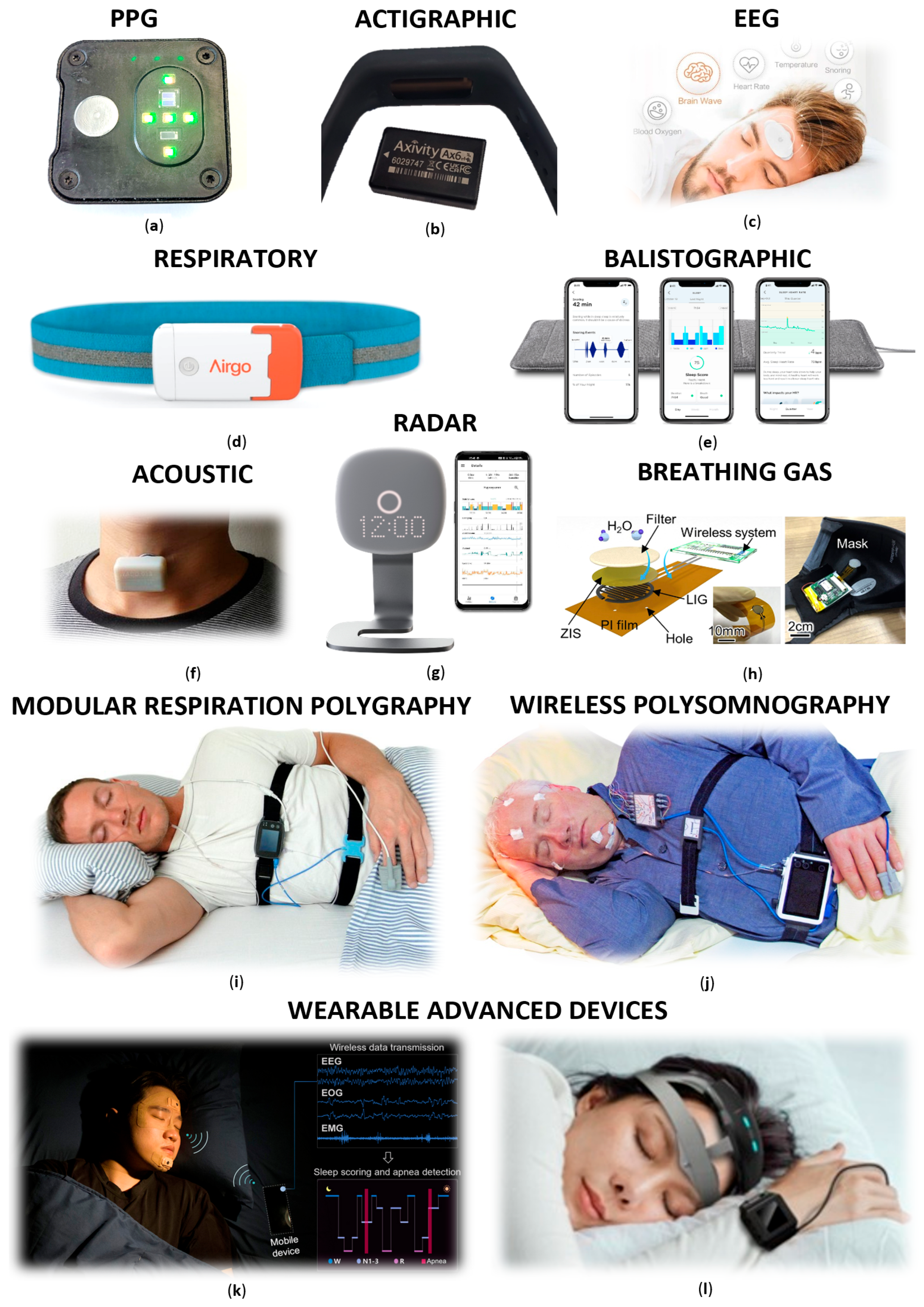
Table 1.
Basic PSG parameters of adults and their physiological values [5].
Table 1.
Basic PSG parameters of adults and their physiological values [5].
| Sleep parameter | Duration 1 |
|---|---|
| Sleep onset latency | ≤ 30 min |
| NREM 1 | 3 – 5% |
| NREM 2 | 45 – 55% |
| NREM 3 | 10 – 20% |
| REM | 20 – 25% |
| REM sleep latency | 60 - 100 min |
| Wakefulness after sleep onset | 1 – 5% |
| Sleep efficiency | > 85% |
1 In minutes or % of sleep duration.
| Classification of severity of sleep-related breathing disorders | AHI 1 |
|---|---|
| Without sleep – related breathing disorders | < 5 |
| Mild severity | 5 ≥ AHI < 15 |
| Moderate severity | 15 ≥ AHI <30 |
| Severe severity | ≥ 30 |
1 Apnoea-hypopnoea index.
| NREM 1 | NREM 2 | NREM 3 | REM | |
|---|---|---|---|---|
| EEG | Characterized by LAMF 1 activity with predominant theta waves (4 – 7 Hz). Alpha activity dissipates, and typical vertex sharp waves lasting up to 0.5 s are visible. | Typical theta waves (4 – 7 Hz) with low to medium amplitude. Presence of sleep spindles (short bursts of 11 – 16 Hz) and K-complexes (sharp delta waves lasting 1 s), which play key roles in sleep maintenance and memory consolidation. Phase duration about 25 minutes and lengthens with each cycle, comprising about 45% of TST 2. | Characterized by slow delta waves (0.5 – 3.5 Hz) with high amplitudes of at least 75 μV in frontal leads. Delta waves constitute more than 20% of the duration of an EEG epoch. | Desynchronized EEG activity with sawtooth waves of 2 – 4 Hz and moderate amplitude, appearing in small clusters in frontal leads. Dream activity occurs with an emotional undertone. REM sleep consolidates memory traces and strengthens memory. |
| HR | Slight decrease compared to wakefulness. | Decrease of 5 – 8% compared to wakefulness. | Decrease of 5 – 8% compared to wakefulness. | Irregular. |
| HRV | Overall HRV increases, LF component decreases, LF/HF ratio decreases, HF component increases. | Overall HRV increases, LF component decreases, LF/HF ratio decreases, HF component increases. |
HRV peaks, LF components are at their lowest, LF/HF ratio decreases, HF components are at its highest. | Significant decrease in HRV, LF component increases, LF/HF ratio increases, HF component decreases. |
| RR | Slower rate compared to wakefulness, regular. | Slower rate compared to wakefulness, regular. | Slow and regular. | Rate equal or higher than wakefulness, breathing becomes shallow. |
| BP | Decrease compared to wakefulness, less pronounced than in NREM 2 and NREM 3. | Decrease of 5 – 14% compared to wakefulness. | More significant decrease than in NREM 2, 5 – 14% lower than wakefulness. | Increase of approximately 5% compared to NREM sleep. |
| CBT | Decrease compared to wakefulness. | Decrease compared to wakefulness. | Largest temperature drop. | Increase compared to NREM sleep. |
1 Low mixed frequency, 2 Total sleep time.
Disclaimer/Publisher’s Note: The statements, opinions and data contained in all publications are solely those of the individual author(s) and contributor(s) and not of MDPI and/or the editor(s). MDPI and/or the editor(s) disclaim responsibility for any injury to people or property resulting from any ideas, methods, instructions or products referred to in the content. |
© 2024 by the authors. Licensee MDPI, Basel, Switzerland. This article is an open access article distributed under the terms and conditions of the Creative Commons Attribution (CC BY) license (http://creativecommons.org/licenses/by/4.0/).
Copyright: This open access article is published under a Creative Commons CC BY 4.0 license, which permit the free download, distribution, and reuse, provided that the author and preprint are cited in any reuse.


