Preprint
Review

This version is not peer-reviewed.

Thrombocytopenia in Sepsis

A peer-reviewed article of this preprint also exists.

Submitted:

01 January 2025

Posted:

03 January 2025

You are already at the latest version

Abstract

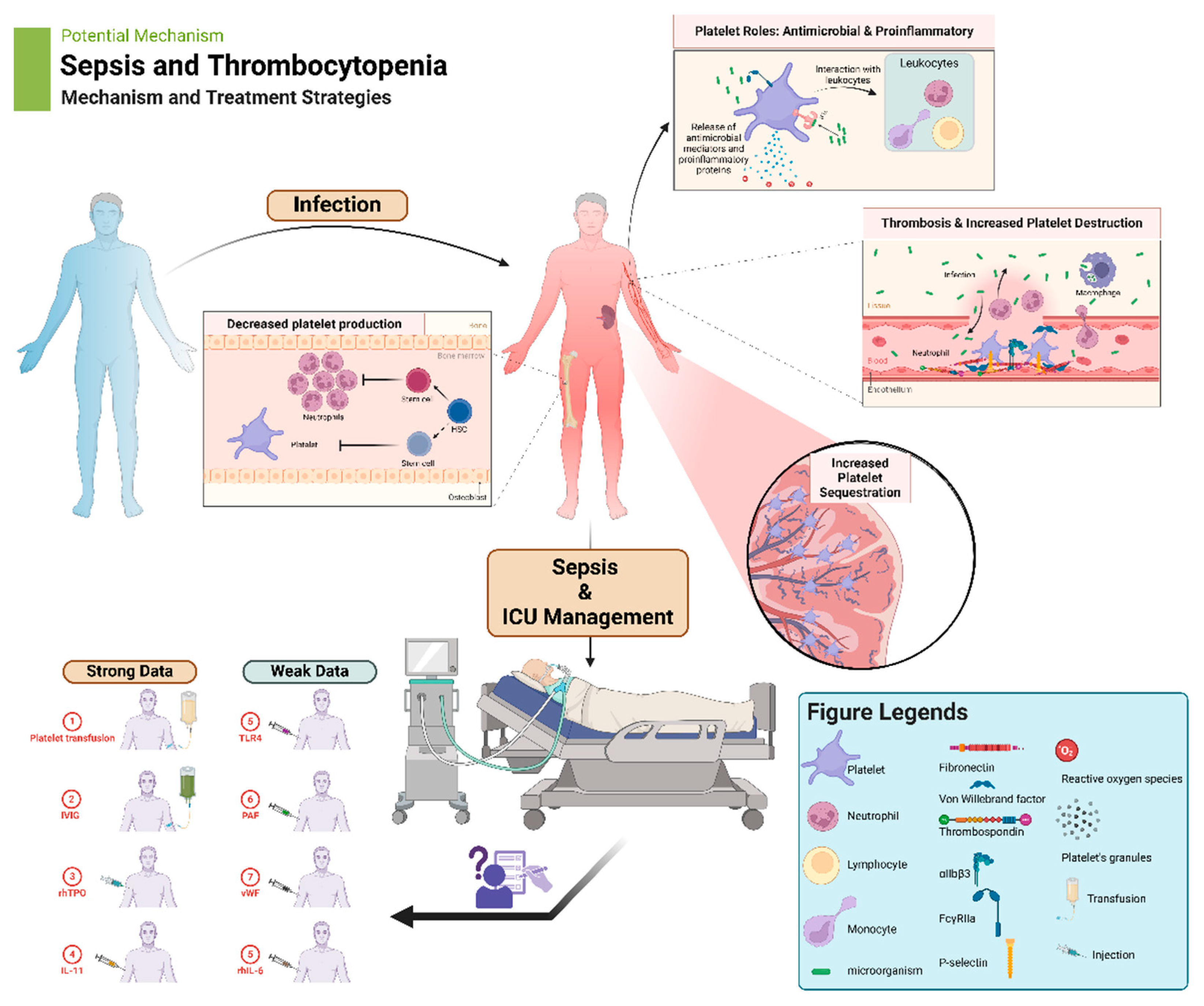
Platelets, traditionally known for their role in hemostasis, have emerged as key players in immune response and inflammation. Sepsis, a life-threatening condition characterized by systemic inflammation, often presents with thrombocytopenia, which at times can be significant. Platelets contribute to the inflammatory response by interacting with leukocytes, endothelial cells, and the innate immune system. However, excessive platelet activation and consumption can lead to thrombocytopenia and exacerbate the severity of sepsis. Understanding the multifaceted roles of platelets in sepsis is crucial for developing effective therapeutic strategies. Targeting platelet-mediated inflammatory responses and promoting platelet production may offer potential avenues for improving outcomes in septic patients with thrombocytopenia. Future research should focus on elucidating the mechanisms underlying platelet dysfunction in sepsis and exploring novel therapeutic approaches to optimize platelet function and mitigate inflammation. This review explores the intricate relationship between platelets, inflammation, and thrombosis in the context of sepsis.

Keywords: 
;  ;  ;  ;  ;  ;  ;  

1. Introduction

Platelets are small (2-5 µm) anucleated cellular elements essential to maintenance of coagulation homeostasis that survive in circulation for up to 10 days following release from the bone marrow in their immature forms, which then derive into smaller functional mature platelets in the vasculature [1,2]. Notably, platelets are also derived from megakaryocytes found in the lungs which may account for about half of all platelets found in peripheral circulation with the capacity to further reconstitute platelet counts when significant thrombocytopenia develops [3,4]. Platelet functions can be impaired under innate or acquired conditions that affect the ability of platelets to properly maintain homeostasis or to assist physiologic non-hemostatic processes [5].
In the setting of tissue injury, platelets through a number of its receptors adhere to the exposed subendothelial extracellular matrix at the site of injury, specifically to collagen, von Willebrand factor, laminin, fibronectin and thrombospondin triggering multiple signaling cascades that strengthen platelet adherence [1,6]. Further enhancement of platelet aggregation and recruitment to the injury site occurs through platelet-platelet interactions mediated by the integrin receptor αIIbβ3 which initiates the formation of a fibrin-rich hemostatic plug [1,6], as well as FcγRIIa among other receptors which incidentally is also important during active bacterial clearance [7]. As a result, the focus of this review will be to understand basic concepts of how platelets exert their non-hemostatic functions in the setting of infection such as in sepsis.

2. Additional Platelet Physiological Roles

Use of platelet transfusions to relieve thrombocytopenia or to address platelet dysfunction is one the most common interventions occurring in clinical practice across medical disciplines. However, the effect of platelet transfusions go far beyond coagulation exemplifying their importance to immunity, inflammatory processes, promoting neoplastic angiogenesis and metastasis [8]. Platelets are capable of modulating immune responses either through their interaction with endothelial cells and leukocytes [9,10], or through release of antimicrobial mediators stored within platelets in response to infection [11,12]. Indeed, it has been reported that platelets in their granules contain more than 300 bioactive proteins that span functions in immunity, inflammation, cell growth and proliferation to name a few [3,13]. Furthermore, it has become increasingly evident that there in an interplay between the immune system, inflammation and cancer, and that platelets play a major role in the convergence between these [14]. Along these lines, platelets’ α-granules contain fibrinogen, von Willebrand factor, platelet factor (PF)-4, coagulation factors, immunoglobulins, growth factors, protease inhibitors, while dense-granules are rich in adenosine diphosphate, serotonin, nucleotides, histamine, dopamine among others [3,13]. These are some examples of mediators found in platelets that carry out functions in different physiologic responses through the body.
Platelets can be seen as the “swiss-army knife” in circulation. Through its conserved role in immune defense, pathological conditions such as heparin-induced thrombocytopenia (HIT), serves to illustrate how the intricate interplay between their activation, immune mechanisms/dysfunction, and clinical complications can disrupt normal homeostasis. HIT is a known complication of using heparin as anticoagulation caused by development of anti-PF4-heparin antibodies. Formation of such antibodies to PF4-heparin complexes as seen in HIT may be indicative of an evolutionarily conserved immune defense mechanism. PF4 found in platelets’ α-granules is actively released during platelet activation, binding directly to polyanions on bacteria leading to antibody formation with specificity for PF4-polyanion complexes that targets bacteria, opsonizing them and enhancing their phagocytosis; however, these antibodies can also cross-react with epitopes of PF4-heparin antigenic complexes leading to their detection with commercially available anti-PF4 assays even in the absence of prior heparin exposure [15]. Thus, PF4 may represent an opsonin to defend against bacteria and antibodies detected during HIT testing can be seen as a bacteria-targeted humoral immune response [15]. Evidence for this can be found in the rapid formation of IgG antibodies to PF4-hepain occurring soon after first heparin exposure, suggesting that IgM was pre-formed at the time heparin was given and that bacterial surface polyanions are the original offending antigen [16,17]. Furthermore, the ability of PF4 to bind with increasing avidity progressively truncated forms of lipopolysaccharide (LPS) found on the surface of Gram-negative bacteria, as shown by the improved binding to phosphates on lipid A molecules devoid of the O antigen and the LPS core, typifies epitopes that mimic PF4-heparin complexes [18]. This provides evidence of a conserved anti-bacterial immune mechanism mediated by platelets’ PF4 [18].
Platelets presence is essential at the crossroads between inflammation and thrombosis. This relationship is indicated by results obtained from annexin knockout mice. In this model, reperfusion injuries show greater platelet adherence, and administration of annexin A1 directly upregulated thromboxane B and modulated phosphatidylserine expression on platelets resulting in cerebral protection through both reduction of platelet activation and enhanced phagocytosis by neutrophils [19]. Likewise, platelets through their toll-like receptors (TLR) and adhesion molecules sense their immediate microenvironment, and function to recruit neutrophils and monocytes to infection sites while stimulating inflammatory cascades [20-22]. Platelets also modulate macrophage responses so that pro-inflammatory mediators release is reduced, thus rescuing mice from septic shock via a cyclooxygenase 1-dependent signaling pathway [23]. These effects, however, can be affected by the age of the donor since platelets from older subjects have shown to both aggregate monocytes and worsen pro-inflammatory responses [24]. Collectively, these findings stress that the interplay between inflammation and thrombosis is highly complex and will require extensive investigation.

3. Immune-Associated Platelet Functions in Sepsis

Platelets are quantitatively the most abundant cellular blood element and represent a native surveillance system hunting for foreign molecules [25]. As a result, thrombocytopenia seen in sepsis may represent a process during which platelets that came into contact with foreign mediators/organisms are actively removed from circulation, or are consumed due to tissue damage caused by the infection. For example, in viral infections such as dengue, decreases in platelet counts are observed soon after exposure to the virus with thrombocytopenia becoming apparent as early as day 4 [26]. Specifically, the number of dengue viral copies inside platelets correlate with C3 and IgG binding, increased surface P-selectin expression indicative of platelet activation, and enhanced clot formation [26]. These infected platelets are in turn phagocytosed by monocytes leading to their clearance from circulation [26].
During sepsis, epidermal growth factor receptor on platelets is central to their activation so that platelets can attract and augment macrophages’ immune function through production of reactive oxygen species (ROS), while concurrently upregulating their pro-inflammatory macrophage M1 phenotype through increased expression of inducible nitric oxide synthase and CD64 resulting in bacteria clearance [24]. Similarly, platelet collagen receptor glycoprotein VI (GPVI) has been shown to be essential to host defense as indicated by data from GPVI-/- knockout mice which have impaired platelet activation and platelet-leukocyte complex formation leading to increased Klebsiella pneumoniae growth and infection [27]. This is of importance since GPVI is a member of the immunoglobulin receptor superfamily and the major receptor for collagen, a prime trigger for platelet activation [28]. Platelets also efficiently directly kill Gram positive bacteria in an FcγRIIa-independent manner as indicated by results using Staphylococcus aureus, and this process does not require PF4 opsonization but release of bactericidal mediators [29]. This contrasts to results obtained with Gram negative shiga-toxin -producing Escherichia coli O157:H7 infection which reduced CD47 expression on platelets in a TLR-dependent manner resulting in enhanced platelet phagocytosis by activated macrophages [30].
Platelets express Fc receptors for antigen recognition [31,32], as well as pro-inflammatory mediators IL1-β and CD40 ligand (L) [33,34]. The latter is important since the adaptive immune response requires CD40 and soluble CD40L, both of which are actively secreted by activated platelets in response to microbial infection [14,35,36]. Furthermore, platelets readily activate proteins of the complement pathway which further activate and recruit additional platelets to the site of infection [37]. Concomitant interaction of platelets with hepatic Kupffer cells represent an additional mechanism by which microbial-platelet clearance occurs [38]. Therefore, the phagocytic capabilities of platelets constitute an evolutionary preserved immune mechanism since thrombocytes of non-mammalian lower vertebrates readily internalize bacteria which are then quickly killed through phagolysosomal fusion [39,40].
Platelet-leukocyte contact also depends on expression of cell adhesion molecules such as P-selectin, and receptors that bridge endothelial cells communication with leukocytes [41]. In regard to how activated platelets stimulate monocytes via cytokines such as TGF-β, it has been shown that CD16 expression on monocytes is induced by platelets causing them to develop into intermediate CD14+CD16+ cells that subsequently differentiate into M2 macrophages capable of antibody-dependent cellular phagocytosis [42]. Platelets are also necessary for formation of neutrophil extracellular traps (NET) which are partly triggered by platelet-leukocyte interactions and the release of proinflammatory mediators from platelets, thus indirectly trapping bacteria, viruses and fungi diminishing their propagation [43]. This occurs through binding of platelet TLR4 to bacterial LPS leading to platelet-neutrophil binding which induces NET formation [44]. This has been confirmed in human infections with either Gram-negative or Gram-positive bacterial infections [45]. Platelets’ phosphatidylinositol 3-kinase catalytic subunit p110β has also been shown to be important for platelet-neutrophil/monocyte binding that when deficient or inhibited results in bacterial dissemination [46]. These results are significant since p110β has also been shown to be involved in tumorigenesis and inflammation [47,48], providing evidence that these processes are closely associated under physiological conditions.
Platelet interactions with bacteria that either induces phagocytosis or activates platelets to initiate an immune response can be subdivided into three major categories which are: direct interaction, indirect, and interactions mediated by secreted mediators. These subsequently lead to three distinct mechanisms: adhesion, phagocytosis, and activation [49,50]. An example of direct platelet activation by bacteria is the binding of the Hs antigen expressed by strains of Streptococci to platelet’s GP1b [51]. An example of indirect interaction is binding of bacterial protein A to plasma’s von Willebrand factor followed by binding of the complex to Gp1b [52]. Alternatively, an example of the third category is when bacteria secrete immunogens that initiate a thrombin-like cascade through activation of protease-activated receptor-1 [53]. This activation leads to subsequent release of bactericidal mediators present in platelet granules, in addition to formation of platelet-bacteria thrombi as seen in sepsis and disseminated intravascular coagulation [49].
By now it should not come as a surprise that the role of platelets as bonafide immune cells is coming into focus since the cellular ancestor of platelets and leukocytes, the thrombocyte, is pivotal to the immune systems of fish and birds [14,48]. It is with this knowledge that platelets’ role during infection should be seen as multifaceted and not driven by their hemostatic function. Their central place as part of an evolutionarily-preserved immune response to defend the body from infectious threats should expand our understanding of additional effects when they are used during transfusions, and especially when patients are treated for their thrombocytopenia.

4. Clinical Implications

Thrombocytopenia in sepsis holds significant clinical importance, serving not just as a laboratory finding but as a vital prognostic indicator that can profoundly impact patient management and outcomes [54,55,56,57]. Studies have shown that septic patients with thrombocytopenia face a higher risk of adverse outcomes, including bleeding complications, prolonged intensive care unit (ICU) stays, and overall increased mortality, compared to subjects with normal platelet counts [58]. For instance, a retrospective study involving 7,981 patients revealed that a ≤10% reduction in platelet counts was associated with significantly lower 30-day mortality and shorter ICU stays compared to those with >10% reduction [59]. This study highlighted that thrombocytopenia in septic shock is associated with higher mortality, with case-fatality rates proportionally increasing with greater severity of thrombocytopenia.
The degree of thrombocytopenia in sepsis also directly correlates with the presence and worsening of multi-organ dysfunction [55]. This correlation implies that correction of the thrombocytopenia as well as targeted antimicrobial coverage has the potential to be the therapeutic intervention that can more significantly improve prognosis and influence outcomes during sepsis (Figure 1). Additional factors contributing to development of thrombocytopenia in sepsis include higher sequential organ failure assessment scores, low PaO2/FiO2 ratios, and requirement for high vasopressor doses [57]. The poor response to platelet transfusions in sepsis, as indicated by low corrected count increments [60], is influenced by increased platelet consumption or destruction, fever, and conditions like splenomegaly and disseminated intravascular coagulation (DIC) that may develop [60,61]. As a result, a clear understanding of these factors is crucial for optimizing transfusion strategies and improving patient outcomes. However, despite the diminished platelet transfusion response, some studies have suggested that outcomes such as mortality and red blood cell (RBC) transfusion needs do not differ between patients with good and poor platelet increments [62].
Even though the management of thrombocytopenia in sepsis remains controversial due to conflicting evidence for and against platelet transfusions, there is an understanding among clinicians and researchers that this needs to be addressed to improve outcomes [63,64]. Further research is fundamental to establish a unified approach to manage sepsis-associated thrombocytopenia (SAT), that takes under consideration its multifactorial nature. Thus, emerging approaches in treating sepsis, such as the use of intravenous immunoglobulins (IVIG) and even administration of recombinant human thrombopoietin (rhTPO), show promise in enhancing platelet production and possibly reducing their destruction, offering potential new strategies for managing thrombocytopenia in such setting [65,66].

5. Management of Thrombocytopenia

5.1. Platelet Transfusions

The Association for the advancement of Blood and Biotherapies has provided recommendations for platelet transfusion thresholds for hospitalized patients, specifically, transfusion for platelet counts ≤10 × 10⁹/L to prevent spontaneous bleeding, prophylactic transfusion for counts <20 × 10⁹/L for central venous catheter placement, and platelet transfusions when patient is <50 × 10⁹/L neurological surgery among others. [67]. However, guidance that is specific for SAT is lacking.
The majority of clinical trials looking at platelet transfusions in setting of infection have focused on hematology patients, creating a significant data gap for septic populations [68]. In this setting, prophylactic platelet transfusion in patients with severe thrombocytopenia (10-50 × 109/L) showed reduced bleeding incidents in transfused patients even among those in intensive care units [68]. Retrospective studies, however, have found conflicting results in which higher platelet transfusion thresholds (20–50 × 10⁹/L) compared to lower thresholds (<20 × 109/L) were associated with reduced mortality [63], while others saw increased mortality, fewer ICU-free days, and similar RBC transfusion rates [64]. Likewise, looking at the impact of platelet transfusions on mortality in sepsis patients with severe thrombocytopenia (≤50 × 10⁹/L), it has been reported that platelet transfusions in this group were associated with higher in-hospital mortality rates but that these had no effect in either 90-day mortality or ICU length of stay [69]. Thus, platelet transfusions in sepsis patients present a complex picture in regard to overall therapeutic benefit. When adverse outcomes to transfusions occur, they are made worse by the frailty of recipients, severity of underlying clinical condition, possible complications such as volume overload compounded by excessive transfusions and infusion of fluids, transfusion reactions, increased risk of thrombosis, and immune dysfunction [64,70-72]. Moreover, platelet transfusions can also contribute to microvascular dysfunction by exacerbating the activation of the coagulation cascade while simultaneously inhibit anticoagulation and fibrinolysis [64,71,73].
Despite these stated risks, platelet transfusions may offer benefits to still to be defined groups of patients with sepsis by improving endothelial barrier function, reducing inflammation through their immunomodulatory effects, and promoting tissue repair [74,75]. As a result, having a clear understanding of the etiology of thrombocytopenia—whether due to hypersplenism, bone marrow suppression, medication-induced (HIT), or secondary to dilutional effects—is essential for optimizing clinical management. Under specific circumstances, strategies such as leukoreduction, platelet additive solutions, or human leucocyte antigens (HLA)- and human platelet alloantigens (HPA)-matched transfusions could improve count increments but their benefit in sepsis patients still require formal investigation [73,76].

5.2. Intravenous Immunoglobulins (IVIG)

The administration of IVIG as a potential therapeutic approach for thrombocytopenia, particularly when immune-mediated platelet destruction is suspected may not be without merit [77]. IVIG modulates the immune response by disrupting autoantibody-mediated platelet clearance [78], making it a plausible treatment option in SAT [79,80], especially when mechanisms mirror those encountered in immune thrombocytopenia [81,82]. It is believed that IVIG is able to do this through its anti-idiotypic effect over circulating antibodies. In this case, elevated levels of platelet-associated IgG observed in septic thrombocytopenia further support its use and future research should establish its role as a therapeutic approach [83].
Results from a randomized blinded clinical trial found that IVIG significantly increased platelet counts in patients with septic thrombocytopenia, increasing counts by 411% in the IVIG group versus 261% in the placebo group by day 9 [81]. Additionally, a report of a septic COVID-19 patient with severe immune thrombocytopenia showed a rapid platelet count recovery and improved oxygenation after combined IVIG with corticosteroid therapy without occurrence of adverse events [84]. Along these lines, reports of pregnant women with thrombocytopenia and signs of infection showing a >60% improvement in counts after receiving IVIG with low-dose rhTPO highlights its therapeutic potential in specific patient populations [80].
In contrast, IVIG therapy in severe fever with thrombocytopenia syndrome (SFTS) has been associated with higher fatality rates and significant adverse events, such as worsening viral loads and suppressed immune cell counts [85]. These findings, derived from a retrospective study, warrant cautious interpretation due to its methodological limitations, including single-center design, SFTS patient selection, and lack of randomization. Interestingly, IgM-enriched IVIG significantly reduced mortality, hospital stays, and APACHE II scores in sepsis patients, showing greater efficacy compared to standard IVIG [86]. Even when higher doses of IVIG were used, therapeutic benefit has been reported through the rapid inhibition of platelet activation, lending support to a broader role in managing thrombocytopenia occurring secondary to immune dysregulation [87,88].
Altogether, IVIG shows promise in rapidly increasing platelet counts and improving outcomes in selected patients with SAT [81,84,86]. Its use may be particularly beneficial for those at high risk of bleeding or undergoing invasive procedures. However, careful patient selection and further studies are needed to establish its true efficacy and safety either as a standalone therapy or in combination with other treatments.

5.3. Recombinant Human Thrombopoietin (rhTPO)

This recombinant grown factor effectively increases platelet counts with minimal side effects by promoting differentiation and maturation of bone marrow stem cells into megakaryocytes, thereby enhancing platelet production without altering their morphology or function [89-92]. These benefits have been well demonstrated during chemotherapy-induced, immune-mediated, and sepsis-associated thrombocytopenia, resulting in overall lower transfusion dependency [89,93-95]. A meta-analysis of ten randomized controlled trials involving 681 patients with SAT showed that rhTPO significantly increased platelet counts, decreased 28-day mortality, reduced number of platelet units transfused, reduced transfusion volumes, and shortened ICU stays compared to conventional antibiotic therapy alone or in combination with IVIG [65]. Furthermore, retrospective data of 213 patients lend support to rhTPO use, showing greater platelet count increases specially in patients with initial counts ≤30 × 10⁹/L and APACHE II scores >15 [92]. Even though this growth factor is costly, the reduction in ICU stays may provide a strong incentive to its use even when 28-day mortality may remained unchanged [92].

5.4. Recombinant Human IL-11

Interleukin-11 (IL-11), a cytokine interacting with hematopoietic and non-hematopoietic cells, has demonstrated therapeutic potential in systemic inflammatory conditions including sepsis [96,97]. Specifically, its strong thrombopoietic effect favors its use as potential therapy for SAT [97,98]. A study of 105 patients found that treatment with IL-11 significantly improved platelet counts and reduced IL-6 levels between days 3 and 14 [99]. The therapy also downregulated the sepsis marker procalcitonin and lowered the APACHE II score of patients, in a background of lower 28-day mortality rate compared to conventional therapy [99]. However, IL-11's role in treating SAT remains mostly underexplored. Although preliminary studies suggest its efficacy in light of improved clinical outcomes, the data is still limited and randomized clinical trials are needed to validate its therapeutic potential in septic patients [100].

5.5. Recombinant Human IL-6

Elevated IL-6 levels, a hallmark of sepsis, correlate with disease severity and act as diagnostic and prognostic markers [101]. Notably, there is an inverse relationship between thrombocytopenia and cytokine activation in sepsis [102], prompting exploration of anti-IL-6 therapies to mitigate inflammation and restore platelet counts since this cytokine is likely a key driver of the cytokine storm in sepsis. Tocilizumab (TCZ), a recombinant humanized monoclonal antibody targeting the IL-6 receptor, has shown potential in managing sepsis, particularly in COVID-19 patients [103,104]. By inhibiting both membranous and soluble IL-6 receptors, TCZ suppresses the IL-6-mediated inflammatory cascade [105]. However, TCZ's role is complex; while it may dampen cytokine-driven platelet consumption, reports of TCZ-induced thrombocytopenia raise concerns [103]. This physiologic duality underscores the need for further research into its safety and efficacy in septic patients, especially those with existing thrombocytopenia. With this in mind, the RESCUE trial aiming to further evaluate IL-6 inhibition on patients with inflammation and atherosclerotic disease via another monoclonal antibody ziltivekimab will provide needed evidence to establish if inhibition of this cytokine proves beneficial to patients [106].

5.6. TLR4 Inhibition

TLR4 is a key membrane-spanning protein in the innate immune system, recognizing pathogen-associated molecular patterns such as LPS from Gram-negative bacteria [107,108]. Expressed on various cell types, including platelets, TLR4 activation triggers intracellular signaling cascades, leading to NF-kB pathway activation that results in release of pro-inflammatory cytokines. This process contributes to systemic inflammation and sepsis progression, positioning TLR4 as a potential therapeutic target for modulating sepsis-associated inflammation [21]. TAK-242, a TLR4 antagonist, has shown promise as adjunct therapy for thrombocytopenia in sepsis by mitigating LPS-induced systemic inflammation, thus reducing end-organ damage and slowing sepsis progression [109-111]. Its ability to specifically target inflammatory pathways underscores its potential to improve outcomes in septic patients with thrombocytopenia.
Activation of TLR4 on platelets by LPS also triggers pro-thrombotic and pro-coagulant responses including platelet aggregation, ATP release, P-selectin expression, and platelet-neutrophil aggregate formation [112]. These mechanisms contribute to both inflammation, coagulopathy, and DIC seen in sepsis. TLR4 also interacts with CD14, damage-associated molecular patterns (e.g., HMGB1), and NETs, amplifying inflammatory and coagulation pathways that link thrombocytopenia and sepsis progression [110-112]. Despite mechanistic evidence supporting TLR4's role in thrombocytopenia in sepsis, clinical trials investigating its therapeutic potential remain lacking. Future studies will be needed to translate these findings into effective clinical interventions.

5.7. Platelet-Activating Factor (PAF) Inhibition

PAF's role in LPS-induced thrombocytopenia and neutropenia was shown by a study looking at 42 dogs treated with LPS, PAF, or saline, with some animals being pre-treated with TCV-309, a potent PAF antagonist. Thrombocytopenia and neutropenia occurred in all but the saline group, with TCV-309 significantly reducing LPS-induced thrombocytopenia, suggesting PAF as a mediator and potential therapeutic target for SAT [113]. PAF appears central to severe sepsis, where bacterial endotoxins trigger its production, leading to hypotension, organ damage, and thrombocytopenia [113]. Use of recombinant PAF-acetylhydrolase demonstrated reduced mortality in severe sepsis patients, underscoring its therapeutic potential [114]. Moreover, PAF receptor (PAF-R) activation exacerbates inflammatory responses in infectious diseases, such as dengue fever, where it worsens thrombocytopenia and increases mortality [115]. Also, its role in inflammation related to HIV and SARS-CoV-2 further emphasizes its relevance across infectious conditions [115]. Thus, targeting PAF-R represents a promising approach for treating SAT. Clinical trials to study the safety and efficacy of PAF inhibitors in the setting of sepsis are justified.

5.8. Von Willebrand Factor (vWF)-Binding Function

Sepsis disrupts the delicate balance between vWF and the enzyme known as a disintegrin and metalloproteinase with thrombospondin type 1 motif 13 (ADAMTS13), leading to lower ADAMTS13 activity in a background of elevated vWF levels [116,117]. This imbalance results from increased vWF and removal of ADAMTS13 from the circulation rather than intrinsic changes in ADAMTS13 itself which when persisting even post-ICU augments thrombotic risks [116]. Studies in Staphylococcus aureus-associated sepsis further emphasize this imbalance's impact. In this research, high vWF and low ADAMTS13 activity correlate with severe illness in humans, while in mice, vWF deficiency improves survival and ADAMTS13 deficiency worsens it [118]. Similarly, GPIb-IX, appears to be important for vWF-mediated platelet adhesion, emerges as a therapeutic target. In LPS-induced sepsis models, a dysfunctional GPIbα mutant or the synthetic vWF-binding inhibitor MPαC significantly reduced mortality, thrombosis, and platelet loss [119]. These findings position vWF-mediated adhesion as a target for development of therapies in SAT.

6. Discussion

Thrombocytopenia in sepsis is a complex clinical complication with significant implications for patient outcomes. It presents frequently in sepsis, affecting nearly half of septic patients and serving as a critical and negative prognostic marker [120,121]. While traditionally viewed as primarily one of the most important hemostatic mediators [122], platelets play a crucial role in the immune response to infection [123]. Thus, thrombocytopenia has profound immune repercussions in patients battling infections. By directly interacting with pathogens, modulating inflammatory responses, and promoting immune cell function, platelets role in both innate and adaptive immunity cannot be denied [124].
The pathogenetic mechanisms seen in sepsis can involve decreased platelet production due to bone marrow suppression, increased destruction through DIC, and platelet sequestration in the spleen and other organs [120,121,125]. The totality of findings presented here makes it evidently clear of the complex interplay of pathophysiological mechanisms and clinical impact have over the possible management strategies needed for thrombocytopenia in sepsis [125]. Clinical implications are profound; thrombocytopenia correlates with higher risks of bleeding, prolonged ICU stays, and increased mortality [121]. Specially, since studies consistently show that even a modest reduction in platelet count is associated with worse outcomes, emphasizing the importance of frequent monitoring and timely intervention [59]. Therefore, the cumulative data underscore the complexity of thrombocytopenia in sepsis and the necessity for targeted management strategies.
A critical aspect of managing septic patients is the consideration of platelet transfusions. While platelet transfusions are often employed to treat thrombocytopenia, their efficacy in sepsis has remained controversial [126,127]. The timing of transfusion, number of platelet units given, and underlying disease severity could influence outcomes. While some studies have suggested benefits in reducing bleeding and improving survival, particularly with higher transfusion thresholds [63,127], others report no improvements in outcomes instead suggesting increased mortality among recipients, possibly due to the severity of illness and complications such as transfusion-related infections, immune dysfunction and the immunomodulatory effects mediated by platelets themselves [64,128].
Emerging therapies such as rhTPO and immunomodulatory agents offer potential for enhancing platelet production and reducing destruction [127]. As pointed out earlier in the text, IVIG have shown promise in this regard, particularly in cases where immune-mediated platelet destruction is suspected. Randomized controlled trials and case studies highlight IVIG's ability to significantly increase platelet counts and improve clinical outcomes in SAT are encouraging [80,81]. However, the effectiveness of IVIG is not universal. For instance, in SFTS, IVIG treatment was associated with increased mortality and adverse immunological effects, highlighting the need for careful patient selection and monitoring after its use [85]. In addition to transfusions and IVIG, adjunctive therapies targeting specific pathways involved in platelet production and destruction are potential therapies that can help patients. These include agents that are immunomodulatory in nature, inhibit platelet receptor interactions, and address underlying conditions such as hypersplenism and bone marrow suppression (Figure 1) [113,114,116]. The use of high-dose IVIG in conditions like HIT illustrates the potential for targeted therapies to mitigate platelet activation and limit platelet consumption [87].
The management of thrombocytopenia in sepsis likely requires a tailored approach that considers individual patient factors and the underlying pathophysiology of a patient’s presentation. Any research looking at this patient population, should account for as many of these variables as possible and recruit patients accordingly so that results can be extrapolated to address the diverse nature of thrombocytopenia in sepsis. Future research ought to include robust randomized clinical trials that establish the benefits of those approaches mentioned in this review. Addition of platelet function tests and advanced diagnostic tools could further refine treatment strategies, ensuring that interventions are timely, effective and safe. Nevertheless, the data is clear that the armamentarium needed to treat thrombocytopenia in setting of infection is likely to grow in the coming years.

7. Conclusions

In conclusion, thrombocytopenia in sepsis is a critical and complex condition with significant implications for patient management and outcomes. A comprehensive understanding of its pathophysiology and clinical impact, coupled with evidence-based management strategies, is essential for improving patient care. Future studies should aim to clarify the role of various treatment modalities, including platelet transfusions and adjunctive therapies as those presented here, in optimizing outcomes for septic patients with thrombocytopenia.

Author Contributions

AS researched the topic, reviewed literature, and co-wrote the manuscript. AM researched the topic and co-wrote the manuscript. RWM developed the topic, reviewed literature, co-wrote the manuscript, and critically revised the manuscript to its final version. All authors have read and agree with the content of the manuscript’s final version.

Funding

No funding was received for this review.

Institutional Review Board Statement

Not applicable.

Informed Consent Statement

Not applicable.

Data Availability Statement

All relevant data has been provided in the text of this review.

Conflicts of Interest

The authors declare no conflicts of interest.

References

  1. Holinstat, M. Normal platelet function. Cancer Metastasis Rev 2017, 36, 195–198. [Google Scholar] [CrossRef]
  2. Hong, H.; Xiao, W.; Maitta, R.W. Steady increment of immature platelet fraction is suppressed by irradiation in single-donor platelet components during storage. PLoS One 2014, 9, e85465. [Google Scholar] [CrossRef]
  3. Repsold, L.; Joubert, A.M. Platelet Function, Role in Thrombosis, Inflammation, and Consequences in Chronic Myeloproliferative Disorders. Cells 2021, 10. [Google Scholar] [CrossRef]
  4. Lefrancais, E.; Ortiz-Munoz, G.; Caudrillier, A.; Mallavia, B.; Liu, F.; Sayah, D.M.; Thornton, E.E.; Headley, M.B.; David, T.; Coughlin, S.R.; et al. The lung is a site of platelet biogenesis and a reservoir for haematopoietic progenitors. Nature 2017, 544, 105–109. [Google Scholar] [CrossRef] [PubMed]
  5. Nurden, A.; Nurden, P. Advances in our understanding of the molecular basis of disorders of platelet function. J Thromb Haemost 2011, 9 Suppl 1, 76–91. [Google Scholar] [CrossRef]
  6. Broos, K.; Feys, H.B.; De Meyer, S.F.; Vanhoorelbeke, K.; Deckmyn, H. Platelets at work in primary hemostasis. Blood Rev 2011, 25, 155–167. [Google Scholar] [CrossRef] [PubMed]
  7. Arman, M.; Krauel, K.; Tilley, D.O.; Weber, C.; Cox, D.; Greinacher, A.; Kerrigan, S.W.; Watson, S.P. Amplification of bacteria-induced platelet activation is triggered by FcgammaRIIA, integrin alphaIIbbeta3, and platelet factor 4. Blood 2014, 123, 3166–3174. [Google Scholar] [CrossRef]
  8. Leslie, M. Cell biology. Beyond clotting: the powers of platelets. Science 2010, 328, 562–564. [Google Scholar] [CrossRef]
  9. Elzey, B.D.; Tian, J.; Jensen, R.J.; Swanson, A.K.; Lees, J.R.; Lentz, S.R.; Stein, C.S.; Nieswandt, B.; Wang, Y.; Davidson, B.L.; et al. Platelet-mediated modulation of adaptive immunity. A communication link between innate and adaptive immune compartments. Immunity 2003, 19, 9–19. [Google Scholar] [CrossRef] [PubMed]
  10. Diacovo, T.G.; Puri, K.D.; Warnock, R.A.; Springer, T.A.; von Andrian, U.H. Platelet-mediated lymphocyte delivery to high endothelial venules. Science 1996, 273, 252–255. [Google Scholar] [CrossRef]
  11. Yeaman, M.R.; Bayer, A.S. Antimicrobial peptides from platelets. Drug Resist Updat 1999, 2, 116–126. [Google Scholar] [CrossRef]
  12. Tang, Y.Q.; Yeaman, M.R.; Selsted, M.E. Antimicrobial peptides from human platelets. Infect Immun 2002, 70, 6524–6533. [Google Scholar] [CrossRef] [PubMed]
  13. Smyth, S.S.; McEver, R.P.; Weyrich, A.S.; Morrell, C.N.; Hoffman, M.R.; Arepally, G.M.; French, P.A.; Dauerman, H.L.; Becker, R.C.; Platelet Colloquium, P. Platelet functions beyond hemostasis. J Thromb Haemost 2009, 7, 1759–1766. [Google Scholar] [CrossRef]
  14. Franco, A.T.; Corken, A.; Ware, J. Platelets at the interface of thrombosis, inflammation, and cancer. Blood 2015, 126, 582–588. [Google Scholar] [CrossRef]
  15. Krauel, K.; Potschke, C.; Weber, C.; Kessler, W.; Furll, B.; Ittermann, T.; Maier, S.; Hammerschmidt, S.; Broker, B.M.; Greinacher, A. Platelet factor 4 binds to bacteria, [corrected] inducing antibodies cross-reacting with the major antigen in heparin-induced thrombocytopenia. Blood 2011, 117, 1370–1378. [Google Scholar] [CrossRef] [PubMed]
  16. Greinacher, A.; Kohlmann, T.; Strobel, U.; Sheppard, J.A.; Warkentin, T.E. The temporal profile of the anti-PF4/heparin immune response. Blood 2009, 113, 4970–4976. [Google Scholar] [CrossRef] [PubMed]
  17. Neuhaus, F.C.; Baddiley, J. A continuum of anionic charge: structures and functions of D-alanyl-teichoic acids in gram-positive bacteria. Microbiol Mol Biol Rev 2003, 67, 686–723. [Google Scholar] [CrossRef]
  18. Krauel, K.; Weber, C.; Brandt, S.; Zahringer, U.; Mamat, U.; Greinacher, A.; Hammerschmidt, S. Platelet factor 4 binding to lipid A of Gram-negative bacteria exposes PF4/heparin-like epitopes. Blood 2012, 120, 3345–3352. [Google Scholar] [CrossRef] [PubMed]
  19. Senchenkova, E.Y.; Ansari, J.; Becker, F.; Vital, S.A.; Al-Yafeai, Z.; Sparkenbaugh, E.M.; Pawlinski, R.; Stokes, K.Y.; Carroll, J.L.; Dragoi, A.M.; et al. Novel Role for the AnxA1-Fpr2/ALX Signaling Axis as a Key Regulator of Platelet Function to Promote Resolution of Inflammation. Circulation 2019, 140, 319–335. [Google Scholar] [CrossRef] [PubMed]
  20. Kapur, R.; Semple, J.W. Platelets as immune-sensing cells. Blood Adv 2016, 1, 10–14. [Google Scholar] [CrossRef] [PubMed]
  21. Vallance, T.M.; Zeuner, M.T.; Williams, H.F.; Widera, D.; Vaiyapuri, S. Toll-Like Receptor 4 Signalling and Its Impact on Platelet Function, Thrombosis, and Haemostasis. Mediators Inflamm 2017, 2017, 9605894. [Google Scholar] [CrossRef]
  22. Andonegui, G.; Kerfoot, S.M.; McNagny, K.; Ebbert, K.V.; Patel, K.D.; Kubes, P. Platelets express functional Toll-like receptor-4. Blood 2005, 106, 2417–2423. [Google Scholar] [CrossRef]
  23. Xiang, B.; Zhang, G.; Guo, L.; Li, X.A.; Morris, A.J.; Daugherty, A.; Whiteheart, S.W.; Smyth, S.S.; Li, Z. Platelets protect from septic shock by inhibiting macrophage-dependent inflammation via the cyclooxygenase 1 signalling pathway. Nat Commun 2013, 4, 2657. [Google Scholar] [CrossRef]
  24. Luo, S.; Xu, R.; Xie, P.; Liu, X.; Ling, C.; Liu, Y.; Zhang, X.; Xia, Z.; Chen, Z.; Tang, J. EGFR of platelet regulates macrophage activation and bacterial phagocytosis function. J Inflamm (Lond) 2024, 21, 10. [Google Scholar] [CrossRef] [PubMed]
  25. Clemetson, K.J. Platelets and pathogens. Cell Mol Life Sci 2010, 67, 495–498. [Google Scholar] [CrossRef]
  26. Ojha, A.; Nandi, D.; Batra, H.; Singhal, R.; Annarapu, G.K.; Bhattacharyya, S.; Seth, T.; Dar, L.; Medigeshi, G.R.; Vrati, S.; et al. Platelet activation determines the severity of thrombocytopenia in dengue infection. Sci Rep 2017, 7, 41697. [Google Scholar] [CrossRef]
  27. Claushuis, T.A.M.; de Vos, A.F.; Nieswandt, B.; Boon, L.; Roelofs, J.; de Boer, O.J.; van 't Veer, C.; van der Poll, T. Platelet glycoprotein VI aids in local immunity during pneumonia-derived sepsis caused by gram-negative bacteria. Blood 2018, 131, 864–876. [Google Scholar] [CrossRef] [PubMed]
  28. Dutting, S.; Bender, M.; Nieswandt, B. Platelet GPVI: a target for antithrombotic therapy?! Trends Pharmacol Sci 2012, 33, 583–590. [Google Scholar] [CrossRef] [PubMed]
  29. Wolff, M.; Handtke, S.; Palankar, R.; Wesche, J.; Kohler, T.P.; Kohler, C.; Gruel, Y.; Hammerschmidt, S.; Greinacher, A. Activated platelets kill Staphylococcus aureus, but not Streptococcus pneumoniae-The role of FcgammaRIIa and platelet factor 4/heparinantibodies. J Thromb Haemost 2020, 18, 1459–1468. [Google Scholar] [CrossRef] [PubMed]
  30. Guo, Y.L.; Liu, D.Q.; Bian, Z.; Zhang, C.Y.; Zen, K. Down-regulation of platelet surface CD47 expression in Escherichia coli O157:H7 infection-induced thrombocytopenia. PLoS One 2009, 4, e7131. [Google Scholar] [CrossRef]
  31. King, M.; McDermott, P.; Schreiber, A.D. Characterization of the Fc gamma receptor on human platelets. Cell Immunol 1990, 128, 462–479. [Google Scholar] [CrossRef] [PubMed]
  32. Qian, K.; Xie, F.; Gibson, A.W.; Edberg, J.C.; Kimberly, R.P.; Wu, J. Functional expression of IgA receptor FcalphaRI on human platelets. J Leukoc Biol 2008, 84, 1492–1500. [Google Scholar] [CrossRef] [PubMed]
  33. Lindemann, S.; Tolley, N.D.; Dixon, D.A.; McIntyre, T.M.; Prescott, S.M.; Zimmerman, G.A.; Weyrich, A.S. Activated platelets mediate inflammatory signaling by regulated interleukin 1beta synthesis. J Cell Biol 2001, 154, 485–490. [Google Scholar] [CrossRef]
  34. Henn, V.; Slupsky, J.R.; Grafe, M.; Anagnostopoulos, I.; Forster, R.; Muller-Berghaus, G.; Kroczek, R.A. CD40 ligand on activated platelets triggers an inflammatory reaction of endothelial cells. Nature 1998, 391, 591–594. [Google Scholar] [CrossRef] [PubMed]
  35. Danese, S.; Katz, J.A.; Saibeni, S.; Papa, A.; Gasbarrini, A.; Vecchi, M.; Fiocchi, C. Activated platelets are the source of elevated levels of soluble CD40 ligand in the circulation of inflammatory bowel disease patients. Gut 2003, 52, 1435–1441. [Google Scholar] [CrossRef]
  36. Yacoub, D.; Hachem, A.; Theoret, J.F.; Gillis, M.A.; Mourad, W.; Merhi, Y. Enhanced levels of soluble CD40 ligand exacerbate platelet aggregation and thrombus formation through a CD40-dependent tumor necrosis factor receptor-associated factor-2/Rac1/p38 mitogen-activated protein kinase signaling pathway. Arterioscler Thromb Vasc Biol 2010, 30, 2424–2433. [Google Scholar] [CrossRef] [PubMed]
  37. Del Conde, I.; Cruz, M.A.; Zhang, H.; Lopez, J.A.; Afshar-Kharghan, V. Platelet activation leads to activation and propagation of the complement system. J Exp Med 2005, 201, 871–879. [Google Scholar] [CrossRef] [PubMed]
  38. Wong, C.H.; Jenne, C.N.; Petri, B.; Chrobok, N.L.; Kubes, P. Nucleation of platelets with blood-borne pathogens on Kupffer cells precedes other innate immunity and contributes to bacterial clearance. Nat Immunol 2013, 14, 785–792. [Google Scholar] [CrossRef] [PubMed]
  39. Nagasawa, T.; Nakayasu, C.; Rieger, A.M.; Barreda, D.R.; Somamoto, T.; Nakao, M. Phagocytosis by Thrombocytes is a Conserved Innate Immune Mechanism in Lower Vertebrates. Front Immunol 2014, 5, 445. [Google Scholar] [CrossRef]
  40. Meseguer, J.; Esteban, M.A.; Rodriguez, A. Are thrombocytes and platelets true phagocytes? Microsc Res Tech 2002, 57, 491–497. [Google Scholar] [CrossRef]
  41. Koupenova, M.; Clancy, L.; Corkrey, H.A.; Freedman, J.E. Circulating Platelets as Mediators of Immunity, Inflammation, and Thrombosis. Circ Res 2018, 122, 337–351. [Google Scholar] [CrossRef]
  42. Lee, S.J.; Yoon, B.R.; Kim, H.Y.; Yoo, S.J.; Kang, S.W.; Lee, W.W. Activated Platelets Convert CD14(+)CD16(-) Into CD14(+)CD16(+) Monocytes With Enhanced FcgammaR-Mediated Phagocytosis and Skewed M2 Polarization. Front Immunol 2020, 11, 611133. [Google Scholar] [CrossRef] [PubMed]
  43. Carestia, A.; Kaufman, T.; Schattner, M. Platelets: New Bricks in the Building of Neutrophil Extracellular Traps. Front Immunol 2016, 7, 271. [Google Scholar] [CrossRef]
  44. Clark, S.R.; Ma, A.C.; Tavener, S.A.; McDonald, B.; Goodarzi, Z.; Kelly, M.M.; Patel, K.D.; Chakrabarti, S.; McAvoy, E.; Sinclair, G.D.; et al. Platelet TLR4 activates neutrophil extracellular traps to ensnare bacteria in septic blood. Nat Med 2007, 13, 463–469. [Google Scholar] [CrossRef]
  45. Carestia, A.; Kaufman, T.; Rivadeneyra, L.; Landoni, V.I.; Pozner, R.G.; Negrotto, S.; D'Atri, L.P.; Gomez, R.M.; Schattner, M. Mediators and molecular pathways involved in the regulation of neutrophil extracellular trap formation mediated by activated platelets. J Leukoc Biol 2016, 99, 153–162. [Google Scholar] [CrossRef] [PubMed]
  46. Schrottmaier, W.C.; Kral-Pointner, J.B.; Salzmann, M.; Mussbacher, M.; Schmuckenschlager, A.; Pirabe, A.; Brunnthaler, L.; Kuttke, M.; Maier, B.; Heber, S.; et al. Platelet p110beta mediates platelet-leukocyte interaction and curtails bacterial dissemination in pneumococcal pneumonia. Cell Rep 2022, 41, 111614. [Google Scholar] [CrossRef]
  47. Mateo, J.; Ganji, G.; Lemech, C.; Burris, H.A.; Han, S.W.; Swales, K.; Decordova, S.; DeYoung, M.P.; Smith, D.A.; Kalyana-Sundaram, S.; et al. A First-Time-in-Human Study of GSK2636771, a Phosphoinositide 3 Kinase Beta-Selective Inhibitor, in Patients with Advanced Solid Tumors. Clin Cancer Res 2017, 23, 5981–5992. [Google Scholar] [CrossRef]
  48. Weyrich, A.S.; Zimmerman, G.A. Platelets: signaling cells in the immune continuum. Trends Immunol 2004, 25, 489–495. [Google Scholar] [CrossRef] [PubMed]
  49. Cox, D. Bacteria-platelet interactions. J Thromb Haemost 2009, 7, 1865–1866. [Google Scholar] [CrossRef]
  50. Fitzgerald, J.R.; Foster, T.J.; Cox, D. The interaction of bacterial pathogens with platelets. Nat Rev Microbiol 2006, 4, 445–457. [Google Scholar] [CrossRef]
  51. Kerrigan, S.W.; Jakubovics, N.S.; Keane, C.; Maguire, P.; Wynne, K.; Jenkinson, H.F.; Cox, D. Role of Streptococcus gordonii surface proteins SspA/SspB and Hsa in platelet function. Infect Immun 2007, 75, 5740–5747. [Google Scholar] [CrossRef]
  52. O'Seaghdha, M.; van Schooten, C.J.; Kerrigan, S.W.; Emsley, J.; Silverman, G.J.; Cox, D.; Lenting, P.J.; Foster, T.J. Staphylococcus aureus protein A binding to von Willebrand factor A1 domain is mediated by conserved IgG binding regions. FEBS J 2006, 273, 4831–4841. [Google Scholar] [CrossRef]
  53. Lourbakos, A.; Yuan, Y.P.; Jenkins, A.L.; Travis, J.; Andrade-Gordon, P.; Santulli, R.; Potempa, J.; Pike, R.N. Activation of protease-activated receptors by gingipains from Porphyromonas gingivalis leads to platelet aggregation: a new trait in microbial pathogenicity. Blood 2001, 97, 3790–3797. [Google Scholar] [CrossRef] [PubMed]
  54. Sharma, B.; Sharma, M.; Majumder, M.; Steier, W.; Sangal, A.; Kalawar, M. Thrombocytopenia in septic shock patients--a prospective observational study of incidence, risk factors and correlation with clinical outcome. Anaesth Intensive Care 2007, 35, 874–880. [Google Scholar] [CrossRef] [PubMed]
  55. Vanderschueren, S.; De Weerdt, A.; Malbrain, M.; Vankersschaever, D.; Frans, E.; Wilmer, A.; Bobbaers, H. Thrombocytopenia and prognosis in intensive care. Crit Care Med 2000, 28, 1871–1876. [Google Scholar] [CrossRef]
  56. Ghimire, S.; Ravi, S.; Budhathoki, R.; Arjyal, L.; Hamal, S.; Bista, A.; Khadka, S.; Uprety, D. Current understanding and future implications of sepsis-induced thrombocytopenia. Eur J Haematol 2021, 106, 301–305. [Google Scholar] [CrossRef] [PubMed]
  57. Sharma, B.; Sharma, M.; Majumder, M.; Steier, W.; Sangal, A.; Kalawar, M. Thrombocytopenia in Septic Shock Patients—A Prospective Observational Study of Incidence, Risk Factors and Correlation with Clinical Outcome. Anaesthesia and Intensive Care 2007, 35, 874–880. [Google Scholar] [CrossRef] [PubMed]
  58. Jiménez-Zarazúa, O.; González-Carrillo, P.L.; Vélez-Ramírez, L.N.; Alcocer-León, M.; Salceda-Muñoz, P.A.T.; Palomares-Anda, P.; Nava-Quirino, O.A.; Escalante-Martínez, N.; Sánchez-Guzmán, S.; Mondragón, J.D. Survival in septic shock associated with thrombocytopenia. Heart & Lung 2021, 50, 268–276. [Google Scholar] [CrossRef]
  59. Liu, X.; Yin, W.; Li, Y.; Qin, Y.; Zou, T. Association between minimal decrease in platelet counts and outcomes in septic patients: a retrospective observational study. BMJ Open 2023, 13, e069027. [Google Scholar] [CrossRef]
  60. Prasad Sahu, D.; Wasnik, M.; Kannauje, P.K. Factors Influencing Corrected Count Increment After Platelet Transfusion in Thrombocytopenic Patients. Cureus 2023, 15, e46161. [Google Scholar] [CrossRef] [PubMed]
  61. Reizine, F.; Le Marec, S.; Le Meur, A.; Consigny, M.; Berteau, F.; Bodenes, L.; Geslain, M.; McQuilten, Z.; Le Niger, C.; Huntzinger, J.; et al. Prophylactic platelet transfusion response in critically ill patients: a prospective multicentre observational study. Critical Care 2023, 27, 373. [Google Scholar] [CrossRef] [PubMed]
  62. Reizine, F.; Le Marec, S.; Le Meur, A.; Consigny, M.; Berteau, F.; Bodenes, L.; Geslain, M.; McQuilten, Z.; Le Niger, C.; Huntzinger, J.; et al. Prophylactic platelet transfusion response in critically ill patients: a prospective multicentre observational study. Crit Care 2023, 27, 373. [Google Scholar] [CrossRef] [PubMed]
  63. Zhou, W.; Fan, C.; He, S.; Chen, Y.; Xie, C. Impact of Platelet Transfusion Thresholds on Outcomes of Patients With Sepsis: Analysis of the MIMIC-IV Database. Shock 2022, 57, 486–493. [Google Scholar] [CrossRef]
  64. He, S.; Fan, C.; Ma, J.; Tang, C.; Chen, Y. Platelet Transfusion in Patients With Sepsis and Thrombocytopenia: A Propensity Score-Matched Analysis Using a Large ICU Database. Front Med (Lausanne) 2022, 9, 830177. [Google Scholar] [CrossRef]
  65. Zhang, J.; Lu, Z.; Xiao, W.; Hua, T.; Zheng, Y.; Yang, M. Efficacy and Safety of Recombinant Human Thrombopoietin on Sepsis Patients With Thrombocytopenia: A Systematic Review and Meta-Analysis. Front Pharmacol 2020, 11, 940. [Google Scholar] [CrossRef]
  66. Zhou, Z.; Feng, T.; Xie, Y.; Huang, P.; Xie, H.; Tian, R.; Qian, B.; Wang, R. The effect of recombinant human thrombopoietin (rhTPO) on sepsis patients with acute severe thrombocytopenia: a study protocol for a multicentre randomised controlled trial (RESCUE trial). BMC Infectious Diseases 2019, 19, 780. [Google Scholar] [CrossRef] [PubMed]
  67. Platelet Transfusion: A Clinical Practice Guideline From the AABB. Annals of Internal Medicine 2015, 162, 205–213. [CrossRef]
  68. van Baarle, F.L.F.; van de Weerdt, E.K.; van der Velden, W.; Ruiterkamp, R.A.; Tuinman, P.R.; Ypma, P.F.; van den Bergh, W.M.; Demandt, A.M.P.; Kerver, E.D.; Jansen, A.J.G.; et al. Platelet Transfusion before CVC Placement in Patients with Thrombocytopenia. N Engl J Med 2023, 388, 1956–1965. [Google Scholar] [CrossRef] [PubMed]
  69. Wu, S.; Chen, Q.; Pan, J.; Zhou, A. Platelet transfusion and mortality in patients with sepsis-induced thrombocytopenia: A propensity score matching analysis. Vox Sanguinis 2022, 117, 1187–1194. [Google Scholar] [CrossRef]
  70. Khan, H.; Belsher, J.; Yilmaz, M.; Afessa, B.; Winters, J.L.; Moore, S.B.; Hubmayr, R.D.; Gajic, O. Fresh-frozen plasma and platelet transfusions are associated with development of acute lung injury in critically ill medical patients. Chest 2007, 131, 1308–1314. [Google Scholar] [CrossRef]
  71. Goel, R.; Ness, P.M.; Takemoto, C.M.; Krishnamurti, L.; King, K.E.; Tobian, A.A.R. Platelet transfusions in platelet consumptive disorders are associated with arterial thrombosis and in-hospital mortality. Blood 2015, 125, 1470–1476. [Google Scholar] [CrossRef] [PubMed]
  72. Levy, J.H.; Neal, M.D.; Herman, J.H. Bacterial contamination of platelets for transfusion: strategies for prevention. Crit Care 2018, 22, 271. [Google Scholar] [CrossRef]
  73. Chen, B.-Z.; Xia, R. Pro-inflammatory effects after platelet transfusion: a review. Vox Sanguinis 2020, 115, 349–357. [Google Scholar] [CrossRef] [PubMed]
  74. Barry, M.; Pati, S. Targeting repair of the vascular endothelium and glycocalyx after traumatic injury with plasma and platelet resuscitation. Matrix Biology Plus 2022, 14, 100107. [Google Scholar] [CrossRef] [PubMed]
  75. Ho-Tin-Noé, B.; Demers, M.; Wagner, D.D. How platelets safeguard vascular integrity. J Thromb Haemost 2011, 9 Suppl 1, 56–65. [Google Scholar] [CrossRef] [PubMed]
  76. Mohammadi, A.; Youssef, D.; Mohammadi, A. Unusual Presentation of Corynebacterium Endocarditis in a Patient Without Conventional Risk Factors: A Case Report. Cureus 2024, 16, e54970. [Google Scholar] [CrossRef]
  77. Heitink-Pollé, K.M.J.; Uiterwaal, C.; Porcelijn, L.; Tamminga, R.Y.J.; Smiers, F.J.; van Woerden, N.L.; Wesseling, J.; Vidarsson, G.; Laarhoven, A.G.; de Haas, M.; et al. Intravenous immunoglobulin vs observation in childhood immune thrombocytopenia: a randomized controlled trial. Blood 2018, 132, 883–891. [Google Scholar] [CrossRef]
  78. Hansen, R.J.; Balthasar, J.P. Intravenous immunoglobulin mediates an increase in anti-platelet antibody clearance via the FcRn receptor. Thromb Haemost 2002, 88, 898–899. [Google Scholar] [CrossRef]
  79. Barsam, S.J.; Psaila, B.; Forestier, M.; Page, L.K.; Sloane, P.A.; Geyer, J.T.; Villarica, G.O.; Ruisi, M.M.; Gernsheimer, T.B.; Beer, J.H.; et al. Platelet production and platelet destruction: assessing mechanisms of treatment effect in immune thrombocytopenia. Blood 2011, 117, 5723–5732. [Google Scholar] [CrossRef]
  80. He, Y.; Fu, H.; Zhu, X.; Huang, Q.; Chen, Q.; Wu, J.; Chen, Y.; Zhao, P.; Liu, K.-Y.; Zhang, X. The Combination of Intravenous Immunoglobulin (IVIG) and Low Does Recombinant Human Thrombopoietin (rhTPO) for the Management of Corticosteroid/IVIG Monotherapy-Resistant Immune Thrombocytopenia in Pregnancy. Blood 2023, 142, 1213–1213. [Google Scholar] [CrossRef]
  81. Burns, E.R.; Lee, V.; Rubinstein, A. Treatment of septic thrombocytopenia with immune globulin. Journal of Clinical Immunology 1991, 11, 363–368. [Google Scholar] [CrossRef]
  82. Matzdorff, A.; Meyer, O.; Ostermann, H.; Kiefel, V.; Eberl, W.; Kühne, T.; Pabinger, I.; Rummel, M. Immune Thrombocytopenia - Current Diagnostics and Therapy: Recommendations of a Joint Working Group of DGHO, ÖGHO, SGH, GPOH, and DGTI. Oncology Research and Treatment 2018, 41, 1–30. [Google Scholar] [CrossRef] [PubMed]
  83. Burns, E.R.; Lee, V.; Rubinstein, A. Treatment of septic thrombocytopenia with immune globulin. J Clin Immunol 1991, 11, 363–368. [Google Scholar] [CrossRef]
  84. Martincic, Z.; Skopec, B.; Rener, K.; Mavric, M.; Vovko, T.; Jereb, M.; Lukic, M. Severe immune thrombocytopenia in a critically ill COVID-19 patient. International Journal of Infectious Diseases 2020, 99, 269–271. [Google Scholar] [CrossRef]
  85. Zhang, S.S.; Du, J.; Cui, N.; Yang, X.; Zhang, L.; Zhang, W.X.; Yue, M.; Wu, Y.X.; Yang, T.; Zhang, X.A.; et al. Clinical efficacy of immunoglobulin on the treatment of severe fever with thrombocytopenia syndrome: a retrospective cohort study. EBioMedicine 2023, 96, 104807. [Google Scholar] [CrossRef]
  86. Pan, B.; Sun, P.; Pei, R.; Lin, F.; Cao, H. Efficacy of IVIG therapy for patients with sepsis: a systematic review and meta-analysis. Journal of Translational Medicine 2023, 21, 765. [Google Scholar] [CrossRef]
  87. Warkentin, T.E. High-dose intravenous immunoglobulin for the treatment and prevention of heparin-induced thrombocytopenia: a review. Expert Review of Hematology 2019, 12, 685–698. [Google Scholar] [CrossRef]
  88. Padmanabhan, A.; Jones, C.G.; Pechauer, S.M.; Curtis, B.R.; Bougie, D.W.; Irani, M.S.; Bryant, B.J.; Alperin, J.B.; Deloughery, T.G.; Mulvey, K.P.; et al. IVIg for Treatment of Severe Refractory Heparin-Induced Thrombocytopenia. Chest 2017, 152, 478–485. [Google Scholar] [CrossRef] [PubMed]
  89. Kuter, D.J. Managing thrombocytopenia associated with cancer chemotherapy. Oncology (Williston Park) 2015, 29, 282–294. [Google Scholar] [PubMed]
  90. Wu, Q.; Ren, J.; Wu, X.; Wang, G.; Gu, G.; Liu, S.; Wu, Y.; Hu, D.; Zhao, Y.; Li, J. Recombinant human thrombopoietin improves platelet counts and reduces platelet transfusion possibility among patients with severe sepsis and thrombocytopenia: a prospective study. J Crit Care 2014, 29, 362–366. [Google Scholar] [CrossRef] [PubMed]
  91. Zhang, J.; Lu, Z.; Xiao, W.; Hua, T.; Zheng, Y.; Yang, M. Efficacy and Safety of Recombinant Human Thrombopoietin on Sepsis Patients With Thrombocytopenia: A Systematic Review and Meta-Analysis. Frontiers in Pharmacology 2020, 11. [Google Scholar] [CrossRef] [PubMed]
  92. Liu, Y.; Jin, G.; Sun, J.; Wang, X.; Guo, L. Recombinant human thrombopoietin in critically ill patients with sepsis-associated thrombocytopenia: A clinical study. International Journal of Infectious Diseases 2020, 98, 144–149. [Google Scholar] [CrossRef]
  93. Vadhan-Raj, S.; Verschraegen, C.F.; Bueso-Ramos, C.; Broxmeyer, H.E.; Kudelkà, A.P.; Freedman, R.S.; Edwards, C.L.; Gershenson, D.; Jones, D.; Ashby, M.; et al. Recombinant human thrombopoietin attenuates carboplatin-induced severe thrombocytopenia and the need for platelet transfusions in patients with gynecologic cancer. Ann Intern Med 2000, 132, 364–368. [Google Scholar] [CrossRef]
  94. Wang, S.; Yang, R.; Zou, P.; Hou, M.; Wu, D.; Shen, Z.; Lu, X.; Li, Y.; Chen, X.; Niu, T.; et al. A multicenter randomized controlled trial of recombinant human thrombopoietin treatment in patients with primary immune thrombocytopenia. Int J Hematol 2012, 96, 222–228. [Google Scholar] [CrossRef]
  95. Wang, C.; Zhang, B.; Wang, S.; Zhang, J.; Liu, Y.; Wang, J.; Fan, Z.; Lv, Y.; Zhang, X.; He, L.; et al. Recombinant human thrombopoietin promotes hematopoietic reconstruction after severe whole body irradiation. Scientific Reports 2015, 5, 12993. [Google Scholar] [CrossRef]
  96. Dorner, A.J.; Goldman, S.J.; Keith, J.C., Jr. Interleukin-11. BioDrugs 1997, 8, 418–429. [Google Scholar] [CrossRef]
  97. Du, X.; Williams, D.A. Interleukin-11: review of molecular, cell biology, and clinical use. Blood 1997, 89, 3897–3908. [Google Scholar] [CrossRef] [PubMed]
  98. Sands, B.E.; Bank, S.; Sninsky, C.A.; Robinson, M.; Katz, S.; Singleton, J.W.; Miner, P.B.; Safdi, M.A.; Galandiuk, S.; Hanauer, S.B.; et al. Preliminary evaluation of safety and activity of recombinant human interleukin 11 in patients with active Crohn's disease. Gastroenterology 1999, 117, 58–64. [Google Scholar] [CrossRef]
  99. Wan, B.; Zhang, H.; Fu, H.; Chen, Y.; Yang, L.; Yin, J.; Wan, Y.; Shi, Y. Recombinant human interleukin-11 (IL-11) is a protective factor in severe sepsis with thrombocytopenia: A case-control study. Cytokine 2015, 76, 138–143. [Google Scholar] [CrossRef]
  100. Du, X.; Williams, D.A. Interleukin-11: Review of Molecular, Cell Biology, and Clinical Use. Blood 1997, 89, 3897–3908. [Google Scholar] [CrossRef] [PubMed]
  101. Song, J.; Park, D.W.; Moon, S.; Cho, H.J.; Park, J.H.; Seok, H.; Choi, W.S. Diagnostic and prognostic value of interleukin-6, pentraxin 3, and procalcitonin levels among sepsis and septic shock patients: a prospective controlled study according to the Sepsis-3 definitions. BMC Infect Dis 2019, 19, 968. [Google Scholar] [CrossRef]
  102. Li, C.; Ture, S.K.; Nieves-Lopez, B.; Blick-Nitko, S.K.; Maurya, P.; Livada, A.C.; Stahl, T.J.; Kim, M.; Pietropaoli, A.P.; Morrell, C.N. Thrombocytopenia Independently Leads to Changes in Monocyte Immune Function. Circulation Research 2024, 134, 970–986. [Google Scholar] [CrossRef]
  103. Zhang, S.; Li, L.; Shen, A.; Chen, Y.; Qi, Z. Rational Use of Tocilizumab in the Treatment of Novel Coronavirus Pneumonia. Clinical Drug Investigation 2020, 40, 511–518. [Google Scholar] [CrossRef] [PubMed]
  104. Interleukin-6 Receptor Antagonists in Critically Ill Patients with Covid-19. New England Journal of Medicine 2021, 384, 1491–1502. [CrossRef]
  105. Cheng, J.; Zeng, H.; Chen, H.; Fan, L.; Xu, C.; Huang, H.; Tang, T.; Li, M. Current knowledge of thrombocytopenia in sepsis and COVID-19. Frontiers in Immunology 2023, 14. [Google Scholar] [CrossRef]
  106. Ridker, P.M.; Devalaraja, M.; Baeres, F.M.M.; Engelmann, M.D.M.; Hovingh, G.K.; Ivkovic, M.; Lo, L.; Kling, D.; Pergola, P.; Raj, D.; et al. IL-6 inhibition with ziltivekimab in patients at high atherosclerotic risk (RESCUE): a double-blind, randomised, placebo-controlled, phase 2 trial. The Lancet 2021, 397, 2060–2069. [Google Scholar] [CrossRef] [PubMed]
  107. Soares, J.B.; Pimentel-Nunes, P.; Roncon-Albuquerque, R.; Leite-Moreira, A. The role of lipopolysaccharide/toll-like receptor 4 signaling in chronic liver diseases. Hepatol Int 2010, 4, 659–672. [Google Scholar] [CrossRef]
  108. Kawai, T.; Akira, S. The role of pattern-recognition receptors in innate immunity: update on Toll-like receptors. Nature Immunology 2010, 11, 373–384. [Google Scholar] [CrossRef]
  109. Li, Y.; Feng, G. TLR4 inhibitor alleviates sepsis-induced organ failure by inhibiting platelet mtROS production, autophagy, and GPIIb/IIIa expression. J Bioenerg Biomembr 2022, 54, 155–162. [Google Scholar] [CrossRef]
  110. Aslam, R.; Speck, E.R.; Kim, M.; Crow, A.R.; Bang, K.W.A.; Nestel, F.P.; Ni, H.; Lazarus, A.H.; Freedman, J.; Semple, J.W. Platelet Toll-like receptor expression modulates lipopolysaccharide-induced thrombocytopenia and tumor necrosis factor-α production in vivo. Blood 2006, 107, 637–641. [Google Scholar] [CrossRef] [PubMed]
  111. Carnevale, R.; Cammisotto, V.; Bartimoccia, S.; Nocella, C.; Castellani, V.; Bufano, M.; Loffredo, L.; Sciarretta, S.; Frati, G.; Coluccia, A.; et al. Toll-Like Receptor 4-Dependent Platelet-Related Thrombosis in SARS-CoV-2 Infection. Circulation Research 2023, 132, 290–305. [Google Scholar] [CrossRef] [PubMed]
  112. Schattner, M. Platelet TLR4 at the crossroads of thrombosis and the innate immune response. Journal of Leukocyte Biology 2019, 105, 873–880. [Google Scholar] [CrossRef] [PubMed]
  113. Tsuchiya, R.; Kyotani, K.; Scott, M.A.; Nishizono, K.; Ashida, Y.; Mochizuki, T.; Kitao, S.; Yamada, T.; Kobayashi, K. Role of platelet activating factor in development of thrombocytopenia and neutropenia in dogs with endotoxemia. Am J Vet Res 1999, 60, 216–221. [Google Scholar] [CrossRef]
  114. Schuster, D.P.; Metzler, M.; Opal, S.; Lowry, S.; Balk, R.; Abraham, E.; Levy, H.; Slotman, G.; Coyne, E.; Souza, S.; et al. Recombinant platelet-activating factor acetylhydrolase to prevent acute respiratory distress syndrome and mortality in severe sepsis: Phase IIb, multicenter, randomized, placebo-controlled, clinical trial*. Critical Care Medicine 2003, 31, 1612–1619. [Google Scholar] [CrossRef]
  115. Harishkumar, R.; Hans, S.; Stanton, J.E.; Grabrucker, A.M.; Lordan, R.; Zabetakis, I. Targeting the Platelet-Activating Factor Receptor (PAF-R): Antithrombotic and Anti-Atherosclerotic Nutrients. Nutrients 2022, 14, 4414. [Google Scholar] [CrossRef]
  116. Singh, K.; Kwong, A.C.; Madarati, H.; Kunasekaran, S.; Sparring, T.; Fox-Robichaud, A.E.; Liaw, P.C.; Kretz, C.A. Characterization of ADAMTS13 and von Willebrand factor levels in septic and non-septic ICU patients. PLoS One 2021, 16, e0247017. [Google Scholar] [CrossRef]
  117. Peetermans, M.; Meyers, S.; Liesenborghs, L.; Vanhoorelbeke, K.; De Meyer, S.F.; Vandenbriele, C.; Lox, M.; Hoylaerts, M.F.; Martinod, K.; Jacquemin, M.; et al. Von Willebrand factor and ADAMTS13 impact on the outcome of Staphylococcus aureus sepsis. Journal of Thrombosis and Haemostasis 2020, 18, 722–731. [Google Scholar] [CrossRef]
  118. Peetermans, M.; Meyers, S.; Liesenborghs, L.; Vanhoorelbeke, K.; De Meyer, S.F.; Vandenbriele, C.; Lox, M.; Hoylaerts, M.F.; Martinod, K.; Jacquemin, M.; et al. Von Willebrand factor and ADAMTS13 impact on the outcome of Staphylococcus aureus sepsis. J Thromb Haemost 2020, 18, 722–731. [Google Scholar] [CrossRef]
  119. Yin, H.; Stojanovic-Terpo, A.; Xu, W.; Corken, A.; Zakharov, A.; Qian, F.; Pavlovic, S.; Krbanjevic, A.; Lyubimov, A.V.; Wang, Z.J.; et al. Role for platelet glycoprotein Ib-IX and effects of its inhibition in endotoxemia-induced thrombosis, thrombocytopenia, and mortality. Arterioscler Thromb Vasc Biol 2013, 33, 2529–2537. [Google Scholar] [CrossRef]
  120. Vardon-Bounes, F.; Ruiz, S.; Gratacap, M.P.; Garcia, C.; Payrastre, B.; Minville, V. Platelets Are Critical Key Players in Sepsis. Int J Mol Sci 2019, 20. [Google Scholar] [CrossRef]
  121. Venkata, C.; Kashyap, R.; Farmer, J.C.; Afessa, B. Thrombocytopenia in adult patients with sepsis: incidence, risk factors, and its association with clinical outcome. J Intensive Care 2013, 1, 9. [Google Scholar] [CrossRef] [PubMed]
  122. Ali, N.; Auerbach, H.E. New-onset acute thrombocytopenia in hospitalized patients: pathophysiology and diagnostic approach. Journal of Community Hospital Internal Medicine Perspectives 2017, 7, 157–167. [Google Scholar] [CrossRef] [PubMed]
  123. Qu, M.; Liu, Q.; Zhao, H.G.; Peng, J.; Ni, H.; Hou, M.; Jansen, A.J.G. Low platelet count as risk factor for infections in patients with primary immune thrombocytopenia: a retrospective evaluation. Ann Hematol 2018, 97, 1701–1706. [Google Scholar] [CrossRef]
  124. Elzey, B.D.; Tian, J.; Jensen, R.J.; Swanson, A.K.; Lees, J.R.; Lentz, S.R.; Stein, C.S.; Nieswandt, B.; Wang, Y.; Davidson, B.L.; et al. Platelet-Mediated Modulation of Adaptive Immunity: A Communication Link between Innate and Adaptive Immune Compartments. Immunity 2003, 19, 9–19. [Google Scholar] [CrossRef] [PubMed]
  125. Cheng, J.; Zeng, H.; Chen, H.; Fan, L.; Xu, C.; Huang, H.; Tang, T.; Li, M. Current knowledge of thrombocytopenia in sepsis and COVID-19. Front Immunol 2023, 14, 1213510. [Google Scholar] [CrossRef]
  126. Ning, S.; Liu, Y.; Barty, R.; Cook, R.; Rochwerg, B.; Iorio, A.; Warkentin, T.E.; Heddle, N.M.; Arnold, D.M. The association between platelet transfusions and mortality in patients with critical illness. Transfusion 2019, 59, 1962–1970. [Google Scholar] [CrossRef]
  127. Hamada, S.R.; Garrigue, D.; Nougue, H.; Meyer, A.; Boutonnet, M.; Meaudre, E.; Culver, A.; Gaertner, E.; Audibert, G.; Vigué, B.; et al. Impact of platelet transfusion on outcomes in trauma patients. Critical Care 2022, 26, 49. [Google Scholar] [CrossRef] [PubMed]
  128. He, S.; Fan, C.; Ma, J.; Tang, C.; Chen, Y. Platelet Transfusion in Patients With Sepsis and Thrombocytopenia: A Propensity Score-Matched Analysis Using a Large ICU Database. Frontiers in Medicine 2022, 9. [Google Scholar] [CrossRef] [PubMed]
Figure 1. This figure illustrates the mechanisms of sepsis-related thrombocytopenia, including decreased platelet production, increased destruction, and sequestration. It also outlines treatment strategies such as platelet transfusion, IVIG, rhTPO, IL-11, and targeting TLR4, PAF, and vWF pathways. Abbreviations: HSC (Hematopoietic Stem Cell), IVIG (Intravenous Immunoglobulin), rhTPO (recombinant human Thrombopoietin), IL-11 (Interleukin-11), TLR4 (Toll-Like Receptor 4), PAF (Platelet-Activating Factor), vWF (Von Willebrand Factor), FcγRIIa (Fc Gamma Receptor IIa), aIIbβ3 (Integrin Alpha-IIb Beta-3), rhIL-6 (recombinant human Interleukin-6). Figures were generated with BioRender.com.
Preprints 144864 g001
Disclaimer/Publisher’s Note: The statements, opinions and data contained in all publications are solely those of the individual author(s) and contributor(s) and not of MDPI and/or the editor(s). MDPI and/or the editor(s) disclaim responsibility for any injury to people or property resulting from any ideas, methods, instructions or products referred to in the content.
Copyright: This open access article is published under a Creative Commons CC BY 4.0 license, which permit the free download, distribution, and reuse, provided that the author and preprint are cited in any reuse.
Prerpints.org logo

Preprints.org is a free preprint server supported by MDPI in Basel, Switzerland.

Subscribe

Disclaimer

Terms of Use

Privacy Policy

Privacy Settings

© 2025 MDPI (Basel, Switzerland) unless otherwise stated