Preprint
Article

Effects of High-Dose Prednisone on the Gastrointestinal Microbiota of Healthy DogsEffects of High-Dose Prednisone on the Gastrointestinal Microbiota of Healthy Dogs

Submitted:

14 January 2025

Posted:

15 January 2025

You are already at the latest version

A peer-reviewed article of this preprint also exists.

Abstract

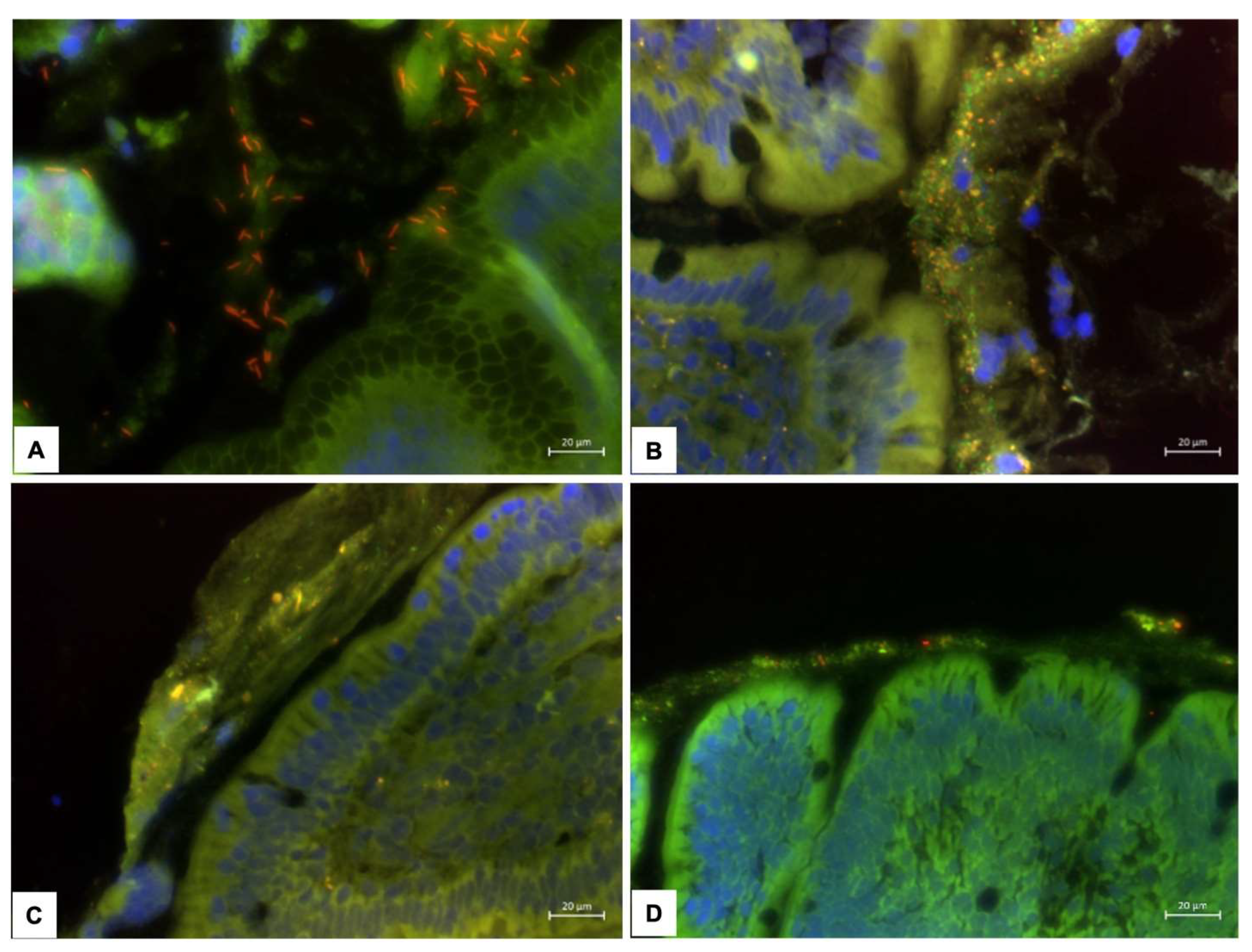
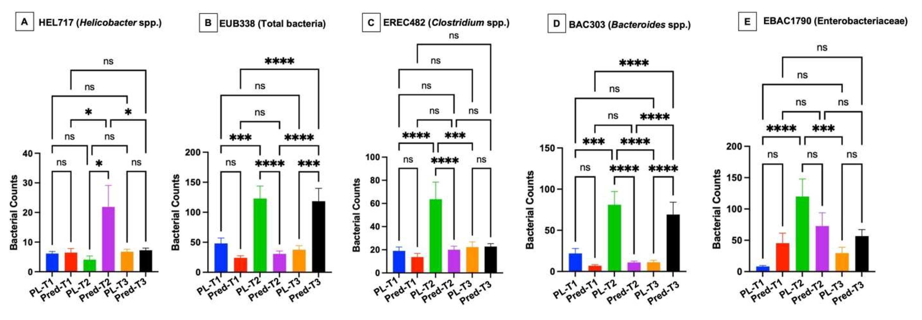
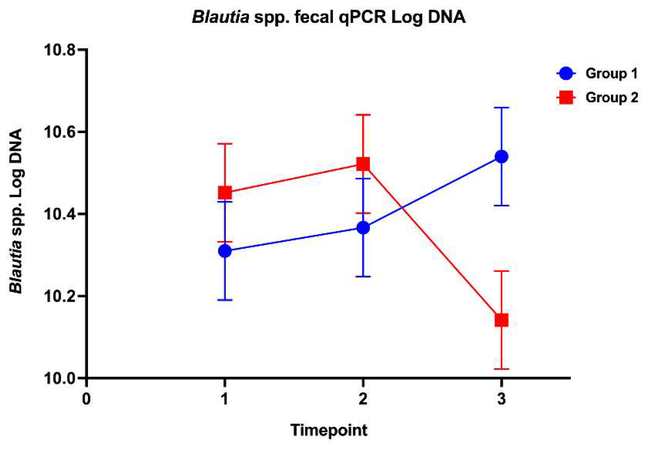
The effects of high-dose glucocorticoids on the gastrointestinal microbiota of healthy dogs are unknown. This study’s aim was to investigate the effects of immunosuppressive doses of prednisone on the fecal microbiota and the gastric and duodenal mucosal microbiota in healthy dogs. Twelve healthy adult dogs were enrolled into a randomized, double-blinded, placebo-controlled trial. Dogs were evaluated on days 0, 14, and 28 following treatments with either prednisone (2 mg/kg/d) or placebo. Outcome measures included: 1) composition and abundance of the fecal microbiota (via high throughput sequencing of the 16S rRNA gene and qPCR-based dysbiosis index [DI]) and 2) spatial distribution of the gastric and duodenal mucosal microbiota using fluorescence in situ hybridization (FISH). No significant difference in alpha and beta diversity or amplicon sequence variants of the fecal microbiota was observed between treatment groups. Blautia spp. concentrations via qPCR were significantly decreased between prednisone group timepoints 2 and 3. Compared to placebo group dogs, prednisone group dogs showed significantly increased gastric mucosal helicobacters and increased mucosal associated total bacteria and Bacteroides in duodenal biopsies over the treatment period. Results indicate that immunosuppressive dosages of prednisone alter the mucosal microbiota of healthy dogs in a time-dependent manner which may disrupt mucosal homeostasis.

Keywords: 
;  ;  ;  ;  ;  

1. Introduction

Glucocorticoids (GCs) are used clinically for their anti-inflammatory and immunosuppressive actions but are associated with multiple side effects [1]. Common side effects include polydipsia, polyuria, vomiting, and diarrhea [2]. High-dose glucocorticoid therapy may cause gastric erosion and ulceration in healthy dogs and people [3,4,5,6]. The mechanism of action for GC-induced injury includes decreased gastric emptying via prostaglandin E2 deficiency, gastric hyperacidity, and oxidative injury to the gastric mucosa [3,5,7]. Moreover, GC-induced gastric mucosal injury may disrupt normal gastric emptying and predispose to alterations in the gastrointestinal (GI) microbiota that are different than eubiosis observed in GI health [5].
Despite their frequent use by clinicians to treat immune-mediated diseases, there is little information regarding the effects of exogenous GCs on the gut microbiota. Several studies in rodent models have demonstrated GC-induced changes in the intestinal (fecal) microbiota that vary among species, subjects, and study design [8,9,10,11,12,13]. A prevailing pattern that emerges is that exogenous GCs increase the abundance of Firmicutes [12,14,15] and Actinobacteria [12,14,16] while the abundance of Bacteroidetes decreases [10,12,14,15]. Importantly, these data were generated using different GCs (prednisone, prednisolone, dexamethasone and hydrocortisone), suggesting a common mechanism of action. In one clinical trial involving dogs diagnosed with chronic inflammatory enteropathy (CIE, formally known as inflammatory bowel disease), therapy with prednisone and metronidazole was associated with altered intestinal abundance of select bacterial groups [17]. In another study, oral administration of prednisone (1 mg/kg) for 14 days to healthy dogs had no effect on fecal bacterial diversity or composition [18]. While these previous canine studies investigated the association between GC administration and the fecal microbiota, the impact of GCs on the gastric and duodenal mucosal microbiota of healthy dogs is unknown.
The objective of this study was to investigate the effects of immunosuppressive doses of prednisone (2 mg/kg/d PO) compared to placebo on the fecal and mucosal microbiota of healthy dogs. We hypothesized that high-dose prednisone administration would alter the gastric and duodenal mucosal microbiota but not the fecal microbiota of healthy dogs.

2. Materials and Methods

2.1. Animals

Archived gastric and duodenal mucosal biopsies obtained from 12 healthy laboratory-reared dogs that participated in a randomized, double-blinded, placebo-controlled trial were analyzed.[19] The animal use/clinical trial protocol was reviewed and approved by the IACUC committee at the University of Tennessee, Knoxville (protocol number 2283).

2.2. Study Design

Dogs were stratified by age and then randomized to either placebo or prednisone treatment groups. Dogs were acclimated to their surroundings for 14 days (days -13 to 0), followed by a 28-day treatment period (days 1-28). All dogs received water ad libitum and were fed a balanced canine commercial dry ration throughout the treatment schedule. Placebo group dogs were administered lactose-containing gelatin capsules (LetCo Medical, Decatur AL), and glucocorticoid group dogs received prednisone (West-Ward Pharmaceuticals Corp., Eatontown NJ) at a dosage of 2 mg/kg q24h PO. All treatments were administered in small meatballs (Purina ONE SmartBlend Healthy Puppy Lamb and Long Grain Rice; Nestle, Switzerland) once daily by an individual blinded to treatment groups. Naturally, voided feces were collected on treatment days 2 to 0, 12 to 14, and 26 to 28.
Esophagogastroduodenoscopy was performed and multiple endoscopic biopsies from the stomach and duodenum were obtained at each of the three timepoints. Mucosal biopsies were placed in 10% neutral buffered formalin, routinely processed and paraffin embedded as a tissue block for H&E histopathologic and fluorescence in situ hybridization (FISH) analyses.

2.3. Mucosal Microbiome Analysis

Formalin-fixed embedded tissue sections were prepared for fluorescence in situ hybridization (FISH) as previously described [20,21,22]. Briefly, sections were mounted on glass slides, deparaffinized and then air-dried prior to hybridization. Cy-3 or FITC labeled FISH probes were reconstituted with DNAse-free water and diluted to a working concentration of 5 ng/μL. Specific probes targeting the most common bacterial isolates from the stomach [23] and small intestine [24,25], as well as the universal bacterial probe Eub338, were applied to tissues (Supplemental Table 1). The probes selected for the stomach targeted Helicobacter spp., Streptococcus spp., and Lactobacillus spp., while probes for the small intestine targeted Clostridium spp., Enterobacteriaceae, and Bacteroides spp. Tissue sections were bathed in 30 μL of DNA–probe mix for 12 hours at 54°C, washed and rinsed, allowed to air-dry, and then mounted with SlowFade Gold mounting media (Life Technologies, Carlsbad, CA). Quantification of fluorophore-labeled (Cy3- or FITC-) bacterial populations present within the adherent mucus was performed using Metamorph® automated software as previously described [22,26,27].

2.4. Fecal Microbiome Analysis

Genomic DNA was extracted from 100 mg of feces for each time point using a commercially available DNA extraction kit (PowerSoil R®, Mo Bio, Carlsbad, CA) according to the manufacturer’s instructions. Amplification and sequencing of the V4 variable region (primers 515F/806R) of the 16S rRNA gene were performed on a MiSeq (Illumina) at MR DNA, as described previously. The software QIIME was used for the processing and analysis of sequences. The raw sequence data was de-multiplexed, and low-quality reads were filtered using default parameters. Chimeric sequences were detected using USEARCH and removed prior to further analysis, and sequences were then assigned to operational taxonomic units (OTUs) using an open-reference OTU picking protocol in QIIME against the Greengenes database. The OTU table was rarefied to 35,000 sequences per sample.
Beta diversity for level 2 investigation included Actinobacteria spp., Bacteroidetes spp., Deferribacteres spp., Firmicutes spp., Fusobacteria spp., Proteobacteria spp., and Tenericutes spp. Additional investigation for amplicon sequence variants was performed with a focus on Clostridium clusters IV and XIV (Lactobacillus spp., Paraprevotella spp., Bacteroides spp., Helicobacter spp., Actinomyces spp., Bifidobacterium spp., family Lachnospiraceae, Ruminococcus spp., Megamonas spp., Blautia spp., Roseburia spp., Coprococcus spp., Clostridium spp., family Ruminococcaceae, and Ruminococcus spp.). Oligonucleotide primers and probes, as well as respective annealing temperatures of primers, are summarized in Supplemental and Table 2.
Quantitative PCR was performed for selected bacterial groups (total bacterial, Faecalibacterium spp., Turicibacter spp., Streptococcus spp., Escherichia coli, Blautia spp., Fusobacterium spp., and Clostridium hiranonis) using extracted DNA as has been previously described [28] (Supplemental Table 2). Briefly, 2 µl of normalized DNA (final concentration: 5 ng/ µl) was combined with 5 µl of a DNA-binding dye (SsoFast EvaGreen supermix; Bio-Rad Laboratories, CA, USA), 0.4 µl each of a forward and reverse primer (final concentration: 400 nM), and 2.6 µl of PCR water to achieve a total reaction volume of 10 µl. Data were expressed as log amount of DNA (fg) for each bacterial group per 10 ng of isolated total DNA.
The dysbiosis index (DI) was calculated from the quantitative PCR analyses. The DI summarizes fecal abundance of 7 bacterial taxa and total bacteria. A DI >2 indicates a significant shift in overall microbiota diversity, <0 is normal and indicates no significant shifts in overall microbiota diversity, and 0-2 indicates mild to moderate shifts in overall microbiota diversity, as shown previously [28,29]. The higher the dysbiosis index, the greater the severity of dysbiosis.

2.5. Statistical and Data Analysis

Tabular data was organized by probe used and treatment group. Descriptive statistics were generated for each response measure. Normality of data was assessed visually by histograms and Q-Q plots. Global changes in microbiota communities (beta diversity) between individuals were determined using unweighted Unifrac distance metrics; principal coordinates analysis (PCoA) plots and rarefaction curves were plotted using QIIME software. The ANOSIM function in PRIMER 6 (PRIMER-E Ltd., Ivybridge, UK) was used to compare beta diversity metrics across time and between treatment groups [30].
Mixed model, split-plot repeated measures ANOVAs that include fixed effects of treatment, time, and treatment-by-time interaction were used to compare quantitative bacterial counts for each bacterial genus in the feces, Shannon indices, goods coverage, the Chao 1 metric, and the dysbiosis index between treatment groups. Dogs nested within groups were included as a random effect in all mixed model analyses. Model assumptions regarding normally distributed residuals were verified with the Shapiro-Wilk test for normality and QQ plots. Model assumptions regarding equality of variances were verified with Levene’s Test for Equality of Variances. Differences in least squares means were determined for bacteria counts and relative abundances with significant main effect or interaction terms. Only bacteria taxa that were present in at least 50% of dogs in ≥ 1 group at ≥ 1 time point were included in statistical analyses. Non-normally distributed data were logarithmically or rank-transformed, as necessary, to meet underlying statistical assumptions. If logarithmic transformation was required, .05 was added to all values. P-values were corrected for multiple comparisons on each phylogenetic level for microbiome evaluations using Benjamini & Hochberg’s False Discovery Rate (FDR). Comparison of numbers of mucosal bacteria was performed with GraphPad Prism 9 (version 9.4.1) (https://graphpad.com/; accessed on 2 September 2022) using one-way ANOVA followed by Šídák’s multiple comparisons test. A P-value of <0.05 was considered significant for all analyses. Publicly accessible software packages (http://www.qiime.org; MedCalc 15.8: MedCalc, Ostend, Belgium; SAS 9.4 release TS1M3: SAS Institute Inc., Cary, NC, USA) were used for all microbial community analyses.

3. Results

3.1. Animals

The placebo group included 2 castrated males, 1 intact male, and 3 intact females with a median age of 3.5 years (range 2 – 6 years), whereas the prednisone group was comprised of 2 castrated males, 2 intact males, and 2 intact females with a median age of 3 years (range 2 – 6 years). Pooled samples for microbiome analysis for 2 dogs at timepoint 2 (1 in each group) were comprised of only 2 fecal samples. The clinical, clinicopathologic, and gastrointestinal changes associated with administration of high-dose prednisone to the healthy dogs of this study have been previously reported [31].

3.2. Mucosal Microbiota

The microbiota of the gastric mucosa was colonized by Helicobacter spp., located within the gastric mucus layer, representing over 95% of the total bacterial load in biopsy specimens (Figure 1). While there was no difference in the numbers of Helicobacter spp. between dog groups at timepoint 1, prednisone group dogs had significantly increased (P < 0.05) numbers of helicobacters at timepoint 2 compared to placebo group dogs and compared to prednisone group dogs at timepoints 1 and 3 (Figure 2 A). There was no difference in the numbers of helicobacters between dog groups at timepoint 3. Very few (<10 bacteria/10 representative fields) streptococci and lactobacilli were visualized in gastric tissues.
The mucosal microbiota in the duodenum of healthy dogs was patchy in distribution and most abundant in adherent mucus (Figure 1). There were no differences in the numbers of bacteria between dog groups at timepoint 1. However, the numbers of total bacteria, Clostridia, Bacteroides, and Enterobacteriaceae were significantly increased (P < 0.05) in placebo group dogs compared to prednisone group dogs at timepoint 2 and compared to bacterial numbers in placebo group dogs at timepoint 1 (Figure 2 B-E). The numbers of Bacteroides in placebo group dogs at timepoint 2 exceeded (P < 0.05) their numbers at timepoint 3. The numbers of total bacteria and Bacteroides were greatest (P < 0.05) in prednisone group dogs at timepoint 3 as compared to placebo group dogs at timepoint 3 and compared to prednisone group dogs at timepoints 1 and 2 (Figure 2 B and D).

3.3. Fecal Microbiota

Alpha diversity did not differ between groups or among time points for Shannon index, observed amplicon sequence variant (ASV), or Chao 1 metrics (P ≥ 0.05 for all, respectively), as noted in Supplemental Table 3. Beta diversity and additional investigation for amplicon sequence variants did not differ significantly between groups or among time points (R = -0.105, P = 0.991 for weighted UniFrac distance (Supplemental Table 4).
Abundances for taxa evaluated by qPCR remained within their respective reference intervals for all dogs at all time points. However, a significant group by time interaction (P = .042, F-value 3.72) was noted for Blautia spp. Based on post-hoc analysis, this reflected significantly decreased Blautia spp. abundance at timepoint 3 for dogs in the prednisone group (Figure 3). The dysbiosis index was within the reference interval for all samples, with no significant differences between groups or among time points (Supplemental Figure 1).

4. Discussion

Glucocorticoids are powerful anti-inflammatory, immune-modulating drugs for treatment of inflammatory conditions (chronic enteropathies, rhinitis, immune-mediated hemolytic anemia) as well as orthopedic, dermatologic, and ophthalmologic disorders [32,33,34,35,36,37,38]. While the common side effects of polyuria, polydipsia, bodyweight gain or loss, and GI mucosal injury are often clinically apparent, GCs have also been shown to alter the composition of the intestinal (fecal) microbiota [9,10,11,12,13]. Our data suggest that dogs administered immunosuppressive doses of prednisone showed alterations in select groups of gastric and duodenal mucosal microbiota. Compared to placebo group dogs, dogs administered prednisone had increased numbers of helicobacters in the stomach and increased numbers of total bacteria and Bacteroides in the duodenum over the treatment period. Alpha and beta diversity of the fecal microbiota, as well as the DI, did not differ between treatment groups or time points. However, the fecal abundance of Blautia spp. was decreased at timepoint 3. In a separate study, the effects of metronidazole or prednisolone (1 mg/kg PO q 24h) on the fecal microbiome of healthy dogs were investigated before (day 0) after (day 14) treatment and 14 and 28 days after drug cessation [18]. No effect of prednisone on the fecal microbiota was observed. Like previous reports[39,40], metronidazole significantly altered the composition of some bacterial groups on day 14 compared with other time points. The data obtained in this earlier clinical study and our current research suggests that different GCs used short term at either anti-inflammatory or immunosuppressive levels do not significantly alter the fecal microbiota of healthy dogs.
The gut microbiota (e.g., bacteria, archaea, fungi, protozoa, and viruses) plays an important role in host health and disease [41,42,43]. It forms an essential component of the intestinal epithelial barrier, contributes to host metabolism, protects against pathogens, and influences development of the mucosal immune system [18,39]. Previous studies have identified a core intestinal microbiota composed of several phyla, including Actinobacteria, Bacteroidetes, Firmicutes, Fusobacteria, and Proteobacteria, in the fecal samples of healthy dogs [39,44]. Within this core community, several major taxa are considered beneficial and belong to the phylum Firmicutes, such as Clostridia and Bacilli, many of which are important short-chain fatty acid producers, including Faecalibacterium [45,46]. Other members of the resident microbiota, such as the family Enterobacteriaceae, are normally present in the small intestine in small numbers but are increased in the feces and mucosa of dogs with chronic inflammatory enteropathy (CIE) due to intestinal inflammation and associated dysbiosis [47,48,49].
The literature on the mucosal microbiota of healthy dogs is less extensive. Most studies have reported the mucosal microbiota of healthy (control) dogs compared to the microbiota in dogs with chronic gastrointestinal diseases. In separate studies, the mucosal microbiota of dogs with CIE was investigated in endoscopic biopsies [49,50] or cytologic brushings [24,48] of the duodenum by performing 454-pyrosequencing or evaluating gene clone libraries. General patterns of mucosal dysbiosis in diseased dogs included reduced biodiversity with increased numbers of Proteobacteria and decreased numbers of Fusobacteria, Clostridia, and Bacteroidaceae. Using FISH, invasive Escherichia coli (E. coli) are found within inflamed colonic mucosa of dogs with granulomatous colitis [26,51,52]. Other FISH studies have shown that ileal and/or colonic tissues of dogs with CIE harbor a dysbiosis characterized by increased numbers of mucosal Enterobacteriaceae and E. coli compared to control tissues [22,26,27,53]. Moreover, depletion of colonic surface and crypt bacteria (e.g., Helicobacter spp. and Akkermansia spp.) were observed in dogs with CIE [53].
Two FISH studies have evaluated the treatment effects of GCs on the mucosal microbiota in dogs with chronic gastroenteritis. Atherly et al. used a six-probe array (targeting Bifidobacterium spp, Enterobacteriaceae, Faecalibacterium spp., Lactobacillus spp., Streptococcus spp., and total bacteria) to investigate the mucosal microbiota of dogs with CIE treated with an elimination diet and immunosuppressive doses of prednisone for 8 weeks [54]. The spatial distribution of mucosal bacteria was significantly different following prednisone therapy, with increased numbers of Bifidobacteria, Faecalibacterium, and Streptococci present within adherent mucus. A second study, using a similar trial design and probe set, compared the treatment of dogs with CIE using prednisone or a multi-strain probiotic [27]. Results showed that prednisone-treated dogs had increased numbers of mucosal Bifidobacteria compared to dogs receiving probiotics. The mechanisms responsible for modulation of the intestinal microbiota by GCs remain poorly defined but may include changes in mucus (qualitative and quantitative), altered production of antimicrobial peptides and secretory IgA, increased intestinal permeability, and modulation of the NOD-like receptor family pyrin domain containing 6 (NLRP6) inflammasome [8,55,56,57].
The hypothalamic-pituitary-adrenal (HPA) axis and endogenous GC secretion are functionally influenced by a normal microbiota, as evidenced by the exaggerated response of acute stress in different rodent models [58,59,60]. For example, germ-free rats demonstrate altered neuroendocrine and behavioral responses to acute stress as compared to specific-pathogen-free male rats, accompanied by increased levels of corticosterone in plasma [59]. Germ-free mice undergoing restraint have increased levels of corticosterone, indicative of stress associated with the restraint procedure [61]. Other animal models have shown that neonatal rats exposed to probiotics early after birth are protected against elevated HPA responses and intestinal barrier dysfunction [62]. Stress has been shown to increase serum corticosteroid levels, alter the murine microbiome, and decrease levels of intestinal lactobacilli while increasing levels of E. coli and pseudomonas. [63,64] Stress also increases the expression of bacterial virulence genes, which can negatively affect intestinal function [63]. Finally, the role of environmental stress in a large animal model has been recently investigated. The prebiotic gallnut tannic acid was shown to ameliorate the stress-induced inflammatory response, fecal dysbiosis, and altered metabolome in laboratory beagles by targeting the intestinal microbiota [65]. These collective findings in different animal models suggest a complex interaction between endogenous and exogenous GCs and the GI microbiota, whose details remain poorly defined.
There are some limitations in our study, with the first being the small sample size of the groups with only 6 dogs in each cohort. The short study duration of 28 days may have underestimated the treatment effects of high-dose GCs on the intestinal microbiota if they were administered for a longer period. Our selection of FISH probes used to identify mucosal bacteria may have missed alterations in other microbial community members affected by diet and GC administration. While the same diet (having an identical macronutrient composition) was fed to both cohorts, it remains possible that other dietary factors may have affected mucosal bacterial composition in the prednisone group dogs [66].
In conclusion, immunosuppressive doses of prednisone administered to healthy dogs have little effect on the fecal microbiota, including the DI. In contrast, dogs administered prednisone showed variable but significant alterations in mucosal gastric Helicobacters and duodenal total bacteria and Bacteroides over the treatment period. Microbiota from mucosal samples more clearly reflect the underlying microbial alterations in response to high-dose prednisone treatment, as compared with fecal samples.

Supplementary Materials

The following supporting information can be downloaded at the website of this paper posted on Preprints.org.

Author Contributions

Conceptualization, S.G., J.C.W., J.S.S., D.K.S., and A.E.J.; methodology, S.G., J.C.W., J.S.S., D.K.S., S.M., E.L., S.V., J.S.S., and A.E.J.; software, J.C.W., J.S.S., D.K.S.; validation, S.G., J.C.W., J.S.S., D.K.S., and A.E.J.; formal analysis, S.G., J.C.W., J.S.S., D.K.S., S.M., E.L., S.V., and A.E.J.; investigation, S.G., J.C.W., J.S.S., D.K.S., S.M., E.L., S.V., and A.E.J.; resources, J.C.C., J.S.S., D.K.S., and A.E.J.; data curation, S.G., J.C.W., J.S.S., D.K.S., and A.E.J.; writing—original draft preparation, S.G., J.C.W., J.S.S., D.K.S., and A.E.J.; writing—review and editing, S.G., J.C.W., J.S.S., D.K.S., and A.E.J.; visualization, D.K.S. and A.E.J.; supervision; J.C.W., J.S.S., D.K.S., and A.E.J.; project administration, J.C.W. and A.E.J.; funding acquisition, J.C.W. and A.E.J. All authors have read and agreed to the published version of the manuscript.

Funding

This research was funded by a University of Tennessee clinical research grant and a VCS Research Incentive Grant from Iowa State University.

Institutional Review Board Statement

The animal study protocol (original clinical trial) was approved by the IACUC committee at the University of Tennessee, Knoxville (protocol number 2283).

Informed Consent Statement

Not required.

Data Availability Statement

The data presented in this study are available in the tables and figures.

Acknowledgments

The authors acknowledge the support of the Department of Veterinary Clinical Sciences (VCS) Core Lab.

Conflicts of Interest

None.

References

  1. Viviano, K. R. Glucocorticoids, Cyclosporine, Azathioprine, Chlorambucil, and Mycophenolate in Dogs and Cats: Clinical Uses, Pharmacology, and Side Effects. The Veterinary clinics of North America. Small animal practice 2022, 52(3), 797–817. [Google Scholar] [CrossRef] [PubMed]
  2. Elkholly, D. A.; Brodbelt, D. C.; Church, D. B.; Pelligand, L.; Mwacalimba, K.; Wright, A. K.; O’Neill, D. G. Side Effects to Systemic Glucocorticoid Therapy in Dogs Under Primary Veterinary Care in the UK. Front Vet Sci 2020, 7, 515. [Google Scholar] [CrossRef] [PubMed]
  3. Filaretova, L.; Podvigina, T.; Bagaeva, T.; Morozova, O. From gastroprotective to ulcerogenic effects of glucocorticoids: role of long-term glucocorticoid action. Curr Pharm Des 2014, 20(7), 1045–50. [Google Scholar] [CrossRef] [PubMed]
  4. Rohrer, C. R.; Hill, R. C.; Fischer, A.; Fox, L. E.; Schaer, M.; Ginn, P. E.; Casanova, J. M.; Burrows, C. F. Gastric hemorrhage in dogs given high doses of methylprednisolone sodium succinate. American journal of veterinary research 1999, 60(8), 977–81. [Google Scholar] [CrossRef] [PubMed]
  5. Tsukamoto, A.; Ohno, K.; Maeda, S.; Nakashima, K.; Fukushima, K.; Fujino, Y.; Hori, M.; Tsujimoto, H. Effect of mosapride on prednisolone-induced gastric mucosal injury and gastric-emptying disorder in dog. J Vet Med Sci 2012, 74(9), 1103–8. [Google Scholar] [CrossRef] [PubMed]
  6. Rak, M. B.; Moyers, T. D.; Price, J. M.; Whittemore, J. C. Clinicopathologic and gastrointestinal effects of administration of prednisone, prednisone with omeprazole, or prednisone with probiotics to dogs: A double-blind randomized trial. J Vet Intern Med 2023, 37(2), 465–475. [Google Scholar] [CrossRef]
  7. Das, D.; Bandyopadhyay, D.; Bhattacharjee, M.; Banerjee, R. K. Hydroxyl radical is the major causative factor in stress-induced gastric ulceration. Free Radic Biol Med 1997, 23(1), 8–18. [Google Scholar] [CrossRef] [PubMed]
  8. Tena-Garitaonaindia, M.; Arredondo-Amador, M.; Mascaraque, C.; Asensio, M.; Marin, J. J. G.; Martínez-Augustin, O.; Sánchez de Medina, F. Modulation of intestinal barrier function by glucocorticoids: Lessons from preclinical models. Pharmacol Res 2022, 177, 106056. [Google Scholar] [CrossRef] [PubMed]
  9. Schepper, J. D.; Collins, F.; Rios-Arce, N. D.; Kang, H. J.; Schaefer, L.; Gardinier, J. D.; Raghuvanshi, R.; Quinn, R. A.; Britton, R.; Parameswaran, N. Involvement of the gut microbiota and barrier function in glucocorticoid-induced osteoporosis. Journal of Bone and Mineral Research 2020, 35(4), 801–820. [Google Scholar] [CrossRef] [PubMed]
  10. Wu, T.; Yang, L.; Jiang, J.; Ni, Y.; Zhu, J.; Zheng, X.; Wang, Q.; Lu, X.; Fu, Z. Chronic glucocorticoid treatment induced circadian clock disorder leads to lipid metabolism and gut microbiota alterations in rats. Life Sciences 2018, 192, 173–182. [Google Scholar] [CrossRef]
  11. He, Z.; Kong, X.; Shao, T.; Zhang, Y.; Wen, C. Alterations of the Gut Microbiota Associated With Promoting Efficacy of Prednisone by Bromofuranone in MRL/lpr Mice. Frontiers in microbiology 2019, 10. [Google Scholar] [CrossRef]
  12. Huang, E. Y.; Inoue, T.; Leone, V. A.; Dalal, S.; Touw, K.; Wang, Y.; Musch, M. W.; Theriault, B.; Higuchi, K.; Donovan, S. Using corticosteroids to reshape the gut microbiome: implications for inflammatory bowel diseases. Inflammatory bowel diseases 2015, 21(5), 963–972. [Google Scholar] [CrossRef] [PubMed]
  13. Li, J.; Yang, M.; Lu, C.; Han, J.; Tang, S.; Zhou, J.; Li, Y.; Ming, T.; Wang, Z. J.; Su, X., Tuna bone powder alleviates glucocorticoid-induced osteoporosis via coregulation of the NF-κB and Wnt/β-catenin signaling pathways and modulation of gut microbiota composition and metabolism. Molecular nutrition & food research 2020, 64 (5), 1900861.
  14. Qi, X.-Z.; Tu, X.; Zha, J.-W.; Huang, A.-G.; Wang, G.-X.; Ling, F., Immunosuppression-induced alterations in fish gut microbiota may increase the susceptibility to pathogens. Fish & Shellfish Immunology 2019, 88, 540-545.
  15. Tourret, J.; Willing, B. P.; Dion, S.; MacPherson, J.; Denamur, E.; Finlay, B. B. Immunosuppressive treatment alters secretion of ileal antimicrobial peptides and gut microbiota, and favors subsequent colonization by uropathogenic Escherichia coli. Transplantation 2017, 101(1), 74–82. [Google Scholar] [CrossRef] [PubMed]
  16. Cheng, D.; Chang, H.; Ma, S.; Guo, J.; She, G.; Zhang, F.; Li, L.; Li, X.; Lu, Y. Tiansi liquid modulates gut microbiota composition and tryptophan–kynurenine metabolism in rats with hydrocortisone-induced depression. Molecules 2018, 23(11), 2832. [Google Scholar] [CrossRef] [PubMed]
  17. Rossi, G.; Pengo, G.; Caldin, M.; Palumbo Piccionello, A.; Steiner, J. M.; Cohen, N. D.; Jergens, A. E.; Suchodolski, J. S. Comparison of microbiological, histological, and immunomodulatory parameters in response to treatment with either combination therapy with prednisone and metronidazole or probiotic VSL#3 strains in dogs with idiopathic inflammatory bowel disease. PloS one 2014, 9(4), e94699. [Google Scholar]
  18. Igarashi, H.; Maeda, S.; Ohno, K.; Horigome, A.; Odamaki, T.; Tsujimoto, H. Effect of oral administration of metronidazole or prednisolone on fecal microbiota in dogs. PloS one 2014, 9(9), e107909. [Google Scholar] [CrossRef]
  19. Whittemore, J. C.; Mooney, A. P.; Price, J. M.; Thomason, J. Clinical, clinicopathologic, and gastrointestinal changes from aspirin, prednisone, or combination treatment in healthy research dogs: A double-blind randomized trial. J Vet Intern Med 2019. [Google Scholar] [CrossRef]
  20. Cassmann, E.; White, R.; Atherly, T.; Wang, C.; Sun, Y.; Khoda, S.; Mosher, C.; Ackermann, M.; Jergens, A. Alterations of the Ileal and Colonic Mucosal Microbiota in Canine Chronic Enteropathies. PLoS One 2016, 11(2), e0147321. [Google Scholar] [CrossRef] [PubMed]
  21. Jergens, A. E.; Pressel, M.; Crandell, J.; Morrison, J. A.; Sorden, S. D.; Haynes, J.; Craven, M.; Baumgart, M.; Simpson, K. W. Fluorescence in situ hybridization confirms clearance of visible Helicobacter spp. associated with gastritis in dogs and cats. J Vet Intern Med 2009, 23(1), 16–23. [Google Scholar] [CrossRef] [PubMed]
  22. Sahoo, D. K.; Allenspach, K.; Mochel, J. P.; Parker, V.; Rudinsky, A. J.; Winston, J. A.; Bourgois-Mochel, A.; Ackermann, M.; Heilmann, R. M.; Köller, G.; Yuan, L.; Stewart, T.; Morgan, S.; Scheunemann, K. R.; Iennarella-Servantez, C. A.; Gabriel, V.; Zdyrski, C.; Pilla, R.; Suchodolski, J. S.; Jergens, A. E., Synbiotic-IgY Therapy Modulates the Mucosal Microbiome and Inflammatory Indices in Dogs with Chronic Inflammatory Enteropathy: A Randomized, Double-Blind, Placebo-Controlled Study. Veterinary sciences 2022, 10 (1).
  23. Garcia-Mazcorro, J. F.; Suchodolski, J. S.; Jones, K. R.; Clark-Price, S. C.; Dowd, S. E.; Minamoto, Y.; Markel, M.; Steiner, J. M.; Dossin, O. Effect of the proton pump inhibitor omeprazole on the gastrointestinal bacterial microbiota of healthy dogs. FEMS Microbiol Ecol 2012, 80(3), 624–36. [Google Scholar] [CrossRef] [PubMed]
  24. Suchodolski, J. S.; Camacho, J.; Steiner, J. M. Analysis of bacterial diversity in the canine duodenum, jejunum, ileum, and colon by comparative 16S rRNA gene analysis. FEMS Microbiol Ecol 2008, 66(3), 567–78. [Google Scholar] [CrossRef] [PubMed]
  25. Suchodolski, J. S. Companion animals symposium: microbes and gastrointestinal health of dogs and cats. J Anim Sci 2011, 89(5), 1520–30. [Google Scholar] [CrossRef]
  26. Cassmann, E.; White, R.; Atherly, T.; Wang, C.; Sun, Y.; Khoda, S.; Mosher, C.; Ackermann, M.; Jergens, A. Alterations of the Ileal and Colonic Mucosal Microbiota in Canine Chronic Enteropathies. PloS one 2016, 11(2), e0147321–e0147321. [Google Scholar] [CrossRef]
  27. White, R.; Atherly, T.; Guard, B.; Rossi, G.; Wang, C.; Mosher, C.; Webb, C.; Hill, S.; Ackermann, M.; Sciabarra, P.; Allenspach, K.; Suchodolski, J.; Jergens, A. E. Randomized, controlled trial evaluating the effect of multi-strain probiotic on the mucosal microbiota in canine idiopathic inflammatory bowel disease. Gut microbes 2017, 8(5), 451–466. [Google Scholar] [CrossRef]
  28. AlShawaqfeh, M. K.; Wajid, B.; Minamoto, Y.; Markel, M.; Lidbury, J. A.; Steiner, J. M.; Serpedin, E.; Suchodolski, J. S., A dysbiosis index to assess microbial changes in fecal samples of dogs with chronic inflammatory enteropathy. FEMS Microbiol Ecol 2017, 93 (11).
  29. Sung, C.-H.; Pilla, R.; Chen, C.-C.; Ishii, P. E.; Toresson, L.; Allenspach-Jorn, K.; Jergens, A. E.; Summers, S.; Swanson, K. S.; Volk, H.; Schmidt, T.; Stuebing, H.; Rieder, J.; Busch, K.; Werner, M.; Lisjak, A.; Gaschen, F. P.; Belchik, S. E.; Tolbert, M. K.; Lidbury, J. A.; Steiner, J. M.; Suchodolski, J. S. Correlation between Targeted qPCR Assays and Untargeted DNA Shotgun Metagenomic Sequencing for Assessing the Fecal Microbiota in Dogs. Animals 2023, 13(16), 2597. [Google Scholar] [CrossRef]
  30. Clarke, K. R. Non-parametric multivariate analyses of changes in community structure. Australian journal of ecology 1993, 18(1), 117–143. [Google Scholar] [CrossRef]
  31. Whittemore, J. C.; Mooney, A. P.; Price, J. M.; Thomason, J. Clinical, clinicopathologic, and gastrointestinal changes from administration of clopidogrel, prednisone, or combination in healthy dogs: A double-blind randomized trial. Journal of Veterinary Internal Medicine 2019, 33(6), 2618–2627. [Google Scholar] [CrossRef] [PubMed]
  32. LeVine, D. N.; Goggs, R.; Kohn, B.; Mackin, A. J.; Kidd, L.; Garden, O. A.; Brooks, M. B.; Eldermire, E. R. B.; Abrams-Ogg, A.; Appleman, E. H.; Archer, T. M.; Bianco, D.; Blois, S. L.; Brainard, B. M.; Callan, M. B.; Fellman, C. L.; Haines, J. M.; Hale, A. S.; Huang, A. A.; Lucy, J. M.; O’Marra, S. K.; Rozanski, E. A.; Thomason, J. M.; Walton, J. E.; Wilson, H. E. ACVIM consensus statement on the treatment of immune thrombocytopenia in dogs and cats. Journal of Veterinary Internal Medicine 2024, 38(4), 1982–2007. [Google Scholar] [CrossRef] [PubMed]
  33. Jergens, A. E.; Heilmann, R. M. Canine chronic enteropathy-Current state-of-the-art and emerging concepts. Front Vet Sci 2022, 9, 923013. [Google Scholar] [CrossRef] [PubMed]
  34. Swann, J. W.; Garden, O. A.; Fellman, C. L.; Glanemann, B.; Goggs, R.; LeVine, D. N.; Mackin, A. J.; Whitley, N. T. ACVIM consensus statement on the treatment of immune-mediated hemolytic anemia in dogs. Journal of Veterinary Internal Medicine 2019, 33(3), 1141–1172. [Google Scholar] [CrossRef]
  35. Windsor, R. C.; Johnson, L. R. Canine chronic inflammatory rhinitis. Clin Tech Small Anim Pract 2006, 21(2), 76–81. [Google Scholar] [CrossRef] [PubMed]
  36. Rhoades, A. C.; Vernau, W.; Kass, P. H.; Herrera, M. A.; Sykes, J. E. Comparison of the efficacy of prednisone and cyclosporine for treatment of dogs with primary immune-mediated polyarthritis. Journal of the American Veterinary Medical Association 2016, 248(4), 395–404. [Google Scholar] [CrossRef] [PubMed]
  37. Saridomichelakis, M. N.; Favrot, C.; Jackson, H. A.; Bensignor, E.; Prost, C.; Mueller, R. S. A proposed medication score for long-term trials of treatment of canine atopic dermatitis sensu lato. The Veterinary record 2021, 188(5), e19. [Google Scholar] [CrossRef]
  38. Holmberg, B.; Maggs, D. The use of corticosteroids to treat ocular inflammation. Vet Clin North Am Small Anim Pract. 2004, 34(3), 693–705. [Google Scholar] [CrossRef] [PubMed]
  39. Pilla, R.; Gaschen, F. P.; Barr, J. W.; Olson, E.; Honneffer, J.; Guard, B. C.; Blake, A. B.; Villanueva, D.; Khattab, M. R.; AlShawaqfeh, M. K.; Lidbury, J. A.; Steiner, J. M.; Suchodolski, J. S. Effects of metronidazole on the fecal microbiome and metabolome in healthy dogs. J Vet Intern Med 2020, 34(5), 1853–1866. [Google Scholar] [CrossRef] [PubMed]
  40. Stübing, H.; Suchodolski, J. S.; Reisinger, A.; Werner, M.; Hartmann, K.; Unterer, S.; Busch, K., The Effect of Metronidazole versus a Synbiotic on Clinical Course and Core Intestinal Microbiota in Dogs with Acute Diarrhea. Veterinary sciences 2024, 11 (5).
  41. Hooper, L. V.; Wong, M. H.; Thelin, A.; Hansson, L.; Falk, P. G.; Gordon, J. I., Molecular analysis of commensal host-microbial relationships in the intestine. Science (New York, N.Y.) 2001, 291 (5505), 881-4.
  42. Hooda, S.; Minamoto, Y.; Suchodolski, J. S.; Swanson, K. S. Current state of knowledge: the canine gastrointestinal microbiome. Anim Health Res Rev 2012, 13(1), 78–88. [Google Scholar] [CrossRef]
  43. Ziese, A. L.; Suchodolski, J. S. Impact of Changes in Gastrointestinal Microbiota in Canine and Feline Digestive Diseases. The Veterinary clinics of North America. Small animal practice 2021, 51(1), 155–169. [Google Scholar] [CrossRef]
  44. Honneffer, J. B.; Minamoto, Y.; Suchodolski, J. S. Microbiota alterations in acute and chronic gastrointestinal inflammation of cats and dogs. World journal of gastroenterology 2014, 20(44), 16489–97. [Google Scholar] [CrossRef] [PubMed]
  45. Garcia-Mazcorro, J. F.; Lanerie, D. J.; Dowd, S. E.; Paddock, C. G.; Grützner, N.; Steiner, J. M.; Ivanek, R.; Suchodolski, J. S. Effect of a multi-species synbiotic formulation on fecal bacterial microbiota of healthy cats and dogs as evaluated by pyrosequencing. FEMS Microbiol Ecol 2011, 78(3), 542–54. [Google Scholar] [CrossRef] [PubMed]
  46. Garcia-Mazcorro, J. F.; Dowd, S. E.; Poulsen, J.; Steiner, J. M.; Suchodolski, J. S. Abundance and short-term temporal variability of fecal microbiota in healthy dogs. Microbiologyopen 2012, 1(3), 340–7. [Google Scholar] [CrossRef]
  47. Minamoto, Y.; Minamoto, T.; Isaiah, A.; Sattasathuchana, P.; Buono, A.; Rangachari, V. R.; McNeely, I. H.; Lidbury, J.; Steiner, J. M.; Suchodolski, J. S. Fecal short-chain fatty acid concentrations and dysbiosis in dogs with chronic enteropathy. J Vet Intern Med 2019, 33(4), 1608–1618. [Google Scholar] [CrossRef]
  48. Xenoulis, P. G.; Palculict, B.; Allenspach, K.; Steiner, J. M.; Van House, A. M.; Suchodolski, J. S. Molecular-phylogenetic characterization of microbial communities imbalances in the small intestine of dogs with inflammatory bowel disease. FEMS Microbiol Ecol 2008, 66(3), 579–89. [Google Scholar] [CrossRef] [PubMed]
  49. Suchodolski, J. S.; Xenoulis, P. G.; Paddock, C. G.; Steiner, J. M.; Jergens, A. E., Molecular analysis of the bacterial microbiota in duodenal biopsies from dogs with idiopathic inflammatory bowel disease. Vet Microbiol 2010, 142 (3-4), 394-400.
  50. Suchodolski, J. S.; Dowd, S. E.; Wilke, V.; Steiner, J. M.; Jergens, A. E. 16S rRNA gene pyrosequencing reveals bacterial dysbiosis in the duodenum of dogs with idiopathic inflammatory bowel disease. PloS one 2012, 7(6), e39333–e39333. [Google Scholar] [CrossRef] [PubMed]
  51. Craven, M.; Mansfield, C. S.; Simpson, K. W. Granulomatous colitis of boxer dogs. The Veterinary clinics of North America. Small animal practice 2011, 41(2), 433–45. [Google Scholar] [CrossRef] [PubMed]
  52. Mansfield, C. S.; James, F. E.; Craven, M.; Davies, D. R.; O’Hara, A. J.; Nicholls, P. K.; Dogan, B.; MacDonough, S. P.; Simpson, K. W. Remission of histiocytic ulcerative colitis in Boxer dogs correlates with eradication of invasive intramucosal Escherichia coli. J Vet Intern Med 2009, 23(5), 964–9. [Google Scholar] [CrossRef] [PubMed]
  53. Giaretta, P. R.; Suchodolski, J. S.; Jergens, A. E.; Steiner, J. M.; Lidbury, J. A.; Cook, A. K.; Hanifeh, M.; Spillmann, T.; Kilpinen, S.; Syrjä, P.; Rech, R. R. Bacterial Biogeography of the Colon in Dogs With Chronic Inflammatory Enteropathy. Veterinary pathology 2020, 57(2), 258–265. [Google Scholar] [CrossRef] [PubMed]
  54. Atherly, T.; Rossi, G.; White, R.; Seo, Y.-J.; Wang, C.; Ackermann, M.; Breuer, M.; Allenspach, K.; Mochel, J. P.; Jergens, A. E., Glucocorticoid and dietary effects on mucosal microbiota in canine inflammatory bowel disease. PloS one 2019, 14 (12).
  55. Alverdy, J.; Aoys, E. The effect of glucocorticoid administration on bacterial translocation. Evidence for an acquired mucosal immunodeficient state. Ann Surg 1991, 214(6), 719–23. [Google Scholar] [CrossRef] [PubMed]
  56. Spitz, J.; Hecht, G.; Taveras, M.; Aoys, E.; Alverdy, J. The effect of dexamethasone administration on rat intestinal permeability: the role of bacterial adherence. Gastroenterology 1994, 106(1), 35–41. [Google Scholar] [CrossRef]
  57. Sun, Y.; Zhang, M.; Chen, C. C.; Gillilland, M., 3rd; Sun, X.; El-Zaatari, M.; Huffnagle, G. B.; Young, V. B.; Zhang, J.; Hong, S. C.; Chang, Y. M.; Gumucio, D. L.; Owyang, C.; Kao, J. Y., Stress-induced corticotropin-releasing hormone-mediated NLRP6 inflammasome inhibition and transmissible enteritis in mice. Gastroenterology 2013, 144 (7), 1478-87, 1487.e1-8.
  58. Clarke, G.; Grenham, S.; Scully, P.; Fitzgerald, P.; Moloney, R. D.; Shanahan, F.; Dinan, T. G.; Cryan, J. F. The microbiome-gut-brain axis during early life regulates the hippocampal serotonergic system in a sex-dependent manner. Mol Psychiatry 2013, 18(6), 666–73. [Google Scholar] [CrossRef] [PubMed]
  59. Crumeyrolle-Arias, M.; Jaglin, M.; Bruneau, A.; Vancassel, S.; Cardona, A.; Daugé, V.; Naudon, L.; Rabot, S. Absence of the gut microbiota enhances anxiety-like behavior and neuroendocrine response to acute stress in rats. Psychoneuroendocrinology 2014, 42, 207–17. [Google Scholar] [CrossRef]
  60. Sudo, N.; Chida, Y.; Aiba, Y.; Sonoda, J.; Oyama, N.; Yu, X. N.; Kubo, C.; Koga, Y. Postnatal microbial colonization programs the hypothalamic-pituitary-adrenal system for stress response in mice. J Physiol 2004, 558 (Pt 1) Pt 1, 263–75. [Google Scholar] [CrossRef]
  61. Vagnerová, K.; Vodička, M.; Hermanová, P.; Ergang, P.; Šrůtková, D.; Klusoňová, P.; Balounová, K.; Hudcovic, T.; Pácha, J. Interactions Between Gut Microbiota and Acute Restraint Stress in Peripheral Structures of the Hypothalamic-Pituitary-Adrenal Axis and the Intestine of Male Mice. Front Immunol 2019, 10, 2655. [Google Scholar] [CrossRef]
  62. Gareau, M. G.; Jury, J.; MacQueen, G.; Sherman, P. M.; Perdue, M. H. Probiotic treatment of rat pups normalises corticosterone release and ameliorates colonic dysfunction induced by maternal separation. Gut 2007, 56(11), 1522–8. [Google Scholar] [CrossRef] [PubMed]
  63. Lutgendorff, F.; Akkermans, L. M.; Söderholm, J. D. The role of microbiota and probiotics in stress-induced gastro-intestinal damage. Curr Mol Med 2008, 8(4), 282–98. [Google Scholar] [CrossRef] [PubMed]
  64. O’Mahony, S. M.; Marchesi, J. R.; Scully, P.; Codling, C.; Ceolho, A. M.; Quigley, E. M.; Cryan, J. F.; Dinan, T. G. Early life stress alters behavior, immunity, and microbiota in rats: implications for irritable bowel syndrome and psychiatric illnesses. Biol Psychiatry 2009, 65(3), 263–7. [Google Scholar] [CrossRef] [PubMed]
  65. Yang, K.; Jian, S.; Wen, C.; Guo, D.; Liao, P.; Wen, J.; Kuang, T.; Han, S.; Liu, Q.; Deng, B. Gallnut Tannic Acid Exerts Anti-stress Effects on Stress-Induced Inflammatory Response, Dysbiotic Gut Microbiota, and Alterations of Serum Metabolic Profile in Beagle Dogs. Front Nutr 2022, 9, 847966. [Google Scholar] [CrossRef] [PubMed]
  66. Pilla, R.; Suchodolski, J. S. The Role of the Canine Gut Microbiome and Metabolome in Health and Gastrointestinal Disease. Frontiers in veterinary science 2020, 6, 498–498. [Google Scholar] [CrossRef] [PubMed]
Figure 1. Three-color FISH identifies mucosal bacteria present in canine gastric and duodenal endoscopic biopsies. Specific bacterial groups (Helicobacter, Clostridia, Bacteroides, and Enterobacteriaceae) hybridizing with Cy3 appear orange. All other bacteria hybridizing with the universal probe (EUB-FITC) appear green. DAPI stained mucosal epithelia stain blue. Panel A - gastric Helicobacter from a prednisone group dog, Panel B - duodenal Bacteroides from a prednisone group dog, Panel C - duodenal Enterobacteriaceae from a placebo group dog, and Panel D - duodenal Clostridia from a placebo group dog. All images at 400x magnification.
Preprints 146211 g001
Figure 2. Numbers of mucosal bacteria in gastric and duodenal biopsies of healthy dogs over the treatment schedule. Data expessed as mean + standard deviation. Gastric helicobacters (Panel A), duodenal total bacteria (Panel B), Clostridia (Panel C), Bacteroides (Panel D), Enterobacteriaiceae (Panel E). PL = placebo group, Pred = prednisone group, T1 = timepoint 1, T2 = timepoint 2, T3 = timepoint 3. **** significantly different at P value < 0.0001, *** significantly different at P value < 0.001, * significantly different at P value < 0.05. ns = no significant difference.
Preprints 146211 g002
Figure 3. Blautia spp. fecal qPCR Log DNA change between animals receiving placebo (group 1) versus prednisone (group 2) at baseline (timepoint 1, day -2 – 0), midway through study period (timepoint 2, day 12 – 14), and at the end of a 28-day treatment schedule (timepoint, day 26 – 28).
Preprints 146211 g003
Disclaimer/Publisher’s Note: The statements, opinions and data contained in all publications are solely those of the individual author(s) and contributor(s) and not of MDPI and/or the editor(s). MDPI and/or the editor(s) disclaim responsibility for any injury to people or property resulting from any ideas, methods, instructions or products referred to in the content.
Copyright: This open access article is published under a Creative Commons CC BY 4.0 license, which permit the free download, distribution, and reuse, provided that the author and preprint are cited in any reuse.

Downloads

124

Views

69

Comments

0

Subscription

Notify me about updates to this article or when a peer-reviewed version is published.

Email

Prerpints.org logo

Preprints.org is a free preprint server supported by MDPI in Basel, Switzerland.

Subscribe

© 2025 MDPI (Basel, Switzerland) unless otherwise stated