Submitted:
26 January 2025
Posted:
27 January 2025
You are already at the latest version
Abstract
Food safety is directly related to human health and has attracted intense attention all over the world. Surface-enhanced Raman scattering (SERS), as a rapid and selective technique, has been widely applied in monitoring food safety. SERS substrates, as an essential factor for sensing design, greatly influence the analytical performance. Currently, nanostructure-based SERS substrates have garnered significant interest due to their excellent merits in improving the sensitivity, specificity, and stability, holding great potential for rapid and accurate sensing toward food contaminants in complex matrices. This review summarizes the fundamentals of Raman spectroscopy and the used nanostructures for designing SERS platform, including precious metal nanoparticles, metal-organic frameworks, polymers, and semiconductors. Moreover, it introduces the mechanisms and applications of nanostructures in enhancing SERS signals for monitoring hazardous substances, such as foodborne bacteria, pesticide and veterinary drug residues, food additives, illegal adulterants, and packaging material contamination. Finally, the review offers broad prospects in sustainable, environmentally friendly, and large-scale analysis to evaluate food safety.
Keywords:
Surface-enhanced Raman scatter
; nanostructures
; sensing platform
; chemical mechanism
; food safety
; hazardous substances
1. Introduction
Food safety is a critical issue in the global public health domain, directly impacting consumer health[1,2,3]. With the increasing complexity of modern food production and supply chains, the contamination of harmful substances in food has become more pronounced[4,5]. The harmful substances are mentioned as microorganisms[6,7,8,9,10], chemical substances[11,12,13,14,15], and physical contaminants[16,17,18,19] that may be introduced during food harvest, processing, storage, and sale[20,21,22,23,24,25]. As mentioned, microbiological contamination, such as bacteria[3,26,27,28,29,30], viruses[31,32,33,34], and fungi[35,36,37,38], can lead to outbreaks of foodborne diseases, posing significant challenges to public health safety[23,39,40]. Chemical contamination, including residues of veterinary drugs[39,41,42] and pesticides[43,44,45,46], heavy metals pollution[47,48,49,50,51,52], food additives[53,54,55,56] and illegal adulterants[57,58,59], is also an issue that cannot be overlooked in food safety. Long-term intake of these chemical substances may have chronic effects on human health, including carcinogenesis, neurological damage, and other health issues[60,61]. Physical contamination, such as glass, metal, rocks, fish bones[62], and packaging contamination[17,18], although not usually causing acute health risks, can cause physical injury and discomfort to consumers.
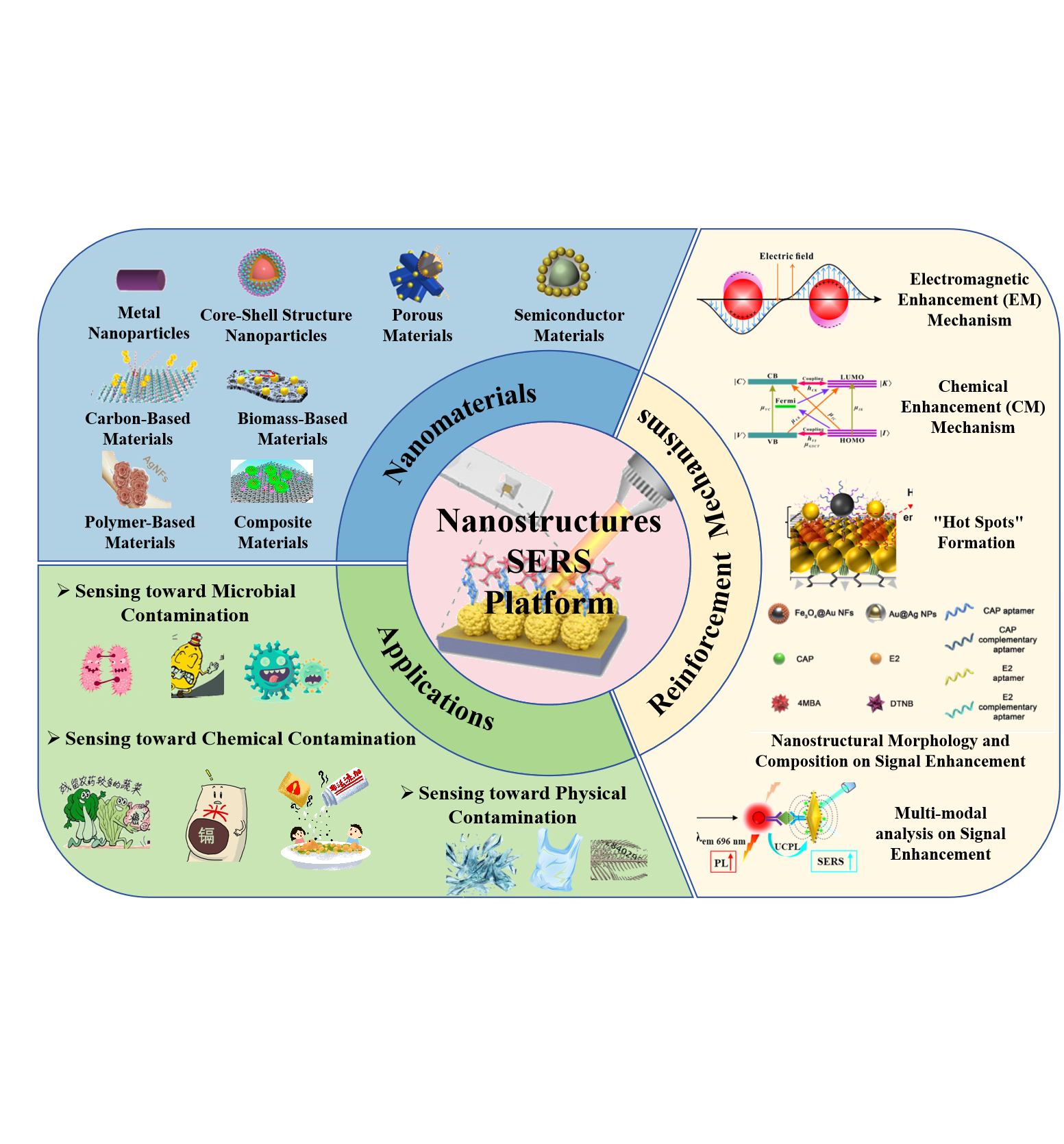
Scheme 1.
Overview of Nanostructures as SERS Substrates: Nanomaterials, Reinforcement Mechanisms, and Applications in Food Hazard Detection.
Scheme 1.
Overview of Nanostructures as SERS Substrates: Nanomaterials, Reinforcement Mechanisms, and Applications in Food Hazard Detection.
To protect consumers from the potential hazards, governments and international organizations have established food safety standards[63,64,65,66]. Although the accuracy of traditional methods toward food safety assessment, they often have some limitations, such as high cost, long duration, laborious operation, and inability to achieve on-site analysis[67,68,69,70]. Therefore, the development of rapid and sensitive technologies is crucial for improving the efficiency of food safety management. Fortunately, Surface-enhanced Raman spectroscopy (SERS) has shown great potential in the field of food safety, which can provide detailed information on molecular vibrations of trace components in complex samples. [71,72,73,74]. SERS pattern, with its high sensitivity, rapid response, and molecular fingerprint characteristics, provides a powerful solution for detecting various harmful substances in food. Consequently, great efforts have been devoted to SERS development, aiming to promptly monitor microorganisms, chemical substances, and physical contaminants in assessing food safety risks.
To design a successful SERS strategy, the sensing platform is considered as a key foctor on analytical performance. In this respect, nanostructures, due to their unique physical and chemical properties[21,75,76,77,78,79], are very popular in SERS platform development for enhancing Raman signals. For example, metal nanostructures such as gold (Au) and silver (Ag) nanoparticles, have a localized surface plasmon resonance (LSPR) effect and can generate strong electromagnetic fields on sensing platform, greatly amplifying the Raman scattering signals of nearby molecules. With help of nanostructures, SERS platform has been applied in detecting mycotoxins[80,81,82], pesticide residues[45,83,84,85,86], and illegal additives[87,88] with high sensitivity for ensuring food safety, in which nanostructures not only amplify SERS signal but also serve as carriers and markers [89,90].
Herein, this review summarized the analytical mechanism of SERS patterns. Moreover, the enhancement behavior of nanostructures (e.g., metal-based, carbon-based nanostructures)toward Raman signals was explored for further improving the sensitivity of SERS platform. Benefitting from the merits of nanostructures, SERS mode was applied in monitoring food hazards, contributing to ensuring the efficiency of food safety.
2. Analytical Mechanism of SERS
2.1. Basic Theory of SERS
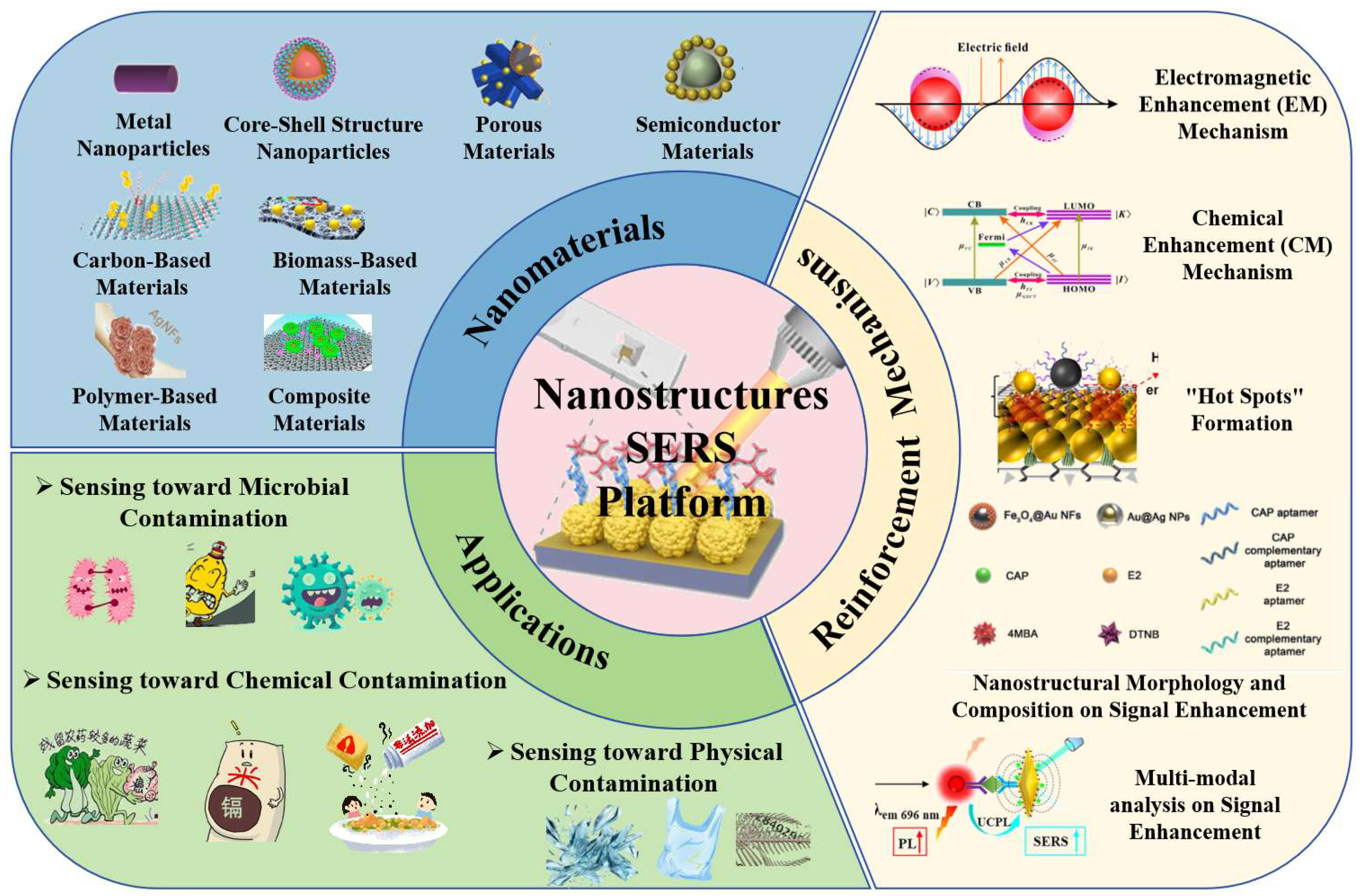
As known, when a beam of light strikes an uneven substrate, the light can be reflected, scattered, or absorbed (Figure 1A). During the mentioned process, the scattered part undergoes a change in frequency, it is named as Raman scattering[91]; while the frequency remains unchanged, it is Rayleigh scattering. From a microscopic perspective, when an excited molecule absorbs the energy of a photon, it can transition to a virtual state, and then return to the ground state, resulting in Rayleigh scattering. Nevertheless, when the molecule reaches another energy level, it is called Raman scattering, in which it can be classified as anti-Stokes scattering (the molecule absorbs energy) and Stokes scattering (the molecule loses energy)[92]. The energy difference between two photons is given as Equation (1):
where represents the frequency shift in the Raman scattering spectrum.
Unlike typical Raman spectra, the intensity of SERS spectra is dependent on the interaction of rough metal surfaces (Ag or Au nanoparticles) with the target molecule[93]. Specifically, SERS has the following characteristics: (1) high sensitivity, with an intensity that is 7 to 14 orders of magnitude greater than that of spontaneous Raman scattering; (2) excellent selectivity, selective amplification of certain normal vibrations associated with a given electronic absorption band. With these merits, SERS offers an effective technique for studying the vibrational and electronic structures of chromophores within biomolecules or entire cells, and it can be applied in the fields of life sciences and food to investigate specific chromophores that play crucial roles in biological processes.
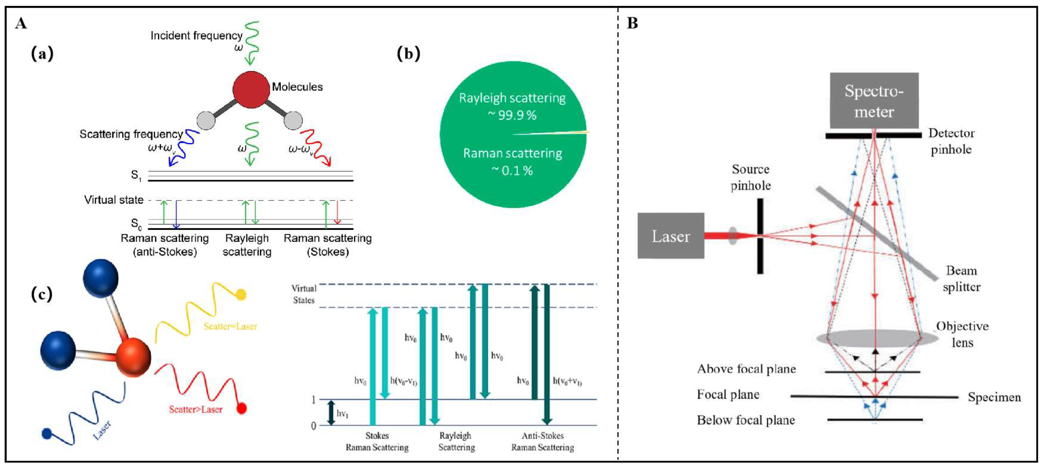
Figure 1.
(A) Schematic diagram of (a) Raman scattering effect; (b) Photon scattering of Rayleigh scattering accounting for about 99.9% , Raman scatteringabout 0.1%; (c) Transition energy level of Stokes, anti-Stokes and Rayleigh scattering[91,94,95]. (B) Schematic setup of confocal Raman microscope[96].
2.2. Raman Signal Acquisition
Raman microscopy[96], which integrates Raman spectroscopy with an optical microscope, enables the acquisition of Raman scattering through an objective lens (Figure 1B). Meanwhile, confocal Raman microscopy directs a point light source through pinhole and beam splitter, focusing it to diffraction-limited spot on the specimen via objective lens. The scattered or emitted light is collected, collimated, and passed through detector pinhole before reaching a spectrometer. The detector pinhole serves as depth selector, rejecting out-of-focus light and selectively gathering signals from the focal plane. Especially, confocal microscopes have emerged as valuable analytical tools, offering superior depth resolution and enhanced image contrast through the suppression of stray light.
Raman imaging could exhibit chemical composition by integrating spatial (x, y dimensions) and spectral (wavelength dimension) information from Raman spectroscopy. There are two primary Raman imaging methodologies: scanning and wide-field imaging. As for scanning imaging, often facilitated by confocal microscopy, is conducted as follows: (1) Point Scanning: This method collects Raman spectra at each spatial location sequentially. The sample is precisely moved across a high-precision stage, controlling both lateral and axial positions. It offers high spectral resolution and full spectral capture, but is time-intensive and susceptible to laser-induced sample damage[96]. (2) Line Scanning: Broadening the spatial range of each scan with a laser line, this technique gathers a line of spatial and spectral data per measurement. The sample is automated along a stage axis perpendicular to the laser line. Despite the reduced Raman signal intensity, line scanning maintains high spectral resolution and is more time-efficient than point scanning[96]. Regarding wide-field Raman imaging, the entire sample area is simultaneously illuminated, enabling the acquisition of spatial data in a single, non-contact scan. Among the developed wide-field imaging, area scanning involves the selection of a specific Raman scattering spectral slice for analysis[96]. While this approach offers speed, it is limited by the complexity of spectral data discrimination.
2.3. SERS Superiority
The primary constraint of traditional Raman spectroscopy is the inherently weak signal, with only about 1 in 106 to 108 photons being inelastically scattered. This limitation can impede the detection of molecules at low concentrations, thereby restricting its utility in food industry applications[97]. SERS overcomes the shortcomings by employing metal-based sensing platform , greatly amplifying the Raman signal of even single molecule by several orders of magnitude, typically from 107 to 1014. Specially, the assay principle involves the interaction between rough metal surfaces and target molecules, in which the inelastic light scattering of the target molecules is enhanced[98]. The enhancement factor can reach up to 1014, thereby enabling single-molecule detection in certain cases. The development of SERS has also promoted the advancement of various other spectroscopic techniques.
According to whether the molecule(e.g., 4-Mercaptobenzoic acid (4-MBA), 4-aminothiolphenol(4-ATP)) is labeled with Raman or not, SERS patterns are categorized into two main strategies: label-free (direct) and label-based (indirect) SERS (Figure 2A). As for the former, label-free SERS could trace the intrinsic fingerprint of the analyte through its direct interaction with the sensing substrate, possessing the merits of simplicity, rapidity, cost-effectiveness, and lack of interference from other components. As for the latter, label-based SERS exploits signal tags that consist of specific Raman reporter molecules for capturing analytes . This approach offers the benefits of multiplex analysis, enhanced sensitivity, and improved repeatability over the label-free pattern[96,99,100,101].
SERS enhancement is accomplished through two mechanisms (Figure 2B): electromagnetic (EM) and chemical enhancement (CM)[98]. The main point in the EM mechanism is to couple incident light with plasmonic nanostructures LSPRs such that the secondary electric field efficiently concentrates the EM field. When molecules are in close vicinity to plasmonic nanostructures, the amplified EM field enhances the SERS intensity (Figure 2C). The EM mechanism is considered to be the predominant contributor to SERS signal amplification with enhancement factor from 104 to 107. When molecules bind directly to plasmonic surface to form charge-transfer structure, CM is associated with the intrinsic chemical composition of the analyte, with enhancement factor usually in the order of 10 to 100[102]. With merits of wider range of excitation wavelengths, less photobleaching susceptibility, and the ability in multiplex assay, SERS is well qualified for tracing single molecule with Raman signal enhancement up to 1010. As for SERS system, there are EM and CM enhancement (Figure 2D). The collective effects of EM and CM on SERS signal intensity (ISERS) can be articulated as shown in Equation (2)[103,104].
where I0 is the incident light intensity, N is the number of Raman probe molecules irradiated on sensing substrate. As seen from the above formula, EFEM, EFCM, N, and I0 were the key factors on SERS signal strength. When calculating the total EF, EFEM and EFCM are usually not separated, and EF value can be calculated according to the following equation:
where NNOR is molecule number in laser irradiation volume, NSERS is total number of molecules adsorbed on sensing platform, NA is Avogadro’s constant, SLAS represents the laser spot area. V is the volume of the solution, and SCON is the contact area of the molecules, ISERS indicates Raman signal strength of the molecule to be tested based on the enhanced substrate, INOR is Raman signal strength obtained from normal substrate. CSERS is molecular concentration based on SERS substrate. CNOR is based on the molecular concentration of normal substrate [103,104].
2.4. Spectral Statistics Characteristics
Raman spectral information is of vital importance for revealing subtle analyte details[105,106]. The related statistical analysis involves preprocessing Raman data to isolate diagnostic bands, constructing classification or predictive models, and subsequently deploying the models for analyte determination. Remarkably, data preprocessing is essential for eliminating spectral noise, artifacts, and irrelevant signals arising from environmental factors and instrumental imperfections[72,92,107]. The used techniques include spike removal, wavenumber calibration, intensity normalization, smoothing, background subtraction, normalization, and dimensionality reduction.
Chemometric analysis of Raman spectra facilitates the extraction of distinct information for sample classification and differentiation. According to the availability of prior knowledge, the models are categorized into unsupervised and supervised pattern recognition . In both, unsupervised pattern recognition, such as principal component analysis (PCA), uncovers underlying structures within unlabeled data and is often paired with clustering algorithms for sample classification. Supervised pattern recognition constructs a mathematical model based on a dataset of known categories, enabling the classification of unknown samples. Popular discriminant methods consist of linear discriminant, partial least squares discrimination, support vector machines (SVM), and artificial neural networks[108].
Additionally, predictive models ascertain the relationship of variables to forecast parameters of new observations. The selection of appropriate mathematical model is critical, and consequently, various evaluation metrics have been developed to refine model parameters and assess performance. For example, the determination coefficient (R²) and root mean square error (RMSE) are used in optimizing model accuracy and reliability. Figure 3 summarizes some best practices for spectral statistics characteristics in SERS at various stages of experiments.
3. Sensitizing Effect of Nanostructures Toward SERS Assay
With the rapid development of nanotechnology, the nanostructures have been endowed with outstanding magnetic, electrical, optical, mechanical, and catalytic properties[109,110,111,112,113,114] and consequently, have attracted enormous attention to SERS strategy for signal enhancement[115,116].
3.1. Classification and Preparation of Nanostructures in SERS
3.1.1. Classification of Nanostructures for Enhancing SERS Signals
The performance of SERS substrates is dependent on their ability for Raman signal enhancement. Fortunately, nanostructures could meet the demand for performance improvement of SERS analysisdue to their unique optical properties. As shown in Table 1 and Figure 4, the used nanostructures in SERS strategy comprise the following categories[117,118,119].
As confirmed, the used nanostructures significantly enhance the intensity and sensitivity of SERS signals by providing abundant “hot spot” areas, increasing the adsorption of target molecules, and adjusting surface plasmon resonance behavior[132,133,134]
Figure 4.
(A) Schematic illustration of SERS measurements using gold nanorods[7]. (B) Schematic diagram of SERS toward patulin based on NAR-SERS substrate (Au@Ag/4-ATP/Cys)[122]. (C) Schematic illustration of the synthesis of Au@Ag/COF SERS substrate[123]. (D) Schematic illustration of fluorescence and SERS bimodal nanosensor for Phthalate acid esters[13]. (E) Schematic illustration of SERS aptasensor based on AuNP dimers/MXenes assemblies [127]. (F) One-Pot Synthesis of a Three-Dimensional Au-Decorated Cellulose Nanocomposite as a Surface-Enhanced Raman Scattering Sensor [128]. (G) AgNFs@MS in SERS detection [129]. (H) Schematic of the working principle of the AEF-SERS platform [124].
3.1.2. Preparation of Nanostructure-Sensitized SERS Substrates
As a mode dependent on the interaction of a rough metal surface with the target molecule for signal output, the sensitivity of SERS often relies on metal-based nanostructures. The designed methods[21,75,135] of nanostructure-sensitized SERS substrates mainly include the following:
The sol-gel method can be used to prepare metal nanoparticles or nanocomposites, allowing control over particle size, morphology, and distribution on SERS sensing platform. By adjusting the metal precursors and reductants in the solution, nanoparticles of various shapes (such as spherical, rod-like, etc.) can be synthesized.
- 2.
The chemical reduction method typically uses the reductant to convert metal salts into metal nanoparticles on SERS platform. For instance, the reductants such as citric acid, sodium borohydride, and hydrogen gas, are used to prepare gold, silver, or other metal nanoparticles. The size and morphology of these nanoparticles can be controlled by adjusting the concentration of reductants, reaction time, temperature, etc.[138]
- 3.
These methods are commonly used to prepare nanostructures with precise morphology and size, such as metal nanorods and nanocolumns. They can precisely control the morphology of the SERS substrate, thereby enhancing the assay performance.
- 4.
Using polymer templates, silica templates, etc., metal nanoparticles with regular structures can be prepared for SERS design. The template method can prepare the desired metal structures through solvent removal, chemical etching, and is often used to manufacture periodic nanostructures.
- 5.
The thermal evaporation method involves heating metal materials to evaporate them and depositing them on a cooled substrate to form metal nanoparticles. This method can be used to prepare gold and silver films, and consequently is commonly used in the preparation of SERS substrates .
- 6.
The solution chemistry method includes reduction reactions in metal salt solution and SERS surface modification,. By changing the pH value, temperature, and reaction time, metal nanoparticles could be prepared with different shapes and sizes.
- 7.
The electrochemical method reduces metal ions to form nanoparticles on the sensing platform through electrolysis and electrodeposition. This method with good controllability is suitable for the preparation of large-area SERS substrates.
- 8.
The self-assembly method utilizes the self-assembling properties of molecules, nanoparticles, or polymers to generate structures with ordered arrangements on sensing platform. This method is often used to manufacture nanostructures with specific shapes and morphology, suitable for enhancing the Raman effect.
Generally, the performance of SERS substrates depends on the selection, morphology, and surface structure. Metal nanoparticles, metal nanorods, metal nanoshells, two-dimensional materials, and carbon nanostructures are widely used in the design of SERS substrates. Various synthesis methods, such as chemical reduction, template methods, and photolithography, can control the morphology, shape, and distribution of nanostructures as needed, further enhancing SERS effect.
3.2. Enhancement Behavior of Nanostructure-Sensitized SERS
Benefiting from the merits of nanostructures, Raman signal could be enhanced by the following behavior including electromagnetic enhancement, chemical enhancement, “hot spots” formation; while the nanostructural property and measure model also could affect Raman effect.
3.2.1. Electromagnetic Enhancement Mechanism
The Electromagnetic enhancement (EM) mechanism is an important way to enhance signals. Upon irradiation with light, metal nanostructures induce SPR, resulting in a markedly enhanced electromagnetic field at SERS platform. LSPR (Figure 5A) can significantly amplify the Raman signal of molecules in the vicinity. The enhancement factor ranges from 104 to 1010 (Figure 5B).
3.2.2. Chemical Enhancement Mechanism
As one enhancement behavior of nanostructure-sensitized SERS, chemical enhancement (CM) mechanism involves charge transfer (Figure 5C) between the sensing substrate and adsorbed molecules. The enhancement effect typically ranges from 10 to 103. This includes non-resonant enhancement, resonant enhancement, and photo-induced charge transfer. As shown in Figure 5D[148], electrons are excited from valence band of TiO2 and ZnO to their surface state energy levels (Ess), and then transfer to the lowest unoccupied molecular orbital (LUMO) of the ENR molecule for SERS signal enhancement. As depicted in Figure 8D, the Ess of ZnO is lower than that of TiO2, facilitating the injection of photoexcited electrons from TiO2 into ZnO and subsequent transfer to ENR’s LUMO, forming a “C”-shaped charge transfer pathway. Concurrently, photogenerated holes in ZnO’s valence band can migrate to TiO2. This process effectively inhibits electron-hole recombination in TiO2, providing additional charge transfer to adsorbed molecules and amplifying the SERS signal. Collectively, due to strong interfacial interaction and high carrier concentration, efficient carrier separation from the heterostructure also contribute to the enhanced SERS signal.
3.2.3. “Hot Spots” Formation
In specific regions of metal nanostructures, such as the tips, edges, or gaps, the intensity of the electromagnetic field is particularly high[89,149]. These areas are referred to as “hot spots” (Figure 6A,B).When molecules are located in these “hot spots”, Raman signals are greatly enhanced, which has a significant effect on improving SERS s.
3.2.4. Nanostructural Morphology and Composition on Signal Enhancement
The size and shape of nanostructures have a significant impact on their SERS activity (Figure 6C)[151]. Nanostructures of appropriate size and shape can optimize the enhancement of the electromagnetic field[150], thereby increasing the intensity of Raman signal. The design of nanostructures, such as nanostars, nanorods, and nanoshells, can increase the number and intensity of “hot spots,” thereby enhancing the SERS signal. Meanwhile, chemical functionalization of nanostructures (Figure 7) can enhance their interaction with target molecules, improving the sensitivity and selectivity of the Raman signal[152]. By modifying specific functional groups or biomolecules (such as antibodies, aptamers) on nanostructure surface, the recognition toward analytes and signal enhancement can be achieved. Chen et al.[93] constructed a SERS aptamer sensor consisting of signal probe that uses Au@4-MBA@Ag for loading aptamer. In the absence of target analytes, the probes bind to the sensing platform through aptamer duplex formation, creating SERS “hot spots” and strong Raman signal due to surface plasmon resonance.
In summary, nanostructures could enhance Raman signal through multiple mechanisms including SPR effect, electromagnetic enhancement, chemical enhancement, formation of “hot spots,” size and shape effects, surface functionalization, and combination with other detection modes, thereby achieving high-sensitivity and high-selectivity of SERS platform.
3.2.5. Multi-Modal Analysis on Signal Enhancement
With help of nanostructures, SERS can be integrated with other detection modes (such as fluorescence, electrochemistry) to achieve multi-modal analysis, which can enhance Raman signals by leveraging the multiple functionalities of nanostructures (Figure 7). Wei et al.[130] designed a SERS/electrochemiluminescence (ECL) bi-modal sensor for the detection of Vibrio vulnificus(Figure 7B). The substrate of SERS-ECL dual-mode sensor contains conductive nanostructure with large specific surface area, which can adsorb more AuNPs and in turn improve SERS signal. At the same time, the nanostructure has superior charge transfer capability for SERS signal enhancement . Currently, it has also been found that electrochemical sensing can greatly improve the performance of SERS pattern. To be special, the electrochemical sensing inteface can adsorb or desorb charged substances, allowing for maximum sensing area coverage or desorption of the target. The reproducibility of the SERS substrate is greatly improved.
4. Nanostructure-Sensitized SERS toward Harmful Substances in Food
Food safety incidents not only pose a threat to consumers’ health, but also severely hinder the progress of food supply chain. It is widely acknowledged that the development of rapid and reliable technologies ensures the timely identification and control of food hazards. Among the developed patterns, SERS holds potential advantages for non-destructive and rapid detection in food safety evaluation. The physicochemical properties of SERS substrates play important roles in sensing performance[89]. However, traditional SERS substrates based on single noble metals suffer from poor sensing stability, limited specificity, and consequently suitable for sensitive detection in complex food scenarios. With the emergence of various functional nanostructures, the development of nanohybrid-sensitized platforms represents a promising frontier for SERS technology in high-sensitivity, interference-free, multiplexing, and reliable food monitoring. The advantages of nanostructure-sensitized SERS for the detection of hazardous substances in food are shown in Table 2.
With the merits, nanostructure-sensitized SERS offers a highly potential tool in field of food safety testing.
4.1. Nanostructure-Sensitized SERS Toward Microbial Contamination in Food
Microbiological contamination remains a urgent issue in the global food safety domain[156,157]. Such contamination encompasses bacteria, viruses, fungi, and parasites, potentially originating from cross-contamination during agricultural production, processing, or transportation[158]. Common foodborne pathogens include Salmonella, Listeria, Escherichia coli(E. coli ), and Staphylococcus aureus(S. Aureus)[159,160]. Outbreaks of foodborne diseases not only impact public health but also result in significant economic losses Traditional microbial assay methods, such as culture-based testing and polymerase chain reaction(PCR)[161,162] are highly accurate but often time-consuming, requiring complex preparation. To enhance assay efficiency, researchers have developed rapid methods based on technologies like SERS, ELISA, and electrochemical sensors. In particular, SERS technology has shown immense potential in microbial detection due to its high sensitivity and specificity. Employing metal nanostructures as sensing platform, SERS is capable of detecting microbial markers at single-molecule level. Notably, SERS technology could be applied to perform in non-aqueous environments, which is particularly important for monitoring microbial in low-moisture foods.
4.1.1. Sensing Toward Foodborne Pathogens
Foodborne pathogens are bacteria that can use food as a carrier and produce harmful effects on the body after consumption.[163,164] Foodborne pathogens are considered as the main cause of food poisoning and infectious disease epidemics. Therefore, effective detection of foodborne pathogens is indispensable for safeguarding public health safety. Pan et al.[26] developed a SERS substrate based on gold nanorods for tracing E. coli in low-moisture foods (Figure 9A), in which the sensing platform was prepared through physical stamping, magnetron sputtering, and electrochemical deposition for Raman signal enhancement. By conjugating 4-ATP functionalized gold nanorods with antibodies, SERS nano-probes were formed that could specifically recognize antigens on E. coli surface. Upon the immunoreaction occurs, they bring enormous nano-probes onto the sensing platform, resulting in the enhancement of Raman spectral signals. The enhancement effect is several orders of magnitude stronger than the unamplified signals (typical SERS enhancement is 105–106 times).
Zhao et al.[165] fabricated Fe3O4@SiO2-Au nanocomposites with both magnetic and Raman properties, which were used as capture probes to separate bacteria(Figure 8B). Gold nanostars, due to their multiple sharp branches generating lots of hot spots, exhibit stronger SERS effects compared to gold nanorods or gold nanospheres. By combining graphene oxide (GO) with gold nanostars, the large surface area of GO and the high SERS activity of gold nanostars were leveraged to further improve the sensitivity of bacterial analysis. During the detection process, the construction of a “capture probe/bacteria/SERS label” sandwich structure achieved dual SERS signal enhancement, significantly improving the assay sensitivity toward the target bacteria. Huang et al.[166] proposed a strategy based on polyphenolic chemistry, utilizing metal-phenolic networks (MPNs) to construct SERS nanotags encapsulating silver nanoparticles (AgNPs@4-mercaptobenzonitrile@MPNs)(Figure 8C). The mentioned MPNs not only serve as a protective layer to improve the stability of SERS tags but also provide non-specific bacterial targeting capabilities due to their interactions with bacterial cell wall components, such as lipopolysaccharides and peptidoglycans. AgNPs with surface plasmon resonance properties, can effectively enhance Raman scattering signals of nearby molecules. Moreover, AgNPs were used as the core of SERS platform, boosting Raman signals through their LSPR characteristics. MPNs, as a protective layer, can reduce the interference of environmental factors on SERS tags, improving the shelf stability of the tags without sacrificing signal intensity. Such protective encapsulation is crucial for the practical application of SERS tags. SERS nanotags combined with MPNs can rapidly and sensitively detect foodborne pathogens, such as E. coli O157:H7 and S. aureus. These literatures demonstrate the significant role of nanostructures in enhancing Raman signals for bacteria analysis, thereby beneficial for the efficiency of food safety testing.
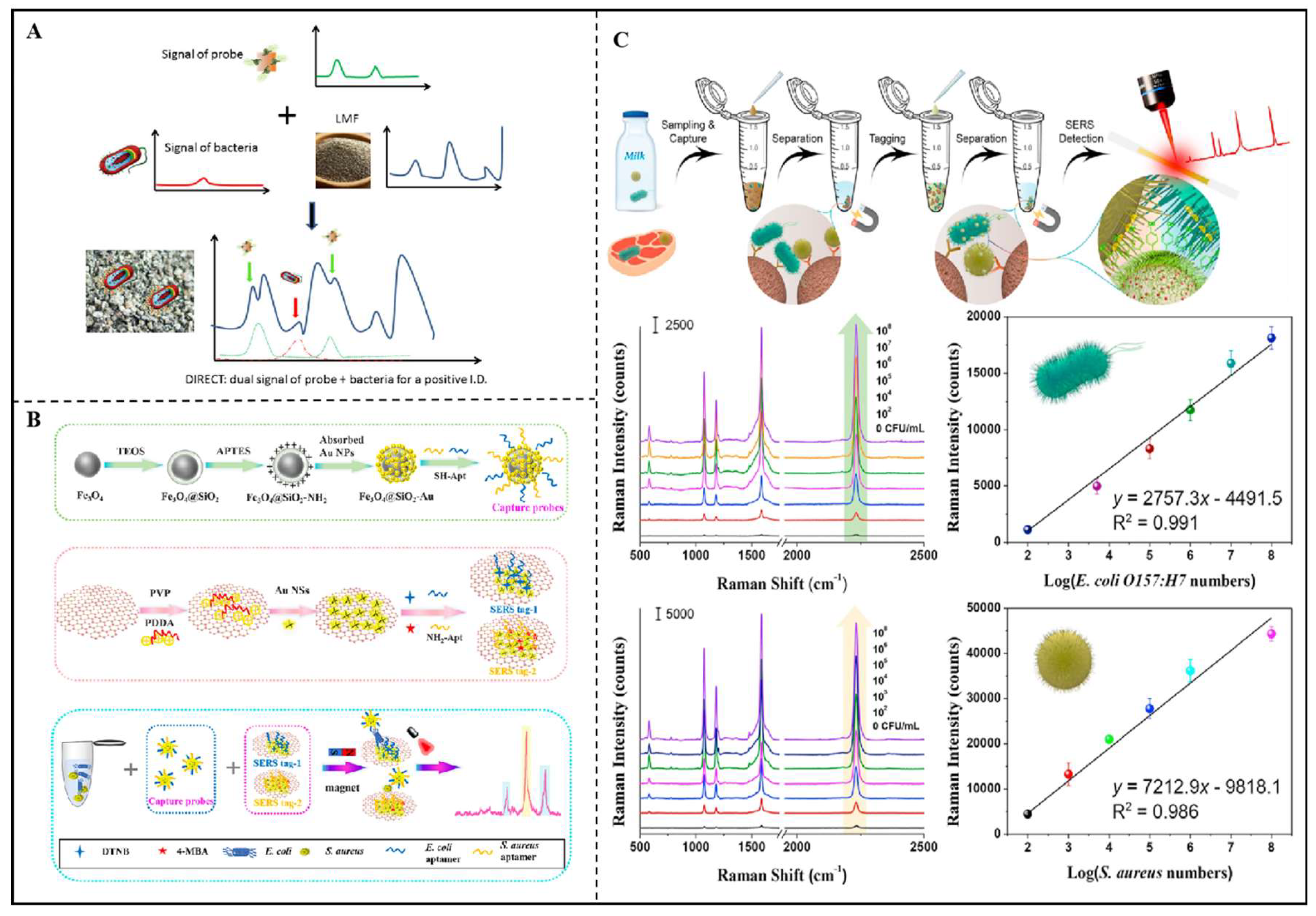
Figure 8.
(A) Scheme of the Dual Immunological Raman-Enabled Crosschecking Test assay, identification of peaks attributed to bacteria (red) and probes (green) yields positive detection of food contamination[26]. (B) Schematic diagram of preparation of magnetic capture probes for simultaneously detecting E. coli and S. aureus[165]. (C) Schematic illustration of foodborne bacteria assay with MPN-functionalized SERS tags[166].
4.1.2. Sensing Toward Fungi, Molds, and Their Toxins
SERS is a technique that utilizes metal nanostructures, such as silver and gold, to enhance Raman scattering signal. This technology significantly amplifies the intensity of Raman signal by adsorbing target molecules, such as patulin (PAT), onto the surface of metal nanoparticles. Ma et al.[122] used cysteine (Cys)-mediated nucleophilic addition reaction to detect PAT in apples. The SPR effect of Au@Ag generated a strong electromagnetic field under laser irradiation, which amplified Raman scattering signals of nearby molecules. The formed complex of PAT and Cys anchored to Au@Ag/4-ATP surface, leading to a decrease in Raman signal of 4-ATP (Figure 9A). The variability in signal was inversely correlated with PAT concentration of, allowing for quantitative detection of PAT. The introduction of chemometric algorithms could effectively extract hidden spectral information, improving the predictive performance. Olvera-Aripez et al.[167] used different filamentous fungi (e.g., Botrytis cinerea, Trichoderma atroviride, Trichoderma asperellum, Alternaria sp. and Ganoderma sessile) to synthesize AuNPs. In this respect, he SPR absorption of AuNPs is significantly boosted as it was close to the wavelength of the laser, thereby significantly amplifying the Raman scattered light. Using AuNPs synthesized by fungi, the mentioned SERS sensors achieved high-sensitivity assay of specific molecules by enhancing Raman signal. By selecting appropriate fungi and adjusting synthesis conditions, the optimal AuNPs provided a simple, low-cost green alternative pattern for designing SERS platform. In addition, Wei et al.[168] established a sensing platform based on SERS and fluorescence dual-signal for monitoring aflatoxin B1 (AFB1), in which CdTe quantum dots (QDs) were used as fluorescence signal source (Figure 9B). The used AuNPs served not only as sensing substrate for Raman enhancement but also as the acceptor for fluorescence resonance energy transfer (FRET). By combining quantum dot-decorated SiO2 with AuNPs, satellite nanostructures with high SERS and low fluorescence signals were formed. Furthermore, the surface plasmon resonance effect of AuNPs generated a local electromagnetic field enhancement on the of metal nanostructure surface. In the absence of AFB1, the FRET effect between QDs and AuNPs led to the quenching of fluorescence signals. When AFB1 was present, AuNPs could detach from QD-SNPs, resulting in the recovery of fluorescence signals and a decrease in SERS signals. The reported nanostructures improved Raman signal through LSPR and FRET effects, achieving high sensitivity for AFB1 assay. Using Raman signal molecules and fluorescent satellite nanostructures, the technology exhibited excellent sensitivity, accuracy, and stability, and could be applied in actual samples. Xie et al.[169] proposed a tri-mode lateral flow immunoassay based on tailored hollow porous gold nanoflowers (HPGN) for the sensitive detection of AFB1(Figure 9C). The unique nanostructure of HPGN, possessing hollow and porous nature, provided superior performance compared to traditional AuNPs. As a Raman reporter molecule, 4-MBA was fixed on HPGN surface through Au-S bonding, endowing HPGN with Raman activity. The work constructed an LFIA platform based on HPGN for high sensitivity and selectivity assay of AFB1 through a tri-mode strategy that congregating SERS activity, photothermal effect, and color change.
Yao et al.[154] proposed a dual-mode aptasensor based on electroactive and SERS-active Ag@Cu2O NPs for tetrodotoxin (TTX) detection (Figure 9D). The Ag@Cu2O core-shell nanostructures were prepared by depositing a Cu2O shell on Ag NPs surface. Ag NPs exhibited optimal plasmonic properties in the ultraviolet-visible region, while Cu2O NPs also showed good Raman properties due to the electron transfer process between Cu2O NPs and Raman molecules. The Cu2O shell, with its large dielectric constant, is beneficial for reducing the plasmon attenuation of Ag NPs and improving SERS performance. This dual-mode sensor not only retained the inherent advantages of a single mode but also provided two independent detection signals that, , greatly improving the accuracy of the detection results. The sensor was used for TTX detection in actual samples (fish meat), showing good recovery rates.
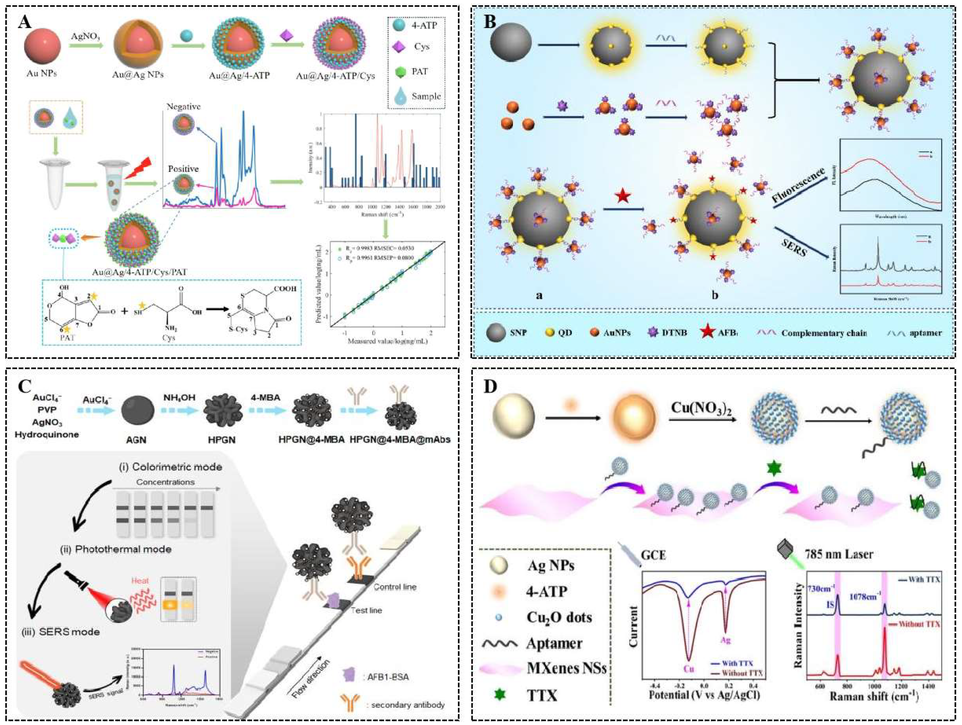
Figure 9.
(A) Schematic diagram of SERS toward patulin based on NAR-SERS substrate (Au@Ag/4-ATP/Cys)[122]. (B) Schematic diagram of designing Raman and fluorescence dual-mode aptasensing toward AFB1[168]. (C) Synthesis of HPGN@4-MBA@mAbs and its application in tri-mode LFIA of AFB1[169]. (D) Schematic Illustration of electroactive and SERS-Active Ag@Cu2O -engineered electrochemical/SERS dual-mode aptasensing toward tetrodotoxin[154].
4.1.3. Sensing Toward Viruses
Peng et al.[170] combined Ag@Au core-shell nanoparticles and hybridization chain reaction (HCR) to achieve highly sensitive detection of hepatitis C virus (HCV) nucleic acids (Figure 10A). Using strong SERS activity of AgNPs with chemical stability of AuNPs, a strong electromagnetic field was generated through surface plasmon resonance effects for Raman scattering signal enhancement. Upon binding of the target HCV nucleic acids to Ag@AuNPs, the HCR reaction was triggered, further enhancing SERS signal. By recording the signals with Raman spectrometer, high-sensitivity and selective detection of HCV nucleic acids was achieved. Y Park et al.[34] constructed a signal amplification system based on SERS for the detection of norovirus (NoV) (Figure 10B).The system employed Au/Ag core/shell satellite nanostructures, in which this structural design improved electromagnetic field intensity 4-MBA served as a Raman tag for signal output. By integrating magnetic nanoparticles (MNPs) with anti-NoV antibodies, a sandwich structure was formed to capture NoV, enabling concentration-dependent SERS signal detection. The system exhibited a detection limit of 0.76 fg/mL for NoV-like particles (NoV-LP), which was approximately 107 times more sensitive than commercial ELISA kits. This SERS-based system demonstrated the potential for highly sensitive detection of Norovirus. Wu et al.[171] synthesized silver nanoparticles with specific SPR properties by chemical reduction. These particles could generate a strong electromagnetic field under laser irradiation, enhancing Raman scattering signal of nearby molecules. By covering silver surface with gold shell to form core-shell structure (Ag@AuNPs), SERS signal was improved. Notably, methanol solvent was introduced into an aqueous system of silver nanoparticles containing iodide ions. Methanol acted as an anticoagulant to maintain the system stable at low temperatures and improved the nanoparticles’ ability to form “hotspots,” which was suitable for virus detection. By introducing the virus sample to SERS substrate, the interaction between virus molecules and nanoparticles could form “hotspots,” enhancing Raman signal of virus molecules. The study successfully obtained stable SERS signals of biomolecules at -20°C low-temperature conditions, which was particularly important for virus assay on cold chain food packaging (Figure 10C).
4.2. Nanostructure-Sensitized SERS toward Chemical Contamination in Food
The effective analysis of chemical contamination is an essential component of food safety, involving the assay of pesticides, veterinary drugs, and their metabolites in agricultural and livestock products[15]. Technologies[172,173,174]such as high-performance liquid chromatography (HPLC), gas chromatography (GC), liquid chromatography-mass spectrometry (LC-MS), and gas chromatography-mass spectrometry (GC-MS) have been widely applied in monitoring chemical contamination. Emerging detection technologies like SERS have garnered attention for their high sensitivity and rapid response capabilities. However, enhancing the sensitivity and selectivity of Raman pattern for certain low-concentration or structurally similar compounds remains a challenge. With the advancement of chemometrics and machine learning technologies, the data analysis capabilities of Raman spectroscopy have been strengthened, improving the assay accuracy[175,176]. More comprehensive Raman spectroscopy databases and standard libraries could support the identification and quantification of more compounds. The efficient sample pretreatment and analysis methods offered useful tools for simultaneous detection of multiple agrochemical and veterinary drug residues at ppt (part-per-trillion) level[89,177].
4.2.1. Sensing Toward Pesticide Residues
He et al.[178] proposed a droplet trapping band-based SERS pattern for r monitoring contaminants in food. Gold nanostructures (Au nanodendrites) with sharp edges and tips was exploited as sensing substrate by electrochemical deposition (Figure 11A), providing abundant SERS hotspot regions. A strip with specific micropores was designed to capture and enrich the target substances in microdroplets via capillary action, thereby amplifying Raman signal. The wettability difference between micropores and gold nanostructures allowed for rapid capture and fixation of microdroplets, facilitating direct sample collection from food surfaces. The large surface area and three-dimensional structure of gold nanostructures further amplified Raman signals, enabling high sensitivity and convenient assay of pesticide residues. Hu et al.[120] detailed SERS pattern for pesticide residue detection based on self-modeled mixed analysis (SMA). The high-density array of gold nanorods was used as SERS substrate via organic-water interfacial self-assembly method (Figure 11B). The SMA mode processed the spectral data of mixed pesticides to extract pure component spectra. By contacting pesticide residues on fruits’ surface with SERS substrate, the useful strategy exhibited good performance for rapid and non-destructive assay of mixed pesticide residues on fruit surfaces through the formation of hotspot regions. Wang et al.[84] described a assay pattern that used metal nanostructures to enhance Raman signals. The SERS substrate was designed by assembling gold cores on polystyrene microspheres, modifying the surface with a Raman internal standard (1,4-benzenedithiol), and subsequently growing a gold shell on this internal standard (Figure 11C). Three-dimensional hotspot regions were constructed, primarily located between the hydrophobic gold film and the composite particles, as well as between the particles and within the core-shell structure. The enhancement of SERS signal primarily arose from electromagnetic effect, where localized surface plasmon resonance occured when light frequency matched the oscillation frequency of electrons,. The hydrophobic interface facilitated the enrichment and capture of analytes. This mode demonstrated high sensitivity and stability in tracing pesticide residues on the surfaces of cucumbers, apples, and fish, s beneficial for low-cost and effective SERS platform toward various pesticide residues. Chen et al.[83] used golden decahedral nanoparticles (Au DNs) with special geometries as SERS active substratesby seed growth method, which made them produce multiple “hot spot” regions(Figure 11D). In the experiments, Au DNs were employed to detect six types of triazole pesticide residues on the surfaces of beans, apples, and vegetables, verifying significant potential for real sample assay with in-situ and multiplex pattern.
4.2.2. Sensing Toward Veterinary Drug Residues
Liang et al.[42] introduced spiky gold-silver nanoparticles (spiky Au@Ag NPs) and bismuth ditungstate (Bi2WO6) composite membranes as SERS platform, which were used for detecting a variety of pesticide and veterinary drug residues with high sensitivity (Figure 12A). Gold nanoparticles were synthesized through a chemical reduction method, and a silver shell was grown on Au surface, forming a spiky structure rich in “hotspot” regions for electromagnetic field enhancement. The incorporation of Bi2WO6 further improved SERS signal and promoted charge transfer from spiky Au@Ag NPs to target molecules through forming “donor-bridge-acceptor” system, thereby amplifying SERS signal. The composite film was validated for its high sensitivity in detecting pesticide and veterinary drug residues in real samples. Wang et al.[177] introduced a Janus-labeled AuNPs-based SERS technique combined with immunochromatographic assay (ICA) for monitoring veterinary drug residues (Figure 12B). The Janus-labeled Au nanoparticles had two functional regions: a monoclonal antibody for the specific recognition of target molecules and a DTNB molecule for SERS signal output. This design allowed the nanoparticles to produce a strong SERS signal when recognizing target molecules such as sulfamethazine (SM2). The precise fabrication of nanogaps created hotspot regions that enhanced SERS signal. By integrating ICA technology, this method achieved rapid, quantitative, and ultrasensitive detection of SM2 residues in milk. Experimental validation demonstrated that this method exhibited high sensitivity, easy to operate, and rapid results, indicating its good application prospects.
Hu et al.[179] described a magnetic Fe3O4-WO3-X@AuNPs sensing toward albendazole (ABZ) and streptomycin (SPT) in meat samples(Figure 12C). The prepared Fe3O4-WO3-X@AuNPs enhanced SERS signal through EM and CM, in which AuNPs provided EM enhancement and WO3-X contributed to CT enhancement. The recognition mechanism between the sensor and target molecules was investigated through charge transfer model. The magnetic properties of Fe3O4-WO3-X@AuNPs allowed for rapid separation and enrichment of target molecules, reducing interference from the sample matrix and improving the accuracy of SERS analysis. The experimental results indicated that this method held promising applications for the rapid, sensitive, and selective assay of veterinary drug residues in meat, well qualified for monitoring food safety. Li et al.[41] introduced a dual-mode pattern to construct AuNPs/Cu-TCPP(Fe) nanosheets with peroxidase activity and SERS effect, which combined with colorimetric and SERS sensing toward veterinary drug residues. The strategy was applied to detect LEV in actual milk sampleswith good recovery rates and stability. Tu et al.[180] introduced a SERS label (MDAu@Ag) to enable the simultaneous detection of multiple veterinary drug residues in complex samples (Figure 12D). The nanostructure tag consisted of three components: a 200 nm Fe3O4 magnetic core with strong magnetic responsiveness; a 1 nm PEI interlayer for built-in nanogaps and multiple efficient hotspots; and two layers of Raman reporter molecules (DTNB and MBA) for SERS signal amplification. The application of MDAu@Ag in LFA system allowed for the direct capture of target drug residues from real food samples, thus eliminating cumbersome pretreatment process and improving assay sensitivity. Additionally, spiking recovery experiment was conducted to validate the reliability of SERS pattern, showcasing strong potential for practical applications.
4.2.3. Sensing Toward Heavy Metals
Heavy metal ions are easy to enter food and drinking water through natural environmental pollution, industrial emission, and chemical fertilizer[181,182] Owing to the high toxicity, easy enrichment, difficult degradation of heavy metals, it is necessary to develop effective strategy toward heavy metal monitoring[183,184]. Li et al.[123] used Au@Ag NPs and covalent organic frameworks (COFs) complexes as SERS substrates (Figure 13A). Au@Ag NPs generated strong electromagnetic fields (E-fields) on COFs surface, in which the large specific surface area and high adsorption capacity of COFs contributed to E-fields enhancement for SERS signal amplification. Y-shaped DNA (containing two embedded probes, Y1 and Y2) was used to shorten the distance between Raman tag and SERS substrate, thereby enhancing the coupling effect of E-fields and improving assay sensitivity. The sensor exhibited good recovery for monitoring Hg2+ in real samples. Chen et al.[185] used the functional AgNPs to trace Mn2+ (Figure 13B). In the presence of Mn2+, MNA-MA-AgNPs aggregated and then reduced the distance between noble metal particles, thereby enhancing the SERS signal. This enhancement behavior was due to the so-called “gap hotspots effect”, in which the intensity of the electromagnetic field increased obviously.[186]
Mohamed[187] applied the special structure and SERS activity of helical CNTs to achieve high sensitivity and selectivity for detecting heavy metal ions (Hg2+/Cd2+) in water. The formed structure of spiral CNTs on the screw nail surface increased the number of SERS-active hot spots,. The chemical modification of CNTs could improve their adsorption capacity for metal ions through chemical bonding. Dong et al.[137] proposed a novel magnetic SERS tag (Fe@RAu) for ultrasensitive immunochromatographic detection (ICA) to monitor cadmium ions (Cd2+) in complex samples. The combination of a large Fe3O4 core and a coarse gold shell provided a robust magnetic response and high SERS activity (Figure 13C), enabling a fast, low-cost sensing toward heavy metal contamination in aqueous environment.
4.2.4. Sensing Toward Food Additives and Illicit Adulterants
Food additives are a type of natural or chemical synthetic substances to improve the food flavor, texture, and storability. The normal use within the permitted range does not cause harm to humans; but exceeding usage limit, it can be toxic and carcinogenic effects on humans[188]. Hence, it is important to develop rapid screening technology for food additives. Ramachandran et al.[189] exploited the special structure of ZnO nanorods and Ag nanotriangles to achieve high sensitivity and selectivity for detecting food additive 2,6-di-tert-butyl-p-hydroxytoluene (BHT) through synergistic effect of CT and EM effects. ZnO nanorods, grown on stainless steel substrate via hydrothermal synthesis, possessed high surface-to-volume ratio, chemical activity, and the ability to directly transfer charge carriers. Ag nanotriangles were sensitized on the surface of ZnO nanorods to boost electromagnetic field coupling and amplification of local fields, thereby achieving a multi-fold sensitivity increase in sensitivity. The junctions between ZnO nanorods and Ag nanotriangles promoted charge transfer and the enhancement of electromagnetic fields, holding significant importance for ensuring food safety.
Li et al.[107] applied precious metal nanostructures such as gold and silver as SERS substrates for the detection of trace additives and contaminants in food. By controlling the shape, size, and arrangement of metal nanostructures, the effect of local electromagnetic fields could be enhanced. Modification of specific molecules or ligands onto metal nanostructures could improve selectivity and sensitivity for the analytes, in which surface plasmon resonance occurred when irradiated with specific light wavelengths for Raman signal output. Lu et al.[190] designed the heterostructure (AgNTs/TNA) composed of silver triangle nanoparticles (AgNTs) and TiO2 nanorod arrays (TNA) as the active SERS substrate toward sensing food additives.. AgNTs, with their sharp edges, strongly improved the local electromagnetic field for creating effective hotspots. TNA contributed to forming strong electromagnetic enhancement areas. The combination of AgNTs and TNA provided both electromagnetic and chemical mechanism effects. Additionally, AgNTs/TNA heterostructure could generate electrons and holes that reacted with oxygen to produce superoxide radicals, oxidizing and breaking down probe molecules, allowing for substrate regeneration and reusability.
Hu et al.[87] utilized a pNIPAM@Au NRs (poly(N-isopropylacrylamide)@Au NRs) nanocomposite with temperature-controlled, tunable plasma behavior and long-term stability as SERS substrate (Figure 14A). Under laser irradiation of a specific wavelength, gold nanorods could act as photothermal converters to quickly generate local heating, triggering the phase transition of pNIPAM. By controlling the loading of Au NRs on pNIPAM template, the optical properties and SERS performance were improved to detect the illicit additive Malachite Green (MG) in aquatic products (Figure 14B). Under the optimal the temperature and laser wavelength, the sensitive detection of MG was achieved with detection limit of 0.73 ng g-1 (Figure 14C), which met the minimum required performance limit (MRPL) set by the European Commission. Xu et al.[191] exploited femtosecond laser-induced plasma-assisted ablation (fs LIPAA) to prepare SERS substrate for monitoring contaminants such as melamine in food. Silicon nanoparticles were generated on glass substrates by fs LIPAA technology, and then silver thin films (Ag NPs/glass) were deposited on them by electron beam evaporator to form hot spots with high uniformity. Wang et al.[192] achieved a highly sensitive detection of PAE plasticizers in food additives by designing a specific combination of two-dimensional silver plates and nano-silver sols(Figure 14D). This silver plate provided a homogeneous metal surface that amplified Raman signal of sample molecules. The sol of silver nanoparticles was used as synergistic agent with 2D silver plates to further enhance Raman scattering. The addition of silver nanoparticles increases the number of hot spots in the system, thereby increasing the intensity of the Raman signal. Li et al.[107] utilized a portable Raman spectrometer (Figure 14E) to compare the changes in Raman spectral characteristic peaks of pure persimmon cakes and adulterated persimmon cakes; machine learning was employed for rapid qualitative assay of titanium dioxide adulteration on the surface of persimmon cakes. This method was not only non-destructive but also highly accurate, making it suitable for on-site analysis.
4.3. Nanostructure-Sensitized SERS Toward Physical Contamination in Food
Physical contamination in food refers to the pollution of food by non-biological and non-chemical substances, which may affect the safety and quality of food and even pose a threat to human health. Table 3 lists some of the common types of physical contamination found in food.
Microplastic, as a kind of plastic particles smaller than 5 mm but larger than 0.1 µm, is considered as a threat to foods and beverages. Identification of microplastic particles from complex matrices can be challenging due to their various sizes, shapes, and polymer types. Yu et al.[17] exploited a liquid-liquid interface strategy to assemble micro-/nanoplastics and gold nanoparticles (GNPs) into dense and uniform plasma arrays (Figure 15A), thereby enabling rapid and sensitive detection of trace nanoplastics . To be special, Ethyl acetate was selected as the organic phase to participate in self-assembly process due to its polarity being close to that of water, which helpd to accelerate the self-assembly process and produce the strongest SERS signals. The presence of micro-/nanoplastics effectively promoted the self-assembly process of GNPs at oil-water interface, increasing the number of molecules in hot spots for SERS signal amplification. The aromatic rings in micro-/nanoplastics interacted strongly with the gold nano-surface through π-metal interactions, promoting the adsorption of more micro-/nanoplastics at liquid-liquid interface. The PCA algorithm was utilized to process SERS data to distinguish and identify various micro-/nanoplastics components in aquatic environments as well as in edible oils (Figure 15B).
Song et al.[62] took advantage of the difference between fish bones and fish meat on Raman spectra, and achieved high sensitivity and high accuracy for discerning fish bones in fish meat (Figure 15C). The Fuzzy Rough Set Model (FRSTCA) based on Thermoelectric Charge Algorithm was used to select the optimal wavelength, and the Support Vector Data Description (SVDD) model was established for the detection of fishbones. Spectral Angle Mapping (SAM) images were converted to binary images for distinguishing fishbone pixels and backgrounds. By calculating the number of pixels of each component in the binary image and correlating it with the actual component concentration, the quantitative detection of fish bones was realized. This technology could effectively detect fish bones with a depth of less than 2.5 mm, and the assay accuracy reached 90.5%, provided a useful technical means for food safety testing.
5. Conclusions
In this review, we discuss the important role of nanostructures in enhancing Raman signals, in particular the application of nanostructure-sensitized SERS in food safety evaluation. By analyzing the latest research findings, this review summarizes the critical role of nanostructures in SERS technology and how they can help improve the efficiency and accuracy. In addition, nanostructures has also promoted the application of SERS technology in conjunction with other assay technologies, providing an efficient and low-cost solution for food safety testing. The application of nanostructures in SERS technology not only improves the efficiency and accuracy of food safety assay, but also provides new ideas and directions for the development of food safety evaluation technology in the future. With the continuous progress of nanostructure technology and the continuous improvement of SERS technology, its application prospect in food safety testing will be broader.
Author Contributions
Z.L. is the leading author and prepared this review paper. R.Y. and H.C. provided critical feedback. X.Z. provided critical feedback and revised this review. All authors have read and agreed to the published version of the manuscript.
Funding
This research received no external funding.
Acknowledgments
Thank you to my mentors and fellow students for their support.
Conflicts of Interest
The authors declare no conflict of interest.
References
- W. Jiang, Q. Wang, et al., Surface-enhanced Raman spectroscopy substrates for monitoring antibiotics in dairy products: Mechanisms, advances, and prospects[J], Comprehensive Reviews in Food Science and Food Safety (2024) 23(6). [CrossRef]
- L. Wu, Y. Li, et al., A dual-mode optical sensor for sensitive detection of saxitoxin in shellfish based on three-in-one functional nanozymes[J], Journal of Food Composition and Analysis (2024) 130. [CrossRef]
- G. Wu, H. Qiu, et al., Nanomaterials-based fluorescent assays for pathogenic bacteria in food-related matrices[J], Trends in Food Science & Technology (2023) 142. [CrossRef]
- S. Liu, Y. Lou, et al., Aroma characteristics of volatile compounds brought by variations in microbes in winemaking[J], Food Chemistry (2023) 420. [CrossRef]
- S.-H. Song, Z.-F. Gao, et al., Aptamer-Based Detection Methodology Studies in Food Safety[J], Food Analytical Methods (2019) 12(4) 966-990. [CrossRef]
- S. Liu, Y. Zhu, et al., Characteristic substance analysis and rapid detection of bacteria spores in cooked meat products by surface enhanced Raman scattering based on Ag@AuNP array substrate[J], Analytica Chimica Acta (2024) 1308. [CrossRef]
- Z. Guo, M. Wang, et al., Label-free surface enhanced Raman scattering spectroscopy for discrimination and detection of dominant apple spoilage fungus[J], International Journal of Food Microbiology (2021) 338. [CrossRef]
- O.M. Atta, S. Manan, et al., Biobased materials for active food packaging: A review[J], Food Hydrocolloids (2022) 125. [CrossRef]
- M.M. Nasiru, E.B. Frimpong, et al., Dielectric barrier discharge cold atmospheric plasma: Influence of processing parameters on microbial inactivation in meat and meat products[J], Comprehensive Reviews in Food Science and Food Safety (2021) 20(3) 2626-2659. [CrossRef]
- Y. Xu, W. Zhang, et al., Impedimetric aptasensor based on highly porous gold for sensitive detection of acetamiprid in fruits and vegetables[J], Food Chemistry (2020) 322. [CrossRef]
- Z. Guo, P. Chen, et al., Detection of Heavy Metals in Food and Agricultural Products by Surface-enhanced Raman Spectroscopy[J], Food Reviews International (2021) 39(3) 1440-1461. [CrossRef]
- Z. Guo, P. Chen, et al., Determination of lead in food by surface-enhanced Raman spectroscopy with aptamer regulating gold nanoparticles reduction[J], Food Control (2022) 132. [CrossRef]
- Y. Rong, S. Ali, et al., Development of a bimodal sensor based on upconversion nanoparticles and surface-enhanced Raman for the sensitive determination of dibutyl phthalate in food[J], Journal of Food Composition and Analysis (2021) 100. [CrossRef]
- X. Zhang, Y. Zhou, et al., Switchable aptamer-fueled colorimetric sensing toward agricultural fipronil exposure sensitized with affiliative metal-organic framework[J], Food Chemistry (2023) 407. [CrossRef]
- W. Wu, W. Ahmad, et al., An upconversion biosensor based on inner filter effect for dual-role recognition of sulfadimethoxine in aquatic samples[J], Food Chemistry (2024) 437. [CrossRef]
- Z. Yue, X. Liu, et al., Reducing microplastics in tea infusions released from filter bags by pre-washing method: Quantitative evidences based on Raman imaging and Py-GC/MS[J], Food Chemistry (2024) 445. [CrossRef]
- F. Yu, C. Qu, et al., Liquid Interfacial Coassembly of Plasmonic Arrays and Trace Hydrophobic Nanoplastics in Edible Oils for Robust Identification and Classification by Surface-Enhanced Raman Spectroscopy[J], Journal of Agricultural and Food Chemistry (2023) 71(39) 14342-14350. [CrossRef]
- Y. Shi, L. Yi, et al., Visual characterization of microplastics in corn flour by near field molecular spectral imaging and data mining[J], Science of The Total Environment (2023) 862. [CrossRef]
- C. Xie, W. Zhou, A Review of Recent Advances for the Detection of Biological, Chemical, and Physical Hazards in Foodstuffs Using Spectral Imaging Techniques[J], Foods (2023) 12(11). [CrossRef]
- S. Liu, M. Zhang, et al., Multifunctional Metal–Organic Frameworks Driven Three-Dimensional Folded Paper-Based Microfluidic Analysis Device for Chlorpyrifos Detection[J], Journal of Agricultural and Food Chemistry (2024) 72(25) 14375-14385. [CrossRef]
- Y. Zou, Y. Shi, et al., Quantum dots as advanced nanomaterials for food quality and safety applications: A comprehensive review and future perspectives[J], Comprehensive Reviews in Food Science and Food Safety (2024) 23(3). [CrossRef]
- M. Mehedi Hassan, P. He, et al., Rapid detection and prediction of chloramphenicol in food employing label-free HAu/Ag NFs-SERS sensor coupled multivariate calibration[J], Food Chemistry (2022) 374. [CrossRef]
- Z. Dong, J. Lu, et al., Antifouling molecularly imprinted membranes for pretreatment of milk samples: Selective separation and detection of lincomycin[J], Food Chemistry (2020) 333. [CrossRef]
- J. Mei, F. Zhao, et al., A review on the application of spectroscopy to the condiments detection: from safety to authenticity[J], Critical Reviews in Food Science and Nutrition (2021) 62(23) 6374-6389. [CrossRef]
- Y. Zhang, M.M. Hassan, et al., A solid-phase capture probe based on upconvertion nanoparticles and inner filter effect for the determination of ampicillin in food[J], Food Chemistry (2022) 386. [CrossRef]
- C. Pan, B. Zhu, et al., A Dual Immunological Raman-Enabled Crosschecking Test (DIRECT) for Detection of Bacteria in Low Moisture Food[J], Biosensors (2020) 10(12). [CrossRef]
- H. Wang, L. Zhu, et al., Recent Advances in Nanomaterials-Based Sensing Platforms for the Determination of Multiple Bacterial Species: A Minireview[J], Analytical Letters (2023) 57(6) 920-939. [CrossRef]
- S. Wu, J.P. Hulme, Recent Advances in the Detection of Antibiotic and Multi-Drug Resistant Salmonella: An Update[J], International Journal of Molecular Sciences (2021) 22(7). [CrossRef]
- S. Wu, N. Duan, et al., Surface-enhanced Raman spectroscopic–based aptasensor for Shigella sonnei using a dual-functional metal complex-ligated gold nanoparticles dimer[J], Colloids and Surfaces B: Biointerfaces (2020) 190. [CrossRef]
- Y. Wang, Z. Hu, et al., Non-thermal Microbial Inactivation of Honey Raspberry Wine Through the Application of High-Voltage Electrospray Technology[J], Food and Bioprocess Technology (2022) 15(1) 177-189. [CrossRef]
- C. Zhang, L. Huang, et al., Magnetic surface-enhanced Raman scattering (MagSERS) biosensors for microbial food safety: Fundamentals and applications[J], Trends in Food Science & Technology (2021) 113 366-381. [CrossRef]
- R. Lu, Z. Liu, et al., Nitric Oxide Enhances Rice Resistance to Rice Black-Streaked Dwarf Virus Infection[J], Rice (2020) 13(1). [CrossRef]
- Y. Xie, X. Du, et al., Seasonal occurrence and abundance of norovirus in pre- and postharvest lettuce samples in Nanjing, China[J], Lwt (2021) 152. [CrossRef]
- E.Y. Park, S. Maehata, et al., Signal-amplified surface-enhanced Raman scattering using core/shell satellite nanoparticles for norovirus detection[J], Microchimica Acta (2024) 191(9). [CrossRef]
- M. Song, I.M. Khan, et al., Research Progress of Optical Aptasensors Based on AuNPs in Food Safety[J], Food Analytical Methods (2021) 14(10) 2136-2151. [CrossRef]
- H. Zhang, N.A. Serwah Boateng, et al., Unravelling the fruit microbiome: The key for developing effective biological control strategies for postharvest diseases[J], Comprehensive Reviews in Food Science and Food Safety (2021) 20(5) 4906-4930. [CrossRef]
- M.M. Hassan, X. Yi, et al., Recent advancements of optical, electrochemical, and photoelectrochemical transducer-based microfluidic devices for pesticide and mycotoxins in food and water[J], Trends in Food Science & Technology (2023) 142. [CrossRef]
- Z. Guo, C. Guo, et al., Identification of the apple spoilage causative fungi and prediction of the spoilage degree using electronic nose[J], Journal of Food Process Engineering (2021) 44(10). [CrossRef]
- X. Shi, C. He, et al., Mo-doped Co LDHs as Raman enhanced substrate for detection of roxarsine[J], Analytica Chimica Acta (2024) 1318. [CrossRef]
- M. Liang, G. Zhang, et al., Paper-Based Microfluidic Chips for Food Hazard Factor Detection: Fabrication, Modification, and Application[J], Foods (2023) 12(22). [CrossRef]
- C. Li, M. Song, et al., Selection of aptamer targeting levamisole and development of a colorimetric and SERS dual-mode aptasensor based on AuNPs/Cu-TCPP(Fe) nanosheets[J], Talanta (2023) 251. [CrossRef]
- D. Liang, Y. Xu, et al., Plasmonic metal NP-bismuth composite film with amplified SERS activity for multiple detection of pesticides and veterinary drugs[J], Chemical Engineering Journal (2023) 474. [CrossRef]
- H. Li, M. Mehedi Hassan, et al., Investigation of nonlinear relationship of surface enhanced Raman scattering signal for robust prediction of thiabendazole in apple[J], Food Chemistry (2021) 339. [CrossRef]
- T. Jiao, M. Mehedi Hassan, et al., Quantification of deltamethrin residues in wheat by Ag@ZnO NFs-based surface-enhanced Raman spectroscopy coupling chemometric models[J], Food Chemistry (2021) 337. [CrossRef]
- M. Marimuthu, K. Xu, et al., Safeguarding food safety: Nanomaterials-based fluorescent sensors for pesticide tracing[J], Food Chemistry (2025) 463. [CrossRef]
- M. Li, X. Hong, et al., Ultrasensitive monitoring strategy of PCR-like levels for zearalenone contamination based DNA barcode[J], Journal of the Science of Food and Agriculture (2021) 101(11) 4490-4497. [CrossRef]
- J. Wang, W. Ahmad, et al., Landing microextraction sediment phase onto surface enhanced Raman scattering to enhance sensitivity and selectivity for chromium speciation in food and environmental samples[J], Food Chemistry (2020) 323. [CrossRef]
- M.M. Hassan, W. Ahmad, et al., Rapid detection of mercury in food via rhodamine 6G signal using surface-enhanced Raman scattering coupled multivariate calibration[J], Food Chemistry (2021) 358. [CrossRef]
- W. Lu, X. Dai, et al., Fenton-like catalytic MOFs driving electrochemical aptasensing toward tracking lead pollution in pomegranate fruit[J], Food Control (2025) 169. [CrossRef]
- A.G. Mukherjee, K. Renu, et al., Heavy Metal and Metalloid Contamination in Food and Emerging Technologies for Its Detection[J], Sustainability (2023) 15(2). [CrossRef]
- X. Wang, X. Hu, et al., A simple and sensitive electrochemical sensing based on amine-functionalized metal–organic framework and polypyrrole composite for detection of lead ions in meat samples[J], Journal of Food Measurement and Characterization (2024) 18(7) 5813-5825. [CrossRef]
- X. Huang, C. Huang, et al., Allosteric switch for electrochemical aptasensor toward heavy metals pollution of Lentinus edodes sensitized with porphyrinic metal-organic frameworks[J], Analytica Chimica Acta (2023) 1278. [CrossRef]
- A.N. Dominguez, L.E. Jimenez, et al., Rapid detection of pyraclostrobin fungicide residues in lemon with surface-enhanced Raman spectroscopy[J], Journal of Food Measurement and Characterization (2023) 17(6) 6350-6362. [CrossRef]
- D. Balram, K.-Y. Lian, et al., Ultrasensitive detection of cytotoxic food preservative tert-butylhydroquinone using 3D cupric oxide nanoflowers embedded functionalized carbon nanotubes[J], Journal of Hazardous Materials (2021) 406. [CrossRef]
- F. Sun, P. Li, et al., Carbon nanomaterials-based smart dual-mode sensors for colorimetric and fluorescence detection of foodborne hazards[J], Trends in Food Science & Technology (2024) 152. [CrossRef]
- M.M. Hassan, Y. Xu, et al., Recent advances of nanomaterial-based optical sensor for the detection of benzimidazole fungicides in food: a review[J], Critical Reviews in Food Science and Nutrition (2021) 63(16) 2851-2872. [CrossRef]
- J. Li, M. Li, et al., Necklace-like Te-Au reticula platform with three dimensional hotspots Surface-Enhanced Raman Scattering (SERS) sensor for food hazards analysis[J], Spectrochimica Acta Part A: Molecular and Biomolecular Spectroscopy (2024) 311. [CrossRef]
- Y. Zhao, X. Jing, et al., Surface-Enhanced Raman Scattering-Active Plasmonic Metal Nanoparticle-Persistent Luminescence Material Composite Films for Multiple Illegal Dye Detection[J], Analytical Chemistry (2021) 93(25) 8945-8953. [CrossRef]
- E.S. Okeke, T.P.C. Ezeorba, et al., Analytical detection methods for azo dyes: A focus on comparative limitations and prospects of bio-sensing and electrochemical nano-detection[J], Journal of Food Composition and Analysis (2022) 114. [CrossRef]
- N. Rong, S. He, et al., Coupled magnetic nanoparticle-mediated isolation and single-cell image recognition to detect Bacillus’ cell size in soil[J], European Journal of Soil Science (2022) 73(3). [CrossRef]
- J. Dai, M. Bai, et al., Advances in the mechanism of different antibacterial strategies based on ultrasound technique for controlling bacterial contamination in food industry[J], Trends in Food Science & Technology (2020) 105 211-222. [CrossRef]
- S. Song, Z. Liu, et al., Detection of fish bones in fillets by Raman hyperspectral imaging technology[J], Journal of Food Engineering (2020) 272. [CrossRef]
- Y. Sun, N. Zhang, et al., Competitive immunosensor for sensitive and optical anti-interference detection of imidacloprid by surface-enhanced Raman scattering[J], Food Chemistry (2021) 358. [CrossRef]
- D. Thapliyal, M. Karale, et al., Current Status of Sustainable Food Packaging Regulations: Global Perspective[J], Sustainability (2024) 16(13). [CrossRef]
- N.R. Abdul Halim, H. Hashim, et al., Food safety regulations implementation and their impact on food security level in Malaysia: A review[J], International Food Research Journal (2024) 31(1) 20-31. [CrossRef]
- A.-A. Cioca, L. Tušar, et al., Food Risk Analysis: Towards a Better Understanding of “Hazard” and “Risk” in EU Food Legislation[J], Foods (2023) 12(15). [CrossRef]
- F.J. Díaz-Galiano, M. Murcia-Morales, et al., Economic poisons: A review of food contact materials and their analysis using mass spectrometry[J], TrAC Trends in Analytical Chemistry (2024) 172. [CrossRef]
- Y.-X. Gu, T.-C. Yan, et al., Recent developments and applications in the microextraction and separation technology of harmful substances in a complex matrix[J], Microchemical Journal (2022) 176. [CrossRef]
- E.D. Tsochatzis, J. Alberto Lopes, et al., Development and validation of a multi-analyte GC-MS method for the determination of 84 substances from plastic food contact materials[J], Analytical and Bioanalytical Chemistry (2020) 412(22) 5419-5434. [CrossRef]
- X. Zhang, J. Yang, et al., A facile molecularly imprinted column coupled to GC-MS/MS for sensitive and selective determination of polycyclic aromatic hydrocarbons and study on their migration in takeaway meal boxes[J], Talanta (2022) 243. [CrossRef]
- J. Wang, Q. Chen, et al., Insights into chemometric algorithms for quality attributes and hazards detection in foodstuffs using Raman/surface enhanced Raman spectroscopy[J], Comprehensive Reviews in Food Science and Food Safety (2021) 20(3) 2476-2507. [CrossRef]
- H. Jiang, Y. He, et al., Quantitative Detection of Acid Value During Edible Oil Storage by Raman Spectroscopy: Comparison of the Optimization Effects of BOSS and VCPA Algorithms on the Characteristic Raman Spectra of Edible Oils[J], Food Analytical Methods (2021) 14(9) 1826-1835. [CrossRef]
- Z. Chen, Y. Sun, et al., Facile synthesis of Au@Ag core–shell nanorod with bimetallic synergistic effect for SERS detection of thiabendazole in fruit juice[J], Food Chemistry (2022) 370. [CrossRef]
- R. Fakhlaei, A.A. Babadi, et al., Application, challenges and future prospects of recent nondestructive techniques based on the electromagnetic spectrum in food quality and safety[J], Food Chemistry (2024) 441. [CrossRef]
- A. Hassane Hamadou, J. Zhang, et al., Modulating the glycemic response of starch-based foods using organic nanomaterials: strategies and opportunities[J], Critical Reviews in Food Science and Nutrition (2022) 63(33) 11942-11966. [CrossRef]
- H. Li, W. Sheng, et al., Recent advances in rare earth ion-doped upconversion nanomaterials: From design to their applications in food safety analysis[J], Comprehensive Reviews in Food Science and Food Safety (2023) 22(5) 3732-3764. [CrossRef]
- X. Zhang, Z. Wang, et al., H-Bond Modulation Mechanism for Moisture-driven Bacteriostat Evolved from Phytochemical Formulation[J], Advanced Functional Materials (2023) 34(13). [CrossRef]
- X. Zhang, Z. Wang, et al., Competitive electrochemical sensing for cancer cell evaluation based on thionine-interlinked signal probes[J], The Analyst (2023) 148(4) 912-918. [CrossRef]
- X. Zhang, Y. Zhou, et al., Energy difference-driven ROS reduction for electrochemical tracking crop growth sensitized with electron-migration nanostructures[J], Analytica Chimica Acta (2024) 1304. [CrossRef]
- J. Liu, X. Zhang, et al., Multiomics analysis reveals that peach gum colouring reflects plant defense responses against pathogenic fungi[J], Food Chemistry (2022) 383. [CrossRef]
- J. Qiu, H. Gu, et al., A diverse Fusarium community is responsible for contamination of rice with a variety of Fusarium toxins[J], Food Research International (2024) 195. [CrossRef]
- M.M. Hassan, M. Zareef, et al., SERS based sensor for mycotoxins detection: Challenges and improvements[J], Food Chemistry (2021) 344. [CrossRef]
- Z. Chen, R. Tan, et al., SERS detection of triazole pesticide residues on vegetables and fruits using Au decahedral nanoparticles[J], Food Chemistry (2024) 439. [CrossRef]
- Y.-H. Wang, C. Huang, et al., 3D hot spot construction on the hydrophobic interface with SERS tags for quantitative detection of pesticide residues on food surface[J], Food Chemistry (2025) 463. [CrossRef]
- X. Zhang, X. Huang, et al., Bioinspired nanozyme enabling glucometer readout for portable monitoring of pesticide under resource-scarce environments[J], Chemical Engineering Journal (2022) 429. [CrossRef]
- X. Zhang, Z. Wang, et al., Uniform stain pattern of robust MOF-mediated probe for flexible paper-based colorimetric sensing toward environmental pesticide exposure[J], Chemical Engineering Journal (2023) 451. [CrossRef]
- B. Hu, D.-W. Sun, et al., A dynamically optical and highly stable pNIPAM @ Au NRs nanohybrid substrate for sensitive SERS detection of malachite green in fish fillet[J], Talanta (2020) 218. [CrossRef]
- K. Chao, S. Dhakal, et al., Raman and IR spectroscopic modality for authentication of turmeric powder[J], Food Chemistry (2020) 320. [CrossRef]
- B. Li, S. Liu, et al., Nanohybrid SERS substrates intended for food supply chain safety[J], Coordination Chemistry Reviews (2023) 494. [CrossRef]
- L. Wang, X. Huang, et al., Characterization of the volatile flavor profiles of Zhenjiang aromatic vinegar combining a novel nanocomposite colorimetric sensor array with HS-SPME-GC/MS[J], Food Research International (2022) 159. [CrossRef]
- A. Ali, E.E. Nettey-Oppong, et al., Miniaturized Raman Instruments for SERS-Based Point-of-Care Testing on Respiratory Viruses[J], Biosensors (2022) 12(8). [CrossRef]
- R. Beeram, K.R. Vepa, et al., Recent Trends in SERS-Based Plasmonic Sensors for Disease Diagnostics, Biomolecules Detection, and Machine Learning Techniques[J], Biosensors (2023) 13(3). [CrossRef]
- J. Chen, H. Lin, et al., Improving the detection accuracy of the dual SERS aptasensor system with uncontrollable SERS “hot spot” using machine learning tools[J], Analytica Chimica Acta (2024) 1307. [CrossRef]
- M. Usman, J.-W. Tang, et al., Recent advances in surface enhanced Raman spectroscopy for bacterial pathogen identifications[J], Journal of Advanced Research (2023) 51 91-107. [CrossRef]
- L. Lin, X. Bi, et al., Surface-enhanced Raman scattering nanotags for bioimaging[J], Journal of Applied Physics (2021) 129(19). [CrossRef]
- M. Petersen, Z. Yu, et al., Application of Raman Spectroscopic Methods in Food Safety: A Review[J], Biosensors (2021) 11(6). [CrossRef]
- Y. Wu, Y. Wang, et al., Surface-enhanced Raman scattering-based strategies for tumor markers detection: A review[J], Talanta (2024) 280. [CrossRef]
- X. Bi, D.M. Czajkowsky, et al., Digital colloid-enhanced Raman spectroscopy by single-molecule counting[J], Nature (2024) 628(8009) 771-775. [CrossRef]
- L. Cai, G. Fang, et al., Label-Free Surface-Enhanced Raman Spectroscopic Analysis of Proteins: Advances and Applications[J], International Journal of Molecular Sciences (2022) 23(22). [CrossRef]
- R. Pilot, R. Signorini, et al., A Review on Surface-Enhanced Raman Scattering[J], Biosensors (2019) 9(2). [CrossRef]
- F. Pisano, M. Masmudi-Martín, et al., Vibrational fiber photometry: label-free and reporter-free minimally invasive Raman spectroscopy deep in the mouse brain[J], Nature Methods (2024). [CrossRef]
- Harshita, H.F. Wu, et al., Recent advances in nanomaterials-based optical sensors for detection of various biomarkers (inorganic species, organic and biomolecules)[J], Luminescence (2022) 38(7) 954-998. [CrossRef]
- Y. Zhao, On the Measurements of the Surface-Enhanced Raman Scattering Spectrum: Effective Enhancement Factor, Optical Configuration, Spectral Distortion, and Baseline Variation[J], Nanomaterials (2023) 13(23). [CrossRef]
- E.C. Le Ru, B. Auguié, Enhancement Factors: A Central Concept during 50 Years of Surface-Enhanced Raman Spectroscopy[J], ACS Nano (2024) 18(14) 9773-9783. [CrossRef]
- T. Guan, H. Yang, et al., Storage period affecting dynamic succession of microbiota and quality changes of strong-flavor Baijiu Daqu[J], Lwt (2021) 139. [CrossRef]
- W. Zhang, A. Wang, et al., Nondestructive measurement of kiwifruit firmness, soluble solid content (SSC), titratable acidity (TA), and sensory quality by vibration spectrum[J], Food Science & Nutrition (2020) 8(2) 1058-1066. [CrossRef]
- J. Li, L. Zhang, et al., Rapid qualitative detection of titanium dioxide adulteration in persimmon icing using portable Raman spectrometer and Machine learning[J], Spectrochimica Acta Part A: Molecular and Biomolecular Spectroscopy (2023) 290. [CrossRef]
- X. Wu, X. Liang, et al., Non-Destructive Techniques for the Analysis and Evaluation of Meat Quality and Safety: A Review[J], Foods (2022) 11(22). [CrossRef]
- N. Baig, I. Kammakakam, et al., Nanomaterials: a review of synthesis methods, properties, recent progress, and challenges[J], Materials Advances (2021) 2(6) 1821-1871. [CrossRef]
- X. Zhang, Y. Zhou, et al., Simple-easy electrochemical sensing mode assisted with integrative carbon-based gel electrolyte for in-situ monitoring of plant hormone indole acetic acid[J], Food Chemistry (2025) 467. [CrossRef]
- Z.-B. Chen, H.-H. Jin, et al., Recent advances on bioreceptors and metal nanomaterials-based electrochemical impedance spectroscopy biosensors[J], Rare Metals (2022) 42(4) 1098-1117. [CrossRef]
- M. Shoaib, H. Li, et al., Emerging MXenes-based aptasensors: A paradigm shift in food safety detection[J], Trends in Food Science & Technology (2024) 151. [CrossRef]
- M.W. Iqbal, T. Riaz, et al., Fucoidan-based nanomaterial and its multifunctional role for pharmaceutical and biomedical applications[J], Critical Reviews in Food Science and Nutrition (2022) 64(2) 354-380. [CrossRef]
- S.S. Ali, M.S. Moawad, et al., Efficacy of metal oxide nanoparticles as novel antimicrobial agents against multi-drug and multi-virulent Staphylococcus aureus isolates from retail raw chicken meat and giblets[J], International Journal of Food Microbiology (2021) 344. [CrossRef]
- Z. Tian, D. Xu, et al., Highly ordered nanocavity as photonic-plasmonic-polaritonic resonator for single molecule miRNA SERS detection[J], Biosensors and Bioelectronics (2024) 254. [CrossRef]
- Z. Tian, Z. Zhang, Photonic–plasmonic resonator for SERS biodetection[J], The Analyst (2024) 149(11) 3123-3130. [CrossRef]
- Y. Ying, Z. Tang, et al., Material design, development, and trend for surface-enhanced Raman scattering substrates[J], Nanoscale (2023) 15(26) 10860-10881. [CrossRef]
- K. Chang, Y. Zhao, et al., Advances in metal-organic framework-plasmonic metal composites based SERS platforms: Engineering strategies in chemical sensing, practical applications and future perspectives in food safety[J], Chemical Engineering Journal (2023) 459. [CrossRef]
- Z. Guo, Y. Zheng, et al., Flexible Au@AgNRs/MAA/PDMS-based SERS sensor coupled with intelligent algorithms for in-situ detection of thiram on apple[J], Sensors and Actuators B: Chemical (2024) 404. [CrossRef]
- B. Hu, D.-W. Sun, et al., Rapid nondestructive detection of mixed pesticides residues on fruit surface using SERS combined with self-modeling mixture analysis method[J], Talanta (2020) 217. [CrossRef]
- K. Wang, D.-W. Sun, et al., Two-dimensional Au@Ag nanodot array for sensing dual-fungicides in fruit juices with surface-enhanced Raman spectroscopy technique[J], Food Chemistry (2020) 310. [CrossRef]
- L. Ma, Q. Xu, et al., Surface-enhanced Raman scattering sensor based on cysteine-mediated nucleophilic addition reaction for detection of patulin[J], Microchemical Journal (2024) 204. [CrossRef]
- Y. Li, N. Zhou, et al., A dual-signaling surface-enhanced Raman spectroscopy ratiometric strategy for ultrasensitive Hg2+ detection based on Au@Ag/COF composites[J], Food Chemistry (2024) 456. [CrossRef]
- Q. Ding, J. Wang, et al., Quantitative and Sensitive SERS Platform with Analyte Enrichment and Filtration Function[J], Nano Letters (2020) 20(10) 7304-7312. [CrossRef]
- X. Gao, Y. Liu, et al., A facile dual-mode SERS/fluorescence aptasensor for AFB1 detection based on gold nanoparticles and magnetic nanoparticles[J], Spectrochimica Acta Part A: Molecular and Biomolecular Spectroscopy (2024) 315. [CrossRef]
- J. Chen, X. Cao, et al., Functionalized MXene (Ti3C2TX) Loaded with Ag Nanoparticles as a Raman Scattering Substrate for Rapid Furfural Detection in Baijiu[J], Foods (2024) 13(19). [CrossRef]
- Z. Wu, D.-W. Sun, et al., Ti3C2Tx MXenes loaded with Au nanoparticle dimers as a surface-enhanced Raman scattering aptasensor for AFB1 detection[J], Food Chemistry (2022) 372. [CrossRef]
- X. Hu, B. Yang, et al., One-Pot Synthesis of a Three-Dimensional Au-Decorated Cellulose Nanocomposite as a Surface-Enhanced Raman Scattering Sensor for Selective Detection and in Situ Monitoring[J], ACS Sustainable Chemistry & Engineering (2021) 9(8) 3324-3336. [CrossRef]
- P. Hu, X. Zhang, et al., A SERS-based point-of-care testing approach for efficient determination of diquat and paraquat residues using a flexible silver flower-coated melamine sponge[J], Food Chemistry (2024) 454. [CrossRef]
- L. Liu, Y. Wang, et al., Research progress of dual-mode sensing technology strategy based on SERS and its application in the detection of harmful substances in foods[J], Trends in Food Science & Technology (2024) 148. [CrossRef]
- Y. Xu, L. Shi, et al., SERS-Active Composites with Au–Ag Janus Nanoparticles/Perovskite in Immunoassays for Staphylococcus aureus Enterotoxins[J], ACS Applied Materials & Interfaces (2022) 14(2) 3293-3301. [CrossRef]
- X. Tang, Q. Hao, et al., Exploring and Engineering 2D Transition Metal Dichalcogenides toward Ultimate SERS Performance[J], Advanced Materials (2024) 36(19). [CrossRef]
- H. Ma, S.-Q. Pan, et al., Surface-Enhanced Raman Spectroscopy: Current Understanding, Challenges, and Opportunities[J], ACS Nano (2024) 18(22) 14000-14019. [CrossRef]
- Y. Hang, A. Wang, et al., Plasmonic silver and gold nanoparticles: shape- and structure-modulated plasmonic functionality for point-of-caring sensing, bio-imaging and medical therapy[J], Chemical Society Reviews (2024) 53(6) 2932-2971. [CrossRef]
- Y. Sun, X. Zheng, et al., Research advances of SERS analysis method based on silent region molecules for food safety detection[J], Microchimica Acta (2023) 190(10). [CrossRef]
- M. Chen, J. Zhang, et al., Hybridizing Silver Nanoparticles in Hydrogel for High-Performance Flexible SERS Chips[J], ACS Applied Materials & Interfaces (2022) 14(22) 26216-26224. [CrossRef]
- H. Dong, W. Bai, et al., Fabrication of Raman reporter molecule–embedded magnetic SERS tag for ultrasensitive immunochromatographic monitoring of Cd ions and clenbuterol in complex samples[J], Colloids and Surfaces A: Physicochemical and Engineering Aspects (2024) 702. [CrossRef]
- L. Jiang, W. Wei, et al., A tailorable and recyclable TiO2 NFSF/Ti@Ag NPs SERS substrate fabricated by a facile method and its applications in prohibited fish drugs detection[J], Journal of Food Measurement and Characterization (2022) 16(4) 2890-2898. [CrossRef]
- R. Liu, J. Wang, et al., Reusable Ag SERS substrates fabricated by tip-based mechanical lithography[J], Optical Materials (2024) 156. [CrossRef]
- J. Peng, P. Liu, et al., Templated synthesis of patterned gold nanoparticle assemblies for highly sensitive and reliable SERS substrates[J], Nano Research (2022) 16(4) 5056-5064. [CrossRef]
- Y. Zhou, S. Yang, Highly sensitive, reproducible, and stable core–shell MoN SERS substrate synthesized via sacrificial template method[J], Spectrochimica Acta Part A: Molecular and Biomolecular Spectroscopy (2025) 327. [CrossRef]
- M.-C. Yang, T.-Y. Chien, et al., Reproducible SERS substrates manipulated by interparticle spacing and particle diameter of gold nano-island array using in-situ thermal evaporation[J], Spectrochimica Acta Part A: Molecular and Biomolecular Spectroscopy (2023) 303. [CrossRef]
- Z. Zhou, A. Xie, et al., Vacuum-assisted thermal evaporation deposition for the preparation of AgNPs/NF 3D SERS substrates and their applications[J], New Journal of Chemistry (2023) 47(46) 21225-21231. [CrossRef]
- N. Zhang, L. Cui, et al., Fabrication of blue silver substrate with 10 nm grains by an electrochemical deposition and application in SERS[J], Journal of Electroanalytical Chemistry (2023) 946. [CrossRef]
- R. Peng, T. Zhang, et al., Self-Assembly of Strain-Adaptable Surface-Enhanced Raman Scattering Substrate on Polydimethylsiloxane Nanowrinkles[J], Analytical Chemistry (2024) 96(26) 10620-10629. [CrossRef]
- H. Yang, H. Mo, et al., Observation of single-molecule Raman spectroscopy enabled by synergic electromagnetic and chemical enhancement[J], PhotoniX (2024) 5(1). [CrossRef]
- B. Yang, G. Chen, et al., Chemical Enhancement and Quenching in Single-Molecule Tip-Enhanced Raman Spectroscopy[J], Angewandte Chemie International Edition (2023) 62(13). [CrossRef]
- H. Zhang, Y. Tang, et al., A new semiconductor heterojunction SERS substrate for ultra-sensitive detection of antibiotic residues in egg[J], Food Chemistry (2024) 431. [CrossRef]
- B. Yin, W.K.H. Ho, et al., Magnetic-Responsive Surface-Enhanced Raman Scattering Platform with Tunable Hot Spot for Ultrasensitive Virus Nucleic Acid Detection[J], ACS Applied Materials & Interfaces (2022) 14(3) 4714-4724. [CrossRef]
- K. Yang, K. Zhu, et al., Ti3C2Tx MXene-Loaded 3D Substrate toward On-Chip Multi-Gas Sensing with Surface-Enhanced Raman Spectroscopy (SERS) Barcode Readout[J], ACS Nano (2021) 15(8) 12996-13006. [CrossRef]
- Q. Hao, Y. Chen, et al., Mechanism Switch in Surface-Enhanced Raman Scattering: The Role of Nanoparticle Dimensions[J], The Journal of Physical Chemistry Letters (2024) 15(28) 7183-7190. [CrossRef]
- L. Wang, X. Huang, et al., Applications of surface functionalized Fe3O4 NPs-based detection methods in food safety[J], Food Chemistry (2021) 342. [CrossRef]
- Y. Zhao, L. Shi, et al., “Add on” Dual-Modal Optical Immunoassay by Plasmonic Metal NP-Semiconductor Composites[J], Analytical Chemistry (2021) 93(6) 3250-3257. [CrossRef]
- J. Yao, Z. Jin, et al., Electroactive and SERS-Active Ag@Cu2O NP-Programed Aptasensor for Dual-Mode Detection of Tetrodotoxin[J], ACS Applied Materials & Interfaces (2023) 15(7) 10240-10249. [CrossRef]
- H. He, D.-W. Sun, et al., A SERS-Fluorescence dual-signal aptasensor for sensitive and robust determination of AFB1 in nut samples based on Apt-Cy5 and MNP@Ag-PEI[J], Talanta (2023) 253. [CrossRef]
- S. Tang, B. He, et al., A dual-signal mode electrochemical aptasensor based on tetrahedral DNA nanostructures for sensitive detection of citrinin in food using PtPdCo mesoporous nanozymes[J], Food Chemistry (2024) 460. [CrossRef]
- X. Xu, S. Lu, et al., Hydrogel/MOF Dual-Modified Photoelectrochemical Biosensor for Antibiofouling and Biocompatible Dopamine Detection[J], Langmuir (2024) 40(20) 10718-10725. [CrossRef]
- H. Yan, B. He, et al., Electrochemical aptasensor based on CRISPR/Cas12a-mediated and DNAzyme-assisted cascade dual-enzyme transformation strategy for zearalenone detection[J], Chemical Engineering Journal (2024) 493. [CrossRef]
- J. Li, X. Zhou, et al., The Effects of Carbendazim on Acute Toxicity, Development, and Reproduction in Caenorhabditis elegans[J], Journal of Food Quality (2020) 2020 1-6. [CrossRef]
- Y. Zhang, C. Zhao, et al., Ultrasensitive Analysis of Escherichia coli O157:H7 Based on Immunomagnetic Separation and Labeled Surface-Enhanced Raman Scattering with Minimized False Positive Identifications[J], Journal of Agricultural and Food Chemistry (2024) 72(40) 22349-22359. [CrossRef]
- Y. Suzuki, H. Shimizu, et al., Simultaneous detection of various pathogenic Escherichia coli in water by sequencing multiplex PCR amplicons[J], Environmental Monitoring and Assessment (2023) 195(2). [CrossRef]
- M. Gao, F. Tan, et al., Rapid detection method of bacterial pathogens in surface waters and a new risk indicator for water pathogenic pollution[J], Scientific Reports (2024) 14(1). [CrossRef]
- Z. Wu, Y. Chen, et al., Endoprotein-activating DNAzyme assay for nucleic acid extraction- and amplification-free detection of viable pathogenic bacteria[J], Biosensors and Bioelectronics (2024) 266. [CrossRef]
- R. Deng, J. Bai, et al., Nanotechnology-leveraged nucleic acid amplification for foodborne pathogen detection[J], Coordination Chemistry Reviews (2024) 506. [CrossRef]
- W. Zhao, S. Yang, et al., Ultrasensitive dual-enhanced sandwich strategy for simultaneous detection of Escherichia coli and Staphylococcus aureus based on optimized aptamers-functionalized magnetic capture probes and graphene oxide-Au nanostars SERS tags[J], Journal of Colloid and Interface Science (2023) 634 651-663. [CrossRef]
- L. Huang, D.-W. Sun, et al., Reproducible, shelf-stable, and bioaffinity SERS nanotags inspired by multivariate polyphenolic chemistry for bacterial identification[J], Analytica Chimica Acta (2021) 1167. [CrossRef]
- J. Olvera-Aripez, S. Camacho-López, et al., Biosynthesis of gold nanoparticles by fungi and its potential in SERS[J], Bioprocess and Biosystems Engineering (2024) 47(9) 1585-1593. [CrossRef]
- J. Wei, Y. He, et al., Satellite nanostructures composed of CdTe quantum dots and DTNB-labeled AuNPs used for SERS-fluorescence dual-signal detection of AFB1[J], Food Control (2024) 156. [CrossRef]
- G. Xie, L. Liu, et al., Development of tri-mode lateral flow immunoassay based on tailored porous gold nanoflower for sensitive detection of aflatoxin B1[J], Food Bioscience (2024) 61. [CrossRef]
- R. Peng, W. Qi, et al., Development of surface-enhanced Raman scattering-sensing Method by combining novel Ag@Au core/shell nanoparticle-based SERS probe with hybridization chain reaction for high-sensitive detection of hepatitis C virus nucleic acid[J], Analytical and Bioanalytical Chemistry (2024) 416(10) 2515-2525. [CrossRef]
- Y. Wu, X. Chen, et al., Low-Temperature Substrate: Detection of Viruses on Cold Chain Food Packaging Based on Surface-Enhanced Raman Spectroscopy[J], ACS Materials Letters (2024) 6(10) 4649-4657. [CrossRef]
- T. Chen, W. Liang, et al., Screening and identification of unknown chemical contaminants in food based on liquid chromatography–high-resolution mass spectrometry and machine learning[J], Analytica Chimica Acta (2024) 1287. [CrossRef]
- Q. Sun, Y. Dong, et al., A review on recent advances in mass spectrometry analysis of harmful contaminants in food[J], Frontiers in Nutrition (2023) 10. [CrossRef]
- X. Su, Z. Chen, et al., Ratiometric immunosensor with DNA tetrahedron nanostructure as high-performance carrier of reference signal and its applications in selective phoxim determination for vegetables[J], Food Chemistry (2022) 383. [CrossRef]
- Y. Ding, Y. Sun, et al., SERS-Based Biosensors Combined with Machine Learning for Medical Application**[J], ChemistryOpen (2023) 12(1). [CrossRef]
- Y. Dong, J. Hu, et al., Advances in machine learning-assisted SERS sensing towards food safety and biomedical analysis[J], TrAC Trends in Analytical Chemistry (2024) 180. [CrossRef]
- Y. Wang, M. Zou, et al., Ultrasensitive and selective detection of sulfamethazine in milk via a Janus-labeled Au nanoparticle-based surface-enhanced Raman scattering-immunochromatographic assay[J], Talanta (2024) 267. [CrossRef]
- X. He, S. Yang, et al., Microdroplet-captured tapes for rapid sampling and SERS detection of food contaminants[J], Biosensors and Bioelectronics (2020) 152. [CrossRef]
- W. Hu, L. Xia, et al., Fe3O4-WO3−X@AuNPs for magnetic separation, enrichment and surface-enhanced Raman scattering analysis all-in-one of albendazole and streptomycin in meat samples[J], Sensors and Actuators B: Chemical (2024) 402. [CrossRef]
- J. Tu, T. Wu, et al., Introduction of multilayered magnetic core–dual shell SERS tags into lateral flow immunoassay: A highly stable and sensitive method for the simultaneous detection of multiple veterinary drugs in complex samples[J], Journal of Hazardous Materials (2023) 448. [CrossRef]
- L. Jiang, M.M. Hassan, et al., Evolving trends in SERS-based techniques for food quality and safety: A review[J], Trends in Food Science & Technology (2021) 112 225-240. [CrossRef]
- P. Yue, M. Zhang, et al., Eco-friendly epoxidized Eucommia ulmoides gum based composite coating with enhanced super-hydrophobicity and corrosion resistance properties[J], Industrial Crops and Products (2024) 214. [CrossRef]
- X. Xia, C. Zhou, et al., Tb3+-nucleic acid probe-based label-free and rapid detection of mercury pollution in food[J], Food Science and Human Wellness (2024) 13(2) 993-998. [CrossRef]
- M. Li, H. Liu, et al., Ultrasensitive label-free electrochemical aptasensor for Pb2+ detection exploiting Exo III amplification and AgPt/GO nanocomposite-enhanced transduction[J], Talanta (2024) 276. [CrossRef]
- Q. Chen, J. Tang, et al., SERS scaffold based on silver nanoparticles with multi-ingredient heavy metal ligands for the determination of Mn(II)[J], Colloid and Polymer Science (2023) 301(8) 949-956. [CrossRef]
- P. Chen, L. Yin, et al., Green reduction of silver nanoparticles for cadmium detection in food using surface-enhanced Raman spectroscopy coupled multivariate calibration[J], Food Chemistry (2022) 394. [CrossRef]
- M. Shaban, In-Situ SERS Detection of Hg2+/Cd2+ and Congo Red Adsorption Using Spiral CNTs/Brass Nails[J], Nanomaterials (2022) 12(21). [CrossRef]
- Y. Sun, H. Tang, et al., Raman spectroscopy for food quality assurance and safety monitoring: a review[J], Current Opinion in Food Science (2022) 47. [CrossRef]
- K. Ramachandran, A. Hamdi, et al., Synergism induced sensitive SERS sensing to detect 2,6-Di-t-butyl-p-hydroxytoluene (BHT) with silver nanotriangles sensitized ZnO nanorod arrays for food security applications[J], Surfaces and Interfaces (2022) 35. [CrossRef]
- Z. Lu, W. Wei, et al., Improved SERS performance of a silver triangular nanoparticle/TiO2 nanoarray heterostructure and its application for food additive detection[J], New Journal of Chemistry (2022) 46(15) 7070-7077. [CrossRef]
- L. Xu, H. Liu, et al., Fabrication of SERS substrates by femtosecond LIPAA for detection of contaminants in foods[J], Optics & Laser Technology (2022) 151. [CrossRef]
- H. Wang, C. Wang, et al., Preparation of SERS substrate with 2D silver plate and nano silver sol for plasticizer detection in edible oil[J], Food Chemistry (2023) 409. [CrossRef]
- B.S. Michaels, T. Ayers, et al., Potential for Glove Risk Amplification via Direct Physical, Chemical, and Microbiological Contamination[J], Journal of Food Protection (2024) 87(7). [CrossRef]
- M. Pakdel, A. Olsen, et al., A Review of Food Contaminants and Their Pathways Within Food Processing Facilities Using Open Food Processing Equipment[J], Journal of Food Protection (2023) 86(12). [CrossRef]
- A. Thakali, J.D. MacRae, et al., Composition and contamination of source separated food waste from different sources and regulatory environments[J], Journal of Environmental Management (2022) 314. [CrossRef]
Figure 2.
(A) (a) Indirect protocol. SERS tag with antibodies for selectively binding to the analyte , while Raman reporter for signal output. (b) Direct protocol. The analyte is adsorbed on the nanostructures and detected through its own Raman spectra[100]. (B) (a) Schematic comparison of the Raman and SERS phenomena. (b) SERS electromagnetic and chemical enhancements[91]. (C)LSPR on plasmonic nanostructures[95]. (a) Scheme of electron clouds oscillating opposite from the direction of electric field. (b) FDTD calculation of the on-resonance (wavelength labeled on the top of each figure) normalized electric field (|E|) distribution of (i) nanosphere, (ii) nanocube, and (iii) nanotriangle. (D) SERS effect[95]. (a) Scheme of electric field hot spot. (b) Two major enhancement mechanisms of SERS: (i) Electromagnetic enhancement: extinction spectrum showing the plasmonic on-resonance (cyan shaded) and off-resonance (gray shaded) wavelengths. (ii) Chemical enhancement: Jablonski diagram illustrating the enhancement from the molecular resonance (cyan) compared with the off-resonance situation (gray).

Figure 3.
Steps for best practices for quantitative and qualitative detection using SERS to overcome the challenges at each stage with a goal of real-world applications[92].
Figure 3.
Steps for best practices for quantitative and qualitative detection using SERS to overcome the challenges at each stage with a goal of real-world applications[92].
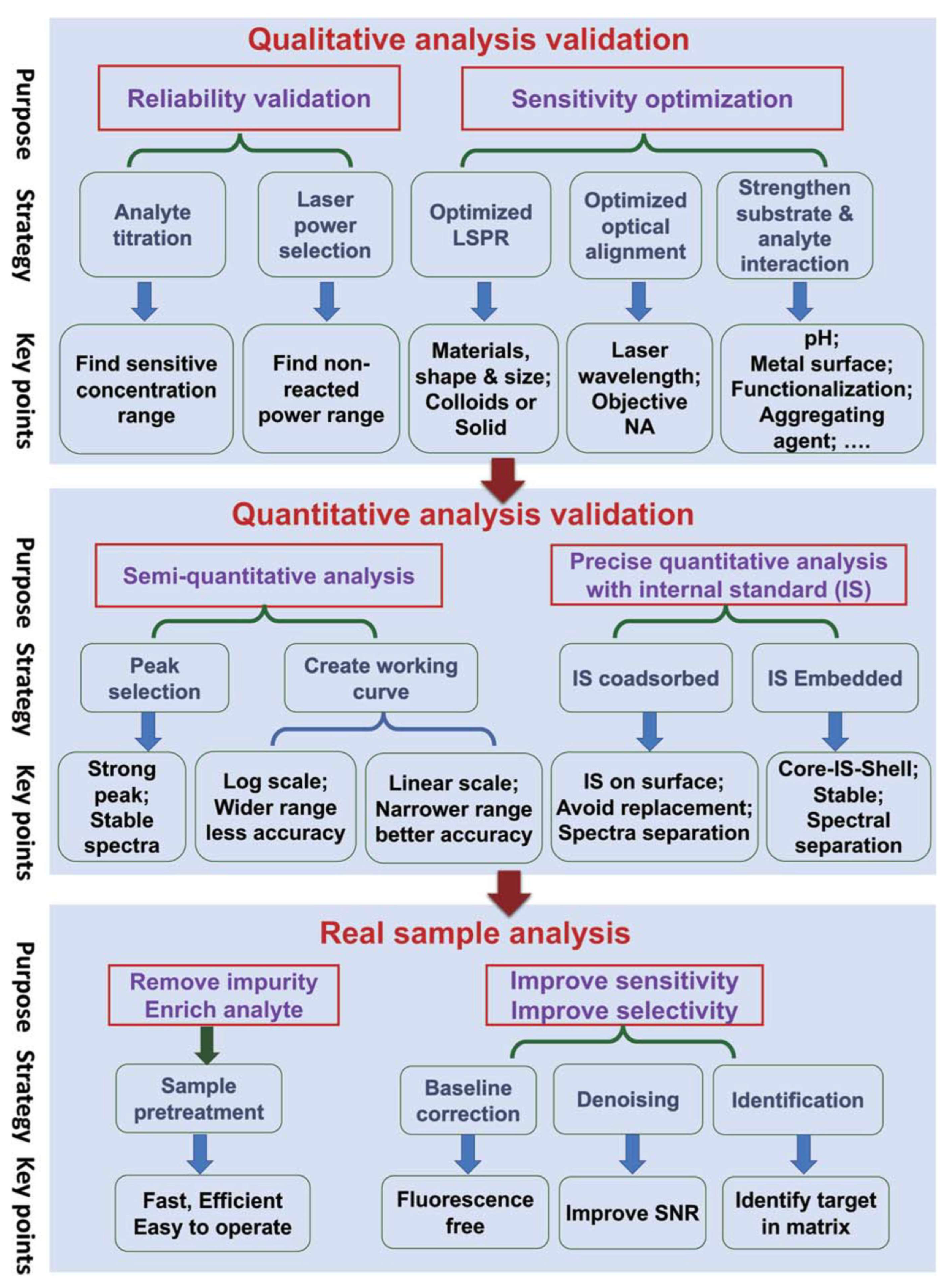
Figure 5.
Illustration of (A,B) electromagnetic mechanism (EM)[1,146] and (C,D) chemical mechanism (CM)[147,148] for SERS. HOMO, highest occupied molecular orbital; LUMO, lowest unoccupied molecular orbital; CB, conduction band; VB, valence band.
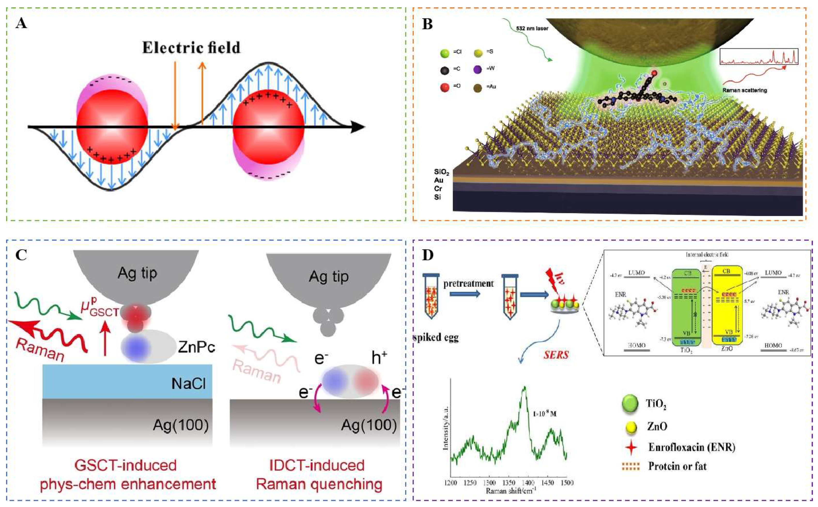
Figure 6.
(A,B) Schematic illustration of SERS mechanisms dominated by hotspots, electromagnetic enhancement, chemical enhancement[89,149]. (C) Preparation and characterization of colloidal assembly based 3D SERS substrate[150].
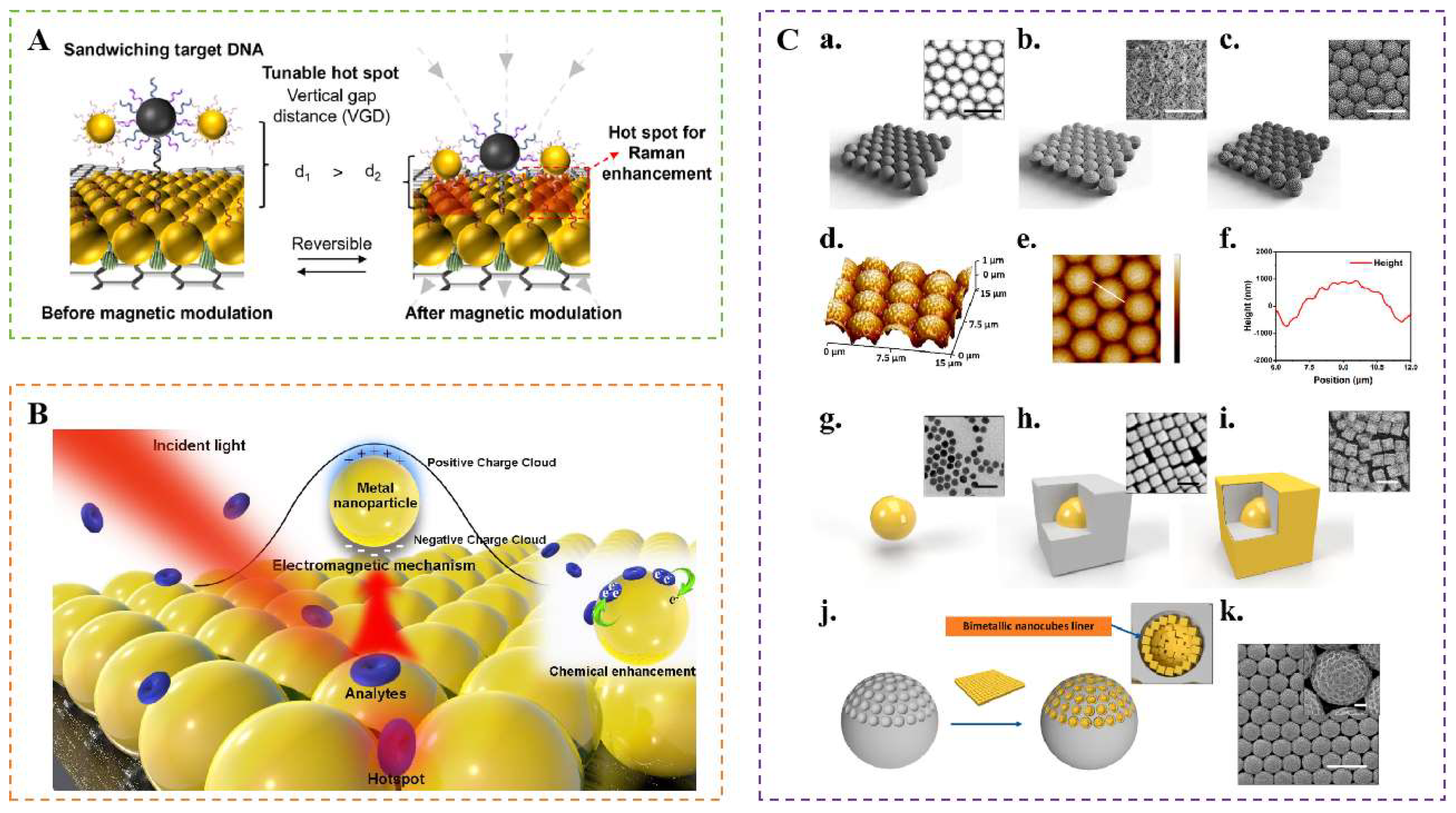
Figure 7.
(A) Schematic Illustration of the construction of “Add on” dual-modal optical immunoassay [153]. (B) AuNRs@Ti3C2Tx-mediated SERS-electrochemical dual-mode sensor[130]. (C) Schematic illustration of electroactive and SERS-Active Ag@Cu2O NP-engineered electrochemical/SERS Dual-mode aptasensors[154]. (D) Principle of SERS-fluorescence dual-signal aptasensor for AFB1 determination[155].
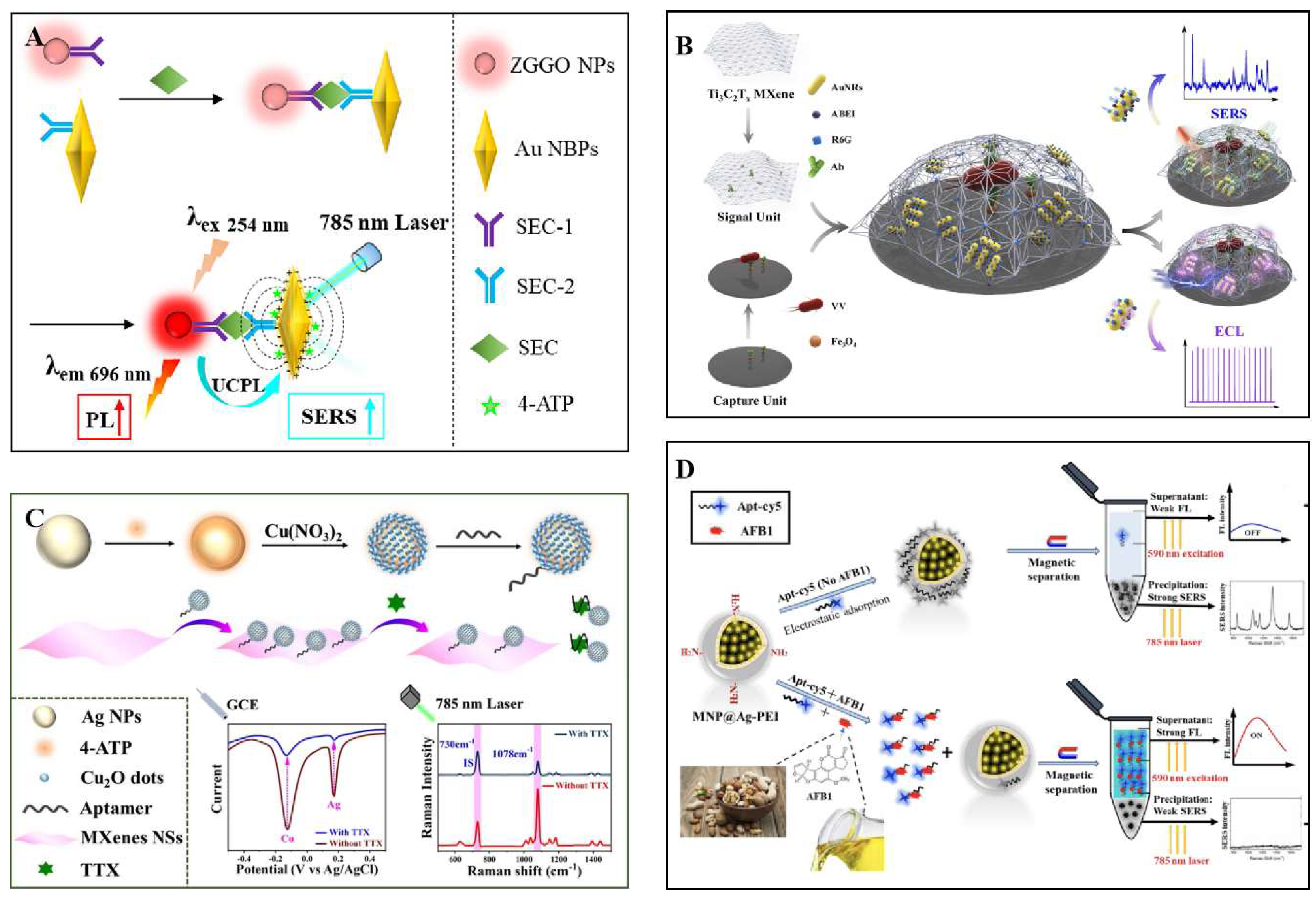
Figure 10.
(A) Schematic illustration of (a) construction of SERS probes, (b) preparation of the assay plate, and (c) SERS-sensing principle[170]. (B)Detection of NoV-LP in PBS using Palmtop Raman spectrometer[34]. (C) Schematic Diagram of designng silver-enhanced substrate, outer packaging of cold chain transportation, and conceptual schematic diagram of relationship between virus sample and “hot spots”generated by silver-enhanced substrate, SERS toward genomic proteins[171].
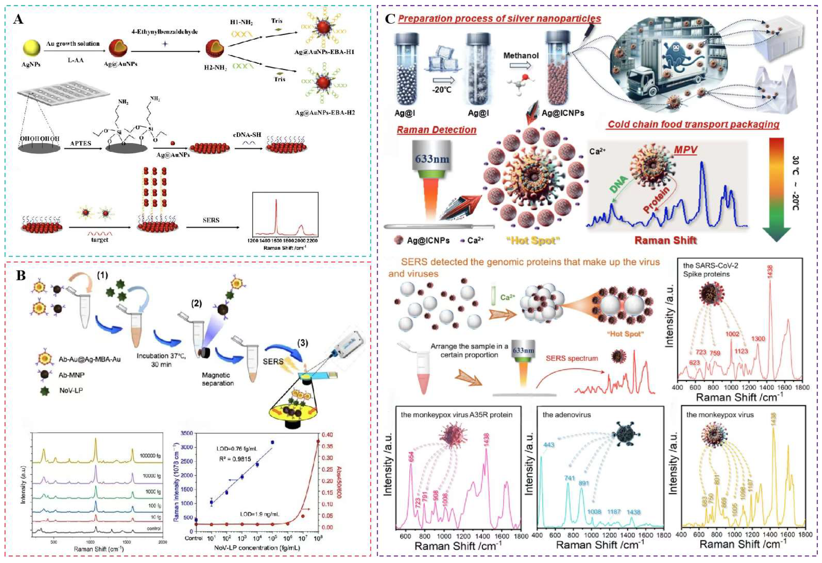
Figure 11.
(A) Tape-based SERS sensors toward on-hand detection of food contaminants[178]. (B) Schematic of interfacial self-assembly of Au NRs array toward multiple pesticides on fruit surface based on SERS and self-modelling mixture analysis [120]. (C) The schematic of PS@Au@1,4-BDT@Au synthesis and pesticide detection[84]. (D) Illustration of Au decahedral nanoparticles for SERS assay of triazole pesticide residues in vegetables and fruits[83].
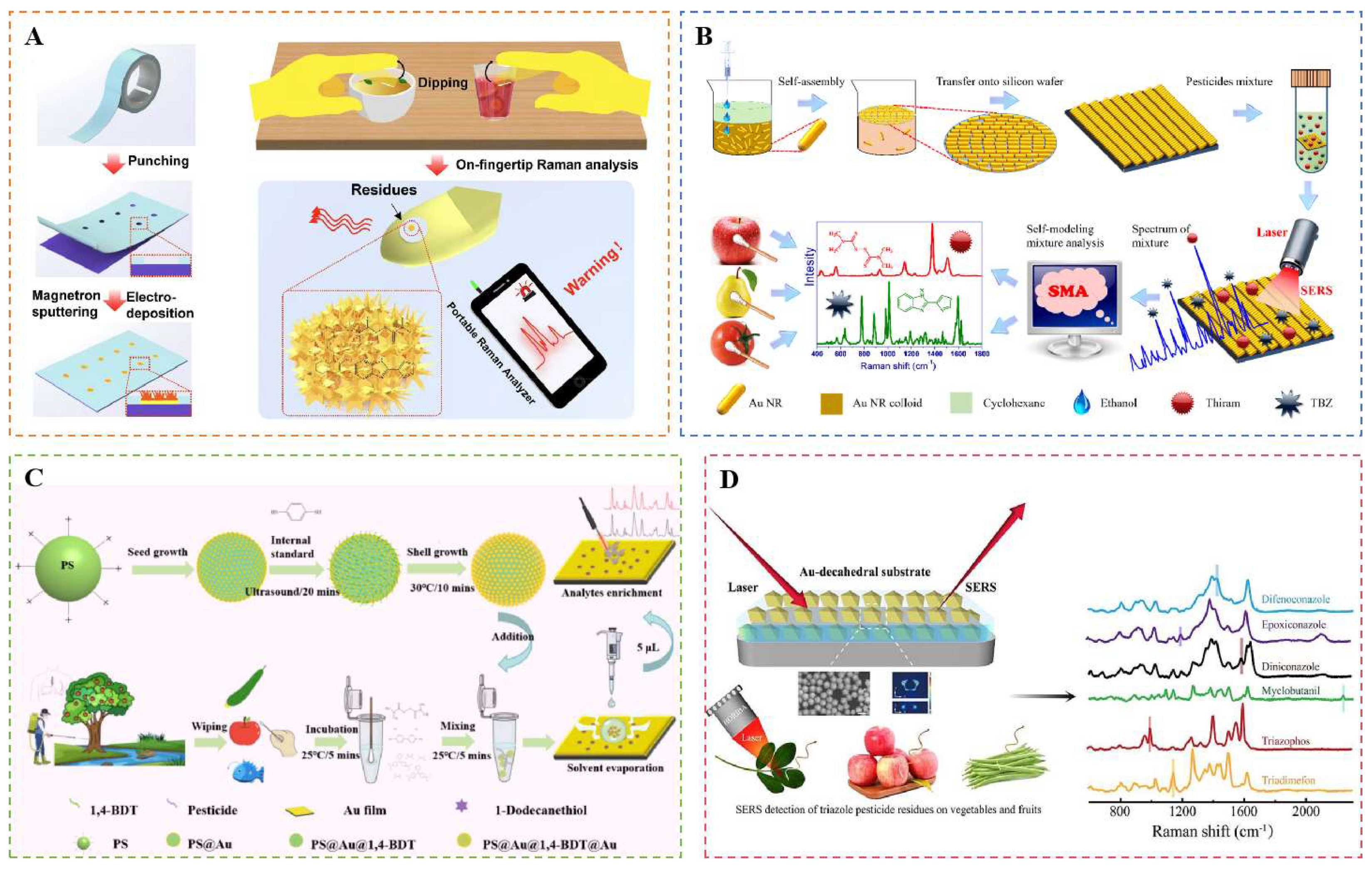
Figure 12.
(A) Schematic illustration of spiky Au@Ag NPs-Bi2WO6 film for SERS sensing toward pesticides and veterinary drugs[42]. (B) Schematic diagram of SERS-ICA based on mAbAuNpDTNB nanostructures. (a) The preparation process of mAbAuNpDTNB SERS tags. (b) Schematic testing process of the SERS-ICA strip for SM2[177].(C) Fe3O4-WO3-X @AuNPs for SERS sensing toward albendazole and streptomycin in meat samples via magnetic separation-enrichment-detection all-in-one[179]. (D) Schematic representation of (a) synthesis of (i) 24 nm Au@Ag NPs and (ii) multilayered MDAu@Ag tags with dual layers of Au@Ag and Raman dyes, (b) preparation of immuno-MDAu@Ag SERS tags, and (c) design of MDAu@Ag-based SERS-LFA for simultaneous detection of four veterinary drugs[180].
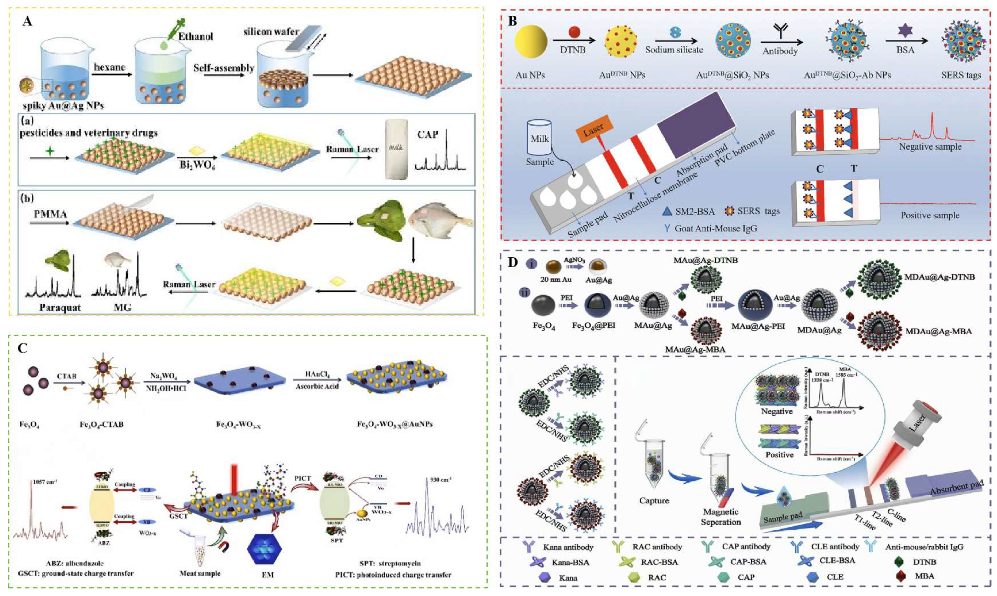
Figure 13.
(A) Schematic illustration of dual-signaling SERS ratiometric platform for Hg2+ detection[123].(B) Schematic diagram of Mn²⁺-induced aggregation of MNA-MA-AgNPson SERS sensing toward Mn²⁺[185]. (C) Schematic of DTNB-embedded Fe@RAu on MagSERS-ICA toward simultaneous monitoring of CLE and Cd2+ in complex samples[137].
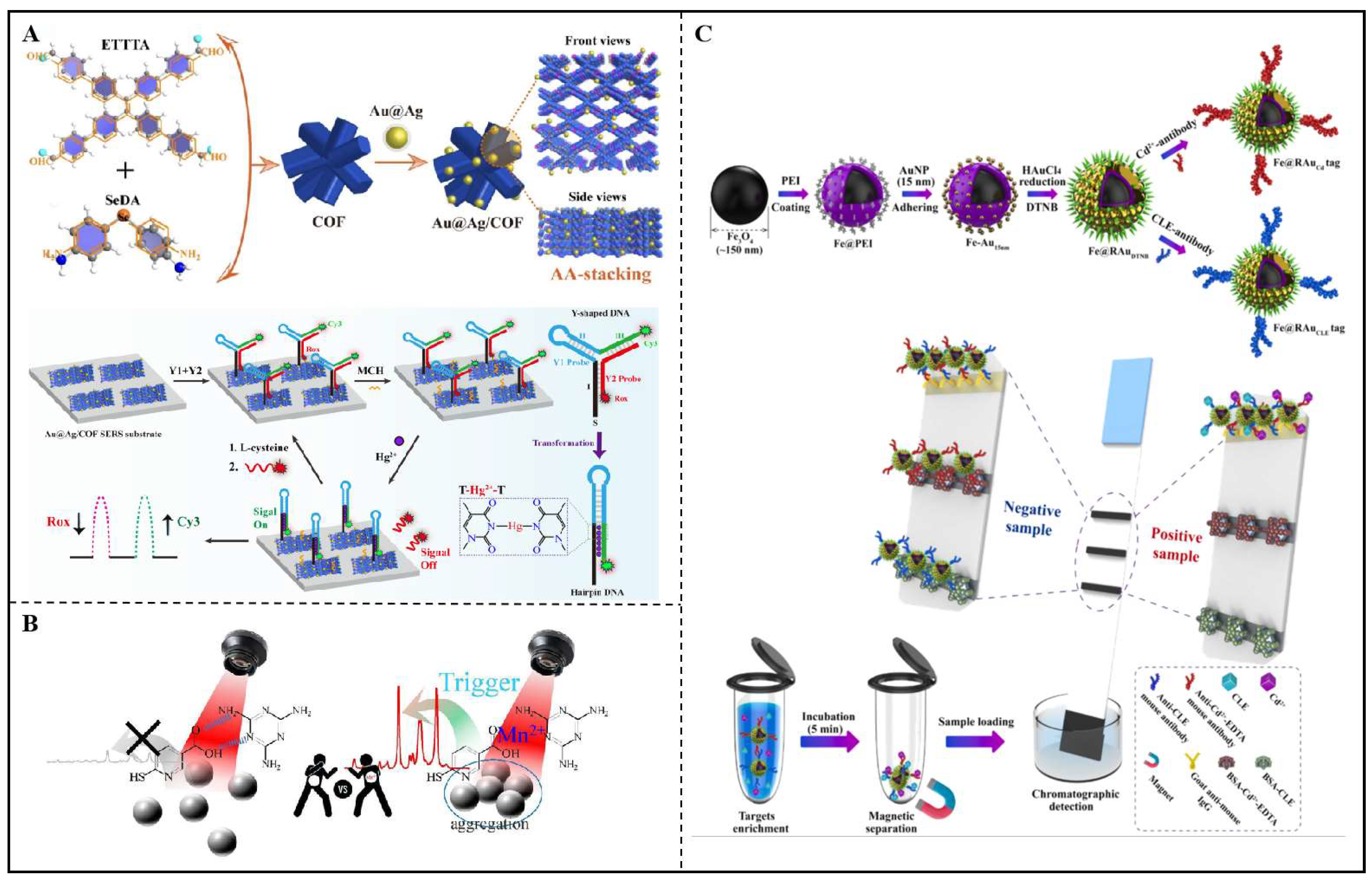
Figure 14.
Preparation of pNIPAM@Au for optical and SERS analysis. (A) Fabrication and characteristic procedures of pNIPAM, Au NRs, and pNIPAM@Au NRs[87]. (B) Synergistic versus competitive mechanism of SERS (hot-spots) and SERRS (LSPR bands matching) effect when using 785 nm (NIR range) and 633 nm (Vis range) laser excitations at swollen (25℃) or collapsed (50 ℃) states[87]. (C) Detection of MG in fish tissues using pNIPAM@Au as SERS substrates[87]. (D) Schematic diagram of SERS sensing toward plasticizer in oil[192]. (E) Schematic diagram of detection of titanium dioxide adulteration in persimmon frosting [107].
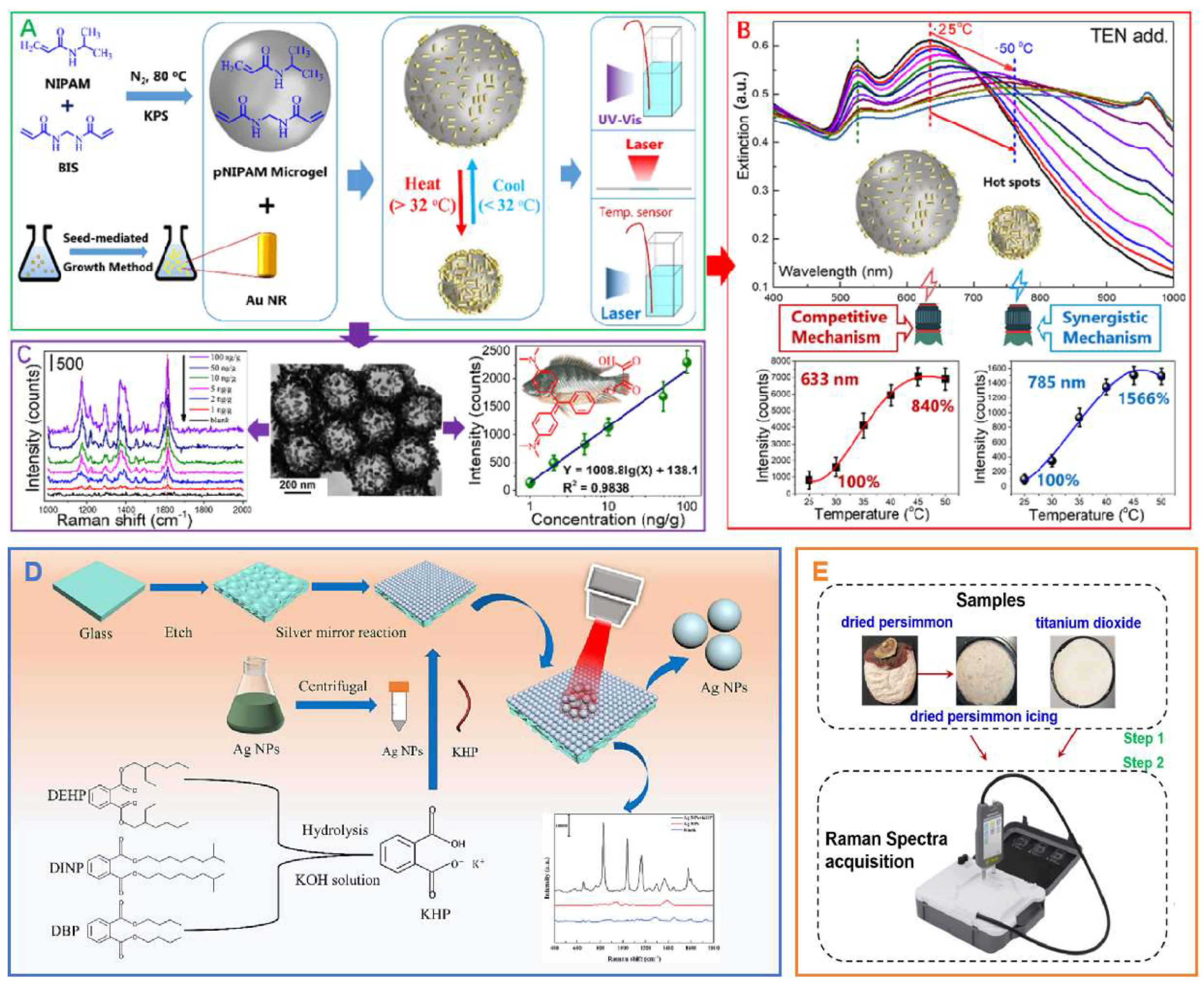
Figure 15.
(A) Illustration of LICA of Plasmonic GNPs and Nanoplastics. (A1) Before Coassembly, GNPs Were Distributed in the Bulk Aqueous Solution. (A2) Adsorption Behavior Occurred between GNPs and Nanoplastics. (A3) Coassembly of GNPs and Nanoplastics at the Ethyl Acetate−Water Interface Induced by Acetone. (B1−B3) SEM Image Corresponding to A1−A3. (B4) Enlarged SEM Image of B3[17]. (B) Schematic diagram of simultaneous detection of multi-component nanoplastics in tap water using liquid interfacial array transferred to a silicon wafer[17]. (C) (a) color image, (b) original, and (c) preprocessed Raman images at 961 cm-1,Preprocessed Raman spectra of fish meat and fish bones (at different depths)[62].
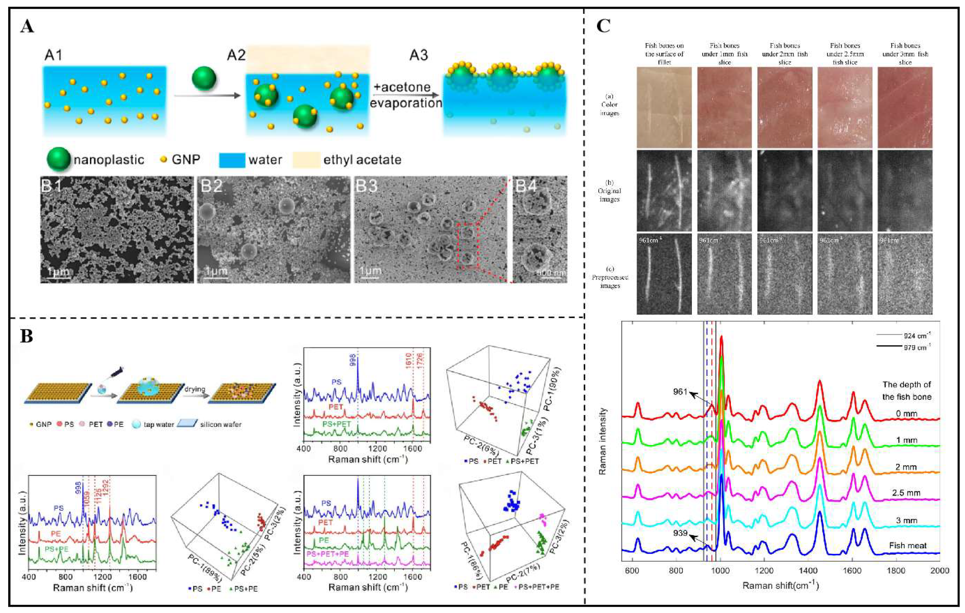

Table 1.
Commonly used nanostructures in SERS platform.
| Classification | Specific Materials | Characteristics |
Metal Nanostructures[7,120]
|
Au, Ag, Metal Alloy | Widely used in SERS substrates due to their excellent LSPR property. |
Core-Shell Structure [13,121,122]
|
Au@Ag, Ag@Au | Optimizing SERS signals by adjusting the properties of the outer layer metal. |
Porous Materials[39,123,124]
|
Porous carbon (PC), Porous silicon (PS), Metal-organic frameworks (MOFs) | High specific surface area and good molecular sieving effects, effectively adsorbing target molecules and enhancing SERS signals. |
Semiconductor Nanostructures[13,125]
|
Titanium dioxide (TiO2), Zinc oxide (ZnO) | Weak in SERS activity on their own, but can enhance signal strength when combined with metal nanoparticles. |
Carbon-Based Nanostructures[126,127]
|
Graphene and its derivatives (e.g., reduced graphene oxide) | Excellent conductivity and large specific surface area, effectively enhancing SERS signals. |
Polymer-Based Nanostructures[128]
|
Functionalized polymers (e.g., PMMA, PDMS) | Combining with metal nanoparticles to form composition for enhancing SERS signals. |
Biomass-Based Nanostructures[129]
|
Natural polymers (e.g., chitosan, gelatin) | Biocompatibility and tunability allow them to combine with metal nanoparticles for enhancing signals. |
Composite Nanostructures[13,123,124,130,131]
|
Combination of different nanostructures (e.g., metals with semiconductors, metals with porous materials) | Achieving synergistic effects to further enhance SERS signals. |
Table 2.
The advantages of nanostructure-sensitized SERS for detection of hazardous substances in food[89,133,134].
| Advantage | Description | Advantage | Description |
| High Sensitivity Detection |
SERS technology uses the surface plasmon resonance of metal nanoparticles to significantly enhance the Raman signals of adsorbed molecules, enabling highly sensitive detection of trace hazardous substances in food. |
Real-Time Monitoring Capability | Combined with portable devices, SERS technology can achieve real-time monitoring of hazardous substances during food processing and storage. |
| Rapid Response | SERS technology can provide rapid assay results, which is crucial for immediate response and management of food safety incidents. | Data Traceability | The Raman spectra provided by SERS have unique fingerprint characteristics, aiding in tracing contamination sources and food safety traceability. |
| No Need for Labeling andPretreatment | SERS detection does not require complex sample pre-treatment or labeling; it can directly test food samples, simplifying operational process. | Strong Environmental Adaptability | Nanostructures can be used under various environmental conditions, enhancing the application potential of SERS technology in diverse food testing scenarios. |
| High Selectivity | SERS technology exhibits high selectivity, enabling the rapid quantitative or qualitative detection of hazardous substances in complex food matrices. | Cost-Effectiveness | Although the initial investment may be high, SERS technology reduces the costs associated with repeated testing and erroneous results, making it cost-effective in the long run. |
| Multiplex Detection Capability |
SERS technology is capable of detecting multiple hazardous substances simultaneously, enhancing the efficiency and scope of detection. |
Biocompatibility | Selecting appropriate nanostructures ensures that SERS detection is safe for both food and operators, avoiding secondary contamination. |
| Classification | Sources | |
| Foreign matter contamination |  |
Metal fragments, glass fragments, plastic fragments, stones, sand particles, and more. May originate from wear of processing equipment or packaging materials, environmental pollution during raw material collection or processing. |
| Radioactive contamination |  |
Contamination caused by radioactive substances, which may enter the food chain through soil and water sources. |
| Noise pollution |  |
Long-term exposure to noise may impact the work efficiency and psychological health of food processing personnel, indirectly affecting food quality. |
| Light pollution |  |
Excessive lighting may affect food storage conditions, leading to spoilage or a decrease in nutritional value. |
| Thermal pollution |  |
Inappropriate temperatures may cause food spoilage during storage and transportation. |
| Mechanical impurities |  |
Lubricating oils, metal shavings, and other substances from mechanical equipment may be mixed into food. |
| Packaging material contamination |  |
Certain chemicals from packaging materials may migrate into food, causing physical contamination. |
| Natural impurities |  |
Naturally occurring impurities in food raw materials, such as small stones in grains, small insects in fruits and vegetables, etc. |
| Human negligence |  |
During food processing, tools, equipment parts, and other foreign objects may be inadvertently mixed into food due to human error. |
Disclaimer/Publisher’s Note: The statements, opinions and data contained in all publications are solely those of the individual author(s) and contributor(s) and not of MDPI and/or the editor(s). MDPI and/or the editor(s) disclaim responsibility for any injury to people or property resulting from any ideas, methods, instructions or products referred to in the content. |
© 2025 by the authors. Licensee MDPI, Basel, Switzerland. This article is an open access article distributed under the terms and conditions of the Creative Commons Attribution (CC BY) license (http://creativecommons.org/licenses/by/4.0/).
Copyright: This open access article is published under a Creative Commons CC BY 4.0 license, which permit the free download, distribution, and reuse, provided that the author and preprint are cited in any reuse.


