Submitted:
26 February 2025
Posted:
27 February 2025
You are already at the latest version
Abstract
Cancer cachexia is a multifactorial syndrome characterized by progressive muscle wasting, fat loss, and metabolic disruptions, commonly observed in cancer patients with advanced disease. It significantly impairs quality of life (QoL), reduces survival rates, and complicates treatment. The pathophysiology of cancer cachexia involves complex host-tumor interactions that lead to decreased appetite, metabolic shifts, and systemic inflammation. These alterations trigger muscle and fat degradation, making cachexia a critical component of cancer progression, particularly in patients with upper gastrointestinal, pancreatic, and lung cancers. Inflammatory markers, such as C-reactive protein, are key indicators of cachexia, and systemic inflammation plays a crucial role in the development of cachexia. Despite its severe impact, there are currently no standardized treatments for cancer cachexia. Clinical trials on immunomodulatory approaches have evaluated the beneficial potential of treating cancer cachexia. Immunomodulation, particularly through targeting multiple inflammatory cytokines, has shown benefit in mitigating cachexia-associated symptoms. Notably, the use of a newly identified immunomodulator, R-ketorolac, in animal models has demonstrated significant improvements in survival. Therefore, future research should focus on understanding the relationship between immune dysfunction and cachexia and developing personalized immunomodulation-based treatment strategies. Immunomodulators that repair destructive immune dysfunction hold promise for reversing cachexia symptoms, overall cancer treatment efficacy, patient survival, and QoL.
Keywords:
Cachexia
; cancer
; immunomodulation
; inflammation
; anorexia
; metabolism
Introduction
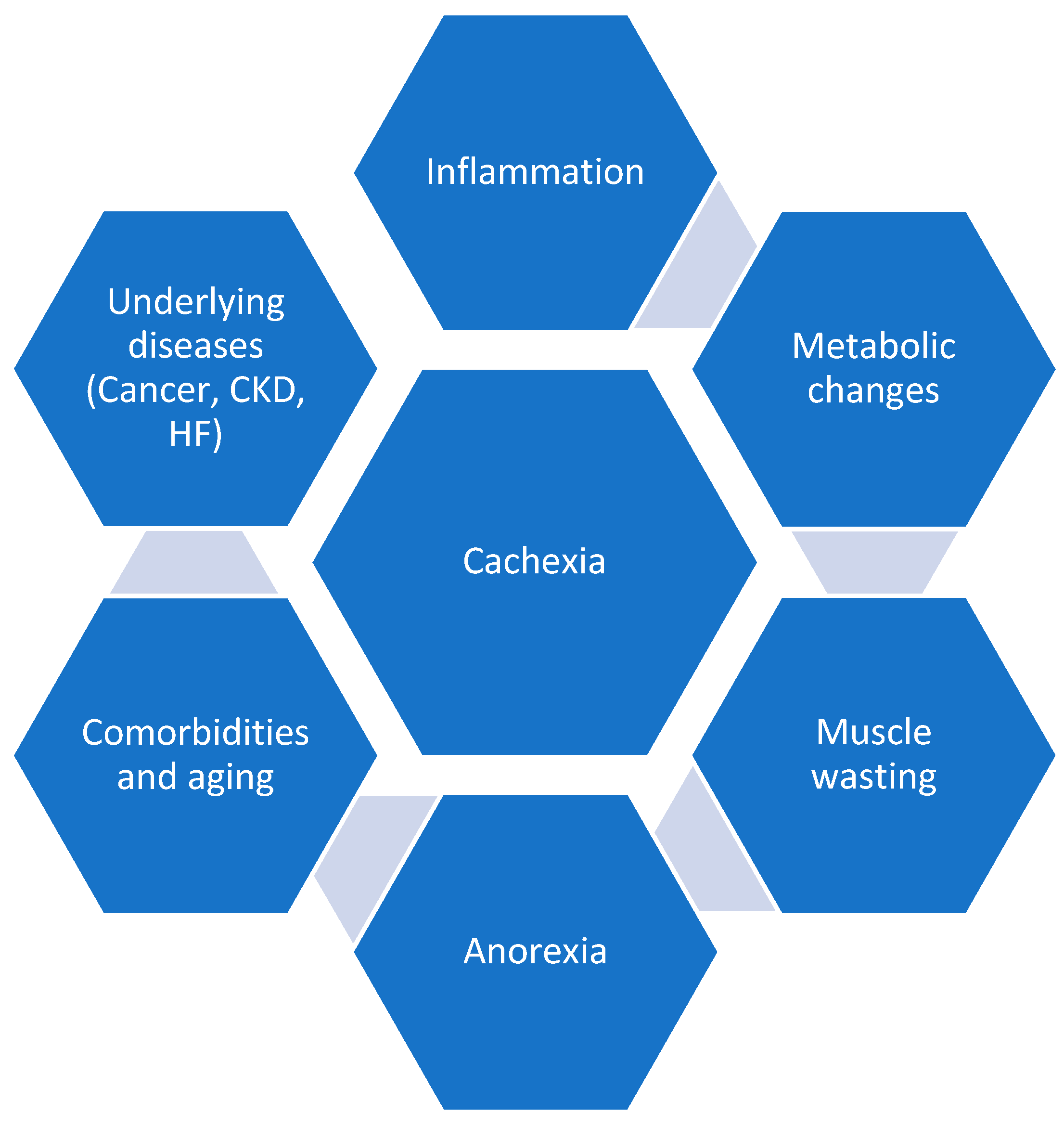
Cachexia, characterized by a decrease in fat and muscle mass, along with weight loss, is commonly observed in patients with advanced cancer and may be a primary indicator of underlying malignancy. Multiple pathophysiological occurrences, such as reduced food intake, decreased physical activity, systemic inflammation, and activation of catabolism, seem to occur simultaneously, along with weight loss (Figure 1). Therefore, the pathophysiology of cachexia has been described as a host-tumor interaction, which is a result of immune dysfunction that causes the brain to reduce appetite, redirect metabolism, affect gastrointestinal function, and induce fatigue [1,2]. As per the European Society for Medical Oncology (ESMO) Clinical Practice Guidelines, cachexia is defined as a subtype of malnutrition that is disease-related, identified using malnutrition screening, and has at least one phenotypical criteria such as loss of or low body mass [A1, weight loss >5% in six months; A2, body mass index (BMI) <20 kg/m2; A3, low muscle mass] and systemic inflammation [1].
Cancer cachexia develops in approximately up to 80% of patients with advanced cancer, and the subsequent 1-year mortality rate ranges from 20 to 60% [3]. Cachexia is most prevalent and severe in upper gastrointestinal tract cancers, with pancreatic and liver cancer-associated cachexia occurring in almost 70 to 85% of the patients. Further, more than 50% of the patients with head and neck cancer, gastroesophageal cancer, or non-small cell lung cancer (NSCLC) suffer from cachexia and is to a lesser extent in patients with breast (25%) and prostate cancer (15%). As the disease progresses, the severity of the weight loss is believed to increase as well [3].
Patients with cancer usually report loss of appetite, anorexia, nausea, and vomiting due to chemotherapy. Further, the lactic acid produced by tumors or inflammatory cytokines activated due to host-tumor interactions may also result in decreased appetite, all causing weight loss and manifesting into metabolic changes [3]. Cachexia is generally a combination of various factors, such as inflammatory disturbances, hyperlipidemia, hyperglycemia, and loss of fat and muscle [4]. Cancer cachexia is associated with muscle wasting, caused by an increased protein-degradation rate and reduced protein synthesis [3].
Moreover, patients with cancer cachexia also experience metabolic and histopathological alterations in the adipose tissue, resulting in wasting and causing muscle loss. White adipose tissue (WAT) undergoes browning, causing systemic and local catabolic state, eventually leading to lipolysis and adipokine secretion. This also ultimately causes energy expenditure and imbalance [5]. In patients with advanced cancer, low muscle mass serves as an independent predictor of immobility and mortality [6]. Another critical component of cachexia is systemic inflammation, often indicated by serum C-reactive protein (CRP) [6]. This inflammation also affects the catabolic processes, causing anorexia and decreasing nutrient intake, resulting in loss of skeletal muscle and adipose tissue [4]. Loss of skeletal muscle mass in cancer patients, in turn, causes increased fatigue and poor quality of life (QoL) [4].
Cachexia syndrome comprises three stages based on its severity, namely pre-cachexia, cachexia, and refractory cachexia. While patients experiencing simple weight loss (<5%) along with clinical symptoms such as anorexia and glucose intolerance are considered to be pre-cachexia [3], the cachexia stage is indicated by the presence of one or more symptoms such as weight loss >5% over last six months, weight loss >2% plus BMI <20 kg/m2 [7]. Further, the refractory cachexia stage is defined as weight loss >15% over the last 6 months and BMI <23 kg/m2 or weight loss >20% in the past 6 months plus BMI <27 kg/m2 [7]. Further, the refractory stage is usually considered terminal and often associated with limited self-care or complete disability. At this stage, patients are unlikely to benefit from weight management treatments, and medical efforts focus mainly on addressing complications related to cachexia, underscoring the importance of early detection [3].
The assessment of cancer cachexia primarily focuses on nutritional status, body weight/composition, QoL, and associated biomarkers. Since inadequate intake is a common issue, it is essential to screen for malnutrition in patients with advanced cancer [8]. Various molecules are associated with cachexia, such as cytokines, neurotransmitters, tumor-derived factors, and neuropeptides [4]. Biomarkers of cancer cachexia, such as albumin, CRP, and various inflammatory cytokines, are key research areas. Tumor-produced lipid and protein mobilization factors activate inflammation, which disrupts protein metabolism and leads to the breakdown of proteins, carbohydrates, and lipids. The clinical impact of cancer cachexia is thus seen in the endocrine and metabolic functions, heart, gastrointestinal tract, muscle mass, bone, and brain [8]. The increased levels of pro-inflammatory cytokines thus result in tissue degradation and muscle wasting in cancer cachexia, also causing an imbalance in immune response. Therefore, immunomodulation strategies in cancer cachexia may be beneficial in modulating the immune response and reducing inflammation, thereby mitigating the cachexia-associated symptoms [9].
Scoring systems like the modified Glasgow Prognostic Score (mGPS), cancer staging score (CSS), and cancer cachexia scoring system (CASCO) help assess cachexia stages and predict patient survival, though biomarkers can be influenced by factors such as sex, age, and underlying diseases [8]. Although inflammation-driven immunoregulation plays a crucial role in cachexia, the immunological factors involved in its progression are still not well understood [2]. Additionally, despite the effect of cachexia on the QoL and life span of cancer patients, there are currently no targeted or standardized treatment options [3]. To improve muscle mass, enhance overall health, and boost tolerance to antitumor therapies, cancer cachexia treatment must follow a comprehensive, personalized, structured, and ongoing approach [8].
Therefore, this review aims to explore the multifactorial nature of cancer cachexia by examining its development, manifestations, influencing factors (such as age, gender, comorbidities, and cancer stage and type), and the potential of immunomodulation in treating it.
Exploring Cachexia—Inflammation, Immunomodulation, and Metabolism
Cancer cachexia, generally observed in patients with late-stage cancer, is a result of cancer-associated immune dysfunction and inflammation, resulting in a cytokine storm that can also cause multiple organ failure. It typically manifests as loss of appetite, weakness, fatigue, and anemia, causing poor QoL, unresponsiveness to treatment, and early mortality [2]. The pathophysiology of cancer cachexia comprises three important mechanisms, namely, metabolic dysregulation, negative energy balance, and neurohormonal imbalances [4]. Inflammation plays a crucial part in the development of cachexia and is usually associated with a negative nitrogen balance, weight loss, and increased consumption of energy [4]. Further, cancer disrupts immune system function, and immune tolerance failure leads to autoimmunity. In advanced malignancy, overproduction of cytokines and autoantibodies contributes to muscle wasting, particularly through IL-1β, IL-6, and TNF-α, which are linked to cancer cachexia [10]. Overall, the immune reaction observed in cachexia comprises various components, such as inflammatory factors, non-inflammatory factors (TGFβ, adipokine, myokine, PTHrP), immunometabolic checkpoints (PDL1, Adenosine, B7-H3, VSIG4), immunometabolic processes (carbon and protein metabolism), neuroimmune responses of central nervous system and peripheral nervous system and gut-immune responses (intestinal barrier disruption, gut microbiota and gut hormone) [11].
Inflammatory mediators are expressed by immune and non-immune cells when activated by damage-associated or pathogen-associated molecular patterns [10]. Cytokines trigger JAK-STAT (Janus kinase/ signal transducer and activator of transcription), NF-κB, and other pathways, leading to catabolic processes in muscles and adipose tissue, including mitochondrial dysfunction, reactive oxygen species production, and inflammation through pathways like JNK (Jun N-terminal kinase), NLRP3 (nucleotide-binding domain, leucine-rich–containing family, pyrin domain–containing-3), PI3K (phosphatidylinositol-3 kinase), MAPK (mitogen-activated protein kinase), and NF-κB. Elevated inflammatory cytokines (IFN-α, IL-8) and growth factors are observed in colorectal cancer, along with increased actin and collagen deposits. Further, growth differentiation factor 15 (GDF15), found in malignant tumors, is also linked to cachexia-related weight loss [10]. Moreover, a meta-analysis reported cancer cachexia reduces the efficacy of immune checkpoint inhibitors. The metabolic alterations associated with cachexia can impair the ability of patients to mount an effective anti-tumor immune response [12]. Conversely, some individuals may be less prone to developing cachexia, such as those with a loss of function mutation in the P-selectin (SELP) gene, which reduces the chances of cachexia.
Studies in rodent models also demonstrate that cachexia can be significantly reduced, even in advanced malignancies, without affecting tumor progression [13]. Moreover, innate immune cells, including macrophages, myeloid-derived suppressor cells (MDSCs), and adaptive immune cells like T cells, contribute to cancer cachexia. In pancreatic cancer models, M2 macrophage infiltration promotes muscle degradation via STAT3 signaling. Conversely, depleting M2 macrophages reduces inflammation and muscle atrophy, highlighting their damaging role in cancer cachexia [5]. Conversely, T cell infiltration offers protection against cachexia, with CD4+ Treg cells protecting muscle fibers from atrophy. Further, CD8+ T cells are linked to reduced activity in pathways that regulate muscle mass maintenance, including ubiquitin-proteasome, catabolic signaling, apoptosis, and autophagy. Therefore, immune cells can either cause or protect against cachexia, and the development of cancer cachexia depends on the immune system, thus highlighting the need for studying the interactions between cytokines, immune cells, and pro-cachectic factors [5].
As per a study by Vanhoutte et al., patients with cachexia demonstrate a short overall survival (OS) rate, and the prediction for worse survival depends on variables such as gender, primary tumor location, and occurrence of metastatic disease [14]. Immunomodulation plays a crucial role in managing cancer cachexia by regulating the immune system's response to both cancer and cachexia itself. Strategies such as immune checkpoint inhibitors, cancer vaccines, and cell-based therapies can alter immune pathways, potentially influencing inflammation and muscle wasting associated with cachexia [9]. Tumor cells have increased programmed cell death 1 (PD-1) and programmed death-ligand 1 (PD-L1) expression, which are correlated to enhanced OS and progression-free survival (PFS) in patients with cancer. In a study by Miyawaki et al., cancer cachexia was seen to reduce the impact of PD-L1 expression in patients with advanced NSCLC, thus demonstrating a desensitizing effect on PD-1 or PD-L1 inhibitors. Therefore, combining anti-cachectic treatments with PD-1 or PD-L1 inhibitors may reduce the desensitizing impact of cancer cachexia and improve the effectiveness of these inhibitors [15]. Further, the understanding of the underlying mechanisms for both innate and adaptive immune response facilitates the use of these molecules as immunomodulators [9].
Additionally, therapies like chimeric antigen receptor (CAR) T-cell therapy and dendritic cell-based vaccines can help restore immune function and target tumor cells more effectively. However, immunomodulation must be carefully managed, as it can also exacerbate cachexia or impact treatment outcomes, necessitating personalized approaches for cancer patients with cachexia to improve therapy effectiveness and minimize side effects [9]. Additionally, some factors influence cachexia prevalence, such as cancer stage, sex, age, genetic risk, comorbidities, and treatment-related catabolic effects. Further, genotypic differences also contribute to cachexia susceptibility, but more research is needed in this area [13].
Age
Age significantly impacts the development of cachexia, particularly through changes in muscle and adipose tissue, heightened inflammation, and immune system dysregulation. Aging is associated with the loss of muscle mass and function, also called sarcopenia [16], which shares similarities with cancer-related cachexia and is characterized by overactive protein breakdown, decreased protein synthesis, and chronic low-grade inflammation. Aging also leads to immunosenescence, a state of immune dysfunction marked by a high number of senescent T cells, reduced natural immunity, and increased pro-inflammatory cytokines like IL-6 and TNFα. These inflammatory markers contribute to muscle wasting by disrupting muscle regeneration and promoting a catabolic environment. Moreover, the secretion of IL-15 and IL-7, which normally help regulate immune responses, declines with age, impairing the immune system’s ability to counteract muscle loss. The accumulation of senescent cells and their secretory phenotype further exacerbates muscle deterioration by influencing myofiber morphology and function. Immunomodulation, particularly targeting senescent cells with senolytic therapies, has shown potential in mitigating age-related muscle decline and improving muscle mass and function [9]. As per a study conducted on mice, age played a role in aggravating weight loss and affected the development and progression of cachexia. Further, the patients’ cohort (n=14) in this study also demonstrated a significant association between weight loss and the circulating cytokines, which were lost among the older patients, demonstrating cachexia biomarkers may also provide varying prognostic values depending on the age [17].
Gender
Gender plays a significant role in the progression and severity of cancer cachexia, with notable differences observed between males and females. Males are more likely to experience muscle depletion (sarcopenia) and greater reductions in muscle mass and strength compared to females with cancer cachexia. A study of 441 cancer patients reported that 61% of men exhibited muscle depletion, while only 31% of women had muscle depletion [13,18]. In addition, male cancer patients tend to have more pronounced reductions in muscle mass, strength, and power, especially in the lower limbs, which correlate with poorer quality of life. Conversely, these muscle-related declines are less pronounced in females. Additionally, inflammation also affects males and females differently. In a study, male mice with cancer cachexia exhibited higher levels of serum proinflammatory cytokines, such as IL-6, which are associated with muscle wasting, whereas females exhibited a different response to these cytokines, with higher IL-6 levels not necessarily leading to accelerated cachexia progression, suggesting a gender-specific difference in immune response [18,19]. These gender-based differences in muscle loss, inflammatory response, and overall cachexia progression indicate that gender should be considered when evaluating cachexia and developing targeted treatment strategies [18].
Additionally, the gender differences in cancer cachexia are also reflected in the muscle fiber composition and their response to muscle wasting. In both mice and humans, skeletal muscle fibers are classified into distinct types based on contraction speed, fatigue resistance, and metabolic properties: slow oxidative (type 1) and fast-twitch (type 2A, 2X, and 2B) fibers. Generally, men have a higher proportion of type 2 fibers, which are less resistant to fatigue, compared to women, who tend to have a greater proportion of type 1 fibers, which are more fatigue-resistant. This difference in muscle fiber composition may influence the susceptibility to cancer-induced muscle wasting [18,20].
In male mice with Colon-26 (C-26) tumors, type 2 fibers were more prone to cancer-induced muscle loss, while in female mice, type 1 fiber exhibited greater atrophy, accompanied by an increase in fast-twitch type 2B fibers. These divergent responses to muscle wasting suggest that the impact of cancer cachexia may vary between the sexes, potentially due to differences in fiber type composition. However, studies in human patients, such as those with upper gastrointestinal or pancreatic cancer, did not show selective fiber atrophy by type, indicating that the phenomenon may be more complex or species-specific [18,21].
Further, in patients >60 years old, cancer cachexia is more frequent in males (40–60%) as compared to females (40–50%). Various internal (hormonal levels and molecular differences) and external (delayed diagnosis, diet, smoking, alcohol consumption, lifestyle) factors may be responsible for variations between genders [4]. Another gender-specific analysis revealed increased IL-6 levels in both genders, with increased platelets in males and elevated free fatty acids in females. In female cancer cachexia patients, plasma IL-6 and free fatty acids concentrations were positively correlated. These pro-inflammatory cytokines (such as IL-6) are vital for immunomodulation in cachexia, altering the immune responses and causing inflammation [9]. Significant differences were found between gastric and colorectal cancer patients with cachexia: gastric cancer patients had elevated IL-6, while colorectal cancer patients showed higher levels of IL-6, TNF-a, platelets, white blood cell count, free fatty acids, and ApoA. The study thus emphasized the need for gender-specific analysis in cancer cachexia and further research on the mechanisms behind abnormal fat metabolism [2].
Co-Morbidities
Cachexia can occur due to various underlying diseases, such as cancer, heart failure, chronic obstructive pulmonary disorder (COPD), chronic inflammation, anorexia, insulin resistance, renal failure, and anemia [22]. Cancer cachexia is also associated with various organs, such as the gut, skeletal muscle, heart, and brain [5]. A paraneoplastic syndrome such as pancreatic cancer-associated diabetes mellitus (PCDM) may also be a comorbidity observed before cachexia [23]. According to a retrospective study, comorbidities were observed in 50% of the patients with cancer cachexia, the most common being COPD (23%), followed by high blood pressure (12%), coronary insufficiency (8%), diabetes (4%) and respiratory failure (3%) [24]. Therefore, cachexia arises not in isolation but within a complex context of comorbid conditions, responses to cancer treatment, toxicity, pain and other symptoms [13].
Cancer Type
Recent studies have shown that the prevalence and severity of cancer cachexia vary depending on the type and stage of cancer [16]. Certain types of cancers, such as pancreatic, lung, ovarian, liver, oesophageal, and brain tumors, pose a challenge for early detection as they do not exhibit noticeable symptoms in their initial stages and are not easily detectable through routine screening [25]. Hence, the risk of cachexia at later stages also increases. Patients with cancer cachexia experience inflammation resulting from increased cytokine levels (such as TNFs and ILs) [3,10]. The production and secretion of such mediators may vary based on the tumor type, along with the magnitude of the systemic inflammatory response, regulation of cytokines, and the risk of developing cachexia [16]. Some of these tumor-derived mediators include glucocorticoids, which increase skeletal muscle protein catabolism and decrease protein synthesis, thus resulting in muscle atrophy. Further, TNF-α is a key pro-inflammatory cytokine that promotes muscle and adipose wasting and contributes to insulin resistance and systemic inflammation in cancer cachexia. Additionally, IL-6 levels are elevated in cancer cachexia, affecting the immune response, metabolism, and muscle wasting. GDF15 is associated with anorexia and muscle wasting, with the circulating levels often correlated with cancer cachexia severity. These pro-inflammatory cytokines are involved in various physiological processes, such as inflammation and immune responses, and thus play a crucial role in immunomodulation in cancer cachexia [9]. Finally, the parathyroid hormone-related peptide is associated with hypercalcemia and bone resorption, indirectly promoting skeletal muscle wasting [16].
Cancer Stage
Cachexia can occur at all stages of cancer; however, the prevalence is higher in the later stages [3]. The occurrence and severity of cachexia are based on tumor type, location, and stage. Various potential biomarkers, such as CRP, hemoglobin, interleukins, etc., are indicators of cachexia and can differ depending on stage and inflammation [16,26]. Therefore, immunomodulation of these markers may be beneficial in reducing the progression of cancer cachexia [10].
Therapy for Cancer Cachexia
Cancer cachexia is a complex condition characterized by anorexia, skeletal muscle wasting, and metabolic changes, making it difficult to treat. Early diagnosis and intervention are crucial, as the pathogenesis of cancer cachexia is not fully understood [5]. However, as per the literature, various immunomodulatory mechanisms have been found to be involved in cachexia; therefore, targeted immunomodulation could serve as a potential therapy to limit the progression of cancer-associated cachexia [10]. Due to the lack of effective treatment for cancer cachexia, a comprehensive treatment strategy combining pharmacological therapy with nutritional support, exercise, and psychosocial interventions is believed to be necessary to address muscle atrophy associated with cancer [5]. Multimodal treatment, comprising a combination of nutrition, exercise, psychological, oncological, and palliative, should be utilized for the treatment of cachexia [1,27] before specific therapy for cancer cachexia gets approved. Current clinical trials focus on exploring new treatment agents, with promising results anticipated for the near future. Key therapies include exercise, which has been shown to reduce muscle degradation, improve muscle function, and enhance insulin sensitivity.
Exercise is the only recommended behavioral treatment for cancer cachexia and is beneficial in enhancing muscle strength and lean body mass while reducing inflammatory markers. Various types of exercise, such as aerobic, resistance, and flexibility training, can help manage muscle atrophy [5,22]. Combining exercise with nutrition therapy is particularly effective, with high-protein diets being recommended to counteract the hypercatabolic state of cachexia. Further, appetite stimulants, such as steroids and progestational agents, have been used to manage appetite loss, with medications like megestrol acetate improving body weight, albeit with increased fat mass rather than lean muscle [5]. As per the ESMO guidelines, corticosteroids, progestins, and olanzapine are recommended for improving appetite in patients with advanced cancers [1]. Clinical trials also explore ghrelin agonists (NCT00933361) like anamorelin, which have shown promising results in increasing appetite and muscle mass [5]. As per the ROMANA 1 and ROMANA 2 trials, patients with NSCLC demonstrated significant improvement in body weight on treatment with anamorelin [8].
Anti-inflammatory drugs targeting cytokines, including TNF-α, are a key focus in cancer cachexia treatment. However, results have been mixed, with some therapies like thalidomide showing promise in reducing weight loss, while others like pentoxifylline and infliximab have been less effective [5,8]. Inhibiting myostatin and its analogue activin A offers another strategy to counter muscle wasting, with agents like ActRIIB showing potential in preventing muscle loss and improving lifespan in animal models. Other approaches, such as using testosterone or selective androgen receptor modulators, have also shown promise in promoting muscle growth [5]. GDF15, which is high in patients with cachexia, resulting in anorexia, is also a therapeutic target in cachexia treatment. Anti-GDF15 humanized monoclonal antibodies such as ponsegromab, NGM120, and AV380 are being investigated in various randomized trials (NCT05546476; NCT04068896; NCT04725474) [28]. Additionally, natural compounds, including resveratrol and curcumin, have demonstrated anti-cancer and anti-wasting effects by regulating muscle protein synthesis and inflammation, offering potential adjunct therapies for cancer cachexia [5].
Various studies in rat cachexia models also demonstrate the effectiveness of melanocortin-4 receptor (MC4R) for its anticachectic properties. The MC4R antagonist was observed to stimulate food intake, reduce weight loss, and preserve fat mass in rat models [5,8]. Additionally, nonsteroidal anti-inflammatory (NSAID) agents are also associated with improved outcomes in body weight or QoL of patients [29]. In our study [10], the effects of R-ketorolac (RK), the inactive component of an NSAID drug, were investigated on cancer cachexia and survival rates in tumor-bearing mice. Mice inoculated with cancer cells showed significant muscle and fat loss, systemic inflammation, and reduced quality of life. Upon induction of cachexia, RK treatment improved survival rates, with 100% survival in RK-treated mice compared to just 10% in untreated mice. RK also reduced body weight loss, muscle and fat wasting, and systemic levels of the pro-inflammatory cytokine IL-6. The treatment also protected against T-lymphopenia, with beneficial effects observed only in mice with functional T-cells. These positive outcomes were independent of COX inhibition, distinguishing it from other NSAIDs and making it a promising therapeutic candidate for managing cancer cachexia [10]. Recently, a "proof of concept" trial for R-Ketorolac, KetoROCX (NCT05336266), has been completed, showing efficacy in reversing cancer cachexia symptoms in patients with advanced pancreatic ductal adenocarcinoma [2,30] Therefore, immunomodulatory strategies should be emphasized to maximize anti-cachexia and anticancer activity, taking into consideration age-related factors that may affect the immune response, such as immune checkpoint overexpression and T-cell exhaustion [9].
Conclusion and Future Directions
Cancer cachexia remains a multifaceted and challenging condition that significantly impacts patients' QoL, survival rates, and response to treatment. It arises from a complex interplay of metabolic dysregulation, inflammation, immune dysfunction, and systemic factors influenced by the type and stage of cancer, age, gender, and comorbidities. Current therapies targeting these mechanisms, such as anti-inflammatory agents, exercise, and nutritional support, show promise but often yield mixed results. The heterogeneity in cachexia progression among individuals thus highlights the need for personalized treatment approaches, with a focus on improving muscle mass, reducing inflammation, and most importantly addressing the underlying immune dysfunction.
Future research should, therefore, focus on identifying novel biomarkers to predict cachexia onset and progression, especially considering the variability of responses based on genetic, environmental, and treatment-related factors. The complex relationship between immune responses and cachexia remains largely unexplored. Future research on cachexia should combine immunology with insights from evolutionary medicine and systems biology. Advances in immunomodulation, including small molecule immunomodulators and novel cytokine inhibitors, offer the potential for mitigating the effect of cachexia, thereby improving patient outcomes. As demonstrated in our research [10], RK-induced immunomodulation has the potential to reverse cancer cachexia and increase survival rate. Therefore, future research to explore extensively the potential of immunomodulation strategies for treating cancer cachexia is warranted. Additionally, understanding the gender- and age-specific differences in cachexia will be crucial for developing more effective and tailored interventions. Moreover, clinical studies involving both animal models and human trials are crucial for further validation of the diagnoses, assessments, and treatment options. Focused research on the intricate relationships between immune cells, inflammation, and metabolic processes will significantly increase our understanding of the role of immunomodulation in tissue metabolism during cachexia.
Disclosures
The authors declare no conflict of interest.
References
- Arends, J.; Strasser, F.; Gonella, S.; Solheim, T.S.; Madeddu, C.; Ravasco, P.; Buonaccorso, L.; de van der Schueren, M.A.E.; Baldwin, C.; Chasen, M.; et al. Cancer cachexia in adult patients: ESMO Clinical Practice Guidelines(☆). ESMO Open 2021, 6, 100092. [Google Scholar] [CrossRef] [PubMed]
- Zhang, L.; Bonomi, P.D. Immune System Disorder and Cancer-Associated Cachexia. Cancers (Basel) 2024, 16. [Google Scholar] [CrossRef] [PubMed]
- Gaafer, O.U.; Zimmers, T.A. Nutrition challenges of cancer cachexia. JPEN J Parenter Enteral Nutr 2021, 45 (Suppl. S2), 16–25. [Google Scholar] [CrossRef] [PubMed]
- Mariean, C.R.; Tiuca, O.M.; Mariean, A.; Cotoi, O.S. Cancer Cachexia: New Insights and Future Directions. Cancers (Basel) 2023, 15. [Google Scholar] [CrossRef]
- Setiawan, T.; Sari, I.N.; Wijaya, Y.T.; Julianto, N.M.; Muhammad, J.A.; Lee, H.; Chae, J.H.; Kwon, H.Y. Cancer cachexia: Molecular mechanisms and treatment strategies. J Hematol Oncol 2023, 16, 54. [Google Scholar] [CrossRef]
- Fearon, K.; Strasser, F.; Anker, S.D.; Bosaeus, I.; Bruera, E.; Fainsinger, R.L.; Jatoi, A.; Loprinzi, C.; MacDonald, N.; Mantovani, G.; et al. Definition and classification of cancer cachexia: An international consensus. Lancet Oncol 2011, 12, 489–495. [Google Scholar] [CrossRef]
- Blum, D.; Stene, G.B.; Solheim, T.S.; Fayers, P.; Hjermstad, M.J.; Baracos, V.E.; Fearon, K.; Strasser, F.; Kaasa, S.; Euro, I. Validation of the Consensus-Definition for Cancer Cachexia and evaluation of a classification model--a study based on data from an international multicentre project (EPCRC-CSA). Ann Oncol 2014, 25, 1635–1642. [Google Scholar] [CrossRef]
- Ni, J.; Zhang, L. Cancer Cachexia: Definition, Staging, and Emerging Treatments. Cancer Manag Res 2020, 12, 5597–5605. [Google Scholar] [CrossRef]
- Penna, F.; Rubini, G.; Costelli, P. Immunomodulation: A new approach to cancer cachexia, potentially suitable for aging. Mol Aspects Med 2024, 100, 101318. [Google Scholar] [CrossRef]
- Zhang, L. A New Study Uncovering the Cause of Health Deterioration and Mortality in Late-stage Cancer Patients. Journal of Cancer Immunology 2024, 6, 119–124. [Google Scholar] [CrossRef]
- Wu, Q.; Liu, Z.; Li, B.; Liu, Y.E.; Wang, P. Immunoregulation in cancer-associated cachexia. J Adv Res 2024, 58, 45–62. [Google Scholar] [CrossRef] [PubMed]
- Yu, Y.; Yan, L.; Huang, T.; Wu, Z.; Liu, J. Cancer cachexia reduces the efficacy of immune checkpoint inhibitors in cancer patients. Aging (Albany NY) 2024, 16, 5354. [Google Scholar] [CrossRef] [PubMed]
- Baracos, V.E.; Martin, L.; Korc, M.; Guttridge, D.C.; Fearon, K.C.H. Cancer-associated cachexia. Nat Rev Dis Primers 2018, 4, 17105. [Google Scholar] [CrossRef] [PubMed]
- Vanhoutte, G.; van de Wiel, M.; Wouters, K.; Sels, M.; Bartolomeeussen, L.; De Keersmaecker, S.; Verschueren, C.; De Vroey, V.; De Wilde, A.; Smits, E.; et al. Cachexia in cancer: What is in the definition? BMJ Open Gastroenterol 2016, 3, e000097. [Google Scholar] [CrossRef]
- Miyawaki, T.; Naito, T.; Kodama, A.; Nishioka, N.; Miyawaki, E.; Mamesaya, N.; Kawamura, T.; Kobayashi, H.; Omori, S.; Wakuda, K.; et al. Desensitizing Effect of Cancer Cachexia on Immune Checkpoint Inhibitors in Patients With Advanced NSCLC. JTO Clin Res Rep 2020, 1, 100020. [Google Scholar] [CrossRef]
- Siddiqui, J.A.; Pothuraju, R.; Jain, M.; Batra, S.K.; Nasser, M.W. Advances in cancer cachexia: Intersection between affected organs, mediators, and pharmacological interventions. Biochim Biophys Acta Rev Cancer 2020, 1873, 188359. [Google Scholar] [CrossRef]
- Geppert, J.; Walth, A.A.; Terron Exposito, R.; Kaltenecker, D.; Morigny, P.; Machado, J.; Becker, M.; Simoes, E.; Lima, J.; Daniel, C.; et al. Aging Aggravates Cachexia in Tumor-Bearing Mice. Cancers (Basel) 2021, 14. [Google Scholar] [CrossRef]
- Zhong, X.; Zimmers, T.A. Sex Differences in Cancer Cachexia. Curr Osteoporos Rep 2020, 18, 646–654. [Google Scholar] [CrossRef]
- Hetzler, K.L.; Hardee, J.P.; Puppa, M.J.; Narsale, A.A.; Sato, S.; Davis, J.M.; Carson, J.A. Sex differences in the relationship of IL-6 signaling to cancer cachexia progression. Biochim Biophys Acta 2015, 1852, 816–825. [Google Scholar] [CrossRef]
- Schiaffino, S.; Reggiani, C. Fiber types in mammalian skeletal muscles. Physiol Rev 2011, 91, 1447–1531. [Google Scholar] [CrossRef]
- Acharyya, S.; Butchbach, M.E.; Sahenk, Z.; Wang, H.; Saji, M.; Carathers, M.; Ringel, M.D.; Skipworth, R.J.; Fearon, K.C.; Hollingsworth, M.A.; et al. Dystrophin glycoprotein complex dysfunction: A regulatory link between muscular dystrophy and cancer cachexia. Cancer Cell 2005, 8, 421–432. [Google Scholar] [CrossRef] [PubMed]
- Watanabe, H.; Oshima, T. The Latest Treatments for Cancer Cachexia: An Overview. Anticancer Res 2023, 43, 511–521. [Google Scholar] [CrossRef] [PubMed]
- Liao, W.C.; Chen, P.R.; Huang, C.C.; Chang, Y.T.; Huang, B.S.; Chang, C.C.; Wu, M.S.; Chow, L.P. Relationship between pancreatic cancer-associated diabetes and cachexia. J Cachexia Sarcopenia Muscle 2020, 11, 899–908. [Google Scholar] [CrossRef]
- Khézami, N.; Bachouch, I.; Bouguerra, K.; Mrassi, H.; Zargouni, A.; Belloumi, N.; Habouria, C.; Chermiti, F.; Fenniche, S. Impact of cancer cachexia on the course of chemotherapy in patients with advanced lung cancer. Eur Respiratory Soc: 2022.
- Holtedahl, K. Challenges in early diagnosis of cancer: The fast track. Scand J Prim Health Care 2020, 38, 251–252. [Google Scholar] [CrossRef]
- Karuppannan, M.; Muthanna, F.M.; Mohd Fauzi, F. Breaking Down Cachexia: A Narrative Review on the Prevalence of Cachexia in Cancer Patients and Its Associated Risk Factors. Nutrition and Cancer 2024, 76, 404–418. [Google Scholar] [CrossRef]
- Fearon, K.; Arends, J.; Baracos, V. Understanding the mechanisms and treatment options in cancer cachexia. Nat Rev Clin Oncol 2013, 10, 90–99. [Google Scholar] [CrossRef]
- Kadakia, K.C.; Hamilton-Reeves, J.M.; Baracos, V.E. Current Therapeutic Targets in Cancer Cachexia: A Pathophysiologic Approach. Am Soc Clin Oncol Educ Book 2023, 43, e389942. [Google Scholar] [CrossRef]
- Roeland, E.J.; Bohlke, K.; Baracos, V.E.; Bruera, E.; Del Fabbro, E.; Dixon, S.; Fallon, M.; Herrstedt, J.; Lau, H.; Platek, M. Management of cancer cachexia: ASCO guideline. Journal of Clinical Oncology 2020, 38, 2438–2453. [Google Scholar] [CrossRef]
- Levi, A.; Ebia, M.; Geller, T.; Jorgensen, K.; Zhang, L.; Osipov, A.; Gong, J.; Hendifar, A.E. Effect of ketorolac on serum GDF-15 and IL-8 in patients with pancreatic cancer with cachexia. American Society of Clinical Oncology: 2025.
Figure 1.
Factors affecting Cachexia; CKD, Chronic kidney disease; HF, Heart failure.
Disclaimer/Publisher’s Note: The statements, opinions and data contained in all publications are solely those of the individual author(s) and contributor(s) and not of MDPI and/or the editor(s). MDPI and/or the editor(s) disclaim responsibility for any injury to people or property resulting from any ideas, methods, instructions or products referred to in the content. |
© 2025 by the authors. Licensee MDPI, Basel, Switzerland. This article is an open access article distributed under the terms and conditions of the Creative Commons Attribution (CC BY) license (http://creativecommons.org/licenses/by/4.0/).
Copyright: This open access article is published under a Creative Commons CC BY 4.0 license, which permit the free download, distribution, and reuse, provided that the author and preprint are cited in any reuse.


