Preprint
Review

This version is not peer-reviewed.

Smart Nanocarriers in Cosmeceuticals through Advanced Delivery Systems

A peer-reviewed article of this preprint also exists.

Submitted:

10 March 2025

Posted:

11 March 2025

You are already at the latest version

Abstract

Nanomaterials have revolutionized various biological applications, including cosmeceuticals, enabling the development of smart nanocarriers for enhanced skin delivery. This review focuses on the role of nanotechnologies in skincare and treatments, providing a concise overview of smart nanocarriers, including thermo-, pH-, and multi-stimuli sensitive systems, focusing on their design, fabrication, and applications in cosmeceuticals. These nanocarriers offer controlled release of active ingredients, addressing challenges like poor skin penetration and ingredient instability. This work discusses the unique properties and advantages of various nanocarrier types, highlighting their potential in addressing diverse skin concerns. Furthermore, we address the critical aspect of biocompatibility, examining potential health risks associated with nanomaterials. Finally, this review highlights current challenges, including precise control of drug release, scalability, and the transition from in vitro to in vivo applications. We also discuss future perspectives, such as the integration of digital technologies and artificial intelligence for personalized skincare to further advance in the technology of smart nanocarriers in cosmeceuticals.

Keywords: 
;  ;  ;  

1. Introduction

Cosmetics, designed for external skin applications to enhance appearance and provide protection, have encountered challenges such as poor skin retention, limited penetration, and ingredient instability [1,2,3]. To address these issues, advanced skin delivery systems have emerged, enabling controlled and targeted delivery of active ingredients [4,5,6]. Notably, the introduction of liposome-based anti-aging lotions by Christian Dior in the late 1980s marked the beginning of nanoparticle exploration in cosmetics [1,4,7,8]. Nanoparticles, with their enhanced surface area-to-volume ratio and nanoscale size, offer superior skin penetration and improved product quality compared to larger particles, opening new avenues for enhancing the efficacy, safety, and aesthetic appeal of cosmeceuticals [9,10,11].
The application of nanotechnology, which is defined by ASTM E56 as technologies that manipulate or incorporate materials with at least one dimension between 1 and 100 nanometers, has revolutionized cosmeceutical development (Figure 1) [12,13]. Nanotechnology enables precise and controlled drug release from nanocarriers, improving stability and facilitating targeted delivery based on interactions between components, drug formulation, and the carrier matrix [14,15,16]. Consequently, the cosmeceutical industry has experienced rapid expansion, driven by significant advancements in nanotechnology that contribute to innovative skincare and cosmetic products [17,18].
The early 2000s witnessed a surge in interest in “nanocosmetics,” the application of nanotechnology to cosmetics [19]. This burgeoning field attracted major cosmetic companies and smaller firms, accelerating research and product development. While the term “nanocosmetics” is now commonplace, its precise definition and the associated benefits and drawbacks have remained a topic of ongoing discussion [20]. Even today, comprehensively explaining the scope and advancement of nanocosmetics poses a challenge [21]. Meanwhile, cosmeceuticals are increasingly popular to meet the consumers’ demands for enhancing the appearance and health of the skin [22]. Cosmeceuticals are cosmetic products that contain biologically active ingredients that are intended to provide medical or therapeutic benefits [23]. They contain ingredients that are purported to have effects beyond simple cosmetic enhancement, such as anti-aging, skin repair, or protection from environmental damage [24]. The active ingredients in cosmeceuticals include vitamins, antioxidants, peptides, and other bioactive compounds [25]. Consequently, the concept of nanocosmeceuticals revolves around the application of nanotechnology to enhance the effectiveness of cosmetic and cosmeceutical products [26].
Today, the field has progressed significantly, with smart nanocarriers designed to respond to deliver active ingredients to specific skin layers in a controlled manner by sensing to internal and external stimuli, such as pH, temperature, or light, ensuring on-demand release of the agents. This responsiveness is crucial for addressing diverse skin concerns, from aging and hyperpigmentation to acne and UV protection. [21,27,28,29]. This targeted delivery enhances the efficacy of skincare products like anti-aging creams, sunscreens, and acne treatments. Common examples include lipid nanoparticles, polymeric nanoparticles, and nanocapsules, which encapsulate active ingredients for release based on specific skin conditions [30,31,32].
This review aims to provide a focused overview of the current state of smart nanocarriers used in cosmeceuticals, focusing on their design, fabrication, and application. We will delve into the various types of smart, stimuli responsive nanocarriers, including thermos, pH-sensitive, and multistimuli responsive nanocarriers, highlighting their unique properties and advantages. Furthermore, we will discuss the biocompatibility of these advanced delivery systems in terms of their potential health risks. Finally, current challenges and future perspectives are also presented. By synthesizing current research and identifying key trends and challenges, this review seeks to provide valuable insights for researchers, industry professionals, and consumers interested in the future of smart nanocarriers in cosmeceuticals.

2. Smart Nanomaterials

Smart nanomaterials such stmuli sensitive nanocarriers represent a cutting-edge approach to biological applications, leveraging the unique properties of nanoscale materials to enhance product efficacy and delivery [34]. Cosmeceuticals uses smart nanomaterials that respond to external stimuli such as temperature and/or pH. These stimuli-responsive materials are crucial for achieving the desired controlled release and functionality of cosmeceuticals. Nanomaterials, whether natural or synthetic, are designed to interact with living tissues such as skin and must be non-toxic and biocompatible. Consequently, a diverse range of smart biomaterials has been investigated as promising candidates for cosmeceuticals, each exhibiting unique properties and responsiveness to specific stimuli.

Thermo-Sensitive Nanocarriers

Thermo-sensitive drug delivery is one of the most extensively studied stimuli-responsive approaches and has been widely investigated as a transdermal drug delivery system (TDDS) [35,36,37]. Among these, nanocarrier-based transdermal delivery systems such as liposomes, solid lipid nanoparticles (SLNs), or polymeric nanogels or nanoparticles (usually poly(N-isopropyl acrylamide), PNIPAM) that exhibit a lower critical solution temperature (LCST) [38,39] These materials exhibits a volume phase transition at a certain temperature (VPTT); a unique, reversible volume change in water near its LCST (32-35°C), which is driven by a coil-to-globule transition in the polymer network strands [40,41]. Below the LCST, the polymers become hydrophilic and adopt an extended, coiled structure, leading to swelling and a change in shape, whereas they become water insoluble, resulting in gel formation [30,42]. Many studies have utilized this thermo-responsiveness for controlled release of loaded active ingredients from nanocarriers on topical applications when the surrounding temperature shifts [43]. Among those nanocarriers, thermo-responsive lipid-based nanoparticles have been studied for dermal and topical applications since they allow sustained delivery of encapsulated active ingredients to the skin as the temperature varied depending on the skin depth [44,45]. For example, Kang et al., developed thermosensitive SLNs for efficient delivery and improved dermal distribution of encapsulated active ingredients, which are difficult to permeate and poorly water soluble, into deep skin layers. They demonstrated that thermosensitive SLNs are excellent topical drug delivery systems confirmed verified by ex vivo and in vivo experiments [46]. Therefore, thermosensitive lipid-based nanoparticles represent a promising and innovative drug delivery system for dermal applications.
PNIPAM has been an increasingly popular choice as a thermosensitive polymeric building block in a nanocarrier for transdermal delivery of bioactive ingredients [47,48,49]. For example, Osorio-Blanco et al. developed thermos-sensitive polymeric nanocapsules (NCs) around the silica nanoparticles (NPs), resulting in the formation of SiO2@NGs as a skin penetration enhancer for skin hydration [50]. N-Isopropylacryl amide (NIPAM) in combination with N-isopropylmethacryl amide (NIPMAM) with different ratios served as thermos-responsive building blocks and dendritic polyglycerol (dPG) as a crosslinker (Figure 2a). These NCs revealed a volume phase transition temperature (VPTT) around 40 °C, enabling a controlled drug release at higher temperatures. The images of the skin sections showed that the samples treated with nanocarriers exhibited a higher fluorescence signal than samples treated with the aqueous dye solution or treaed with nanocarrier without NPs (Figure 2b). A similar strategy was adopted for better transdermal delivery of nanogels fabricated with oligo ethylene glycol (OEG) and dPG as a thermoresponsive polymeric building block and a macro-crosslinker, respectively [38].
Figure 2. (a) Schematic design of thermosensitive nanocarriers using silica nanoparticles, (b) representative images of skin section, showing the different intensity of the penetration dye. Reproduced from [50] under a creative common attribution 4.0 (https://creativecommons.org/licenses/by/4.0/).
Preprints 151849 g002
In addition, a recent study developed a thermoresponsive nanocarriers using mesoporous silica nanoparticles (MSNs) coated with PNIPAM for controlled quercetin delivery to the skin and they showed promising outcomes of the system as an efficient approach for the controlled delivery of antioxidants using a thermal sensitive nanocarrier [51].
Although PNIPAM is the preferred polymer building block as thermosensitive nanocarriers, other polymeric networks have been explored, which include poly(ethylene glycol) methacrylate (PEGDMA) [38], polygycerol derivatives [52] and poly(N-viylcaprolactam) (PVCL) [53]. The nanocarriers fabricated with these polymeric building blocks exhibited the potential for advanced dermal delivery of bioactive ingredients. For example, Calderon and colleagues developed precisely engineered, highly biocompatible, thermosensitive nanogels (tNGs) using oligo ethylene glycol (OEG) as a thermosensitive component and dendritic polyglycerol (dPG) as a crosslinker. The size and volume phase transition temperature (VPTT) of These tNGs can be carefully controlled by surfactant concentration, crosslinker acrylation degree and feed, and OEGMA feed ratio. Preliminary uptake studies of Rhd labeled NGs into human skin demonstrated temperature-dependent internalization of these systems and better penetration in the epidermis than non-thermosensitive counterparts [38]. Furthermore, triblock copolymers poly(ethylene oxide)-b-poly(propylene oxide)-b-poly(ethylene oxide)(PEO-PPO-PEO), known as Poloxamers or Pluronics, have been extensively used for constructing thermosensitive nanocarriers for the delivery of cosmeceuticals due to their excellent biocompatibility approved by US FDA for certain biomedical applications [52,54].

pH-Sensitive Nanocarriers

The natural pH of the healthy skin ranges pH 4-6 depending on the conditions of body and environment and the maintenance of low acidity of the skin is essential for important skin functions such as homeostasis or the integrity and cohesion of the stratum corneum (SC) [55]. Inflammatory skin conditions often exhibit different pH levels compared to healthy skin [56]. An elevated skin pH compromises the skin’s protective barrier and microbiome, increasing the risk of infection and inflammation, which results in several skin disorders, including atopic dermatitis [57], and acne [58]. The pH-sensitive carriers can be designed to release their therapeutic payload specifically in these altered pH environments, maximizing drug efficacy at the target site [59,60]. Consequently, researchers have been increasingly exploring pH-sensitive nanocarriers for skin treatment for targeted and controlled drug delivery of active ingredients in skin treatments due to such key advantages [61,62,63,64]. For instance, a research group reported an excellent delivery system containing pH-responsive gold nanoparticle-stabilized liposomes for topical antimicrobial delivery. As the delivery system effectively enabled the controlled release of nanoparticle-stabilized liposomes into the bacterial culture, leading to pH dependent fusion with the bacterial membrane, they demonstrated the feasibility of the pH sensitive nanoparticles an emerging drug delivery platform for topical applications [65].
The pH-responsive carriers mainly rely on functional groups that either accept or lose a proton depending on the surrounding acidity [32,66]. These polymers are categorized as either anionic or cationic. A recent study investigated the feasibility of specific pH sensitive nanoparticles for a transdermal targeted delivery of biomolecules to the affected skin [67]. They used poly(methacrylic acid-co-methyl methacrylate, 1:1) also known as Eudragit L 100 as a polymeric building block to produce a pH sensitive nanocarrier and demonstrated the enhanced cutaneous penetration of dexamethasone into the skin, analyzed by electron paramagnetic resonance (EPR) and confocal laser scanning microscopy (CLSM) (Figure 3). Similarly, another study used the same pH sensitive carriers to deliver hydrocortisone loaded microparticles in a controlled manner to treat atopic dermatitis skin where the pH is elevated, compared to normal skin pH (5.0~5.5). They demonstrated that the incorporation of these microparticles into Carbopol and HPMC-based gel formulations showed good stability and pH-responsive permeation into porcine skin [68].
Figure 3. Penetration and release of active ingredients from pH sensitive nanoparticles. reproduced from [67] under a creative common attribution 4.0 (https://creativecommons.org/licenses/by/4.0/).
Preprints 151849 g003
The pH-sensitive nanocarriers can also be designed to respond to subtle pH changes, allowing precise control over the timing and rate of active agents to the site, which is particularly valuable for chronic skin conditions that require sustained drug delivery [19,69]. For instance, Jung et al., developed pH-sensitive ceramide imbedded PLGA nanocarriers with chitosan coating (Chi-PLGA/Cer) to overcome the limitation of its hydrophobic nature of ceramide and side effects of excessive treatment of skin conditions such as atopic dermatitis (AD). The nanocarrier systems were able to enhance initial adherence to the skin and prevent the initial burst release of ceramide and were degraded by the weakly acidic skin, resulting in controlled release of ceramide (Figure 4) [70]. It is important to recognize that the benefits of pH-sensitive nanocarriers in skincare often intertwine. Improved drug stability and enhanced penetration frequently occur together [71].
Figure 4. (a) Schematics of design of Chitosan-PLGA/Ceramide treatment on atopic dermatitis (AD) lesion and electron microscopic images of the shape of (b) PLGA nanoparticles, (c) chitosan coating and (e) shrinkage of PLGA. Reproduced from [67] under a creative common attribution 4.0 (https://creativecommons.org/licenses/by/4.0/).
Preprints 151849 g004

Other Stimuli-Sensitive Nanocarriers

Besides thermos-sensitive and pH-sensitive nanocarriers, other stimuli-sensitive nanocarriers have been explored for transdermal delivery systems. For example, a redox-responsive poly(ethylene glycol)-block-poly(lactide) (PEG-block-PLA) polymeric nanocarrier containing a disulfide bone was developed to deliver an anti-aging agent (retinol). The authors demonstrated the redox-sensitive behavior of the nanocarriers in the presence of glutathione, susceptible to break of disulfide bone of the nanocarriers [72]. In addition, enzyme-sensitive nanocarriers can be designed for transdermal delivery systems since the skin is known to have high enzyme activity, which can be utilized when designing delivery of active ingredients under the biological environment [73]. As an example, Kim et al., designed a nanocarrier by conjugating a genetically engineered epidermal growth factor (EGF) containing matrix metalloproteinase (MMP) cleavage site, to a nonwoven poly(ε-caprolactone)(PCL) fiber mat to release EGF only in the presence of the enzyme. They showed that the enzyme-sensitive nanofibers significantly increased migration and proliferation of human keratinocytes in the presence of MMP-9, as compared to the control [74]. An electro-sensitive nanocarrier can also be prepared for the controlled release of active ingredients as a transdermal drug delivery system. For this purpose, Im et al., developed an electro-sensitive nanocarrier fabricated by a semi-interpenetrating polymer network (IPN) containing multi-walled carbon nanotubes responsible for electro-sensitivity. The drug release was observed to increase proportionally with the applied electric voltage, attributed to the voltage-induced dissolution of polyethylene oxide within the semi-IPN [75].

Multiple Stimuli-Sensitive Systems

For enhanced efficiency of smart nanocarriers, one may consider combining multiple stimuli-responsive properties of nanocarriers for controlled transdermal delivery of active cosmeceutical ingredients. The materials can be designed to respond to multiple stimuli such as temperature, pH and redox potential for improved specificity and more precisely controlled delivery of bioactive agents [76]. For example, Yamazaki and colleagues developed dual-stimuli responsive liposomes modified with pH and temperature sensitive polymers for controlled transdermal delivery (Figure 5) [77]. They were able to show pH and temperature dependence of the controlled release model active agent (calcein) from the smart liposomes modified with the polymers. Therefore, these smart NPs may have the potential usefulness for better delivery system for cosmeceuticals or transdermal therapeutics.
Figure 5. Dual stimuli responsive liposomes for transdermal drug delivery system, which responds to body temperature at the epidermis and acidic pH at the endosome. Reproduced from [77] under a creative common attribution 4.0 (https://creativecommons.org/licenses/by/4.0/) .
Preprints 151849 g005
Furthermore, temperature sensitive PNIPAM can be combined with pH-sensitive monomers such as methacrylic acid (MAA), acrylic acid (AAc) or hyaluronic acid (HA) for the development of dual stimuli responsive transdermal drug delivery systems [61,78,79]. For this purpose, Abu Samah and Heard developed temperature and pH sensitive polyNIPAM copolymerized with AAc termed as poly(NIPAM-co-AAc) nanogels to enhance transdermal delivery of caffeine. They revealed that the permeation data of caffeine-loaded poly(NIPAM-co-AAc) demonstrated the enhanced delivery of the loaded caffeine across the epidermis in comparison to the saturated solution of caffeine by 3.5 orders of magnitude [80].
In certain cases, pH and redox sensitivity can be used simultaneously since a pH gradient and oxidative environment coexist in certain pathological skin conditions. For example, a recent study produced a redox/pH-sensitive nanocarrier, engineered by Eudragit E100-cystamine (EuE100-cyst) and phospholipids for transdermal drug delivery systems, triggered by glutathione (GSH) and low pH [81]. The results demonstrated an effective transdermal therapeutic efficacy for controlled release of corticosteroid through a pig skin model. However, the fabrication of multistimuli-responsive systems often requires too complex processes, thus the cost-effective ways for large scale production must be developed before commercial realization.

4. Biocompatibility of Nanocarriers

While nanocarriers offer significant benefits in cosmetics, their safety concerns cannot be overlooked. Rigorous research, regulatory oversight, and transparent communication are essential to ensure the safe use of nanocarriers in cosmetic products. Balancing innovation with safety will be a key to the sustainable growth of nanotechnology in the cosmetics industry [82]. The use of stimuli-sensitive nanocarriers in cosmeceuticals offers numerous potential benefits as they can perform certain cosmeceutical functions, but it also raises significant safety considerations. Concerns about potential health risks from nanocarriers remain significant, primarily due to the limited availability of long-term toxicological data and the presence of contradictory research results [83,84].
Currently, there is no globally accepted consensus that identifies nanomaterials as cosmeceutical ingredients. In the US, the FDA has not yet defined nanomaterials in terms of regulatory perspective and has stated that “the current framework for safety assessment is sufficiently robust and flexible to be appropriate for a variety of materials, including nanomaterials” [85]. However, scientists implicitly defined nanomaterials as a term referring to a material or final product are designed to have at least one dimension between approximately 1 and 100 nanometers (Figure 1) [15,21]. This is based on the definition given by some important organizations, such as the International American Society for Testing and Materials (ASTM), which is recognized worldwide for the development of international standards. In addition, the ASTM published the first formalized definition of nanotechnology: “any technology that measures, manipulates or incorporates materials and/or resources from 1 to 100 nm”. This concept is very similar to the National Nanotechnology Initiative (NNI) definition: “nanotechnology is the development, understanding and control of materials at the nanoscale, ranging from 1 to 100 nm” [86]. Moreover, the FDA published three comprehensive guidance documents concerning the safety issues of nanotechnology: two of them are related to cosmetics [87]. Based on their recommendations, the FDA-regulated products including cosmetics, involve the application of nanotechnology, which concern both the size of the particles and the properties/phenomena depending on size: (1) if a material or final product is designed to have at least one external dimension, or internal or surface structure, in the nanoscale range (approximately 1 nm to 100 nm)”; (2) if “a material or final product is designed to exhibit properties or phenomena, including physical or chemical properties or biological effects, which are attributable to its size, even if these dimensions are outside the nanoscale range, down to one micrometer (1000 nm) [88].
Although nanomaterials are a new class of biomaterials and offer opportunities for better cosmeceutical functions, they are subject to thorough screening to ensure the safety of consumer health [89]. The production and use of nanomaterials may result in unknown health risks since the exposures of biological systems to nanomaterials of this size have not been adequately studied [82]. Furthermore, a nanomaterial may have different biological interactions than the same material in larger dimensions [90]. Properties of nanomaterials such as small size, large surface area, high reactivity that make them unique and impart tremendous potential for technological advances, are also the very properties that may be responsible for adverse effects [91,92]. For example, the small size of nanoparticles allows them to penetrate deeper layers of the skin, potentially reaching viable cells and even the bloodstream, which raises concerns about the potential for systemic exposure and accumulation of nanomaterials in the body [93]. How nanomaterials enter the body is critical to assessing their safety. While cutaneous exposure is the primary route for cosmetics, it’s unclear that the nanomaterials penetrate through the stratum corneum, which is the outermost layer of the epidermis [94]. Skin conditions (e.g., damaged skin, eczema, psoriasis) can significantly influence nanoparticle penetration, resulting in increasing health risk. In addition, special attention should also be paid to exposures by inhalation as well as ingestion of nanomaterials [95].
Due to the unique properties of nanomaterials in cosmetics, which drive product function, consumers may face potential health risks. Therefore, rigorous safety reviews are essential for each nanomaterial, which must include testing on nano-specific characteristics, such as skin penetration and inhalation risks [96]. The FDA safety assessments should consider factors like physicochemical properties, size distribution, shape, solubility, and potential impurities. Furthermore, it’s crucial to determine possible exposure routes and gather comprehensive toxicological data, including dermal penetration, inhalation, genotoxicity, and irritation potential [97]. In addition, the ASTM guidelines offer thorough validation of nanomaterials for in vitro cytotoxicity (ASTM E2526) using two methods such as 3-(4,5-Dimethylthiazolyl-2)-2,5-diphenyltetrazolium bromide (MTT) reduction and lactate dehydrogenase (LDH) release. They also include in vitro inflammation test (ASTM E2525) to determine nanoparticle stimulation on the inhibition of the maturation of certain bone marrow cells, which may be particularly sensitive to nano-scale materials.

5. Challenges and Future Perspectives

Despite remarkable advancements of smart nanomaterials-based delivery systems in cosmeceuticals, a majority of stimuli-responsive nanocarriers are still in the early stages of development and the optimization of the synthesis procedures is needed before they can be available to consumers. First, it is very difficult to achieve precise control over the timing and amount of active ingredients required for optimal efficacy. Thus, the need for a precise control over the “response” to the applied “stimulus” makes their clinical translation challenging [29,98]. In addition, unwanted, or premature release of the active ingredients before the stimulus is applied to the carriers is an also major challenge [99]. Furthermore, skin conditions (skin pH, temperature, and moisture levels) can fluctuate, affecting the release of active ingredients, making it challenging to ensure consistent and predictable responses from the nanocarriers [100]. Therefore, designing nanocarriers that release active ingredients at the desired rate and duration remains a significant challenge. Maintaining the stability of active ingredients and stimuli-responsive nanocarriers under various storage conditions is essential since active ingredients and nanocarrier materials can degrade over time, affecting product efficacy and safety [101]. While numerous stimuli-responsive nanosystems have been evaluated in vitro, there is a significant gap in between in vitro and in vivo applications, especially for topical or transdermal applications, demanding urgent attention, an aspect that needs immediate focus [102]. The scalability and manufacturing stimuli-sensitive nanocarriers at a large scale, which can be complex and expensive, require tremendous efforts and financial burden, hence developing cost-effective and scalable production processes should be essential for commercial viability [103].
While several major challenges are yet to be overcome, the future of smart stimuli-sensitive nanocarriers in cosmeceuticals holds immense potential, driven by ongoing advancements in nanotechnology and a growing demand for personalized and effective skincare solutions. Future research should focus on overcoming current challenges while addressing potential risks to ensure consumer safety and product efficacy. For example, nanocarriers will be integrated with digital technologies, such as wearable sensors and mobile apps, to provide real-time monitoring of skin health and personalized skincare recommendations [104,105]. Artificial intelligence (AI), especially generative AI will be used to analyze vast amounts of data and develop optimized nanocarrier formulations for specific skin concerns [106,107,108].

Conflicts of Interest

The authors declare no conflicts of interest.

References

  1. V. Gupta et al., “Nanotechnology in Cosmetics and Cosmeceuticals-A Review of Latest Advancements,” (in eng), Gels, vol. 8, no. 3, Mar 10 2022. [CrossRef]
  2. Z. A. A. Aziz et al., “Role of Nanotechnology for Design and Development of Cosmeceutical: Application in Makeup and Skin Care,” (in eng), Front Chem, vol. 7, p. 739, 2019. [CrossRef]
  3. H. Zhou et al., “Current Advances of Nanocarrier Technology-Based Active Cosmetic Ingredients for Beauty Applications,” (in eng), Clin Cosmet Investig Dermatol, vol. 14, pp. 867-887, 2021. [CrossRef]
  4. S. Martel-Estrada, A. Morales-Cardona, C. Vargas-Requena, J. Rubio-Lara, C. Martínez-Pérez, and F. Jimenez-Vega, “Delivery systems in nanocosmeceuticals,” (in English), Reviews on Advanced Materials Science, Review vol. 61, no. 1, pp. 901-930, DEC 27 2022 2022. [CrossRef]
  5. N. Golubovic-Liakopoulos, S. R. Simon, and B. Shah, “Nanotechnology use with cosmeceuticals,” (in eng), Semin Cutan Med Surg, vol. 30, no. 3, pp. 176-80, Sep 2011. [CrossRef]
  6. Y. Mohammed et al., “Advances and future perspectives in epithelial drug delivery,” (in English), Advanced Drug Delivery Reviews, Review vol. 186, JUL 2022 2022, Art no. ARTN 114293. [CrossRef]
  7. C. Puglia and D. Santonocito, “Cosmeceuticals: Nanotechnology-Based Strategies for the Delivery of Phytocompounds,” (in English), Current Pharmaceutical Design, Review vol. 25, no. 21, pp. 2314-2322, 2019 2019. [CrossRef]
  8. C. Marianecci et al., “Niosomes from 80s to present: the state of the art,” (in eng), Adv Colloid Interface Sci, vol. 205, pp. 187-206, Mar 2014. [CrossRef]
  9. L. Katz, K. Dewan, and R. Bronaugh, “Nanotechnology in cosmetics,” (in English), Food and Chemical Toxicology, Article vol. 85, pp. 127-137, NOV 2015 2015. [CrossRef]
  10. Mihranyan, N. Ferraz, and M. Stromme, “Current status and future prospects of nanotechnology in cosmetics,” (in English), Progress in Materials Science, Review vol. 57, no. 5, pp. 875-910, JUN 2012 2012. [CrossRef]
  11. M. Raszewska-Famielec and J. Flieger, “Nanoparticles for Topical Application in the Treatment of Skin Dysfunctions-An Overview of Dermo-Cosmetic and Dermatological Products,” (in eng), Int J Mol Sci, vol. 23, no. 24, Dec 15 2022. [CrossRef]
  12. M. Sethi, R. Rana, S. Sambhakar, and M. Chourasia, “Nanocosmeceuticals: Trends and Recent Advancements in Self Care,” (in English), Aaps Pharmscitech, Review vol. 25, no. 3, FEB 29 2024 2024, Art no. ARTN 51. [CrossRef]
  13. Pareek et al., “Advancing lipid nanoparticles: A pioneering technology in cosmetic and dermatological treatments,” (in English), Colloid and Interface Science Communications, Article vol. 64, JAN 2025 2025, Art no. ARTN 100814. [CrossRef]
  14. G. Fytianos, A. Rahdar, and G. Kyzas, “Nanomaterials in Cosmetics: Recent Updates,” (in English), Nanomaterials, Review vol. 10, no. 5, MAY 2020 2020, Art no. ARTN 979. [CrossRef]
  15. S. Gupta, R. Bansal, N. Jindal, and A. Jindal, “Nanocarriers and nanoparticles for skin care and dermatological treatments,” (in eng), Indian Dermatol Online J, vol. 4, no. 4, pp. 267-72, Oct 2013. [CrossRef]
  16. R. Tenchov, R. Bird, A. E. Curtze, and Q. Zhou, “Lipid NanoparticlesFrom Liposomes to mRNA Vaccine Delivery, a Landscape of Research Diversity and Advancement,” ACS Nano, vol. 15, no. 11, pp. 16982-17015, 2021/11/23 2021. [CrossRef]
  17. C. Oliveira, C. Coelho, J. A. Teixeira, P. Ferreira-Santos, and C. M. Botelho, “Nanocarriers as Active Ingredients Enhancers in the Cosmetic Industry-The European and North America Regulation Challenges,” (in eng), Molecules, vol. 27, no. 5, Mar 3 2022. [CrossRef]
  18. F. Antunes, P. Pereira, C. Reis, and P. Rijo, “Nanosystems for Skin Delivery: From Drugs to Cosmetics,” (in eng), Curr Drug Metab, vol. 18, no. 5, pp. 412-425, 2017. [CrossRef]
  19. K. Khezri, M. Saeedi, and S. Dizaj, “Application of nanoparticles in percutaneous delivery of active ingredients in cosmetic preparations,” (in English), Biomedicine & Pharmacotherapy, Review vol. 106, pp. 1499-1505, OCT 2018 2018. [CrossRef]
  20. T. Piluk, G. Faccio, S. Letsiou, R. Liang, and M. Freire-Gormaly, “A critical review investigating the use of nanoparticles in cosmetic skin products,” (in English), Environmental Science-Nano, Review vol. 11, no. 9, pp. 3674-3692, SEP 12 2024 2024. [CrossRef]
  21. Y. R. Maghraby, A. H. Ibrahim, R. M. El-Shabasy, and H. M. E.-S. Azzazy, “Overview of Nanocosmetics with Emphasis on those Incorporating Natural Extracts,” ACS Omega, vol. 9, no. 34, pp. 36001-36022, 2024/08/27 2024. [CrossRef]
  22. K. I. Martin and D. A. Glaser, “Cosmeceuticals: the new medicine of beauty,” (in eng), Mo Med, vol. 108, no. 1, pp. 60-3, Jan-Feb 2011.
  23. Goyal et al., “Bioactive-Based Cosmeceuticals: An Update on Emerging Trends,” (in eng), Molecules, vol. 27, no. 3, Jan 27 2022. [CrossRef]
  24. 24 A. Pandey, G. K. Jatana, and S. Sonthalia, “Cosmeceuticals,” in StatPearls. Treasure Island (FL): StatPearls Publishing Copyright © 2025, StatPearls Publishing LLC., 2025.
  25. M. Manela-Azulay and E. Bagatin, “Cosmeceuticals vitamins,” (in eng), Clin Dermatol, vol. 27, no. 5, pp. 469-74, Sep-Oct 2009. [CrossRef]
  26. S. Pandey, D. Bawiskar, and V. Wagh, “Nanocosmetics and Skin Health: A Comprehensive Review of Nanomaterials in Cosmetic Formulations,” (in eng), Cureus, vol. 16, no. 1, p. e52754, Jan 2024. [CrossRef]
  27. M. P. Vinardell and M. Mitjans, “Nanocarriers for Delivery of Antioxidants on the Skin,” Cosmetics, vol. 2, no. 4, pp. 342-354doi: 10.3390/cosmetics2040342.
  28. L. Van Gheluwe, I. Chourpa, C. Gaigne, and E. Munnier, “Polymer-Based Smart Drug Delivery Systems for Skin Application and Demonstration of Stimuli-Responsiveness,” (in English), Polymers, Review vol. 13, no. 8, APR 2021 2021, Art no. ARTN 1285. [CrossRef]
  29. D. Liu, F. Yang, F. Xiong, and N. Gu, “The Smart Drug Delivery System and Its Clinical Potential,” (in eng), Theranostics, vol. 6, no. 9, pp. 1306-23, 2016. [CrossRef]
  30. S. Mura, J. Nicolas, and P. Couvreur, “Stimuli-responsive nanocarriers for drug delivery,” (in English), Nature Materials, Review vol. 12, no. 11, pp. 991-1003, NOV 2013 2013. [CrossRef]
  31. S. Klee, M. Farwick, and P. Lersch, “Triggered release of sensitive active ingredients upon response to the skin’s natural pH,” (in English), Colloids and Surfaces a-Physicochemical and Engineering Aspects, Article vol. 338, no. 1-3, pp. 162-166, APR 15 2009 2009. [CrossRef]
  32. C. Nastiti, T. Ponto, Y. Mohammed, M. S. Roberts, and H. A. E. Benson, “Novel Nanocarriers for Targeted Topical Skin Delivery of the Antioxidant Resveratrol,” (in eng), Pharmaceutics, vol. 12, no. 2, Jan 29 2020. [CrossRef]
  33. S. Dubey, A. Dey, G. Singhvi, M. Pandey, V. Singh, and P. Kesharwani, “Emerging trends of nanotechnology in advanced cosmetics,” (in English), Colloids and Surfaces B-Biointerfaces, Article vol. 214, JUN 2022 2022, Art no. ARTN 112440. [CrossRef]
  34. K. Elkhoury, P. Koçak, A. Kang, E. Arab-Tehrany, J. Ellis Ward, and S. R. Shin, “Engineering Smart Targeting Nanovesicles and Their Combination with Hydrogels for Controlled Drug Delivery,” Pharmaceutics, vol. 12, no. 9. [CrossRef]
  35. R. Parhi, “Development and optimization of pluronic® F127 and HPMC based thermosensitive gel for the skin delivery of metoprolol succinate,” (in English), Journal of Drug Delivery Science and Technology, Article vol. 36, pp. 23-33, DEC 2016 2016. [CrossRef]
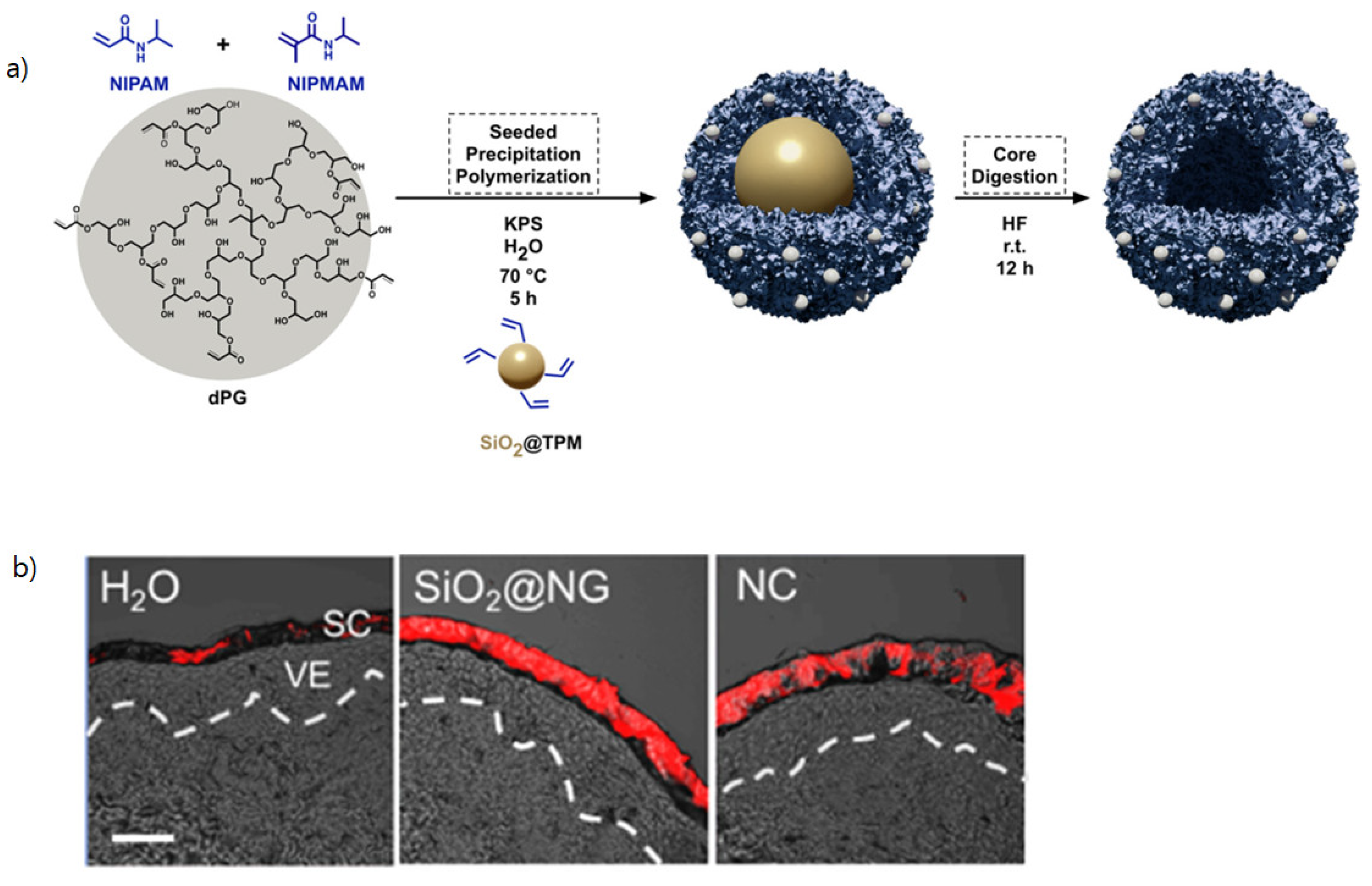
  36. F. Rancan, M. Giulbudagian, J. Jurisch, U. Blume-Peytavi, M. Calderón, and A. Vogt, “Drug delivery across intact and disrupted skin barrier: Identification of cell populations interacting with penetrated thermoresponsive nanogels,” (in eng), Eur J Pharm Biopharm, vol. 116, pp. 4-11, Jul 2017. [CrossRef]
  37. R. C. Op ’t Veld et al., “Thermosensitive biomimetic polyisocyanopeptide hydrogels may facilitate wound repair,” (in eng), Biomaterials, vol. 181, pp. 392-401, Oct 2018. [CrossRef]
  38. M. Asadian-Birjand et al., “Engineering thermoresponsive polyether-based nanogels for temperature dependent skin penetration,” (in English), Polymer Chemistry, Article vol. 6, no. 32, pp. 5827-5831, 2015 2015. [CrossRef]
  39. M. Kang, S. Hong, and J. Kim, “Release property of microgels formed by electrostatic interaction between poly(N-isopropylacrylamide-co-methacrylic acid) and poly(N-isopropylacrylamide-co-dimethylaminoethylmethacrylate),” (in English), Journal of Applied Polymer Science, Article vol. 125, no. 3, pp. 1993-1999, AUG 5 2012 2012. [CrossRef]
  40. N. Ferreira, L. Ferreira, V. Cardoso, F. Boni, A. Souza, and M. Gremiao, “Recent advances in smart hydrogels for biomedical applications: From self-assembly to functional approaches,” (in English), European Polymer Journal, Review vol. 99, pp. 117-133, FEB 2018 2018. [CrossRef]
  41. M. Neumann et al., “Stimuli-Responsive Hydrogels: The Dynamic Smart Biomaterials of Tomorrow,” (in eng), Macromolecules, vol. 56, no. 21, pp. 8377-8392, Nov 14 2023. [CrossRef]
  42. Gandhi, A. Paul, S. Sen, and K. Sen, “Studies on thermoresponsive polymers: Phase behaviour, drug delivery and biomedical applications,” (in English), Asian Journal of Pharmaceutical Sciences, Review vol. 10, no. 2, pp. 99-107, APR 2015 2015. [CrossRef]
  43. D. D. Ghosh and G. Chakrabarti, “Applications of Targeted Nano Drugs and Delivery Systems,” in Thermoresponsive Drug Delivery Systems, Characterization and Application, D. D. Ghosh and G. Chakrabarti Eds. Oxford, UK: Elsevier, 2019, pp. 133-155.
  44. Y. Ding, S. M. Pyo, and R. H. Müller, “smartLipids(®) as third solid lipid nanoparticle generation - stabilization of retinol for dermal application,” (in eng), Pharmazie, vol. 72, no. 12, pp. 728-735, Dec 1 2017. [CrossRef]
  45. M. Schäfer-Korting, W. Mehnert, and H. C. Korting, “Lipid nanoparticles for improved topical application of drugs for skin diseases,” (in eng), Adv Drug Deliv Rev, vol. 59, no. 6, pp. 427-43, Jul 10 2007. [CrossRef]
  46. J. Kang et al., “Preparation and evaluation of tacrolimus-loaded thermosensitive solid lipid nanoparticles for improved dermal distribution,” (in English), International Journal of Nanomedicine, Article vol. 14, pp. 5381-5396, 2019 2019. [CrossRef]
  47. C. Gerecke et al., “Biocompatibility and characterization of polyglycerol-based thermoresponsive nanogels designed as novel drug-delivery systems and their intracellular localization in keratinocytes,” (in English), Nanotoxicology, Article vol. 11, no. 2, pp. 267-277, MAR 2017 2017. [CrossRef]
  48. S. Jadhav, D. Scalarone, V. Brunella, E. Ugazio, S. Sapino, and G. Berlier, “Thermoresponsive copolymer-grafted SBA-15 porous silica particles for temperature-triggered topical delivery systems,” (in English), Express Polymer Letters, Article vol. 11, no. 2, pp. 96-105, FEB 2017 2017. [CrossRef]
  49. M. Witting et al., “Thermosensitive dendritic polyglycerol-based nanogels for cutaneous delivery of biomacromolecules,” (in English), Nanomedicine-Nanotechnology Biology and Medicine, Article vol. 11, no. 5, pp. 1179-1187, JUL 2015 2015. [CrossRef]
  50. E. Osorio-Blanco et al., “Polyglycerol-Based Thermoresponsive Nanocapsules Induce Skin Hydration and Serve as a Skin Penetration Enhancer,” (in English), Acs Applied Materials & Interfaces, Article vol. 12, no. 27, pp. 30136-30144, JUL 8 2020 2020. [CrossRef]
  51. E. Ugazio et al., “Thermoresponsive mesoporous silica nanoparticles as a carrier for skin delivery of quercetin,” (in English), International Journal of Pharmaceutics, Article vol. 511, no. 1, pp. 446-454, SEP 10 2016 2016. [CrossRef]
  52. M. Pelegrino, D. de Araújo, and A. Seabra, “S-nitrosoglutathione-containing chitosan nanoparticles dispersed in Pluronic F-127 hydrogel: Potential uses in topical applications,” (in English), Journal of Drug Delivery Science and Technology, Article vol. 43, pp. 211-220, FEB 2018 2018. [CrossRef]
  53. Zavgorodnya, C. Carmona-Moran, V. Kozlovskaya, F. Liu, T. Wick, and E. Kharlampieva, “Temperature-responsive nanogel multilayers of poly(N-vinylcaprolactam) for topical drug delivery,” (in English), Journal of Colloid and Interface Science, Article vol. 506, pp. 589-602, NOV 15 2017 2017. [CrossRef]
  54. P. Chen, H. Zhang, S. Cheng, G. Zhai, and C. Shen, “Development of curcumin loaded nanostructured lipid carrier based thermosensitive <i>in situ</i> gel for dermal delivery,” (in English), Colloids and Surfaces a-Physicochemical and Engineering Aspects, Article vol. 506, pp. 356-362, OCT 5 2016 2016. [CrossRef]
  55. H. Lambers, S. Piessens, A. Bloem, H. Pronk, and P. Finkel, “Natural skin surface pH is on average below 5, which is beneficial for its resident flora,” (in eng), Int J Cosmet Sci, vol. 28, no. 5, pp. 359-70, Oct 2006. [CrossRef]
  56. E. Jones, C. Cochrane, and S. Percival, “The Effect of pH on the Extracellular Matrix and Biofilms,” (in English), Advances in Wound Care, Review vol. 4, no. 7, pp. 431-439, JUL 2015 2015. [CrossRef]
  57. B. Eberlein-König et al., “Skin surface pH, stratum corneum hydration, trans-epidermal water loss and skin roughness related to atopic eczema and skin dryness in a population of primary school children,” (in eng), Acta Derm Venereol, vol. 80, no. 3, pp. 188-91, May 2000. [CrossRef]
  58. N. Schürer, “pH and Acne,” (in eng), Curr Probl Dermatol, vol. 54, pp. 115-122, 2018. [CrossRef]
  59. M. Karimi et al., “pH-Sensitive stimulus-responsive nanocarriers for targeted delivery of therapeutic agents,” (in English), Wiley Interdisciplinary Reviews-Nanomedicine and Nanobiotechnology, Review vol. 8, no. 5, pp. 696-716, SEP-OCT 2016 2016. [CrossRef]
  60. M. Abri Aghdam et al., “Recent advances on thermosensitive and pH-sensitive liposomes employed in controlled release,” Journal of Controlled Release, vol. 315, pp. 1-22, 2019/12/10/ 2019. [CrossRef]
  61. Banerjee, D. Mishra, T. Das, and T. Maiti, “Wound pH-Responsive Sustained Release of Therapeutics from a Poly(NIPAAm-co-AAc) Hydrogel,” (in English), Journal of Biomaterials Science-Polymer Edition, Article vol. 23, no. 1-4, pp. 111-132, 2012 2012. [CrossRef]
  62. H. Jeong, S. Nam, J. Song, and S. Park, “Synthesis and physicochemical properties of pH-sensitive hydrogel based on carboxymethyl chitosan/2-hydroxyethyl acrylate for transdermal delivery of nobiletin,” (in English), Journal of Drug Delivery Science and Technology, Article vol. 51, pp. 194-203, JUN 2019 2019. [CrossRef]
  63. L. Soriano-Ruiz et al., “Design and evaluation of a multifunctional thermosensitive poloxamer-chitosan-hyaluronic acid gel for the treatment of skin burns,” (in eng), Int J Biol Macromol, vol. 142, pp. 412-422, Jan 1 2020. [CrossRef]
  64. F. Alexis, E. Pridgen, L. Molnar, and O. Farokhzad, “Factors affecting the clearance and biodistribution of polymeric nanoparticles,” (in English), Molecular Pharmaceutics, Article|Proceedings Paper vol. 5, no. 4, pp. 505-515, JUL-AUG 2008 2008. [CrossRef]
  65. W. Gao et al., “Hydrogel containing nanoparticle-stabilized liposomes for topical antimicrobial delivery,” (in eng), ACS Nano, vol. 8, no. 3, pp. 2900-7, Mar 25 2014. [CrossRef]
  66. T. Yoshida, T. C. Lai, G. S. Kwon, and K. Sako, “pH- and ion-sensitive polymers for drug delivery,” (in eng), Expert Opin Drug Deliv, vol. 10, no. 11, pp. 1497-513, Nov 2013. [CrossRef]
  67. P. Dong et al., “pH-sensitive Eudragit® L 100 nanoparticles promote cutaneous penetration and drug release on the skin,” (in English), Journal of Controlled Release, Article vol. 295, pp. 214-222, FEB 2019 2019. [CrossRef]
  68. Rizi, R. Green, M. Donaldson, and A. Williams, “Using pH Abnormalities in Diseased Skin to Trigger and Target Topical Therapy,” (in English), Pharmaceutical Research, Article vol. 28, no. 10, pp. 2589-2598, OCT 2011 2011. [CrossRef]
  69. C. Shields et al., “Encapsulation and controlled release of retinol from silicone particles for topical delivery,” (in English), Journal of Controlled Release, Article vol. 278, pp. 37-48, MAY 28 2018 2018. [CrossRef]
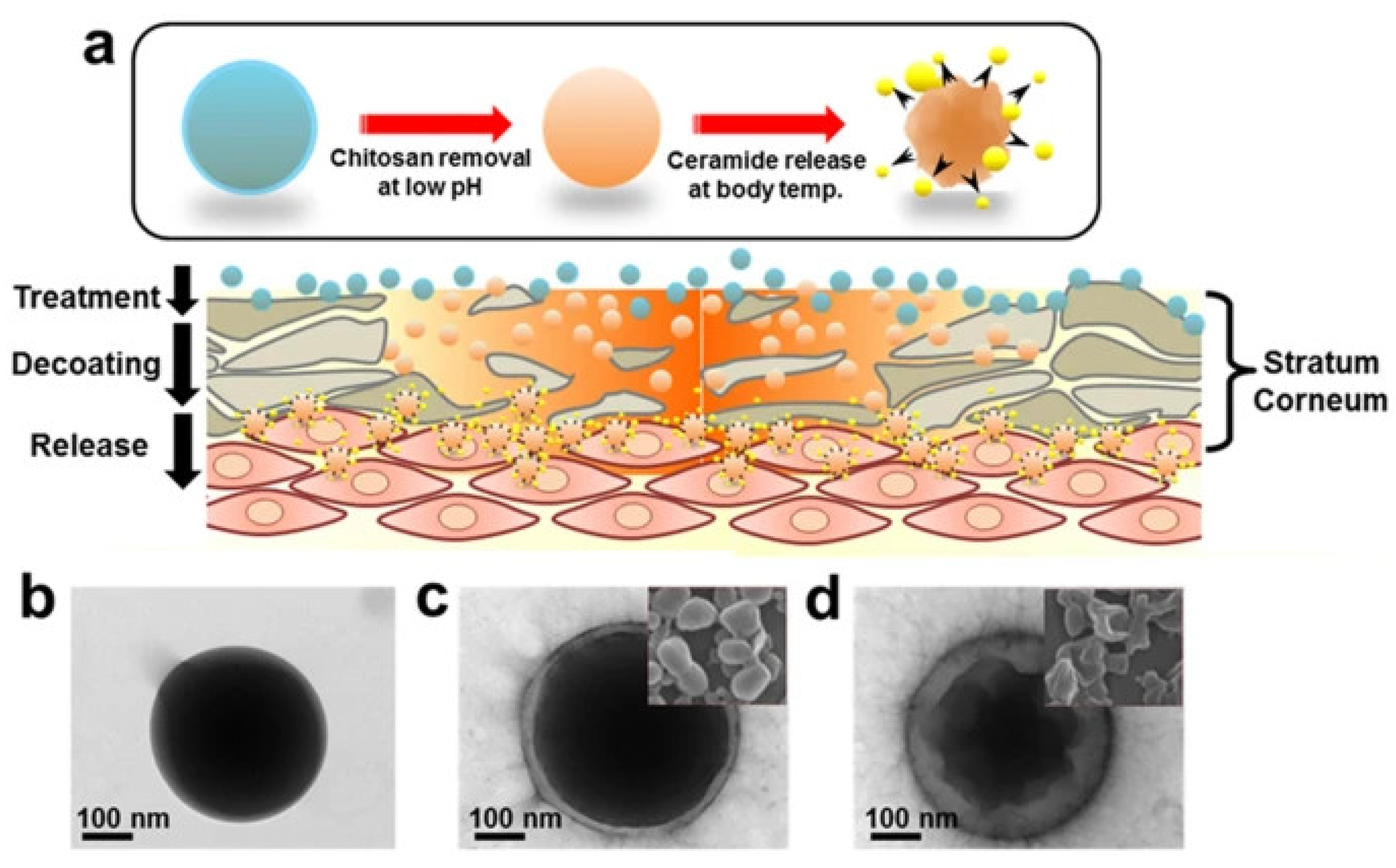
  70. S. Jung et al., “Thermodynamic Insights and Conceptual Design of Skin-Sensitive Chitosan Coated Ceramide/PLGA Nanodrug for Regeneration of Stratum Corneum on Atopic Dermatitis,” (in English), Scientific Reports, Article vol. 5, DEC 15 2015 2015, Art no. ARTN 18089. [CrossRef]
  71. Y. Kang et al., “Nanocarrier-Based Transdermal Drug Delivery Systems for Dermatological Therapy,” (in English), Pharmaceutics, Review vol. 16, no. 11, NOV 2024 2024, Art no. ARTN 1384. [CrossRef]
  72. Van Gheluwe, E. Buchy, I. Chourpa, and E. Munnier, “Three-Step Synthesis of a Redox-Responsive Blend of PEG-block-PLA and PLA and Application to the Nanoencapsulation of Retinol,” (in eng), Polymers (Basel), vol. 12, no. 10, Oct 14 2020. [CrossRef]
  73. U. Ahmad, Z. Ahmad, A. A. Khan, J. Akhtar, S. P. Singh, and F. J. Ahmad, “Strategies in Development and Delivery of Nanotechnology Based Cosmetic Products,” (in eng), Drug Res (Stuttg), vol. 68, no. 10, pp. 545-552, Oct 2018. [CrossRef]
  74. S. E. Kim, P. W. Lee, and J. K. Pokorski, “Biologically Triggered Delivery of EGF from Polymer Fiber Patches,” (in eng), ACS Macro Lett, vol. 6, no. 6, pp. 593-597, 2017. [CrossRef]
  75. J. S. Im, B. Bai, and Y. S. Lee, “The effect of carbon nanotubes on drug delivery in an electro-sensitive transdermal drug delivery system,” (in eng), Biomaterials, vol. 31, no. 6, pp. 1414-9, Feb 2010. [CrossRef]
  76. R. Kim, S. L. Lee, and S. N. Park, “Properties and in vitro drug release of pH- and temperature-sensitive double cross-linked interpenetrating polymer network hydrogels based on hyaluronic acid/poly (N-isopropylacrylamide) for transdermal delivery of luteolin,” (in eng), Int J Biol Macromol, vol. 118, no. Pt A, pp. 731-740, Oct 15 2018. [CrossRef]
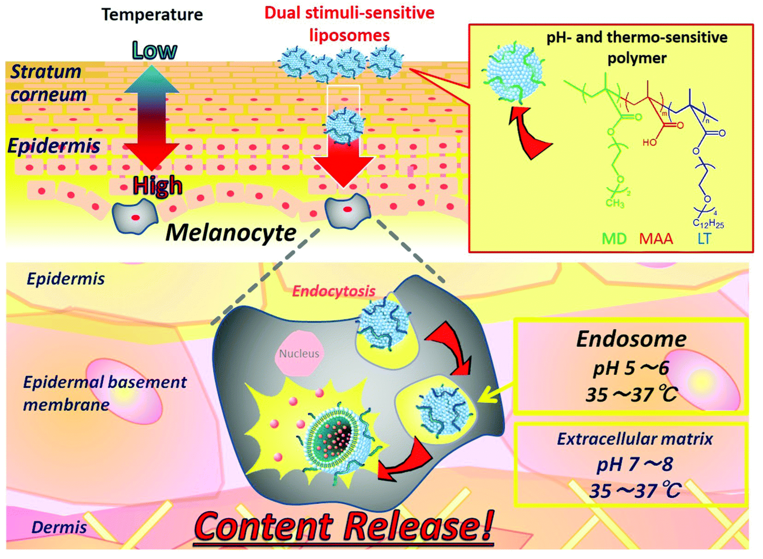
  77. Yamazaki et al., “Dual-stimuli responsive liposomes using pH- and temperature-sensitive polymers for controlled transdermal delivery,” (in English), Polymer Chemistry, Article vol. 8, no. 9, pp. 1507-1518, MAR 7 2017 2017. [CrossRef]
  78. Kim, S. Lee, and S. Park, “Properties and <i>in vitro</i> drug release of pH- and temperature-sensitive double cross-linked interpenetrating polymer network hydrogels based on hyaluronic acid/poly (N-isopropylacrylamide) for transdermal delivery of luteolin,” (in English), International Journal of Biological Macromolecules, Article vol. 118, pp. 731-740, OCT 15 2018 2018. [CrossRef]
  79. V. Lopez, J. Hadgraft, and M. Snowden, “The use of colloidal microgels as a (trans)dermal drug delivery system,” (in English), International Journal of Pharmaceutics, Article vol. 292, no. 1-2, pp. 137-147, MAR 23 2005 2005. [CrossRef]
  80. Abu Samah and C. Heard, “Enhanced <i>in vitro</i> transdermal delivery of caffeine using a temperature- and pH-sensitive nanogel, poly(NIPAM-<i>co</i>-AAc),” (in English), International Journal of Pharmaceutics, Article vol. 453, no. 2, pp. 630-640, SEP 10 2013 2013. [CrossRef]
  81. S. Mavuso et al., “<i>In Vitro</i>, <i>Ex Vivo</i>, and <i>In Vivo</i> Evaluation of a Dual pH/Redox Responsive Nanoliposomal Sludge for Transdermal Drug Delivery,” (in English), Journal of Pharmaceutical Sciences, Article vol. 107, no. 4, pp. 1028-1036, APR 2018 2018. [CrossRef]
  82. Ferraris, C. Rimicci, S. Garelli, E. Ugazio, and L. Battaglia, “Nanosystems in Cosmetic Products: A Brief Overview of Functional, Market, Regulatory and Safety Concerns,” (in English), Pharmaceutics, Review vol. 13, no. 9, SEP 2021 2021, Art no. ARTN 1408. [CrossRef]
  83. Hubbs et al., “Nanotoxicology-A Pathologist’s Perspective,” (in English), Toxicologic Pathology, Review vol. 39, no. 2, pp. 301-324, FEB 2011 2011. [CrossRef]
  84. M. Mogharabi, M. Abdollahi, and M. Faramarzi, “Toxicity of nanomaterials; an undermined issue,” (in English), Daru-Journal of Pharmaceutical Sciences, Editorial Material vol. 22, AUG 15 2014 2014, Art no. ARTN 59. [CrossRef]
  85. S. L. Schneider and H. W. Lim, “A review of inorganic UV filters zinc oxide and titanium dioxide,” (in eng), Photodermatol Photoimmunol Photomed, vol. 35, no. 6, pp. 442-446, Nov 2019. [CrossRef]
  86. Melo, M. Amadeu, M. Lancellotti, L. de Hollanda, and D. Machado, “THE ROLE OF NANOMATERIALS IN COSMETICS: NATIONAL AND INTERNATIONAL LEGISLATIVE ASPECTS,” (in English), Quimica Nova, Article vol. 38, no. 4, pp. 599-603, MAY 2015 2015. [CrossRef]
  87. M. Ajazzuddin, G. Jeswani, and A. Kumar Jha, “Nanocosmetics: Past, Present and Future Trends,” Recent Patents on Nanomedicine, vol. 5, no. 1, pp. 3-11, // 2015.
  88. Guidance, “Guidance for Industry Considering Whether an FDA-Regulated Product Involves the Application of Nanotechnology,” (in English), Biotechnology Law Report, Article vol. 30, no. 5, pp. 613-616, SEP 2011 2011. [CrossRef]
  89. Buzea, Pacheco, II, and K. Robbie, “Nanomaterials and nanoparticles: sources and toxicity,” (in eng), Biointerphases, vol. 2, no. 4, pp. Mr17-71, Dec 2007. [CrossRef]
  90. Joudeh and D. Linke, “Nanoparticle classification, physicochemical properties, characterization, and applications: a comprehensive review for biologists,” Journal of Nanobiotechnology, vol. 20, no. 1, p. 262, 2022/06/07 2022. [CrossRef]
  91. T. Gebel et al., “Manufactured nanomaterials: categorization and approaches to hazard assessment,” (in English), Archives of Toxicology, Review vol. 88, no. 12, pp. 2191-2211, DEC 2014 2014. [CrossRef]
  92. W. W. Chan et al., “Grand Plans for Nano,” (in eng), ACS Nano, vol. 9, no. 12, pp. 11503-5, Dec 22 2015. [CrossRef]
  93. J. Shokri, “Nanocosmetics: benefits and risks,” (in English), Bioimpacts, Editorial Material vol. 7, no. 4, pp. 207-208, 2017 2017. [CrossRef]
  94. M. Schneider, F. Stracke, S. Hansen, and U. F. Schaefer, “Nanoparticles and their interactions with the dermal barrier,” (in eng), Dermatoendocrinol, vol. 1, no. 4, pp. 197-206, Jul 2009. [CrossRef]
  95. M. Wang, X. Lai, L. Shao, and L. Li, “Evaluation of immunoresponses and cytotoxicity from skin exposure to metallic nanoparticles,” (in eng), Int J Nanomedicine, vol. 13, pp. 4445-4459, 2018. [CrossRef]
  96. Groso, A. Petri-Fink, A. Magrez, M. Riediker, and T. Meyer, “Management of nanomaterials safety in research environment,” (in English), Particle and Fibre Toxicology, Review vol. 7, DEC 10 2010 2010, Art no. ARTN 40. [CrossRef]
  97. Drasler, P. Sayre, K. Steinhäuser, A. Petri-Fink, and B. Rothen-Rutishauser, “In vitro approaches to assess the hazard of nanomaterials (vol 8, pg 99, 2017),” (in English), Nanoimpact, Correction vol. 9, pp. 51-51, JAN 2018 2018. [CrossRef]
  98. J. H. Lee and Y. Yeo, “Controlled drug release from pharmaceutical nanocarriers,” Chemical Engineering Science, vol. 125, pp. 75-84, 2015/03/24/ 2015. [CrossRef]
  99. S. Adepu and S. Ramakrishna, “Controlled Drug Delivery Systems: Current Status and Future Directions,” (in eng), Molecules, vol. 26, no. 19, Sep 29 2021. [CrossRef]
  100. M. Gupta, U. Agrawal, and S. P. Vyas, “Nanocarrier-based topical drug delivery for the treatment of skin diseases,” (in eng), Expert Opin Drug Deliv, vol. 9, no. 7, pp. 783-804, Jul 2012. [CrossRef]
  101. H. T. Phan and A. J. Haes, “What Does Nanoparticle Stability Mean?,” The Journal of Physical Chemistry C, vol. 123, no. 27, pp. 16495-16507, 2019/07/11 2019. [CrossRef]
  102. N. Akombaetwa, A. B. Ilangala, L. Thom, P. B. Memvanga, B. A. Witika, and A. B. Buya, “Current Advances in Lipid Nanosystems Intended for Topical and Transdermal Drug Delivery Applications,” (in eng), Pharmaceutics, vol. 15, no. 2, Feb 15 2023. [CrossRef]
  103. R. John, J. Monpara, S. Swaminathan, and R. Kalhapure, “Chemistry and Art of Developing Lipid Nanoparticles for Biologics Delivery: Focus on Development and Scale-Up,” Pharmaceutics, vol. 16, no. 1. [CrossRef]
  104. J. Lee and K. H. Kwon, “Future value and direction of cosmetics in the era of metaverse,” Journal of Cosmetic Dermatology, vol. 21, no. 10, pp. 4176-4183, 2022/10/01 2022. [CrossRef]
  105. Ahmad Tarar, U. Mohammad, and K. S. S, “Wearable Skin Sensors and Their Challenges: A Review of Transdermal, Optical, and Mechanical Sensors,” (in eng), Biosensors (Basel), vol. 10, no. 6, May 28 2020. [CrossRef]
  106. Elder, M. Cappelli, C. Ring, and N. Saedi, “Artificial intelligence in cosmetic dermatology: An update on current trends,” (in English), Clinics in Dermatology, Article vol. 42, no. 3, pp. 216-220, MAY-JUN 2024 2024. [CrossRef]
  107. Kania, K. Montecinos, and D. Goldberg, “Artificial intelligence in cosmetic dermatology,” (in English), Journal of Cosmetic Dermatology, Article vol. 23, no. 10, pp. 3305-3311, OCT 2024 2024. [CrossRef]
  108. Vatiwutipong, S. Vachmanus, T. Noraset, and S. Tuarob, “Artificial Intelligence in Cosmetic Dermatology: A Systematic Literature Review,” (in English), Ieee Access, Review vol. 11, pp. 71407-71425, 2023 2023. [CrossRef]
Figure 1. The schematic diagram of different nanoscale materials ranging between 1 ~ 100 nm. Organic nanoparticles (polymers, dendrimers); Inorganic nanoparticles (calcium phosphate, gold nanoparticles); Organic/Inorganic hybrids (functionalized gold nanoparticles, nanocomposites); Carbon based (functionalized fullerenes); Liposomes and biological nanoparticles (protein and nucleic acid based). Reproduced from [33] under a creative common attribution 4.0 (https://creativecommons.org/licenses/by/4.0/).
Preprints 151849 g001
Disclaimer/Publisher’s Note: The statements, opinions and data contained in all publications are solely those of the individual author(s) and contributor(s) and not of MDPI and/or the editor(s). MDPI and/or the editor(s) disclaim responsibility for any injury to people or property resulting from any ideas, methods, instructions or products referred to in the content.
Copyright: This open access article is published under a Creative Commons CC BY 4.0 license, which permit the free download, distribution, and reuse, provided that the author and preprint are cited in any reuse.
Prerpints.org logo

Preprints.org is a free preprint server supported by MDPI in Basel, Switzerland.

Subscribe

Disclaimer

Terms of Use

Privacy Policy

Privacy Settings

© 2025 MDPI (Basel, Switzerland) unless otherwise stated