Preprint
Article

This version is not peer-reviewed.

Are Deep Tissue Cultures a Reliable Alternative to Bone Biopsy for Diagnosing Diabetic Foot Osteomyelitis? A Comparative Diagnostic Study

A peer-reviewed article of this preprint also exists.

Submitted:

11 March 2025

Posted:

12 March 2025

You are already at the latest version

Abstract

Background: Diabetic foot osteomyelitis (DFO) is a serious complication of diabetic foot ulcers (DFUs) that contributes to high morbidity and an increased risk of lower extremity amputation. While bone biopsy cultures are considered the gold standard for identifying causative pathogens, their invasive nature limits widespread clinical use. This study evaluates the microbiological concordance between deep tissue and bone cultures in diagnosing DFO. Methods: A retrospective analysis was con-ducted on 107 patients with DFO who underwent simultaneous deep tissue and bone biopsy cultures. Patient demographics, ulcer classification, and microbiological culture results were recorded. The agreement between deep tissue and bone cultures was as-sessed to determine the diagnostic utility of deep tissue sampling. Results: The overall concordance between deep tissue and bone cultures was 51.8%. Staphylococcus aureus was the most frequently isolated pathogen in both culture types and had the highest agreement rate (44.4%). Concordance rates were lower for Gram-negative bacteria (31.9%) and other Gram-positive microorganisms (24.2%). In 21.2% of cases, patho-gens were isolated only from deep tissue cultures, while 16.5% had positive bone cul-tures but negative deep tissue cultures. Conclusions: Deep tissue cultures demonstrate moderate agreement with bone biopsy cultures when diagnosing DFO, particularly for Staphylococcus aureus. While bone biopsy remains the gold standard diagnosis tool, deep tissue cultures may provide clinically useful information when bone sampling is not feasible. Further studies are needed to improve non-invasive diagnostic methods for DFO.

Keywords: 
;  ;  ;  ;  ;  ;  

1. Introduction

Diabetes mellitus (DM) is a metabolic disorder that has become an increasing global health burden and is now recognized as one of the most significant public health challenges worldwide. Current epidemiological data estimate that approximately 828 million individuals worldwide are living with diabetes, reflecting an increase of 630 million cases since 1990 [1]. This dramatic rise in diabetes prevalence has resulted in a parallel increase in diabetes-related complications, thereby placing an escalating burden on healthcare systems.
DM is associated with a wide range of complications that contribute to increased morbidity and mortality, with diabetic foot ulcers (DFUs) representing a particularly significant clinical challenge due to their impact on lower extremities [2]. It is estimated that 19–34% of individuals with diabetes will develop a DFU at some point in their lifetime, with up to 60% of these ulcers becoming complicated by infection. The severity of these infections varies, and it is estimated that 20–60% of diabetic foot infections involve underlying bone, leading to diabetic foot osteomyelitis (DFO) [3,4,5]. DFO is a serious and complex complication associated with an increased risk of amputation, prolonged antibiotic therapy, delayed wound healing, high recurrence rates, and extended hospitalization periods, making its accurate diagnosis and appropriate management crucial for improving patient outcomes [4,5].
The presence or absence of osteomyelitis in DFU is a critical determinant of ulcer prognosis. Accurate diagnosis of osteomyelitis is essential for optimizing treatment strategies, preventing unnecessary and prolonged antibiotic use, and minimizing the need for invasive surgical interventions. [6]. Early and definitive diagnosis is essential to avoid inappropriate therapeutic decisions and prevent disease progression. However, the absence of universally accepted diagnostic criteria and the variability among currently available diagnostic tests complicate the clinical decision-making process [7,8]. Although a combination of clinical assessment, radiological imaging, and laboratory testing is considered to enhance diagnostic accuracy, existing evidence highlights the limitations of each method, and the most effective diagnostic approach remains uncertain [9]. Moreover, emerging molecular diagnostic techniques and advanced imaging modalities, such as positron emission tomography-computed tomography (PET-CT) and magnetic resonance imaging (MRI), have been proposed to improve diagnostic precision, but their cost, availability, and standardization remain challenging in routine clinical settings[10] .
Microbiological and histopathological analysis of bone biopsy specimens is considered the gold standard for diagnosing osteomyelitis, enabling the precise identification of causative pathogens and their antibiotic susceptibility profiles [11,12]. However, bone biopsy may not always be feasible in routine clinical practice due to several limitations, including the requirement for an experienced clinical team, specialized equipment, and the potential risk of procedural complications such as fractures or contamination of adjacent tissues. Additionally, disparities in accessibility to bone biopsy procedures across different healthcare settings and geographical regions further complicate its widespread adoption[13].
Although international guidelines recommend tailoring antimicrobial therapy based on bone biopsy culture results, real-world clinical practice often relies on soft tissue culture results to guide treatment decisions. Given the potential spread of infection from superficial soft tissue to bone in DFO, deep tissue cultures obtained from the ulcer base have been proposed as an alternative diagnostic tool. However, their ability to reliably reflect bone infection remains controversial [14].
In this study, we aimed to evaluate whether deep tissue cultures serve as a reliable alternative to bone biopsy cultures in the diagnosis of DFO. By analyzing the microbiological concordance between deep tissue and bone biopsy cultures, we seek to contribute to the development of more effective and practical diagnostic strategies for improving DFO management and treatment approaches. Additionally, we aim to explore the clinical implications of adopting deep tissue cultures as a diagnostic tool and their potential role in guiding targeted antibiotic therapy, thereby optimizing patient outcomes while addressing the limitations associated with bone biopsy procedures[15].

2. Materials and Methods

2.1. Ethical Approval

The study was approved by the Ethics Committee of Ankara Bilkent City Hospital (TABED 1-24-820). All participants were provided with detailed information regarding the study's purpose and design, and both verbal and written informed consent were obtained. All study procedures were conducted in accordance with the ethical standards of the institutional review board and the principles of the 1964 Declaration of Helsinki and its subsequent revisions.

2.2. Study Population

This retrospective study analyzed patients who were followed up in the Chronic Wound Unit of our hospital between 1 January 2024 and 1 July 2024 due to diabetic foot wounds.
The Chronic Wound Unit operates with a multidisciplinary approach and serves as a tertiary referral center, evaluating and treating approximately 400 patients per month.
A total of 107 patients aged 18 years or older were included in the study based on the following criteria:
  • Diagnosis of diabetic foot infection;
  • Positive probe-to-bone (PTB) test (performed using a sterile blunt metal probe; considered positive when bone was palpable through the ulcer);
  • Radiographic evidence of osteomyelitis (presence of suggestive findings in initial or follow-up X-rays);
  • Absence of clinical signs of Charcot’s neuroarthropathy;
  • Concurrent collection of deep tissue and bone culture samples in the operating room during hospitalization;
  • Not receiving antibiotic therapy at the time of hospital admission
The demographic characteristics of the patients were recorded, including age, sex, comorbidities, prior antibiotic use, and classification of the ulcer according to the Infectious Diseases Society of America/International Working Group of the Diabetic Foot (IDSA/IWGDF) classification system, along with the causative microorganisms isolated from deep tissue and bone cultures.
All data were obtained from the hospital’s electronic medical record system.
Figure 1. (a) Diabetic toe osteomyelitis with positive probe-to-bone test. (b): Same patient, X-ray image consistent with osteomyelitis. Cortical irregularities, osteolytic lesions, and soft tissue edema are observed at the level of the interphalangeal joints.
Preprints 152059 g001
Figure 2. (a) A diabetic foot ulcer on the plantar surface with a positive probe-to- bone test, consistent with osteomyelitis. (b) Same patient, X-ray image consistent with osteomyelitis. Phalanges and metatarsal bones show osteolytic lesions, cortical erosion, and irregularities.
Preprints 152059 g002

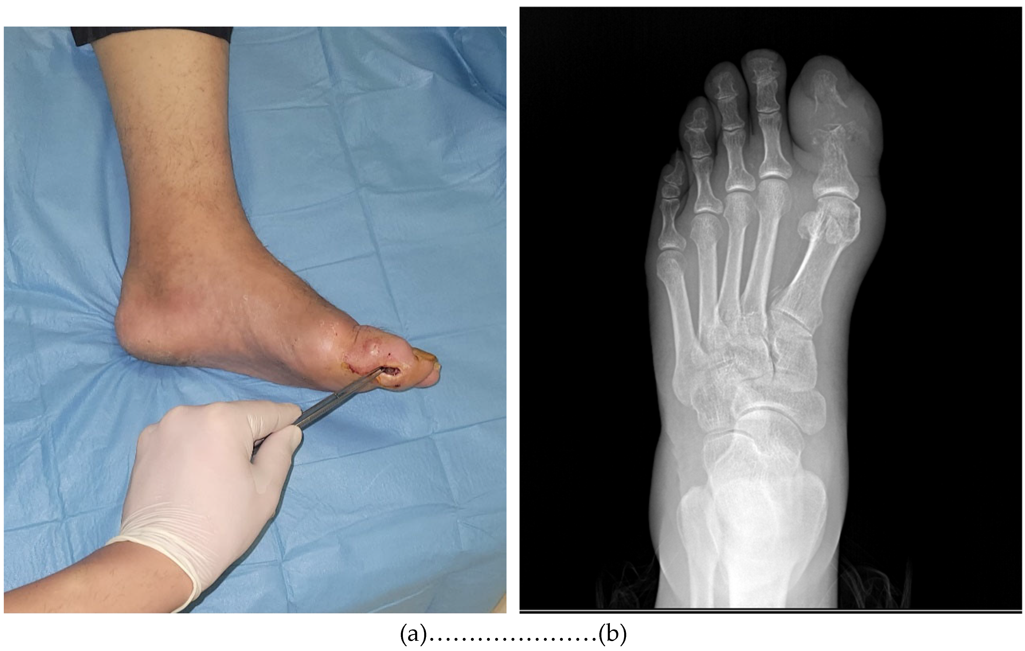
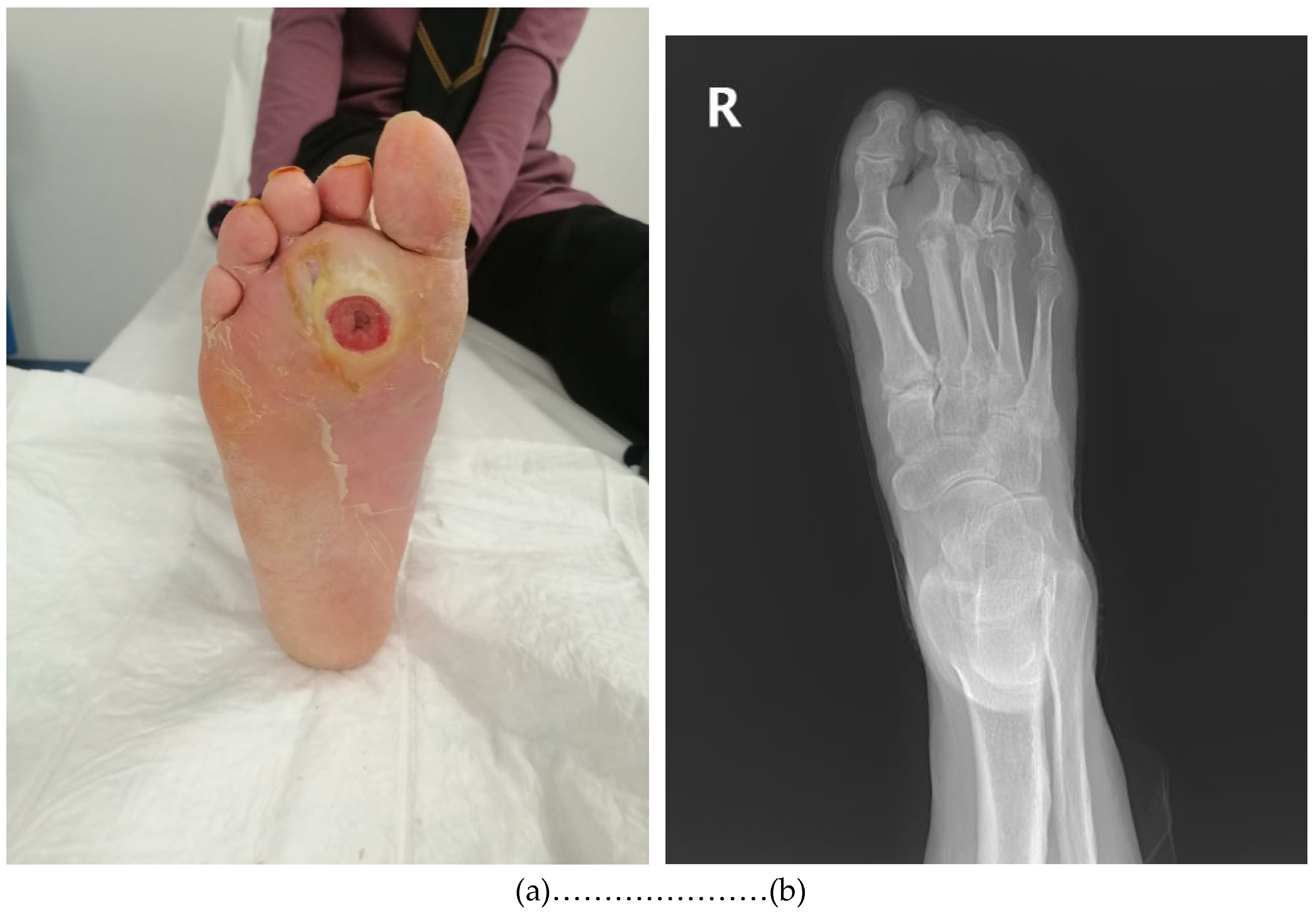
2.3. Specimen Collection

The IDSA/IWGDF classification system [16] was used to grade the DFUs. Simultaneous deep tissue and bone samples were collected intraoperatively under peripheral nerve blockade by the same team. Following the removal of necrotic tissues and superficial debris from the ulcer base, deep tissue biopsy specimens measuring approximately 4–5 mm in diameter were obtained under aseptic conditions using a sterile scalpel. Bone samples were collected either during surgical debridement or amputation in cases where infected bone was excised. Throughout the sampling process, strict aseptic conditions were maintained to minimize the contamination risk. To prevent cross-contamination, surgical instruments and gloves were changed as necessary and potential contamination sources were eliminated. Standard culturing techniques were used and all conditions were similar for all patient samples. Our hospital has an in-house microbiology laboratory. All the collected specimens were placed into a transport medium and sent to our hospital microbiology laboratory for culturing within 30 min of data collection.

2.4. Microbiological Analysis

The identification of isolates in tissue and bone specimen cultures and antibiotic susceptibility tests were performed with a VITEK-2 (bioMérieux, Marcy-I’Ètoile, France) automated identification device. Methicillin resistance in Staphylococcus isolates was evaluated with cefoxitin. Resistance rates and MIC (minimal inhibitory concentration) values were determined according to EUCAST (the European Committee on Antimicrobial Susceptibility Testing) standards [17]. Anaerobic bacteria were not included in this study because anaerobic cultures were not performed in our laboratory.

2.5.Statistical Analysis

Descriptive statistical analysis was performed using IBM SPSS Statistics 25 software (SPSS Inc., Chicago, IL, USA, 2011). Age is expressed as the mean ± standard deviation. Sex, frequency of comorbid diseases and situations, grade of ulcers, identified microorganisms, and concordance between deep tissue and bone sample cultures are expressed as numbers and percentages.

3. Results

A total of 107 patients were included in the study. The mean age was 63.4 ± 12.3 years, and74.8% (n = 80) of the participants were male. All the patients had at least one comorbidity. The most common comorbid conditions included hypertension (93.5%), peripheral neuropathy (91.5%), peripheral vascular obstruction (89.7%), and atherosclerosis (75.7%) Prior exposure to antibiotics was noted in 96.3% of patients (n = 103). (Table 1).
According to the IDSA/IWGDF ulcer classification system, grade 3 ulcers were the most prevalent (64.5%), followed by grade 4 (30.8%) and grade 2 (4.7%) ulcers (Table 2).
Deep tissue cultures were available for 107 patients, and bone cultures were available for 105 of the patients. All infections were monomicrobial with a dominant pathogen identified in every culture that showed microbial growth. In total ,85 patients had microorganism growth in any cultures.
The most identified pathogen in both the deep tissue and bone cultures was Staphylococcus aureus (S. aureus). It was isolated from 13.1% of the patients (Table 3). The second most common pathogen in deep tissue cultures was Escherichia coli, while the second most common agent in bone cultures was Klebsiella pneumoniae.
In total, 85 patients had microorganism growth in any cultures. While the same pathogen was isolated from both the deep tissue and bone cultures in 51.8% of the patients. In contrast, in 10.6% of cases, there was a discordance between the pathogens isolated from deep tissue and bone samples. In 21.2% of patients, bacterial growth was detected only in the deep tissue culture, while no growth was observed in the bone culture. Similarly, in 16.5% of patients, growth was observed only in the bone culture, whereas no pathogen was detected in the deep tissue culture (Table 4).
As mentioned above, 51.8% cases had concordance between the results from the deep tissue and bone cultures. When the concordance was evaluated according to the identified pathogen, the highest concordance between the deep tissue and bone cultures was in patients were S. aureus (44.4%) was identified, followed by Gram-negative (31.9%) and then other Gram-positive microorganisms (24.2%) (Table 5).

4. Discussion

This study aims to evaluate the microbiological concordance between deep tissue and bone biopsy cultures in the diagnosis of DFO and to analyze the diagnostic accuracy of deep tissue cultures. The accurate microbiological diagnosis of DFO is crucial for determining appropriate treatment protocols.
In our study, the overall concordance rate between deep tissue and bone biopsy cultures was found to be 51.8%. This finding indicates that in one out of every two patients, the same pathogen was isolated in both culture types. The highest concordance rate was observed in patients with S. aureus isolation (44.4%), while lower concordance rates were found in Gram-negative bacteria (31.9%) and other Gram-positive microorganisms (24.2%). These findings suggest that although deep tissue cultures have a limited but still meaningful diagnostic value compared to bone biopsy, their limitations should be considered, particularly in microorganisms with lower concordance rates. The use of deep tissue cultures alone may not always be sufficient for an accurate diagnosis of osteomyelitis.
DFIs are a common health problem in individuals with diabetes, often leading to serious complications such as amputation. These infections typically originate from an open wound in the skin and soft tissue but, in most cases, progress to involve the underlying bone, resulting in osteomyelitis. DFO is an infection that develops as a consequence of long-standing diabetes and is frequently associated with advanced peripheral neuropathy, peripheral arterial disease, foot deformities, and inadequate foot care (18,19). In our study, the majority of patients were also found to have these two factors .Compared to the rates reported in the literature (20,21,22,23), the higher prevalence observed in our clinic may be attributed to the fact that our center serves as a national referral facility for advanced-stage patients. In particular, the majority of admitted patients presented with stage three and four ulcers, which contributed to the increased severity of infection and ischemic processes.
The management of DFO often involves surgical debridement, long-term antibiotic therapy, or a combination of both [24,25,26]. However, the effectiveness of antibiotic treatment relies on the accurate identification of causative pathogens. To confirm the diagnosis of osteomyelitis and accurately determine the responsible microorganisms, bone biopsy culture is considered the gold standard. However, the invasive nature of bone biopsy, technical challenges, and the risk of complications limit its integration into routine clinical practice [27,28].
As a less invasive and more practical alternative, deep tissue cultures are frequently used in clinical practice. Slater et al. emphasized that, due to the limitations of bone biopsy, there is an increasing reliance on soft tissue cultures in clinical settings [29]. However, the extent to which deep tissue cultures accurately represent osteomyelitis pathogens and their diagnostic reliability remains controversial.In particular, determining the diagnostic accuracy of deep tissue cultures becomes crucial in cases where bone biopsy is not feasible. In this context, studies evaluating the diagnostic reliability of deep tissue cultures are becoming increasingly important for establishing optimal treatment strategies in the management of DFO.
Studies evaluating the diagnostic concordance between bone biopsy cultures, which are considered the gold standard for osteomyelitis diagnosis, and other tissue cultures have reported varying concordance rates. Senneville et al. found a 22.5% concordance between superficial swab cultures and percutaneous bone biopsy cultures, demonstrating that superficial swabs are not a reliable method for diagnosing osteomyelitis [30]. A prospective study from India, which included 144 patients, reported a concordance rate of only 38.2% between bone biopsy and superficial swab cultures [31]. Liu et al. found a 42.8% concordance between deep tissue and bone biopsy cultures [32]. In our study, the concordance rate was 51.8%, which is higher than previously reported studies. Similarly, Ertuğrul et al. reported a 49% concordance rate in a prospective study involving 45 patients [33]. A systematic review conducted in 2020 reported that the concordance rates between bone and soft tissue cultures ranged from 19% to 42%, with the highest concordance observed for S. aureus [34].
These findings indicate that deep tissue cultures should not be completely disregarded; however, they do not serve as a full alternative to bone biopsy. Nevertheless, there are studies supporting the reliability of deep tissue cultures. Malone et al. reported a concordance rate of 73.5% between deep tissue cultures and either surgical or percutaneous bone biopsies and suggested that deep tissue cultures should not be overlooked, particularly in centers where bone biopsy is not feasible [35].
In the literature, some studies have proposed sinus tract cultures as an alternative diagnostic method for osteomyelitis. A non-randomized, prospective study from Switzerland, which included 54 cases of osteomyelitis, demonstrated that two consecutive sinus tract cultures could accurately predict the causative pathogen in monomicrobial infections [36].Similarly, in a cross-sectional, prospectively designed study by Soomro et al., which included 90 patients with chronic osteomyelitis, it was suggested that sinus tract cultures might provide valuable microbiological information when interpreted cautiously. However, the study also emphasized that sinus tract cultures carry a high risk of contamination and should therefore be carefully evaluated in clinical decision-making [37].
In our study, S. aureus was identified as the most frequently isolated pathogen in both bone biopsy and deep tissue cultures. This finding is consistent with the study conducted by Hartemann-Heurtier and Senneville (2008) and supports the notion that S. aureus is one of the primary causative pathogens in DFO [38]. Additionally, the highest concordance rate between bone biopsy and deep tissue cultures was observed in cases with S.aureus isolation (44.4%). Notably, the most recent guidelines of the IWGDF emphasize that, in cases where a single virulent pathogen particularly S. Aureus is isolated from an aseptically obtained deep soft tissue sample, a bone biopsy may not be necessary [39]. This recommendation aligns with the findings of our study, suggesting that deep tissue cultures may hold diagnostic value in cases dominated by S. aureus. However, bone biopsy remains essential for the identification of other pathogens, particularly in polymicrobial infections, where advanced diagnostic techniques may be more appropriate.
When the pathogens isolated from deep tissue and bone cultures of the patients included in our study were classified as Gram-positive and Gram-negative microorganisms, Gram-negative microorganisms were found to be more frequently isolated. The most commonly detected Gram-negative bacteria were Escherichia coli and Klebsiella pneumoniae. Therefore, when determining the empirical treatment for osteomyelitis, it should be considered that Gram-negative bacteria are the predominant microorganisms. When evaluating the concordance rates between bone biopsy and deep tissue cultures, the lower concordance rate observed for Gram-negative bacteria (31.9%) suggests that deep tissue cultures may have limitations in accurately identifying the causative pathogens of osteomyelitis. Pellizzer et al. (2001) reported that wound cultures are often insufficient in isolating Gram-negative bacteria, which may lead to inappropriate antibiotic therapy and treatment failures [40].
In our study, 21.2% of patients had positive deep tissue cultures but negative bone cultures, whereas 16.5% had positive bone cultures but negative deep tissue cultures. This discrepancy may be attributed to the heterogeneous distribution of infection within anatomical compartments or the effects of prior antibiotic exposure. Although patients were not receiving antibiotics at the time of sample collection, the retrospective design of our study limits the ability to assess the precise impact of previous antimicrobial use on the culture results.
Antibiotic therapy can significantly affect culture outcomes. Prior studies have shown that previous antibiotic use may lead to false-negative culture results in both bone and deep tissue samples, and discontinuing antibiotics before biopsy increases the likelihood of pathogen detection [41,42]. However, the optimal antibiotic-free interval before biopsy remains uncertain, with some experts recommending a minimum of several days, ideally two weeks, before obtaining bone biopsy specimens [42]. In our study, most patients had a history of prior antibiotic use, but antibiotics were discontinued at the time of sample collection. However, due to the retrospective nature of this study, the exact duration of antibiotic cessation could not be determined, representing a study limitation.
In our study, anaerobic bacteria were not isolated, likely due to the limitations in anaerobic bacterial culture conditions in our hospital laboratory. Despite numerous studies on DFI over the past two decades, the true prevalence of anaerobic pathogens in DFI remains uncertain. This uncertainty is largely attributed to the lack of standardization in bacterial culture methods across studies. Factors such as the type of sample collected for analysis, the conditions and efficiency of sample transport to the microbiology laboratory, and the processing methods used significantly influence the detection rate of anaerobic bacteria [43]. In our study, no bacterial growth was observed in 39.3% of deep tissue cultures and 33.3% of bone cultures. Considering the high prevalence of peripheral arterial occlusion in our patient cohort, it is plausible that anaerobic bacteria could be the causative pathogens in these culture-negative cases. If anaerobic bacteria had been successfully cultured, the concordance rate between bone and tissue cultures might have been higher. In this context, replacing conventional culture techniques with advanced molecular diagnostic methods could enhance the identification of anaerobic bacteria in the etiology of DFI, providing a more accurate assessment of their role. [44].
Traditional culture methods may be insufficient for detecting anaerobic bacteria and fastidious pathogens. In recent years, molecular techniques such as 16S rRNA gene sequencing and metagenomic next-generation sequencing (mNGS) have emerged as promising tools with the potential to identify DFO pathogens with higher sensitivity [45,46]. These methods offer several advantages, including the ability to detect pathogens even in culture-negative cases, improved identification of polymicrobial infections, and the potential for personalized antimicrobial therapy.However, high costs, the need for specialized laboratory infrastructure, uncertainties in clinical interpretation, and the inability to distinguish between viable and non-viable bacteria remain significant limitations to their routine use [45,46,47]. Future studies should focus on evaluating the correlation between these tests and clinical outcomes, assessing the impact of combining them with conventional culture methods on diagnostic accuracy, and conducting cost-effectiveness analyses to determine their feasibility for routine clinical implementation.
This study has some important limitations. First, its retrospective design may have introduced selection bias and confounding factors. Second, our patient population was limited to a single center, restricting the generalizability of microbiological findings to other geographic regions. Third, only conventional culture methods were used, without incorporating molecular diagnostic approaches, potentially underestimating the presence of anaerobic or difficult-to-culture microorganisms. Lastly, while we assessed microbiological concordance, we did not evaluate the direct impact of culture-based diagnostic strategies on patient outcomes in a prospective manner, which limits our ability to correlate diagnostic accuracy with clinical efficacy.
The most recent guidelines of the IWGDF [39] use continue to recommend bone biopsy as the preferred diagnostic method for DFO. In the future, the integration of advanced imaging techniques and novel microbiological diagnostic methods into clinical practice may provide less invasive and more practical alternatives for the diagnosis of osteomyelitis. However, the reliability of these methods must be validated, and their standardization for clinical use remains essential. At present, bone biopsy continues to be regarded as the gold standard, particularly for culture-based diagnosis.

5. Conclusions

This study demonstrates that the microbiological concordance between deep tissue and bone biopsy cultures in the diagnosis of diabetic foot osteomyelitis remains limited. The concordance rate of 51.8% suggests that deep tissue cultures may provide guidance in clinical decision-making, particularly in cases with Staphylococcus aureus isolation. However, the low concordance rates observed in Gram-negative bacteria and other pathogens indicate that deep tissue cultures alone may not be sufficient for the diagnosis of osteomyelitis. Therefore, there remains a need for diagnostic methods that provide reliability comparable to bone biopsy cultures while being less invasive and more easily integrated into routine clinical practice. Although novel molecular diagnostic techniques and advanced imaging modalities have the potential to improve the diagnostic process of DFO, further studies are required to validate their effectiveness and clinical utility.
In conclusion, further research is necessary to develop diagnostic strategies that enhance accuracy, improve clinical applicability, and optimize patient management in DFO. Specifically, future studies should focus on determining the diagnostic reliability of deep tissue cultures in different patient populations, comparing the efficacy of molecular and advanced diagnostic approaches with bone biopsy, and evaluating their impact on clinical outcomes.

Author Contributions

Methodology, S.U., B.C., M.A.; Formal analysis, S.U., B.C., M.A.; Data curation, S.U., B.C., İ.K.; Writing—original draft, S.U., B.C., M.A., İ.K.; Writing—review & editing, S.U., B.C., İ.K. and M.A. All authors have read and agreed to the published version of the manuscript.

Funding

This research received no external funding.

Institutional Review Board Statement

The study protocol was approved by the Ankara Bilkent City Hospital Ethics Committee (Approval date: 18 December 2024, approval number: TABED 1-24-820).

Informed Consent Statement

Not applicable.

Data Availability Statement

All data needed to support the conclusions are present in the paper. Raw data are available from the corresponding author, S.U., upon reasonable request.

Conflicts of Interest

The authors declare no conflict of interest.

References

  1. NCD Risk Factor Collaboration (NCD-RisC). Worldwide trends in diabetes prevalence and treatment from 1990 to 2022: A pooled analysis of 1108 population-representative studies with 141 million participants. Lancet 2024, 404, 2077–2093. [Google Scholar] [CrossRef] [PubMed]
  2. Tomic, D.; Shaw, J.E.; Magliano, D.J. The burden and risks of emerging complications of diabetes mellitus. Nat. Rev. Endocrinol. 2022, 18, 525–539. [Google Scholar] [CrossRef] [PubMed]
  3. Malhotra, R.; Chan, C.S.; Nather, A. Osteomyelitis in the diabetic foot. Diabet. Foot Ankle 2014, 5, 24445. [Google Scholar] [CrossRef] [PubMed]
  4. Armstrong, D.G.; Boulton, A.J.; Bus, S.A. Diabetic Foot Ulcers and Their Recurrence. N. Engl. J. Med. 2017, 376, 2367–2375. [Google Scholar] [CrossRef] [PubMed]
  5. Senneville, E.; Albalawi, Z.; van Asten, S.A.; Abbas, Z.G.; Allison, G.; Aragon-Sanchez, J.; Embil, J.M.; Lavery, L.A.; Alhasan, M.; Oz, O.; et al. Diagnosis of infection in the foot of patients with diabetes: A systematic review. Diabetes Metab. Res. Rev. 2024, 40, e3723. [Google Scholar] [CrossRef] [PubMed]
  6. Mutluoglu, M.; Sivrioglu, A.K.; Eroglu, M.; Uzun, G.; Turhan, V.; Ay, H.; Lipsky, B.A. The implications of the presence of osteomyelitis on outcomes of infected diabetic foot wounds. Scand. J. Infect. Dis. 2013, 45, 497–503. [Google Scholar] [CrossRef] [PubMed]
  7. Ansert, E.A.; Tarricone, A.N.; Coye, T.L.; Crisologo, P.A.; Truong, D.; Suludere, M.A.; Lavery, L.A. Update of biomarkers to diagnose diabetic foot osteomyelitis: A meta-analysis and systematic review. Wound Repair. Regen. 2024, 32, 366–376. [Google Scholar] [CrossRef]
  8. Aragón-Sánchez, J.; Lipsky, B.A.; Lázaro-Martínez, J.L. Diagnosing diabetic foot osteomyelitis: Is the combination of probe-to-bone test and plain radiography sufficient for high-risk inpatients? Diabet. Med. 2011, 28, 191–194. [Google Scholar] [CrossRef]
  9. Meyr, A.J.; Seo, K.; Khurana, J.S.; Choksi, R.; Chakraborty, B. Level of Agreement with a Multi-Test Approach to the Diagnosis of Diabetic Foot Osteomyelitis. J. Foot Ankle Surg. 2018, 57, 1137–1139. [Google Scholar] [CrossRef]
  10. Lauri, C.; Tamminga, M.; Glaudemans, A.W.J.M.; Juárez Orozco, L.E.; Erba, P.A.; Jutte, P.C.; Lipsky, B.A.; IJzerman, M.J.; Signore, A.; Slart, R.H.J.A. Detection of osteomyelitis in the diabetic foot by imaging techniques: A systematic review and meta-analysis comparing MRI, white blood cell scintigraphy, and FDG-PET. Diabetes Care 2017, 40, 1111–1120. [Google Scholar] [CrossRef]
  11. de Jager, E.; Levine, A.A.; Udyavar, N.R.; Burstin, H.R.; Bhulani, N.; Hoyt, D.B.; Ko, C.Y.; Weissman, J.S.; Britt, L.D.; Haider, A.H.; Maggard-Gibbons, M.A. Disparities in surgical access: A systematic literature review, conceptual model, and evidence map. J. Am. Coll. Surg. 2019, 228, 276–298. [Google Scholar] [CrossRef]
  12. Cortes-Penfield, N.W.; Armstrong, D.G.; Brennan, M.B.; Fayfman, M.; Ryder, J.H.; Tan, T.W.; Schechter, M.C. Evaluation and Management of Diabetes-related Foot Infections. Clin. Infect. Dis. 2023, 77, e1–e13. [Google Scholar] [CrossRef] [PubMed]
  13. Dinh, M.T.; Abad, C.L.; Safdar, N. Diagnostic accuracy of the physical examination and imaging tests for osteomyelitis underlying diabetic foot ulcers: Meta-analysis. Clin. Infect. Dis. 2008, 47, 519–527. [Google Scholar] [CrossRef] [PubMed]
  14. Armstrong, D.G.; Tan, T.W.; Boulton, A.J.M.; Bus, S.A. Diabetic Foot Ulcers: A Review. JAMA 2023, 330, 62–75. [Google Scholar] [CrossRef] [PubMed]
  15. Gramberg, M.C.T.T.; Lagrand, R.S.; Sabelis, L.W.E.; den Heijer, M.; de Groot, V.; Nieuwdorp, M.; Kortmann, W.; Sieswerda, E.; Peters, E.J.G. Using a BonE BiOPsy (BeBoP) to determine the causative agent in persons with diabetes and foot osteomyelitis: Study protocol for a multicentre, randomised controlled trial. Trials 2021, 22, 517. [Google Scholar] [CrossRef]
  16. Monteiro-Soares, M.; Hamilton, E.J.; Russell, D.A.; Srisawasdi, G.; Boyko, E.J.; Mills, J.L.; Jeffcoate, W.; Game, F. Guidelines on the classification of foot ulcers in people with diabetes (IWGDF 2023 update). Diabetes Metab. Res. Rev. 2024, 40, e3648. [Google Scholar] [CrossRef]
  17. The European Committee on Antimicrobial Susceptibility Testing. Breakpoint tables for interpretation of MICs and zone diameters. Version 15.0, 2025. Available online: https://www.eucast.org.
  18. Coşkun, B.; Ayhan, M.; Ulusoy, S. Relationship between Prognostic Nutritional Index and Amputation in Patients with Diabetic Foot Ulcer. Diagnostics 2024, 14, 738. [Google Scholar] [CrossRef] [PubMed]
  19. Lipsky, B.A.; Uçkay, İ. Treating Diabetic Foot Osteomyelitis: A Practical State-of-the-Art Update. Medicina 2021, 57, 339. [Google Scholar] [CrossRef] [PubMed]
  20. Prompers, L.; Huijberts, M.; Apelqvist, J.; Jude, E.; Piaggesi, A.; Bakker, K.; Edmonds, M.; Holstein, P.; Jirkovska, A.; Mauricio, D.; et al. High prevalence of ischaemia, infection and serious comorbidity in patients with diabetic foot disease in Europe. Baseline results from the Eurodiale study. Diabetologia 2007, 50, 18–25. [Google Scholar] [CrossRef] [PubMed]
  21. Fitridge, R.; Chuter, V.; Mills, J.; Hinchliffe, R.; Azuma, N.; Behrendt, C.A.; Boyko, E.J.; Conte, M.S.; Humphries, M.; Kirksey, L.; et al. The intersocietal IWGDF, ESVS, SVS guidelines on peripheral artery disease in people with diabetes and a foot ulcer. Diabetes Metab. Res. Rev. 2024, 40, e3686. [Google Scholar] [CrossRef]
  22. Elafros, M.A.; Andersen, H.; Bennett, D.L.; Savelieff, M.G.; Viswanathan, V.; Callaghan, B.C.; Feldman, E.L. Towards prevention of diabetic peripheral neuropathy: Clinical presentation, pathogenesis, and new treatments. Lancet Neurol. 2022, 21, 922–936. [Google Scholar] [CrossRef] [PubMed]
  23. Deng, H.; Li, B.; Shen, Q.; Zhang, C.; Kuang, L.; Chen, R.; Wang, S.; Ma, Z.; Li, G. Mechanisms of diabetic foot ulceration: A review. J. Diabetes 2023, 15, 299–312. [Google Scholar] [CrossRef] [PubMed]
  24. Gariani, K.; Pham, T.T.; Kressmann, B.; Jornayvaz, F.R.; Gastaldi, G.; Stafylakis, D.; Philippe, J.; Lipsky, B.A.; Uçkay, I. Three Weeks Versus Six Weeks of Antibiotic Therapy for Diabetic Foot Osteomyelitis: A Prospective, Randomized, Noninferiority Pilot Trial. Clin. Infect. Dis. 2021, 73, e1539–e1545. [Google Scholar] [CrossRef] [PubMed]
  25. Sohrabi, K.; Belczyk, R. Surgical Treatment of Diabetic Foot and Ankle Osteomyelitis. Clin. Podiatr. Med. Surg. 2022, 39, 307–319. [Google Scholar] [CrossRef]
  26. Yammine, K.; Mouawad, J.; Abou Orm, G.; Assi, C.; Hayek, F. The diabetic sausage toe: Prevalence, presentation and outcomes. Int. Wound J. 2024, 21, e70002. [Google Scholar] [CrossRef]
  27. Couturier, A.; Chabaud, A.; Desbiez, F.; Descamps, S.; Petrosyan, E.; Letertre-Gilbert, P.; Mrozek, N.; Vidal, M.; Tauveron, I.; Maqdasy, S.; et al. Comparison of microbiological results obtained from per-wound bone biopsies versus transcutaneous bone biopsies in diabetic foot osteomyelitis: A prospective cohort study. Eur. J. Clin. Microbiol. Infect. Dis. 2019, 38, 1287–1291. [Google Scholar] [CrossRef]
  28. Senneville, É.; Lipsky, B.A.; Abbas, Z.G.; Aragón-Sánchez, J.; Diggle, M.; Embil, J.M.; Kono, S.; Lavery, L.A.; Malone, M.; van Asten, S.A.; Urbančič-Rovan, V.; et al. Diagnosis of infection in the foot in diabetes: A systematic review. Diabetes Metab. Res. Rev. 2020, 36 (Suppl. S1), e3281. [Google Scholar] [CrossRef]
  29. Slater, R.A.; Lazarovitch, T.; Boldur, I.; Ramot, Y.; Buchs, A.; Weiss, M.; Hindi, A. Swab cultures accurately identify bacterial pathogens in diabetic foot wounds not involving bone. Diabet. Med. 2004, 21, 705–709. [Google Scholar] [CrossRef]
  30. Senneville, E.; Melliez, H.; Beltrand, E.; Legout, L.; Valette, M.; Cazaubiel, M.; Cordonnier, M.; Caillaux, M.; Yazdanpanah, Y.; Mouton, Y. Culture of percutaneous bone biopsy specimens for diagnosis of diabetic foot osteomyelitis: Concordance with ulcer swab cultures. Clin. Infect. Dis. 2006, 42, 57–62. [Google Scholar] [CrossRef]
  31. Elamurugan, T.P.; Jagdish, S.; Kate, V.; Chandra Parija, S. Role of bone biopsy specimen culture in the management of diabetic foot osteomyelitis. Int. J. Surg. 2011, 9, 214–216. [Google Scholar] [CrossRef] [PubMed]
  32. Liu, J.; Liu, L.; Li, Y.; Cai, Z.; Zhang, H. Concordance of bone culture and deep tissue culture during the operation of diabetic foot osteomyelitis and clinical characteristics of patients. Eur. J. Trauma. Emerg. Surg. 2023, 49, 2579–2588. [Google Scholar] [CrossRef] [PubMed]
  33. Ertugrul, M.B.; Baktiroglu, S.; Salman, S.; Unal, S.; Aksoy, M.; Berberoglu, K.; Calangu, S. Pathogens isolated from deep soft tissue and bone in patients with diabetic foot infections. J. Am. Podiatr. Med. Assoc. 2008, 98, 290–295. [Google Scholar] [CrossRef]
  34. Senneville, E.M.; Lipsky, B.A.; van Asten, S.A.V.; Peters, E.J. Diagnosing diabetic foot osteomyelitis. Diabetes Metab. Res. Rev. 2020, 36 (Suppl. S1), e3250. [Google Scholar] [CrossRef]
  35. Malone, M.; Bowling, F.L.; Gannass, A.; Jude, E.B.; Boulton, A.J. Deep wound cultures correlate well with bone biopsy culture in diabetic foot osteomyelitis. Diabetes Metab. Res. Rev. 2013, 29, 546–550. [Google Scholar] [CrossRef]
  36. Bernard, L.; Uçkay, I.; Vuagnat, A.; Assal, M.; Stern, R.; Rohner, P.; Hoffmeyer, P. Two consecutive deep sinus tract cultures predict the pathogen of osteomyelitis. Int. J. Infect. Dis. 2010, 14, e390–e393. [Google Scholar] [CrossRef]
  37. Soomro, S.; Siddiqi, M.A.; Taufiq, I. Diagnostic value of sinus tract culture versus intraoperative bone culture in patients with chronic osteomyelitis. J. Pak. Med. Assoc. 2016, 66 (Suppl. S3), S109–S111. [Google Scholar] [PubMed]
  38. Hartemann-Heurtier, A.; Senneville, E. Diabetic foot osteomyelitis. Diabetes Metab. 2008, 34, 87–95. [Google Scholar] [CrossRef]
  39. Senneville, É.; Albalawi, Z.; van Asten, S.A.; Abbas, Z.G.; Allison, G.; Aragón-Sánchez, J.; Embil, J.M.; Lavery, L.A.; Alhasan, M.; Oz, O.; Uçkay, I.; et al. IWGDF/IDSA guidelines on the diagnosis and treatment of diabetes-related foot infections (IWGDF/IDSA 2023). Diabetes Metab. Res. Rev. 2024, 40, e3687. [Google Scholar] [CrossRef]
  40. Pellizzer, G.; Strazzabosco, M.; Presi, S.; Furlan, F.; Lora, L.; Benedetti, P.; Bonato, M.; Erle, G.; De Lalla, F. Deep tissue biopsy vs. superficial swab culture monitoring in the microbiological assessment of limb-threatening diabetic foot infection. Diabet. Med. 2001, 18, 822–827. [Google Scholar] [CrossRef]
  41. Manas, A.B.; Taori, S.; Ahluwalia, R.; Slim, H.; Manu, C.; Rashid, H.; Kavarthapu, V.; Edmonds, M.; Vas, P.R.J. Admission Time Deep Swab Specimens Compared with Surgical Bone Sampling in Hospitalized Individuals with Diabetic Foot Osteomyelitis and Soft Tissue Infection. Int. J. Low. Extrem. Wounds. 2021, 20, 300–308. [Google Scholar] [CrossRef] [PubMed]
  42. Macauley, M.; Adams, G.; Mackenny, P.; Kubelka, I.; Scott, E.; Buckworth, R.; Biddiscombe, C.; Aitkins, C.; Lake, H.; Matthews, V.; et al. Microbiological evaluation of resection margins of the infected diabetic foot ulcer. Diabet. Med. 2021, 38, e14440. [Google Scholar] [CrossRef] [PubMed]
  43. Charles, P.G.; Uçkay, I.; Kressmann, B.; Emonet, S.; Lipsky, B.A. The role of anaerobes in diabetic foot infections. Anaerobe 2015, 34, 8–13. [Google Scholar] [CrossRef] [PubMed]
  44. van Asten, S.A.; La Fontaine, J.; Peters, E.J.; Bhavan, K.; Kim, P.J.; Lavery, L.A. The microbiome of diabetic foot osteomyelitis. Eur. J. Clin. Microbiol. Infect. Dis. 2016, 35, 293–298. [Google Scholar] [CrossRef] [PubMed]
  45. Malone, M.; Johani, K.; Jensen, S.O.; Gosbell, I.B.; Dickson, H.G.; Hu, H.; Vickery, K. Next Generation DNA Sequencing of Tissues from Infected Diabetic Foot Ulcers. EBioMedicine. 2017, 21, 142–149. [Google Scholar] [CrossRef] [PubMed]
  46. Johani, K.; Fritz, B.G.; Bjarnsholt, T.; Lipsky, B.A.; Jensen, S.O.; Yang, M.; Dean, A.; Hu, H.; Vickery, K.; Malone, M. Understanding the microbiome of diabetic foot osteomyelitis: Insights from molecular and microscopic approaches. Clin. Microbiol. Infect. 2019, 25, 332–339. [Google Scholar] [CrossRef]
  47. Noor, S.; Raghav, A.; Parwez, I.; Ozair, M.; Ahmad, J. Molecular and culture-based assessment of bacterial pathogens in subjects with diabetic foot ulcer. Diabetes Metab. Syndr. 2018, 12, 417–421. [Google Scholar] [CrossRef]
Table 1. Patient characteristics.
Age, mean ± SD * 63.449 ± 12.363
Sex (male), n (%) 80 (74.8%)
Any comorbid disease, n (%)
Diabetes Mellitus 107 (100%)
Hypertension 100 (93.5%)
Peripheral neuropathy 98 (91.5%)
Peripheral vascular obstruction 96 (89.7%)
Atherosclerosis 81 (75.7%)
Congestive heart failure 31 (29%)
Chronic renal failure 26 (24.3%)
Venous stasis 4 (3.7%)
Chronic obstructive pulmonary disease 1 (0.9%)
Cerebrovascular accident 1 (0.9%)
Hepatitis 1 (0.9%)
Lymphedema 1 (0.9%)
Previous antibiotic use 103 (96.3%)
* Abbreviation: SD = standard deviation.
Table 2. Grade of ulcerations in the study population.
IDSA/IWGDF Classification No (%)
2 5 (4.7)
3 69 (64.5)
4 33 (30.8)
Table 3. Pathogens identified in deep tissue and bone biopsy cultures of patients.
Deep Tissue Cultures (107), n (%) Bone Cultures (105), n (%)
Staphylococcus aureus 14 (13.1) Staphylococcus aureus 13 (12.2)
MRSA 6 (5.6) MRSA 7 (6.7)
Escherichia coli 9 (8.4) Klebisella pneumoniae 9 (8.4)
Klebisella pneumoniae 5 (4.7) Streptococcusspp. 8 (7.5)
Proteus spp. 5 (4.7) Escherichia coli 7 (6.5)
Pseudomonas aeruginosa 5 (4.7) Corynebacterium striatum 6 (5.6)
Corynebacterium striatum 4 (3.7) Proteus spp. 6 (5.6)
Morganella morgagnii 3 (2.8) Coagulase-negative staphylococci 5 (4.7)
Enterococcus faecalis 3 (2.8) Pseudomonas aeruginosa 5 (4.7)
A.baumannii 3 (2.8) Citrobacter spp. 3 (2.8)
Streptococcus spp. 3 (2.8) Providencia 2 (1.9)
Coagulase-negative staphylococci 2 (1.9) Morganella morgagnii 2 (1.9)
Citrobacter spp. 2 (1.9) Candida spp. 1 (0.9)
Enterobacter cloacae 2 (1.9) Enterobacter cloacae 1 (0.9)
Achromobacter 1 (0.9) Helcococcus kunzii 1 (0.9)
Helcococcus kunzii 1 (0.9) Ralstonia picketti 1 (0.9)
Providencia 1 (0.9) No growth 35 (33.3)
Serratia marcescens 1 (0.9)
Candida spp. 1 (0.9)
No growth 42 (39.3)
Total 107 (100) Total 105(100)
Table 4. Concordance of deep tissue and bone biopsy cultures, n (%).
Same microorganism isolated 44 (51.8)
Different microorganisms isolated 9 (10.6)
Deep Tissue culture only 18 (21.2)
Bone culture only 14 (16.5)
Table 5. Concordance between tissue and bone sample cultures according to microorganism type.
Total Deep Tissue Bone Biopsy Correlation,n (%)
S. aureus 27 14 13 12(44.4)
Other Gram-positive 33 13 20 8(24.2)
Gram-negative 73 37 36 23(31.5)
Disclaimer/Publisher’s Note: The statements, opinions and data contained in all publications are solely those of the individual author(s) and contributor(s) and not of MDPI and/or the editor(s). MDPI and/or the editor(s) disclaim responsibility for any injury to people or property resulting from any ideas, methods, instructions or products referred to in the content.
Copyright: This open access article is published under a Creative Commons CC BY 4.0 license, which permit the free download, distribution, and reuse, provided that the author and preprint are cited in any reuse.
Prerpints.org logo

Preprints.org is a free preprint server supported by MDPI in Basel, Switzerland.

Subscribe

Disclaimer

Terms of Use

Privacy Policy

Privacy Settings

© 2025 MDPI (Basel, Switzerland) unless otherwise stated