Preprint
Review

This version is not peer-reviewed.

Optimizing Biologic Therapy for the Prevention of Post-Operative Recurrence in Crohn’s Disease: Current Evidence and Future Perspectives

A peer-reviewed article of this preprint also exists.

Submitted:

12 March 2025

Posted:

13 March 2025

You are already at the latest version

Abstract

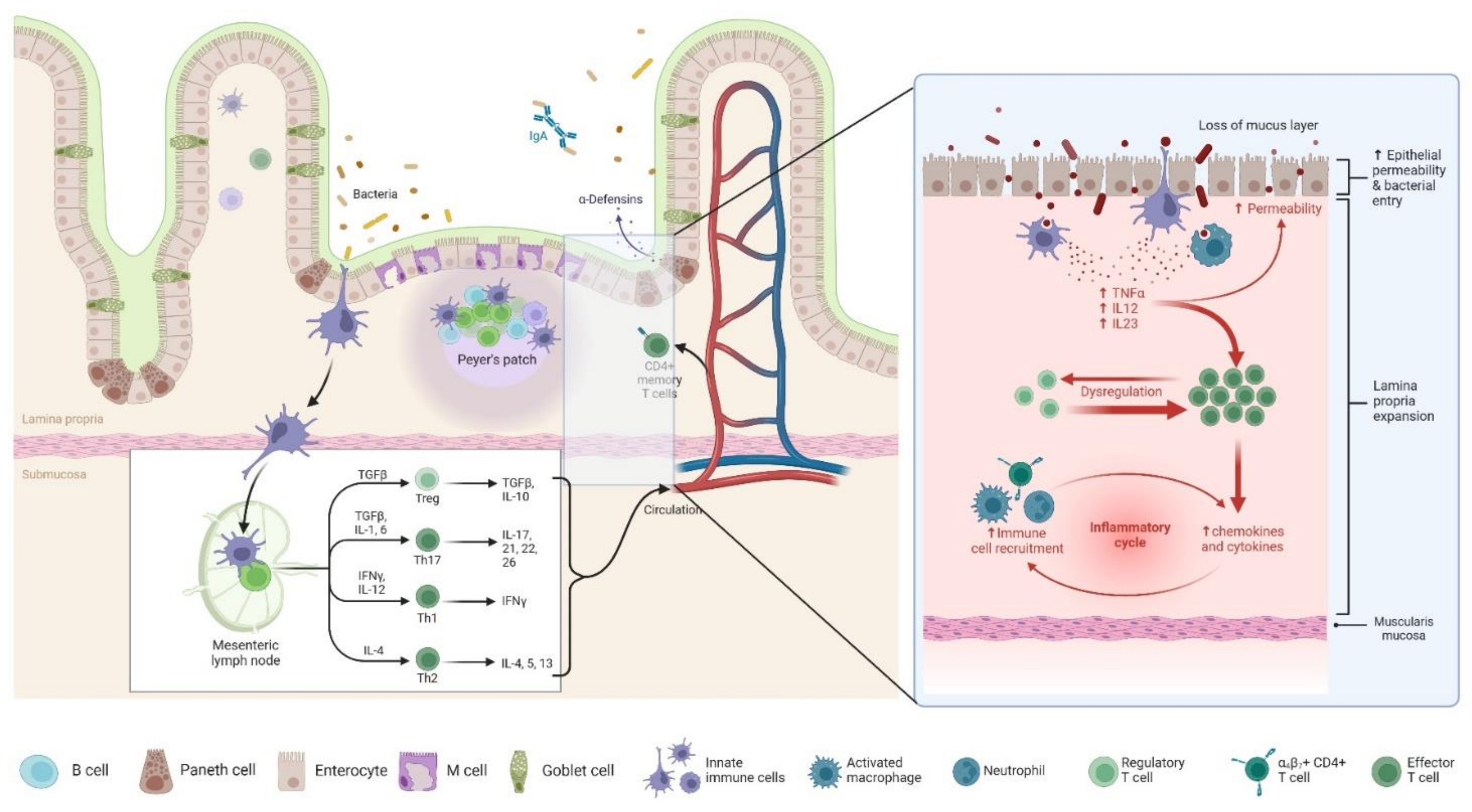
Crohn’s disease (CD) imposes a substantial burden on patients due to its chronic, relapsing nature, often necessitating surgical intervention. However, surgery is not curative, and post-operative recurrence (POR) remains a major clinical challenge, with up to 80% of patients developing endoscopic recurrence within one year if left untreated. The pathophysiology of POR is multifactorial, involving dysregulated immune responses, gut microbiota alterations, and mucosal healing impairment, highlighting the need for targeted therapeutic strategies. This review aims to explore the current landscape of POR management, focusing on biologic therapies and emerging advanced treatments. Conventional management relies on early prophylactic therapy with anti-TNF agents such as infliximab and adalimumab, which have demonstrated efficacy in reducing endoscopic and clinical recurrence. However, newer biologics, including IL-23 inhibitors (risankizumab) and Janus kinase (JAK) inhibitors (upadacitinib), have shown promise in CD management, though their role in POR remains underexplored. The lack of direct clinical evidence for advanced biologics in POR prevention, combined with inter-individual variability in treatment response, underscores the need for further research. Future directions should focus on optimizing therapeutic strategies through personalized medicine, identifying predictive biomarkers, and conducting robust trials to establish the efficacy of novel agents in POR prevention. A tailored, evidence-driven approach is essential to improving long-term outcomes and minimizing disease recurrence in post-operative CD patients.

Keywords: 
;  ;  ;  ;  

1. Introduction

Crohn's disease (CD) is a chronic inflammatory bowel disease (IBD) characterized by relapsing and remitting symptoms, leading to significant morbidity and a decreased quality of life [1]. The disease can affect any part of the gastrointestinal tract, most commonly the terminal ileum and colon, resulting in symptoms such as abdominal pain, diarrhea, weight loss, and fatigue [2]. The etiology of CD is multifactorial, involving genetic predisposition, environmental factors, and an abnormal immune response [3,4].
Surgical intervention plays a crucial role in the management of CD, particularly when complications arise or medical therapy fails to control the disease [5,6]. Indications for surgery include strictures causing bowel obstruction, fistulas, abscesses, and perforations [7]. Despite advancements in medical therapies, up to 80% of patients with CD will require surgical resection during their lifetime [8]. However, surgery is not curative; the disease often recurs at or near the site of anastomosis.
Postoperative recurrence (POR) of CD remains a significant challenge in clinical practice. Endoscopic evidence of recurrence can be observed in up to 70% of patients within one year after ileocolonic resection, even in the absence of clinical symptoms [9]. Clinical recurrence typically follows endoscopic recurrence and can lead to complications necessitating further surgical interventions. Risk factors for POR include smoking, penetrating disease phenotype, prior intestinal surgery, and the presence of perianal disease [10]. Given the high incidence of POR in CD and the associated complications, there is a critical need for effective preventive and therapeutic strategies [11]. Emerging advanced biologic therapies, such as upadacitinib, risankizumab, and tofacitinib, have shown promise in managing moderate to severe CD [12]. This review aimed to evaluate the efficacy and safety of these agents in the context of POR, providing insights to inform clinical practice and guide future research.

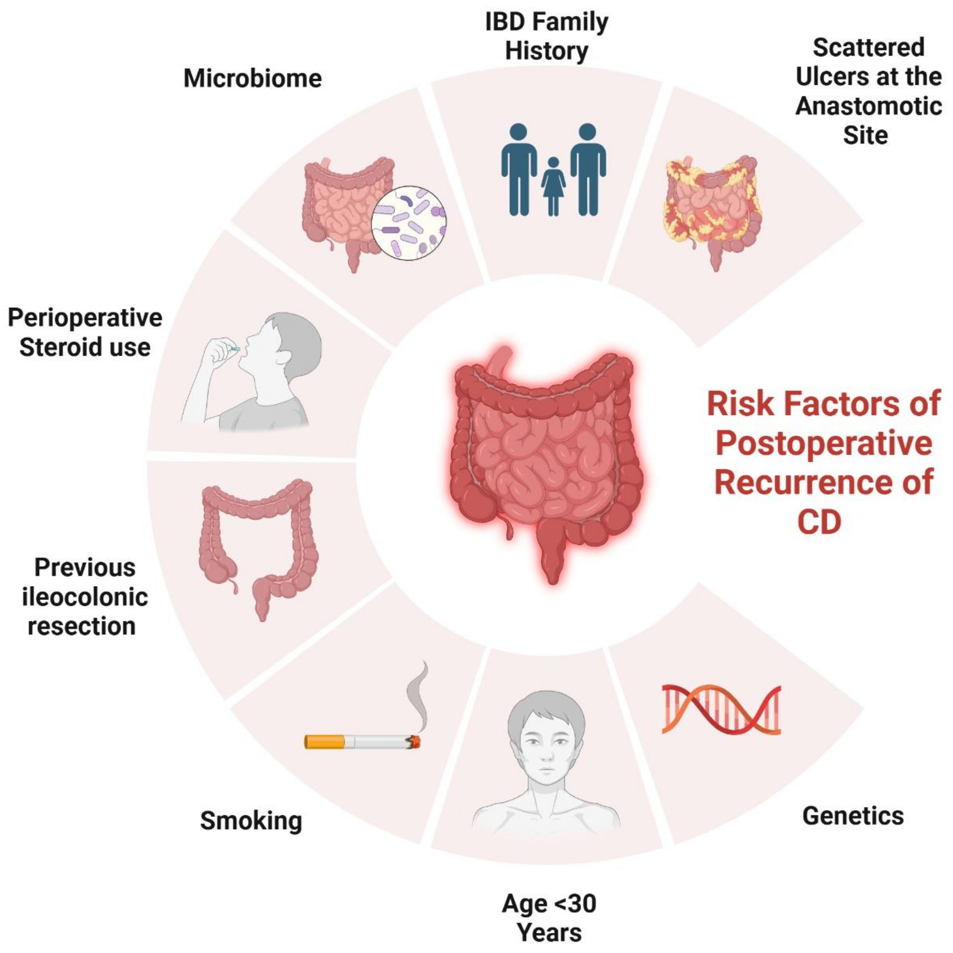
2. Risk Factors Contributing to POR

Several risk factors have been identified that contribute to the likelihood of postoperative recurrence in CD (Figure 1).

2.1. Genetical Factors

Mutations in the NOD2/CARD15 gene have been linked to the development and progression of CD, particularly in predicting ileal stenotic disease and the need for early surgical intervention [13]. Among these, the 1007fs mutation has been associated with an increased risk of POR, with Renda et al reporting a hazard ratio (HR) of 2.9 (95% CI 1.1-7.3, P=0.03) [14]. However, conflicting results have been observed, as studies by Manconi et al and Fowler et al did not find a significant association [15,16]. Beyond NOD2, additional genetic loci have been evaluated. Individuals with reduced IL-10 production were more likely to experience endoscopic POR, though no specific IL-10 promoter polymorphism was identified [17]. The IRGFM polymorphism was correlated with frequent intestinal resections [18], while CARD8 mutations in homozygous individuals were linked to a higher risk of surgical recurrence (OR 7.56, 95% CI 1.13-50.37, P=0.036) [19]. Moreover, SMAD3 gene mutations were found to elevate the likelihood of surgical recurrence (HR 4.04, 95% CI 1.77-9.21, P=0.001) [15]. While these genetic markers may provide insight into CD prognosis, further studies are required to establish their predictive accuracy.

2.2. Surgical and Histological-Related Factors

Regarding surgical approach, laparoscopic resection is preferred due to its advantages in recovery and cosmesis, but no significant difference in recurrence rates has been observed between laparoscopic and open surgery, as demonstrated in RCTs by Stocchi et al. [20] and Eshuis et al. [21], along with a meta-analysis by Patel et al [22]. Strictureplasty, a bowel-sparing procedure, is a viable alternative to resection in select patients. However, recurrence rates vary depending on technique. A meta-analysis by Tichansky et al found that Heineke-Mikulicz (HM) strictureplasty was associated with higher recurrence rates compared to the Finney technique [23], while Reese et al reported a non-significant increase in surgical recurrence for strictureplasty alone (38% vs. 31%, OR 1.36, 95% CI 0.96-1.93, P=0.09) compared to resection [24]. Furthermore, blood transfusion has been investigated for its immunosuppressive effects, with recent data from a 318-patient cohort linking transfusion to increased risks of endoscopic recurrence (HR 2.08, 95% CI 1.38-3.14, P<0.001) and surgical recurrence (HR 3.43, 95% CI 1.92-6.13, P<0.001) [25]. Lastly, prior resections are a well-established risk factor for POR. A randomized trial by McLeod et al found an OR of 1.78 (95% CI 1.06-2.90, P=0.028) for endoscopic recurrence and OR 2 (95% CI 1.14-3.60, P=0.0016) for surgical recurrence in patients with prior resections [26].
The type of anastomosis plays a significant role in POR. Studies suggest that side-to-side anastomosis (SSA), particularly the stapled SSA, may be associated with lower recurrence rates compared to end-to-end anastomosis (EEA) [27,28]. However, an RCT by McLeod et al found no significant difference in recurrence rates between SSA and EEA, with endoscopic recurrence at 12 months reported as 42.5% for EEA and 37.9% for SSA (P=0.55), and clinical recurrence at 21.9% and 22.7%, respectively (P=0.92) [26]. Similarly, a meta-analysis by Simillis et al (661 patients) found no significant difference between the two techniques in perioperative recurrence or reoperation rates [29]. A novel Kono-S anastomosis, designed to minimize ischemia and bacterial overgrowth, has shown low recurrence rates in preliminary studies, but further research is needed [30]. In terms of extent and resection margins, a landmark RCT by Fazio et al demonstrated that limited resection margins (2 cm from the diseased bowel) were not associated with higher recurrence rates than wide resections (12 cm) [31]. While the length of resected bowel does not consistently predict recurrence, European Crohn’s and Colitis Organisation (ECCO) guidelines identify extensive resection (>50 cm) as a predictive factor for POR [32]. These findings highlight the importance of surgical strategy in managing CD, emphasizing the need for individualized decision-making.
The role of granulomas in POR remains debated. Early studies suggested a protective effect, with Glass et al associating granulomas with a lower POR risk [33], while Champers reported a better prognosis when granulomas were found in the large bowel and anus [34]. However, more recent research indicates an increased risk. A retrospective Mayo Clinic study (89 patients) found granulomas linked to higher POR risk (HR 2.89, 95% CI 1.26-6.64, P=0.01) [35]. A meta-analysis by Similis et al (22 studies) concluded that granulomatous CD increases the risk of recurrence (OR 1.37, 95% CI 1.02-1.84, P=0.04) and reoperation (OR 2.38, 95% CI 1.43-3.95, P<0.001), leading ECCO guidelines to recognize granulomas as a POR risk factor [36]. Additionally, lymphatic vessel density has emerged as a potential biomarker. Rahier et al found that decreased lymphatic density in the mucosa and submucosa at the proximal margin was associated with early POR, while a mucosal lymphatic density >7% predicted non-recurrence (sensitivity 81%, specificity 75%) [37]. Further research is needed to validate its prognostic value.
Plexitis has been widely studied for its potential role in predicting POR. A study by Ferrante et al (59 patients) identified myenteric plexitis at the proximal resection margin as a significant predictor of early endoscopic recurrence at 3 months (OR 4.36, 95% CI 1.44-13.23, P=0.008) and 12 months (OR 9.80, 95% CI 1.04-92.70, P=0.041) [38]. However, submucosal plexitis did not show a significant correlation with POR. Subsequent studies provided mixed findings: Sokol et al (171 patients) linked submucosal plexitis (≥3 mastocytes) with early clinical recurrence (HR 1.87, 95% CI 1.00-3.46, P=0.048), while Bressenot et al (67 patients) found an even higher risk when eosinophils (HR 8.02, 95% CI 1.87-34.47, P=0.0054) or lymphocytes (HR 5.84, 95% CI 1.23-27.65, P=0.0269) were present [39,40]. Another study (86 specimens) reported severe myenteric plexitis as a predictor for surgical recurrence (P=0.035) [41]. Despite inconsistencies, French cohort studies and a Belgian prospective study confirmed myenteric plexitis as an independent risk factor for POR, particularly at the proximal margin [42,43].

2.3. Disease-Related Factors

The behavior of the disease significantly influences POR risk. A penetrating phenotype (Montreal-B3) is associated with early POR, as shown in multiple studies, including Sachar et al, where none of the stricturing phenotype (Montreal-B2) patients experienced POR within three years [44]. Similarly, Avidan et al (86 patients) found that penetrating disease significantly increased POR risk and shortened the recurrence interval compared to smoking [45]. Two meta-analyses confirmed these findings: Similis et al (13 studies, 3044 patients) reported that perforating disease increased POR risk (HR 1.50, 95% CI 1.16-1.93, P=0.002) [46], while Pascua et al (12 studies) linked perforating disease to a higher risk of endoscopic POR (OR 1.59, 95% CI 1.37-1.84, P<0.05) [47]. Consequently, ECCO guidelines now include fistulizing disease as a POR risk factor [32]. Perianal disease is also widely accepted as a strong predictor of POR. A Korean study (132 patients) found that perianal lesions were an independent risk factor for reoperation (HR 1.98, 95% CI 1.03-3.78, P=0.039) [48], while a population-based cohort (907 patients) demonstrated a relative risk of 1.6 (95% CI 1.2-2.3, P=0.003) for POR [49]. Disease location also plays a role, with ileal involvement (Montreal-L1) being a predictive factor for a second ileocolonic resection (OR 2.42, 95% CI 1.02-5.78, P=0.05) [50]. Similarly, a John Radcliffe Hospital study (280 patients) found that ileal disease was associated with higher POR rates compared to ileocolonic or colonic involvement [51], and a Netherlands population-based study supported the association between small bowel disease and POR [52]. While some studies link jejunal disease with increased surgical recurrence rates [53], the role of upper gastrointestinal involvement remains debated, with mixed findings from population-based studies [54].

2.4. Patient-Related Factors

Smoking is a well-established, modifiable risk factor for POR in CD. Cottone et al identified smoking as a predictor for endoscopic recurrence (OR 2.2, 95% CI 1.2-3.8) and surgical recurrence (HR 2.0, 95% CI 1.2-2.3), while Unkart et al found smokers had an increased risk of a second ileocolonic resection (HR 2.08, 95% CI 1.11-3.91, P=0.023) [55,56]. A meta-analysis of 16 studies (2,692 patients) confirmed a 2-fold higher risk for clinical recurrence and a 2.5-fold increased risk for surgical recurrence in smokers at 10 years, while smoking cessation reduced the POR risk to that of non-smokers [57]. The dose-response effect has been documented, with heavy smokers (>15 cigarettes/day) having higher recurrence rates (77% vs. 45%, P=0.02) [58]. However, some studies failed to confirm smoking as a risk factor, likely due to small sample sizes or missing smoking history data [50,59]. Sex has not been consistently associated with POR, with studies reporting conflicting results [60,61].

3. Classification and Monitoring of Postoperative Recurrence

POR can manifest in various forms, including endoscopic, clinical, radiologic, and surgical recurrence [62,63]. Endoscopic recurrence is typically the earliest detectable form and is identified through colonoscopy, revealing mucosal lesions at the anastomotic site or in the neoterminal ileum. Clinical recurrence involves the return of symptoms such as abdominal pain, diarrhea, and weight loss, which may or may not correlate with endoscopic findings [64]. Radiologic recurrence is identified through imaging modalities like magnetic resonance enterography or computed tomography enterography, showing structural changes indicative of active disease. Surgical recurrence refers to the necessity for additional surgical intervention due to complications or refractory disease.
Diagnosing POR of CD requires a multifaceted approach incorporating clinical symptoms, biochemical markers in serum and feces, radiological imaging, and endoscopic evaluation [65]. However, differentiating POR from other postoperative complications, such as adhesional obstructions, dysmotility, or bile salt malabsorption-related diarrhea, can be challenging [66]. Endoscopy remains the gold standard for detecting POR, with the Rutgeerts’ score (RS) being the most widely used classification tool in clinical practice and trials [67,68]. The RS categorizes the severity of endoscopic lesions observed at the neoterminal ileum and ileocolonic anastomosis, which serve as predictors for future clinical activity [69]. Research has established that a higher RS score correlates with an increased risk of clinical recurrence over subsequent years, with a significant distinction observed between patients with minimal (i0–i1) versus moderate-to-severe (≥i2) lesions [70,71]. Furthermore, a systematic review by Ble et al. confirmed that patients with endoscopic recurrence (RS ≥ i2) face a markedly higher risk of clinical relapse, demonstrating a relative risk (RR) of 10.7 [64]. Lesions restricted to the anastomosis are categorized as i2a, while the presence of more than five aphthous ulcers or larger skip lesions falls under the i2b classification [72,73,74]. Recent studies have explored the potential differences in clinical outcomes between i2a and i2b lesions [75,76,77]; however, findings have been inconsistent, leaving uncertainty about whether these two subgroups should be managed and monitored with distinct treatment approaches. Guidelines recommend using the RS at 6 months post-surgery to guide CD management. Patients with i0–i1 lesions are at low risk of recurrence [78] and can continue current management with periodic noninvasive markers and repeat endoscopy in 1–3 years [79]. Conversely, i2, i3, or i4 lesions indicate early postoperative recurrence, warranting treatment escalation, often with biologic therapy [80].
Advances in surgical techniques, such as the Kono-S anastomosis, have demonstrated lower endoscopic recurrence rates compared to conventional anastomotic techniques, highlighting the impact of surgical approach on POR risk [81,82]. Given the variability in recurrence patterns, risk stratification tools incorporating surgical, anatomical, and clinical factors are needed [83]. Timing of postoperative ileocolonoscopy is crucial for detecting early recurrence and guiding therapeutic decisions, with six months being the optimal assessment point, as evidenced by the POCER trial [84,85]. Current guidelines recommend follow-up endoscopic evaluations within 6–12 months after surgery, with subsequent assessments guided by clinical and biomarker trends [86,87].
Cross-sectional imaging techniques, such as intestinal ultrasound (IUS) and magnetic resonance enterography (MRE), provide a comprehensive evaluation of the entire bowel wall, overcoming the limitation of endoscopy, which only assesses the mucosal surface [88]. These non-invasive modalities, free from ionizing radiation exposure, are increasingly favored for POR monitoring in CD. A meta-analysis by Yung et al. found that while capsule endoscopy had the highest sensitivity (100%) but lower specificity (69%), MRE demonstrated a sensitivity of 97.3% and specificity of 83.7%, and IUS showed a sensitivity of 83.5% and specificity of 91.5% [89]. The MONITOR index, developed to assess POR through MRE, assigns scores based on bowel wall characteristics, with a cut-off of 1 yielding a sensitivity of 79% and specificity of 55% [90]. Despite its diagnostic utility, MRE’s high cost, limited availability, and contrast requirements restrict its widespread application.
IUS is emerging as a preferred alternative due to its accessibility and real-time imaging capabilities [91]. IUS findings correlate closely with endoscopic RS, and IUS has shown higher diagnostic yield than clinical indices or blood/stool markers in identifying early recurrence [92]. One study concluded that bowel ultrasound has “high diagnostic efficacy” for POR and can be considered a valid non-invasive alternative to ileocolonoscopy [93]. This noninvasive approach can allow earlier detection of subclinical recurrence and potentially reduce the need for routine surveillance colonoscopies [68]. IUS diagnostic accuracy can be enhanced through small intestine contrast ultrasound (SICUS) and contrast-enhanced ultrasonography (CEUS), which provide additional insights into bowel wall inflammation [94]. A meta-analysis by Rispo et al. reported that bowel wall thickness (BWT) >3 mm on IUS had a sensitivity of 82% and specificity of 88% for detecting endoscopic recurrence, while SICUS demonstrated higher sensitivity (99%) but lower specificity (75%) [95]. A multicenter prospective study incorporating IUS and fecal calprotectin (FC) identified BWT per 1-mm increase, mesenteric lymph nodes, and FC ≥50 μg/g as independent predictors of endoscopic recurrence, correctly classifying 75% of patients [68]. Increased power Doppler signal (bowel wall hyperemia) is another hallmark of active disease; recurrent lesions often show marked hypervascularity on IUS, a feature that correlates significantly with endoscopic inflammation [93]. Other ultrasound signs of POR include the loss of normal bowel wall stratification and mesenteric changes such as creeping fat hypertrophy or enlarged lymph nodes [96]. Notably, the presence of prominent mesenteric lymph nodes on IUS was associated with a markedly higher risk of endoscopic recurrence (OR ≈15 in one prospective cohort) [68]. These findings underscore the potential role of IUS and FC in postoperative CD surveillance, potentially reducing the reliance on colonoscopy. However, further large-scale prospective studies are needed to validate these non-invasive monitoring strategies.
Biomarkers, particularly FC and serum markers, offer non-invasive alternatives for disease monitoring, aiding in risk stratification and optimizing endoscopic evaluation timing [97]. FC, a neutrophil-derived protein, strongly correlates with endoscopic and histologic inflammation [98]. A meta-analysis determined that an FC threshold of 150 μg/g had a sensitivity of 70% and specificity of 69% in predicting endoscopic recurrence (RS > i2), though methodological inconsistencies across studies limit its reliability [99]. Research by Boschetti et al. suggested that a lower threshold of 100 μg/g improved sensitivity (95%) but reduced specificity (54%), potentially decreasing the need for colonoscopy by 30% [100]. However, some studies have questioned the accuracy of FC in detecting POR, highlighting the importance of serial measurements over single assessments [101].
Serum biomarkers offer an additional approach to POR surveillance, with better patient adherence than stool-based assays [102]. However, standard inflammatory markers like C-reactive protein (CRP) lack sufficient sensitivity for early recurrence detection [102]. The Endoscopic Healing Index (EHI), a panel measuring 13 proteins related to inflammation and tissue remodeling, has been investigated for its ability to identify endoscopic remission (ER); which was defined as Simple Endoscopic Score for Crohn’s Disease (SES-CD) of ≤ 2 [103]. A validation study demonstrated that an EHI score <20 had high sensitivity (83.2–97.1%) but variable specificity [103]. Further analysis using data from the POCER trial found that at six months post-surgery, EHI and FC had comparable sensitivity (81.8% vs. 90.9%) and negative predictive values (84.0% vs. 91.7%) for detecting recurrence [104]. However, at 18 months, FC was a better predictor than EHI, with a negative predictive value of 89.7% compared to 64.9% [104]. While serum biomarkers show promise, FC remains the preferred tool for non-invasive monitoring of POR, and further research is necessary to validate serum-based markers as independent predictors and assess their clinical utility.

4. Pathophysiology of POR

POR of CD is driven by a complex interplay of microbiological, immunological, and genetic factors [105]. The gut microbiota plays a pivotal role in disease recurrence, as demonstrated by studies linking dysbiosis to increased inflammation following ileocecal resection [106], as shown in Figure 2. Notably, Sokol et al. reported significant shifts in the ileal mucosa-associated microbiota, with an increased presence of Proteobacteria and a reduction in Firmicutes, correlating with endoscopic recurrence [107]. Additionally, alterations in luminal content flow at the anastomotic site, particularly in side-to-side anastomoses, facilitate bacterial proliferation and immune activation, further contributing to POR [108]. Genetic predisposition also influences recurrence risk, with the NOD2 gene playing a crucial role in microbial recognition and immune modulation. A meta-analysis highlighted that patients with NOD2 variants have a significantly higher likelihood of POR (odds ratio: 1.64, p = 0.003), and additional genes such as BACH2, CARD8, and TNFSF15 have been implicated in recurrence risk, though their mechanisms remain undefined [109]. Despite these findings, genetic testing is not yet recommended for routine clinical assessment due to the lack of standardized predictive biomarkers [110].
The immune system’s involvement in POR is also substantial, with both innate and adaptive immunity contributing to disease recurrence. Macrophages, serving as key mediators of inflammation, have been observed in increased numbers in the neoterminal ileum prior to endoscopic lesions, supporting their role in early POR pathogenesis [108]. Additionally, T-cell clonal expansion has been detected in resected bowel mucosa, suggesting that cell-mediated immunity plays a central role in recurrence, particularly among smokers [111]. Recent attention has also been given to the mesentery’s contribution to CD recurrence, with findings indicating that mesenteric excision may influence POR rates. The phenomenon of "creeping fat," characterized by mesenteric adipose hypertrophy and immune cell infiltration, suggests that mesenteric lymph nodes may serve as reservoirs for CD immune memory, potentially facilitating disease recurrence [105]. However, evidence regarding the benefits of mesenteric excision remains inconclusive, with retrospective analyses yielding conflicting results, and current guidelines do not universally recommend routine mesenteric resection during ileocecal resection [112,113].

5. Current Strategies and Guidelines of Prophylaxis

Ileocecal resection is not curative for CD, and a significant proportion of patients experience POR. The pivotal study by Rutgeerts et al. demonstrated that up to 73% of patients develop endoscopic lesions within one-year post-surgery, a finding that has been confirmed in more recent studies [78,114]. However, the optimal strategy to prevent recurrence remains a subject of debate, as approximately 20–30% of patients do not develop endoscopic POR, while 40–50% exhibit only intermediate lesions (Rutgeerts i1–i2), which are associated with a lower risk of clinical and surgical recurrence [115]. Current management approaches include either systematic prophylactic therapy initiated early after surgery or an endoscopy-driven strategy in which treatment is started based on evidence of recurrence detected during a colonoscopy performed 6–12 months postoperatively [115]. International guidelines offer differing recommendations: the ECCO and the British Society of Gastroenterology (BSG) advocate prophylactic therapy for high-risk patients, while the American Gastroenterological Association (AGA) recommends prophylaxis for all patients, regardless of risk factors [32,116,117]. Furthermore, there is variation in defining high-risk patients, with the BSG requiring at least two risk factors, while ECCO and AGA consider a single risk factor sufficient [32,116,117].
Each strategy has its advantages and limitations. Systematic prophylaxis improves medium- to long-term remission rates but risks overtreatment, exposing patients to unnecessary drug-related side effects [115]. Conversely, an endoscopy-driven approach may prevent over- and undertreatment, but categorizing patients into high- and low-risk groups remains challenging. Studies have not definitively resolved this debate. Supporting the BSG guidelines, one study found that only patients with three or more risk factors had significantly higher odds of developing endoscopic POR [118]. In contrast, a retrospective analysis of 376 patients indicated that while prophylaxis reduced endoscopic POR at one year in high-risk patients (HR 0.48, p = 0.04), there was no significant difference in clinical recurrence within three years between the two strategies (HR 1.06, p = 0.82) [119]. Another study observed that in patients with only one risk factor, immediate prophylaxis did not significantly lower endoscopic or severe endoscopic POR rates within 12 months post-surgery [120]. However, a prospective cohort study of 213 CD patients who underwent ileocecal resection found that both low- and high-risk patients who did not receive prophylaxis had higher endoscopic POR rates compared to those who did (45% vs. 16%, p = 0.012; 49% vs. 26%, p = 0.019, respectively) [121]. Additionally, the PORCSE study found that the endoscopic POR rate was higher in the endoscopy-driven group compared to the early prophylaxis group (53.8% vs. 41.5%, p = 0.039), with significantly higher clinical POR (35.7% vs. 17.7%, p = 0.002) [122]. Similarly, a retrospective study reported that the absence of prophylactic therapy was independently associated with increased endoscopic POR risk (OR 0.22; p = 0.0004) and a greater likelihood of hospitalization and surgery at five years [123].

6. Traditional Biological Therapies

Biologic therapies have revolutionized the management of CD, particularly for patients with moderate to severe forms unresponsive to conventional treatments. These therapies are derived from living organisms and specifically target components of the immune system to reduce inflammation. By inhibiting specific cytokines or their receptors, biologics can effectively diminish the inflammatory response characteristic of CD. The advent of advanced biologic agents has expanded treatment options, offering hope for improved disease control and quality of life for patients.

6.1. Anti-Tumor Necrosis Factor (TNF) Agents

Infliximab has shown efficacy in preventing POR in CD. A non-randomized study found infliximab with methotrexate superior to mesalamine (0% vs. 75% POR) [124], and retreatment restored remission in relapsed patients [125]. An RCT showed early infliximab use reduced recurrence at 12 and 36 months [126], while a pair-matched study identified it as the only factor preventing surgical recurrence (HR 0.08, p = 0.03) [127]. The PREVENT trial confirmed a significant reduction in endoscopic recurrence (30.6% vs. 60.0%, p < 0.001), though clinical recurrence differences were not significant (12.9% vs. 20.0%, p = 0.097) [128]. Adalimumab also reduced POR, with one study showing 75% of high-risk patients in remission at 24 months [129] and another reporting low clinical (13.7%) and endoscopic (20.7%) recurrence at one year [130]. However, to the best of our knowledge, there are no placebo-controlled RCTs exist for adalimumab. Comparative studies and meta-analyses found no significant difference between infliximab and adalimumab in preventing recurrence [131,132,133].

6.2. Anti-Integrin and Anti-Interleukin Agents

Data on the effectiveness of vedolizumab (VDZ) and ustekinumab (UST) in preventing POR of CD remain limited. Retrospective comparisons between vedolizumab and anti-TNF agents indicate a lower rate of ER at 6–12 months in vedolizumab-treated patients (25% vs. 66%, p = 0.01), though clinical and biological remission rates were comparable. Notably, vedolizumab was independently associated with a higher risk of endoscopic POR in both univariate and multivariate analyses [134]. Preliminary findings from the REPREVIO trial, a placebo-controlled RCT, demonstrated a significantly higher ER rate in patients receiving vedolizumab compared to placebo (42% vs. 3%, p < 0.001), with treated patients having a 77.8% probability of achieving a better RS than controls (p < 0.0001) [135]. In contrast, ustekinumab showed promise in a small-scale comparative study where patients receiving UST had a lower endoscopic POR rate at six months than those on azathioprine (28% vs. 54.5%, p = 0.029), though the difference was primarily driven by moderate disease (Rutgeerts i2) rather than severe recurrence (≥i3) [136].
Real-world evidence further supports the need for additional studies. A retrospective multicenter analysis of 297 patients receiving biologic prophylaxis for POR found that endoscopic recurrence at one year was 61.8% for UST, 33% for VDZ, and 40.2% for anti-TNF therapy. However, since patients treated with UST or VDZ had higher rates of prior surgery and biologic exposure, adjusted analyses showed comparable risk of POR between groups (VDZ vs. anti-TNF: OR 0.55, 95% CI 0.25–1.19; UST vs. anti-TNF: OR 1.86, 95% CI 0.79–4.38) [137]. Another multicenter cohort study of 278 patients found that only early anti-TNF therapy (within four weeks) was associated with a reduced POR risk, while VDZ and UST did not show significant benefits (anti-TNF: aHR 0.61, 95% CI 0.40–0.93; VDZ: aHR 1.44, 95% CI 0.59–3.56; UST: aHR 2.06, 95% CI 0.84–5.06) [138]. A direct prospective comparison of vedolizumab vs. ustekinumab (40 UST-treated vs. 25 VDZ-treated) found similar clinical POR rates at 12 months (32% vs. 30%) and endoscopic POR rates at 18 months (42% vs. 40%) [139]. These findings highlight the need for further RCTs to clarify the role of ustekinumab and vedolizumab in POR prevention.

7. Advanced Biologic Therapies

7.1. Upadacitinib

The efficacy of upadacitinib in CD was established through two induction trials (U-EXCEL, U-EXCEED) and one maintenance trial (U-ENDURE) [140]. The induction studies included patients with moderate-to-severe CD, defined by frequent soft or liquid stools, an abdominal pain score of ≥2, and an SES-CD of ≥6. In U-EXCEL, 54.6% of patients had failed conventional therapy, while 45.4% had previously failed biologics. In contrast, U-EXCEED exclusively included biologic-experienced patients. The primary efficacy endpoints were clinical remission (CR) [Crohn’s Disease Activity Index (CDAI) <150] and endoscopic response (>50% reduction in SES-CD) at week 12 for induction trials and week 52 for maintenance. Upadacitinib 45 mg daily demonstrated superior rates of CR and endoscopic response versus placebo in both induction studies, with some patients experiencing remission as early as weeks 2 and 4 [140]. In the U-ENDURE maintenance trial, patients who responded in the induction phase were randomized to receive 15 mg, 30 mg upadacitinib, or placebo. By week 52, both treatment arms achieved higher remission rates (15 mg: 37.3%, 30 mg: 47.6%, placebo: 15.1%) and greater ER (15 mg: 27.6%, 30 mg: 40.1%, placebo: 7.3%) compared to placebo [140]. These trials confirmed upadacitinib’s efficacy in inducing and maintaining remission in CD, with rapid onset of action and durable response rates.
In terms of real-world evidence, a multicenter retrospective cohort study assessed the effectiveness and safety of upadacitinib in IBD, across tertiary care centers. The study included 236 adult patients (≥18 years) who had received at least 8 weeks of upadacitinib therapy. In 156 CD patients, steroid-free remission (76.8%, p < 0.001), CR (77.8%, p < 0.001), and clinical response (81.0%) were recorded at week 12, with a mean CDAI reduction from 214.9 to 117.5 (p < 0.001). ER was 19.4%, with a 48.9% endoscopic response and 4.9% mucosal healing. Radiological remission was achieved in 9.1%, with an 85.7% radiologic response, while intestinal ultrasound showed remission in 5.7% and response in 56.7% [141]. These findings highlight upadacitinib’s efficacy in achieving steroid-free remission, clinical response, and mucosal healing, particularly in biologic-resistant patients. Another multicenter retrospective study assessed the real-world effectiveness of selective JAK inhibitors in CD, focusing on bio-experienced patients included 246 patients, with 115 receiving upadacitinib for CD, and a median follow-up of 7.5 months. At week 12, 76.2% of upadacitinib-treated CD patients achieved CR. By week 24, 76.9% achieved corticosteroid-free CR (CSFCR), while 50.0% had ER. At week 52, 66.7% remained in CSFCR, and 54.5% achieved ER. On the other hand, the study highlighted that upadacitinib was less effective in patients with stricturing and penetrating disease, as they were less likely to achieve CR by the end of induction (p = 0.04). Significant reductions in CRP (p < 0.0001) and faecal calprotectin (p < 0.0001) were observed as early as week 2, indicating a rapid anti-inflammatory effect [142]. These findings suggest that selective JAK1 inhibition with upadacitinib is an effective and well-tolerated treatment option for refractory moderate-to-severe CD, particularly in patients without penetrating or stricturing complications.
Moreover, a retrospective multicenter study assessed the real-world persistence, effectiveness, and safety of upadacitinib in IBD patients, with a minimum follow-up of 12 weeks. The study included 100 patients, with 68 diagnosed with CD. Participants had previously received a median of four advanced therapies. CR was achieved in 59% at week 8, 64% at week 12, and 42% at week 52. Dose escalation successfully restored remission in 60% of patients experiencing relapse. Additionally, 80% of patients with active immune-mediated diseases or extraintestinal manifestations showed improvement with upadacitinib therapy [143].
In patients with difficult-to-treat CD, a multicenter observational cohort study included 64 patients with moderate-to-severe CD (Harvey-Bradshaw Index [HBI] >8 or SES-CD >6), all of whom had failed corticosteroids, thiopurines, infliximab, adalimumab, vedolizumab, and ustekinumab. Many had prior surgical interventions (67.2%) and extra-intestinal manifestations (43.8%), with the majority having ileocolonic disease (59.4%). Patients received upadacitinib 45 mg/day for 12 weeks (induction) followed by 30 mg/day for 12 weeks (maintenance). The primary endpoint, CSFCR (HBI ≤3) at 12 weeks, was achieved in 33 patients (51.6%), while clinical response (HBI reduction >3 points) was observed in 46 patients (71.9%). Biochemical remission (FC <150 μg/g and CRP <0.5 mg/dL) was documented in 36.2% of patients, while deep remission (clinical + biochemical remission + transmural healing) occurred in 21.8%. Ultrasonographic assessments at week 12 showed transmural healing in 28.8% of patients and transmural response in 73%. At week 24, among the 50 patients who continued upadacitinib, CR was sustained in 39 patients (78%), while clinical response persisted in 48 patients (96%). Biochemical remission was achieved in 38.3% [144].
Regarding refractory CD in pediatrics, a multicenter retrospective study evaluated the effectiveness and safety of upadacitinib as an induction therapy in pediatric CD across 30 international centers. The study included 100 children (median age: 15.8 years), all previously exposed to at least one biologic, with 89% having failed two or more biologic therapies. After eight weeks of induction therapy, 75% achieved a clinical response, 56% reached CR, and 52% achieved corticosteroid- and exclusive enteral nutrition-free CR. Additionally, 68% showed normalization of CRP, and 58% had FC <150 mcg/g, with 13% achieving both enteral nutrition-free CR and FC remission [145]. Another single-center retrospective cohort study evaluated the real-world efficacy and safety of upadacitinib in pediatric IBD patients (ages 9–20 years). The study included 20 children and adolescents diagnosed with CD (n = 3), UC (n = 13), and IBD-unclassified (IBD-U) (n = 4). The primary outcome was clinical response, defined as a ≥20-point reduction in the Pediatric Ulcerative Colitis Activity Index (PUCAI) or ≥12.5-point decrease in the Pediatric CDAI (PCDAI). At week 8 (UC/IBD-U) and week 12 (CD), 90% of patients achieved clinical response, while 75% reached SF-CR post-induction, with 65% maintaining remission at week 24. PUCAI scores significantly improved in UC/IBD-U patients, and FC levels showed a downward trend post-induction, though CRP levels remained unchanged. Endoscopic response was observed in seven out of eight evaluated cases, with three achieving ER. Two patients discontinued therapy due to surgical interventions (subtotal colectomy and ileostomy for rectal perforation), but no new safety concerns were identified [146]. These findings indicate that upadacitinib is a promising therapeutic option for pediatric IBD patients refractory to approved treatments, with encouraging efficacy and a manageable safety profile.
While there is currently no direct evidence evaluating the efficacy of upadacitinib in preventing POR of CD, its proven effectiveness in inducing and maintaining remission in moderate-to-severe and refractory CD suggests a potential role in this setting. Clinical trials such as U-EXCEL, U-EXCEED, and U-ENDURE have demonstrated significant clinical and ER rates, even in patients with prior treatment failures. Moreover, real-world studies have confirmed upadacitinib's effectiveness in biologic-experienced and highly refractory CD patients, with notable improvements in CSFCR, mucosal healing, and inflammatory markers. In difficult-to-treat CD, upadacitinib has shown promising clinical and biochemical remission rates, as well as transmural healing, which is a crucial marker of long-term disease control. Furthermore, its efficacy in pediatric CD—a population with limited treatment options—reinforces its potential as a viable alternative for patients at high risk of recurrence. Given that POR is driven by persistent inflammation, immune dysregulation, and mucosal barrier dysfunction, the selective JAK1 inhibition offered by upadacitinib may provide an anti-inflammatory effect capable of modifying disease recurrence patterns. Future randomized controlled trials and prospective cohort studies are needed to assess whether upadacitinib could be a viable strategy to prevent POR, particularly in high-risk patients, and to determine its role compared to conventional prophylactic therapies such as thiopurines, anti-TNFs, and IL-12/23 inhibitors.

7.2. Risankizumab

A phase II randomized, placebo-controlled trial evaluated risankizumab for moderate-to-severe CD, enrolling 121 patients, 93% of whom had previously failed TNF-antagonists or vedolizumab. Patients received intravenous risankizumab (600 mg or 200 mg) or placebo at weeks 0, 4, and 8, with CR (CDAI < 150) achieved in 24% (200 mg), 37% (600 mg), and 15.4% (placebo, p = 0.0489). Secondary endpoints, including clinical response (31% vs. 15%, p = 0.0489), ER (17% vs. 3%, p = 0.0015), and deep remission (7% vs. 0%, p = 0.0107), were significantly improved in the risankizumab groups [147]. In an open-label extension study, patients who did not achieve CR and ER at week 12 received risankizumab 600 mg IV every four weeks for an additional 12 weeks, while those in deep remission underwent a 12-week washout period. By week 26, CR rates were 55% in the placebo group, 59% in the 200 mg risankizumab group, and 47% in the 600 mg group. Among 62 patients who maintained CR at week 26, maintenance therapy with 180 mg subcutaneous (SC) risankizumab was initiated, resulting in 71% sustaining CR, 35% achieving ER, and 29% reaching deep remission at week 52 [148]. In a subsequent open-label extension, 65 patients continued risankizumab for a median of 33 months, with over 71% maintaining CR and more than 42% achieving ER by week 112, highlighting its durable long-term efficacy [149].
In 2022, the ADVANCE trial evaluated induction therapy for moderate-to-severe CD in patients with prior biologic or conventional therapy failure [150]. A total of 850 patients were randomized (2:2:1) to receive IV risankizumab (600 mg or 1200 mg) or placebo at weeks 0, 4, and 8. The co-primary endpoints at week 12 were CR (CDAI <150 in the USA; stool frequency ≤2.8 and abdominal pain score ≤1 in non-USA countries) and endoscopic response (>50% reduction in SES-CD or ≥2-point decrease for isolated ileal disease with baseline SES-CD of 4). Risankizumab outperformed placebo across all primary endpoints. CR was achieved in 45% (600 mg, p < 0.0001) and 42% (1200 mg, p < 0.0001) vs. 25% (placebo), while stool frequency and abdominal pain-based remission rates were 43% (600 mg, p < 0.0001) and 41% (1200 mg, p < 0.0001) vs. 22% (placebo). Additionally, 40% (600 mg, p < 0.0001) and 32% (1200 mg, p < 0.0001) of risankizumab-treated patients achieved endoscopic response, compared to 12% on placebo [150].
The MOTIVATE induction trial was a multicenter, randomized, double-masked study that evaluated clinical and ER in patients with CD who had previously failed biologic therapy. A total of 569 patients were randomized 1:1:1 to receive risankizumab 600 mg, 1200 mg, or placebo every 4 weeks. At week 12, CR rates based on CDAI (<150) were 42% (600 mg, p < 0.0001) and 40% (1200 mg, p < 0.0001), significantly higher than the 20% in the placebo group. Similarly, remission based on stool frequency and abdominal pain scores was 35% (600 mg, p = 0.0007) and 40% (1200 mg, p < 0.0001) versus 19% for placebo. Endoscopic response rates at week 12 were also superior in the risankizumab groups (29% and 34%, p < 0.0001) compared to 11% in the placebo group. No significant differences in efficacy were observed between the 600 mg and 1200 mg doses in either the ADVANCE or MOTIVATE trials [151].
The FORTIFY trial enrolled patients who achieved CR in the ADVANCE or MOTIVATE trials. Participants were randomized to receive SC risankizumab (180 mg or 360 mg) or placebo every 8 weeks. At week 52, CR rates were significantly higher in the 180 mg (55%, p = 0.0031) and 360 mg (52%, p = 0.0054) groups compared to placebo (41%). Endoscopic response was also superior in both risankizumab groups (47%, p < 0.0001) vs. 22% in the placebo group. Mucosal healing at week 52 occurred in 31% of patients receiving 360 mg risankizumab SC vs. 10% in the placebo group (p < 0.001), while ER was achieved in 39% of risankizumab-treated patients compared to 13% on placebo (p < 0.001) [152].
Both the ADVANCE and MOTIVATE trials showed that patients receiving risankizumab had greater rates of mucosal healing and ER. In ADVANCE, mucosal healing was achieved at week 12 by 21% of patients receiving risankizumab (600 mg) versus 8% in the placebo group, while in MOTIVATE, rates were 14% vs. 4% for placebo. Similarly, ER was observed in 24% (ADVANCE) and 19% (MOTIVATE) of patients receiving risankizumab, compared to 9% and 4% in the placebo groups, respectively [152].
A direct comparison between ustekinumab and risankizumab in biologic-experienced CD patients was conducted in the SEQUENCE study [153]. The primary endpoint (CDAI-based CR at week 24) demonstrated non-inferiority of risankizumab to ustekinumab with remission rates of 59% vs. 40%, respectively. For the second primary endpoint (ER at week 48, SES-CD ≤4 with a ≥2-point reduction and no subscore >1), risankizumab was significantly superior to ustekinumab (32% vs. 16%, p < 0.0001) [154].
Although no direct studies have evaluated the use of risankizumab for the prevention of POR in CD, its demonstrated efficacy in induction and maintenance trials suggests a potential role in this setting. The consistent achievement of CR, endoscopic response, and mucosal healing in both biologic-naïve and biologic-experienced patients highlights its robust anti-inflammatory effects, which are crucial in preventing disease recurrence following surgical resection. Furthermore, the SEQUENCE trial's findings of risankizumab’s superior endoscopic outcomes compared to ustekinumab further support its potential utility in maintaining post-operative remission. Given that preventing POR requires effective suppression of residual inflammation, risankizumab’s ability to achieve deep and durable remission could make it a valuable option for patients at high risk of recurrence. Future clinical trials specifically investigating its role in POR are warranted to determine its efficacy in this indication and to establish optimal treatment strategies.

8. Future Directions and Research Opportunities

8.1. Ongoing Clinical Trials and Emerging Therapies

Despite the significant progress in biologic therapy for CD, the prevention and management of POR remain major clinical challenges. Ongoing clinical trials are investigating novel therapeutic strategies to optimize POR management, including emerging biologics and small molecules with different mechanisms of action. IL-23 inhibitors, such as risankizumab and mirikizumab, are under evaluation for their role in reducing post-operative inflammation and recurrence. The potential of JAK inhibitors, including upadacitinib, in preventing POR is also an area of active research, given their rapid onset of action and promising efficacy in biologic-experienced patients with CD. Combination therapy approaches are another avenue being explored to enhance the durability of remission post-surgery. Trials assessing the concurrent use of anti-TNF agents with IL-23 inhibitors or JAK inhibitors may provide insights into superior mucosal healing and sustained suppression of inflammation. Additionally, controlled-release formulations of immunomodulators and biologics, designed for localized intestinal targeting, are being developed to improve drug efficacy while minimizing systemic side effects.

8.2. Personalized Medicine Approaches in Biologic Therapy Selection for POR

The selection of biologic therapy for preventing POR remains largely empirical, often based on clinical risk stratification. However, personalized medicine approaches are emerging as critical tools for optimizing post-operative treatment strategies. Stratifying patients based on disease phenotype, previous treatment response, and genetic or immunologic markers may help tailor biologic selection to maximize efficacy. Therapeutic drug monitoring (TDM) has shown promise in optimizing the use of anti-TNF therapy in preventing POR, ensuring adequate drug levels while mitigating the risk of immunogenicity. Additionally, multi-omic profiling, including transcriptomics and microbiome analysis, is being investigated to identify specific patient subgroups who may benefit from alternative biologic pathways, such as IL-23 or JAK-STAT inhibition. The integration of artificial intelligence and machine learning models into clinical decision-making could further enhance individualized treatment selection, improving long-term outcomes in post-operative patients.

8.3. Potential Biomarkers for Predicting Response to Biologics in POR

The identification of reliable biomarkers for predicting biologic response in POR is an essential area of research. Traditional inflammatory markers such as FC and CRP have limited specificity in post-operative settings, necessitating the exploration of novel predictive biomarkers. Several emerging biomarkers, including oncostatin M (OSM), serum IL-23 levels, and gut-specific transcriptomic signatures, are under investigation for their potential to guide biologic selection in preventing POR. Endoscopic and histologic markers, such as the RS and the EHI, are being refined to serve as early indicators of biologic efficacy in post-operative patients. The use of non-invasive imaging modalities, including intestinal ultrasound and MRI-based inflammation markers, may further enhance early detection of POR and allow timely therapeutic adjustments.

9. Conclusions

Despite advances in biologic therapies, the prevention and management of POR in CD remain challenging. While anti-TNF agents, such as infliximab and adalimumab, have been the cornerstone of POR prevention, newer biologics—including IL-23 inhibitors and JAK inhibitors—are emerging as promising alternatives, particularly for biologic-experienced patients. However, direct evidence supporting their use in POR is still limited, highlighting the need for well-designed clinical trials to evaluate their efficacy in the post-operative setting.
Future research should focus on identifying optimal biologic sequencing strategies, evaluating combination therapy approaches, and refining predictive models for patient stratification. The integration of TDM and biomarker-driven treatment selection may improve biologic persistence and efficacy in preventing POR. Additionally, real-world studies assessing the long-term outcomes of IL-23 and JAK inhibitors in preventing surgical recurrence are needed to guide clinical practice. As the landscape of biologic therapy continues to evolve, a more personalized and evidence-based approach to POR management will be essential in improving patient outcomes and reducing the need for repeat surgery.

References

  1. Ha F, Khalil H. Crohn’s disease: a clinical update. Therap Adv Gastroenterol [Internet]. 2015 Nov 16;8(6):352–9. Available online: https://journals.sagepub.com/.
  2. Ranasinghe IR, Tian C, Hsu R. Crohn Disease. In Treasure Island (FL); 2025.
  3. Carrière J, Darfeuille-Michaud A, Nguyen HTT. Infectious etiopathogenesis of Crohn’s disease. World J Gastroenterol. 2014 Sep;20(34):12102–17.
  4. Kong L, Pokatayev V, Lefkovith A, Carter GT, Creasey EA, Krishna C, et al. The landscape of immune dysregulation in Crohn’s disease revealed through single-cell transcriptomic profiling in the ileum and colon. Immunity. 2023 Feb;56(2):444–458.e5.
  5. Meima-van Praag EM, Buskens CJ, Hompes R, Bemelman WA. Surgical management of Crohn’s disease: a state of the art review. Int J Colorectal Dis. 2021 Jun;36(6):1133–45.
  6. Chiarello MM, Pepe G, Fico V, Bianchi V, Tropeano G, Altieri G, et al. Therapeutic strategies in Crohn’s disease in an emergency surgical setting. World J Gastroenterol. 2022 May;28(18):1902–21.
  7. Toh JW, Stewart P, Rickard MJ, Leong R, Wang N, Young CJ. Indications and surgical options for small bowel, large bowel and perianal Crohn’s disease. World J Gastroenterol. 2016 Oct;22(40):8892–904.
  8. Luglio G, Kono T. Surgical Techniques and Risk of Postoperative Recurrence in CD: A Game Changer? Inflamm Intest Dis. 2022 Jan;7(1):21–7.
  9. Fasulo E, D’Amico F, Osorio L, Allocca M, Fiorino G, Zilli A, et al. The Management of Postoperative Recurrence in Crohn’s Disease. J Clin Med. 2023 Dec;13(1).
  10. Ma D, Li Y, Li L, Yang L. Risk factors for endoscopic postoperative recurrence in patients with Crohn’s Disease: a protocol for systematic review and meta-analysis. BMC Gastroenterol [Internet]. 2024 Jun 25;24(1):211. Available online: https://bmcgastroenterol.biomedcentral.com/articles/10.1186/s12876-024-03301-z.
  11. Shah RS, Click BH. Medical therapies for postoperative Crohn’s disease. Therap Adv Gastroenterol [Internet]. 2021 Jan 15;14. [CrossRef]
  12. Ananthakrishnan AN, Murad MH, Scott FI, Agrawal M, Haydek JP, Limketkai BN, et al. Comparative Efficacy of Advanced Therapies for Management of Moderate-to-Severe Ulcerative Colitis: 2024 American Gastroenterological Association Evidence Synthesis. Gastroenterology [Internet]. 2024 Dec 1;167(7):1460–82. [CrossRef]
  13. Economou M, Trikalinos TA, Loizou KT, Tsianos E V., Ioannidis JPA. Differential Effects of NOD2 Variants on Crohn’s Disease Risk and Phenotype in Diverse Populations: A Metaanalysis. Am J Gastroenterol [Internet]. 2004 Dec;99(12):2393–404. Available online: https://journals.lww.com/00000434-200412000-00020.
  14. Renda MC, Orlando A, Civitavecchia G, Criscuoli V, Maggio A, Mocciaro F, et al. The Role of CARD15 Mutations and Smoking in the Course of Crohn’s Disease in a Mediterranean Area. Am J Gastroenterol [Internet]. 2008 Mar;103(3):649–55. Available online: https://journals.lww.com/00000434-200803000-00024.
  15. Fowler SA, Ananthakrishnan AN, Gardet A, Stevens CR, Korzenik JR, Sands BE, et al. SMAD3 gene variant is a risk factor for recurrent surgery in patients with Crohn’s disease. J Crohn’s Colitis [Internet]. 2014 Aug;8(8):845–51. [CrossRef]
  16. Maconi G, Colombo E, Sampietro GM, Lamboglia F, D’Incà R, Daperno M, et al. CARD15 Gene Variants and Risk of Reoperation in Crohn’s Disease Patients. Am J Gastroenterol [Internet]. 2009 Oct 28;104(10):2483–91. Available online: https://journals.lww.com/00000434-200910000-00016.
  17. Meresse, B. Low ileal interleukin 10 concentrations are predictive of endoscopic recurrence in patients with Crohn’s disease. Gut [Internet]. 2002 Jan 1;50(1):25–8. [CrossRef]
  18. Sehgal R, Berg A, Polinski JI, Hegarty JP, Lin Z, McKenna KJ, et al. Mutations in IRGM Are Associated With More Frequent Need for Surgery in Patients With Ileocolonic Crohn’s Disease. Dis Colon Rectum [Internet]. 2012 Feb;55(2):115–21. Available online: https://journals.lww.com/00003453-201202000-00002.
  19. Germain A, Guéant RM, Chamaillard M, Bresler L, Guéant JL, Peyrin-Biroulet L. CARD8 gene variant is a risk factor for recurrent surgery in patients with Crohn’s disease. Dig Liver Dis [Internet]. 2015 Nov;47(11):938–42. Available online: https://linkinghub.elsevier.com/retrieve/pii/S1590865815004193.
  20. Stocchi L, Milsom JW, Fazio VW. Long-term outcomes of laparoscopic versus open ileocolic resection for Crohn’s disease: Follow-up of a prospective randomized trial. Surgery [Internet]. 2008 Oct;144(4):622–8. Available online: https://linkinghub.elsevier.com/retrieve/pii/S0039606008004182.
  21. Eshuis EJ, Slors JFM, Stokkers PCF, Sprangers MAG, Ubbink DT, Cuesta MA, et al. Long-term outcomes following laparoscopically assisted versus open ileocolic resection for Crohn’s disease. Br J Surg [Internet]. 2010 Mar 4;97(4):563–8. Available online: https://academic.oup.com/bjs/article/97/4/563/6150220.
  22. Patel S V, Patel SV, Ramagopalan S V, Ott MC. Laparoscopic surgery for Crohn’s disease: a meta-analysis of perioperative complications and long term outcomes compared with open surgery. BMC Surg [Internet]. 2013 Dec 24;13(1):14. Available online: http://bmcsurg.biomedcentral.com/articles/10.1186/1471-2482-13-14.
  23. Tichansky D, Cagir B, Yoo E, Marcus SM, Fry RD. Strictureplasty for Crohn’s disease. Dis Colon Rectum [Internet]. 2000 Jul;43(7):911–9. Available online: https://journals.lww.com/00003453-200043070-00005.
  24. Reese GE, Purkayastha S, Tilney HS, Von Roon A, Yamamoto T, Tekkis PP. Strictureplasty vs resection in small bowel Crohn’s disease: an evaluation of short-term outcomes and recurrence. Color Dis [Internet]. 2007 Oct 21;9(8):686–94. Available online: https://onlinelibrary.wiley.com/doi/10.1111/j.1463-1318.2006.01114.x.
  25. Li Y, Stocchi L, Rui Y, Liu G, Gorgun E, Remzi FH, et al. Perioperative Blood Transfusion and Postoperative Outcome in Patients with Crohn’s Disease Undergoing Primary Ileocolonic Resection in the “Biological Era.” J Gastrointest Surg [Internet]. 2015 Oct;19(10):1842–51. Available online: https://linkinghub.elsevier.com/retrieve/pii/S1091255X23033814.
  26. McLeod RS, Wolff BG, Ross S, Parkes R, McKenzie M. Recurrence of Crohn’s Disease After Ileocolic Resection Is Not Affected by Anastomotic Type. Dis Colon Rectum [Internet]. 2009 May;52(5):919–27. Available online: https://journals.lww.com/00003453-200905000-00009.
  27. Scarpa M, Ruffolo C, Bertin E, Polese L, Filosa T, Prando D, et al. Surgical predictors of recurrence of Crohn’s disease after ileocolonic resection. Int J Colorectal Dis [Internet]. 2007 Sep 30;22(9):1061–9. Available online: http://link.springer.com/10.1007/s00384-007-0329-4.
  28. Muñoz-Juárez M, Yamamoto T, Wolff BG, Keighley MRB. Wide-lumen stapled anastomosis vs. conventional end-to-end anastomosis in the treatment of Crohn’s disease. Dis Colon Rectum [Internet]. 2001 Jan;44(1):20–5. Available online: https://journals.lww.com/00003453-200144010-00005.
  29. Simillis C, Purkayastha S, Yamamoto T, Strong SA, Darzi AW, Tekkis PP. A Meta-Analysis Comparing Conventional End-to-End Anastomosis vs. Other Anastomotic Configurations After Resection in Crohn’s Disease. Dis Colon Rectum [Internet]. 2007 Oct;50(10):1674–87. Available online: https://journals.lww.com/00003453-200750100-00024.
  30. Kono T, Fichera A, Maeda K, Sakai Y, Ohge H, Krane M, et al. Kono-S Anastomosis for Surgical Prophylaxis of Anastomotic Recurrence in Crohn’s Disease: an International Multicenter Study. J Gastrointest Surg [Internet]. 2016 Apr;20(4):783–90. Available online: https://linkinghub.elsevier.com/retrieve/pii/S1091255X23064788.
  31. Fazio VW, Marchetti F, Church JM, Goldblum JR, Lavery IC, Hull TL, et al. Effect of Resection Margins on the Recurrence of Crohn’s Disease in the Small Bowel. Ann Surg [Internet]. 1996 Oct;224(4):563–73. Available online: http://journals.lww.com/00000658-199610000-00014.
  32. Gionchetti P, Dignass A, Danese S, Magro Dias FJ, Rogler G, Lakatos PL, et al. 3rd European Evidence-based Consensus on the Diagnosis and Management of Crohn’s Disease 2016: Part 2: Surgical Management and Special Situations. J Crohn’s Colitis [Internet]. 2017 Feb 1;11(2):135–49. Available online: https://academic.oup.com/ecco-jcc/article/11/2/135/2456548.
  33. Glass RE, Baker WN. Role of the granuloma in recurrent Crohn’s disease. Gut [Internet]. 1976 Jan 1;17(1):75–7. Available online: https://gut.bmj.com/lookup/doi/10.1136/gut.17.1.75.
  34. Chambers TJ, Morson BC. The granuloma in Crohn’s disease. Gut [Internet]. 1979 Apr 1;20(4):269–74. Available online: https://gut.bmj.com/lookup/doi/10.1136/gut.20.4.269.
  35. Malireddy K, Larson DW, Sandborn WJ, Loftus E V., Faubion WA, Pardi DS, et al. Recurrence and Impact of Postoperative Prophylaxis in Laparoscopically Treated Primary Ileocolic Crohn Disease. Arch Surg [Internet]. 2010 Jan 1;145(1). Available online: http://archsurg.jamanetwork.com/article.aspx?doi=10.1001/archsurg.2009.248.
  36. Simillis C, Jacovides M, Reese GE, Yamamoto T, Tekkis PP. Meta-analysis of the Role of Granulomas in the Recurrence of Crohn Disease. Dis Colon Rectum [Internet]. 2010 Feb;53(2):177–85. Available online: https://journals.lww.com/00003453-201002000-00011.
  37. Rahier JF, Dubuquoy L, Colombel JF, Jouret-Mourin A, Delos M, Ferrante M, et al. Decreased Lymphatic Vessel Density Is Associated With Postoperative Endoscopic Recurrence in Crohn’s Disease. Inflamm Bowel Dis [Internet]. 2013 Sep;19(10):2084–90. Available online: https://academic.oup.com/ibdjournal/article/19/10/2084-2090/4602989.
  38. Ferrante M, de Hertogh G, Hlavaty T, D’Haens G, Penninckx F, D’Hoore A, et al. The Value of Myenteric Plexitis to Predict Early Postoperative Crohn’s Disease Recurrence. Gastroenterology [Internet]. 2006 May;130(6):1595–606. Available online: https://linkinghub.elsevier.com/retrieve/pii/S0016508506003362.
  39. Sokol H, Polin V, Lavergne-Slove A, Panis Y, Treton X, Dray X, et al. Plexitis as a predictive factor of early postoperative clinical recurrence in Crohn’s disease. Gut [Internet]. 2009 Sep 1;58(9):1218–25. Available online: https://gut.bmj.com/lookup/doi/10.1136/gut.2009.177782.
  40. Bressenot A, Chevaux JB, Williet N, Oussalah A, Germain A, Gauchotte G, et al. Submucosal Plexitis as a Predictor of Postoperative Surgical Recurrence in Crohnʼs Disease. Inflamm Bowel Dis [Internet]. 2013 Jul;19(8):1654–61. Available online: https://academic.oup.com/ibdjournal/article/19/8/1654-1661/4603086.
  41. Misteli H, Koh CE, Wang LM, Mortensen NJ, George B, Guy R. Myenteric plexitis at the proximal resection margin is a predictive marker for surgical recurrence of ileocaecal Crohn’s disease. Color Dis [Internet]. 2015 Apr 20;17(4):304–10. Available online: https://onlinelibrary.wiley.com/doi/10.1111/codi.12896.
  42. Decousus S, Boucher AL, Joubert J, Pereira B, Dubois A, Goutorbe F, et al. Myenteric plexitis is a risk factor for endoscopic and clinical postoperative recurrence after ileocolonic resection in Crohn’s disease. Dig Liver Dis [Internet]. 2016 Jul;48(7):753–8. Available online: https://linkinghub.elsevier.com/retrieve/pii/S1590865816000621.
  43. Lemmens B, de Buck van Overstraeten A, Arijs I, Sagaert X, Van Assche G, Vermeire S, et al. Submucosal Plexitis as a Predictive Factor for Postoperative Endoscopic Recurrence in Patients with Crohn’s Disease Undergoing a Resection with Ileocolonic Anastomosis: Results from a Prospective Single-centre Study. J Crohn’s Colitis [Internet]. 2017 Feb;11(2):212–20. Available online: https://academic.oup.com/ecco-jcc/article-lookup/doi/10.1093/ecco-jcc/jjw135.
  44. Sachar DB, Lemmer E, Ibrahim C, Edden Y, Ullman T, Ciardulo J, et al. Recurrence patterns after first resection for stricturing or penetrating Crohnʼs disease. Inflamm Bowel Dis [Internet]. 2009 Jul;15(7):1071–5. Available online: https://academic.oup.com/ibdjournal/article/15/7/1071-1075/4643554.
  45. Avidan B, Sakhnini E, Lahat A, Lang A, Koler M, Zmora O, et al. Risk Factors Regarding the Need for a Second Operation in Patients with Crohn’s Disease. Digestion [Internet]. 2005;72(4):248–53. Available online: https://karger.com/DIG/article/doi/10.1159/000089960.
  46. Simillis C, Yamamoto T, Reese GE, Umegae S, Matsumoto K, Darzi AW, et al. A Meta-Analysis Comparing Incidence of Recurrence and Indication for Reoperation After Surgery for Perforating Versus Nonperforating Crohn’s Disease. Am J Gastroenterol [Internet]. 2008 Jan;103(1):196–205. Available online: https://journals.lww.com/00000434-200801000-00029.
  47. PASCUA M, SU C, LEWIS JD, BRENSINGER C, LICHTENSTEIN GR. Meta-analysis: factors predicting post-operative recurrence with placebo therapy in patients with Crohn’s disease. Aliment Pharmacol Ther [Internet]. 2008 Sep;28(5):545–56. [CrossRef]
  48. Han YM, Kim JW, Koh S, Kim BG, Lee KL, Im JP, et al. Patients with perianal Crohn’s disease have poor disease outcomes after primary bowel resection. J Gastroenterol Hepatol [Internet]. 2016 Aug 30;31(8):1436–42. [CrossRef]
  49. Bernell O, Lapidus A, Hellers G. Risk factors for surgery and recurrence in 907 patients with primary ileocaecal Crohn’s disease. J Br Surg [Internet]. 2000 Dec 1;87(12):1697–701. Available online: https://academic.oup.com/bjs/article/87/12/1697/6268590.
  50. Manser CN, Frei P, Grandinetti T, Biedermann L, Mwinyi J, Vavricka SR, et al. Risk Factors for Repetitive Ileocolic Resection in Patients with Crohnʼs Disease. Inflamm Bowel Dis [Internet]. 2014 Sep;20(9):1548–54. Available online: https://academic.oup.com/ibdjournal/article/20/9/1548-1554/4579123.
  51. Borley NR, Mortensen NJM, Chaudry MA, Mohammed S, Warren BF, George BD, et al. Recurrence After Abdominal Surgery for Crohn’s Disease. Dis Colon Rectum [Internet]. 2002 Mar;45(3):377–83. Available online: https://journals.lww.com/00003453-200245030-00012.
  52. Romberg-Camps MJL, Dagnelie PC, Kester ADM, Hesselink-van de Kruijs MAM, Cilissen M, Engels LGJB, et al. Influence of Phenotype at Diagnosis and of Other Potential Prognostic Factors on the Course of Inflammatory Bowel Disease. Am J Gastroenterol [Internet]. 2009 Feb 27;104(2):371–83. Available online: https://journals.lww.com/00000434-200902000-00020.
  53. Keh C, Shatari T, Yamamoto T, Menon A, Clark MA, Keighley MR. Jejunal Crohn’s disease is associated with a higher postoperative recurrence rate than ileocaecal Crohn’s disease. Color Dis [Internet]. 2005 Jul;7(4):366–8. [CrossRef]
  54. Vester-Andersen MK, Vind I, Prosberg M V., Bengtsson BG, Blixt T, Munkholm P, et al. Hospitalisation, surgical and medical recurrence rates in inflammatory bowel disease 2003–2011—A Danish population-based cohort study. J Crohn’s Colitis [Internet]. 2014 Dec;8(12):1675–83. [CrossRef]
  55. Cottone M, Rosselli M, Orlando A, Oliva L, Puleo A, Cappello M, et al. Smoking habits and recurrence in Crohn’s disease. Gastroenterology [Internet]. 1994 Mar;106(3):643–8. Available online: http://linkinghub.elsevier.com/retrieve/pii/0016508594906971.
  56. Unkart JT, Anderson L, Li E, Miller C, Yan Y, Charles Gu C, et al. Risk Factors for Surgical Recurrence after Ileocolic Resection of Crohn’s Disease. Dis Colon Rectum [Internet]. 2008 Aug;51(8):1211–6. Available online: https://journals.lww.com/00003453-200851080-00004.
  57. Reese GE, Nanidis T, Borysiewicz C, Yamamoto T, Orchard T, Tekkis PP. The effect of smoking after surgery for Crohn’s disease: a meta-analysis of observational studies. Int J Colorectal Dis [Internet]. 2008 Dec 2;23(12):1213–21. Available online: http://link.springer.com/10.1007/s00384-008-0542-9.
  58. Yamamoto T, Allan RN, Keighley MRB. Smoking is a predictive factor for outcome after colectomy and ileorectal anastomosis in patients with Crohn’s colitis. J Br Surg [Internet]. 1999 Aug 1;86(8):1069–70. Available online: https://academic.oup.com/bjs/article/86/8/1069/6269308.
  59. de Barcelos IF, Kotze PG, Spinelli A, Suzuki Y, Teixeira F V., de Albuquerque IC, et al. Factors affecting the incidence of early endoscopic recurrence after ileocolonic resection for Crohn’s disease: a multicentre observational study. Color Dis [Internet]. 2017 Jan 5;19(1). [CrossRef]
  60. Handler M, Dotan I, Klausner JM, Yanai H, Neeman E, Tulchinsky H. Clinical recurrence and re-resection rates after extensive vs. segmental colectomy in Crohn’s colitis: a retrospective cohort study. Tech Coloproctol [Internet]. 2016 May 17;20(5):287–92. [CrossRef]
  61. Polle SW, Slors JFM, Weverling GJ, Gouma DJ, Hommes DW, Bemelman WA. Recurrence after segmental resection for colonic Crohn’s disease. Br J Surg [Internet]. 2005 Aug 17;92(9):1143–9. Available online: https://academic.oup.com/bjs/article/92/9/1143/6144363.
  62. Nos P, Domenech E. Postoperative Crohn’s disease recurrence: a practical approach. World J Gastroenterol. 2008 Sep;14(36):5540–8.
  63. Rivière P, Bislenghi G, Vermeire S, Domènech E, Peyrin-Biroulet L, Laharie D, et al. Postoperative Crohn’s Disease Recurrence: Time to Adapt Endoscopic Recurrence Scores to the Leading Surgical Techniques. Clin Gastroenterol Hepatol [Internet]. 2022 Jun 1;20(6):1201–4. [CrossRef]
  64. Ble A, Renzulli C, Cenci F, Grimaldi M, Barone M, Sedano R, et al. The Relationship Between Endoscopic and Clinical Recurrence in Postoperative Crohn’s Disease: A Systematic Review and Meta-analysis. J Crohns Colitis. 2022 Mar;16(3):490–9.
  65. Spinelli A, Sacchi M, Fiorino G, Danese S, Montorsi M. Risk of postoperative recurrence and postoperative management of Crohn’s disease. World J Gastroenterol. 2011 Jul;17(27):3213–9.
  66. Dragoni G, Allocca M, Myrelid P, Noor NM, Hammoudi N, Rivière P, et al. Results of the eighth scientific workshop of ECCO: Diagnosing postoperative recurrence of Crohn’s disease after an ileocolonic resection with ileocolonic anastomosis. J Crohn’s Colitis. 2023;17(9):1373–86.
  67. Vespa E, Furfaro F, Allocca M, Fiorino G, Correale C, Gilardi D, et al. Endoscopy after surgery in inflammatory bowel disease: Crohn’s disease recurrence and pouch surveillance. Expert Rev Gastroenterol Hepatol. 2020;14(9):829–41.
  68. Furfaro F, D’Amico F, Zilli A, Craviotto V, Aratari A, Bezzio C, et al. Noninvasive Assessment of Postoperative Disease Recurrence in Crohn’s Disease: A Multicenter, Prospective Cohort Study on Behalf of the Italian Group for Inflammatory Bowel Disease. Clin Gastroenterol Hepatol Off Clin Pract J Am Gastroenterol Assoc. 2023 Nov;21(12):3143–51.
  69. Xiong S, He J, Chen B, He Y, Zeng Z, Chen M, et al. A nomogram incorporating ileal and anastomotic lesions separately to predict the long-term outcome of Crohn’s disease after ileocolonic resection. Therap Adv Gastroenterol [Internet]. 2023 Jan 14;16. [CrossRef]
  70. Chongthammakun V, Fialho A, Fialho A, Lopez R, Shen B. Correlation of the Rutgeerts score and recurrence of Crohn’s disease in patients with end ileostomy. Gastroenterol Rep. 2017 Nov;5(4):271–6.
  71. Narula N, Wong ECL, Dulai PS, Marshall JK, Jairath V, Reinisch W. The Performance of the Rutgeerts Score, SES-CD, and MM-SES-CD for Prediction of Postoperative Clinical Recurrence in Crohn’s Disease. Inflamm Bowel Dis. 2023 May;29(5):716–25.
  72. Gecse K, Lowenberg M, Bossuyt P, Rutgeerts PJ, Vermeire S, Stitt L, et al. Sa1198 Agreement Among Experts in the Endoscopic Evaluation of Postoperative Recurrence in Crohn’s Disease Using the Rutgeerts Score. Gastroenterology [Internet]. 2014 May;146(5):S-227. Available online: https://linkinghub.elsevier.com/retrieve/pii/S0016508514608027.
  73. Rivière P, Pekow J, Hammoudi N, Wils P, De Cruz P, Wang CP, et al. Comparison of the Risk of Crohn’s Disease Postoperative Recurrence Between Modified Rutgeerts Score i2a and i2b Categories: An Individual Patient Data Meta-analysis. J Crohn’s Colitis [Internet]. 2023 Mar 18;17(2):269–76. Available online: https://academic.oup.com/ecco-jcc/article/17/2/269/6702795.
  74. Domènech E, Mañosa M, Bernal I, Garcia-Planella E, Cabré E, Piñol M, et al. Impact of azathioprine on the prevention of postoperative Crohnʼs disease recurrence: Results of a prospective, observational, long-term follow-up study. Inflamm Bowel Dis [Internet]. 2008 Apr;14(4):508–13. Available online: https://academic.oup.com/ibdjournal/article/14/4/508-513/4653666.
  75. Ollech JE, Aharoni-Golan M, Weisshof R, Normatov I, Sapp AR, Kalakonda A, et al. Differential risk of disease progression between isolated anastomotic ulcers and mild ileal recurrence after ileocolonic resection in patients with Crohn’s disease. Gastrointest Endosc [Internet]. 2019 Aug;90(2):269–75. Available online: https://linkinghub.elsevier.com/retrieve/pii/S0016510719300720.
  76. Rivière P, Vermeire S, Irles-Depe M, Van Assche G, Rutgeerts P, de Buck van Overstraeten A, et al. No Change in Determining Crohn’s Disease Recurrence or Need for Endoscopic or Surgical Intervention With Modification of the Rutgeerts’ Scoring System. Clin Gastroenterol Hepatol [Internet]. 2019 Jul;17(8):1643–5. Available online: https://linkinghub.elsevier.com/retrieve/pii/S1542356518310796.
  77. Hammoudi N, Auzolle C, Tran Minh ML, Boschetti G, Bezault M, Buisson A, et al. Postoperative Endoscopic Recurrence on the Neoterminal Ileum But Not on the Anastomosis Is Mainly Driving Long-Term Outcomes in Crohn’s Disease. Am J Gastroenterol [Internet]. 2020 Jul;115(7):1084–93. Available online: https://journals.lww.com/10.14309/ajg.0000000000000638.
  78. Rutgeerts P, Geboes K, Vantrappen G, Beyls J, Kerremans R, Hiele M. Predictability of the postoperative course of Crohn’s disease. Gastroenterology [Internet]. 1990 Oct;99(4):956–63. Available online: https://linkinghub.elsevier.com/retrieve/pii/0016508590906136.
  79. Shah RS, Regueiro M, Cohen B, Mahadevan U. A Review on the Management of Postoperative Crohn’s Disease. Pract Gastroenterol. 2024;48(2):20–7.
  80. Annese V, Daperno M, Rutter MD, Amiot A, Bossuyt P, East J, et al. European evidence based consensus for endoscopy in inflammatory bowel disease. J Crohn’s Colitis [Internet]. 2013 Dec;7(12):982–1018. [CrossRef]
  81. Bislenghi G, Vancoillie PJ, Fieuws S, Verstockt B, Sabino J, Wolthuis A, et al. Effect of anastomotic configuration on Crohn’s disease recurrence after primary ileocolic resection: a comparative monocentric study of end-to-end versus side-to-side anastomosis. Updates Surg. 2023;75(6):1607–15.
  82. Kann, B.R. Anastomotic considerations in Crohn’s disease. Clin Colon Rectal Surg. 2023;36(1):63–73.
  83. Scheurlen KM, Parks MA, Macleod A, Galandiuk S. Unmet Challenges in Patients with Crohn’s Disease. J Clin Med. 2023 Aug;12(17).
  84. De Cruz P, Kamm MA, Hamilton AL, Ritchie KJ, Krejany EO, Gorelik A, et al. Crohn’s disease management after intestinal resection: a randomised trial. Lancet (London, England). 2015 Apr;385(9976):1406–17.
  85. De Cruz P, Bernardi M, Kamm MA, Allen PB, Prideaux L, Williams J, et al. Postoperative recurrence of Crohn’s disease: impact of endoscopic monitoring and treatment step-up. Color Dis. 2013;15(2):187–97.
  86. Nguyen GC, Loftus EVJ, Hirano I, Falck-Ytter Y, Singh S, Sultan S. American Gastroenterological Association Institute Guideline on the Management of Crohn’s Disease After Surgical Resection. Gastroenterology. 2017 Jan;152(1):271–5.
  87. Lichtenstein GR, Loftus E V, Isaacs KL, Regueiro MD, Gerson LB, Sands BE. ACG clinical guideline: management of Crohn’s disease in adults. Off J Am Coll Gastroenterol ACG. 2018;113(4):481–517.
  88. Allocca M, Dal Buono A, D’Alessio S, Spaggiari P, Garlatti V, Spinelli A, et al. Relationships Between Intestinal Ultrasound Parameters and Histopathologic Findings in a Prospective Cohort of Patients With Crohn’s Disease Undergoing Surgery. J Ultrasound Med [Internet]. 2023 Aug 6;42(8):1717–28. [CrossRef]
  89. Yung DE, Har-Noy O, Tham YS, Ben-Horin S, Eliakim R, Koulaouzidis A, et al. Capsule Endoscopy, Magnetic Resonance Enterography, and Small Bowel Ultrasound for Evaluation of Postoperative Recurrence in Crohn’s Disease: Systematic Review and Meta-Analysis. Inflamm Bowel Dis [Internet]. 2018 Jan 1;24(1):93–100. Available online: https://academic.oup.com/ibdjournal/article/24/1/93/4757506.
  90. Schaefer M, Laurent V, Grandmougin A, Vuitton L, Bourreille A, Luc A, et al. A Magnetic Resonance Imaging Index to Predict Crohn’s Disease Postoperative Recurrence: The MONITOR Index. Clin Gastroenterol Hepatol [Internet]. 2022 May;20(5):e1040–9. Available online: https://linkinghub.elsevier.com/retrieve/pii/S1542356521006996.
  91. Yebra Carmona J, Poza Cordón J, Suárez Ferrer C, Martín Arranz E, Lucas Ramos J, Andaluz García I, et al. Correlación entre la endoscopia y la ecografía intestinal para la evaluación de la recurrencia posquirúrgica de la enfermedad de Crohn. Gastroenterol Hepatol [Internet]. 2022 Jan;45(1):40–6. Available online: https://linkinghub.elsevier.com/retrieve/pii/S0210570521000893.
  92. Macedo CP, Sarmento Costa M, Gravito-Soares E, Gravito-Soares M, Ferreira AM, Portela F, et al. Role of Intestinal Ultrasound in the Evaluation of Postsurgical Recurrence in Crohn’s Disease: Correlation with Endoscopic Findings. GE Port J Gastroenterol. 2022 May;29(3):178–86.
  93. Yebra Carmona J, Poza Cordón J, Suárez Ferrer C, Martín Arranz E, Lucas Ramos J, Andaluz García I, et al. Correlation between endoscopy and intestinal ultrasound for the evaluation of postoperative recurrence of Crohn’s disease. Gastroenterol y Hepatol (English Ed [Internet]. 2022 Jan;45(1):40–6. Available online: https://linkinghub.elsevier.com/retrieve/pii/S2444382421002339.
  94. Paredes JM, Ripollés T, Cortés X, Moreno N, Martínez MJ, Bustamante-Balén M, et al. Contrast-enhanced ultrasonography: Usefulness in the assessment of postoperative recurrence of Crohn’s disease. J Crohn’s Colitis [Internet]. 2013 Apr;7(3):192–201. [CrossRef]
  95. Rispo A, Imperatore N, Testa A, Nardone OM, Luglio G, Caporaso N, et al. Diagnostic Accuracy of Ultrasonography in the Detection of Postsurgical Recurrence in Crohn’s Disease: A Systematic Review with Meta-analysis. Inflamm Bowel Dis [Internet]. 2018 Apr 23;24(5):977–88. Available online: https://academic.oup.com/ibdjournal/article/24/5/977/4982866.
  96. Connelly, T.M. Predictors of recurrence of Crohn’s disease after ileocolectomy: A review. World J Gastroenterol [Internet]. 2014;20(39):14393. Available online: http://www.wjgnet.com/1007-9327/full/v20/i39/14393.
  97. Azramezani Kopi T, Shahrokh S, Mirzaei S, Asadzadeh Aghdaei H, Amini Kadijani A. The role of serum calprotectin as a novel biomarker in inflammatory bowel diseases: a review study. Gastroenterol Hepatol from bed to bench. 2019;12(3):183–9.
  98. Lee YW, Lee KM, Lee JM, Chung YY, Kim DB, Kim YJ, et al. The usefulness of fecal calprotectin in assessing inflammatory bowel disease activity. Korean J Intern Med. 2019 Jan;34(1):72–80.
  99. Tham YS, Yung DE, Fay S, Yamamoto T, Ben-Horin S, Eliakim R, et al. Fecal calprotectin for detection of postoperative endoscopic recurrence in Crohn’s disease: systematic review and meta-analysis. Therap Adv Gastroenterol. 2018;11:1756284818785571.
  100. Boschetti G, Moussata D, Stefanescu C, Roblin X, Phelip G, Cotte E, et al. Levels of fecal calprotectin are associated with the severity of postoperative endoscopic recurrence in asymptomatic patients with Crohn’s disease. Off J Am Coll Gastroenterol ACG. 2015;110(6):865–72.
  101. Lasson A, Strid H, Ohman L, Isaksson S, Olsson M, Rydström B, et al. Fecal calprotectin one year after ileocaecal resection for Crohn’s disease--a comparison with findings at ileocolonoscopy. J Crohns Colitis. 2014 Aug;8(8):789–95.
  102. Khakoo NS, Lewis A, Roldan GA, Al Khoury A, Quintero MA, Deshpande AR, et al. Patient adherence to fecal calprotectin testing is low compared to other commonly ordered tests in patients with inflammatory bowel disease. Crohn’s Colitis 360. 2021;3(3):otab028.
  103. D’Haens G, Kelly O, Battat R, Silverberg MS, Laharie D, Louis E, et al. Development and Validation of a Test to Monitor Endoscopic Activity in Patients With Crohn’s Disease Based on Serum Levels of Proteins. Gastroenterology. 2020 Feb;158(3):515–526.e10.
  104. Hamilton AL, De Cruz P, Wright EK, Dervieux T, Jain A, Kamm MA. Non-invasive Serological Monitoring for Crohn’s Disease Postoperative Recurrence. J Crohns Colitis. 2022 Dec;16(12):1797–807.
  105. Sensi B, Siragusa L, Efrati C, Petagna L, Franceschilli M, Bellato V, et al. The Role of Inflammation in Crohn’s Disease Recurrence after Surgical Treatment. Cavaliere C, editor. J Immunol Res [Internet]. 2020 Dec 26;2020:1–14. Available online: https://www.hindawi.com/journals/jir/2020/8846982/.
  106. D’Haens GR, Geboes K, Peeters M, Baert F, Penninckx F, Rutgeerts P. Early lesions of recurrent Crohn’s disease caused by infusion of intestinal contents in excluded ileum. Gastroenterology [Internet]. 1998 Feb;114(2):262–7. Available online: https://linkinghub.elsevier.com/retrieve/pii/S0016508598704767.
  107. Sokol H, Brot L, Stefanescu C, Auzolle C, Barnich N, Buisson A, et al. Prominence of ileal mucosa-associated microbiota to predict postoperative endoscopic recurrence in Crohn’s disease. Gut [Internet]. 2020 Mar;69(3):462–72. [CrossRef]
  108. Zorzi F, Monteleone I, Sarra M, Calabrese E, Marafini I, Cretella M, et al. Distinct Profiles of Effector Cytokines Mark the Different Phases of Crohn’s Disease. Chamaillard M, editor. PLoS ONE [Internet]. 2013 Jan 17;8(1):e54562. [CrossRef]
  109. Dang JT, Dang TT, Wine E, Dicken B, Madsen K, Laffin M. The Genetics of Postoperative Recurrence in Crohn Disease: A Systematic Review, Meta-analysis, and Framework for Future Work. Crohn’s Colitis 360 [Internet]. 2021 Apr 1;3(2). [CrossRef]
  110. Rivière P, Bislenghi G, Hammoudi N, Verstockt B, Brown S, Oliveira-Cunha M, et al. Results of the Eighth Scientific Workshop of ECCO: Pathophysiology and Risk Factors of Postoperative Crohn’s Disease Recurrence after an Ileocolonic Resection. J Crohn’s Colitis [Internet]. 2023 Nov 8;17(10):1557–68. Available online: https://academic.oup.com/ecco-jcc/article/17/10/1557/7127328.
  111. Allez M, Auzolle C, Ngollo M, Bottois H, Chardiny V, Corraliza AM, et al. T cell clonal expansions in ileal Crohn’s disease are associated with smoking behaviour and postoperative recurrence. Gut [Internet]. 2019 Nov;68(11):1961–70. [CrossRef]
  112. Mineccia M, Maconi G, Daperno M, Cigognini M, Cherubini V, Colombo F, et al. Has the Removing of the Mesentery during Ileo-Colic Resection an Impact on Post-Operative Complications and Recurrence in Crohn’s Disease? Results from the Resection of the Mesentery Study (Remedy). J Clin Med [Internet]. 2022 Apr 1;11(7):1961. Available online: https://www.mdpi.com/2077-0383/11/7/1961.
  113. Coffey CJ, Kiernan MG, Sahebally SM, Jarrar A, Burke JP, Kiely PA, et al. Inclusion of the Mesentery in Ileocolic Resection for Crohn’s Disease is Associated With Reduced Surgical Recurrence. J Crohn’s Colitis [Internet]. 2018 Nov 9;12(10):1139–50. Available online: https://academic.oup.com/ecco-jcc/article/12/10/1139/4788815.
  114. de Bruyn JR, Bossuyt P, Ferrante M, West RL, Dijkstra G, Witteman BJ, et al. High-Dose Vitamin D Does Not Prevent Postoperative Recurrence of Crohn’s Disease in a Randomized Placebo-Controlled Trial. Clin Gastroenterol Hepatol [Internet]. 2021 Aug;19(8):1573–1582.e5. Available online: https://linkinghub.elsevier.com/retrieve/pii/S1542356520306984.
  115. Ferrante M, Pouillon L, Mañosa M, Savarino E, Allez M, Kapizioni C, et al. Results of the Eighth Scientific Workshop of ECCO: Prevention and Treatment of Postoperative Recurrence in Patients With Crohn’s Disease Undergoing an Ileocolonic Resection With Ileocolonic Anastomosis. J Crohn’s Colitis [Internet]. 2023 Nov 24;17(11):1707–22. Available online: https://academic.oup.com/ecco-jcc/article/17/11/1707/7127335.
  116. Lamb CA, Kennedy NA, Raine T, Hendy PA, Smith PJ, Limdi JK, et al. British Society of Gastroenterology consensus guidelines on the management of inflammatory bowel disease in adults. Gut [Internet]. 2019 Dec;68(Suppl 3):s1–106. [CrossRef]
  117. Nguyen GC, Loftus E V., Hirano I, Falck–Ytter Y, Singh S, Sultan S, et al. American Gastroenterological Association Institute Guideline on the Management of Crohn’s Disease After Surgical Resection. Gastroenterology [Internet]. 2017 Jan;152(1):271–5. Available online: https://linkinghub.elsevier.com/retrieve/pii/S0016508516352854.
  118. Joustra V, Duijvestein M, Mookhoek A, Bemelman W, Buskens C, Koželj M, et al. Natural History and Risk Stratification of Recurrent Crohn’s Disease After Ileocolonic Resection: A Multicenter Retrospective Cohort Study. Inflamm Bowel Dis [Internet]. 2022 Jan 5;28(1):1–8. Available online: https://academic.oup.com/ibdjournal/article/28/1/1/6203395.
  119. Joustra V, van Sabben J, van der does de Willebois E, Duijvestein M, de Boer N, Jansen J, et al. Benefit of Risk-stratified Prophylactic Treatment on Clinical Outcome in Postoperative Crohn’s Disease. J Crohn’s Colitis [Internet]. 2023 Apr 3;17(3):318–28. Available online: https://academic.oup.com/ecco-jcc/article/17/3/318/6702662.
  120. Dragoni G, Castiglione F, Bezzio C, Pugliese D, Spagnuolo R, Viola A, et al. Comparison of two strategies for the management of postoperative recurrence in Crohn’s disease patients with one clinical risk factor: A multicentre IG-IBD study. United Eur Gastroenterol J [Internet]. 2023 Apr 21;11(3):271–81. [CrossRef]
  121. Arkenbosch JHC, Beelen EMJ, Dijkstra G, Romberg-Camps M, Duijvestein M, Hoentjen F, et al. Prophylactic Medication for the Prevention of Endoscopic Recurrence in Crohn’s Disease: a Prospective Study Based on Clinical Risk Stratification. J Crohn’s Colitis [Internet]. 2023 Mar 18;17(2):221–30. Available online: https://academic.oup.com/ecco-jcc/article/17/2/221/6696306.
  122. Geldof J, Truyens M, Hanssens M, Van Gucht E, Holvoet T, Elorza A, et al. Prophylactic Versus Endoscopy-driven Treatment of Crohn’s Postoperative Recurrence: A Retrospective, Multicentric, European Study [PORCSE Study]. J Crohn’s Colitis [Internet]. 2024 Aug 14;18(8):1202–14. Available online: https://academic.oup.com/ecco-jcc/article/18/8/1202/7578736.
  123. D’Amico F, Tasopoulou O, Fiorino G, Zilli A, Furfaro F, Allocca M, et al. Early Biological Therapy in Operated Crohn’s Disease Patients Is Associated With a Lower Rate of Endoscopic Recurrence and Improved Long-term Outcomes: A Single-center Experience. Inflamm Bowel Dis [Internet]. 2023 Apr 3;29(4):539–47. Available online: https://academic.oup.com/ibdjournal/article/29/4/539/6594472.
  124. Sorrentino, D. Infliximab With Low-Dose Methotrexate for Prevention of Postsurgical Recurrence of Ileocolonic Crohn Disease. Arch Intern Med [Internet]. 2007 Sep 10;167(16):1804. Available online: http://archinte.jamanetwork.com/article.aspx?doi=10.1001/archinte.167.16.1804.
  125. Sorrentino D, Paviotti A, Terrosu G, Avellini C, Geraci M, Zarifi D. Low-Dose Maintenance Therapy With Infliximab Prevents Postsurgical Recurrence of Crohn’s Disease. Clin Gastroenterol Hepatol [Internet]. 2010 Jul;8(7):591–599.e1. Available online: https://linkinghub.elsevier.com/retrieve/pii/S1542356510001163.
  126. Yoshida K, Fukunaga K, Ikeuchi H, Kamikozuru K, Hida N, Ohda Y, et al. Scheduled infliximab monotherapy to prevent recurrence of Crohnʼs disease following ileocolic or ileal resection: A 3-year prospective randomized open trial. Inflamm Bowel Dis [Internet]. 2012 Sep;18(9):1617–23. Available online: https://academic.oup.com/ibdjournal/article/18/9/1617-1623/4607752.
  127. Araki T, Uchida K, Okita Y, Fujikawa H, Inoue M, Ohi M, et al. The impact of postoperative infliximab maintenance therapy on preventing the surgical recurrence of Crohn’s disease: a single-center paired case–control study. Surg Today [Internet]. 2014 Feb 6;44(2):291–6. Available online: http://link.springer.com/10.1007/s00595-013-0538-0.
  128. Regueiro M, Feagan BG, Zou B, Johanns J, Blank MA, Chevrier M, et al. Infliximab Reduces Endoscopic, but Not Clinical, Recurrence of Crohn’s Disease After Ileocolonic Resection. Gastroenterology [Internet]. 2016 Jun;150(7):1568–78. Available online: https://linkinghub.elsevier.com/retrieve/pii/S0016508516002936.
  129. Papamichael K, Archavlis E, Lariou C, Mantzaris GJ. Adalimumab for the prevention and/or treatment of post-operative recurrence of Crohn’s disease: A prospective, two-year, single center, pilot study. J Crohn’s Colitis [Internet]. 2012 Oct;6(9):924–31. [CrossRef]
  130. Aguas, M. Adalimumab in prevention of postoperative recurrence of Crohn’s disease in high-risk patients. World J Gastroenterol [Internet]. 2012;18(32):4391. Available online: http://www.wjgnet.com/1007-9327/full/v18/i32/4391.htm.
  131. Gangwani, M.K. Comparing adalimumab and infliximab in the prevention of postoperative recurrence of Crohn’s disease: a systematic review and meta-analysis. Ann Gastroenterol [Internet]. 2023. Available online: http://www.annalsgastro.gr/files/journals/1/earlyview/2023/AnnGastroenterol-36-293-0786.pdf.
  132. Liu C, Li N, Zhan S, Tian Z, Wu D, Li T, et al. Anti -TNFα agents in preventing the postoperative recurrence of Crohn’s disease: Do they still play a role in the biological era? Expert Opin Biol Ther [Internet]. 2021 Nov 2;21(11):1509–24. [CrossRef]
  133. Erős A, Farkas N, Hegyi P, Szabó A, Balaskó M, Veres G, et al. Anti-TNFα agents are the best choice in preventing postoperative Crohn’s disease: A meta-analysis. Dig Liver Dis [Internet]. 2019 Aug;51(8):1086–95. Available online: https://linkinghub.elsevier.com/retrieve/pii/S1590865819306280.
  134. Yamada A, Komaki Y, Patel N, Komaki F, Pekow J, Dalal S, et al. The Use of Vedolizumab in Preventing Postoperative Recurrence of Crohn’s Disease. Inflamm Bowel Dis [Internet]. 2018 Feb 16;24(3):502–9. Available online: https://academic.oup.com/ibdjournal/article/24/3/502/4863704.
  135. D’Haens G, Taxonera C, Lopez-Sanroman A, Nos Mateu P, Danese S, Armuzzi A, et al. OP14 Prevention of postoperative recurrence of Crohn’s disease with vedolizumab: First results of the prospective placebo-controlled randomised trial REPREVIO. J Crohn’s Colitis [Internet]. 2023 Jan 30;17(Supplement_1):i19–i19. Available online: https://academic.oup.com/ecco-jcc/article/17/Supplement_1/i19/7009310.
  136. Buisson A, Nancey S, Manlay L, Rubin DT, Hebuterne X, Pariente B, et al. Ustekinumab is more effective than azathioprine to prevent endoscopic postoperative recurrence in Crohn’s disease. United Eur Gastroenterol J [Internet]. 2021 Jun 5;9(5):552–60. [CrossRef]
  137. Yanai H, Kagramanova A, Knyazev O, Sabino J, Haenen S, Mantzaris GJ, et al. Endoscopic Postoperative Recurrence in Crohn’s Disease After Curative Ileocecal Resection with Early Prophylaxis by Anti-TNF, Vedolizumab or Ustekinumab: A Real-World Multicentre European Study. J Crohn’s Colitis [Internet]. 2022 Dec 5;16(12):1882–92. Available online: https://academic.oup.com/ecco-jcc/article/16/12/1882/6650750.
  138. Axelrad JE, Li T, Bachour SP, Nakamura TI, Shah R, Sachs MC, et al. Early Initiation of Antitumor Necrosis Factor Therapy Reduces Postoperative Recurrence of Crohn’s Disease Following Ileocecal Resection. Inflamm Bowel Dis [Internet]. 2023 Jun 1;29(6):888–97. Available online: https://academic.oup.com/ibdjournal/article/29/6/888/6651946.
  139. Mañosa M, Fernández-Clotet A, Nos P, Martín-Arranz MD, Manceñido N, Carbajo A, et al. Ustekinumab and vedolizumab for the prevention of postoperative recurrence of Crohn’s disease: Results from the ENEIDA registry. Dig Liver Dis [Internet]. 2023 Jan;55(1):46–52. Available online: https://linkinghub.elsevier.com/retrieve/pii/S1590865822006181.
  140. Loftus EVJ, Panés J, Lacerda AP, Peyrin-Biroulet L, D’Haens G, Panaccione R, et al. Upadacitinib Induction and Maintenance Therapy for Crohn’s Disease. N Engl J Med. 2023 May;388(21):1966–80.
  141. Wu H, Xie T, Yu Q, Su T, Zhang M, Wu L, et al. An Analysis of the Effectiveness and Safety of Upadacitinib in the Treatment of Inflammatory Bowel Disease: A Multicenter Real-World Study. Biomedicines. 2025 Jan;13(1).
  142. Farkas B, Bessissow T, Limdi JK, Sethi-Arora K, Kagramanova A, Knyazev O, et al. Real-World Effectiveness and Safety of Selective JAK Inhibitors in Ulcerative Colitis and Crohn’s Disease: A Retrospective, Multicentre Study. J Clin Med. 2024 Dec;13(24).
  143. García MJ, Brenes Y, Vicuña M, Bermejo F, Sierra-Ausín M, Vicente R, et al. Persistence, Effectiveness, and Safety of Upadacitinib in Crohn’s Disease and Ulcerative Colitis in Real Life: Results From a Spanish Nationwide Study (Ureal Study). Am J Gastroenterol. 2024 Nov.
  144. Bezzio C, Franchellucci G, Savarino E V, Mastronardi M, Caprioli FA, Bodini G, et al. Upadacitinib in Patients With Difficult-to-Treat Crohn’s Disease. Crohn’s colitis 360. 2024 Oct;6(4):otae060.
  145. Cohen S, Spencer EA, Dolinger MT, Suskind DL, Mitrova K, Hradsky O, et al. Upadacitinib for Induction of Remission in Paediatric Crohn’s Disease: An International Multicentre Retrospective Study. Aliment Pharmacol Ther. 2025 Feb.
  146. Runde J, Ryan K, Hirst J, Lebowitz J, Chen W, Brown J, et al. Upadacitinib is associated with clinical response and steroid-free remission for children and adolescents with inflammatory bowel disease. J Pediatr Gastroenterol Nutr. 2025 Jan;80(1):133–40.
  147. Feagan BG, Sandborn WJ, D’Haens G, Panés J, Kaser A, Ferrante M, et al. Induction therapy with the selective interleukin-23 inhibitor risankizumab in patients with moderate-to-severe Crohn’s disease: a randomised, double-blind, placebo-controlled phase 2 study. Lancet [Internet]. 2017 Apr;389(10080):1699–709. Available online: https://linkinghub.elsevier.com/retrieve/pii/S0140673617305706.
  148. Feagan BG, Panés J, Ferrante M, Kaser A, D’Haens GR, Sandborn WJ, et al. Risankizumab in patients with moderate to severe Crohn’s disease: an open-label extension study. Lancet Gastroenterol Hepatol [Internet]. 2018 Oct;3(10):671–80. Available online: https://linkinghub.elsevier.com/retrieve/pii/S2468125318302334.
  149. Ferrante M, Feagan BG, Panés J, Baert F, Louis E, Dewit O, et al. Long-Term Safety and Efficacy of Risankizumab Treatment in Patients with Crohn’s Disease: Results from the Phase 2 Open-Label Extension Study. J Crohn’s Colitis [Internet]. 2021 Dec 18;15(12):2001–10. Available online: https://academic.oup.com/ecco-jcc/article/15/12/2001/6291316.
  150. D’Haens G, Panaccione R, Baert F, Bossuyt P, Colombel JF, Danese S, et al. Risankizumab as induction therapy for Crohn’s disease: results from the phase 3 ADVANCE and MOTIVATE induction trials. Lancet [Internet]. 2022 May;399(10340):2015–30. Available online: https://linkinghub.elsevier.com/retrieve/pii/S0140673622004676.
  151. Bossuyt P, Ferrante M, Baert F, Danese S, Feagan BG, Loftus Jr E V, et al. OP36 Risankizumab therapy induces improvements in endoscopic endpoints in patients with Moderate-to-Severe Crohn’s Disease: Results from the phase 3 ADVANCE and MOTIVATE studies. J Crohn’s Colitis [Internet]. 2021 May 27;15(Supplement_1):S033–4. Available online: https://academic.oup.com/ecco-jcc/article/15/Supplement_1/S033/6286077.
  152. Ferrante M, Panaccione R, Baert F, Bossuyt P, Colombel JF, Danese S, et al. Risankizumab as maintenance therapy for moderately to severely active Crohn’s disease: results from the multicentre, randomised, double-blind, placebo-controlled, withdrawal phase 3 FORTIFY maintenance trial. Lancet [Internet]. 2022 May;399(10340):2031–46. Available online: https://linkinghub.elsevier.com/retrieve/pii/S0140673622004664.
  153. ClinicalTrials.gov. Study Comparing Intravenous (IV)/Subcutaneous (SC) Risankizumab to IV/SC Ustekinumab to Assess Change in Crohn’s Disease Activity Index (CDAI) in Adult Participants With Moderate to Severe Crohn’s Disease (CD) (SEQUENCE) [Internet]. 2024. Available online: https://clinicaltrials.gov/study/NCT04524611.
  154. AbbVie News Center. AbbVie’s SKYRIZI® (Risankizumab) Met All Primary and Secondary Endpoints Versus Stelara® (Ustekinumab) in Head-To-Head Study in Crohn’s Disease. [Internet]. 2023. Available online: https://news.abbvie.com/news/press-releases/abbvies-skyrizi-risankizumab-met-all-primary-and-secondary-endpoints-versus-stelara-ustekinumab-in-head-to-head-study-in-crohns-disease.htm.
Figure 1. Risk factors of POR in CD.
Preprints 152199 g001
Figure 2. Pathophysiology of POR in CD; This figure illustrates the immune dysregulation underlying POR in CD/ The left panel represents a normal intestinal mucosa with intact epithelial barriers, antimicrobial defenses (IgA and α-defensins), and organized immune surveillance via Peyer’s patches and mesenteric lymph nodes. Regulatory T cells (Tregs) maintain immune homeostasis by producing anti-inflammatory cytokines (TGF-β, IL-10). The right panel highlights the pathological mechanisms contributing to POR in CD. Loss of the mucus layer and increased epithelial permeability facilitate bacterial translocation, triggering an exaggerated immune response. Activated macrophages release pro-inflammatory cytokines (TNF-α, IL-12, IL-23), leading to dysregulated immune activation, effector T cell expansion, and an inflammatory cycle characterized by enhanced recruitment of immune cells, upregulation of chemokines and cytokines, and lamina propria expansion. The chronic inflammatory response drives mucosal damage and fibrosis, increasing the risk of disease recurrence post-surgery.
Preprints 152199 g002
Disclaimer/Publisher’s Note: The statements, opinions and data contained in all publications are solely those of the individual author(s) and contributor(s) and not of MDPI and/or the editor(s). MDPI and/or the editor(s) disclaim responsibility for any injury to people or property resulting from any ideas, methods, instructions or products referred to in the content.
Copyright: This open access article is published under a Creative Commons CC BY 4.0 license, which permit the free download, distribution, and reuse, provided that the author and preprint are cited in any reuse.
Prerpints.org logo

Preprints.org is a free preprint server supported by MDPI in Basel, Switzerland.

Subscribe

Disclaimer

Terms of Use

Privacy Policy

Privacy Settings

© 2025 MDPI (Basel, Switzerland) unless otherwise stated