
Submitted:
31 March 2025
Posted:
31 March 2025
You are already at the latest version
Abstract
Childhood neuroblastoma (NB) is a malignant tumour that is a member of a class of embryonic tumours that have their origins in sympathoadrenal progenitor cells. There are five stages in the clinical NB staging system: 1, 2A, 2B, 3, 4S, and 4. For those diagnosed with stage 4 neuroblastoma (NBS4) the treatment options are limited with a survival rate of between 40 to 50%. Since 1975, more than 15 targets have been identified in the search for a treatment for high-risk NBS4. This article is concerned with the search for a multi-target drug treatment for high-risk NBS4 and focuses on four possible treatment targets that research has identified as having a role in the development of NBS4 and includes the inhibitors Histone Deacetylase (HDAC), Bromodomain (BRD), Hedgehog (HH), and Tropomyosin Kinase (TRK). Computer-aided drug design and molecular modelling have greatly assisted drug discovery in medicinal chemistry. Computational methods such as molecular docking, homology modelling, molecular dynamics, and quantitative structure-activity relationships (QSAR) are frequently used as part of the process for finding new therapeutic drug targets. Using these methods, 8 compounds (inhibitors) were identified as possible inhibitors for all four targets. Results revealed that all four targets BRD, HDAC, HH and TRK share similar amino acid sequencing that ranges from 80-100% offering the possibility of further testing for multi-target drug use. Two additional targets were also tested as part of this work, Retinoic Acid (RA) and c-Src (Csk) which showed similarity across their receptors of 80-100% but lower that 80% for the other four targets. The work for these two targets is the subject of a paper currently in progress.
Keywords:
Neuroblastoma Stage 4 (NBS4)
; receptors
; compounds
; multi-target drugs
; docking
; cross-docking
; binding interaction
1. Introduction
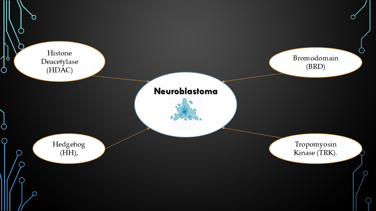
Childhood neuroblastoma (NB) is a malignant tumour that is a member of a class of embryonic tumours that have their origins in sympathoadrenal progenitor cells.[1] There are five stages in the clinical NB staging system: 1, 2A, 2B, 3, 4S, and 4.[1] For those diagnosed with stage 4 neuroblastoma (NBS4) the treatment options are limited with a survival rate of between 40 to 50%.[2] This article is concerned with the search for a multi-target drug treatment for high-risk NBS4 and focuses on four possible treatment targets. Since 1975, more than 15 targets have been identified in the search for a treatment for high-risk NBS4.[3] In this work six targets were originally selected and of these, four demonstrated amino acid sequence similarity of 80-100% and are the focus of this paper. The other two targets are the subject of a paper in progress and showed similarity across their receptors of 80-100% but lower than 80% for the other four targets.
The four targets described here have been investigated for the role they play in the development of NB and include: Histone Deacetylase (HDAC), Bromodomain (BRD), Hedgehog (HH), and Tropomyosin Kinase (TRK). Of these, some are epigenetic such as Bromodomain (BRD), Hedgehog (HH) and Histone Deacetylase (HDAC) that is concerned with heritable changes in the functioning of genes without changes in the DNA sequence. In the case of cancers, it is nearly impossible to reverse genetic alterations, whereas epigenetic changes “can dynamically respond to signals from the physical, biological and social environment.”[4] Others targets investigated for NBS4 include Tyrosine Kinases such as MYCN that is involved in gene amplification in NB. The fourth target in this paper belongs to the Tyrosine Kinase family and is Tropomyosin Receptor Kinase (TRK).
Current treatment for NBS4 involves immunotherapy combined with anti-cancer drugs.[5] With current therapeutic agents meeting with little success in treating NBS4, the search for a new target remains urgent.[6] Currently, treatment agents focus on a one-drug-one-target approach and/or combination therapy, which has had little success in improving survival rates. An alternative approach to the current model is to develop a multi-target drug that interacts with multiple targets with high efficacy to change the disease network. Further perceived benefits to developing a multi-target drug offer the possibility of making "cocktail therapies" or drug combinations redundant [7] leading to less pharmacokinetic and safety profile testing, as the risk of drug-drug interactions would be reduced.[8] Since it is uncommon for multiple targets to mutate simultaneously in different pathways or at various locations within a single cascade pathway, multi-target drugs may also avoid drug resistance brought on by single-target mutations or changes in expression.[9]
This article presents a developmental, multi-targeted drug design approach using computational methods available to medicinal chemistry. Computer-aided drug design and molecular modelling have greatly assisted drug development within the field of medicinal chemistry.[10] Computational methods such as molecular docking, homology modelling, molecular dynamics, and quantitative structure-activity relationships (QSAR) are frequently used as part of the process of finding new therapeutic drug targets.[11] Using these methods, eight compounds (inhibitors) were identified as possible inhibitors for all four targets. Results revealed that all four targets BRD, HDAC, HH and TRK share similar amino acid sequencing that ranges from 80-100% offering the possibility of further testing for their suitability for multi-target drug use. Two additional targets were also tested as part of this work, Retinoic Acid (RA) signalling pathway and c-Src kinase (Csk) which showed similarity across their receptors of 80-100% but were lower that 80% for the other four targets. The work for these two targets is the subject of a paper currently in progress.
1.1. Selected Targets and Why?
- 1)
- Histone Deacetylase (HDAC)
The Histone Deacetylase (HDAC) family comprises of 18 enzymes divided into four classes (I, II, III, and IV) according to their enzymatic activities, subcellular localisation, and homology to yeast HDCA.[12] In the case of HDAC 8 (class I) it was found to be downregulated in NBS4 (Figure 1).[13]
- 2)
- Bromodomain (BRD)
Early in the 1990s, the Brahma gene of Drosophila Melanogaster was found to contain a family of evolutionarily conserved motifs known as Bromodomains (BRD).[14] Numerous studies have been conducted on the Bromodomain and extra terminal (BET) family. It consists of BRDT, BRD2, BRD3, and BRD4, all of which are widely expressed, with the exception of BRDT, which is only expressed in the testis.[15] BRDs bind histone tail acetylated lysines, recognising the acetyl group is essential for recruiting additional chromatin factors and transcriptional machinery, which controls gene transcription.[15] The BET family also functions as a cell cycle regulator with BRD4 regulating the expression of genes necessary for the transition from the M to early G1 phase.[15] Research found that the compound JQ1, a BRD inhibitor, upregulated p27 and the proapoptotic gene BIM while suppressing MYC expression, resulting in G1 cell cycle arrest.[16] Studies on the Bromodomain inhibitor BET762 in vivo have also shown that it has anti-cancer properties.[17]
- 3)
- Hedgehog (HH)
Hedgehog Inhibitors (HHIs) have become a promising new target for cancer therapy.[18] The signalling pathway identified in 1980 [19] was a crucial regulator of growth, patterning formation, and cell migration during embryonic development.[20] The components of the HH signalling pathway are involved in signalling to the transcription factors.[21] One study found that signalling deregulation was observed with Gorlin syndrome and cancers (Figure 2).[22]
- 4)
- Tropomyosin Receptor Kinase (TRK)
The neurotrophin family of peptide hormones activates three related tyrosine kinases known as tropomyosin receptor kinases (TRK) [23] that along with various forms of cancer, are also essential in neurodegenerative diseases. TRK inhibitor development to target cancers driven by NTRK fusion, has gained attention in the past ten years (Figure 3).[24]
1.2. Medicinal Chemistry Approaches to Drug Design
Rational multi-target drug design strategies in recent years have varied from pharmacophore combination, screening, [25] and similar scaffold structure.[26] Each strategy has advantages and disadvantages, along with varying challenges. The purpose of this work is to suggest a possible modified approach. In the search for a multi-target drug for NBS4 using advanced medicinal chemistry software, this approach assesses the possibility of different targets by assessing receptor similarities and the selection of two lead compounds for each target (eight compounds in total) that have demonstrated an inhibitory effect on NBS4 cells, to form the basis of a search for a multi-target drug.
Having identified four targets, two receptors from each target were selected (see Table 1) and a Protein Aligner tool from Samson was used to check for their suitability, with results reporting 80-100% similarity for the four targets. Protein Aligner checks for amino acid sequence similarity between receptors with high similarity between receptors indicating their suitability for use in cross-docking. For the purposes of this work docking involves docking a compound to a receptor known for that target (i.e. BRD) whereas cross-docking is docking the same compound to a receptor that belongs to a different target (i.e. TRK) and vice versa. The aim of cross-docking is to explore the suitability of the selected compound for use as a multi-target drug. This is a process that involves selecting known lead compounds to produce hitlists of compounds and this was done using BROOD [27] as part of the OpenEye suite.
Several BROOD rounds were completed on the 2 lead compounds for each target to produce hitlists using "Shape and Color" and "Shape and Electrostatics." On completion, BROOD ranked each of the hitlists according to BROOD hitlist parameters (see list below) and the top 25 of each hitlist was selected to run on OEDocking[28], AFITT [29], ROCS [30], and VIDA[31]. In addition, the Samson docking suite, including AutoDock Vina Extended and Fitted (Molecular Forecaster) [32], with another docking program, Molegro [33], acted as confirmation for both the docking and cross-docking procedures. From this process 8 to 10 compounds were selected for each target, all of which showed improved parameters compared to the original lead compounds (see Table 3). The compounds from each individual target were then cross-docked with the receptors from the other three using AFITT. From the 35 compounds run only 8 showed potential suitability for multi-target use (two from each list) see Table 6.
To check and compare the compounds (clusters) for toxicity and mutagenicity, Toxicity Estimation Software Tools (TEST) were used to provide the prediction mechanisms of the toxic action of the clusters.[34] The results from TEST showed some similarity with the original lead compounds (Table 7) which also provided the suitability for use. Further work was done to explore possible synthesis routes for each of the eight compounds using the retrosynthesis program Spaya [35].
2. Results
2.1. Medicinal Chemistry Results
Table 1 below contains the selected targets, protein/receptors (Protein Data Bank) and the identified lead compounds.

Table 1.
Selected lead compounds and receptors for each target type by literature review.
| Target | Lead Compound 1 | Lead compound 2 | Protein /Receptor |
|---|---|---|---|
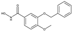
| 1-Histone Deacetylase 8 Inhibitors | 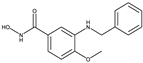 |
 |
2V5X and 2V5W[36] |
| 8b[37] | 20a[37] | ||
| 2-Bromodomain Inhibitors |
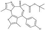 JQ1[15] |
 BET762[15] |
4BJX[38] and 5UY9[39] |
| 3-HH inhibitors |
 BMS-833923[40] |
 Vismodegib[41] |
5L7I[42] and 3N1P[43] |
| 4-Tropomyosin Receptor Kinase Inhibitors |
 GW441759 [44] |
 Compound 10[44] |
4AT3[45] and 3V5Q[46] |
The next stage was to compare the similarity of the proteins (receptors) for all four targets. This was achieved using the Samson Protein Aligner tool, showing that the similarities between the selected proteins ranged from 80% to 100% (Figure 8).
Figure 4.
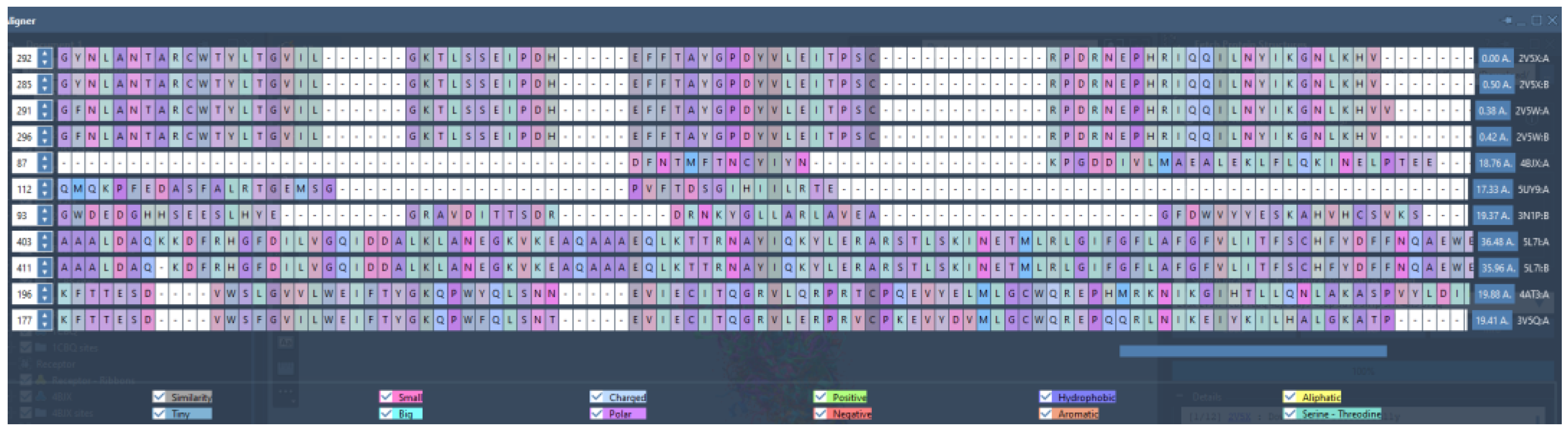
Print screen from Protein Aligner of 2V5W with all the selected receptors from the four targets. The similarity (in grey) was obtained using Protein Aligner by Samson. Zero indicates 100% similarity, and 100% means no similarity. Figure 4 shows that the similarities between the selected proteins range from 80% to 100%.
Figure 4.
Print screen from Protein Aligner of 2V5W with all the selected receptors from the four targets. The similarity (in grey) was obtained using Protein Aligner by Samson. Zero indicates 100% similarity, and 100% means no similarity. Figure 4 shows that the similarities between the selected proteins range from 80% to 100%.
By selecting an active group in the lead compound, the program can produce a hitlist using shape and colour and shape and electrostatics. Table 2 shows how many rounds were performed for each lead compound according to target. The top 25 compounds from the hitlists were selected for the docking studies from each round. Figure 5 shows some of the compounds.
- 1)
- AroRingCt: Number of aromatic rings in the molecule,
- 2)
- ClusterID/IdeaGroup: ClusterID of the molecule;
- 3)
- Colour: The replacement fragment's colour Tanimoto score in comparison to the query fragment;
- 4)
- Combo: Tanimoto combo score for the replacement fragment's shape and colour in comparison to the query fragment;
- 5)
- Egan: The Boolean indicates if the molecule satisfies the Egan bioavailability model;
- 6)
- Fragment: SMILES string of the replacement fragment;
- 7)
- Freq: The replacement fragment's frequency;
- 8)
- fsp3C: The molecule's fraction of sp3 hybridized carbon atoms;
- 9)
- HvyAtoms: Number of heavy atoms in the molecule;
- 10)
- LipinskiDon: Number of Lipinski donors in the molecule;
- 11)
- LipinkskiAcc: Number of Lipinski acceptors in the molecule;
- 12)
- LipinskiFail: Boolean specifying whether the molecule fails Lipinski’s rule of five;
- 13)
- Local strain: Calculated local strain of the molecule;
- 14)
- Molecular TanimotoCombo: Shape + colour Tanimoto combo score of the molecule against the query molecule;
- 15)
- MolWt: Molecular weight of the molecule;
- 16)
- p(active): Belief score of the molecule;
- 17)
- RingCt: Number of ring atoms;
- 18)
- RingRatio: Ratio of the number of ring atoms to the total number of heavy atoms;
- 19)
- Rotors: Number of rotatable bonds in the molecule;
- 20)
- shape: Compare the replacement fragment's Shape Tanimoto score to that of the query fragment;
- 21)
- Source Mols: SMILES strings of the molecules the replacement fragment is part of;
- 22)
- Source Mol Labels: Labels of the molecules the replacement fragment is part of;
- 23)
- tPSA: Calculated topological polar surface area of the molecule;
- 24)
- Veber: Boolean specifying whether the molecule passes the Veber bioavailability model, and
- 25)
- XlogP: Calculated LogP of the molecule [47].
The next stage was to dock each hitlist with their relevant receptors; Figure 7 shows one of the docking outcomes using FRED (OpenEye suite) at the top of the list cluster 22, 1 of 1.
Having completed all the docking using FRED, Molegro, AutoDock Vina, and Fitted only the top compounds for each target were selected (range 8 to 10 see Table 3) to be run for validation on ROCS a program that scores and aligns a database of molecules with a query. The score assigns a number to molecules according to their likelihood of having biological characteristics in common with the query molecule (Figure 8).
Another crystallographic tool used from the OpenEye suite is AFITT (Figure 9). AFITT creates a new combined forcefield that fits small molecules into crystallographic density while preserving superior chemistry by combining the shape and MMFF technologies of OpenEye. In order to verify the refinement, it also offers an interface to external refinement programs, such as real space correlation coefficient calculation (RSCC) and interactive Ramachandran plots.
Selected clusters from all targets were also docked with the Samson Suite, including AutoDock Vina Extended and Fitted by Molecular Forecaster.
Figure 10.
Screenshot from Samson (AutoDock Vina Extended): TRKI (clusters) with 4AT3 before docking (A) and after docking (B).
Figure 10.
Screenshot from Samson (AutoDock Vina Extended): TRKI (clusters) with 4AT3 before docking (A) and after docking (B).
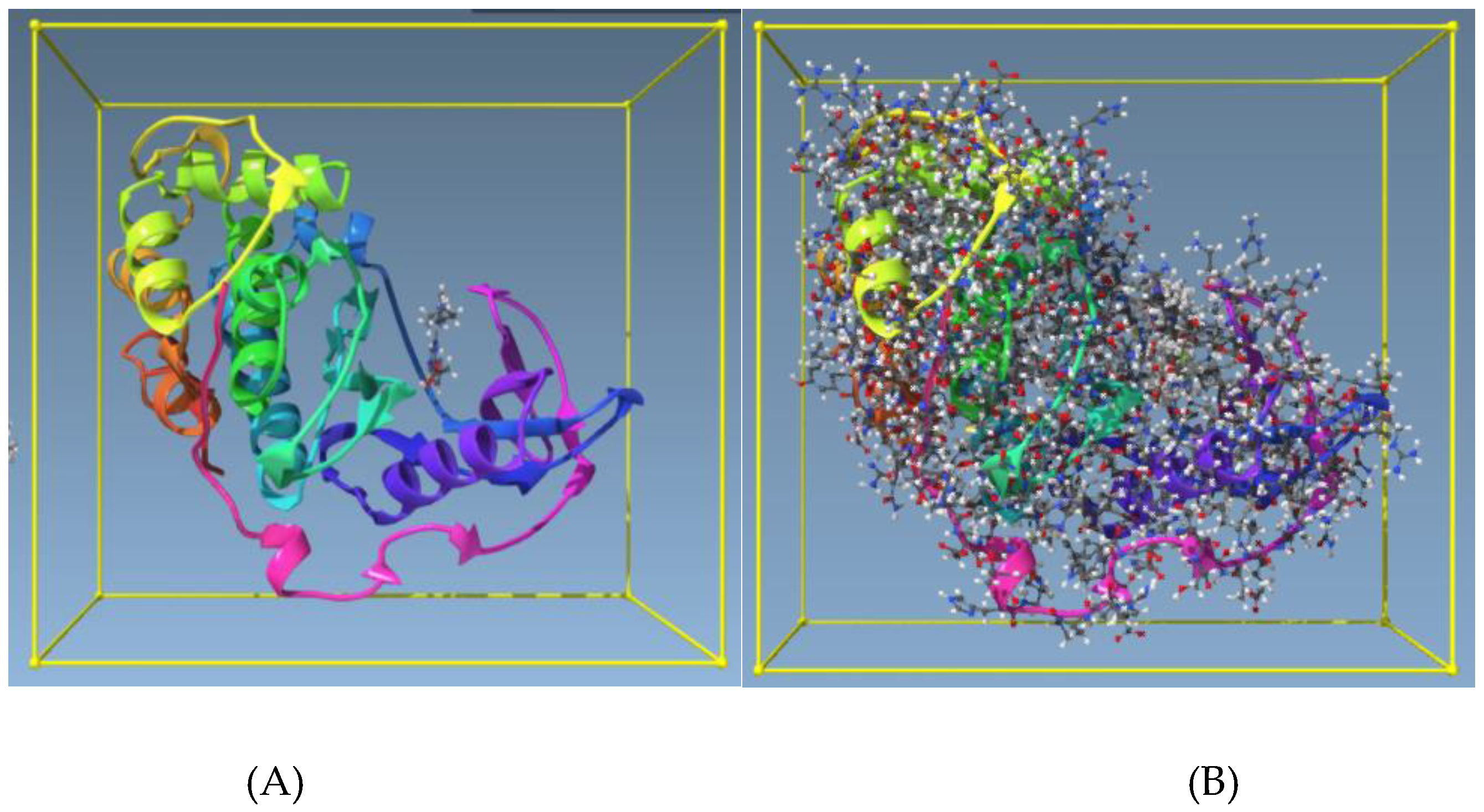
Figure 11.
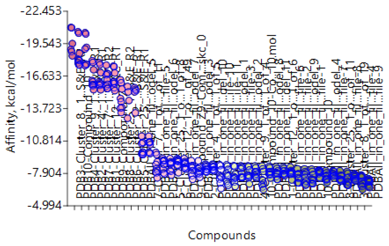
Docking ranking of the TRK receptor 4AT3 with TRKI selected clusters in AutoDock Vina Extended.
Figure 11.
Docking ranking of the TRK receptor 4AT3 with TRKI selected clusters in AutoDock Vina Extended.
All possible clusters (Table 3) were docked with all docking tools: FRED, AFITT, AutoDock Vina Extended, Molegro, and Fitted. Table 4 provides an example.
Table 4.
The top clusters obtained from BROOD BRD receptor 4BJX were docked with AFITT, FRED, AutoDock Vina Extended, Molegro, and Fitted. BET762 and JQ1 were the lead compounds.
Table 4.
The top clusters obtained from BROOD BRD receptor 4BJX were docked with AFITT, FRED, AutoDock Vina Extended, Molegro, and Fitted. BET762 and JQ1 were the lead compounds.
| Column1 | Clusters from Bromodomain (BRD) | AFITT | FRED | AutoDock Vina extended | Molegro | Fitted |
|---|---|---|---|---|---|---|
| 1 | Cluster 3, 1 of 3 | 0.766 | -6.474 | -8.124 | -4.86 | -25.259 |
| 2 | Cluster 25, 1 of 12 | 0.6863 | -8.315 | -8.549 | 89.3 | -26.775 |
| 3 | Cluster 16, 1 of 2 | 0.7356 | -6.337 | -7.542 | 52.9 | -21.687 |
| 4 | Cluster 15, 1 of 2 | 0.553 | -5.308 | -7.048 | 33.28 | -23.624 |
| 5 | Cluster 4, 1 of 4 | 0.4898 | -5.693 | -8.403 | 36.98 | -25.566 |
| 6 | Cluster 24, 1 of 7 | 0.4644 | -8.556 | -8.909 | 80.89 | -30.289 |
| 7 | Cluster 23, 1 of 1 | 0.497 | -5.955 | -7.795 | 9.34 | -24.131 |
| 8 | Cluster 10, 1 of 1 | 0.7103 | -4.672 | -7.71 | 5.23 | -21.734 |
| 9 | BET-762-Lead compound | 0.6691 | -7.528 | -7.971 | 49.84 | -19.709 |
| 10 | JQ1-Lead compound | 0.6084 | -8.133 | -7.006 | 99.29 | -22.161 |
Table 5.
Docking of selected clusters from each target with more than one receptor of the same target. Data represent the best real space correlation coefficient calculation (RSCC).
Table 5.
Docking of selected clusters from each target with more than one receptor of the same target. Data represent the best real space correlation coefficient calculation (RSCC).
| Clusters HDACs | AFITT | Clusters BRD | AFITT | Clusters HH | AFITT | Clusters Tropomyosin | AFITT | ||||
|---|---|---|---|---|---|---|---|---|---|---|---|
| 2V5X | 2V5W | 4BJX | 5UY9 | 5L7I | 3N1P | 4AT3 | 3V5Q | ||||
| Cluster 22, 1 of 22 | 0.652 | 0.541 | Cluster 3, 1 of 3 | 0.77 | 0.42 | Cluster 4, 1 of 1 | 0.552 | 0.336 | Cluster 12, 1 of 6 | 0.638 | 0.438 |
| Cluster 21, 1 of 11 | 0.650 | 0.499 | Cluster 25, 1 of 12 | 0.69 | 0.39 | Cluster 8, 1 of 1 | 0.514 | 0.385 | Cluster 4, 1 of 5 | 0.685 | 0.438 |
| Cluster 16,1 of 65 | 0.650 | 0.541 | Cluster 16, 1 of 2 | 0.74 | 0.33 | Cluster 1, 1 of 3 | 0.521 | 0.330 | Cluster 8, 1 of 19 | 0.435 | 0.438 |
| Cluster 23, 1 of 1 | 0.645 | 0.532 | Cluster 15, 1 of 2 | 0.55 | 0.39 | Cluster 9, 1 of 1 | 0.513 | 0.404 | Cluster 9, 1 of 4 | 0.622 | 0.539 |
| Cluster 1, 1 of 26 | 0.642 | 0.504 | Cluster 4, 1 of 4 | 0.49 | 0.33 | Cluster 8, 1 of 29 | 0.496 | 0.432 | Cluster 25, 1 of 8 | 0.675 | 0.521 |
| Cluster 7, 1 of 26 | 0.635 | 0.527 | Cluster 24, 1 of 7 | 0.46 | 0.37 | Cluster 21, 1 of 99 | 0.493 | 0.340 | Cluster 12, 1 of 49 | 0.630 | 0.627 |
| Cluster 4, 1 of 28 | 0.633 | 0.499 | Cluster 23, 1 of 1 | 0.50 | 0.37 | Cluster 15, 1 of 6 | 0.491 | 0.374 | Cluster 7, 1 of 11 | 0.643 | 0.596 |
| Cluster 10, 1 of 86 | 0.632 | 0.517 | Cluster 10, 1 of 1 | 0.71 | 0.37 | Cluster 23, 1 of 1 | 0.478 | 0.470 | Cluster 25, 1 of 11 | 0.491 | 0.614 |
| Cluster 12, 1 of 50 | 0.631 | 0.524 | BET-762 | 0.67 | 0.39 | Cluster 17, 1 of 1 | 0.471 | 0.364 | Compound Z9 | 0.596 | 0.543 |
| 20A | 0.631 | 0.511 | JQ1 | 0.61 | 0.41 | Cluster 20, 1 of 12 | 0.463 | 0.389 | Compound 10 | 0.560 | 0.505 |
| 8B | 0.629 | 0.516 | Vesmodigib | 0.363 | 0.363 | ||||||
| BMS-833923 | 0.313 |
The next step was cross-docking, which involved docking receptors with various cluster types and comparing the results. Figure 12, Figure 13, Figure 14 and Figure 15 show some of the results from the cross-docking of each target.
Figure 12.
Cross-docking. HDAC Receptor 2V5W with BRD, HH, and TRK of multi-target inhibitors using AutoDock Vina Extended in Samson.
Figure 12.
Cross-docking. HDAC Receptor 2V5W with BRD, HH, and TRK of multi-target inhibitors using AutoDock Vina Extended in Samson.
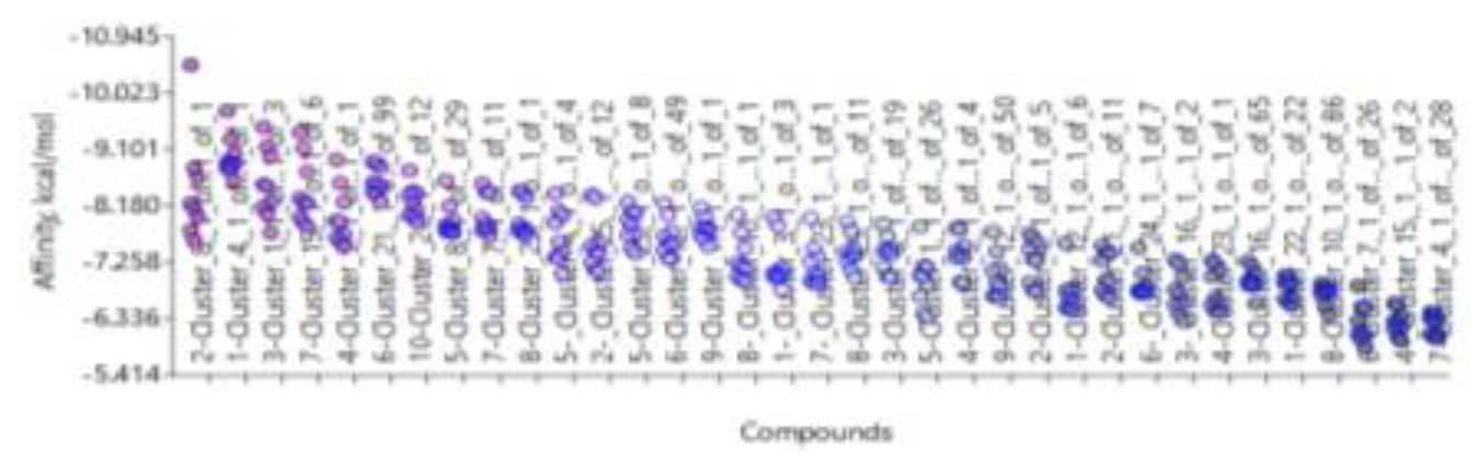
Figure 13.
Cross-docking. BRD Receptor 4BJX with top list HDAC, HH, and TRK multi-target inhibitors using AutoDock Vina Extended in Samson.
Figure 13.
Cross-docking. BRD Receptor 4BJX with top list HDAC, HH, and TRK multi-target inhibitors using AutoDock Vina Extended in Samson.
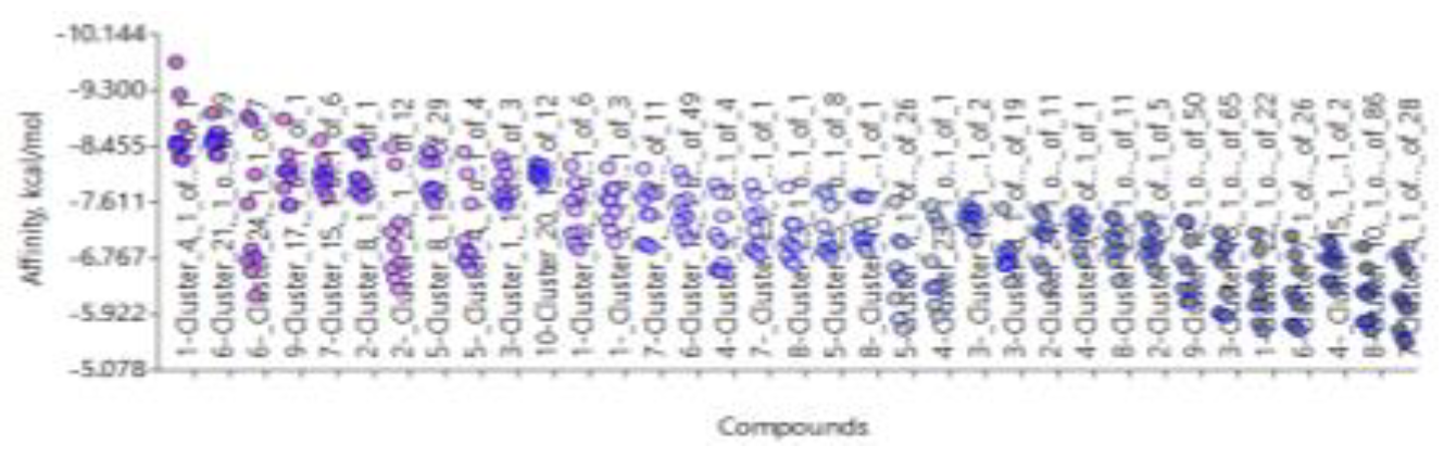
Figure 14.
Cross-docking. HH Receptor 5L7I with HDAC, BRD, and TRK multi-target inhibitors using AutoDock Vina Extended in Samson.
Figure 14.
Cross-docking. HH Receptor 5L7I with HDAC, BRD, and TRK multi-target inhibitors using AutoDock Vina Extended in Samson.
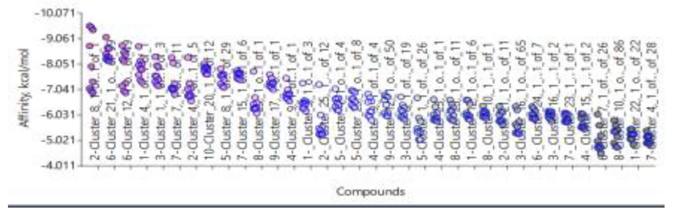
Figure 15.
An example of cross-docking. TRK Receptor 4AT3 with HDAC, HH, and BRD inhibitors using AutoDock Vina Extended in Samson.
Figure 15.
An example of cross-docking. TRK Receptor 4AT3 with HDAC, HH, and BRD inhibitors using AutoDock Vina Extended in Samson.
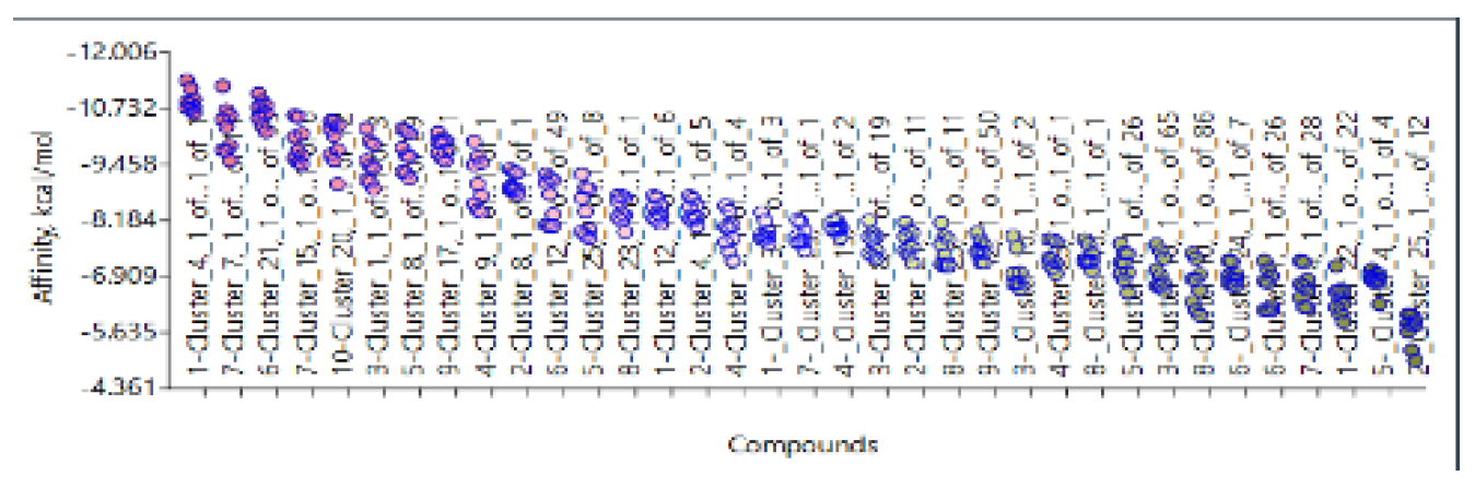
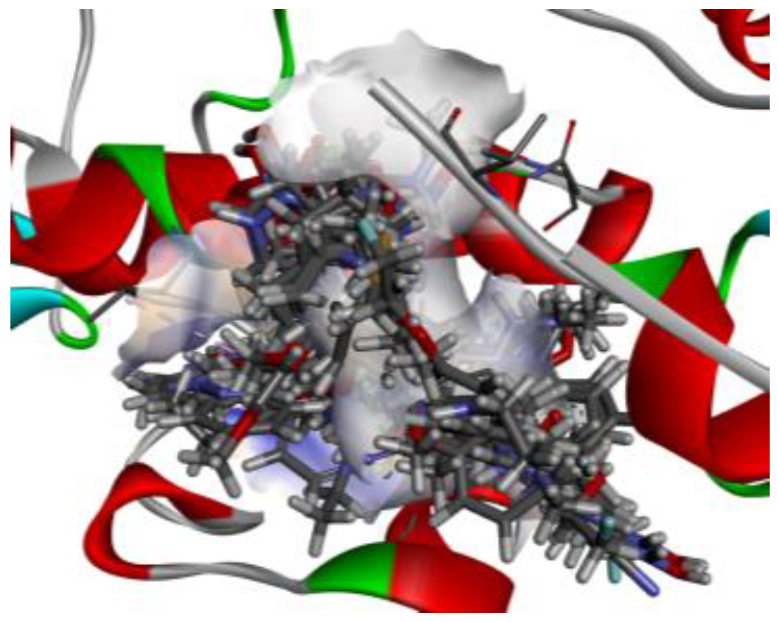
Figure 16.
Docking. HDAC Receptor 2V5x with BRD, HH, and TRK inhibitors using Samson Fitted in BIOVIA Studio Discovery Visualizer.[48].
Figure 16.
Docking. HDAC Receptor 2V5x with BRD, HH, and TRK inhibitors using Samson Fitted in BIOVIA Studio Discovery Visualizer.[48].
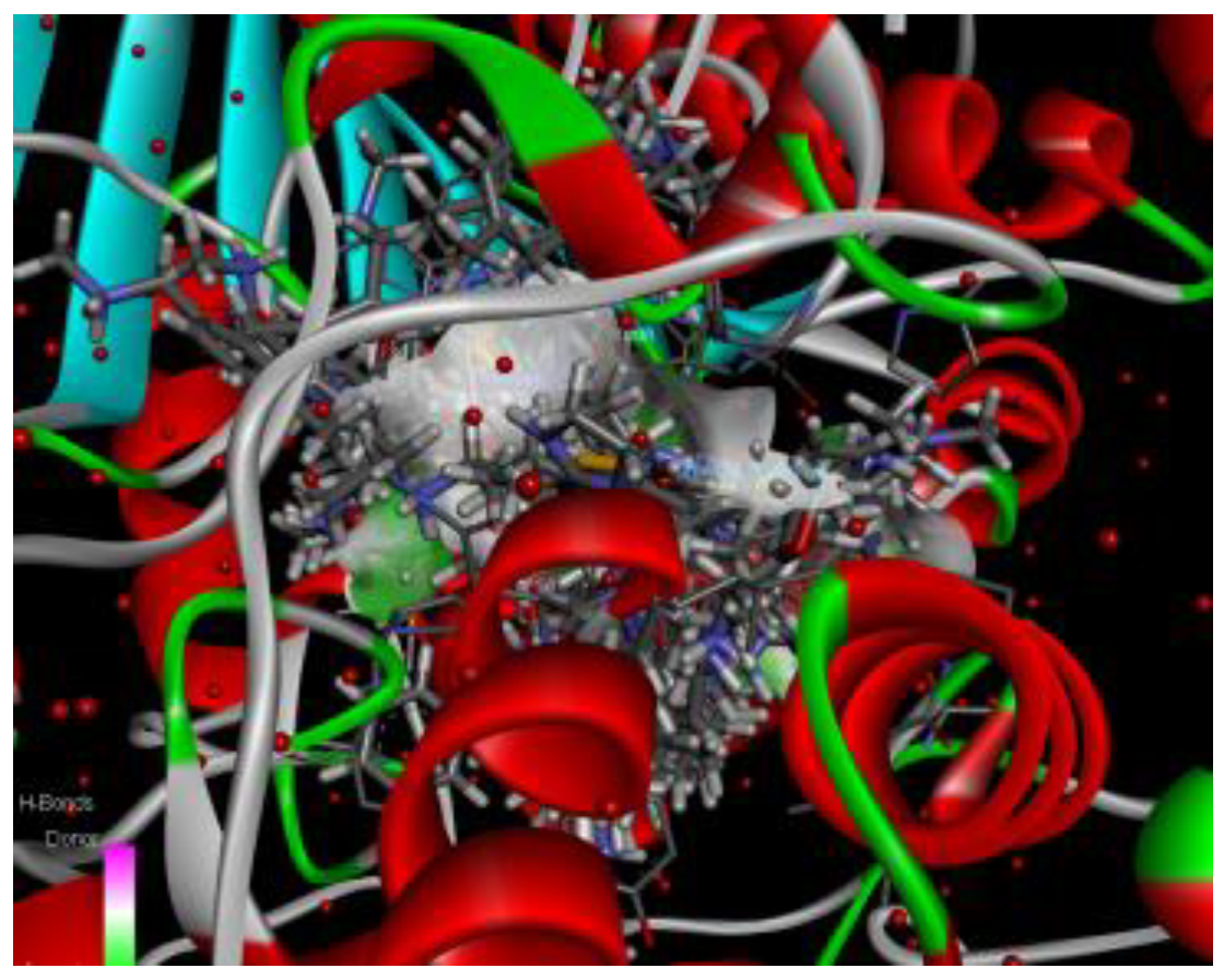
Figure 17.
Docking. HDAC Receptor 2V5x with BRD, HH, and TRK inhibitors using Samson Fitted in BIOVIA Studio Discovery Visualizer.[48].
Figure 17.
Docking. HDAC Receptor 2V5x with BRD, HH, and TRK inhibitors using Samson Fitted in BIOVIA Studio Discovery Visualizer.[48].
Having completed all cross-docking for the receptors and the selected clusters, eight compounds were identified as possible multi-target compounds (see Table 6 below).
- 1)
- Bioconcentration factor: ratio of the chemical concentration in fish as a result of absorption via the respiratory surface to that in water at a steady state.
- 2)
- Ames mutagenicity: A compound is positive for mutagenicity if it induces revertant colony growth in any strain of Salmonella typhimurium.
- 3)
- Oral rat LD50: Amount of chemical (mg/kg body weight) that causes 50% of rats to die after oral ingestion.
- 4)
- 48-hour T. pyriformis IGC50: Concentration of the test chemical in water (mg/L) that causes 50% growth inhibition to Tetrahymena pyriformis after 48 hours.
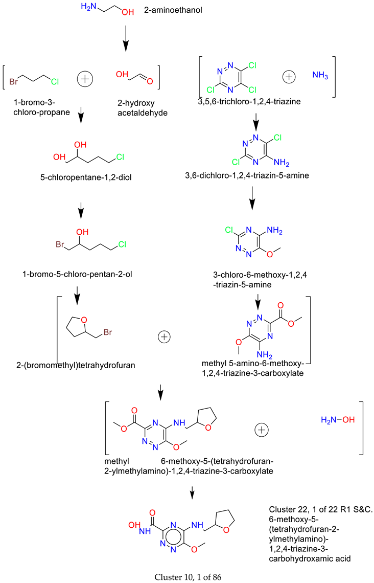
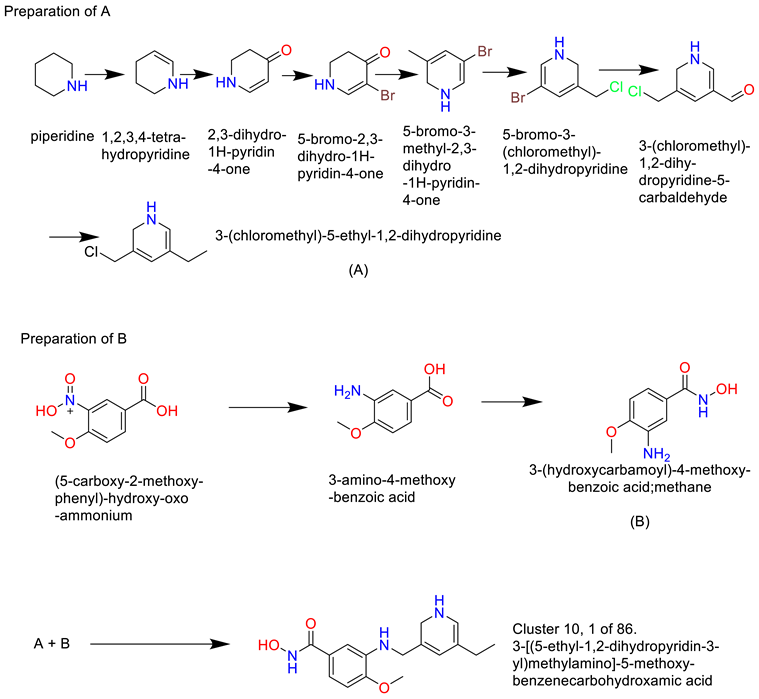
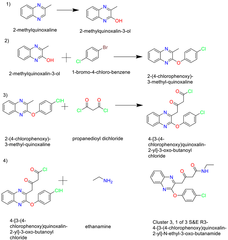
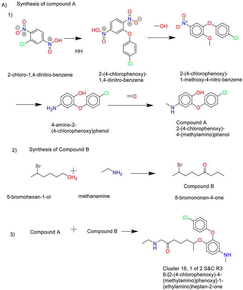
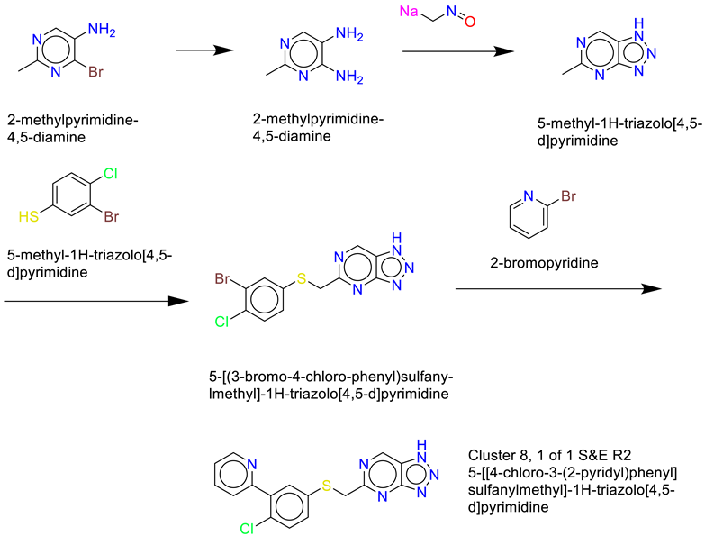
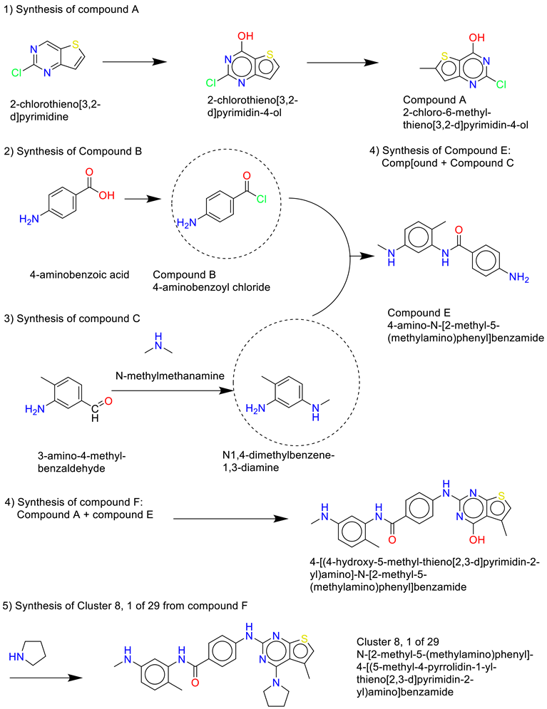
2.2. Retrosynthesis Results Using Spaya
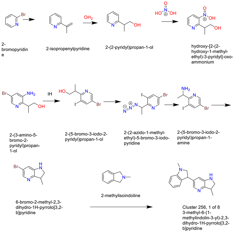
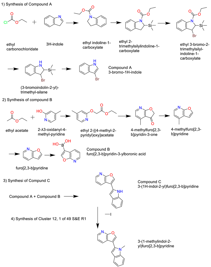
- 1)
- Synthesis of Cluster 22, 1 of 22 R1 S&C[35]
- 2)
- Cluster 10, 1 of 86
- 3)
- Cluster 3, 1 of 3
- 4)
- Cluster 16, 1 of 2
- 5)
- Cluster 8, 1 of 1
- 6)
- Cluster 8, 1 of 29
- 7)
- Cluster 25, 1 of 8
- 8)
- Cluster 12, 1 of 49
3. Discussion
The development of multi-target drugs is described as the process of “taking a well-validated primary target for a given disease and adding secondary activities to enhance efficacy”.[25] Designing such a drug involves fusing the inhibitory actions of two or more drugs into one molecule.[26] Typical approaches used for designing multi-target drugs include “Pharmacophore” and “Screening.” The pharmacophore approach can include processes such as merged-pharmacophore mode, fused-pharmacophore mode, non-cleavable linked pharmacophore, and cleavable pharmacophore. Pharmacophore modelling aims to "strip" functional groups of their true chemical nature in order to categorize them into a small number of pharmacophore types based on their predominant physicochemical characteristics.[49] Difficulties with this method occur due to inadequate or inaccurate conformational sampling, ambiguities in pharmacophore typing (primarily because of uncertainty regarding the tautomeric/protonation status of compounds), computer time limitations in complex molecular overlay calculations, and the selection of inappropriate anchoring points in active sites when ligand cocrystal structures are unavailable.[49] Along with pharmacophore the technique of screening is also used for drug discovery and has four identifiable categories: Fragment-based Drug Discovery (FBDD), High-throughput Screening (HTS), High-content Screening (HCS), and Virtual Screening (VS). For the work described here, a modified version of the Virtual Screening (VS) programme is used to enable the discovery of multi-target compounds (inhibitors). Using computational methods available to medicinal chemistry, computer-aided drug design and molecular modelling have greatly assisted drug design in the field.[10] Computational methods such as molecular docking, homology modelling, molecular dynamics, and quantitative structure-activity relationships (QSAR) are frequently used as part of the process for finding new therapeutic drug targets using computational methods.[11] For this work 8 compounds (inhibitors) were identified as possible inhibitors for four targets: HDAC, BRD, HH and TRK.
Having selected four targets used in the study of NBS4, two receptors from each of the four targets were selected and the similarities of the receptors were compared as representations of the targets (Figure 4). Results indicated 80-100% similarities (using the Protein Aligner program from Samson) confirming the possibility for multi-target use. High similarity between receptors indicates their suitability for use in cross-docking and is a process that involves selecting known lead compounds to produce hitlists of compounds (clusters). Using BROOD [27] as part of the OpenEye suite several BROOD rounds were completed on the lead compounds to produce hitlists with the top 25 of each hitlist selected to run on OEDocking[28], AFITT [29], ROCS [30], and VIDA. The hitlists of compounds (clusters) were docked and the selected compounds, cross-docked. The clusters were docked with more than one docking program as a means to validating the result.
The final ranking of the selected clusters was done on AFITT as the receptor preparation with the tool MakeReceptor gives the user more control over the receptor-creating process. AFITT also has the advantage of real-space fitting of ligands in density, integrated with REFMAC, PHENIX, BUSTER, CNX, and COOT and also fragment and cocktail fitting. In AFFIT it is possible to select more than one ligand to fit generation of high-quality refinement dictionaries for use. This can be done during reciprocal space refinement that includes: the use of forcefield (MMFF); semi-empirical (AM1, PM3) methods during reciprocal space refinement for BUSTER and Phenix; real space fitting of protein residues; proper handling of covalently bonded ligands, and proper handling of multiple occupancy ligands.
The ranking is based not only on the best results but also on the ability of the identified compounds to cross-dock on the receptors of other targets, and it was this process that led to the selection of the eight compounds. Using the tool ROCS provided cluster validation and is based on a large data base search. Toxicity and mutagenicity of the eight compounds were tested using Toxicity Estimation Software Tools (TEST) [34] and the results showed some similarity with the original lead compounds (Table 7). With the aid of the retro-synthesis programme Spaya (IKTOS), the possibility of synthesizing all eight compounds was demonstrated, (see results section) [35]. These results point the way to future work that will focus on preparing and testing the eight compounds in vitro and in vivo.[50]
4. Materials and Methods
4.1. Materials
Computer programs:
- OpenEye Scientific programs, which include various applications, are being used. The suite comprises BROOD, MakeReceptor, FRED, and AFITT.
- Molegro Virtual Docker.
- The Samson suite includes Autodock Vina extended, the Fitted suite by Molecular Forecaster, and Protein Aligner.
- Toxicity Estimation Software Tools (TEST).
- BIOVIA Discovery Studio Visualizer.
- Spaya-retrosynthesis software.
4.2. Method
- Identifying drug targets.
- Selection of two proteins (receptors) for each target and downloading the PDB files and their electron density map from the Protein Data Bank database.
- Comparing the similarities of the receptors. Run protein similarity on Samson (Protein Aligner) to determine suitability.
- Selection of two lead compounds from each type.
- Run the lead compounds on BROOD (from the OpenEye suite) and produce hit lists using Shape and Colour and Shape and Electrostatics.
- Receptor preparations using MakeReceptor from the OpenEye suite.
- Docking the hit compounds with OpenEye suite (FRED), Molegro, and Samson suite (AutoDockVina and Fitted).
- Run cross-docking; each hitlist clusters from one target to the other 3 targets (using their protein/receptor).
- Run hits with AFITT to rank the compounds according to their fitting probabilities.
- Run selected clusters on ROCS.
- Run selected clusters on Toxicity Estimation Software Tools (TEST)
- Run clusters on Spaya to find the best synthesis route.
5. Conclusions
Using Virtual Screening with some modifications, eight compounds were identified as potential inhibitors across four targets (HDAC, BRD, HH, TRK) for the development of multi-target drug treatment of NBS4. The next stage is for the eight compounds to undergo single molecule testing in vivo and in vitro.[50]
Funding
Please add: This research received no external funding.
Data Availability Statement
See the attached folder.
Acknowledgments
I would like to thank Dr. Una Canning for her invaluable support and editing skills and Dr. Corinne Kay for her advice and support since I started working on neuroblastoma. Thanks also to Colin Gaudion for his support in testing some of the programmes used for this work.
Conflicts of Interest
The author’s daughter, Isabella, was diagnosed with NBS4 in January 2003. Isabella relapsed in March 2005 and died in July 2005, a week after her seventh birthday.
References
- Cullinane, C.J.; Burchill, S.A.; Squire, J.A.; O'Leary, J.J.; Lewis, I.J. Molecular Biology and Pathology of Paediatric Cancer; 2003. [Google Scholar]
- Paraboschi, I.; Privitera, L.; Kramer-Marek, G.; Anderson, J.; Giuliani, S. Novel Treatments and Technologies Applied to the Cure of Neuroblastoma. Children (Basel) 2021, 8, 482. [Google Scholar] [CrossRef]
- Cohen, S.; Carpenter, G. Human epidermal growth factor: isolation and chemical and biological properties. Proc. Natl. Acad. Sci. U. S. A. 1975, 72, 1317–1321. [Google Scholar] [CrossRef] [PubMed]
- Inomistova, M.; Khranovska, N.; Skachkova, O. Role of Genetic and Epigenetic Alterations in Pathogenesis of Neuroblastoma; Academic Press, 2019; pp. 23–41. [Google Scholar]
- Bhoopathi, P.; Mannangatti, P.; Emdad, L.; Das, S.K.; Fisher, P.B. The quest to develop an effective therapy for neuroblastoma. J Cell Physiol 2021, 236, 7775–7791. [Google Scholar] [CrossRef]
- Gerges, A.; Canning, U. Neuroblastoma and its Targets Therapies: A Medicinal Chemistry Review. ChemMedChem 2024, 19, e202300535. [Google Scholar] [CrossRef] [PubMed]
- Zhang, W.L.; Pei, J.F.; Lai, L.H. Computational Multitarget Drug Design. Journal of Chemical Information and Modeling 2017, 57, 403–412. [Google Scholar] [CrossRef]
- Anighoro, A.; Bajorath, J.; Rastelli, G. Polypharmacology: Challenges and Opportunities in Drug Discovery. Journal of Medicinal Chemistry 2014, 57, 7874–7887. [Google Scholar] [CrossRef] [PubMed]
- Knight, Z.A.; Lin, H.; Shokat, K.M.; Knight, Z.A.; Lin, H.; Shokat, K.M. Targeting the cancer kinome through polypharmacology. Nature Reviews Cancer 2010, 10, 130–137. [Google Scholar] [CrossRef]
- Ooms, F. Molecular Modeling and Computer Aided Drug Design. Examples of their Applications in Medicinal Chemistry. Current Medicinal Chemistry 2000, 7, 141–158. [Google Scholar] [CrossRef]
- Kumalo, H.M.; Bhakat, S.; Soliman, M.E.S.; Kumalo, H.M.; Bhakat, S.; Soliman, M.E.S. Theory and Applications of Covalent Docking in Drug Discovery: Merits and Pitfalls. Molecules 2015, 20, 1984–2000. [Google Scholar] [CrossRef]
- Bolden, J.E.; Peart, M.J.; Johnstone, R.W. Anticancer activities of histone deacetylase inhibitors. Nat Rev Drug Discov 2006, 5, 769–784. [Google Scholar] [CrossRef]
- Oehme, I.; Deubzer, H.E.; Wegener, D.; Pickert, D.; Linke, J.P.; Hero, B.; Kopp-Schneider, A.; Westermann, F.; Ulrich, S.M.; von Deimling, A.; et al. Histone deacetylase 8 in neuroblastoma tumorigenesis. Clin Cancer Res 2009, 15, 91–99. [Google Scholar] [CrossRef] [PubMed]
- Tamkun, J.W.; Deuring, R.; Scott, M.P.; Kissinger, M.; Pattatucci, A.M.; Kaufman, T.C.; Kennison, J.A. brahma: a regulator of Drosophila homeotic genes structurally related to the yeast transcriptional activator SNF2/SWI2. Cell 1992, 68, 561–572. [Google Scholar] [CrossRef]
- Pérez-Salvia, M.; Esteller, M. Bromodomain inhibitors and cancer therapy: From structures to applications. Epigenetics 2017, 12, 323–339. [Google Scholar] [CrossRef] [PubMed]
- Li, G.-Q.; Guo, W.-Z.; Zhang, Y.; Seng, J.-J.; Zhang, H.-P.; Ma, X.-X.; Zhang, G.; Li, J.; Yan, B.; Tang, H.-W.; et al. Suppression of BRD4 inhibits human hepatocellular carcinoma by repressing MYC and enhancing BIM expression. Oncotarget 2015, 7, 6275. [Google Scholar] [CrossRef] [PubMed]
- Mirguet, O.; Gosmini, R.; Toum, J.; Clement, C.A.; Barnathan, M.; Brusq, J.M.; Mordaunt, J.E.; Grimes, R.M.; Crowe, M.; Pineau, O.; et al. Discovery of epigenetic regulator I-BET762: lead optimization to afford a clinical candidate inhibitor of the BET bromodomains. J Med Chem 2013, 56, 7501–7515. [Google Scholar] [CrossRef]
- Peukert, S.; Miller-Moslin, K. Small-molecule inhibitors of the hedgehog signaling pathway as cancer therapeutics. ChemMedChem 2010, 5, 500–512. [Google Scholar] [CrossRef]
- Nusslein-Volhard, C.; Wieschaus, E. Mutations affecting segment number and polarity in Drosophila. Nature 1980, 287, 795–801. [Google Scholar] [CrossRef]
- Lum, L.; Beachy, P.A. The Hedgehog response network: sensors, switches, and routers. Science 2004, 304, 1755–1759. [Google Scholar] [CrossRef]
- Skoda, A.M.; Simovic, D.; Karin, V.; Kardum, V.; Vranic, S.; Serman, L. The role of the Hedgehog signaling pathway in cancer: A comprehensive review. Bosn J Basic Med Sci 2018, 18, 8–20. [Google Scholar] [CrossRef]
- Goodrich, L.V.; Scott, M.P. Hedgehog and patched in neural development and disease. Neuron 1998, 21, 1243–1257. [Google Scholar] [CrossRef]
- McCarthy, C.; Walker, E. Tropomyosin receptor kinase inhibitors: a patent update 2009 – 2013. Expert Opinion on Therapeutic Patents 2014, 24, 731–744. [Google Scholar] [CrossRef]
- Bailey, J.J.; Jaworski, C.; Tung, D.; Wängler, C.; Wängler, B.; Schirrmacher, R. Tropomyosin receptor kinase inhibitors: an updated patent review for 2016–2019. Expert Opinion on Therapeutic Patents 2020, 30, 1737011. [Google Scholar] [CrossRef] [PubMed]
- Richard Morphy, C.K. , Zoran Rankovic. From magic bullets to designed multiple ligands. Drug Discovery Today 2004, 9, 641–651. [Google Scholar] [CrossRef] [PubMed]
- Li, X.; Li, X.; Liu, F.; Li, S.; Shi, D. Rational Multitargeted Drug Design Strategy from the Perspective of a Medicinal Chemist. J Med Chem 2021, 64, 10581–10605. [Google Scholar] [CrossRef] [PubMed]
- Wang, L.-h.; Evers, A.; Monecke, P.; Naumann, T. Ligand based lead generation - considering chemical accessibility in rescaffolding approaches via BROOD. Journal of Cheminformatics 2012, 4, o20. [Google Scholar] [CrossRef]
- McGann, M. FRED and HYBRID docking performance on standardized datasets. J Comput Aided Mol Des 2012, 26, 897–906. [Google Scholar] [CrossRef]
- Janowski, P.A.; Moriarty, N.W.; Kelley, B.P.; Case, D.A.; York, D.M.; Adams, P.D.; Warren, G.L. Improved ligand geometries in crystallographic refinement using AFITT in PHENIX. Acta Crystallogr D Struct Biol 2016, 72, 1062–1072. [Google Scholar] [CrossRef]
- López-Ramos, M.; Perruccio, F. HPPD: Ligand-and Target-Based Virtual Screening on a Herbicide Target. J Chem Inf Model 2010, 50, 801–814. [Google Scholar] [CrossRef]
- VIDA, 5.0.7.0 ed; OpenEye, Cadence Molecular Sciences: Santa Fe, NM, 2024.
- Moitessier, N.; Pottel, J.; Therrien, E.; Englebienne, P.; Liu, Z.; Tomberg, A.; Corbeil, C.R. Medicinal Chemistry Projects Requiring Imaginative Structure-Based Drug Design Methods. Accounts of Chemical Research 2016, 49, 1646–1657. [Google Scholar] [CrossRef]
- Thomsen, R.; Christensen, M.H. MolDock: A New Technique for High-Accuracy Molecular Docking. Journal of Medicinal Chemistry 2006, 49, 3315–3321. [Google Scholar] [CrossRef]
- Martin T; Harten P; Venkatapathy R; Young, D. T.E.S.T., EPA-ORD-CCTE Clowder: Public Domain Dedication, 2021.
- Parrot, M.; Tajmouati, H.; da Silva, V.B.R.; Atwood, B.R.; Fourcade, R.; Gaston-Mathé, Y.; Do Huu, N.; Perron, Q.; Parrot, M.; Tajmouati, H.; et al. Integrating synthetic accessibility with AI-based generative drug design. Journal of Cheminformatics 2023, 15, 1. [Google Scholar] [CrossRef]
- Vannini, A.; Volpari, C.; Gallinari, P.; Jones, P.; Mattu, M.; Carfí, A.; Francesco, R.D.; Steinkühler, C.; Marco, S.D. Substrate binding to histone deacetylases as shown by the crystal structure of the HDAC8–substrate complex. EMBO reports 2007, 8, 879–884. [Google Scholar] [CrossRef] [PubMed]
- Heimburg, T.; Kolbinger, F.R.; Zeyen, P.; Ghazy, E.; Herp, D.; Schmidtkunz, K.; Melesina, J.; Shaik, T.B.; Erdmann, F.; Schmidt, M.; et al. Structure-Based Design and Biological Characterization of Selective Histone Deacetylase 8 (HDAC8) Inhibitors with Anti-Neuroblastoma Activity. J Med Chem 2017, 60, 10188–10204. [Google Scholar] [CrossRef] [PubMed]
- Wyce, A.; Ganji, G.; Smitheman, K.N.; Chung, C.-w.; Korenchuk, S.; Bai, Y.; Barbash, O.; Le, B.; Craggs, P.D.; McCabe, M.T.; et al. BET Inhibition Silences Expression of MYCN and BCL2 and Induces Cytotoxicity in Neuroblastoma Tumor Models. PLoS One 2023, 8, e72967. [Google Scholar] [CrossRef] [PubMed]
- Hu, X.; Dong, S.-H.; Chen, J.; Zhou, X.Z.; Chen, R.; Nair, S.; Lu, K.P.; Chen, L.-F.; Hu, X.; Dong, S.-H.; et al. Prolyl isomerase PIN1 regulates the stability, transcriptional activity and oncogenic potential of BRD4. Oncogene 2017, 36, 36. [Google Scholar] [CrossRef]
- Siu, L.L.; Papadopoulos, K.; Alberts, S.R.; Kirchoff-Ross, R.; Vakkalagadda, B.; Lang, L.; Ahlers, C.M.; Bennett, K.L.; Tornout, J.M.V. A first-in-human, phase I study of an oral hedgehog (HH) pathway antagonist, BMS-833923 (XL139), in subjects with advanced or metastatic solid tumors. Journal of Clinical Oncology 2010, 28, 15. [Google Scholar] [CrossRef]
- Dlugosz, A.; Agrawal, S.; Kirkpatrick, P. Vismodegib. Nature Reviews Drug Discovery 2012, 11, 437–438. [Google Scholar] [CrossRef]
- Byrne, E.F.X.; Sircar, R.; Miller, P.S.; Hedger, G.; Luchetti, G.; Nachtergaele, S.; Tully, M.D.; Mydock-McGrane, L.; Covey, D.F.; Rambo, R.P.; et al. Structural basis of Smoothened regulation by its extracellular domains. Nature 2016, 535, 7613. [Google Scholar] [CrossRef]
- Kavran, J.M.; Ward, M.D.; Oladosu, O.O.; Mulepati, S.; Leahy, D.J. All Mammalian Hedgehog Proteins Interact with Cell Adhesion Molecule, Down-regulated by Oncogenes (CDO) and Brother of CDO (BOC) in a Conserved Manner *. Journal of Biological Chemistry 2010, 285, 11607–11616. [Google Scholar] [CrossRef]
- Bernard-Gauthier, V.; Aliaga, A.; Aliaga, A.; Boudjemeline, M.; Hopewell, R.; Kostikov, A.; Rosa-Neto, P.; Thiel, A.; Schirrmacher, R. Syntheses and Evaluation of Carbon-11- and Fluorine-18-Radiolabeled pan-Tropomyosin Receptor Kinase (Trk) Inhibitors: Exploration of the 4-Aza-2-oxindole Scaffold as Trk PET Imaging Agents. ACS Chem Neurosci 2014, 6, 260–276. [Google Scholar] [CrossRef]
- Bertrand, T.; Kothe, M.; Liu, J.; Dupuy, A.; Rak, A.; Berne, P.F.; Davis, S.; Gladysheva, T.; Valtre, C.; Crenne, J.Y.; et al. The Crystal Structures of TrkA and TrkB Suggest Key Regions for Achieving Selective Inhibition. Journal of Molecular Biology 2012, 423, 439–453. [Google Scholar] [CrossRef] [PubMed]
- Albaugh, P.; Fan, Y.; Mi, Y.; Sun, F.; Adrian, F.; Li, N.; Jia, Y.; Sarkisova, Y.; Kreusch, A.; Hood, T.; et al. Discovery of GNF-5837, a Selective TRK Inhibitor with Efficacy in Rodent Cancer Tumor Models. ACS Med Chem Lett 2012. [Google Scholar] [CrossRef] [PubMed]
- OpenEye, C.M.S. BROOD. OpenEye, 2023. [Google Scholar]
- Discovery Studio Visualizer, v24.1.0.23298 ed; Dassault Systemes Biovia Corp, 2023.
- Horvath, D. Pharmacophore-Based Virtual Screening. Methods in Molecular Biology 2010. [Google Scholar] [CrossRef]
- Korshavn, K.J.; Jang, M.; Kwak, Y.J.; Kochi, A.; Vertuani, S.; Bhunia, A.; Manfredini, S.; Ramamoorthy, A.; Lim, M.H. Reactivity of Metal-Free and Metal-Associated Amyloid-beta with Glycosylated Polyphenols and Their Esterified Derivatives. Sci Rep 2015, 5, 17842. [Google Scholar] [CrossRef]
Figure 1.
Deacetylation by histone deacetylase 8 (HDAC8) along with inhibition of HDAC8-by-HDAC8 inhibitor. Created in BioRender. Gerges, A. (2025) https://BioRender.com/r11c559.
Figure 1.
Deacetylation by histone deacetylase 8 (HDAC8) along with inhibition of HDAC8-by-HDAC8 inhibitor. Created in BioRender. Gerges, A. (2025) https://BioRender.com/r11c559.
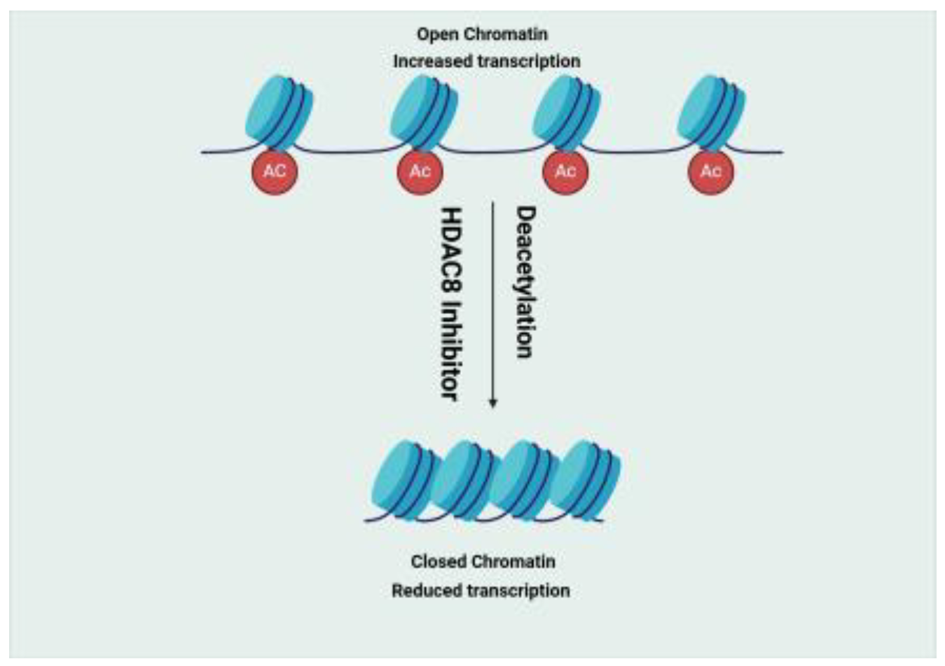
Figure 2.
Hedgehog pathway activation in cancer. Created in BioRender. Gerges, A. (2025) https://BioRender.com/w90s818.
Figure 2.
Hedgehog pathway activation in cancer. Created in BioRender. Gerges, A. (2025) https://BioRender.com/w90s818.
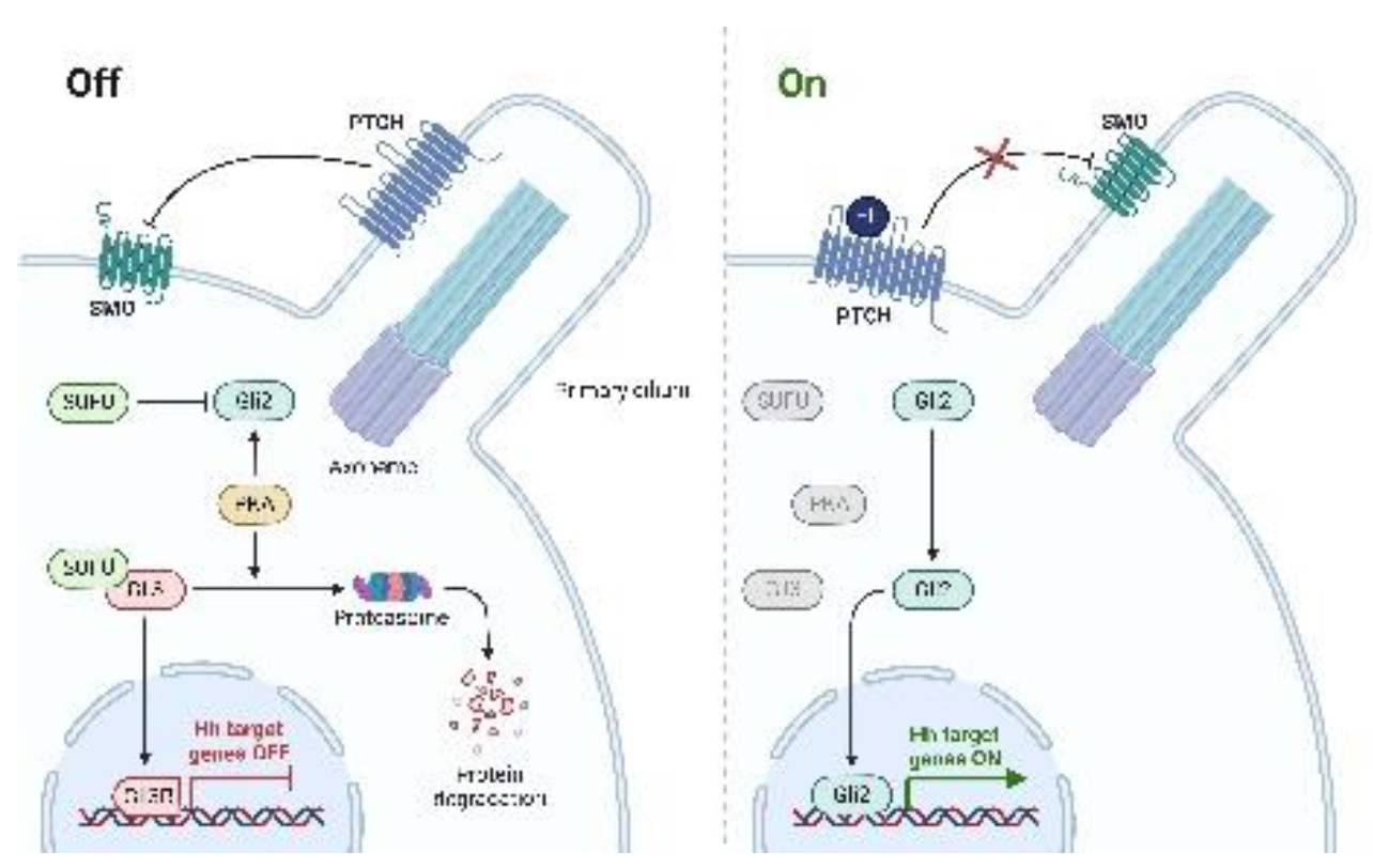
Figure 3.
Receptor Tyrosine Kinases (RTKs). Created in https://BioRender.com.
Figure 3.
Receptor Tyrosine Kinases (RTKs). Created in https://BioRender.com.
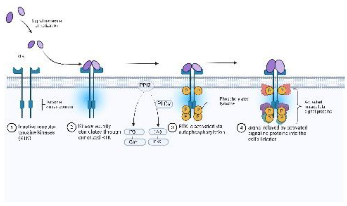
Figure 5.
Print screen of BROOD (Shape and Colour) outcome when using 8b as a lead compound. The figure on the left (A) shows the first 11 compounds made from 8b, with the figure on the right (B) showing another 12.
Figure 5.
Print screen of BROOD (Shape and Colour) outcome when using 8b as a lead compound. The figure on the left (A) shows the first 11 compounds made from 8b, with the figure on the right (B) showing another 12.
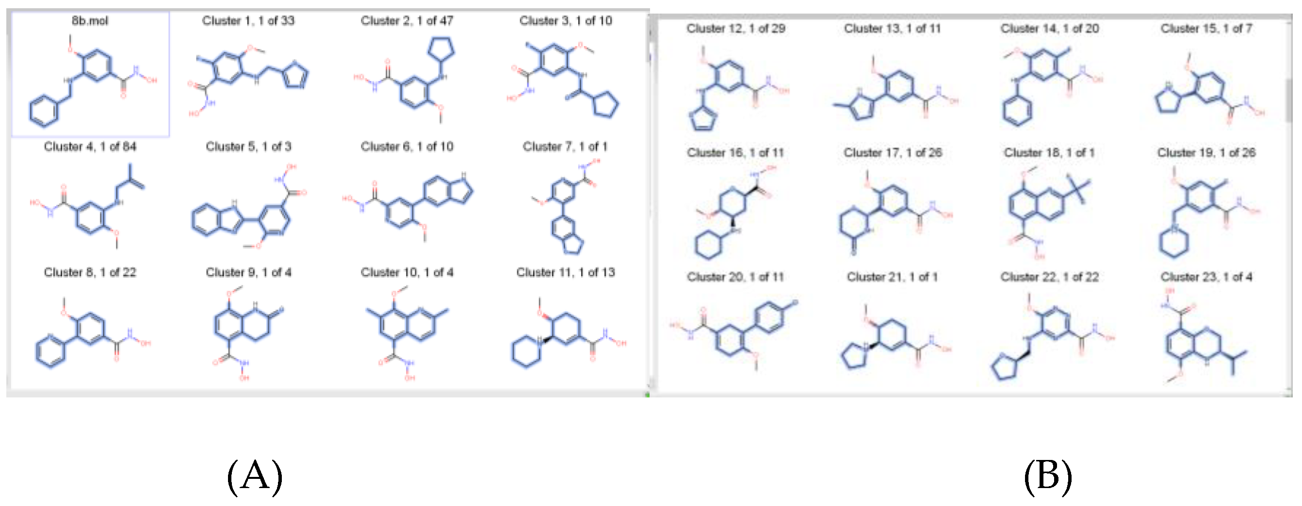
Figure 6.
Print screen is taken from the BROOD hitlist file, which contains the results in CSV format; it does not represent the entire outcome.
Figure 6.
Print screen is taken from the BROOD hitlist file, which contains the results in CSV format; it does not represent the entire outcome.
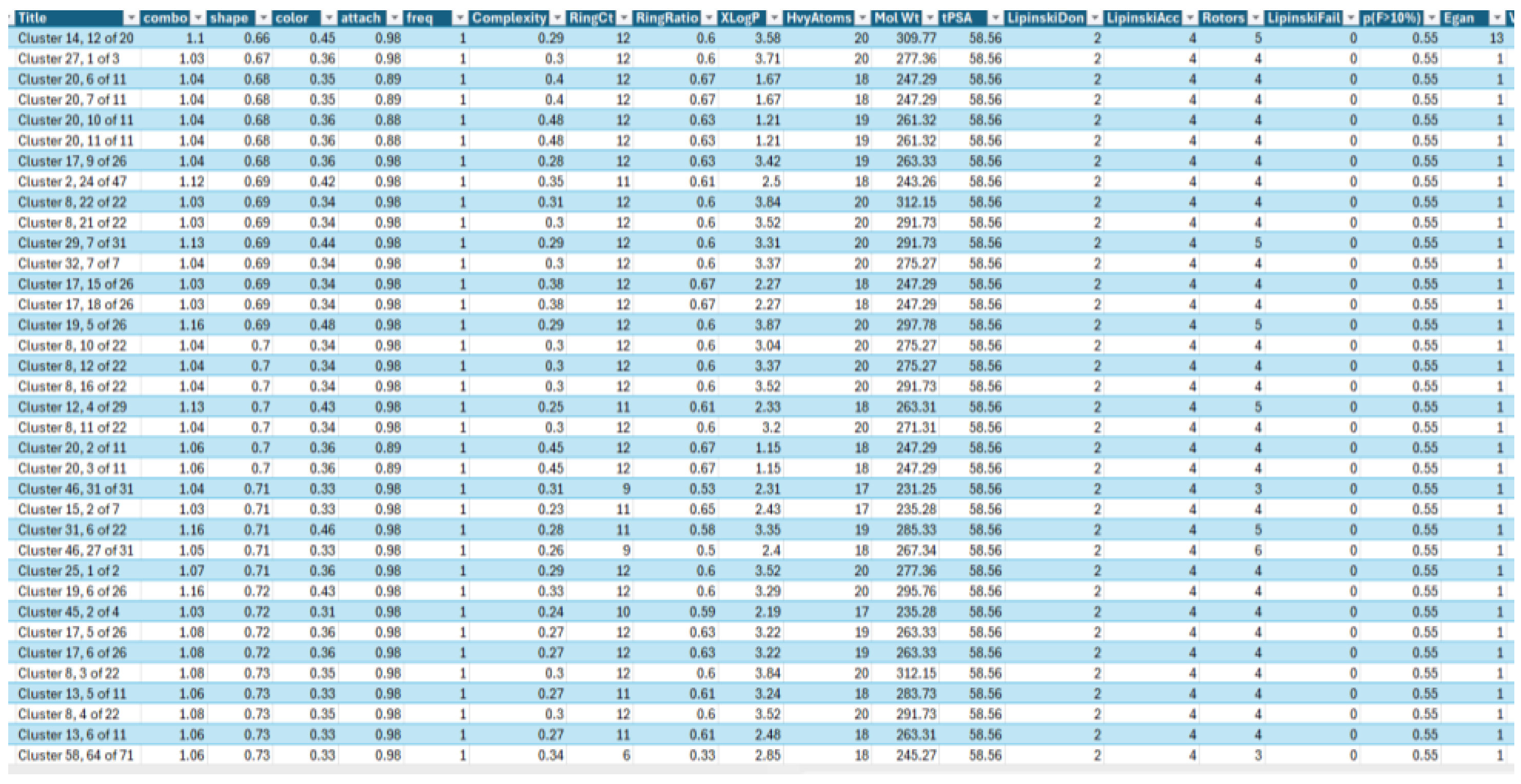
Figure 7.
FRED docking result: HDCA, 2V5W with cluster 22,1 of 1.
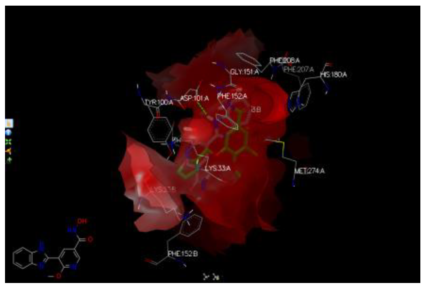
Figure 8.
An example from ROCS outcome: BRDs; BET762.
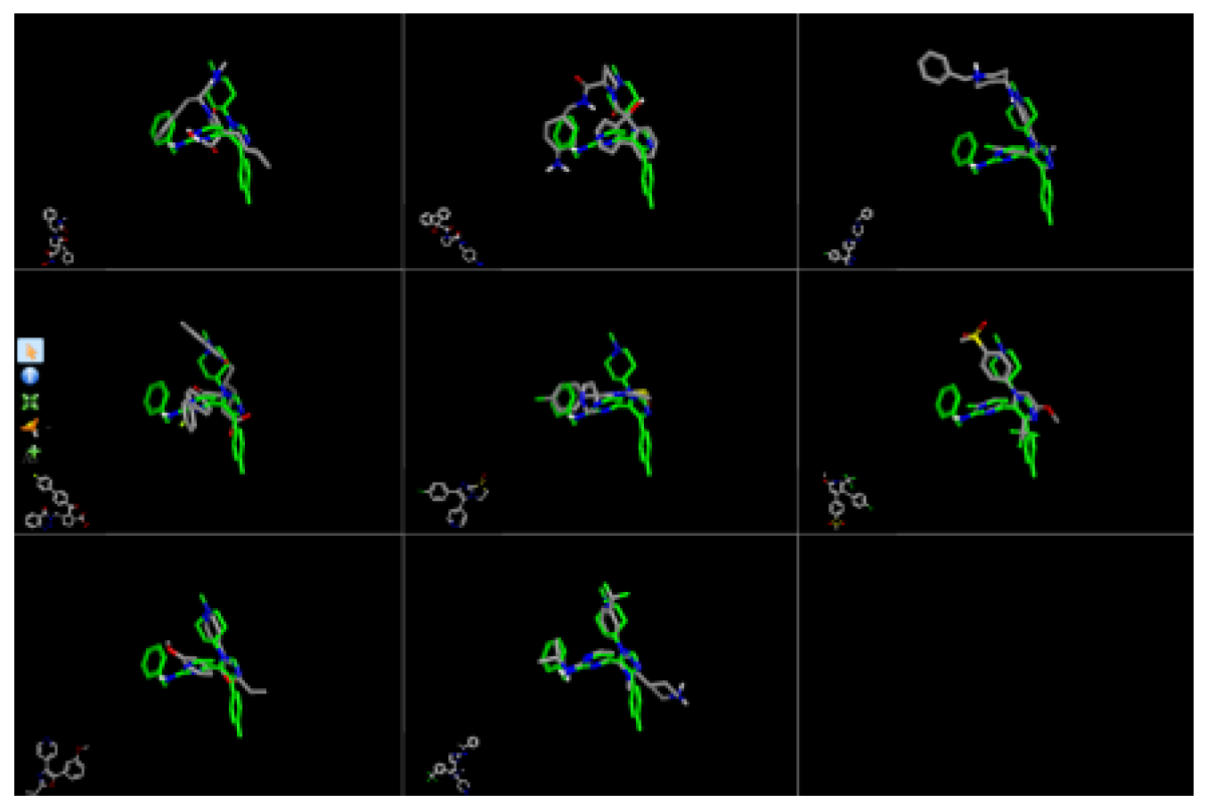
Figure 9.
An example of HH clusters with AFITT; protein 5L7I with HHIs clusters.
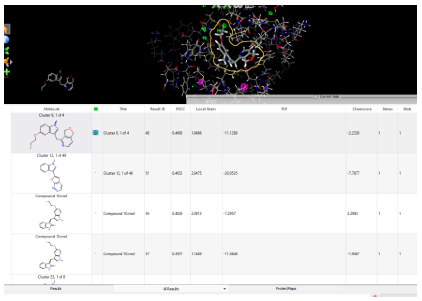
Table 2.
Summary of the rounds on BROOD for generating hitlists for each target. The top 25 compounds were selected from each round.
Table 2.
Summary of the rounds on BROOD for generating hitlists for each target. The top 25 compounds were selected from each round.
| Target | Shape and Colour | Shape and Electrostatics |
|---|---|---|
| 1-Histone Deacetylase 8 Inhibitors (8b and 20a) | 8b-2 rounds 20a-2 rounds |
8b-2 rounds 20a-2 rounds |
| 2-Bromodomain Inhibitors (JQ1 and I-BET762) | JQ1-2 rounds I-BET-762-3 rounds |
JQ1-2 rounds I-BET-762-3 rounds |
| 3-HH inhibitors (BMS-833923 and Vismodegib) | BMS-833923- 2 rounds Vismodegib-2 rounds |
BMS-833923- 2 rounds Vismodegib-2 rounds |
| 4-Tropomyosin Receptor Kinase Inhibitors (GW441759 and 10) | GW441759-2 rounds Compound 10- 2 rounds |
GW441759-2 rounds Compound 10- 2 rounds |
Table 3.
Selected clusters from each target (total n=35).
| HDACIs (n=9) | BRDI (n=8) | HH (n=10) | TRK (n=8) |
|---|---|---|---|
| Cluster 22, 1 of 22 | Cluster 3, 1 of 3 | Cluster 4, 1 of 1 | Cluster 12, 1 of 6 |
| Cluster 21, 1 of 11 | Cluster 25, 1 of 12 | Cluster 8, 1 of 1 | Cluster 4, 1 of 5 |
| Cluster 16,1 of 65 | Cluster 16, 1 of 2 | Cluster 1, 1 of 3 | Cluster 8, 1 of 19 |
| Cluster 23, 1 of 1 | Cluster 15, 1 of 2 | Cluster 9, 1 of 1 | Cluster 9, 1 of 4 |
| Cluster 1, 1 of 26 | Cluster 4, 1 of 4 | Cluster 8, 1 of 29 | Cluster 25, 1 of 8 |
| Cluster 7, 1 of 26 | Cluster 24, 1 of 7 | Cluster 21, 1 of 99 | Cluster 12, 1 of 49 |
| Cluster 4, 1 of 28 | Cluster 23, 1 of 1 | Cluster 15, 1 of 6 | Cluster 7, 1 of 11 |
| Cluster 10, 1 of 86 | Cluster 10, 1 of 1 | Cluster 23, 1 of 1 | Cluster 25, 1 of 8 |
| Cluster 12, 1 of 50 | Cluster 17, 1 of 1 | ||
| Cluster 20, 1 of 12 |
Table 6.
Selected clusters from each type that docked with the rest of the targets (n=8).
| HDACIs (n=2) | BRDI (n=2) | HH (n=2) | TRK (n=2) |
|---|---|---|---|
| Cluster 22, 1 of 22 | Cluster 3, 1 of 3 | Cluster 8, 1 of 1 | Cluster 25, 1 of 8 |
| Cluster 10, 1 of 86 | Cluster 16, 1 of 2 | Cluster 8, 1 of 29 | Cluster 12, 1 of 49 |
Table 7.
Results from the Toxicity Estimation Software Tools (TEST); predictive values.
| Clusters | Bioconcentration Factor1 | Mutagenicity2 | Oral rat LD50-Log10(mol/kg)3 | T. Pyriformis IGC50 (48 hrs)mg/L4 |
|---|---|---|---|---|
| Cluster 22, 1 of 22 | 0.31 | Positive | 1.78 | 3845.78 |
| Cluster 10, 1 of 86 | 5.56 | Positive | 2.67 | 173.52 |
| Cluster 3, 1 of 3 | 12.71 | Positive | 2.52 | 4.61 |
| Cluster 16, 1 of 2 | 46.62 | Negative | 2.66 | 1.91 |
| Cluster 8, 1 of 1 | 27.88 | Negative | 1.70 | 6.33 |
| Cluster 8, 1 of 29 | 8.62 | N/A | 2.52 | N/A |
| Cluster 25, 1 of 8 | 98.26 | Positive | 2.01 | 6.55 |
| Cluster 12, 1 of 49 | 308.45 | Negative | 2.41 | 6.46 |
| 20A | 9.94 | Positive | N/A | 36.39 |
| 8B | 5.14 | Positive | N/A | 29.50 |
| BET-762 | 22.21 | Negative | 2.26 | 2.27 |
| JQ1 | 235.52 | Negative | 2.45 | 0.73 |
| Vesmodigib | 28.48 | Negative | 2.13 | 2.87 |
| BMS-833923 | 11.53 | Positive | 2.38 | N/A |
| Compound Z9 | 25.14 | Positive | 2.20 | 42.96 |
| Compound 10 | 20.85 | Positive | 2.65 | 7.04 |
Disclaimer/Publisher’s Note: The statements, opinions and data contained in all publications are solely those of the individual author(s) and contributor(s) and not of MDPI and/or the editor(s). MDPI and/or the editor(s) disclaim responsibility for any injury to people or property resulting from any ideas, methods, instructions or products referred to in the content. |
© 2025 by the authors. Licensee MDPI, Basel, Switzerland. This article is an open access article distributed under the terms and conditions of the Creative Commons Attribution (CC BY) license (http://creativecommons.org/licenses/by/4.0/).
Copyright: This open access article is published under a Creative Commons CC BY 4.0 license, which permit the free download, distribution, and reuse, provided that the author and preprint are cited in any reuse.


