
Submitted:
20 April 2025
Posted:
21 April 2025
You are already at the latest version
Abstract
Background: Glycocalyx disintegration is associated with adverse outcome in patients with trauma or sepsis. As microvascular dysfunction has an impact on disease progression in chronic heart failure (CHF) patients, we hypothesized that changes in microcirculation might be associated with mortality. Methods: Fifty patients with ischemic and non-ischemic cardiomyopathy and conservative treatment with baseline measurements of the sublingual microcirculation (via Sidestream Darkfield videomicroscopy) were followed up for two years. Glycocalyx thickness was assessed indirectly by calculation of the perfused boundary region (PBR). Results: Loss of glycocalyx was pronounced in non-survivors after one, n=10, and two years, n=16, PBR: 2.05µm (1.88-2.15µm) vs. 1.87µm (1.66-2.03µm) and 2.04 (1.93-2.11) vs. 1.84 (1.62-1.97); p=0.042 and p= 0.003, respectively. Area under the ROC curve for the analysis of the predictive value of PBR on two- year mortality was 0.77 (p=0.003; SE: 0.07, CI (95%): 0.63-0.91). ROC curve analysis determined a PBR of 1.9µm as best predictor for two- year mortality (sensitivity: 0.81; specificity: 0.59). Moreover, multivariate regression analysis revealed PBR and functional capillary density as significant predictor of two- year mortality, p=0.036 and p=0.048, respectively. Conclusion: Glycocalyx disintegration is related to poor overall survival in CHF patients.
Keywords:
glycocalyx
; microcirculation
; capillaries
; cardiomyopathy
; mortality
1. Introduction
Disturbance of the microcirculation promotes the progression of cardiovascular diseases [1,2]. The glycocalyx has a main role in endothelial protection and its disintegration is often associated with inflammation and leads to atherogenic processes [3,4]. Glycocalyx impairment facilitates tissue infiltration by monocytes/ macrophages, polymorphonuclears and lymphocytes [5]. Further, glycocalyx disintegration promotes the formation of tissue oedema, including myocardial tissue compromising heart function [6,7,8,9]. The increased myocardial water content restricts left ventricular contractility, cardiac output and diastolic cardiac function [7,10,11].
Negatively charged proteoglycans are the main components of the glycocalyx and consist of a core protein covalently linked to glycosaminoglycans (GAGs) [12]. The latter are increased in the human plasma during conditions of septic shock [13,14], and of those hyaluronic acid and heparan sulphate are higher in non-survivors [13]. Another component of the glycocalyx, syndecan-1, was measured as marker for glycocalyx disintegration in patients with acute decompensated heart failure admitted to hospital and was predictive for the development of acute kidney injury and mortality [15]. Furthermore, in trauma patients, higher levels of circulating syndecan-1 were associated with increased coagulopathy and mortality [16].
In addition, the importance of an intact endothelial surface layer has become more and more evident during the recent pandemic, as glycocalyx degradation with endothelial dysfunction were reported as a key pathomechanism in severe acute respiratory syndrome coronavirus 2 (SARS-CoV-2) infection [17,18,19,20,21,22].
Previously, we described sublingual microvascular rarefaction in patients with chronic heart failure and optimized guideline-directed medical therapy [23]. However, glycocalyx dimensions did not differ between patients and healthy controls [23]. The aim of the performed follow up was to assess a possible association of the obtained microcirculatory parameters with patient mortality.
2. Methods
We performed a follow up of a previously published cross- sectional mono-centre clinical trial [23]. The study was performed in accordance with the Declaration of Helsinki and the protocol approved by the local Ethics Committee of the Medical University of Vienna. All participants signed a written informed consent.
Mortality status was assessed by the database of the Vienna General Hospital, which is connected to patient files in hospitals of Vienna as well as by telephone follow-up and the Austrian statistic agency (Statistics Austria).
2.1. Microscope Imaging
In vivo sublingual assessment of the microvasculature was performed using a sidestream darkfield videomicroscope (CapiScope HVCS Handheld Video Capillary Microscope, KK Technology, England) as previously published [23,24,25], by one person to avoid inter-observer variability.
The camera is provided with light emitting diodes using a wavelength of 525nm to detect the hemoglobin of circulating red blood cells. The standard lens of the microscope enables a 0.92µm/pixel magnification in 752 x 480 pixels (field of view: 692 x 442). The software for acquisition and calculation of the perfused boundary region (PBR) is supplied by GlycoCheck BV (Maastricht, The Netherlands) and detailed methodology was described previously [25,26]. The camera is placed under the tongue near the frenulum and the software identifies micro-vessels below 30µm of thickness due to the contrast of red blood cells (RBC). RBC column widths are measured in at least 3000 vessel segments. The PBR is the most luminal part of the glycocalyx, which allows for limited penetration of the RBCs [27]. It is located at both sides of the RBC column; to determine its properties, the distance between the median RBC column width (P50) and the outer edge of the RBC- perfused luminal part of the glycocalyx (= perfused diameter) is calculated using the following equation: (perfused diameter-median RBC column width)/2. The increase in PBR reflects glycocalyx destruction [26,27,28]. The average PBR of microvessels between 5-25 µm diameter was used for statistical analyses. The PBR is inversely proportional to the glycocalyx [28]. The measurement and analysis system has shown to achieve reliable results and has been to date used in different clinical studies [27,28,29,30,31,32].
To assess capillary density, the software recognizes all micro-vessels below 30µm of thickness by determination of the red blood cells against the background. Vascular segments (line markers) are placed every 10µm of the vessel length. The recording process continues until a minimum of 3000 vascular segments. After the acquisition, on the first frame of each recording session a total of 21 line markers are placed every 0.5µm of the vascular segments. Only those vessels with an appropriate contrast of more than 60% of all 21 line markers are considered as functional (=valid perfused) vessels. All perfused vessels are referred to as total capillary density. RBC filling percentage is calculated by determining the percentage of vessels with RBCs present during the recording session (corresponding to 40 frames per session) [26]. RBC filling percentage and perfused capillary density are regarded as estimates of microcirculatory perfusion [23,26].
2.2. Statistics
Statistical analysis was performed using the Statistical Package for Social Sciences (IBM Corp. Armonk, NY, Released 2012). Median and interquartile range of continuous variables are shown. Categorical variables are given as number (%). We performed the non-parametric Mann Whitney U tests to detect differences in continuous variables. The chi-square test was used to assess differences in categorical variables. Spearman rank correlation was used to assess correlations.
In addition, receiver-operating characteristic (ROC) curve analyses were performed including standard error (SE) and 95% confidence intervals (CI) and used to graphically depict the relation between mortality and capillary density as well as for calculation of predictive thresholds for capillary density with respect to mortality.
A logistic regression analysis was performed to describe the relationship between functional or total perfused capillary density and mortality.
3. Results
Clinical characteristics of the followed patients at one and two years are given in Table 1.
After one year 10 patients (20%) and after two years 16 patients (32%) died.
At baseline, the PBR was 1.93µm (1.70-2.06µm) in the overall study population [23].
There was a significant inverse correlation of the PBR and RBC filling percentage r= -0.916, p<0.001 [23].
The PBR was significantly higher in patients who did not survive the follow up period: PBR: 2.05µm (1.88-2.15µm) vs. 1.87µm (1.66-2.03µm), p=0.042 after one year and 2.04µm (1.93-2.11µm) vs. 1.84µm (1.62-1.97µm); p= 0.003 after two years, Table 2.
At the time point of 1-year of the follow up there was no difference in RBC filling percentage (71% [70- 74%] vs. 74% [71-78%], p= 0.087), functional (2732µm/mm2 [1820-3141 µm/mm2] vs. 2407µm/mm2 [2085-2736 µm/mm2], p=0.369) or total perfused capillary density (3525µm/mm2 [2410-6435µm/mm2] vs. 3538µm/mm2 [3043- 4497µm/mm2], p=0.971) between survivors and non-survivors, Table 2.
Non-survivors of the 2- year follow up had significantly lower RBC filing percentage, signifying a disturbed microcirculatory perfusion (71% [70- 74%] vs. 75% [71-79%], p=0.028). There was no difference in functional (2630µm/mm2 [2028-2974µm/mm2] vs. 2403µm/mm2 [2068- 2688µm/mm2], p=0.3) or total perfused capillary density (3568 µm/mm2 [2963- 5339µm/mm2] vs. 3538µm/mm2 [3021-4397µm/mm2], p=0.75) between non-survivors and survivors, Table 2.
As reported previously, at baseline PBR correlated with inflammation markers (fibrinogen: r=0.54 and C-reactive protein: r=0.36), platelet count (r=0.38), and with measures of renal/liver function such as estimated glomerular filtration rate (r=-0.34), bilirubin (r=-0.39) and albumin (r=-0.30) in CHF patients, all p<0.05 [23].
Of these markers, non-survivors of the one-year follow up had lower baseline levels of alanine aminotransferase, p=0.019, Table 1. The other parameters did not differ between survivors and non-survivors at one year, Table 1.
In contrast, non-survivors of the two-year follow up had significantly higher baseline NT-proBNP and creatinine levels, with lower estimated glomerular filtration rate (GFR) as compared to survivors, Table 1. Further, higher baseline c-reactive protein and lower levels of albumin and alanine aminotransferase were observed, Table 1.
In a multivariate regression model comprising PBR, functional and total capillary density, NT-proBNP, creatinine, c-reactive protein, albumin and alanine aminotransferase, PBR and functional capillary density remained significantly associated with patient survival at two years, Table 3.
Area under the ROC curve for the analysis of the predictive value of PBR on two- year mortality was 0.77 (p=0.003; SE: 0.07, CI (95%): 0.63-0.91). ROC curve analysis revealed thresholds of 1.9µm for PBR as best predictors for two- year mortality (sensitivity: 0.81; specificity: 0.59), Figure 1A.
4. Discussion
Glycocalyx disintegration is a central component of endothelial dysfunction driving atherosclerosis and cardiovascular diseases [4,33]. The latter are associated with altered microvascular perfusion and endothelial barrier properties, often related to disease progression and severity [23,24,25,34,35,36]. Glycocalyx destruction precedes endothelial dysfunction destabilizing vascular homeostasis [9,21]. Herein, all components of the Virchow’s triad including endothelial integrity, vascular perfusion and coagulation are affected [21]. These processes, though often induced by inflammation, promote themselves a pro-inflammatory and pro-coagulative state eventually leading to tissue oedema, thrombosis, necrosis and also atherosclerosis [9,37]. Moreover, as endothelial function and vascular integrity is disturbed, glycocalyx degradation promotes the progression of cardiovascular diseases [38].
The presented long-term follow up of our preliminary study examining CHF patients shows the significant association of glycocalyx destruction with mortality, despite guideline-directed OMT. In contrast, we previously could not distinguish an influence of the glycocalyx constitution on mortality in CHF patients with VAD therapy [24]. The observed difference might be due to altered haemodynamics and their possible influence on the glycocalyx in VAD patients or possibly due to benefits/ complications associated with the mechanical circulatory support itself [24,39].
In addition to increased glycocalyx disruption, patients, who died during the follow up of our study had also higher markers of inflammation. This observation corresponds to the concept of a pro-inflammatory state affecting glycocalyx integrity with an impact on adverse events [4]. The latter might occur primarily in the microvasculature, often leading to difficult diagnostic processes, limiting patients’ quality of life and eventually promoting disease progression.
The results of our long-term follow up are further in-line with previous reports showing an association between syndecan-1, which was measured as a marker of glycocalyx disruption, and 6-month mortality after acute decompensated heart failure [15]. In this study, Neves et al. investigated 201 patients with acute decompensated heart failure admitted to the emergency department [15]. Herein, syndecan-1 levels correlated with hsCRP and both were independently related to 6-month mortality [15]. Higher plasma levels of syndecan-1 were further associated with the development of acute kidney injury during the hospital stay [15]. As previously reported, we also observed an inverse correlation between PBR and eGFR in our patient group, signifying the occurrence of more pronounced glycocalyx destruction in patients with lower eGFR levels [23]. Furthermore, non survivors of the 2-year follow up had lower glomerular filtration rates.
Higher plasma levels of syndecan-1 were also associated with higher all-cause mortality and rehospitalization in HF patients with preserved ejection fraction, signifying the association of glycocalyx degradation with adverse patient outcome [40].
The glycocalyx is key in regulating tissue homeostasis and its intactness is necessary to maintain the filtration barrier and prevent oedema formation [9,41]. Myocardial oedema formation has been described in heart failure and can be attributed to glycocalyx degradation resulting in microvascular barrier dysfunction [42]. The accumulation of water in interstitial and intracellular compartments evokes cardiomyocyte injury, dysfunction, and in consequence cardiac remodelling [9].
Glycocalyx integrity has also been shown to be crucial in shielding endothelial cells against viral infections like SARS-CoV-2 [22,43]. In this context the shedding of glycocalyx components can be regarded as a main factor accelerating viral entry [21,43]. Endothelial dysfunction and endotheliitis evoked by viral invasion drives thromboinflammation affecting the equilibrium of the Virchow’s triad [21]. Together with changes of plasma viscoelastic properties, microclots occur affecting the perfusion of the capillary network [44]. Moreover, sustained changes in glycocalyx composition contributing to inflammatory and pro-coagulative processes are discussed to imply long- lasting sequelae after COVID-19 infection [9,21,45,46].
Since the glycocalyx represents a fragile structure and preservation of its properties is demanding, therapeutic options remain mainly experimental.
Herein, concepts targeting inflammatory and pro-coagulative pathways are promising to convey glycocalyx protection [47]. Hitherto, medication like sodium-glucose cotransporter 2 (SGLT-2) inhibitors are recommended in heart failure and statins in hyperlipidemia guidelines and are known to exhibit anti- inflammatory properties [48,49]. Moreover, finerenone is discussed to convey glycocalyx structure preservation and protection against COVID-19-associated adverse events in patients with type 2 diabetes and chronic kidney disease [50].
Additionally, experimental approaches covering preconditioning concepts and agents resembling glycocalyx components are under investigation [9].
Moreover, in vivo diagnostic approaches remain challenging. With the use of intravital sublingual capillaroscopy, patients at risk could be identified, which might benefit from further therapy with regard to glycocalyx preservation or restoration. Further studies addressing this question are warranted.
5. Conclusions
In vivo obtained PBR values as indirect measure of the glycocalyx were independently associated with mortality in a long-term follow up of CHF patients. These observations should provide a cornerstone for further studies regarding glycocalyx composition and preservation in health and disease.
Author Contributions
Idea and Conceptualization: P.P.W., M.H. and B.J.; Data Acquisition: P.P.W., C.S., J.P., C.W.; Data Analyses: P.P.W.; Original Draft Preparation: P.P.W.; Review and Editing: M.H., I.M.L., C.S., J.P., C.W., T.G., S.S., R.K., C.W.K., B.J.; Supervision: P.P.W., M.H. and B.J. All authors have contributed substantially to the work. All authors have read and agreed to the published version of the manuscript.
Funding
This research received no external funding.
Institutional Review Board Statement
This study was approved by the Ethics Committee of the Medical University of Vienna, EC-number: 1734/2013.
Informed Consent Statement
Informed consent was obtained from all subjects involved in the study.
Data Availability Statement
Raw data generated and/or analysed during the current study are available from the corresponding author on reasonable request.
Conflict of Interest
The authors declare no conflict of interest.
References
- Strain, W.D.; Paldanius, P.M. Diabetes, cardiovascular disease and the microcirculation. Cardiovasc Diabetol 2018, 17, 57. [Google Scholar] [CrossRef]
- Gutterman, D.D.; Chabowski, D.S.; Kadlec, A.O.; Durand, M.J.; Freed, J.K.; Ait-Aissa, K.; Beyer, A.M. The Human Microcirculation: Regulation of Flow and Beyond. Circ Res 2016, 118, 157–172. [Google Scholar] [CrossRef] [PubMed]
- Reitsma, S.; Slaaf, D.W.; Vink, H.; van Zandvoort, M.A.; oude Egbrink, M.G. The endothelial glycocalyx: composition, functions, and visualization. Pflugers Archiv: European journal of physiology 2007, 454, 345–359. [Google Scholar] [CrossRef]
- Poledniczek, M.; Neumayer, C.; Kopp, C.W.; Schlager, O.; Gremmel, T.; Jozkowicz, A.; Gschwandtner, M.E.; Koppensteiner, R.; Wadowski, P.P. Micro- and Macrovascular Effects of Inflammation in Peripheral Artery Disease-Pathophysiology and Translational Therapeutic Approaches. Biomedicines 2023, 11. [Google Scholar] [CrossRef] [PubMed]
- Song, J.W.; Zullo, J.A.; Liveris, D.; Dragovich, M.; Zhang, X.F.; Goligorsky, M.S. Therapeutic Restoration of Endothelial Glycocalyx in Sepsis. J Pharmacol Exp Ther 2017, 361, 115–121. [Google Scholar] [CrossRef] [PubMed]
- van den Berg, B.M.; Vink, H.; Spaan, J.A. The endothelial glycocalyx protects against myocardial edema. Circ Res 2003, 92, 592–594. [Google Scholar] [CrossRef]
- Mehlhorn, U.; Geissler, H.J.; Laine, G.A.; Allen, S.J. Myocardial fluid balance. European journal of cardio-thoracic surgery: official journal of the European Association for Cardio-thoracic Surgery 2001, 20, 1220–1230. [Google Scholar] [CrossRef]
- Becker, B.F.; Chappell, D.; Jacob, M. Endothelial glycocalyx and coronary vascular permeability: the fringe benefit. Basic Res Cardiol 2010, 105, 687–701. [Google Scholar] [CrossRef]
- Panagiotides, N.G.; Poledniczek, M.; Andreas, M.; Hulsmann, M.; Kocher, A.A.; Kopp, C.W.; Piechota-Polanczyk, A.; Weidenhammer, A.; Pavo, N.; Wadowski, P.P. Myocardial Oedema as a Consequence of Viral Infection and Persistence-A Narrative Review with Focus on COVID-19 and Post COVID Sequelae. Viruses 2024, 16. [Google Scholar] [CrossRef]
- Laine, G.A.; Allen, S.J. Left ventricular myocardial edema. Lymph flow, interstitial fibrosis, and cardiac function. Circ Res 1991, 68, 1713–1721. [Google Scholar] [CrossRef]
- Davis, K.L.; Mehlhorn, U.; Laine, G.A.; Allen, S.J. Myocardial edema, left ventricular function, and pulmonary hypertension. Journal of applied physiology 1995, 78, 132–137. [Google Scholar] [CrossRef] [PubMed]
- Pries, A.R.; Secomb, T.W.; Gaehtgens, P. The endothelial surface layer. Pflugers Archiv: European journal of physiology 2000, 440, 653–666. [Google Scholar] [CrossRef] [PubMed]
- Nelson, A.; Berkestedt, I.; Bodelsson, M. Circulating glycosaminoglycan species in septic shock. Acta Anaesthesiol Scand 2014, 58, 36–43. [Google Scholar] [CrossRef]
- Henrich, M.; Gruss, M.; Weigand, M.A. Sepsis-induced degradation of endothelial glycocalix. ScientificWorldJournal 2010, 10, 917–923. [Google Scholar] [CrossRef]
- Neves, F.M.; Meneses, G.C.; Sousa, N.E.; Menezes, R.R.; Parahyba, M.C.; Martins, A.M.; Liborio, A.B. Syndecan-1 in Acute Decompensated Heart Failure--Association With Renal Function and Mortality. Circ J 2015, 79, 1511–1519. [Google Scholar] [CrossRef] [PubMed]
- Johansson, P.I.; Stensballe, J.; Rasmussen, L.S.; Ostrowski, S.R. A high admission syndecan-1 level, a marker of endothelial glycocalyx degradation, is associated with inflammation, protein C depletion, fibrinolysis, and increased mortality in trauma patients. Ann Surg 2011, 254, 194–200. [Google Scholar] [CrossRef]
- Ackermann, M.; Verleden, S.E.; Kuehnel, M.; Haverich, A.; Welte, T.; Laenger, F.; Vanstapel, A.; Werlein, C.; Stark, H.; Tzankov, A.; et al. Pulmonary Vascular Endothelialitis, Thrombosis, and Angiogenesis in Covid-19. N Engl J Med 2020, 383, 120–128. [Google Scholar] [CrossRef]
- Varga, Z.; Flammer, A.J.; Steiger, P.; Haberecker, M.; Andermatt, R.; Zinkernagel, A.S.; Mehra, M.R.; Schuepbach, R.A.; Ruschitzka, F.; Moch, H. Endothelial cell infection and endotheliitis in COVID-19. Lancet 2020, 395, 1417–1418. [Google Scholar] [CrossRef]
- Wadowski, P.P.; Piechota-Polańczyk, A.; Andreas, M.; Kopp, C.W. Cardiovascular Disease Management in the Context of Global Crisis. International Journal of Environmental Research and Public Health 2023, 20. [Google Scholar] [CrossRef]
- Veraldi, N.; Vivès, R.R.; Blanchard-Rohner, G.; L’Huillier, A.G.; Wagner, N.; Rohr, M.; Beghetti, M.; De Agostini, A.; Grazioli, S. Endothelial glycocalyx degradation in multisystem inflammatory syndrome in children related to COVID-19. Journal of Molecular Medicine 2022, 100, 735–746. [Google Scholar] [CrossRef]
- Wadowski, P.P.; Panzer, B.; Jozkowicz, A.; Kopp, C.W.; Gremmel, T.; Panzer, S.; Koppensteiner, R. Microvascular Thrombosis as a Critical Factor in Severe COVID-19. Int J Mol Sci 2023, 24. [Google Scholar] [CrossRef] [PubMed]
- Wadowski, P.P.; Jilma, B.; Kopp, C.W.; Ertl, S.; Gremmel, T.; Koppensteiner, R. Glycocalyx as Possible Limiting Factor in COVID-19. Front Immunol 2021, 12, 607306. [Google Scholar] [CrossRef] [PubMed]
- Wadowski, P.P.; Hulsmann, M.; Schorgenhofer, C.; Lang, I.M.; Wurm, R.; Gremmel, T.; Koppensteiner, R.; Steinlechner, B.; Schwameis, M.; Jilma, B. Sublingual functional capillary rarefaction in chronic heart failure. Eur J Clin Invest 2018, 48. [Google Scholar] [CrossRef]
- Wadowski, P.P.; Steinlechner, B.; Zimpfer, D.; Schloglhofer, T.; Schima, H.; Hulsmann, M.; Lang, I.M.; Gremmel, T.; Koppensteiner, R.; Zehetmayer, S.; et al. Functional capillary impairment in patients with ventricular assist devices. Sci Rep 2019, 9, 5909. [Google Scholar] [CrossRef]
- Wadowski, P.P.; Kautzky-Willer, A.; Gremmel, T.; Koppensteiner, R.; Wolf, P.; Ertl, S.; Weikert, C.; Schorgenhofer, C.; Jilma, B. Sublingual microvasculature in diabetic patients. Microvasc Res 2020, 129, 103971. [Google Scholar] [CrossRef] [PubMed]
- Lee, D.H.; Dane, M.J.; van den Berg, B.M.; Boels, M.G.; van Teeffelen, J.W.; de Mutsert, R.; den Heijer, M.; Rosendaal, F.R.; van der Vlag, J.; van Zonneveld, A.J.; et al. Deeper penetration of erythrocytes into the endothelial glycocalyx is associated with impaired microvascular perfusion. PLoS One 2014, 9, e96477. [Google Scholar] [CrossRef]
- Martens, R.J.; Vink, H.; van Oostenbrugge, R.J.; Staals, J. Sublingual microvascular glycocalyx dimensions in lacunar stroke patients. Cerebrovascular diseases 2013, 35, 451–454. [Google Scholar] [CrossRef]
- Dane, M.J.; Khairoun, M.; Lee, D.H.; van den Berg, B.M.; Eskens, B.J.; Boels, M.G.; van Teeffelen, J.W.; Rops, A.L.; van der Vlag, J.; van Zonneveld, A.J.; et al. Association of kidney function with changes in the endothelial surface layer. Clinical journal of the American Society of Nephrology: CJASN 2014, 9, 698–704. [Google Scholar] [CrossRef]
- Machin, D.R.; Gates, P.E.; Vink, H.; Frech, T.M.; Donato, A.J. Automated Measurement of Microvascular Function Reveals Dysfunction in Systemic Sclerosis: A Cross-sectional Study. J Rheumatol 2017, 44, 1603–1611. [Google Scholar] [CrossRef]
- Donati, A.; Damiani, E.; Domizi, R.; Romano, R.; Adrario, E.; Pelaia, P.; Ince, C.; Singer, M. Alteration of the sublingual microvascular glycocalyx in critically ill patients. Microvasc Res 2013, 90, 86–89. [Google Scholar] [CrossRef]
- Groen, B.B.; Hamer, H.M.; Snijders, T.; van Kranenburg, J.; Frijns, D.; Vink, H.; van Loon, L.J. Skeletal muscle capillary density and microvascular function are compromised with aging and type 2 diabetes. Journal of applied physiology 2014, 116, 998–1005. [Google Scholar] [CrossRef] [PubMed]
- Koning, N.J.; Vonk, A.B.; Vink, H.; Boer, C. Side-by-Side Alterations in Glycocalyx Thickness and Perfused Microvascular Density During Acute Microcirculatory Alterations in Cardiac Surgery. Microcirculation 2016, 23, 69–74. [Google Scholar] [CrossRef]
- Poledniczek, M.; Neumayer, C.; Kopp, C.W.; Schlager, O.; Gremmel, T.; Jozkowicz, A.; Gschwandtner, M.E.; Koppensteiner, R.; Wadowski, P.P. Micro- and Macrovascular Effects of Inflammation in Peripheral Artery Disease-Pathophysiology and Translational Therapeutic Approaches. Preprints.org 2023. [Google Scholar] [CrossRef] [PubMed]
- Vlahu, C.A.; Lemkes, B.A.; Struijk, D.G.; Koopman, M.G.; Krediet, R.T.; Vink, H. Damage of the endothelial glycocalyx in dialysis patients. J Am Soc Nephrol 2012, 23, 1900–1908. [Google Scholar] [CrossRef] [PubMed]
- Wadowski, P.P.; Schorgenhofer, C.; Rieder, T.; Ertl, S.; Pultar, J.; Serles, W.; Sycha, T.; Mayer, F.; Koppensteiner, R.; Gremmel, T.; et al. Microvascular rarefaction in patients with cerebrovascular events. Microvasc Res 2022, 140, 104300. [Google Scholar] [CrossRef]
- Nieuwdorp, M.; Meuwese, M.C.; Vink, H.; Hoekstra, J.B.; Kastelein, J.J.; Stroes, E.S. The endothelial glycocalyx: a potential barrier between health and vascular disease. Curr Opin Lipidol 2005, 16, 507–511. [Google Scholar] [CrossRef]
- Panzer, B.; Kopp, C.W.; Neumayer, C.; Koppensteiner, R.; Jozkowicz, A.; Poledniczek, M.; Gremmel, T.; Jilma, B.; Wadowski, P.P. Toll-like Receptors as Pro-Thrombotic Drivers in Viral Infections: A Narrative Review. Cells 2023, 12. [Google Scholar] [CrossRef]
- Wadowski, P.P.; Piechota-Polanczyk, A.; Andreas, M.; Kopp, C.W. Cardiovascular Disease Management in the Context of Global Crisis. Int J Environ Res Public Health 2022, 20. [Google Scholar] [CrossRef]
- Netuka, I.; Litzler, P.Y.; Berchtold-Herz, M.; Flecher, E.; Zimpfer, D.; Damme, L.; Sundareswaran, K.S.; Farrar, D.J.; Schmitto, J.D.; Investigators, E.T. Outcomes in HeartMate II Patients With No Antiplatelet Therapy: 2-Year Results From the European TRACE Study. Ann Thorac Surg 2017, 103, 1262–1268. [Google Scholar] [CrossRef]
- Tromp, J.; van der Pol, A.; Klip, I.T.; de Boer, R.A.; Jaarsma, T.; van Gilst, W.H.; Voors, A.A.; van Veldhuisen, D.J.; van der Meer, P. Fibrosis marker syndecan-1 and outcome in patients with heart failure with reduced and preserved ejection fraction. Circ Heart Fail 2014, 7, 457–462. [Google Scholar] [CrossRef]
- Curry, F.E.; Michel, C.C. The Colloid Osmotic Pressure Across the Glycocalyx: Role of Interstitial Fluid Sub-Compartments in Trans-Vascular Fluid Exchange in Skeletal Muscle. Frontiers in Cell and Developmental Biology 2021, 9. [Google Scholar] [CrossRef] [PubMed]
- Vasques-Novoa, F.; Angelico-Goncalves, A.; Alvarenga, J.M.G.; Nobrega, J.; Cerqueira, R.J.; Mancio, J.; Leite-Moreira, A.F.; Roncon-Albuquerque, R., Jr. Myocardial oedema: pathophysiological basis and implications for the failing heart. ESC Heart Fail 2022, 9, 958–976. [Google Scholar] [CrossRef]
- Targosz-Korecka, M.; Kubisiak, A.; Kloska, D.; Kopacz, A.; Grochot-Przeczek, A.; Szymonski, M. Endothelial glycocalyx shields the interaction of SARS-CoV-2 spike protein with ACE2 receptors. Sci Rep 2021, 11, 12157. [Google Scholar] [CrossRef] [PubMed]
- Illibauer, J.; Clodi-Seitz, T.; Zoufaly, A.; Aberle, J.H.; Weninger, W.J.; Foedinger, M.; Elsayad, K. Diagnostic potential of blood plasma longitudinal viscosity measured using Brillouin light scattering. Proc Natl Acad Sci U S A 2024, 121, e2323016121. [Google Scholar] [CrossRef]
- Pretorius, E.; Vlok, M.; Venter, C.; Bezuidenhout, J.A.; Laubscher, G.J.; Steenkamp, J.; Kell, D.B. Persistent clotting protein pathology in Long COVID/Post-Acute Sequelae of COVID-19 (PASC) is accompanied by increased levels of antiplasmin. Cardiovasc Diabetol 2021, 20, 172. [Google Scholar] [CrossRef]
- Panagiotides, N.G.; Zimprich, F.; Machold, K.; Schlager, O.; Muller, M.; Ertl, S.; Loffler-Stastka, H.; Koppensteiner, R.; Wadowski, P.P. A Case of Autoimmune Small Fiber Neuropathy as Possible Post COVID Sequelae. Int J Environ Res Public Health 2023, 20. [Google Scholar] [CrossRef]
- Panagiotides, N.G.; Poledniczek, M.; Andreas, M.; Hulsmann, M.; Kocher, A.A.; Kopp, C.W.; Piechota-Polanczyk, A.; Weidenhammer, A.; Pavo, N.; Wadowski, P.P. Myocardial Oedema as a Consequence of Viral Infection and Persistence-A Narrative Review with Focus on COVID-19 and Post COVID Sequelae. Preprints.org 2023. [Google Scholar] [CrossRef]
- McDonagh, T.A.; Metra, M.; Adamo, M.; Gardner, R.S.; Baumbach, A.; Bohm, M.; Burri, H.; Butler, J.; Celutkiene, J.; Chioncel, O.; et al. 2023 Focused Update of the 2021 ESC Guidelines for the diagnosis and treatment of acute and chronic heart failure. Eur Heart J 2023, 44, 3627–3639. [Google Scholar] [CrossRef] [PubMed]
- Mach, F.; Baigent, C.; Catapano, A.L.; Koskinas, K.C.; Casula, M.; Badimon, L.; Chapman, M.J.; De Backer, G.G.; Delgado, V.; Ference, B.A.; et al. 2019 ESC/EAS Guidelines for the management of dyslipidaemias: lipid modification to reduce cardiovascular risk. Eur Heart J 2020, 41, 111–188. [Google Scholar] [CrossRef]
- Pitt, B.; Agarwal, R.; Anker, S.D.; Ruilope, L.M.; Rossing, P.; Ahlers, C.; Brinker, M.; Joseph, A.; Lambelet, M.; Lawatscheck, R.; et al. Association of Finerenone Use With Reduction in Treatment-Emergent Pneumonia and COVID-19 Adverse Events Among Patients With Type 2 Diabetes and Chronic Kidney Disease: A FIDELITY Pooled Secondary Analysis. JAMA Netw Open 2022, 5, e2236123. [Google Scholar] [CrossRef]
Figure 1.
Receiver-operating characteristic (ROC) curve for the analysis of the predictive value of (A) perfused boundary region (PBR): area under the curve (AUC) = 0.77 ± 0.07 (SE), CI 95%: 0.63–0.91, p = 0.003. (B) functional capillary density: AUC = 0.59 ± 0.09 (SE), CI 95%: 0.41–0.77, p = 0.298 and (C) total perfused capillary density: AUC = 0.53 ± 0.09 (SE), CI 95%: 0.34–0.71, p = 0.747, depicted as blue line, respectively, for mortality at two years. SE, standard error.
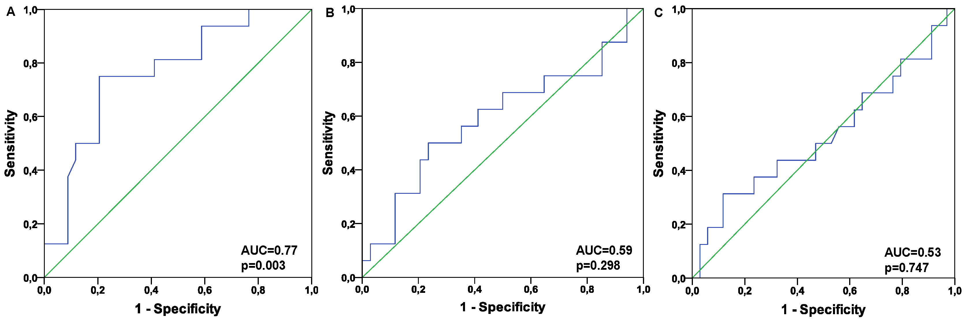

Table 1.
Patients’ characteristics.
| Follow up period of one year | |||
|---|---|---|---|
| Overall Death n=10 | Overall Survival n=40 | p-Value | |
| Age | 69 (62-76) | 70 (59- 77) | 0.952 |
| Sex (m/f) | 8/2 | 36/4 | 0.384 |
| BMI | 29 (24- 32) | 28 (24-32) | 0.574 |
| Fibrinogen (g/L) | 4.2 (3.6 -4.7) | 4.0 (3.6-4.6) | 0.700 |
| Leukocytes (*109/L) | 7.7 (6.5 -9.0) | 7.6 (6.3 -9.1) | 0.849 |
| Platelets (*109/L) | 215 (162 -255) | 200 (180-238) | 0.926 |
| NT-proBNP (pg/mL) | 4005 (2826- 7937) | 2599 (1527-4549) | 0.201 |
| Albumin (g/L) | 42.6 (37.3- 45.3) | 43.4 (40.4- 45.3) | 0.925 |
| Alanine aminotransferase (µmol/s·l) | 0.28 (0.21-0.32) | 0.37 (0.28-0.48) | 0.019 |
| Aspartate aminotransferase (µmol/s·l) | 0.35 (0.28 -0.45) | 0.43 (0.32-0.50) | 0.138 |
| Total bilirubin (µmol/L) | 9.9 (5.9 -16.9) | 12.3 (7.2 -19.9) | 0.586 |
| C- reactive protein (mg/L) | 4.2 (3.1 -7.5) | 5.3 (1.7 -8.9) | 0.780 |
| Serum creatinine (µmol/ L) | 115.8 (98.6- 333.3) | 120.7 (95.3 - 176.8) | 0.432 |
| Estimated glomerular filtration rate (ml/min) | 50.2 (17.1 -62.4) | 51.5 (33.1- 73.4) | 0.343 |
| Follow up period of two years | |||
| Overall Death n=16 | Overall Survival n=34 | p-Value | |
| Age | 74 (65-80) | 70 (57-75) | 0.134 |
| Sex (m/f) | 14/2 | 30/4 | 0.941 |
| BMI | 28.2 (24.6 -31.5) | 27.9 (24.1-32.3) | 0.803 |
| Fibrinogen (g/L) | 4.2 (3.7-5.0) | 4.0 (3.5-4.5) | 0.174 |
| Leukocytes (*109/L) | 7.7 (6.4- 9.2) | 7.6 (6.1-8.8) | 0.542 |
| Platelets (*109/L) | 215 (176-244) | 196 (178-240) | 0.706 |
| NT-proBNP (pg/mL) | 4693 (3377- 11425) | 2202 (1483-4243) | 0.004 |
| Albumin (g/L) | 41.6 (37.4- 44.3) | 44.2 (40.9- 45.9) | 0.037 |
| Alanine aminotransferase (µmol/s·l) | 0.28 (0.23-0.35) | 0.37 (0.28-0.49) | 0.033 |
| Aspartate aminotransferase (µmol/s·l) | 0.35 (0.28-0.48) | 0.43 (0.30-0.50) | 0.275 |
| Total bilirubin (µmol/L) | 10.6 (7.4-16.9) | 12.2 (6.8 -20.3) | 0.881 |
| C- reactive protein (mg/L) | 7.3 (3.9-1.7) | 3.5 (1.6-7.9) | 0.026 |
| Serum creatinine (µmol/ L) | 165 (108-294) | 113 (95 -151) | 0.066 |
| Estimated glomerular filtration rate (ml/min) | 38.8 (19.5-56.1) | 56.8 (42.2-75.8) | 0.045 |
Table 1: Data are presented as median and IQR.
Table 2.
Microvascular parameters.
| Follow up period of one year | |||
| Overall Death n=10 | Overall Survival n=40 | p-Value | |
| PBR (µm) | 2.05 (1.88-2.14) | 1.87 (1.66 -2.03) | 0.042 |
| RBC filling % | 71 (70-74) | 74 (71-78) | 0.087 |
| Functional capillary density (µm/mm2) | 2732 (1820-3141) | 2407 (2085-2736) | 0.369 |
| Total capillary density (µm/mm2) | 3525 (2410-6435) | 3538 (3043-4497) | 0.971 |
| Ratio (%) | 73 (60-85) | 71 (57 -76) | 0.331 |
| Follow up period of two years | |||
| Overall Death n=16 | Overall Survival n=34 | p-Value | |
| PBR (µm) | 2.04 (1.93-2.11) | 1.84 (1.62-1.97) | 0.003 |
| RBC filling % | 71 (70-74) | 75 (71-79) | 0.028 |
| Functional capillary density (µm/mm2) | 2630 (2028-2974) | 2403 (2068-2688) | 0.298 |
| Total capillary density (µm/mm2) | 3568 (2963-5339) | 3538 (3021-4397) | 0.747 |
| Ratio (%) | 73 (57-78) | 71 (57 -77) | 0.771 |
Table 2 Data are presented as median and IQR.
Table 3.
Multivariate regression analyses.
| One year | Two years | |||||
| B | CI | P | B | CI | P | |
| PBR | 4.8 | 0.5-27684 | 0.087 | 5.5 | 1.4-38820.5 | 0.036 |
| Functional capillary density | 0.03 | 1.0-1.1 | 0.083 | 0.3 | 1.0-1.1 | 0.048 |
| Total capillary density | -0.01 | 0.98-1.0 | 0.149 | -0.01 | 0.98-1.0 | 0.064 |
| NT-proBNP | 0 | 1.0-1.0 | 0.487 | 0.0 | 1.0-1.0 | 0.489 |
| Creatinine | -0.002 | 0.98-1.0 | 0.762 | -0.01 | 0.98-1.0 | 0.224 |
| C- reactive protein | -0.05 | 0.4-2.6 | 0.915 | 0.3 | 0.8-2.6 | 0.285 |
| Albumin | 0.1 | 0.8-1.6 | 0.448 | -0.03 | 0.8-1.2 | 0.793 |
| Alanine aminotransferase | -8,3 | 0.0-5.0 | 0.101 | -4.9 | 0-2.5 | 0.099 |
Disclaimer/Publisher’s Note: The statements, opinions and data contained in all publications are solely those of the individual author(s) and contributor(s) and not of MDPI and/or the editor(s). MDPI and/or the editor(s) disclaim responsibility for any injury to people or property resulting from any ideas, methods, instructions or products referred to in the content. |
© 2025 by the authors. Licensee MDPI, Basel, Switzerland. This article is an open access article distributed under the terms and conditions of the Creative Commons Attribution (CC BY) license (http://creativecommons.org/licenses/by/4.0/).
Copyright: This open access article is published under a Creative Commons CC BY 4.0 license, which permit the free download, distribution, and reuse, provided that the author and preprint are cited in any reuse.


