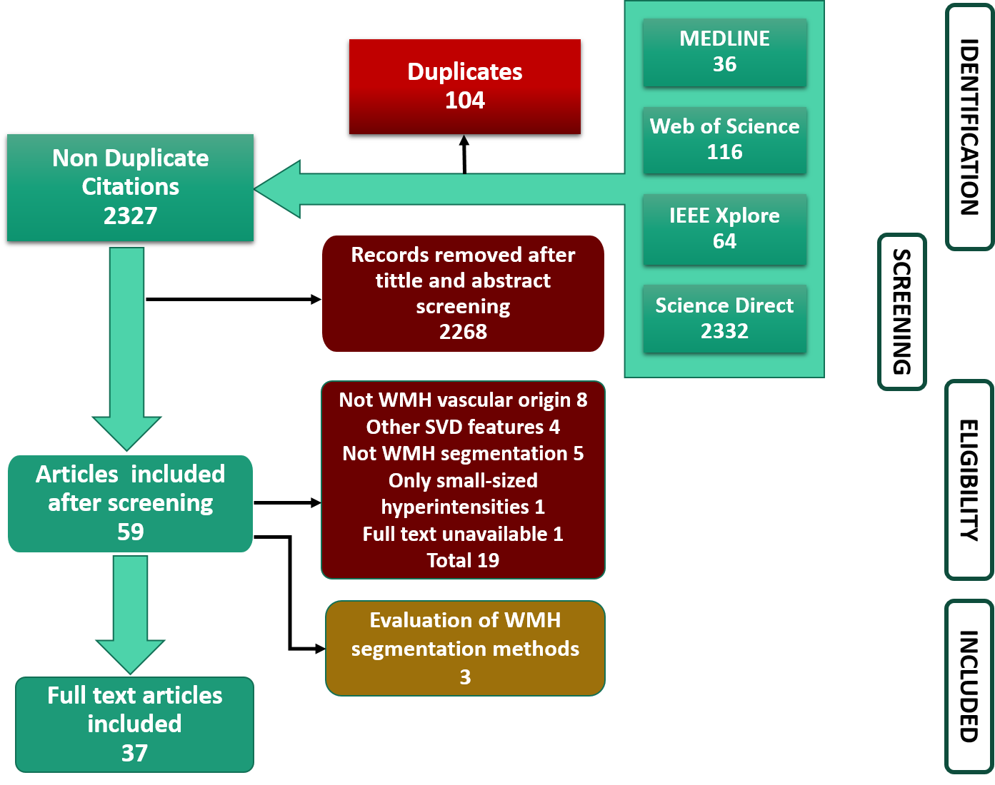
Background: White matter hyperintensities (WMH), of presumed vascular origin, are visible and quantifiable neuroradiological markers of brain parenchymal change. These changes may range from damage secondary to inflammation and other neurological conditions, through to healthy ageing. Fully automatic WMH quantification methods are promising, but still, traditional semi-automatic methods seem to be preferred in clinical research. We systematically reviewed the literature for fully automatic methods developed in the last five years, to assess what are considered state-of-the-art techniques, as well as trends in the analysis of WMH of presumed vascular origin. Method: We registered the systematic review protocol with the International Prospective Register of Systematic Reviews (PROSPERO), registration number - CRD42019132200. We conducted the search for fully automatic methods developed from 2015 to July 2020 on Medline, Science direct, IEE Explore, and Web of Science. We assessed risk of bias and applicability of the studies using QUADAS 2. Results: The search yielded 2327 papers after removing 104 duplicates. After screening titles, abstracts and full text, 37 were selected for detailed analysis. Of these, 16 proposed a supervised segmentation method, 10 proposed an unsupervised segmentation method, and 11 proposed a deep learning segmentation method. Average DSC values ranged from 0.538 to 0.91, being the highest value obtained from an unsupervised segmentation method. Only four studies validated their method in longitudinal samples, and eight performed an additional validation using clinical parameters. Only 8/37 studies made available their method in public repositories. Conclusions: We found no evidence that favours deep learning methods over the more established k-NN, linear regression and unsupervised methods in this task. Data and code availability, bias in study design and ground truth generation influence the wider validation and applicability of these methods in clinical research.

