Künnapuu, J.; Bokharaie, H.; Jeltsch, M. Proteolytic Cleavages in the VEGF Family: Generating Diversity Among Angiogenic VEGFs, Essential for the Activation of Lymphangiogenic VEGFs. Biology2021, 10, 167.
Künnapuu, J.; Bokharaie, H.; Jeltsch, M. Proteolytic Cleavages in the VEGF Family: Generating Diversity Among Angiogenic VEGFs, Essential for the Activation of Lymphangiogenic VEGFs. Biology 2021, 10, 167.
Abstract
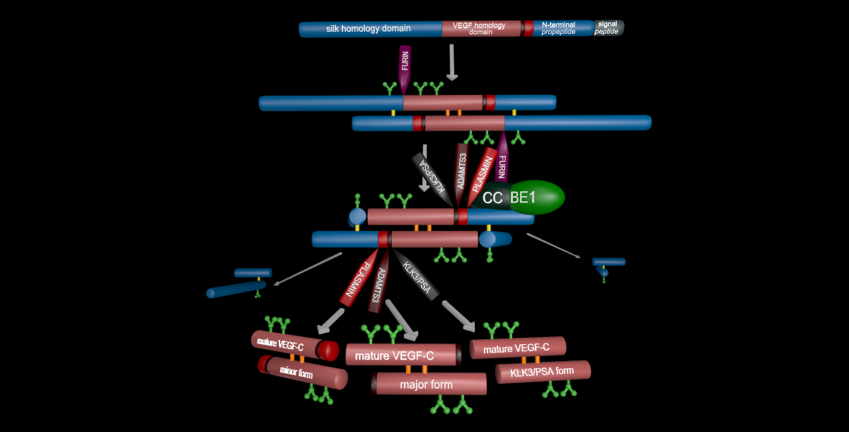
Specific proteolytic cleavages turn on, modify, or turn off the activity of vascular endothelial growth factors (VEGFs). Proteolysis is most prominent among the lymphangiogenic VEGF-C and VEGF-D, which are synthesized as precursors that need to undergo enzymatic removal of their C- and N-terminal propeptides before they can activate their receptors. The activating cleavage of VEGF-C is mediated by at least five different proteases: plasmin, ADAMTS3, prostate-specific antigen, cathepsin D, and thrombin. All of these proteases except for ADAMTS3 can also activate VEGF-D. Processing by different proteases results in distinct forms of the "mature" growth factors, which differ in affinity and receptor activation potential. The “default” VEGF-C-activating enzyme ADAMTS3 does not activate VEGF-D and therefore, VEGF-C and VEGF-D do function in different contexts. VEGF-C itself is also regulated in different contexts by different proteases. During embryonic development, ADAMTS3 activates VEGF-C. In contrast, thrombin and plasmin likely activate VEGF-C/-D during tissue injury-induced lymphangiogenesis, and PSA and cathepsin D perhaps during tumor-associated pathological lymphangiogenesis. Additionally, cathepsin D from saliva might activate latent VEGF-C/-D upon wound licking, thereby accelerating healing. Similar to tyrosine kinase receptors and VEGFs themselves, these activating proteases could be targeted to modulate angiogenesis and lymphangiogenesis in relevant diseases.
Biology and Life Sciences, Biochemistry and Molecular Biology
Copyright:
This is an open access article distributed under the Creative Commons Attribution License which permits unrestricted use, distribution, and reproduction in any medium, provided the original work is properly cited.
Received:
22 January 2021
Commenter:
Béla Tóth
The commenter has declared there is no conflict of interests.
Comment:
Dear Jaana et al.
Overall, very interesting. One thing I noticed is that you do not talk much about the main function of VEGFC (i.e. developmental ADAMTS3-mediated lymphangiogenesis), but you mention the (potential) functions of alternative activation pathways. Did you skip the main function on purpose? The only reason for skipping this is imho the fact that the main function of VEGFC is already the topic of many other reviews.
But you definitely should update the cover image, because it lacks cathepsin D and thrombin, which you mention in the text!
Received:
8 February 2021
Commenter:
Béla Tóth
The commenter has declared there is no conflict of interests.
Comment:
Hi again,
Still something, that you might want to clarify is the role of the two different domains of CCBE1 (i.e. which one being important), because of the results of Le Guen et al. (2014) on the one side and Bos et al. (2011) plus Jeltsch et al. (2014) on the other side, which superficially appear to contradict each other.
The commenter has declared there is no conflict of interests.
Comment:
Thanks Béla,
your comments are welcome and will revise both the image and the text. But I guess you mix Le Guen 2014 with Roukens 2015 (where we have been co-authors). It was Roukens who showed that the acceleration of VEGF-C activation resides within the C-terminal domain of CCBE1. Our data from 2017 (using only the N-terminal domain) shows clearly that it greatly facilitates the assembly of the cleavage complex (consisting of VEGF-C, ADAMTS3 and CCBE1) by recruiting pro-VEGF-C from the liquid phase to the cell surface. However, I have to admit that we still have no clue HOW CCBE1 is doing this (given that its interaction with VEGF-C is so weak that we cannot detect it).
Commenter: Béla Tóth
The commenter has declared there is no conflict of interests.
Overall, very interesting. One thing I noticed is that you do not talk much about the main function of VEGFC (i.e. developmental ADAMTS3-mediated lymphangiogenesis), but you mention the (potential) functions of alternative activation pathways. Did you skip the main function on purpose? The only reason for skipping this is imho the fact that the main function of VEGFC is already the topic of many other reviews.
But you definitely should update the cover image, because it lacks cathepsin D and thrombin, which you mention in the text!
Commenter: Béla Tóth
The commenter has declared there is no conflict of interests.
Still something, that you might want to clarify is the role of the two different domains of CCBE1 (i.e. which one being important), because of the results of Le Guen et al. (2014) on the one side and Bos et al. (2011) plus Jeltsch et al. (2014) on the other side, which superficially appear to contradict each other.
Commenter:
The commenter has declared there is no conflict of interests.
your comments are welcome and will revise both the image and the text. But I guess you mix Le Guen 2014 with Roukens 2015 (where we have been co-authors). It was Roukens who showed that the acceleration of VEGF-C activation resides within the C-terminal domain of CCBE1. Our data from 2017 (using only the N-terminal domain) shows clearly that it greatly facilitates the assembly of the cleavage complex (consisting of VEGF-C, ADAMTS3 and CCBE1) by recruiting pro-VEGF-C from the liquid phase to the cell surface. However, I have to admit that we still have no clue HOW CCBE1 is doing this (given that its interaction with VEGF-C is so weak that we cannot detect it).