Preprint
Case Report
Perimyocarditis as First Manifestation of Systemic Lupus Erythematosus Successfully Treated With Heart Failure and Immunosuppressive Therapy
Altmetrics
Downloads
172
Views
136
Comments
1
A peer-reviewed article of this preprint also exists.
Submitted:
23 January 2023
Posted:
25 January 2023
You are already at the latest version
Alerts
Abstract
Systemic lupus erythematosus (SLE) myocarditis is presumed to be rare but associated with ad-verse outcomes. If SLE diagnosis has not previously been established, its clinical presentation is often unspecific and difficult to recognize. Furthermore, there is a lack of data in the scientific literature regarding myocarditis and its treatment in systemic immune-mediated diseases, leading to its late recognition and undertreatment. We present the case of a young woman whose first lupus manifestations included acute perimyocarditis, among other symptoms and signs that provided clues to the diagnosis of SLE. Transthoracic and speckle tracking echocardiography were helpful in detecting early abnormalities in myocardial wall thickness and contractility while waiting for cardiac magnetic resonance (CMR). Since the patient presented with acute decompensated heart failure (HF), HF treatment was promptly started in parallel with immunosuppressive therapy, with a good response. In the treatment of myocarditis with heart failure, we were guided by the echocardiographic findings, biomarkers for myocardial injury N-terminal pro b-type natriuretic peptide (NT-proBNP) and hs-troponin I, biomarkers for systemic inflammation erythrocyte sedimentation rate (ESR) and C-reactive protein (CRP), and biomarkers for SLE disease activity (Complement C)3, C4, and anti-dsDNA levels.
Keywords:
Subject: Medicine and Pharmacology - Cardiac and Cardiovascular Systems
1. Introduction
Systemic lupus erythematosus is an autoimmune disease more common in women of childbearing age and can affect any organ. Cardiac disease is frequent among SLE patients and can manifest as conduction abnormalities, valvular, pericardial, myocardial, and coronary artery disease, with cardiovascular events being the major cause of premature death [1]. Despite a general decline in SLE mortality over the past decades, cardiovascular mortality remains unchanged [2]. Pericarditis is the most common echocardiographic finding, occurring in more than half of SLE patients [3]. Myocarditis is often asymptomatic with a prevalence between 0.4-16%, but since it can be life-threatening, we must suspect myocarditis in every SLE patient with HF [4]. Cardiac magnetic resonance appears to be the most precise non-invasive diagnostic tool, although not widely available, while right heart catheterization with endomyocardial biopsy (EMB) should be considered when it is not entirely clear that SLE is the cause of myocarditis. Transthoracic echocardiography (TTE) may reveal abnormalities of the left ventricle (LV) systolic and diastolic function, and both global and segmental areas of hypokinesis can be detected. Depending on the degree of myocardial inflammation, chamber size and myocardial wall thickness reflecting interstitial oedema can vary [5]. In addition, speckle tracking echocardiography (STE) can detect subclinical abnormalities in myocardial contractility and could be helpful in diagnosing myocarditis, especially in its initial stages [6]. Moreover, it was shown that localizations of myocarditis area showed on CMR correspond to area with depressed longitudinal strain on echo [6]. Acute myocarditis in lupus is often accompanied by pericarditis, and treatment of both has not been assessed in controlled trials and is so far based on expert opinions and case reports published in the literature. Here we present a patient in whom acute HF due to acute perimyocarditis was the first SLE manifestation and who was successfully treated with high doses of methylprednisolone (MP) and hydroxychloroquine (HCQ) in parallel with standard HF therapy.
2. Case Description
A 30-year-old woman was hospitalized at the Internal Medicine Clinic due to recurrent biliary colic and suspicion of the development of acute calculous cholecystitis with a mildly elevated ESR and CRP. Treatment with analgesics, spasmolytics, and antibiotics was started. Despite the treatment, three days later, the patient became febrile (>38.5 C), developed maculopapular rash on the arms and torso, a malar rash, dyspnea, and chest pain with elevated NT-proBNP (9373 ng/L, normal range: <208.4 ng/L), and hs-troponin I (293 ng/L, normal range:<39 ng/L) without signs of ischemia on ECG (Table 1). The patient also had a low level of albumin, an elevated level of fibrinogen and a normal value of ferritin, as markers of acute inflammation.
Furthermore, abdominal ultrasound at that moment showed no signs of acute cholecystitis, while on TTE there was: hypo-contractility in the basal segment of the anterior, septal, and lateral walls with preserved wall thickness and LV diameter, mildly reduced LV ejection fraction (EF), reduced global longitudinal peak strain (GLPS) -15.1%, moderate mitral regurgitation (MR), diastolic dysfunction (DD) grade 3, and a small pericardial effusion (2-3 mm) (Figure 1 and Figure 2).
Since there was a concern about a possible associated allergic reaction (rash) during the treatment with antibiotics, the patient immediately received a low dose of corticosteroids and antihistamines while extensive evaluation was initiated in parallel with guideline-directed treatment for HF. Blood and urine cultures were negative, as were viral panels for cardiotropic infectious agents. All tumor markers and interferon-gamma release assay for latent tuberculosis infection were in the reference range. A clinical immunologist examined the patient and recommended diagnostic workup under suspicion of SLE. Coronary angiography did not show obstructive coronary artery disease. Since the patient manifested symptomatic HF with a slightly reduced EF but with the possibility of fast deterioration, we introduced diuretics, beta-blockers, aldosterone antagonists, and sacubitril/valsartan as well as ibuprofen due to signs of pericarditis. Interestingly, a few days later, the TTE showed markedly increased thickness of the LV walls with normal EF, GLPS of -19%, mild MR, DD grade 1, and progression of pericardial effusion to 10 mm. The GLPS abnormalities were again found in the basal segments of the anterior, septal, and lateral walls (Figure 3 and Figure 4).
This increased thickness of myocardial walls on second TTE probably reflected interstitial edema due to suspected inflammation, and the rapid EF improvement was explained by better loading conditions due to diuretics and HF therapy. Nevertheless, depressed GLPS was still noted within several segments of the LV, raising suspicion of myocarditis. After immunological workup the diagnosis of SLE was made; the 2019 EULAR/ACR criteria for SLE were fulfilled: leukopenia presented on several occasions before according to available medical documentation, strongly positive ANA, complement consumption - low C3, pericarditis, myocarditis, fever, and acute cutaneous lupus (Table 1). Also, patient reported photosensitivity and recurrent cervical lymphadenopathy and had persistently low lymphocyte count with occasional thrombocytopenia but not less than 100 x 109/L. Therefore, in addition to HF therapy, HCQ 200 mg and MP 2 mg/kg/day were introduced, and ibuprofen was ceased. Soon after initiating the above-mentioned therapy, the patient reported regression of all symptoms, and there was a significant decrease in NT-proBNP, normalization of leukocyte and platelets count, troponin, ESR, CRP, fibrinogen, and the MP was reduced to 0.5 mg/kg/day. Meanwhile, we obtained CMR results that confirmed the myocarditis diagnosis and were comparable with abnormalities detected on TTE and STE. After only 3 weeks of therapy, TTE was completely normal (Figure 5), and we tapered MP to 0.3 mg/kg/day.
Sacubitril/valsartan and aldosterone antagonists were completely discontinued after 6 months, and the dose of MP was gradually decreased with total discontinuation after 12 months (after 6 months MP dose was 8 mg daily). Now, 24 months after the first manifestation of SLE, the patient is still in complete clinical and immunological disease remission and is continuously taking bisoprolol at minimal dosage and 200 mg of HCQ (Table 1).
3. Discussion
Clinicians must be aware of unusual SLE presentations such as acute HF due to SLE-myocarditis with only 0.37% of patients presenting as such, but with potentially fatal outcomes: cardiogenic shock or malignant arrhythmias [7]. The diagnosis of SLE itself, especially SLE-myocarditis, is challenging in clinical practice if the patient has not had a previous diagnosis of lupus or lupus signs and symptoms. Although EMB is considered the gold standard for the diagnosis of myocarditis, it is not a routine procedure because it is invasive and has a risk of sampling error. Consequently, to establish the correct diagnosis of SLE-myocarditis, we must combine the patient's previous medical history, current signs and symptoms, laboratory tests, TTE with STE, CMR, and EMB if available, and exclude other common causes of myocarditis. In our case, we established a diagnosis of SLE according to the 2019 EULAR/ACR classification criteria and after the exclusion of other causes of myocarditis. Since there are no guidelines for the treatment of myocarditis in systemic rheumatic diseases, the treatment is based on the experience of individual rheumatologists and cardiologists, as well as review papers, case reports, and expert opinions published in the literature. In our case, we were guided by the 2016 ESC guidelines for HF valid at that time, the Position statement of the ESC Working Group on Myocardial and Pericardial Disease, EULAR recommendations for the management of SLE, previously reported case reports, and reviews [8,9,10,11,12,13,14,15]. According to the literature, treatment of lupus myocarditis requires an initial period of intensive immunosuppression to decrease disease activity, followed by a longer period of less intensive therapy to consolidate the response and prevent relapses [9,12]. Our patient was initially treated with a high dose of MP: 2 mg/kg/day (with the possible implication of early-started low-dose corticosteroids due to rash and concern of an allergic reaction), and the high dose was reduced to a moderate dose (0.3 mg/kg/day) after TTE and laboratory confirmation of myocardial recovery with further gradual dose reduction. Ibuprofen was stopped since non-steroidal anti-inflammatory drugs (NSAIDs) could enhance inflammation and increase mortality, which has been shown in animal models of myocarditis, although recent findings suggest that treatment with NSAIDs does not affect outcome in patients with acute myocarditis or myopericarditis [10,11,16]. In our opinion, patients with HF due to SLE should be treated in parallel with HF guideline-directed therapy to reduce the risk of further myocardial damage and potential complications, although the optimal duration of the treatment is not yet well-established. Also, the optimal duration of glucocorticoid therapy is not known in lupus myocarditis, but we were guided by the patient's clinical signs and symptoms and laboratory and TTE findings. The treatment should be started as soon as possible to prevent further damage by suppressing the inflammation that leads to heart remodeling and fibrosis. Furthermore, in relation to changes detected on TTE with STE, a combination of widely available laboratory parameters/biomarkers such as troponin and NT-proBNP (biomarkers of myocardial injury), anti-dsDNA, C3 and C4 (biomarkers of SLE disease activity), and ESR, CRP, albumin, and fibrinogen (biomarkers of systemic inflammation) could be used not only for early diagnosis but also for SLE-myocarditis follow-up and therapy management [17,18]. In the absence of a rapid response to glucocorticoids, another immunosuppressant such as cyclophosphamide or rituximab should be introduced into the therapy [14,19]. Also, we must bear in mind that treatment of myocarditis in systemic autoimmune or autoinflammatory disease should be tailored to each individual patient depending on which organs are simultaneously affected by the disease. We believe that timely-initiated immunosuppressive glucocorticoid therapy in parallel with HF therapy with neurohumoral blockade was the key to successful treatment in the case of our patient.
4. Conclusions
In patients with organ-threatening diseases, such as acute perimyocarditis with heart failure in an SLE flare, the choice of treatment could be high-dose MP with proven fast immunosuppression in combination with guideline-directed heart failure therapy to suppress myocardial inflammation and preserve LV function. In the treatment of SLE-myocarditis we should be guided by the findings on TTE with STE, and biomarkers of myocardial injury, lupus activity, and systemic inflammation. Clinical trials with larger numbers of patients conducted by the cardiologist and clinical immunologist/rheumatologist are necessary to provide guidelines for diagnosing and managing myocarditis in autoimmune and autoinflammatory systemic diseases.
Author Contributions
Conceptualization, M.I.M. and P.G.R.; methodology, P.K. and Z.P.; validation, M.I.M., E.G. and P.G.R.; formal analysis, M.I.M. and P.K..; investigation, M.I.M.; P.G.R.; I.Z.K. and Z.P.; data curation, M.I.M. and L.I.; writing—original draft preparation, M.I.M. and L.I.; writing—review and editing, E.G., I.Z.K. and P.K.; supervision M.I.M. All authors have read and agreed to the published version of the manuscript.
Funding
This research received no external funding.
Institutional Review Board Statement
Since only normal clinical practice is described in this case report, formal ethical approval by the Independent Review Board was not required in accordance with the policy of our institution.
Informed Consent Statement
Written informed consent has been obtained from the patient to publish this paper.
Data Availability Statement
The original data generated and analyzed for this study are included in the published article. Further inquiries can be directed to the corresponding author.
Conflicts of Interest
The authors declare no conflict of interest.
References
- Tincani, A.; Rebaioli, C.B., Taglietti, M.; Shoenfeld, Y. Heart involvement in systemic lupus erythematosus, anti-phospholipid syndrome and neonatal lupus. Rheumatology (Oxford) 2006, 45 (Suppl. S4), iv8-iv13. [CrossRef]
- Björnådal, L.; Yin, L.; Granath, F.; Klareskog, L.; Ekbom, A. Cardiovascular disease a hazard despite improved prognosis in patients with systemic lupus erythematosus: results from a Swedish population based study 1964-95. J Rheumatol 2004, 31, 713–719. [Google Scholar] [PubMed]
- Dein, E.; Douglas, H.; Petri, M.; Law, G.; Timlin, H. Pericarditis in Lupus. Cureus 2019, 11, e4166. [Google Scholar] [CrossRef] [PubMed]
- Tani, C.; Elefante, E.; Arnaud, L.; Barreira, S.C.; Bulina, I.; Cavagna, L.; Costedoat-Chalumeau, N.; Doria, A.; Fonseca, J.E.; Franceschini, F.; Fredi, M.; Iaccarino, L.; Limper, M.; Majnik, J.; Nagy, G.; Pamfil, C.; Rednic, S.; Reynolds, J.A.; Tektonidou, M.G.; Troldborg, A.; Zanframundo, G.; Mosca, M. Rare clinical manifestations in systemic lupus erythematosus: a review on frequency and clinical presentation. Clin Exp Rheumatol 2022, 40, Suppl 134(5):93-102. [CrossRef]
- Felker, G. M.; Boehmer, J. P.; Hruban, R. H.; Hutchins, G. M.; Kasper, E. K.; Baughman, K. L.; Hare, J. M. Echocardiographic findings in fulminant and acute myocarditis. Journal of the American College of Cardiology 2000, 36, 227–232. [Google Scholar] [CrossRef] [PubMed]
- Uziębło-Życzkowska, B.; Mielniczuk, M.; Ryczek, R.; Krzesiński, P. Myocarditis successfully diagnosed and controlled with speckle tracking echocardiography. Cardiovascular ultrasound 2020, 18, 19. [Google Scholar] [CrossRef] [PubMed]
- Tanwani, J.; Tselios, K.; Gladman, D. D.; Su, J.; Urowitz, M. B. Lupus myocarditis: a single center experience and a comparative analysis of observational cohort studies. Lupus 2018, 27, 1296–1302. [Google Scholar] [CrossRef] [PubMed]
- Ponikowski, P.; Voors, A. A.; Anker, S. D.; Bueno, H.; Cleland, J. G.; Coats, A. J.; Falk, V.; González-Juanatey, J. R.; Harjola, V. P.; Jankowska, E. A.; Jessup, M.; Linde, C.; Nihoyannopoulos, P.; Parissis, J. T.; Pieske, B.; Riley, J. P.; Rosano, G. M.; Ruilope, L. M.; Ruschitzka, F.; Rutten, F. H., … Document Reviewers. 2016 ESC Guidelines for the diagnosis and treatment of acute and chronic heart failure: The Task Force for the diagnosis and treatment of acute and chronic heart failure of the European Society of Cardiology (ESC). Developed with the special contribution of the Heart Failure Association (HFA) of the ESC. European journal of heart failure 2016, 18, 891–975.
- Caforio, A. L. P.; Adler, Y.; Agostini, C.; Allanore, Y.; Anastasakis, A.; Arad, M.; Böhm, M.; Charron, P.; Elliott, P. M.; Eriksson, U.; Felix, S. B.; Garcia-Pavia, P.; Hachulla, E.; Heymans, S.; Imazio, M.; Klingel, K.; Marcolongo, R.; Matucci Cerinic, M.; Pantazis, A.; Plein, S. … Linhart, A. Diagnosis and management of myocardial involvement in systemic immune-mediated diseases: a position statement of the European Society of Cardiology Working Group on Myocardial and Pericardial Disease. European heart journal 2017, 38, 2649–2662. [CrossRef]
- Caforio, A. L.; Pankuweit, S.; Arbustini, E.; Basso, C.; Gimeno-Blanes, J.; Felix, S. B.; Fu, M.; Heliö, T.; Heymans, S.; Jahns, R.; Klingel, K.; Linhart, A.; Maisch, B.; McKenna, W.; Mogensen, J.; Pinto, Y. M.; Ristic, A.; Schultheiss, H. P.; Seggewiss, H.; Tavazzi, L., … European Society of Cardiology Working Group on Myocardial and Pericardial Diseases. Current state of knowledge on aetiology, diagnosis, management, and therapy of myocarditis: a position statement of the European Society of Cardiology Working Group on Myocardial and Pericardial Diseases. European heart journal 2013, 34, 2636–2648. [CrossRef]
- Adler, Y.; Charron, P.; Imazio, M.; Badano, L.; Barón-Esquivias, G.; Bogaert, J.; Brucato, A.; Gueret, P.; Klingel, K.; Lionis, C.; Maisch, B.; Mayosi, B.; Pavie, A.; Ristic, A. D.; Sabaté Tenas, M.; Seferovic, P.; Swedberg, K.; Tomkowski, W., and ESC Scientific Document Group. 2015 ESC Guidelines for the diagnosis and management of pericardial diseases: The Task Force for the Diagnosis and Management of Pericardial Diseases of the European Society of Cardiology (ESC)Endorsed by: The European Association for Cardio-Thoracic Surgery (EACTS). European heart journal 2015, 36, 2921–2964.
- Fanouriakis, A.; Kostopoulou, M.; Alunno, A.; Aringer, M.; Bajema, I.; Boletis, J. N.; Cervera, R.; Doria, A.; Gordon, C.; Govoni, M.; Houssiau, F.; Jayne, D.; Kouloumas, M.; Kuhn, A., Larsen, J. L.; Lerstrøm, K.; Moroni, G.; Mosca, M.; Schneider, M.; Smolen, J. S. … Boumpas, D. T. 2019 update of the EULAR recommendations for the management of systemic lupus erythematosus. Annals of the rheumatic diseases 2019, 78, 736–745.
- Durrance, R. J.; Movahedian, M.; Haile, W.; Teller, K.; Pinsker, R. Systemic Lupus Erythematosus Presenting as Myopericarditis with Acute Heart Failure: A Case Report and Literature Review. Case reports in rheumatology 2019, 2019, 6173276. [Google Scholar] [CrossRef] [PubMed]
- Zhang, L.; Zhu, Y. L.; Li, M. T.; Gao, N.; You, X.; Wu, Q. J.; Su, J. M.; Shen, M.; Zhao, L. D.; Liu, J. J.; Zhang, F. C.; Zhao, Y.; Zeng, X. F. Lupus Myocarditis: A Case-Control Study from China. Chinese medical journal 2015, 128, 2588–2594. [Google Scholar] [CrossRef] [PubMed]
- Xibillé-Friedmann, D.; Pérez-Rodríguez, M.; Carrillo-Vázquez, S.; Álvarez-Hernández, E.; Aceves, F. J., Ocampo-Torres, M. C.; García-García, C.; García-Figueroa, J. L.; Merayo-Chalico, J.; Barrera-Vargas, A.; Portela-Hernández, M.; Sicsik, S.; Andrade-Ortega, L.; Rosales-Don Pablo, V. M.; Martínez, A.; Prieto-Seyffert, P.; Pérez-Cristóbal, M.; Saavedra, M. Á.; Castro-Colín, Z.; Ramos, A.; … Barile-Fabris, L. A. Clinical practice guidelines for the treatment of systemic lupus erythematosus by the Mexican College of Rheumatology. Guía de práctica clínica para el manejo del lupus eritematoso sistémico propuesta por el Colegio Mexicano de Reumatología. Reumatologia clinica 2019, 15, 3–20.
- Mirna, M.; Schmutzler, L.; Topf, A.; Boxhammer, E.; Sipos, B.; Hoppe, U. C.; Lichtenauer, M. Treatment with Non-Steroidal Anti-Inflammatory Drugs (NSAIDs) Does Not Affect Outcome in Patients with Acute Myocarditis or Myopericarditis. Journal of cardiovascular development and disease 2022, 9, 32. [Google Scholar] [CrossRef] [PubMed]
- Gulhar, R.; Ashraf, M. A.; Jialal, I. (2022). Physiology, Acute Phase Reactants. In StatPearls. StatPearls Publishing.
- Suresh, A.; Martens, P.; Tang, W. H. W. Biomarkers for Myocarditis and Inflammatory Cardiomyopathy. Current heart failure reports 2022, 19, 346–355. [Google Scholar] [CrossRef] [PubMed]
- Zagelbaum Ward, N. K.; Linares-Koloffon, C.; Posligua, A.; Gandrabur, L.; Kim, W. Y.; Sperber, K.; Wasserman, A.; Ash, J. Cardiac Manifestations of Systemic Lupus Erythematous: An Overview of the Incidence, Risk Factors, Diagnostic Criteria, Pathophysiology and Treatment Options. Cardiology in review 2022, 30, 38–43. [Google Scholar] [CrossRef] [PubMed]
Figure 1.
Initial transthoracic echocardiography showing left ventricular wall thickness (IVS, LPW) within normal range in parasternal long axis (PLAX) view. IVS interventricular septum, LA left atrium, LV left ventricle, LPW left posterior wall, MV mitral valve, PE pericardial effusion.
Figure 1.
Initial transthoracic echocardiography showing left ventricular wall thickness (IVS, LPW) within normal range in parasternal long axis (PLAX) view. IVS interventricular septum, LA left atrium, LV left ventricle, LPW left posterior wall, MV mitral valve, PE pericardial effusion.
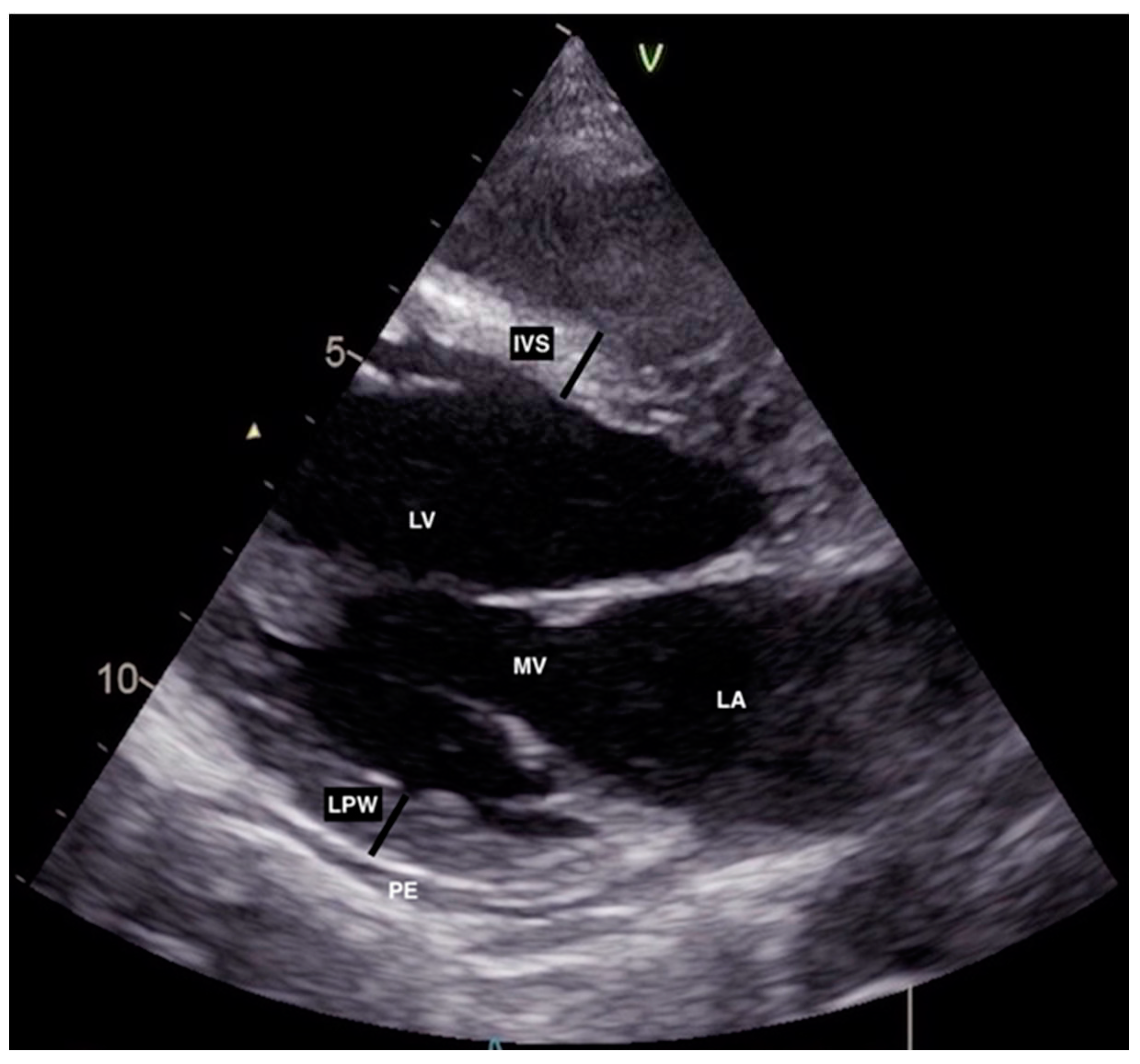
Figure 2.
Initial transthoracic echocardiography with mildly reduced left ventricular ejection fraction (EF) calculated by Simpson biplane method and reduced global longitudinal peak strain (GLPS) with reduced values in basal segment of the anterior, septal, and lateral walls.
Figure 2.
Initial transthoracic echocardiography with mildly reduced left ventricular ejection fraction (EF) calculated by Simpson biplane method and reduced global longitudinal peak strain (GLPS) with reduced values in basal segment of the anterior, septal, and lateral walls.
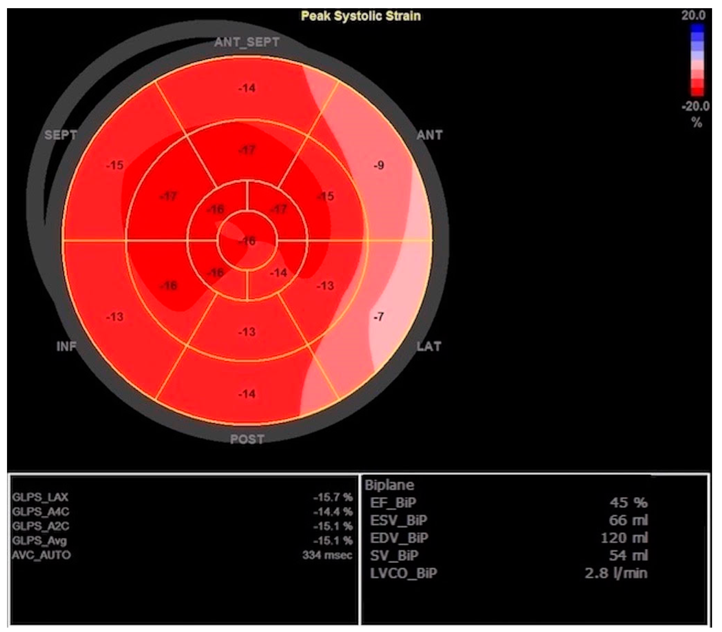
Figure 3.
Second transthoracic echocardiography showing markedly increased thickness of left ventricle (LV) walls (IVS, LPW) in parasternal long axis (PLAX) view. AV aortic valve, IVS interventricular septum, LA left atrium, LPW left posterior wall, MV mitral valve, PE pericardial effusion, RV right ventricle.
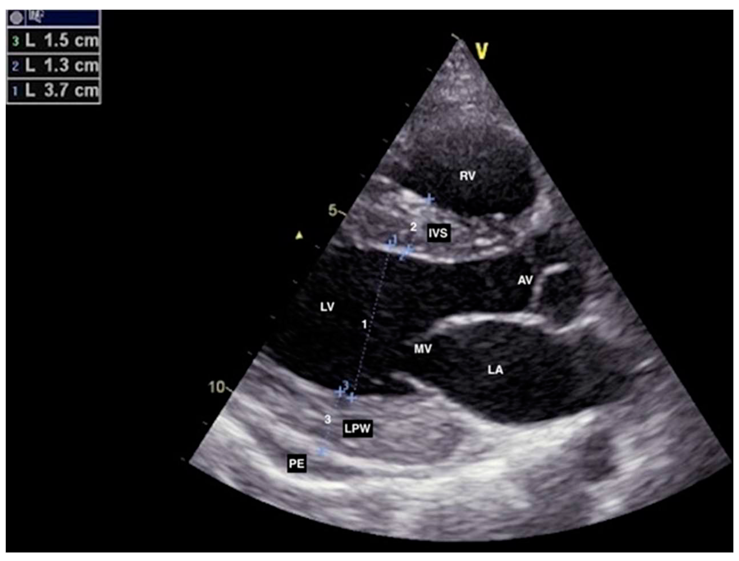
Figure 4.
Second transthoracic echocardiography with improved left ventricle ejection fraction (EF) and improved global longitudinal peak strain (GLPS) but still noticeable reduced regional strain values.
Figure 4.
Second transthoracic echocardiography with improved left ventricle ejection fraction (EF) and improved global longitudinal peak strain (GLPS) but still noticeable reduced regional strain values.
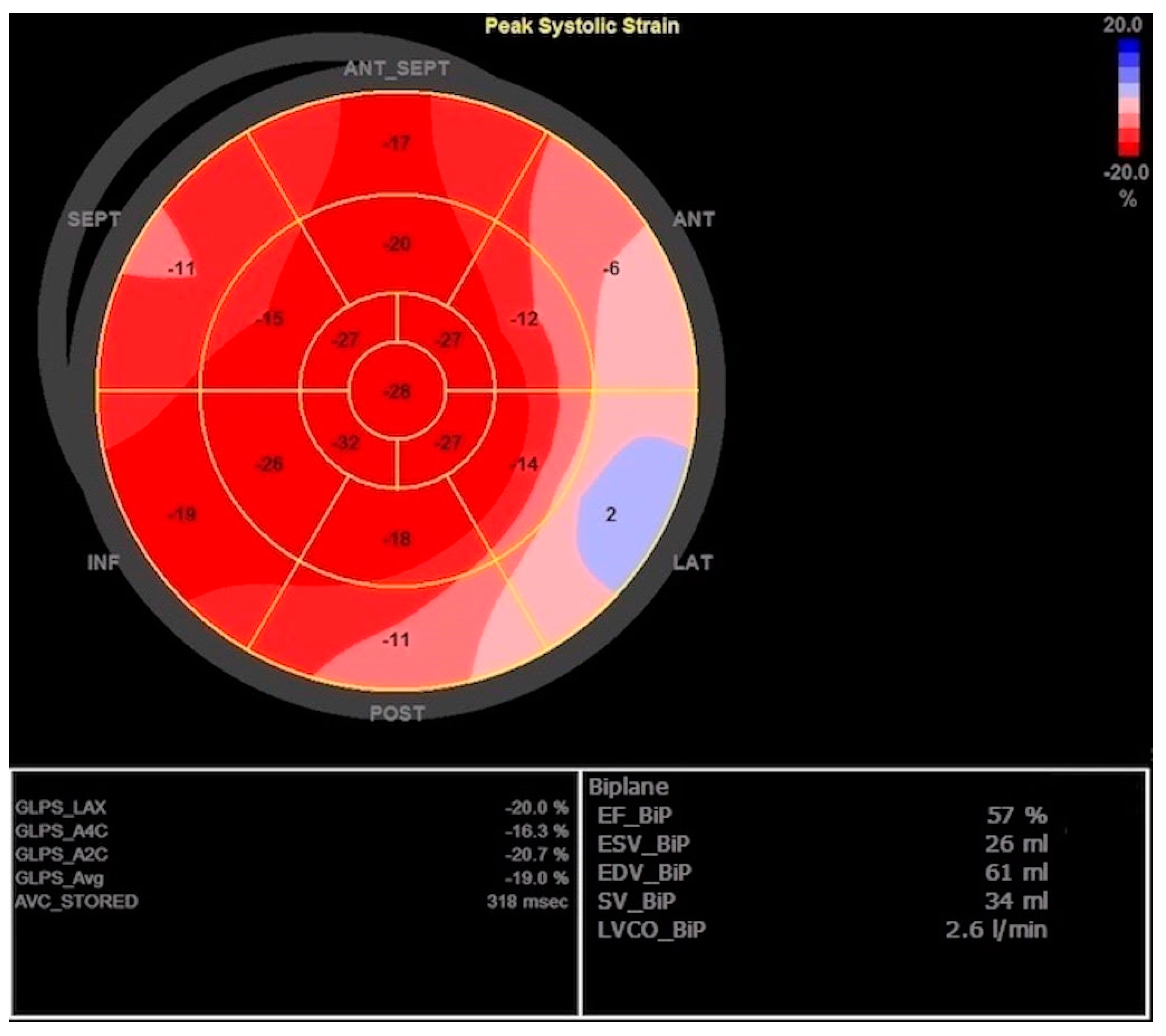
Figure 5.
Completely recovered left ventricle ejection fraction (EF) and global longitudinal peak strain (GLPS) with no signs of hypokinesis 3 weeks after initiating guideline-directed heart failure therapy concomitantly with immunosuppressive therapy.
Figure 5.
Completely recovered left ventricle ejection fraction (EF) and global longitudinal peak strain (GLPS) with no signs of hypokinesis 3 weeks after initiating guideline-directed heart failure therapy concomitantly with immunosuppressive therapy.
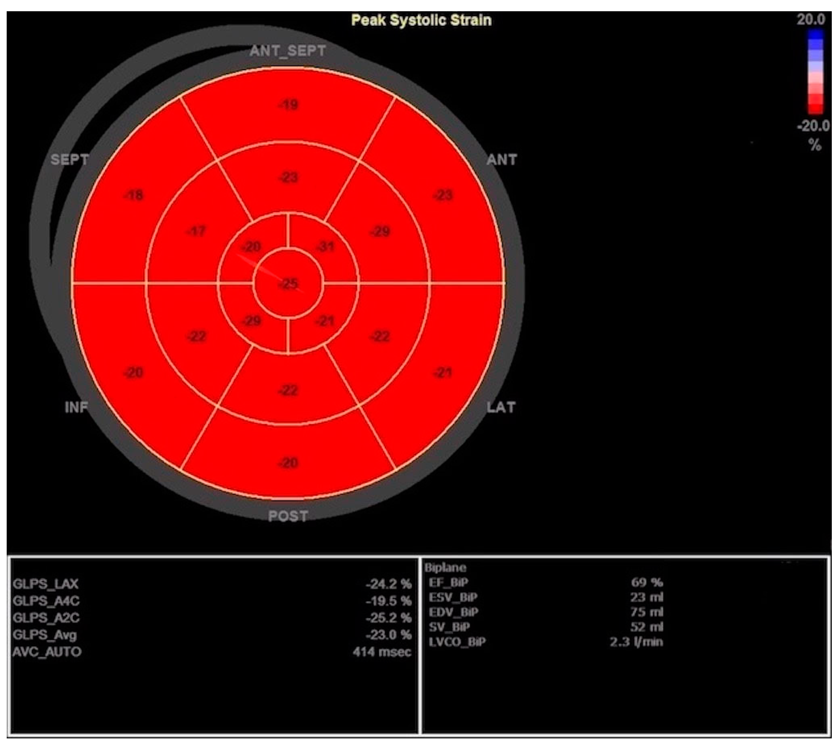
Table 1.
Laboratory data upon admission and 24 months after initiation of heart failure and immunosuppressive therapy.
Table 1.
Laboratory data upon admission and 24 months after initiation of heart failure and immunosuppressive therapy.
| Reference range | Admission | After 24 months | |
|---|---|---|---|
| White blood cell count (109/L) | 3.4-9.7 | 3.1 | 6.9 |
| Hemoglobin (g/L) | 138-175 | 116 | 137 |
| Platelets (109/L) | 158-424 | 132 | 193 |
| CRP (mg/dL) | <5.0 | 26.5 | 3.5 |
| ESR (mm/3.6ks) | 4-24 | 28 | 11 |
| Albumin (g/L) | 41-51 | 36 | 44 |
| Ferritin (µg/L) | 10-158 | 61 | 12 |
| Fibrinogen (g/L) | 1.8-3.5 | 3.9 | 2.2 |
| Procalcitonin (µg/L) | 0.5-2 | 1.30 | <0.5 |
| Creatinine (µmol/L) | 49-90 | 68 | 51 |
| 24-hour urine protein (mg/24h) | <150 | 54 | 145 |
| hs-troponin I (ng/L) | <39 | 293 | 36 |
| NT-proBNP (ng/L) | <208.4 | 9373 | 135 |
| Direct antiglobulin test | - | Negative | - |
| C3 (g/L) | 0.89-1.87 | 0.72 | 1.0 |
| C4 (g/L) | 0.17-0.38 | 0.17 | 0.28 |
| Antinuclear antibody* | Positive>1 | >55 | - |
| Anti-dsDNA antibody* (IU/ml) | Positive>15 | <0.7 | 1.2 |
| Anti-Smith (Sm) antibody | - | - | - |
| Anticardiolipin antibody IgM* (MPL-U/mL) | Positive>40 | 1.1 | 0.7 |
| Anticardiolipin antibody IgG* (GPL-U/mL) | Positive>40 | 1.2 | 0.9 |
| Beta2-Glycoprotein I IgM* (EliA U/mL) | Positive>10 | <2.4 | <2.4 |
| Beta2-Glycoprotein I IgG* (EliA U/mL) | Positive>10 | <0.8 | <0.8 |
| Lupus anticoagulant | - | Negative | - |
CRP C-reactive protein, ESR Erythrocyte sedimentation rate, NT-proBNP N-terminal pro b-type natriuretic peptide, C Complement, *FEIA Phadia 200.
Disclaimer/Publisher’s Note: The statements, opinions and data contained in all publications are solely those of the individual author(s) and contributor(s) and not of MDPI and/or the editor(s). MDPI and/or the editor(s) disclaim responsibility for any injury to people or property resulting from any ideas, methods, instructions or products referred to in the content. |
© 2023 by the authors. Licensee MDPI, Basel, Switzerland. This article is an open access article distributed under the terms and conditions of the Creative Commons Attribution (CC BY) license (http://creativecommons.org/licenses/by/4.0/).
Copyright: This open access article is published under a Creative Commons CC BY 4.0 license, which permit the free download, distribution, and reuse, provided that the author and preprint are cited in any reuse.
Echocardiography myocardial work and cardiac biomarkers indicate subclinical systolic myocardial dysfunction in patients with systematic lupus erythematous
Nikolaos P.E. Kadoglou
et al.
,
2024
Systematic Aetiological Assessment of Myocarditis: A Prospective Cohort Study
Vincent Michel
et al.
,
2024
Cardiac MRI as a Risk Stratification Tool in COVID -19 Myocarditis
Olga Nedeljković-Arsenović
et al.
,
2024
MDPI Initiatives
Important Links
© 2024 MDPI (Basel, Switzerland) unless otherwise stated


