Submitted:
04 March 2023
Posted:
06 March 2023
You are already at the latest version
Abstract
Keywords:
1. Introduction
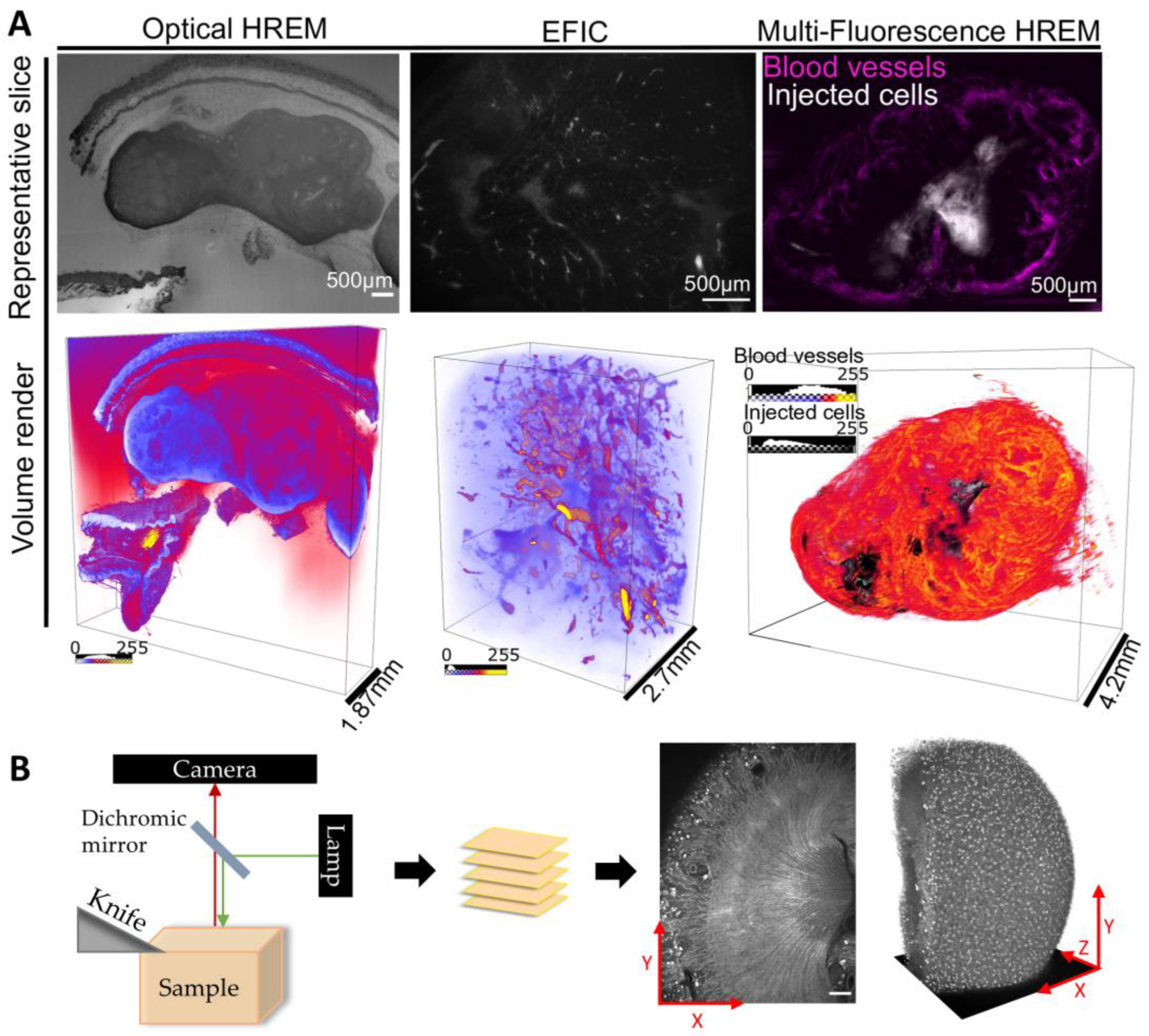
1.1. Background on Episcopic Imaging Techniques
1.2. The importance of quantitative image analysis
1.3. Methods of quantitative imaging
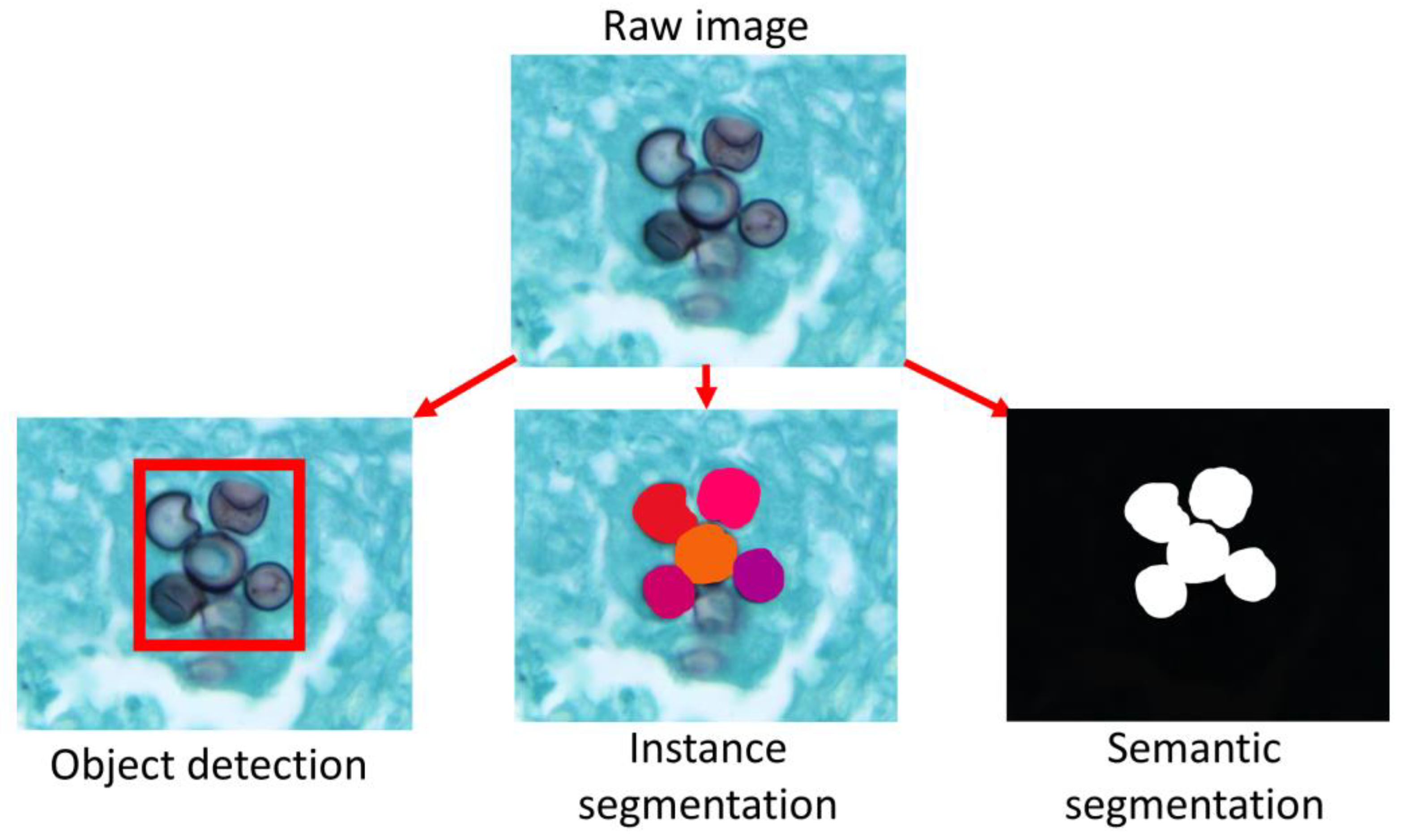
2. Methods
- Year
- Contrast type
- Structure under investigation
- Model organism
-
Was annotation performed?
- If yes: was this segmentation, object detection, or both?
- Was quantitative data presented?
- Method of annotation /quantification – organized into categories determined during analysis
- Software used
- Was the raw image data deposited in an online repository?
- The type of publication (paper, conference paper, thesis)
3. Results
3.1. Trends in prevalence of quantification
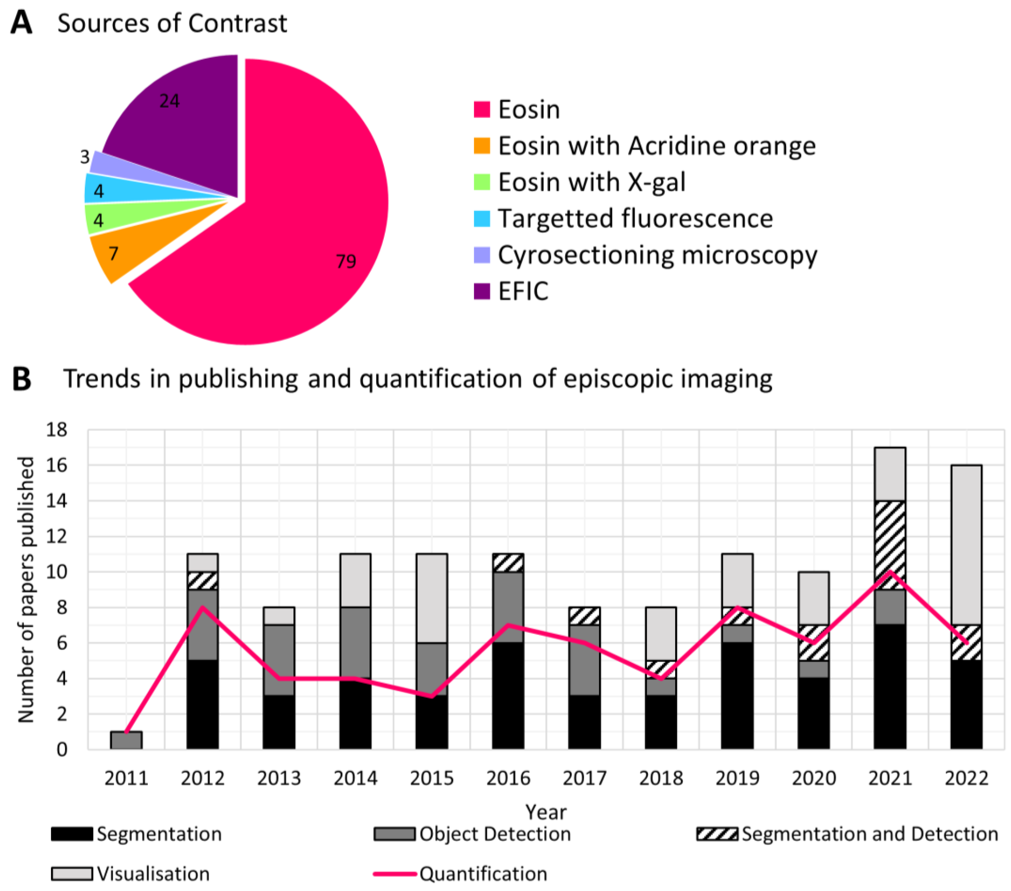
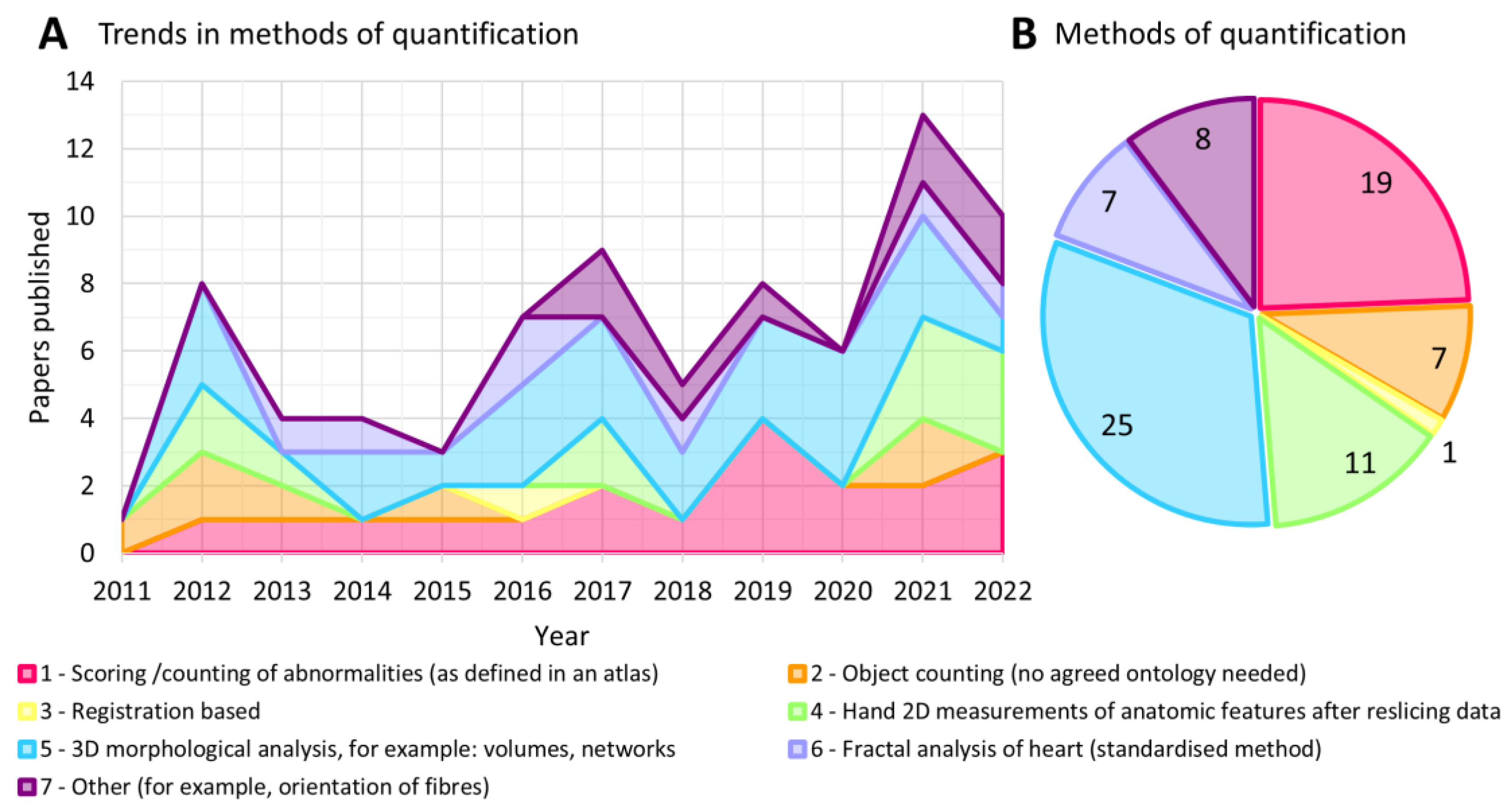
3.2. Trends in methods of quantification
- Scoring /counting of abnormalities (as defined in an atlas)
- Object counting (no agreed ontology needed)
- Registration based
- Hand 2D measurements of anatomic features after reslicing data
- 3D morphological analysis, for example: volumes, networks
- Fractal analysis of heart (standardized method)
- Other (for example, orientation of fibers)
3.3. Methods of image annotation
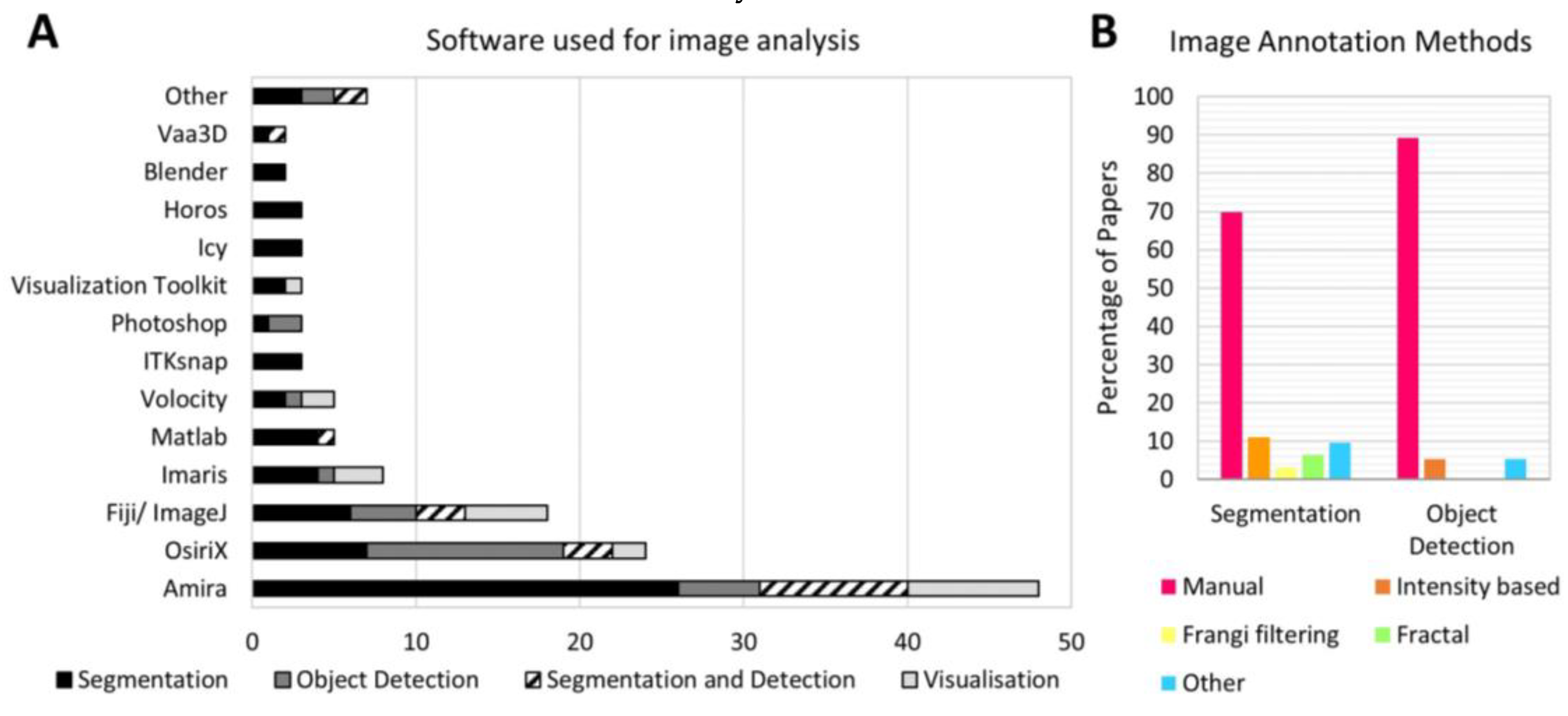
4. Discussion
6. Future Directions
Supplementary Materials
Author Contributions
Funding
Institutional Review Board Statement
Informed Consent Statement
Data Availability Statement
Conflicts of Interest
References
- Weninger, W.J.; Geyer, S.H.; Mohun, T.J.; Rasskin-Gutman, D.; Matsui, T.; Ribeiro, I.; Costa, L.D.F.; Izpisúa-Belmonte, J.C.; Müller, G.B. High-Resolution Episcopic Microscopy: A Rapid Technique for High Detailed 3D Analysis of Gene Activity in the Context of Tissue Architecture and Morphology. Anat Embryol (Berl) 2006, 211, 213–221. [Google Scholar] [CrossRef]
- Walsh, C.; Holroyd, N.A.; Finnerty, E.; Ryan, S.G.; Sweeney, P.W.; Shipley, R.J.; Walker-Samuel, S. Multifluorescence High-Resolution Episcopic Microscopy for 3D Imaging of Adult Murine Organs. Adv Photonics Res 2021, 2, 2100110. [Google Scholar] [CrossRef]
- Weninger, W.J.; Mohun, T.J. Three-Dimensional Analysis of Molecular Signals with Episcopic Imaging Techniques. In Methods in molecular biology (Clifton, N.J.); Humana Press Inc: Totowa, NJ, 2007; Volume 411, pp. 35–46. ISBN 1064-3745. [Google Scholar]
- Brown, S.D.M.; Moore, M.W.; Baldock, R.; Bhattacharya, S.; Copp, A.J.; Hemberger, M.; Houart, C.; Hurles, M.E.; Robertson, E.; Smith, J.C.; et al. Towards an Encyclopaedia of Mammalian Gene Function: The International Mouse Phenotyping Consortium. Dis Model Mech 2012, 5, 289–292. [Google Scholar] [CrossRef]
- Waheed, A.A.; Rao, K.S.; Gupta, P.D. Mechanism of Dye Binding in the Protein Assay Using Eosin Dyes. Anal Biochem 2000, 287, 73–79. [Google Scholar] [CrossRef]
- Weninger, W.J.; Mohun, T. Phenotyping Transgenic Embryos: A Rapid 3-D Screening Method Based on Episcopic Fluorescence Image Capturing. Nat Genet 2002, 30, 59–65. [Google Scholar] [CrossRef]
- Hakimzadeh, N.; van Lier, M.G.J.T.B.; van Horssen, P.; Daal, M.; Ly, D.H.; Belterman, C.; Coronel, R.; Spaan, J.A.E.; Siebes, M. Selective Subepicardial Localization of Monocyte Subsets in Response to Progressive Coronary Artery Constriction. Am J Physiol Heart Circ Physiol 2016, 311, H239–H250. [Google Scholar] [CrossRef]
- Symvoulidis, P.; Cruz Pérez, C.; Schwaiger, M.; Ntziachristos, V.; Westmeyer, G.G. Serial Sectioning and Multispectral Imaging System for Versatile Biomedical Applications. 2014 IEEE 11th International Symposium on Biomedical Imaging, ISBI 2014 2014, 890–893. [Google Scholar] [CrossRef]
- Geyer, S.H.; Nöhammer, M.M.; Mathä, M.; Reissig, L.; Tinhofer, I.E.; Weninger, W.J. High-Resolution Episcopic Microscopy (HREM): A Tool for Visualizing Skin Biopsies. Microscopy and Microanalysis 2014, 20, 1356–1364. [Google Scholar] [CrossRef]
- Geyer, S.H.; Nöhammer, M.M.; Tinhofer, I.E.; Weninger, W.J. The Dermal Arteries of the Human Thumb Pad. J Anat 2013, 223, 603–609. [Google Scholar] [CrossRef]
- Tinhofer, I.E.; Zaussinger, M.; Geyer, S.H.; Meng, S.; Kamolz, L.P.; Tzou, C.H.J.; Weninger, W.J. The Dermal Arteries in the Cutaneous Angiosome of the Descending Genicular Artery. J Anat 2018, 232, 979–986. [Google Scholar] [CrossRef]
- Frangi, A.F.; Niessen, W.J.; Vincken, K.L.; Viergever, M.A. Multiscale Vessel Enhancement Filtering. Lecture Notes in Computer Science (including subseries Lecture Notes in Artificial Intelligence and Lecture Notes in Bioinformatics) 1998, 1496, 130–137. [Google Scholar] [CrossRef]
- Mohun, T.; Adams, D.J.; Baldock, R.; Bhattacharya, S.; Copp, A.J.; Hemberger, M.; Houart, C.; Hurles, M.E.; Robertson, E.; Smith, J.C.; et al. Deciphering the Mechanisms of Developmental Disorders (DMDD): A New Programme for Phenotyping Embryonic Lethal Mice. Dis Model Mech 2013, 6, 562–566. [Google Scholar] [CrossRef]
- Dupays, L.; Shang, C.; Wilson, R.; Kotecha, S.; Wood, S.; Towers, N.; Mohun, T. Sequential Binding of MEIS1 and NKX2-5 on the Popdc2 Gene: A Mechanism for Spatiotemporal Regulation of Enhancers during Cardiogenesis. Cell Rep 2015, 13, 183–195. [Google Scholar] [CrossRef]
- Geyer, S.H.; Reissig, L.F.; Hüsemann, M.; Höfle, C.; Wilson, R.; Prin, F.; Szumska, D.; Galli, A.; Adams, D.J.; White, J.; et al. Morphology, Topology and Dimensions of the Heart and Arteries of Genetically Normal and Mutant Mouse Embryos at Stages S21–S23. J Anat 2017, 231, 600–614. [Google Scholar] [CrossRef]
- Sweeney, P.W.; Walsh, C.; Walker-Samuel, S.; Shipley, R.J. A Three-Dimensional, Discrete-Continuum Model of Blood Flow in Microvascular Networks. bioRxiv 2022, 2022.11.23.517681. [Google Scholar] [CrossRef]
- Mohun, T.; Adams, D.J.; Baldock, R.; Bhattacharya, S.; Copp, A.J.; Hemberger, M.; Houart, C.; Hurles, M.E.; Robertson, E.; Smith, J.C.; et al. Deciphering the Mechanisms of Developmental Disorders (DMDD): A New Programme for Phenotyping Embryonic Lethal Mice. DMM Disease Models and Mechanisms 2013, 6, 562–566. [Google Scholar] [CrossRef]
- Weninger, W.J.; Geyer, S.H.; Martineau, A.; Galli, A.; Adams, D.J.; Wilson, R.; Mohun, T.J. Phenotyping Structural Abnormalities in Mouse Embryos Using High-Resolution Episcopic Microscopy. Dis Model Mech 2014, 7, 1143–1152. [Google Scholar] [CrossRef]
- Wilson, R.; McGuire, C.; Mohun, T.; Adams, D.; Baldock, R.; Bhattacharya, S.; Collins, J.; Fineberg, E.; Firminger, L.; Galli, A.; et al. Deciphering the Mechanisms of Developmental Disorders: Phenotype Analysis of Embryos from Mutant Mouse Lines. Nucleic Acids Res 2016, 44, D855–D861. [Google Scholar] [CrossRef]
- Captur, G.; Karperien, A.L.; Hughes, A.D.; Francis, D.P.; Moon, J.C. The Fractal Heart — Embracing Mathematics in the Cardiology Clinic. Nat Rev Cardiol 2017, 14, 56. [Google Scholar] [CrossRef] [PubMed]
- Captur, G.; Ho, C.Y.; Schlossarek, S.; Kerwin, J.; Mirabel, M.; Wilson, R.; Rosmini, S.; Obianyo, C.; Reant, P.; Bassett, P.; et al. The Embryological Basis of Subclinical Hypertrophic Cardiomyopathy. Scientific Reports 2016 6:1 2016, 6, 1–11. [Google Scholar] [CrossRef] [PubMed]
- Cerqueira, M.D.; Weissman, N.J.; Dilsizian, V.; Jacobs, A.K.; Kaul, S.; Laskey, W.K.; Pennell, D.J.; Rumberger, J.A.; Ryan, T.; Verani, M.S. Standardized Myocardial Segmentation and Nomenclature for Tomographic Imaging of the Heart. Circulation 2002, 105, 539–542. [Google Scholar] [CrossRef]
- Garcia-Canadilla, P.; Mohun, T.J.; Bijnens, B.; Cook, A.C. Detailed Quantification of Cardiac Ventricular Myocardial Architecture in the Embryonic and Fetal Mouse Heart by Application of Structure Tensor Analysis to High Resolution Episcopic Microscopic Data. Front Cell Dev Biol 2022, 10. [Google Scholar] [CrossRef]
- Garcia-Canadilla, P.; Cook, A.C.; Mohun, T.J.; Oji, O.; Schlossarek, S.; Carrier, L.; McKenna, W.J.; Moon, J.C.; Captur, G. Myoarchitectural Disarray of Hypertrophic Cardiomyopathy Begins Pre-Birth. J Anat 2019, 235, 962–976. [Google Scholar] [CrossRef]
- le Garrec, J.F.; Domínguez, J.N.; Desgrange, A.; Ivanovitch, K.D.; Raphaël, E.; Bangham, J.A.; Torres, M.; Coen, E.; Mohun, T.J.; Meilhac, S.M. A Predictive Model of Asymmetricmorphogenesis from 3D Reconstructionsof Mouse Heart Looping Dynamics. Elife 2017, 6. [Google Scholar] [CrossRef]
- Goyal, A.; Lee, J.; Lamata, P.; van den Wijngaard, J.; van Horssen, P.; Spaan, J.; Siebes, M.; Grau, V.; Smith, N.P. Model-Based Vasculature Extraction from Optical Fluorescence Cryomicrotome Images. IEEE Trans Med Imaging 2013, 32, 56–72. [Google Scholar] [CrossRef]
- Horssen, P. Multiscale Analysis of Coronary Branching and Collateral Connectivity: Coupling Vascular Structure and Perfusion in 3D. 2012.
- Yap, C.H.; Liu, X.; Pekkan, K. Characterizaton of the Vessel Geometry, Flow Mechanics and Wall Shear Stress in the Great Arteries of Wildtype Prenatal Mouse. PLoS One 2014, 9, e86878. [Google Scholar] [CrossRef]
- Wu, J.; He, Y.; Yang, Z.; Guo, C.; Luo, Q.; Zhou, W.; Chen, S.; Li, A.; Xiong, B.; Jiang, T.; et al. 3D BrainCV: Simultaneous Visualization and Analysis of Cells and Capillaries in a Whole Mouse Brain with One-Micron Voxel Resolution. Neuroimage 2014, 87, 199–208. [Google Scholar] [CrossRef]
- Xiao, H.; Peng, H. APP2: Automatic Tracing of 3D Neuron Morphology Based on Hierarchical Pruning of a Gray-Weighted Image Distance-Tree. Bioinformatics 2013, 29, 1448–1454. [Google Scholar] [CrossRef] [PubMed]
- Walsh, C.; Holroyd, N.; Shipley, R.; Walker-Samuel, S. Asymmetric Point Spread Function Estimation and Deconvolution for Serial-Sectioning Block-Face Imaging. Communications in Computer and Information Science 2020, 1248 CCIS, 235–249. [Google Scholar] [CrossRef]
- Gindes, L.; Matsui, H.; Achiron, R.; Mohun, T.; Ho, S.Y.; Gardiner, H. Comparison of Ex-Vivo High-Resolution Episcopic Microscopy with in-Vivo Four-Dimensional High-Resolution Transvaginal Sonography of the First-Trimester Fetal Heart. Ultrasound in Obstetrics and Gynecology 2012, 39, 196–202. [Google Scholar] [CrossRef] [PubMed]
- Maurer, B.; Geyer, S.H.; Weninger, W.J. A Chick Embryo with a yet Unclassified Type of Cephalothoracopagus Malformation and a Hypothesis for Explaining Its Genesis. Journal of Veterinary Medicine Series C: Anatomia Histologia Embryologia 2013, 42, 191–200. [Google Scholar] [CrossRef]
- Francis, R.J.B.; Christopher, A.; Devine, W.A.; Ostrowski, L.; Lo, C. Congenital Heart Disease and the Specification of Left-Right Asymmetry. Am J Physiol Heart Circ Physiol 2012, 302, 2102–2111. [Google Scholar] [CrossRef]
- Geyer, S.H.; Maurer, B.; Dirnbacher, K.; Weninger, W.J. Dimensions of the Great Intrathoracic Arteries of Early Mouse Fetuses of the C57BL/6 Strain. The Open Anatomy Journal 2012, 4, 1–6. [Google Scholar] [CrossRef]
- Geyer, S.H.; Maurer, B.; Ptz, L.; Singh, J.; Weninger, W.J. High-Resolution Episcopic Microscopy Data-Based Measurements of the Arteries of Mouse Embryos: Evaluation of Significance and Reproducibility under Routine Conditions. Cells Tissues Organs 2012, 195, 524–534. [Google Scholar] [CrossRef]
- Keady, B.T.; Samtani, R.; Tobita, K.; Tsuchya, M.; San Agustin, J.T.; Follit, J.A.; Jonassen, J.A.; Subramanian, R.; Lo, C.W.; Pazour, G.J. IFT25 Links the Signal-Dependent Movement of Hedgehog Components to Intraflagellar Transport. Dev Cell 2012, 22, 940–951. [Google Scholar] [CrossRef]
- Lescroart, F.; Mohun, T.; Meilhac, S.M.; Bennett, M.; Buckingham, M. Lineage Tree for the Venous Pole of the Heart: Clonal Analysis Clarifies Controversial Genealogy Based on Genetic Tracing. Circ Res 2012, 111, 1313–1322. [Google Scholar] [CrossRef]
- Stauber, M.; Laclef, C.; Vezzaro, A.; Page, M.E.; Ish-Horowicz, D. Modifying Transcript Lengths of Cycling Mouse Segmentation Genes. Mech Dev 2012, 129, 61–72. [Google Scholar] [CrossRef] [PubMed]
- Anderson, R.H.; Chaudhry, B.; Mohun, T.J.; Bamforth, S.D.; Hoyland, D.; Phillips, H.M.; Webb, S.; Moorman, A.F.M.; Brown, N.A.; Henderson, D.J. Normal and Abnormal Development of the Intrapericardial Arterial Trunks in Humans and Mice. Cardiovasc Res 2012, 95, 108–115. [Google Scholar] [CrossRef] [PubMed]
- Sizarov, A.; Lamers, W.H.; Mohun, T.J.; Brown, N.A.; Anderson, R.H.; Moorman, A.F.M. Three-Dimensional and Molecular Analysis of the Arterial Pole of the Developing Human Heart. J Anat 2012, 220, 336–349. [Google Scholar] [CrossRef] [PubMed]
- Captur, G.; Flett, A.; Barison, A.; Wilson, R.; Sado, D.; Cook, C.; McKenna, W.J.; Mohun, T.J.; Muthurangu, V.; Elliott, P.; et al. A New Definition of Left Ventricular Compaction/Noncompaction - the New Gold-Standard? Journal of Cardiovascular Magnetic Resonance 2013 15:1 2013, 15, 1–2. [Google Scholar] [CrossRef]
- Hjeij, R.; Lindstrand, A.; Francis, R.; Zariwala, M.A.; Liu, X.; Li, Y.; Damerla, R.; Dougherty, G.W.; Abouhamed, M.; Olbrich, H.; et al. ARMC4 Mutations Cause Primary Ciliary Dyskinesia with Randomization of Left/Right Body Asymmetry. The American Journal of Human Genetics 2013, 93, 357–367. [Google Scholar] [CrossRef] [PubMed]
- Liu, X.; Francis, R.; Kim, A.J.; Ramirez, R.; Chen, G.; Subramanian, R.; Anderton, S.; Kim, Y.; Wong, L.; Morgan, J.; et al. Interrogating Congenital Heart Defects with Noninvasive Fetal Echocardiography in a Mouse Forward Genetic Screen. Circ Cardiovasc Imaging 2014, 7, 31–42. [Google Scholar] [CrossRef] [PubMed]
- Geyer, S.H.; Weninger, W.J. Metric Characterization of the Aortic Arch of Early Mouse Fetuses and of a Fetus Featuring a Double Lumen Aortic Arch Malformation. Annals of Anatomy 2013, 195, 175–182. [Google Scholar] [CrossRef]
- Weninger, W.J.; Geyer, S.H. Three-Dimensional (3D) Visualisation of the Cardiovascular System of Mouse Embryos and Fetus. The Open Cardiovascular Imaging Journal 2009, 1, 1–12. [Google Scholar] [CrossRef]
- Kim, A.J.; Francis, R.; Liu, X.; Devine, W.A.; Ramirez, R.; Anderton, S.J.; Wong, L.Y.; Faruque, F.; Gabriel, G.C.; Leatherbury, L.; et al. Micro-Computed Tomography Provides High Accuracy Congenital Heart Disease Diagnosis in Neonatal and Fetal Mice. Circ Cardiovasc Imaging 2013, 6, 551. [Google Scholar] [CrossRef]
- Liu, X.; Francis, R.; Tobita, K.; Kim, A.; Leatherbury, L.; Lo, C.W. Ultra-High Frequency Ultrasound Biomicroscopy and High Throughput Cardiovascular Phenotyping in a Large Scale Mouse Mutagenesis Screen. 2013, 8593, 25–32. [Google Scholar] [CrossRef]
- Cui, C.; Chatterjee, B.; Lozito, T.P.; Zhang, Z.; Francis, R.J.; Yagi, H.; Swanhart, L.M.; Sanker, S.; Francis, D.; Yu, Q.; et al. Wdpcp, a PCP Protein Required for Ciliogenesis, Regulates Directional Cell Migration and Cell Polarity by Direct Modulation of the Actin Cytoskeleton. PLoS Biol 2013, 11, e1001720. [Google Scholar] [CrossRef]
- Krishnan, A.; Samtani, R.; Dhanantwari, P.; Lee, E.; Yamada, S.; Shiota, K.; Donofrio, M.T.; Leatherbury, L.; Lo, C.W. A Detailed Comparison of Mouse and Human Cardiac Development. Pediatric Research 2014 76:6 2014, 76, 500–507. [Google Scholar] [CrossRef] [PubMed]
- Captur, G.; Mohun, T.J.; Finocchiaro, G.; Wilson, R.; Levine, J.; Conner, L.; Lopes, L.; Patel, V.; Sado, D.; Li, C.; et al. Advanced Assessment of Cardiac Morphology and Prediction of Gene Carriage by CMR in Hypertrophic Cardiomyopathy - the HCMNet/UCL Collaboration. Journal of Cardiovascular Magnetic Resonance 2014 16:1 2014, 16, 1–3. [Google Scholar] [CrossRef]
- Tsuchiya, M.; Yamada, S. High-Resolution Histological 3D-Imaging: Episcopic Fluorescence Image Capture Is Widely Applied for Experimental Animals. Congenit Anom (Kyoto) 2014, 54, 250–251. [Google Scholar] [CrossRef]
- Sivaguru, M.; Fried, G.A.; Miller, C.A.H.; Fouke, B.W. Multimodal Optical Microscopy Methods Reveal Polyp Tissue Morphology and Structure in Caribbean Reef Building Corals. J Vis Exp 2014, 51824. [Google Scholar] [CrossRef] [PubMed]
- Wiedner, M.; Tinhofer, I.E.; Kamolz, L.P.; Seyedian Moghaddam, A.; Justich, I.; Liegl-Atzwanger, B.; Bubalo, V.; Weninger, W.J.; Lumenta, D.B. Simultaneous Dermal Matrix and Autologous Split-Thickness Skin Graft Transplantation in a Porcine Wound Model: A Three-Dimensional Histological Analysis of Revascularization. Wound Repair Regen 2014, 22, 749–754. [Google Scholar] [CrossRef] [PubMed]
- Rana, M.S.; Théveniau-Ruissy, M.; de Bono, C.; Mesbah, K.; Francou, A.; Rammah, M.; Domínguez, J.N.; Roux, M.; Laforest, B.; Anderson, R.H.; et al. Tbx1 Coordinates Addition of Posterior Second Heart Field Progenitor Cells to the Arterial and Venous Poles of the Heart. Circ Res 2014, 115, 790–799. [Google Scholar] [CrossRef] [PubMed]
- Anderson, R.H.; Spicer, D.E.; Brown, N.A.; Mohun, T.J. The Development of Septation in the Four-Chambered Heart. Anat Rec 2014, 297, 1414–1429. [Google Scholar] [CrossRef] [PubMed]
- Takaishi, R.; Aoyama, T.; Zhang, X.; Higuchi, S.; Yamada, S.; Takakuwa, T. Three-Dimensional Reconstruction of Rat Knee Joint Using Episcopic Fluorescence Image Capture. Osteoarthritis Cartilage 2014, 22, 1401–1409. [Google Scholar] [CrossRef] [PubMed]
- Chen, C.T.; Hehnly, H.; Yu, Q.; Farkas, D.; Zheng, G.; Redick, S.D.; Hung, H.F.; Samtani, R.; Jurczyk, A.; Akbarian, S.; et al. A Unique Set of Centrosome Proteins Requires Pericentrin for Spindle-Pole Localization and Spindle Orientation. Current Biology 2014, 24, 2327–2334. [Google Scholar] [CrossRef] [PubMed]
- Anderson, R.H.; Mohun, T.J.; Brown, N.A. Clarifying the Morphology of the Ostium Primum Defect. J Anat 2015, 226, 244–257. [Google Scholar] [CrossRef] [PubMed]
- Geyer, S.H.; Tinhofer, I.E.; Lumenta, D.B.; Kamolz, L.P.; Branski, L.; Finnerty, C.C.; Herndon, D.N.; Weninger, W.J. High-Resolution Episcopic Microscopy (HREM): A Useful Technique for Research in Wound Care. Annals of Anatomy - Anatomischer Anzeiger 2015, 197, 3–10. [Google Scholar] [CrossRef]
- Notari, M.; Hu, Y.; Sutendra, G.; Dedeić, Z.; Lu, M.; Dupays, L.; Yavari, A.; Carr, C.A.; Zhong, S.; Opel, A.; et al. IASPP, a Previously Unidentified Regulator of Desmosomes, Prevents Arrhythmogenic Right Ventricular Cardiomyopathy (ARVC)-Induced Sudden Death. Proc Natl Acad Sci U S A 2015, 112, E973–E981. [Google Scholar] [CrossRef]
- Damerla, R.R.; Cui, C.; Gabriel, G.C.; Liu, X.; Craige, B.; Gibbs, B.C.; Francis, R.; Li, Y.; Chatterjee, B.; San Agustin, J.T.; et al. Novel Jbts17 Mutant Mouse Model of Joubert Syndrome with Cilia Transition Zone Defects and Cerebellar and Other Ciliopathy Related Anomalies. Hum Mol Genet 2015, 24, 3994. [Google Scholar] [CrossRef]
- Boussommier-Calleja, A.; Li, G.; Wilson, A.; Ziskind, T.; Scinteie, O.E.; Ashpole, N.E.; Sherwood, J.M.; Farsiu, S.; Challa, P.; Gonzalez, P.; et al. Physical Factors Affecting Outflow Facility Measurements in Mice. Invest Ophthalmol Vis Sci 2015, 56, 8331–8339. [Google Scholar] [CrossRef] [PubMed]
- Gardiner, H.M.; Matsui, H.; Ho, S.Y.; Mohun, T.J. Postmortem High-Resolution Episcopic Microscopy (HREM) of Small Human Fetal Hearts. Ultrasound in Obstetrics & Gynecology 2015, 45, 492–493. [Google Scholar] [CrossRef] [PubMed]
- Zhang, X.; Aoyama, T.; Takaishi, R.; Higuchi, S.; Yamada, S.; Kuroki, H.; Takakuwa, T. Spatial Change of Cruciate Ligaments in Rat Embryo Knee Joint by Three-Dimensional Reconstruction. PLoS One 2015, 10, e0131092. [Google Scholar] [CrossRef] [PubMed]
- Ivins, S.; Chappell, J.; Vernay, B.; Suntharalingham, J.; Martineau, A.; Mohun, T.J.; Scambler, P.J. The CXCL12/CXCR4 Axis Plays a Critical Role in Coronary Artery Development. Dev Cell 2015, 33, 455–468. [Google Scholar] [CrossRef] [PubMed]
- Jenner, F.; van Osch, G.J.V.M.; Weninger, W.; Geyer, S.; Stout, T.; van Weeren, R.; Brama, P. The Embryogenesis of the Equine Femorotibial Joint: The Equine Interzone. Equine Vet J 2015, 47, 620–622. [Google Scholar] [CrossRef] [PubMed]
- al Harbi, Y. 3D Modelling and Tissue-Level Morphology of Trapeziometacarpal Joint. 2016.
- Zak, J.; Vives, V.; Szumska, D.; Vernet, A.; Schneider, J.E.; Miller, P.; Slee, E.A.; Joss, S.; Lacassie, Y.; Chen, E.; et al. ASPP2 Deficiency Causes Features of 1q41q42 Microdeletion Syndrome. Cell Death & Differentiation 2016 23:12 2016, 23, 1973–1984. [Google Scholar] [CrossRef] [PubMed]
- Huang, Y.C.; Chen, F.; Li, X. Clarification of Mammalian Cloacal Morphogenesis Using High-Resolution Episcopic Microscopy. Dev Biol 2016, 409, 106. [Google Scholar] [CrossRef]
- Henkelman, R.M.; Friedel, M.; Lerch, J.P.; Wilson, R.; Mohun, T. Comparing Homologous Microscopic Sections from Multiple Embryos Using HREM. Dev Biol 2016, 415, 1. [Google Scholar] [CrossRef] [PubMed]
- Tretter, J.T.; Steffensen, T.; Westover, T.; Anderson, R.H.; Spicer, D.E. Developmental Considerations with Regard to So-Called Absence of the Leaflets of the Arterial Valves. Cardiol Young 2017, 27, 302–311. [Google Scholar] [CrossRef]
- Tsutsumi, R.; Yamada, S.; Agata, K. Functional Joint Regeneration Is Achieved Using Reintegration Mechanism in Xenopus Laevis. Regeneration 2016, 3, 26. [Google Scholar] [CrossRef]
- Lana-Elola, E.; Watson-Scales, S.; Slender, A.; Gibbins, D.; Martineau, A.; Douglas, C.; Mohun, T.; Fisher, E.M.; Tybulewicz, V.L. Genetic Dissection of Down Syndrome-Associated Congenital Heart Defects Using a New Mouse Mapping Panel. Elife 2016, 5. [Google Scholar] [CrossRef] [PubMed]
- Dickinson, M.E.; Flenniken, A.M.; Ji, X.; Teboul, L.; Wong, M.D.; White, J.K.; Meehan, T.F.; Weninger, W.J.; Westerberg, H.; Adissu, H.; et al. High-Throughput Discovery of Novel Developmental Phenotypes. Nature 2016 537:7621 2016, 537, 508–514. [Google Scholar] [CrossRef] [PubMed]
- Captur, G.; Wilson, R.; Bennett, M.F.; Luxán, G.; Nasis, A.; de la Pompa, J.L.; Moon, J.C.; Mohun, T.J. Morphogenesis of Myocardial Trabeculae in the Mouse Embryo. J Anat 2016, 229, 314–325. [Google Scholar] [CrossRef]
- Geyer, S.H.; Reissig, L.; Rose, J.; Wilson, R.; Prin, F.; Szumska, D.; Ramirez-Solis, R.; Tudor, C.; White, J.; Mohun, T.J.; et al. A Staging System for Correct Phenotype Interpretation of Mouse Embryos Harvested on Embryonic Day 14 (E14.5). J Anat 2017, 230, 710–719. [Google Scholar] [CrossRef]
- Pokhrel, N.; Ben-Tal Cohen, E.; Genin, O.; Sela-Donenfeld, D.; Cinnamon, Y. Cellular and Morphological Characterization of Blastoderms from Freshly Laid Broiler Eggs. Poult Sci 2017, 96, 4399–4408. [Google Scholar] [CrossRef]
- Wilson, R.; Geyer, S.H.; Reissig, L.; Rose, J.; Szumska, D.; Hardman, E.; Prin, F.; McGuire, C.; Ramirez-Solis, R.; White, J.; et al. Highly Variable Penetrance of Abnormal Phenotypes in Embryonic Lethal Knockout Mice. Wellcome Open Res 2017, 1, 1. [Google Scholar] [CrossRef] [PubMed]
- Zhou, Z.; Wang, J.; Guo, C.; Chang, W.; Zhuang, J.; Zhu, P.; Li, X. Temporally Distinct Six2-Positive Second Heart Field Progenitors Regulate Mammalian Heart Development and Disease. Cell Rep 2017, 18, 1019–1032. [Google Scholar] [CrossRef] [PubMed]
- Girkin, C.A.; Fazio, M.A.; Yang, H.; Reynaud, J.; Burgoyne, C.F.; Smith, B.; Wang, L.; Downs, J.C. Variation in the Three-Dimensional Histomorphometry of the Normal Human Optic Nerve Head With Age and Race: Lamina Cribrosa and Peripapillary Scleral Thickness and Position. Invest Ophthalmol Vis Sci 2017, 58, 3759. [Google Scholar] [CrossRef]
- Kim, Y.-J.; Osborn, D.P.; Lee, J.-Y.; Araki, M.; Araki, K.; Mohun, T.; Känsäkoski, J.; Brandstack, N.; Kim, H.-T.; Miralles, F.; et al. WDR11-Mediated Hedgehog Signalling Defects Underlie a New Ciliopathy Related to Kallmann Syndrome. EMBO Rep 2018, 19, 269–289. [Google Scholar] [CrossRef]
- Engelkes, K.; Friedrich, F.; Hammel, J.U.; Haas, A. A Simple Setup for Episcopic Microtomy and a Digital Image Processing Workflow to Acquire High-Quality Volume Data and 3D Surface Models of Small Vertebrates. Zoomorphology 2018, 137, 213–228. [Google Scholar] [CrossRef]
- Ertl, J.; Pichlsberger, M.; Tuca, A.C.; Wurzer, P.; Fuchs, J.; Geyer, S.H.; Maurer-Gesek, B.; Weninger, W.J.; Pfeiffer, D.; Bubalo, V.; et al. Comparative Study of Regenerative Effects of Mesenchymal Stem Cells Derived from Placental Amnion, Chorion and Umbilical Cord on Dermal Wounds. Placenta 2018, 65, 37–46. [Google Scholar] [CrossRef] [PubMed]
- Pokhrel, N.; Cohen, E.B.T.; Genin, O.; Ruzal, M.; Sela-Donenfeld, D.; Cinnamon, Y. Effects of Storage Conditions on Hatchability, Embryonic Survival and Cytoarchitectural Properties in Broiler from Young and Old Flocks. Poult Sci 2018, 97, 1429–1440. [Google Scholar] [CrossRef] [PubMed]
- Gershon, E.; Hadas, R.; Elbaz, M.; Booker, E.; Muchnik, M.; Kleinjan-Elazary, A.; Karasenti, S.; Genin, O.; Cinnamon, Y.; Gray, P.C. Identification of Trophectoderm-Derived Cripto as an Essential Mediator of Embryo Implantation. Endocrinology 2018, 159, 1793–1807. [Google Scholar] [CrossRef] [PubMed]
- Perez-Garcia, V.; Fineberg, E.; Wilson, R.; Murray, A.; Mazzeo, C.I.; Tudor, C.; Sienerth, A.; White, J.K.; Tuck, E.; Ryder, E.J.; et al. Placentation Defects Are Highly Prevalent in Embryonic Lethal Mouse Mutants. Nature 2018 555:7697 2018, 555, 463–468. [Google Scholar] [CrossRef] [PubMed]
- Paun, B.; Bijnens, B.; Cook, A.C.; Mohun, T.J.; Butakoff, C. Quantification of the Detailed Cardiac Left Ventricular Trabecular Morphogenesis in the Mouse Embryo. Med Image Anal 2018, 49, 89–104. [Google Scholar] [CrossRef] [PubMed]
- Izhaki, A.; Alvarez, J.P.; Cinnamon, Y.; Genin, O.; Liberman-Aloni, R.; Eyal, Y. The Tomato BLADE ON PETIOLE and TERMINATING FLOWER Regulate Leaf Axil Patterning along the Proximal-Distal Axes. Front Plant Sci 2018, 9. [Google Scholar] [CrossRef]
- Kohl, A.; Golan, N.; Cinnamon, Y.; Genin, O.; Chefetz, B.; Sela-Donenfeld, D. A Proof of Concept Study Demonstrating That Environmental Levels of Carbamazepine Impair Early Stages of Chick Embryonic Development. Environ Int 2019, 129, 583–594. [Google Scholar] [CrossRef]
- de Franco, E.; Watson, R.A.; Weninger, W.J.; Wong, C.C.; Flanagan, S.E.; Caswell, R.; Green, A.; Tudor, C.; Lelliott, C.J.; Geyer, S.H.; et al. A Specific CNOT1 Mutation Results in a Novel Syndrome of Pancreatic Agenesis and Holoprosencephaly through Impaired Pancreatic and Neurological Development. Am J Hum Genet 2019, 104, 985–989. [Google Scholar] [CrossRef] [PubMed]
- Poopalasundaram, S.; Richardson, J.; Scott, A.; Donovan, A.; Liu, K.; Graham, A. Diminution of Pharyngeal Segmentation and the Evolution of the Amniotes. Zoological Lett 2019, 5, 1–9. [Google Scholar] [CrossRef]
- Montandon, S.A.; Fofonjka, A.; Milinkovitch, M.C. Elastic Instability during Branchial Ectoderm Development Causes Folding of the Chlamydosaurus Erectile Frill. Elife 2019, 8. [Google Scholar] [CrossRef]
- Dupays, L.; Towers, N.; Wood, S.; David, A.; Stuckey, D.J.; Mohun, T. Furin, a Transcriptional Target of NKX2-5, Has an Essential Role in Heart Development and Function. PLoS One 2019, 14, e0212992. [Google Scholar] [CrossRef] [PubMed]
- Franck, G.; Even, G.; Gautier, A.; Salinas, M.; Loste, A.; Procopio, E.; Gaston, A.T.; Morvan, M.; Dupont, S.; Deschildre, C.; et al. Haemodynamic Stress-Induced Breaches of the Arterial Intima Trigger Inflammation and Drive Atherogenesis. Eur Heart J 2019, 40, 928–937. [Google Scholar] [CrossRef] [PubMed]
- Cinnamon, Y.; Genin, O.; Yitzhak, Y.; Riov, J.; David, I.; Shaya, F.; Izhaki, A. High-Resolution Episcopic Microscopy Enables Three-Dimensional Visualization of Plant Morphology and Development. Plant Direct 2019, 3, e00161. [Google Scholar] [CrossRef] [PubMed]
- Phillips, H.M.; Stothard, C.A.; Shaikh Qureshi, W.M.; Kousa, A.I.; Alberto Briones-Leon, J.; Khasawneh, R.R.; O’Loughlin, C.; Sanders, R.; Mazzotta, S.; Dodds, R.; et al. Pax9 Is Required for Cardiovascular Development and Interacts with Tbx1 in the Pharyngeal Endoderm to Control 4th Pharyngeal Arch Artery Morphogenesis. Development (Cambridge) 2019, 146. [Google Scholar] [CrossRef]
- Desgrange, A.; Lokmer, J.; Marchiol, C.; Houyel, L.; Meilhac, S.M. Standardised Imaging Pipeline for Phenotyping Mouse Laterality Defects and Associated Heart Malformations, at Multiple Scales and Multiple Stages. DMM Disease Models and Mechanisms 2019, 12. [Google Scholar] [CrossRef]
- Reissig, L.F.; Herdina, A.N.; Rose, J.; Gesek, B.M.; Lane, J.L.; Prin, F.; Wilson, R.; Hardman, E.; Galli, A.; Tudor, C.; et al. The Col4a2em1(IMPC)Wtsi Mouse Line: Lessons from the Deciphering the Mechanisms of Developmental Disorders Program. Biol Open 2019, 8. [Google Scholar] [CrossRef] [PubMed]
- Vanyai, H.K.; Prin, F.; Guillermin, O.; Marzook, B.; Boeing, S.; Howson, A.; Saunders, R.E.; Snoeks, T.; Howell, M.; Mohun, T.J.; et al. Control of Skeletal Morphogenesis by the Hippo-YAP/TAZ Pathway. Development (Cambridge) 2020, 147. [Google Scholar] [CrossRef]
- Bryant, D.; Seda, M.; Peskett, E.; Maurer, C.; Pomeranz, G.; Ghosh, M.; Hawkins, T.A.; Cleak, J.; Datta, S.; Hariri, H.; et al. Diverse Species-Specific Phenotypic Consequences of Loss of Function Sorting Nexin 14 Mutations. Scientific Reports 2020 10:1 2020, 10, 1–11. [Google Scholar] [CrossRef] [PubMed]
- Johnson, A.L.; Schneider, J.E.; Mohun, T.J.; Williams, T.; Bhattacharya, S.; Henderson, D.J.; Phillips, H.M.; Bamforth, S.D. Early Embryonic Expression of AP-2α Is Critical for Cardiovascular Development. J Cardiovasc Dev Dis 2020, 7. [Google Scholar] [CrossRef]
- Dabiri, B.; Kampusch, S.; Geyer, S.H.; Le, V.H.; Weninger, W.J.; Széles, J.C.; Kaniusas, E. High-Resolution Episcopic Imaging for Visualization of Dermal Arteries and Nerves of the Auricular Cymba Conchae in Humans. Front Neuroanat 2020, 14. [Google Scholar] [CrossRef]
- Loomba, R.S.; Tretter, J.T.; Mohun, T.J.; Anderson, R.H.; Kramer, S.; Spicer, D.E. Identification and Morphogenesis of Vestibular Atrial Septal Defects. Journal of Cardiovascular Development and Disease 2020, Vol. 7, Page 35 2020, 7, 35. [Google Scholar] [CrossRef] [PubMed]
- Long, H.K.; Osterwalder, M.; Welsh, I.C.; Hansen, K.; Davies, J.O.J.; Liu, Y.E.; Koska, M.; Adams, A.T.; Aho, R.; Arora, N.; et al. Loss of Extreme Long-Range Enhancers in Human Neural Crest Drives a Craniofacial Disorder. Cell Stem Cell 2020, 27, 765–783. [Google Scholar] [CrossRef]
- Stothard, C.A.; Mazzotta, S.; Vyas, A.; Schneider, J.E.; Mohun, T.J.; Henderson, D.J.; Phillips, H.M.; Bamforth, S.D. Pax9 and Gbx2 Interact in the Pharyngeal Endoderm to Control Cardiovascular Development. J Cardiovasc Dev Dis 2020, 7. [Google Scholar] [CrossRef] [PubMed]
- Desgrange, A.; le Garrec, J.F.; Bernheim, S.; Bønnelykke, T.H.; Meilhac, S.M. Transient Nodal Signaling in Left Precursors Coordinates Opposed Asymmetries Shaping the Heart Loop. Dev Cell 2020, 55, 413–431. [Google Scholar] [CrossRef]
- Eliyahu, A.; Duman, Z.; Sherf, S.; Genin, O.; Cinnamon, Y.; Abu-Abied, M.; Weinstain, R.; Dag, A.; Sadot, E. Vegetative Propagation of Elite Eucalyptus Clones as Food Source for Honeybees (Apis Mellifera); Adventitious Roots versus Callus Formation. Isr J Plant Sci 2020, 67, 83–97. [Google Scholar] [CrossRef]
- Wilde, S.; Feneck, E.M.; Mohun, T.J.; Logan, M.P.O. 4D Formation of Human Embryonic Forelimb Musculature. Development (Cambridge) 2021, 148. [Google Scholar] [CrossRef]
- Reissig, L.F.; Geyer, S.H.; Rose, J.; Prin, F.; Wilson, R.; Szumska, D.; Galli, A.; Tudor, C.; White, J.K.; Mohun, T.J.; et al. Artefacts in Volume Data Generated with High Resolution Episcopic Microscopy (HREM). Biomedicines 2021, 9. [Google Scholar] [CrossRef]
- Zopf, L.M.; Heimel, P.; Geyer, S.H.; Kavirayani, A.; Reier, S.; Fröhlich, V.; Stiglbauer-Tscholakoff, A.; Chen, Z.; Nics, L.; Zinnanti, J.; et al. Cross-Modality Imaging of Murine Tumor Vasculature—a Feasibility Study. Mol Imaging Biol 2021, 23, 874–893. [Google Scholar] [CrossRef]
- Azkue, J.J. External Surface Anatomy of the Postfolding Human Embryo: Computer-aided, Three-dimensional Reconstruction of Printable Digital Specimens. J Anat 2021, 239, 1438–1451. [Google Scholar] [CrossRef]
- Jiang, H.; Hooper, C.; Kelly, M.; Steeples, V.; Simon, J.N.; Beglov, J.; Azad, A.J.; Leinhos, L.; Bennett, P.; Ehler, E.; et al. Functional Analysis of a Gene-Edited Mouse Model to Gain Insights into the Disease Mechanisms of a Titin Missense Variant. Basic Res Cardiol 2021, 116, 14. [Google Scholar] [CrossRef]
- Wendling, O.; Hentsch, D.; Jacobs, H.; Lemercier, N.; Taubert, S.; Pertuy, F.; Vonesch, J.-L.; Sorg, T.; di Michele, M.; le Cam, L.; et al. High Resolution Episcopic Microscopy for Qualitative and Quantitative Data in Phenotyping Altered Embryos and Adult Mice Using the New “Histo3D” System. Biomedicines 2021, 9, 767. [Google Scholar] [CrossRef] [PubMed]
- Fazio, M.A.; Gardiner, S.K.; Bruno, L.; Hubbard, M.; Bianco, G.; Karuppanan, U.; Kim, J.; el Hamdaoui, M.; Grytz, R.; Downs, J.C.; et al. Histologic Validation of Optical Coherence Tomography-Based Three-Dimensional Morphometric Measurements of the Human Optic Nerve Head: Methodology and Preliminary Results. Exp Eye Res 2021, 205, 108475. [Google Scholar] [CrossRef] [PubMed]
- Reissig, L.F.; Seyedian Moghaddam, A.; Prin, F.; Wilson, R.; Galli, A.; Tudor, C.; White, J.K.; Geyer, S.H.; Mohun, T.J.; Weninger, W.J. Hypoglossal Nerve Abnormalities as Biomarkers for Central Nervous System Defects in Mouse Lines Producing Embryonically Lethal Offspring. Front Neuroanat 2021, 15. [Google Scholar] [CrossRef]
- Prickett, A.R.; Montibus, B.; Barkas, N.; Amante, S.M.; Franco, M.M.; Cowley, M.; Puszyk, W.; Shannon, M.F.; Irving, M.D.; Madon-Simon, M.; et al. Imprinted Gene Expression and Function of the Dopa Decarboxylase Gene in the Developing Heart. Front Cell Dev Biol 2021, 9. [Google Scholar] [CrossRef] [PubMed]
- Khasawneh, R.R.; Kist, R.; Queen, R.; Hussain, R.; Coxhead, J.; Schneider, J.E.; Mohun, T.J.; Zaffran, S.; Peters, H.; Phillips, H.M.; et al. Msx1 Haploinsufficiency Modifies the Pax9-Deficient Cardiovascular Phenotype. BMC Dev Biol 2021, 21, 14. [Google Scholar] [CrossRef] [PubMed]
- Walsh, C.; Holroyd, N.; Finnerty, E.; Ryan, S.G.; Sweeney, P.W.; Shipley, R.J.; Walker-Samuel, S. Multi-Fluorescence High-Resolution Episcopic Microscopy (MF-HREM) for Three Dimensional Imaging of Adult Murine Organs. bioRxiv 2020, 2020.04.03.023978. [Google Scholar] [CrossRef]
- Mark, M.; Teletin, M.; Wendling, O.; Vonesch, J.L.; Féret, B.; Hérault, Y.; Ghyselinck, N.B. Pathogenesis of Anorectal Malformations in Retinoic Acid Receptor Knockout Mice Studied by HREM. Biomedicines 2021, 9. [Google Scholar] [CrossRef] [PubMed]
- Fofonjka, A.; Milinkovitch, M.C. Reaction-Diffusion in a Growing 3D Domain of Skin Scales Generates a Discrete Cellular Automaton. Nature Communications 2021 12:1 2021, 12, 1–13. [Google Scholar] [CrossRef] [PubMed]
- Fierro, A.T. del; Hamer, B. den; Jansz, N.; Chen, K.; Beck, T.; Vanyai, H.; Benetti, N.; Gurzau, A.D.; Daxinger, L.; Xue, S. SMCHD1 Has Separable Roles in Chromatin Architecture and Gene Silencing That Could Be Targeted in Disease. bioRxiv 2021, 2021.05.12.443934. [Google Scholar] [CrossRef]
- Ghadge, S.K.; Messner, M.; Seiringer, H.; Maurer, T.; Staggl, S.; Zeller, T.; Müller, C.; Börnigen, D.; Weninger, W.J.; Geyer, S.H.; et al. Smooth Muscle Specific Ablation of CXCL12 in Mice Downregulates CXCR7 Associated with Defective Coronary Arteries and Cardiac Hypertrophy. Int J Mol Sci 2021, 22, 5908. [Google Scholar] [CrossRef]
- Macías, Y.; de Almeida, M.C.; Tretter, J.T.; Anderson, R.H.; Spicer, D.E.; Mohun, T.J.; Sánchez-Quintana, D.; Farré, J.; Back Sternick, E. Miniseries 1—Part II: The Comparative Anatomy of the Atrioventricular Conduction Axis. EP Europace 2022, 24, 443–454. [Google Scholar] [CrossRef] [PubMed]
- Mohun, T.J.; Weninger, W.J. Imaging Heart Development Using High-Resolution Episcopic Microscopy. Curr Opin Genet Dev 2011, 21, 573–578. [Google Scholar] [CrossRef]
- Kalisch-Smith, J.I.; Morris, E.C.; Strevens, M.A.A.; Redpath, A.N.; Klaourakis, K.; Szumska, D.; Outhwaite, J.E.; Sun, X.; Vieira, J.M.; Smart, N.; et al. Analysis of Placental Arteriovenous Formation Reveals New Insights Into Embryos With Congenital Heart Defects. Front Genet 2022, 12. [Google Scholar] [CrossRef] [PubMed]
- Mahmoud, M.; Evans, I.; Wisniewski, L.; Tam, Y.; Walsh, C.; Walker-Samuel, S.; Frankel, P.; Scambler, P.; Zachary, I. Bcar1/P130Cas Is Essential for Ventricular Development and Neural Crest Cell Remodelling of the Cardiac Outflow Tract. Cardiovasc Res 2022, 118, 1993. [Google Scholar] [CrossRef] [PubMed]
- Reissig, L.F.; Geyer, S.H.; Winkler, V.; Preineder, E.; Prin, F.; Wilson, R.; Galli, A.; Tudor, C.; White, J.K.; Mohun, T.J.; et al. Detailed Characterizations of Cranial Nerve Anatomy in E14.5 Mouse Embryos/Fetuses and Their Use as Reference for Diagnosing Subtle, but Potentially Lethal Malformations in Mutants. Front Cell Dev Biol 2022, 10. [Google Scholar] [CrossRef] [PubMed]
- de Goederen, V.; Vetter, R.; McDole, K.; Iber, D. Hinge Point Emergence in Mammalian Spinal Neurulation. Proceedings of the National Academy of Sciences 2022, 119. [Google Scholar] [CrossRef] [PubMed]
- Pokhrel, N.; Genin, O.; Sela-Donenfeld, D.; Cinnamon, Y. HREM, RNAseq and Cell Cycle Analyses Reveal the Role of the G2/M-Regulatory Protein, WEE1, on the Survivability of Chicken Embryos during Diapause. Biomedicines 2022, 10, 779. [Google Scholar] [CrossRef] [PubMed]
- Céline Hachoud; Faten Chaabani; Erwan Watrin; Valérie Cormier-Daire; Michel Pucéat Inhibition of TGFβ Pathway Prevents Short Body Size and Cardiac Defects in Nipbl-Deficient Mice, a Mouse Model of Cornelia de Lange Syndrome. bioRxiv 2022. [CrossRef]
- Jacob, A. Ross; Nathaly Arcos-Villacis; Edmund Battey; Cornelis Boogerd; Emilie Marhuenda; Didier Hodzic; Fabrice Prin; Tim Mohun; Norman Catibog; Olga Tapia; et al. Lem2 Is Essential for Cardiac Development by Maintaining Nuclear Integrity. bioRxiv 2022. [Google Scholar] [CrossRef]
- Hikspoors, J.P.J.M.; Macías, Y.; Tretter, J.T.; Anderson, R.H.; Lamers, W.H.; Mohun, T.J.; Sánchez-Quintana, D.; Farré, J.; Back Sternick, E. Miniseries 1—Part I: The Development of the Atrioventricular Conduction Axis. EP Europace 2022, 24, 432–442. [Google Scholar] [CrossRef]
- Anderson, R.H.; Bamforth, S.D. Morphogenesis of the Mammalian Aortic Arch Arteries. Front Cell Dev Biol 2022, 10. [Google Scholar] [CrossRef] [PubMed]
- Gabriel, G.C.; Yagi, H.; Xu, X.; Lo, C.W. Novel Insights into the Etiology, Genetics, and Embryology of Hypoplastic Left Heart Syndrome. 2022, 13, 565–570. [Google Scholar] [CrossRef] [PubMed]
- Marshall, J.J.T.; Cull, J.J.; Alharbi, H.O.; Thin, M.Z.; Cooper, S.T.E.; Barrington, C.; Vanyai, H.; Snoeks, T.; Siow, B.; Suáarez-Bonnet, A.; et al. PKN2 Deficiency Leads Both to Prenatal ‘Congenital’ Cardiomyopathy and Defective Angiotensin II Stress Responses. Biochemical Journal 2022, 479, 1467. [Google Scholar] [CrossRef] [PubMed]
- Luke A., Simpson; Darren, Crowley; Teri, Forey; Helena, Acosta; Zoltan, Ferjentsik; Jodie, Chatfield; Alexander, Payne; Benjamin S., Simpson; Catherine, Redwood; James E., Dixon; et al. The Nanog Epigenetic Remodeling Complex Was Essential to Vertebrate Mesoderm Evolution. bioRxiv 2022. [Google Scholar] [CrossRef]
- Geyer, S.H.; Maurer-Gesek, B.; Reissig, L.F.; Rose, J.; Prin, F.; Wilson, R.; Galli, A.; Tudor, C.; White, J.K.; Mohun, T.J.; et al. The Venous System of E14.5 Mouse Embryos—Reference Data and Examples for Diagnosing Malformations in Embryos with Gene Deletions. J Anat 2022, 240, 11. [Google Scholar] [CrossRef]
Disclaimer/Publisher’s Note: The statements, opinions and data contained in all publications are solely those of the individual author(s) and contributor(s) and not of MDPI and/or the editor(s). MDPI and/or the editor(s) disclaim responsibility for any injury to people or property resulting from any ideas, methods, instructions or products referred to in the content. |
© 2023 by the authors. Licensee MDPI, Basel, Switzerland. This article is an open access article distributed under the terms and conditions of the Creative Commons Attribution (CC BY) license (http://creativecommons.org/licenses/by/4.0/).


