Preprint
Review

Advancements in Implantable Microelectrodes: Opportunities and Risks for Neural Interface Technology

Altmetrics

Downloads

396

Views

235

Comments

0

This version is not peer-reviewed

Submitted:

11 March 2023

Posted:

13 March 2023

You are already at the latest version

Alerts
Abstract
Intracortical microelectrodes that can be implanted have the ability to capture fast-changing neuron action potentials in the living brain. Despite this potential, many obstacles must still be overcome for reliable, long-term, high-quality recordings and accurate analysis of brain activity. These challenges include improving recording quality, enhancing recording stability, increasing recording capacity, and adding multi-functional features. One potential approach to enhance implantable microelectrodes involves the advancement of materials, refinement of implantation methodologies, and augmentation of the number of sites for recording. However, these challenges are still being actively researched, and advancements in microelectrode technology are underway. The difficulties with implantable microelectrodes are evaluated, and solutions are presented from different perspectives. The latest advancements in microelectrode technology are reviewed and condensed, and future possibilities are explored.
Keywords: 
Subject: Engineering  -   Bioengineering

I. INTRODUCTION

The last decade witnessed extensive growth in implantable neural microelectrodes [1] in both research in clinical settings and neuroscience [2]. The foundation of neural engineering goes back to the 1920s when scientists and engineers developed the electroencephalogram (EEG), stimulation of the nervous system in the 1960s resulting in the invention of cochlear implants, and in 1970 onwards, deploying computers to model and control the nervous system. With advancements in semiconductor device fabrication resulting in feature size reduction from 10μm to 2nm, scientific output in the field of neuroprostheses like FES [3] and DBS [4], in neural restoration like BCI and transcranial stimulation [5,6]. As the field expanded, disease-specific interventions were introduced for various diseases such as dystonia [7], depression [8,9], epilepsy [10], Parkinson's disease [11], Alzheimer's [12], and more. Neurostimulation therapies like DBS (deep brain stimulation), Cervical spinal cord stimulation (CSS), and VNS(vagus nerve stimulation) emerged as the last resort options for drug and therapy-resistant epilepsy, anxiety disorder, ALS, and depression. However, the associated costs go upwards of 100,000 USD [13]. This suggests that microelectrodes hold promise for future employment in a closed-loop sensing system that can be implanted. A system of this kind would be able to observe the brain by recording neural activity and reacting to the brain's intentional or objective stimuli to aid in the healing process of patients suffering from neurological conditions or injuries. An analogy of this is a closed-loop electronic fuel injection system we take for granted in our vehicles. The closed-loop electronic fuel injection system uses all the sensors before sending the fuel-air mixture into the engine cylinder leading to more power and fewer pollutants generation [14].
The use of implantable microelectrodes is also crucial for the advancement of brain-machine interfaces, which can allow for the control of prosthetic limbs or autonomous inhibition for seizures; in contrast to DBS, CSS, and VNS implantable microelectrodes, don't sit deep in the brain and also depend on signal processing techniques to recreate the original signals at the exiting end. Although implantable stimulating electrodes are commercially available, the technology for microelectrode recording is still under research and confronts obstacles in:
  • Materials selection like metals, polymers, ceramics, carbon-based materials, composites, biomaterials, and a combination of the above-mentioned.
  • Preparation techniques like laser micromachining, sol-gel, CVD, PVD, electrodeposition, electrospinning, and microfabrication.
  • Electronic circuit designs like amplifiers and preamplifiers, filters, multiplexers, ADCs and DACs, power management, data transmission, control and feedback, and wireless technology.
  • Implantation techniques, like targeted infusion, chronic implantation, and stereotaxic surgery [15].
In order for a microelectrode device to be efficacious when implanted intracortically, it must meet specific requirements based on output, reliability, consistency, and diversity. This review is organized according to the importance of these requirements, which may be considered key research gaps in ascending order:
  • Exceptional Quality Neural Signal Recording with Implantable Microelectrodes.
  • Long-Term Reliability in Chronic Implantation
  • Accurate Brain Signal Decoding through High-Speed, High-Density Recording
  • Multimodal Recording and Stimulation for Versatile Applications and Enhancing Implantable Microelectrodes through Integration with Other Technologies
  • Standardizing the Manufacturing Process for Improved Implantable Microelectrodes Quality and Reliability
The review will cover the four critical features of implantable intracortical microelectrode devices in the order of significance and the scale of desirable fidelity, highlighting the progress in the four key areas mentioned above and the challenges of each. Furthermore, it will present novel techniques that hold promise in fulfilling the criteria for ideal microelectrodes in multiple ways, which may prove crucial for the development of advanced implantable microelectrodes in the future. Towards the end, presently available devices are mentioned for clinical research and commercial aspects.

II. EXCEPTIONAL QUALITY NEURAL SIGNAL RECORDING WITH IMPLANTABLE MICROELECTRODES

An electrode serves as a means to facilitate the flow of electric current into and out of a material or device, which is its primary objective. In the context of neural engineering, implantable microelectrodes acquire electrophysiological signals from neurons, precisely action potentials but also local field potentials (LFPs) and EEGs, which are the fundamental units of neural electrical activity [16]. High-fidelity recorded signals are crucial for accurately analyzing neuronal activity. LPFs can interfere with action potential signals based on many factors and require signal processing and filtering techniques to minimize their impact. In the case of EEGs, there could be interference in the form of common mode noise. The quality of recorded signals is determined by several parameters, including the signal-to-noise ratio (SNR), the ability for single-unit recording, and long-term recording stability. Of these, the ability for single-unit and long-term recording stability depends on the SNR. Therefore, the SNR is a critical parameter for evaluating the quality of recorded signals; it is represented by the equation [17]
SNR = V m a x V m i n 2 × R M S
The SNR is a measure of the ratio of the amplitude of the recording signal to the amplitude of the background noise and is represented by the following equation:
SNR = R M S   o f   t h e   s i g n a l   t r a c e R M S   o f   t h e   n o i s e   t r a c e
The SNR is primarily influenced by the amplitude of the recording signal; for extracellular spike signals, the amplitude can range from a few millivolts to typically around 100 microvolts [18,19]. The level of background noise, which encompasses both thermal noises from the electrode and biological noise, also affects the SNR [20].
Typically, the total baseline noise must be less than 20 microvolts [21] (i.e., the SNR should exceed 5:1 [22]).
Since most of the techniques used in extracting data in the form of signals have been developed using EEG and BCI, here are some standard signal processing techniques used in improving the quality of data.
  • Filtering: removing noise and other unwanted signals from the brain signals. Examples include Kalman and Weiner filters. Kalman filter is a mathematical algorithm that uses a series of measurements and predictions to estimate the state of a system. It is commonly used in applications where real-time data is used to make predictions or control decisions. It improves SNR by combining sensor measurements, system dynamics, and knowledge of the noise characteristics to produce more accurate estimates of the true signal. Weiner filter is a type of filter that is used to remove noise from a signal or to improve the SNR by estimating the signal and noise in a statistical sense and then subtracting the noise from the original signal [23].
  • Artifact reduction: removing artifacts, such as eye movements and muscle activity, can interfere with brain signals. Temporal filtering involves removing high-frequency noise and low-frequency drifts from the EEG signals. Spatial filtering: This involves removing noise from specific electrodes, for example, by using a common average reference. Independent Component Analysis (ICA) is a statistical technique used to separate independent sources of EEG signals. Artifact Subspace Reconstruction (ASR) involves removing artifacts from the EEG signals by projecting them into a subspace of artifact patterns. Removing muscle or eye movement artifacts involves identifying and removing artifacts generated by eye or muscle movements, which can produce large and sudden changes in the EEG signals [24].
  • Feature extraction: identifying and extracting relevant features, such as frequency bands, event-related potentials, or power changes, from the brain signals. The most common features in feature extraction are time domain features, frequency domain features, event-related potentials, power spectral density, spatial features, and temporal features [25].
  • Equalization: adjusting the balance of different frequency bands to optimize the signal quality. Equalization is a signal processing technique that can improve the SNR in electronic and biomedical devices. Equalization involves adjusting the amplitude or phase of different frequency components of a signal to achieve a desired response. There are various techniques, such as linear equalization, adaptive equalization, and non-linear equalization, which are used to improve the SNR. Linear equalization involves adjusting the amplitude or phase of different frequency components of a signal using a fixed filter. Linear equalization can be used to improve the SNR by reducing the level of background noise and enhancing the desired signal. Adaptive equalization is a type of linear equalization that uses an algorithm to adjust the filter coefficients based on the characteristics of the signal and noise. This can help to improve the SNR by adapting the filter to the specific conditions of the signal and noise. Non-linear equalization involves using non-linear processing techniques to adjust the amplitude or phase of different frequency components of a signal. Non-linear equalization can improve the SNR by reducing the level of background noise and enhancing the desired signal in a way that is not possible with linear equalization.
  • Decoding: converting the extracted features into control signals that can be used to control the external device.
  • Classification: using machine learning algorithms, such as support vector machines or neural networks, to categorize the brain signals into different classes, such as left/right-hand movement or object recognition.
  • Feedback: providing visual, auditory, or haptic feedback to the user to inform them of the status of the BCI system and help them improve their control [26].
Each of these techniques plays a vital role in the overall performance of the BCI system, and their application depends on the specific requirements of the BCI system and the desired level of accuracy and control.
The SNR is a function of both biotic and abiotic factors. Biotic factors refer to living organisms or biological components interacting with an implantable device, such as the body's immune response to a foreign object. Abiotic factors refer to non-living components or physical factors affecting an implantable device, such as temperature, humidity, and mechanical forces. In the context of implantable devices, both biotic and abiotic factors must be taken into consideration when designing, testing, and using these devices to ensure they are safe and effective. From the perspective of the device, the SNR is mainly influenced by the material composition, geometry, and morphology of the electrode sites. These factors impact the SNR by altering the interface impedance between the electrode and neural tissue.
Interface impedance is essential in neural implants because it affects the signal-to-noise ratio of the neural recordings. High impedance can lead to a decrease in the amplitude of the neural signals, making them more challenging to detect and interpret. Additionally, high impedance can increase the noise in the recordings, making it more difficult to separate the neural signals from the background noise. Low impedance, on the other hand, can help to increase the amplitude of the neural signals, making them easier to detect and interpret, and can also help to reduce the noise present in the recordings. Therefore, maintaining low interface impedance is crucial for accurate and reliable neural recordings in implantable devices [27].
The impedance at the electrode-tissue interface can be influenced by the surface area of the electrode. A larger surface area results in a lower impedance, as the double-layer capacitance, which controls the total impedance, increases with the surface area. [28] Double-layer capacitance is a type of electrostatic capacitance that occurs at the interface between an electrode and an electrolyte. It arises from the accumulation of ions from the electrolyte at the electrode surface, forming two layers of charge: one close to the electrode (the electrode's "surface charge"), and one close to the electrolyte (the "diffuse layer"). These two charge layers act as a capacitor, storing electrical energy in the form of an electric field.
The formula for the double-layer capacitance (C) can be expressed as
C = ε A d  
Where:
ε is the dielectric constant of the material,
A is the surface area of the electrode
d is the distance between the electrode and the electrolyte solution [104].
There is also an inverse relationship described by the equation connecting the capacitance and impedance.
Z = 1 i ω C b + 1 R b
Where i is the imaginary unit, w is the frequency of the current, C is capacitance, and R is resistance.
There is also parasitic impedance that can have a significant impact on the performance and reliability of the device. Parasitic impedance in implantable electrodes can arise from various sources, including the material properties of the electrode itself, the presence of electrolyte or biofilm on the surface of the electrode, and the mechanical and electrical connection between the electrode and the implantable device.
If the parasitic impedance is too high, it can result in a reduction in the signal-to-noise ratio, increased noise in the recorded signals, and a reduction in the overall performance of the device. To mitigate these effects, engineers often use techniques such as impedance matching and signal conditioning to reduce the impact of parasitic impedance in implantable neural electrodes [105].
One of the prevalent concerns is that the electrode-tissue interface impedance decreases with an increase in electrode size. However, there are limitations to increasing the electrode size [29], such as the need to maintain high selectivity in the recorded signals and single-unit isolation, as well as the requirement for a small implantable device footprint to minimize tissue damage, also the inverse relationship between ease of plantation and smaller electrodes. To enhance the exposed surface area within a compact geometry, the electrode interface is frequently modified using techniques such as microfabrication, nano-patterning, and surface functionalization.
Nano-patterning refers to the process of creating patterns on the nanometer scale to increase the surface area [30]; in microfabrication, it is done on the micrometer scale, whereas surface functionalization refers to the process of modifying the chemical and physical properties of a surface to make it more biocompatible which reduces tissue rejection and inflammation.
The principle of extracellular recording refers to measuring the electrical activity of neurons in the brain by recording the electrical potentials from outside the cell. This is done by placing electrodes in close proximity to the neurons, typically on or near the brain's surface, and measuring the changes in voltage that occur as a result of the electrical activity of the neurons. The electrical activity of the neurons generates action potentials, which are small changes in voltage that propagate along the neuron's surface. These action potentials can be recorded by the electrodes and used to infer the activity of the neurons. The size of the electrode can affect the quality of the recorded signals in extracellular recordings, as a larger electrode size will record from a larger area of the brain and result in the signals from multiple neurons being recorded and averaged together, making it difficult to separate the signals from individual neurons and resulting in a decrease in the signal-to-noise ratio [29]. To maintain high selectivity and a high signal-to-noise ratio, the electrode size is often kept small. However, this can limit the exposed surface area of the electrode. To overcome this limitation, the electrode interface is often modified to increase the surface area while still keeping the overall size of the device small, such as by using high-resolution microelectrodes. Additionally, the device's size needs to be small as it causes less damage to the brain.
Interfacial modification is a technique used to improve the performance of electrodes in implantable devices by increasing the surface area of the electrodes. This holds significance as the interface impedance between the brain tissue and electrode may reduce quickly as the electrode size increases, which can adversely affect the signal-to-noise ratio (SNR) of the device. By increasing the surface area of the electrode, the interfacial impedance can be reduced, which can improve the SNR [106].
There is a wide range of materials available for modifying the interface, such as metals, metal nitrides, carbon materials, metal oxides, and conductive polymers. Each material has its unique properties and can be used in different ways to increase the surface area of the electrode. Metals, such as gold and platinum [31,32,33], are commonly used for interfacial modification due to their excellent conductivity and chemical stability. These metals can be added to the electrode by electroplating, a process in which metal ions are deposited onto the electrode surface to form a thin metal film. By utilizing these materials, it is possible to enhance the effective surface area of the electrode without altering the device's geometry significantly. This, in turn, can lead to a reduction in impedance and an increase in charge storage capacity [34,35].
Metal nitrides, such as titanium nitride [36,37], are also commonly used for interfacial modification due to their high mechanical strength and chemical stability. These materials can be added to the electrode by a process known as physical vapor deposition, which involves heating a solid source of the material to a high temperature and allowing the vaporized atoms to condense onto the electrode surface.
Carbon-based materials, like carbon nanotubes, carbon nanofibers, and graphene, are popular for surface modification because of their high conductivity and high surface area-to-volume ratio. They are added to electrodes through chemical vapor deposition OR CVD, in which the electrode surface is exposed to a mixture of carbon-based gasses, depositing the desired carbon material on the electrode surface [38,39,40].
Metal oxides, such as iridium oxide [41,42], are also used for interfacial modification due to their good biocompatibility and electrochemical stability. These materials can be added to the electrode by a process known as electrodeposition, which involves applying a current to the electrode in an electrolyte solution that contains the desired metal ions.
Interfacial modification is also often done using conductive polymers like poly (3, 4-ethylene dioxythiophene) and polypyrrole due to their high conductivity and biocompatibility [43,44]. These polymers can be added to the electrode by a process in which a polymer is formed by the electrochemical reduction of a monomer at the electrode surface. The process involves using an electrode and a counter electrode, typically made of a conductive material such as carbon or metal, immersed in a solution containing the monomer. A potential difference is applied across the electrodes, which causes the electrons to flow from the electrode to the monomer molecules, reducing them to polymerize [45].
There are two main types of electrochemical polymerization: anodic and cathodic. Anodic electrochemical polymerization involves the oxidation of the monomer at the anode, while cathodic electrochemical polymerization involves the reduction of the monomer at the cathode [46].
One of the main advantages of electrochemical polymerization is that it can be performed in aqueous solutions, which makes it well-suited for the preparation of water-soluble polymers. This is particularly useful in biomedical applications, where aqueous solubility is often desirable [47].
Electrochemical polymerization can also be used to create thin films of polymers on electrode surfaces, which is helpful in applications such as biosensors and bioelectronics. The thickness and properties of the polymer films can be controlled by varying the potential difference, the monomer concentration, and the electrolyte composition.
Overall, electrochemical polymerization is a powerful technique for the preparation of polymers with precise control over the molecular weight, composition, and morphology, which makes it a versatile tool for various applications in materials science, chemistry, and biomedical engineering.
Figure 1. Flexible electrodes with ultrasmall footprints. (a) 3D nanoelectronic network with feature sizes below 10 μm. (b) Bioinspired neuron-like electronics with cross-sectional areas down to 1 × 0.9 μm2. (c) Neurotassel electrodes [91] with a neurite-scale cross-sectional footprint of 3 × 1.5 μm2. (d) A comparison between a conventional implantable electrode and a carbon-based micro-fiber electrode with a cross-sectional diameter of 30 µm. (e) Nanofabricated ultra-flexible electrodes [89] with cross-sectional areas smaller than 10 µm2.
Preprints 69680 g001
Conducting polymers and metal oxides are both types of materials that have unique electrical and optical properties that can be exploited for various applications. These materials are "inherently active" because they have built-in properties that allow them to respond to external stimuli, such as changes in voltage or temperature. This makes them well-suited for use in interfaces, where different materials or systems meet and interact. Because conducting polymers and metal oxides are inherently active, they can be used to improve the properties of interfaces, such as electrical conductivity or optical transparency. For example, metal oxides can be used as a barrier layer in electronic devices to prevent leakage current, while conducting polymers can be used as a transparent conductive layer in solar cells.
Iridium oxide (IrOx) is often combined with iridium (Ir) to form an Ir/IrOx layer on electrodes because of its unique electrochemical properties. Iridium is a highly conductive metal resistant to corrosion and has a low overpotential for the oxygen reduction reaction (ORR) and oxygen evolution reaction (OER) in electrochemical systems. However, Iridium is relatively expensive and brittle, which can limit its practical use. IrOx, on the other hand, is a mixed-valence metal oxide that has a high activity for the ORR and OER but has relatively low conductivity.
By combining Ir and IrOx together, the Ir/IrOx layer can take advantage of the beneficial properties of both materials. Iridium oxide films are prepared by two methods. The first is an electrodeposition method [41], and the second is a sputtered method [49,50]. Both ways are suitable for neural implants as they produce biocompatible materials. The Ir component provides good conductivity and stability, while the IrOx component provides high activity for the ORR and OER. The combination of these two materials also leads to an enhancement of the electro-catalytic activity and stability of the electrodes.
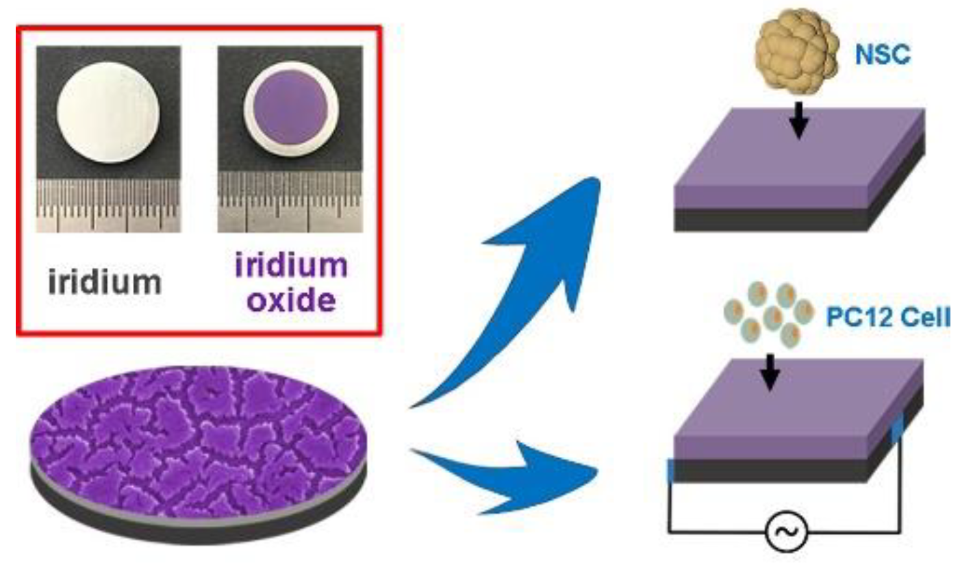
Figure 2. Patterened iridium oxide film shows excellent electrochemical stability, which can be used for neural stem cells and cell culture of primitive nerve cells [48].
Preprints 69680 g002
The ability of Iridium oxide (IrOx) to reversibly convert between Ir3+ and Ir4+ states is one of the critical properties that makes it an attractive material for use in electrochemical applications. The conversion between the Ir3+ and Ir4+ states allows for the storage of charge on the electrode, and this process is known as the "redox reaction." The Ir3+ state has a lower oxidation state, and the Ir4+ state has a higher oxidation state.
During the charging process, electrons are transferred from the electrode to the Ir3+ ions, which then convert to Ir4+ ions. This process increases the number of electrons on the electrode, resulting in a net positive charge and a decrease in the number of Ir3+ ions. This process is reversible, and during the discharging process, electrons are transferred from the Ir4+ ions back to the electrode, resulting in the conversion of Ir4+ ions back to Ir3+ ions. This process decreases the number of electrons on the electrode, resulting in a net negative charge and an increase in the number of Ir3+ ions.
The reversibility of the redox reaction allows for the storage of charge on the electrode, which leads to a high charge storage capacity. In addition, the conversion between Ir3+ and Ir4+ states also leads to a low impedance. Impedance is a measure of the resistance to the flow of electrical current and is often related to the charge-transfer resistance at the electrode/electrolyte interface. The conversion between Ir3+ and Ir4+ states allows for efficient electron transfer at the electrode/electrolyte interface, which leads to a low impedance and high conductivity.
In summary, the ability of IrOx to reversibly convert between Ir3+ and Ir4+ states allows for charge storage on the electrode and low impedance. This process leads to high charge storage capacity and improved conductivity, making IrOx an attractive material for electrochemical applications. Conductive polymers are a class of polymers that have electrical conductivity similar to that of metals. They are achieved by doping the polymer, which is the process of introducing impurities into the polymer to change its electrical properties. Dopants are typically small molecules with electrons that can be easily donated or accepted by the polymer, thus increasing its conductivity. The most common dopants used for conductive polymers are ions of transition metals such as copper, nickel, and gold. The dopants are typically incorporated into the polymer through chemical reactions, and the resulting material exhibits excellent electrical conductivity. However, the conductivity of these materials is not as good as metals, but they are lightweight and flexible and can be used in applications where metals are not suitable [51].
Conductive polymers have been extensively studied for their potential use in biomedical applications, particularly in neural and brain-computer interfaces. One reason is that some conductive polymers are biocompatible, which means that they do not cause adverse reactions when in contact with living tissue. They also have a good affinity towards neurons, meaning they can interact with and stimulate neurons in a specific way [52].
One example of a conductive polymer that has been studied for its biocompatibility and neural affinity is polypyrrole. This polymer is non-toxic to cells and can promote the growth and differentiation of neurons. Additionally, polypyrrole has been used to create neural electrodes successfully implanted in animals, with minimal tissue damage and good electrical recording capabilities [53].
Another example is Polyaniline (PANI), which is a conductive polymer that is biocompatible and has been used in a variety of biomedical applications. It has also been shown to support the growth and survival of neurons and has been used as electrode material in neural implants [54]. These examples demonstrate that conductive polymers can be used in a variety of biomedical applications, particularly in neural and brain-computer interfaces, due to their biocompatibility and neural affinity.
It's important to note that the biocompatibility and neural affinity of conductive polymers depend on the synthesis, chemical structure, and conditions of the final product. Often they are less stable and have inefficient adhesion to metal substrates [55,56]. The research in this field is ongoing to find the best and safe conductive polymers for biomedical applications. There have been reports of novel composite materials made up of conducting polymers and carbon materials exhibiting better stability and electrochemical performance [43].
All these techniques are widely used in the interfacial modification of electrodes to improve the performance of implantable devices, like lowering interfacial impedance, maintaining high SNR, and long-term stability.

III. LONG-TERM RELIABILITY IN CHRONIC IMPLANTATION
Stability is one of the crucial and deciding factors in making or breaking an implantable electrode. Implantable electrodes are expected ideally to function for the user’s lifetime. This issue can be divided into two components. The first is mechanical stability, and the second is biocompatibility in the long run.
The extracellular fluid environment of the body can be toxic for neural implant electrodes due to the presence of various molecules and ions that can damage the electrodes over time. The extracellular fluid comprises multiple ions and molecules, such as chloride, phosphate, potassium, calcium, magnesium, glucose, and enzymes that can corrode or degrade the electrode materials [57]. The issues associated with the mechanical failure of electrodes are cracking, delamination, peeling, and overall degradation [58,59]. Several studies investigated the problems of cracking and delamination and found the design of electrodes, implantation techniques, and mechanical and chemical stress as the factors. It is found that cracking and delamination happen at the electrode microwire’s tip [60].
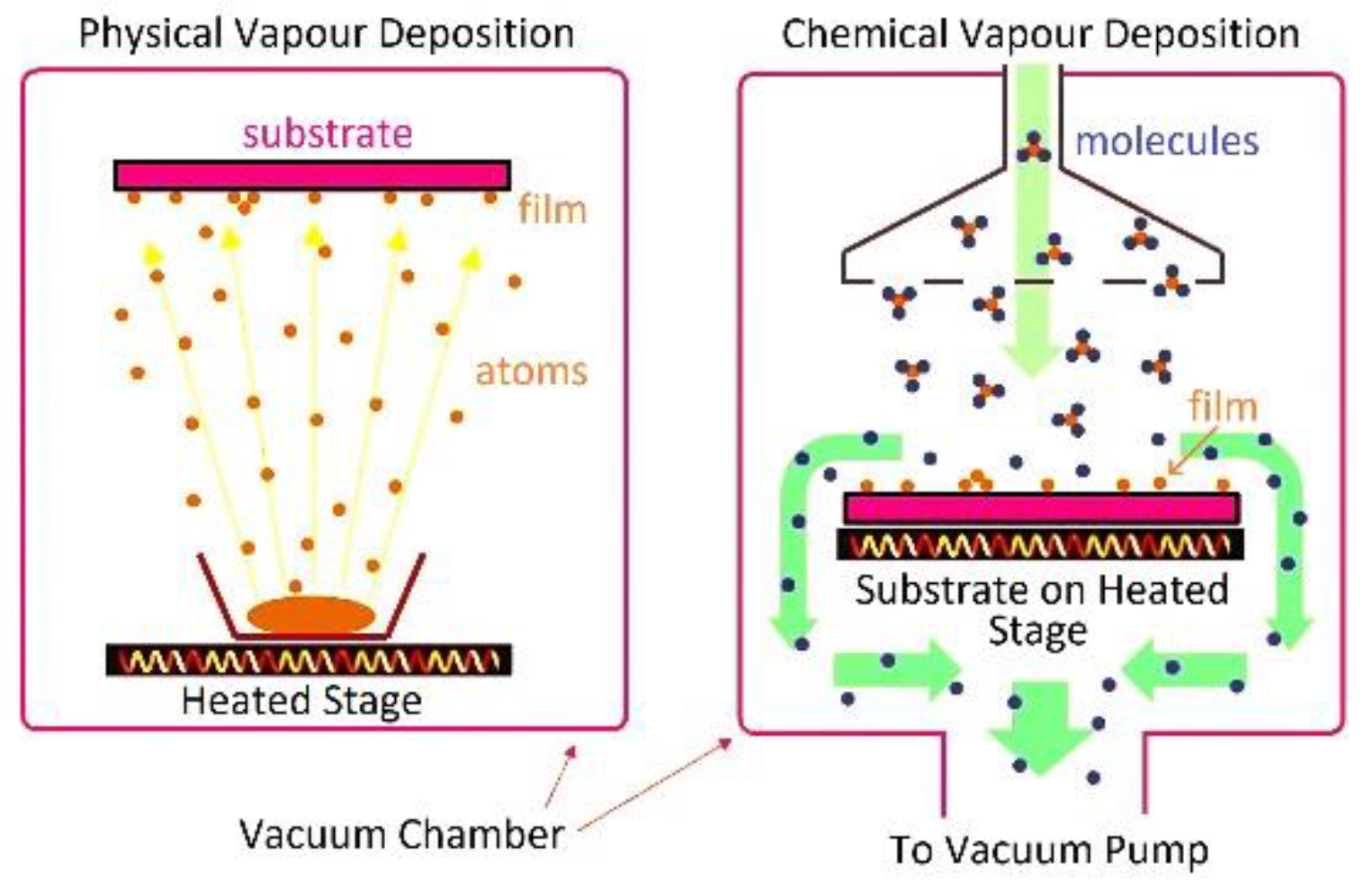
Adhesion-enhancing coating materials, such as titanium (Ti), chromium (Cr), and silane, are frequently used in neural implant electrodes to strengthen the bond between the conductive material and the insulation layer [61,62]. These materials can be applied as a thin layer on either the conductive material or the insulation layer surface to improve the connection between the two. For example, titanium and chromium are metals that can be deposited onto the surface of the conductive material using techniques such as physical vapor deposition (PVD) or chemical vapor deposition (CVD) [63]. These metals can form a strong bond with the insulation layer, which can help to prevent delamination and to crack.
PVD is a process in which a solid material is vaporized and deposited onto a substrate as a thin film. The material to be deposited is typically solid, and the vaporization is typically done through heating or sputtering. PVD is a versatile technique used to deposit a wide range of materials, including metals, alloys, and ceramics [107].
CVD, on the other hand, is a process in which a chemical reaction occurs between a gas and the substrate, resulting in the formation of a thin film. The gases react with the substrate surface to form a thin film. CVD is typically used to deposit many materials, including metals, alloys, ceramics, and semiconductors.
Figure 3. An illustration of differentiating CVD and PVD processes.
Preprints 69680 g003
Both PVD and CVD are vacuum-based techniques, which allow for the growth of high-quality thin films with reasonable control over the thickness and properties of the films. The main difference between the two methods is the way in which the material is deposited on the substrate. PVD is a physical process where the material is deposited on the substrate in the form of a vapor, while CVD is a chemical process where the material is deposited on the substrate through a chemical reaction.
In biomedical engineering, PVD and CVD techniques deposit thin films of biocompatible materials on implantable devices such as neural electrodes, stents, and artificial joints. These films can help to reduce the foreign body response and improve the biocompatibility of the device, which can improve the performance and longevity of the implant [108].
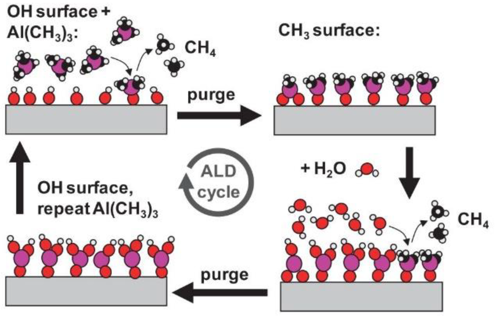
Another process of deposition is called Atomic Layer Deposition (ALD), which is a thin film deposition technique that is often used to coat brain implant electrodes. ALD is a highly controlled and precise method for depositing thin films with precise control over the thickness, composition, and structure of the deposited films. This makes ALD an attractive option for coating brain implants, as it allows for the creation of highly uniform and conformal coatings that can enhance the biocompatibility, stability, and durability of the implant [64].
Figure 4. An illustration of the chemical processes involved in an ALD cycle [68].
Preprints 69680 g004
ALD is also attractive for coating brain implants due to its ability to create ultra-thin films with precisely controlled thickness, which can be essential for maintaining the functionality and performance of the implant. ALD can also be used to deposit biocompatible materials, such as titanium dioxide or hafnium dioxide, which can improve the biocompatibility of the implant and help to reduce the risk of adverse biological reactions.
In the field of biomedical engineering, silanes are used as surface modifying agents for various substrates, such as glass, silicon, metal, and polymers. The silane molecules can be chemically bonded to the substrate surface, forming a siloxane (Si-O-Si) bonding, leading to a hydrophobic surface. This property is helpful for preventing the non-specific adsorption of biomolecules and cells, which is vital in applications such as biosensors and bioelectronics. Silanes are also a coating agent for implantable devices such as neural electrodes. The silane coating can help to reduce the foreign body response and increase the biocompatibility of the device, which can improve the performance and longevity of the implant. Such modification can enhance the bonding between the conductive material and insulation layer, reducing the likelihood of delamination and cracking [109].
It's important to note that the choice of adhesive coating material depends on the specific application, the materials used for the conductive and insulation layer, and the environment the implant will be in. The use of adhesive coating materials can improve the long-term stability of the implantable electrodes, but the research on this topic is ongoing, and new techniques, materials, and designs like thermal annealing [65] and functionalized surface treatment [66] are being used and developed.
Another issue can occur in implantable electrodes when two different metals are in contact with an electrolyte, such as the extracellular fluid in the body. The two metals will have different electrode potentials, and this difference in potential can cause a flow of electrons from one metal to the other, leading to the corrosion of one of the metals. This phenomenon is known as the galvanic cell effect [67].
In the case of implantable electrodes, this effect can occur between the conductive material and the metal of the electrical connector or between the conductive material and the metal of the housing of the implant. The corrosion of the metal can lead to a decrease in the performance of the electrode and can also cause structural damage to the electrode. This issue is avoided by opting for highly corrosion-resistant metal materials like Pt and Ir and going for a metal alloying treatment [59].
Stability is a chief aspect of implantable electrodes, and biocompatibility is one of the critical components of stability. For a device to be considered biocompatible, it is necessary that all materials utilized in its fabrication do not cause harm or toxicity to tissue. Metals such as platinum, iridium, gold, tungsten, and stainless steel are commonly used to construct electrodes due to their favorable properties, including chemical stability, non-toxicity, and high electrical conductivity [69]. Electrode encapsulation is often done using silicon, silicon dioxide, or polymers [70].
Biocompatibility demands that the implantable electrodes trigger a minimal immune response from the surrounding tissue. This immune response is divided into two phases: the initial, short-term response caused by the device's mechanical impact during insertion (acute immune response) and the later, prolonged response due to prolonged exposure to the device (chronic immune response). Factors such as the device's size, insertion speed, tip shape, and surface roughness can impact the acute response, while the chronic response is determined mainly by the tethering mode, brain micro-motion, and mismatching of mechanical properties between the electrodes and brain tissue [71,72,73].
This chronic response can lead to the formation of a glial scar zone [74], a zone of compacted macrophages and astrocytes around the electrode, and can cause degenerative changes in the nerve and prevent neuronal regeneration, resulting in neuronal loss. These mechanisms can affect the quality of long-term recordings and even lead to failure to record. Additionally, chronic blood-brain barrier disruption caused by device implantation may also be a factor in electrode failure [75]. Also, the extracellular environment can trigger an immune response, which can lead to inflammation and the formation of a fibrous capsule around the electrode [76]. This phenomenon is called foreign body reaction (FBR), which is a natural response of the body to a foreign object that has been implanted into the tissue. This immune response is initiated by an unfamiliar object in the tissue, which the body perceives as a threat. The fibrous capsule that forms around the implant helps to isolate it from the surrounding tissue but can also cause mechanical and electrical changes, leading to decreased performance and increased impedance of the implant. Chronic FBR can also result in tissue degeneration and loss, affecting the long-term stability and function of the implant. Encapsulation can lead to a decrease in the electrode's performance and can cause the electrode to move or dislodge from its original position [110].
Additionally, the extracellular environment can change over time, for example, due to changes in the body's pH or glucose levels or the accumulation of enzymes or other molecules. These changes can lead to further damage to the electrode and can decrease its performance over time [111]. It’s a common phenomenon observed by many people worldwide in coffee consumption. When you consume coffee excessively, the caffeine and other compounds in the coffee are absorbed into your bloodstream and distributed throughout your body. As your body metabolizes the coffee, some of these compounds are excreted through your sweat. This can cause your sweat to have a distinct coffee-like smell. The smell is caused by volatile organic compounds, like caffeine, which can be excreted through sweat glands.
Tissue response can severely affect the signal-recording quality of implantable electrodes in multiple ways. Researchers have proposed several solutions to decrease the effect of tissue response on signal recording quality. An approach to implantation involves placing electrodes within a tube containing growth factors that stimulate neuronal growth towards the tube. The recording quality is enhanced by decreasing the distance between the neurons and recording electrodes [77].
An alternative method is to use electrodes constructed from liquid crystal elastomeric material, which allows for recording sites beyond the glial scar area [112]. Direct penetration of the dura mater with electrodes can also decrease damage caused by the removal of the dura mater.
A common approach to minimize the chronic immune response is to make electrodes softer and more flexible. This helps reduce the mismatch in mechanical properties between the electrode and brain tissue and improves the longevity and signal quality of the electrode.
Additionally, the surface modification of the electrode with conductive polymers or metal oxides can reduce the tissue response and improve the signal recording quality. These modifications increase the electrochemical activity and larger surface area of the electrode [113].
Softness and hardness are related to the resistance of a material to being pressed or indented. Soft materials are less resistant to indentation and are considered "softer," while harder materials are more resistant and considered "harder.”
Flexibility and stiffness are related to the resistance of a material to elastic deformation. Flexible materials are less resistant to deformation and are considered "more flexible," while stiffer materials are more resistant and considered "stiffer.”
Compliance pertains to a material's flexibility, which refers to how easily an elastomer bends when subjected to an external force. A material with high compliance is more flexible and is considered "more compliant," while a material with low compliance is less flexible and considered "less compliant."
In implantable electrodes, having high softness and compliance is desirable because it lessens the mismatch between the electrode and brain tissue and reduces damage caused by the electrode. However, it's essential to remember that materials with a high Young's modulus, such as carbon fibers [78] and carbon nanotubes [79], can also be considered compliant as they bend easily under external force.
K = E × w h 3 12
Where E is Young’s modulus, w is the electrode’s width, and h is the electrode’s thickness.
A more compliant electrode does not necessarily mean a more flexible one. Compliance of an electrode can be increased by reducing its cross-sectional size. However, this has little effect on the radial force [80]. Similarly, materials with low stiffness can be flexible, but they are also usually soft, which has led to overlooking the effect of material hardness on tissue response. Some studies propose that using low-density materials can mitigate the inflammatory response by reducing the impact of inertia during the micromotion of the brain [81]. This indicates that the mismatch between electrodes and tissues should consider multiple material characteristics, not just material rigidity.
Improving the compliance of implantable electrodes can enhance their aptness by minimizing the force between the electrode and the surrounding tissue. This can be achieved by using soft tethering, a method in which the electrode is connected to a compliant substrate that flexes with the movement of the surrounding tissue, thereby reducing the forces on the tissue and minimizing tissue damage and the resulting immune response.
The technique of using flexible ribbon cables, known as soft tethering, connects rigid electrodes in the brain to a rigid head stage on the skull. This helps protect the embedded electrodes without disrupting them and lets them move with the brain during micromotion, reducing tissue-electrode interaction and enhancing long-term stability. Additionally, soft tethering improves the encapsulation of the electrodes, further increasing stability and minimizing tissue damage.
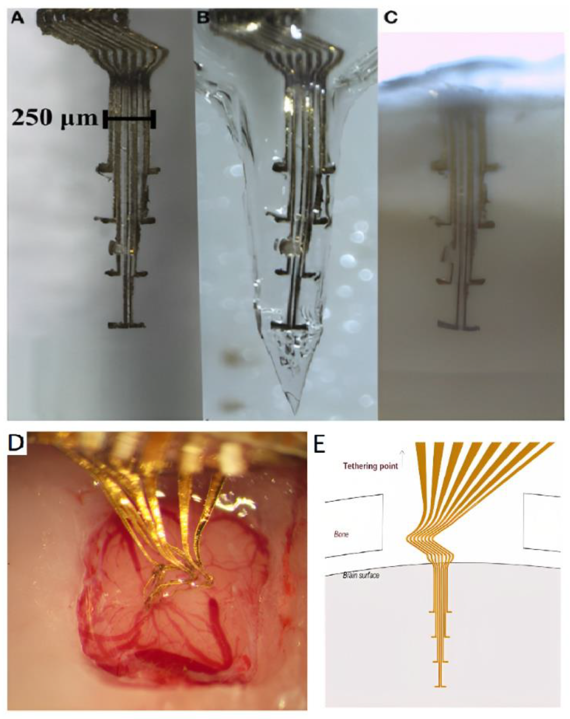
Figure X. (A) An electrode array prior to embedding into a gelatin-based matrix. (B) The same electrode array after being embedded into a gelatin matrix-shaped like a needle. (C) Same electrode array inside a section of clarified brain tissue three weeks post-implantation. (D) Photograph was taken 3.5 h after implantation of the gelatin-embedded electrode, showing that the gelatin has dissolved and the brain surface contracted around the electrode array. (E) A schematic of the 3D flexible electrode array [137].
Preprints 69680 g005
Reducing the footprint of an electrode refers to making it smaller in size. This can be done using nanomaterials such as carbon nanotubes and carbon fiber electrodes. Doing this has many benefits, such as reducing the bending stiffness of the material and reducing the amount of damage that is caused during insertion. It has been shown that reducing the electrode footprint to less than 10 micrometers can reduce macrophage aggregation, reducing the acute immune response. Therefore, using nanomaterials to reduce the footprint of electrodes can lead to a more compliant and biocompatible device.
Various methods can be employed to increase the pliability and flexibility of electrodes utilized in medical implants, with the goal of minimizing the inflammatory response and promoting neuronal growth. One method is to apply a soft, biocompatible coating to the surface of the electrode. Another approach is to use a polymer material with a lower Young's modulus as the substrate for the electrode. Many flexible polymer materials have been used to create these flexible electrodes, such as parylene, polyimide, polydimethylsiloxane (PDMS), SU-8, and graphene. Additionally, there are ultra-small electrodes that have been developed, which leads to enhanced compliance and a minimal chronic immune response with prolonged implantation [83,84,85,86].
Apart from these materials, different materials include Polyethyleneimine (PEI), which is a cationic polymer that has been investigated for its potential use in neural implants. PEI is attractive for use in these applications due to its biocompatibility and low toxicity, which are important factors for ensuring the safety and long-term performance of implantable devices in the body. In addition, PEI can be modified to improve its mechanical properties and drug delivery capabilities, making it a versatile material for neural implants. PEI has been used as a coating material for implantable electrodes to improve their biocompatibility and reduce toxicity. By modifying the surface of the electrodes with PEI, researchers aim to reduce the immune response and enhance the stability of the implant over time [114].
There are challenges in using compliant electrodes, which are flexible and soft, in brain implant devices. One issue is that these electrodes have low buckling strength, making them difficult to insert into the brain without bending. Buckling strength is a measure of the amount of load an object can withstand before collapsing and is related to the material properties of the object, such as its Young's modulus, the moment of inertia, and length. To enhance the buckling strength, researchers have tried to increase the cross-sectional area and shorten the length of the electrodes, but this can come at the cost of sacrificing other performance factors [82].
Many insertion methods have been developed for compliant electrodes to address this problem. These methods can be broadly divided into two categories: reinforcement by a temporary material and insertion with an auxiliary tool. Temporary reinforcement materials can be used to enhance the buckling strength of the electrodes but are eventually degraded in the body. These materials include gelatin, maltose, dextran [82], polyethylene glycol, polyglycolic acid, silk fibroin, and other similar polymers. These materials can be temporarily combined with electrodes by dip-coating or molding; however, these methods have limitations such as poor accuracy, consistency, and the need for additional processing [82,87,88,89].
Silk fibroin is a biopolymer protein that is naturally extracted from the silkworm, specifically from the cocoon of the silkworm. It is a strong, tough, and biocompatible material that is used in a variety of biomedical applications, including tissue engineering, drug delivery, and wound healing [90,91].
Silk fibroin has several unique properties that make it attractive for biomedical applications. It is biocompatible, meaning it is well-tolerated by the body and does not trigger an immune response. It is also biodegradable, meaning it can be naturally broken down by the body over time. Additionally, silk fibroin is strong, flexible, and hydrophobic, which makes it useful for applications that require a material that can maintain its structural integrity in the presence of bodily fluids.
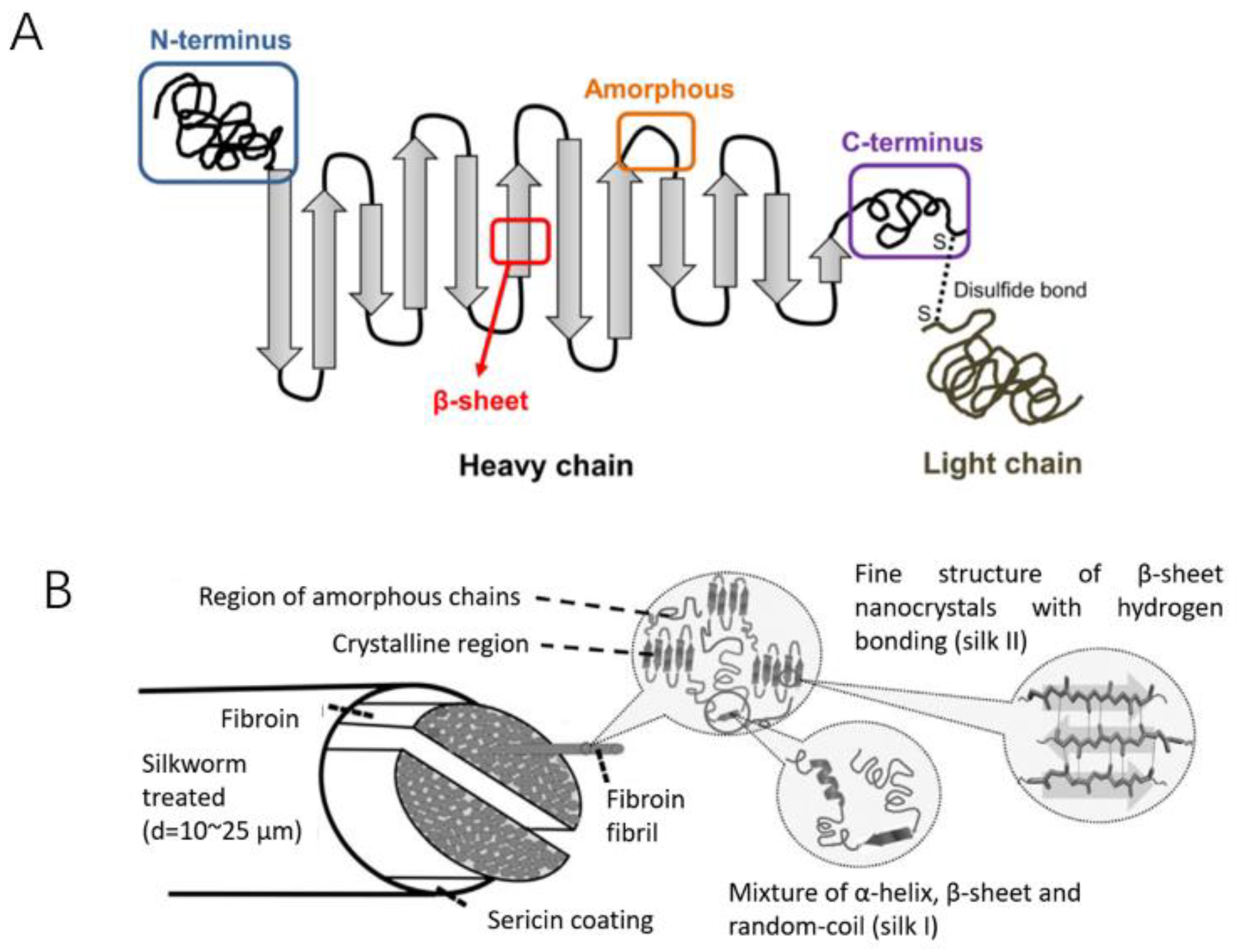
Figure Y. Schematic diagram of the silk structure. (A) heavy chain (i.e., N-terminus, β-sheets, Amorphous, and C-terminus) and a light chain that is linked via disulfide bonds. (B) silkworm thread, overall fibril structure, and silk fibroin polypeptide chains [144].
Preprints 69680 g006
Overall, silk fibroin is an attractive material for biomedical applications because it combines strength, biocompatibility, and biodegradability. The ability to precisely control its processing and properties, as well as its versatility, make it a promising material for the development of new and improved biomedical products [92].
Recently, researchers have integrated the fabrication of these reinforcing materials into the process flow, which allows for batch production, but Young's moduli of these materials are generally very low, which still has a negative impact on the footprint of the electrodes.
The insertion of complaint electrodes into the brain is done by using a rigid auxiliary tool. The method involves attaching the compliant electrode to a rigid tool, such as a silicon probe or a tungsten microneedle, and using the tool to insert the electrode into the brain. Once the electrode is in place, the auxiliary tool is withdrawn, leaving the compliant electrode in place [93].
There are different ways to attach the compliant electrode to the auxiliary tool, such as electrostatic adsorption, water-soluble adhesive bonding, and syringe wrapping. Other methods include mutually matched mechanical structures, such as needle-hole and pulling methods. Certain structures that are specifically engineered can diminish the amount of force needed for insertion and penetrate the dura mater directly to implant the pliant electrode [94,95,96].
Using an auxiliary tool in implantation may have drawbacks compared to using flexible materials. The use of rigid auxiliary tools results in a larger overall implant size than using only a compliant electrode, although still smaller than a reinforced electrode. There's also a risk that the tool could affect the placement of electrodes or even remove them during removal, and the tool's removal may cause additional tissue damage.
There are different methods of inserting compliant electrodes into the brain with minimal damage. One ideal approach would be to use implantation techniques that do not require auxiliary tools or materials. Some researchers have used biological enzymes to soften the cerebrum, which reduces the force required for insertion [97]. However, this method is still experimental and not commonly used. The use of enzymes is thought to be a way to soften the brain tissue to allow for the implantation of electrodes or other devices with less force, which may reduce the risk of tissue damage. However, this approach also carries risks, such as the potential for unintended effects on brain function, and it requires careful control of the enzyme concentration and timing of application to avoid adverse effects. This method is still in the research phase, and it has not been proven to be safe and effective yet.
Certain researchers have employed adaptive materials to create electrodes that possess sufficient strength to penetrate brain tissue prior to implantation, but are capable of softening to match the Young's modulus of brain tissue after being implanted. These materials include smart polymers, nanocomposites, shape-memory polymers, and liquid crystal polymers. However, maintaining their stability during processing can be challenging, and these materials often have limited flexibility [98,99,100].
Another strategy is using electrodes incorporating microfluidic channels. These electrodes can change their stiffness by altering the fluid pressure inside the channels, which allows them to possess properties similar to adaptive materials. Non-contact implantation techniques, such as magnetic and fluidic actuation, can also minimize the damage caused during the implantation of compliant electrodes, though they may have limitations regarding the actuation force and other factors [101].
Stereotaxic surgery is a surgical technique that uses precise coordinates to locate and manipulate specific structures within the brain. It is a highly specialized field that combines neuroscience, neuroanatomy, and surgical precision to perform precise and controlled interventions within the brain.
The technique is based on a three-dimensional coordinate system called the stereotaxic frame, which is used to locate the target area within the brain precisely. This allows for the precise placement of electrodes, cannulas, or other instruments within the brain with millimeter accuracy.
Stereotaxic surgery is commonly used in the field of neuroscience for a variety of purposes, including
  • Deep brain stimulation (DBS) for the treatment of movement disorders such as Parkinson's disease,
  • Lesioning procedures for the treatment of psychiatric and neurological disorders,
  • Placement of electrodes for recording neural activity
  • Delivery of therapeutic agents such as gene therapy vectors or neurotrophins.
Performing stereotaxic surgery necessitates a significant level of proficiency and specific equipment and is generally carried out by specialized neurosurgeons in a dedicated operating room [115].

IV. ACCURATE BRAIN SIGNAL DECODING THROUGH HIGHSPEED, HIGH-DENSITY RECORDING

The multi-electrode recording method is used to observe the electrical activity of several neurons in various brain areas simultaneously. This enhances knowledge of the relationships between multiple neurons, leading to a better comprehension of neural activity and nervous system functions. Improved spatial resolution in recording facilitates the accurate identification of individual neurons from various locations in a large group of neurons [102,103]. Therefore, high-density electrodes with a high data output capacity are crucial in large-scale neural recordings.
For traditional metal and silicon electrodes and newer flexible electrodes, it's important to increase the number of electrodes used for recording and their density to monitor the activity of more neurons in multiple brain regions. This helps to understand the interactions between many neurons, providing a better understanding of neural activity and the complex functions of the nervous system. Flexible electrodes can cover a larger area of the brain because they are not limited by shank spacing and can be implanted more easily due to their flexibility.
Research states the advantages of silicon-based microelectrodes over metal-wire electrodes in the neural recording. It mentions that over the past few decades, there has been a rapid increase in the number of neurons that can be recorded simultaneously by microelectrodes, doubling approximately every seven years [102]. This increase is attributed to the development of silicon-based microelectrodes, which have a well-established advantage over metal-wire electrodes in increasing channels and consistency.
The research also highlights that metal-wire electrodes are typically fabricated by hand bundling. Hand bundling is a process used to fabricate metal microwire electrodes. It is a manual process in which individual metal wires are bundled together by hand to form a single electrode. The wires are typically made of platinum, tungsten, or stainless steel. The wires are carefully aligned and bundled together using tools such as tweezers, clamps, or heat-shrink tubing, to form a bundle of wires with a consistent diameter [116].
Hand bundling is a relatively simple and inexpensive method of fabricating metal microwire electrodes, but it has certain limitations. The primary constraint is the manual nature of the process, which poses challenges in achieving precise consistency and high-density integration. The process depends on the skill of the person doing the bundling, which can lead to variations in the properties of the resulting electrodes. Moreover, the lack of scalability in the process presents difficulties in manufacturing a large number of electrodes with uniform characteristics, thereby hindering the achievement of precise consistency and high-density integration. In contrast, silicon electrodes are fabricated using MEMS (Micro-Electro-Mechanical Systems) processes, such as Utah arrays and Michigan probes. Silicon microelectrodes are made in bulk and can be arranged precisely within a small area. Earlier rigid microelectrodes, in contrast, usually had no more than 100 channels.
This information highlights the advantages of silicon-based microelectrodes in neural recording, particularly in increasing the number of neurons that can be recorded simultaneously and achieving precise consistency and high-density integration. The use of silicon-based microelectrodes has led to significant advances in the field of neural recording and is an important research area in neuroscience and biomedical engineering.
The fabrication of traditional metal and silicon electrodes has advanced, leading to substantial improvements in recording capacity. For example, a microwire electrode bundle was found to create a 65,536-channel recording system in one study, while Neuropixel electrodes integrated 5120 recording sites on a single four-shank probe. On the other hand, compliant electrodes made of carbon fibers or nanotubes currently have only a limited number of recording channels, typically tens of channels. However, flexible electrodes made from polymer materials are compatible with MEMS processes and have already surpassed 1000 recording channels.
The field of neural recording requires high-density and high-throughput electrodes for capturing large-scale neural activity. However, there is no standard definition for these terms. The goal of the Brain Activity Map Project is to record the activity of every single neuron, which would require a significant number of electrodes [117,118]. A study by Adam et al. estimated the number of electrodes needed to record all neurons in a rat brain. The study found that to achieve this goal, at least 750,000 electrodes would be needed with a spacing of around 80 micrometers (μm) if each electrode can sort out 100 individual neurons. On the other hand, if each electrode can sort out only ten neurons, the number of electrodes would increase to 7,500,000 and need to be arranged with a spacing of 40 μm [119].
The previously mentioned calculations regarding the required number of electrodes for recording all neurons in a rat brain is only a theoretical estimate. In actuality, researchers may focus on specific regions of the brain rather than the entire brain. As a result, it is more practical to define "high throughput" and "high density" in a local area around the electrode. It is commonly understood that an electrode's recording range is around 100 μm. Based on this, the region within 100 μm from the implanted electrode can be considered the recordable area. High density would mean that the distance between electrode sites is no greater than 100 μm, and high throughput would mean the ability to record all the neurons in that region.
To define "high density" and "high throughput" in a practical context, the area around an electrode where it can record electrical activity can be considered the recordable region. This is generally estimated to be around 100 micrometers from the electrode. So, if the spacing between electrode sites is less than 100 micrometers, the electrodes can be considered as having high density. High throughput, in this context, would be the ability of the electrodes to record all neurons within the recordable region. In the case of a rat brain, which has a density of around 90,000 neurons per cubic millimeter, each electrode would need to record about 90 neurons. Though it may be challenging to sort such a high number of neurons, reducing the spacing between electrodes can make this task easier [119].
Developments in silicon electrodes have allowed for a reduction in electrode spacing down to 20 μm. This has been made possible through various fabrication techniques to create 3D electrode arrays. These arrays can cover the entire implantation region, making it simpler to attain high-density and high-throughput recordings compared to Utah arrays and metal-wire electrode arrays, which only have electrode sites at the front of the implantation area. Decreasing electrode size allows for a reduction in shank spacing, although it is limited by the volume substitution ratio. This reduction in shank spacing leads to an increase in electrode distribution density, which lowers the sorting requirement for the electrodes to a feasible level.
High-throughput electrodes face the challenge of growing in size while maintaining a desirable range. To overcome this challenge, using MEMS or CMOS processes is preferred to keep the size of these electrodes within acceptable limits. CMOS (Complementary Metal-Oxide-Semiconductor) processes are manufacturing technology used to produce integrated circuits, including microelectrodes for electrophysiological recordings. The CMOS process enables high-density fan-out of electrodes by integrating amplification circuits directly into the electrodes, avoiding the issue of size enlargement between the electrodes and external interconnections. However, CMOS processes are not yet compatible with flexible electrode fabrication, a standard method for packaging high-throughput electrodes. The reduction of the interconnect line widths can be achieved through the use of electron-beam lithography, which has made the widths no larger than 100 nanometers. Electron beam lithography (EBL) is a technology for creating nanoscale patterns and structures by directing a beam of electrons onto a substrate coated with a resist material. The electrons cause the resistance to change its chemical or physical properties, making it more or less sensitive to further processing. The exposed areas of the resist can then be developed or etched away, leaving a pattern on the substrate that can be used as a mask for further processing. EBL is often used to produce high-resolution patterns and structures in microelectronics, nanotechnology, and other fields that require precise control over the size and shape of structures at the nanoscale.
Although challenges exist in decreasing the size of recording sites because of impedance and thermal noise, the recording region's footprint can be constrained by modifying the recording site configuration, such as utilizing multi-shank electrodes or a multilayer wiring technique.
Multi-shank electrodes are a type of neural recording electrode that consists of multiple shanks, or thin, elongated structures, that are bundled together. Each shank contains multiple electrodes that can record electrical activity from the brain or other neural tissue. The multiple shanks are typically bundled together to increase the number of electrodes implanted in the brain while also reducing the overall size of the implant [120].
The process of connecting each recording site on high-throughput electrodes to an amplifier chip or interface poses a challenge called fan-out. Standard commercial chips or interfaces, such as Intan and Omnetics, are used, but the arrangement of the pads must match these devices. This limits the pad size and arrangement, causing enlargement of the interconnects and reducing the recording region size, with the pad-to-site area ratio being around ten or higher.
Michigan probes have a solution to the fan-out challenge in high-throughput electrodes by integrating amplification circuits with recording electrodes using CMOS technology. This enables high-density fan-out. However, this method is not feasible for flexible electrodes, which require a different fabrication process. A common packaging technique for flexible electrodes is to group thousands of channels into threads with a limited number of channels, package each thread with a commercial chip or interface in the plane, and then stack them in another dimension. This only balances the increase in package size and does not change the enlargement of the back-end packaging. When recording throughput increases further, this stacking method becomes impractical.
The drawback of the stacking method used in flexible electrode packaging is low fan-out density, where the area of effective interconnection between electrodes and external circuits takes up too little space in the back-end package. This is due to the design of PCBs, which are made to match commercially available chips/interfaces and therefore have restricted pad arrangements. Most of the board is occupied by wires and other components, leaving only a tiny area for interconnection. Moreover, non-uniform board thickness and the necessity for heat dissipation lead to gaps between modules when stacked, which contributes to an overall larger package size.

V. MULTIMODAL RECORDING AND STIMULATION FOR VERSATILE APPLICATIONS AND ENHANCING IMPLANTABLE MICROELECTRODES THROUGH INTEGRATION WITH OTHER TECHNOLOGIES

 Some key areas of integration include 

Wireless power and data transmission: Implantable microelectrodes typically require some sort of power source, either in the form of a battery or a connection to an external power source. There is a need to develop more efficient and reliable methods for powering these devices, and wireless power transmission is one area of research that holds promise in this regard. Wireless data transmission is also an essential area of research, as it can significantly improve the speed and reliability of data transfer from the implanted electrodes to external devices [121,122,123].
Integration with other neural interfaces: Implantable microelectrodes can be combined with other types of neural interfaces, such as optogenetics, to achieve a more comprehensive understanding of the brain and its functions. For example, implantable microelectrodes can be used to record electrical signals from neurons, while optogenetics can be used to control the activity of neurons using light [124].
Integration with computer systems and robotics: Implantable microelectrodes can be integrated with computer systems and robotics to create closed-loop systems that can interact with the brain in real-time. Implantable electrodes can be employed to capture brain signals and transmit them to a computer for subsequent processing and communication of commands to devices such as robotic arms [125].
Integration with wearable devices: Implantable microelectrodes can be integrated with wearable devices to create closed-loop systems that can be used for rehabilitation and other therapeutic applications. For example, implantable electrodes can be used to record signals from the brain and send them to a wearable device, which can then use this information to provide feedback and support to the user [126].
Apart from electrophysiological recording, various sensor types have been created to monitor brain activity signals, such as neurotransmitter-detecting chemical sensors [129], thermometers to monitor the physiological state of the brain tissue around the electrodes [130], and optical sensors to capture calcium fluorescence signals [131,132]. These sensors can be added to microelectrodes as additional tools for electrophysiological recording. For instance, calcium imaging can be used to record the activity of thousands of neurons simultaneously with high spatial accuracy.
Calcium imaging is a technique used to visualize and measure the activity of neurons in the brain. It is based on the fact that when a neuron is active, it takes calcium ions from the extracellular space. This influx of calcium ions causes a slight increase in the level of fluorescence in the neuron, which can be detected using a microscope.
There are two main types of calcium imaging: intracellular and extracellular. Intracellular calcium imaging involves injecting a calcium-sensitive dye into the neuron, which binds to the calcium ions and changes its fluorescence. Extracellular calcium imaging, on the other hand, uses a dye that binds to the extracellular space and changes its fluorescence in response to changes in the level of calcium ions.
Calcium imaging is a powerful tool for studying neural activity in the brain, as it allows researchers to visualize the activity of thousands of neurons at once [127,128]. It is commonly used in studies of neural development, synaptic plasticity, and neural coding. Calcium imaging, when integrated into microelectrodes, has been used to explore the connection between neural activity and behavior and to examine the function of various neurotransmitters and neuromodulators in the brain. However, it still has several obstacles, such as poor temporal resolution, limited accessibility to certain brain regions, and larger implantation damage.
Anessentialortant aspect of microelectrodes is their ability to stimulate neurons and record signals. Electrical stimulation [133], pharmacological stimulation [134,135], and optogenetic stimulation [136] are commonly used as neuromodulation techniques. Due to its precision and ability to be reversed, optogenetics has gained significant traction as a neuromodulation tool in recent years. Channelrhodopsin-2, known as a typical protein, can introduce sodium ions into cells during neural activity when applying blue light and can artificially induce the activity of target neurons. Recent progress in micro-LED light sources has enabled the development of tiny stimulation devices that can be implanted into the brain in different configurations. However, the in vivo implantation of these light sources still faces challenges related to waterproofing, heat management, and interference from other photoelectric signals and power source.
For example, the waterproof packaging may be unable to keep the device dry and free from electrical short circuits. In addition, the heat dissipation of the device needs to be considered in order to prevent it from damaging the surrounding tissue. Photoelectric crosstalk, which is the leakage of light from one device to another, also needs to be taken into account. Finally, the power supply needs to provide enough power to the implantable device without draining too much power from the organism [138,139].
Photoelectric crosstalk refers to the phenomenon where light used to activate one group of cells in a sample also activates neighboring cells that were not intended to be targeted. This can occur due to the diffraction of light, which causes it to spread out and reach cells that are not directly in the path of the light beam. The crosstalk can also occur due to the absorption of light by pigments or other molecules in the sample, which can re-emit the light in a different direction. This can result in a decrease in the specificity of the optogenetic manipulation, making it more difficult to control the activity of specific cells.
To reduce photoelectric crosstalk, scientists use techniques such as confocal microscopy, which uses a laser beam to focus light on a specific point in the sample and use optical filters to block the spread of light to the surrounding area. Additionally, using smaller and more precise light sources, or using optogenetic tools that are more sensitive to specific wavelengths of light, can also help to minimize crosstalk.
A functional system design is necessary to use microelectrodes in human brain implantation. Some implantable microelectrodes utilize wireless transmission, wireless power supply, and heat dissipation designs, which make them adaptable to different environments and more user-friendly [140,141].

VI. STANDARDIZING THE MANUFACTURING PROCESS FOR IMPROVED IMPLANTABLE MICROELECTRODES QUALITY AND RELIABILITY

There have been some established standards for implantable microelectrodes in the brain. These standards are meant to ensure the quality and reliability of the devices, as well as to ensure the safety of the subjects being implanted with these devices.
For example, the International Electrotechnical Commission (IEC) has established standards for implantable medical electrical devices, including implantable microelectrodes. The International Electrotechnical Commission (IEC) is a global organization that sets standards for a wide range of electrical and electronic products, including medical electrical devices. The IEC has established standards for implantable medical electrical devices, including implantable microelectrodes.
These standards cover various aspects of implantable microelectrodes, including their electrical and mechanical characteristics, biocompatibility, and performance. Some of the key standards established by the IEC include:
Electrical safety: The IEC sets standards for the electrical safety of implantable microelectrodes to ensure that the devices do not pose a risk of electrical shock or fire to the patient.
Biocompatibility: The IEC sets standards for the biocompatibility of implantable microelectrodes to ensure that the devices do not cause adverse reactions or interfere with the normal functioning of the body.
Performance: The IEC sets standards for the performance of implantable microelectrodes to ensure that the devices meet certain minimum requirements for accuracy, reliability, and stability.
Manufacturing processes: The IEC sets standards for the manufacturing processes used to produce implantable microelectrodes to ensure that the devices are produced to consistent standards of quality and reliability.
The IEC standards provide a framework for researchers and manufacturers to develop and use implantable microelectrodes in a safe, ethical, and effective manner. These standards help to ensure that implantable microelectrodes are manufactured to consistent standards of quality and reliability and that they are safe and effective for use in human subjects. These standards cover various aspects of the devices, such as their electrical and mechanical characteristics, biocompatibility, and performance.
In addition, the National Institute of Neurological Disorders and Stroke (NINDS) has established guidelines for the ethical and safe use of implantable microelectrodes in human subjects, which include requirements for informed consent, device safety and reliability, and data protection.
Some of the critical guidelines established by the NINDS include the following:
Informed consent: The NINDS requires that all human subjects receive comprehensive information about the risks and benefits of implantable microelectrodes before undergoing the procedure and that they provide written informed consent.
Device safety and reliability: The NINDS requires that implantable microelectrodes be manufactured to high standards of quality and reliability and that they undergo rigorous testing and quality control procedures.
Data protection: The NINDS requires that all data collected from implantable microelectrodes be treated with confidentiality and that appropriate security measures be taken to protect the privacy of the subjects.
Ethical considerations: The NINDS requires that all studies involving implantable microelectrodes be approved by an Institutional Review Board (IRB) and that they adhere to the principles of the Declaration of Helsinki and the Common Rule.
Monitoring and follow-up: The NINDS requires that all subjects be monitored for adverse events and that appropriate medical care and support be provided as needed.
These guidelines serve as a framework for researchers and manufacturers to develop and use implantable microelectrodes in a safe, ethical, and effective manner. The NINDS guidelines help to ensure that implantable microelectrodes are used in a manner that protects the rights and welfare of human subjects and that the results of these studies are reliable and valid.
Apart from these, there is also the International Organization for Standardization (ISO) - The ISO is another global organization that develops and publishes international standards for a wide range of products and technologies, including implantable neural electrodes. The ISO has developed standards for the performance and safety of these devices, which help to ensure that they are manufactured and used in a manner that is consistent and reliable.
Food and Drug Administration (FDA) - The FDA is the US federal agency responsible for protecting public health by regulating the safety and efficacy of medical devices, including implantable neural electrodes. The FDA provides guidelines and regulations for the development and use of these devices, which help to ensure that they are manufactured and used in a manner that is safe and effective.
European Medical Device Regulation (MDR) - The MDR is a set of regulations developed by the European Union (EU) to ensure the safety and performance of medical devices, including implantable neural electrodes. The MDR provides guidelines and standards for the development and use of these devices, which help to ensure that they are manufactured and used in a manner that is safe and effective for patients in the EU.
Overall, these standards and guidelines serve as a framework for researchers and manufacturers to develop and use implantable microelectrodes in a safe, ethical, and effective manner. However, it is important to note that the field is constantly evolving, and standards and guidelines may change as new technologies and methods are developed.

VII. CURRENTLY AVAILABLE SOLUTIONS

1.
NeuroPace RNS System
The NeuroPace RNS system is a type of implantable neural stimulation device developed by the company NeuroPace. It is a responsive neurostimulation (RNS) system designed to treat patients with epilepsy who have not responded to other treatments. The RNS system is designed to detect seizures as they are beginning to occur and then deliver a small electrical stimulus to the brain to stop the seizure from progressing.
The RNS system consists of a small, implantable device (about the size of a pocket watch) that is placed under the skin of the skull and two thin wires (electrodes) that are surgically placed on the surface of the brain. These electrodes detect the abnormal electrical activity that occurs during a seizure and sends this information to the implantable device. The device then delivers a small electrical stimulus (a current-controlled and a charge-balanced biphasic electrical stimulation) to the brain to stop the seizure from progressing. The adjustability for frequency ranges from 1 to 333hz, and the intensity of current ranges from 1mA to 12mA [142]. It is considered a last resort treatment for patients who have not responded to other treatments and was FDA-approved in 2013. In 2017, NeuroPace marked its 1000th implant.
2.
NeuroNexus
NeuroNexus Technologies is a medical device company that designs, manufactures, and sells advanced neurosensory and neurostimulation devices. The company was founded in 2000 and is based in Ann Arbor, Michigan, USA. NeuroNexus has many implantable and non-implantable neurotechnology products, including microelectrode arrays, flexible probes, cuff electrodes, wireless stimulators, and recording systems.
The company's products are used in various applications, including basic neuroscience research, drug development, and treating neurological disorders such as Parkinson's, epilepsy, and chronic pain. NeuroNexus also provides custom design and manufacturing services for researchers and clinicians working in the field of neural engineering.
One of the main focuses of the company is the development of flexible microelectrode arrays, which are thin, flexible probes that can be inserted into the brain or spinal cord to record neural activity or deliver electrical stimulation. These microelectrodes are made of thin film materials and are designed to conform to the contours of the brain, which helps to minimize the risk of tissue damage and improve the safety and efficacy of the stimulation. They offer up to 256 channel ECoG electrodes array [143].
3.
Imec
Imec is a research and development company based in Belgium specializing in nanoelectronics and digital technologies. They have a research program dedicated to developing advanced neural interfaces, which is focused on creating devices that can record and stimulate the activity of individual neurons in the brain with high spatial and temporal resolution. These devices are intended for use in both research and clinical applications, such as the treatment of neurological disorders and the restoration of sensory and motor function in people with disabilities. They are working with companies like Medtronic, Boston Scientific, and St. Jude Medical to develop implantable devices for applications like deep brain stimulation, spinal cord stimulation, and retinal prostheses.
4.
Neuropixels
Neuropixels electrodes are a type of neural recording electrode designed to record the activity of large numbers of neurons simultaneously. They are developed by the Neuropixels consortium, a collaboration between researchers at the Howard Hughes Medical Institute (HHMI), University College London (UCL), and the University of Cambridge.
Neuropixels electrodes are made up of many individual recording sites, or "pixels," that are spaced closely on a flexible probe. Each pixel contains an electrode that can record the electrical activity of a single neuron. The pixels are arranged in a linear or 2D array on the probe, allowing the electrodes to be inserted into different regions of the brain or spinal cord.
The Neuropixels electrodes are designed to have several key features that make them well-suited for large-scale neural recording. They have a high density of recording sites, which allows them to record the activity of many neurons at once. They are also designed to have low noise levels, so they can record the small electrical signals produced by neurons with high accuracy. Additionally, they are flexible and can be inserted through small openings in the skull, which allows them to record from deep brain regions with minimal invasiveness.
Neuropixels electrodes are widely used in research studies to study brain function and neural circuits. They are particularly useful for studying the activity of large populations of neurons in different regions of the brain and for understanding how these neurons interact with each other to generate behavior.
In summary, Neuropixels electrodes are a type of neural recording electrode designed to record the activity of large numbers of neurons simultaneously. They are developed by the Neuropixels consortium, a collaboration between researchers at the Howard Hughes Medical Institute (HHMI), University College London (UCL), and the University of Cambridge. They have a high density of recording sites, which allows them to record the activity of many neurons at once. They are also designed to have low noise levels, so they can record the small electrical signals produced by neurons with high accuracy. Additionally, they are flexible and can be inserted through small openings in the skull, which allows them to record from deep brain regions with minimal invasiveness. They are particularly useful for studying the activity of large populations of neurons in different regions of the brain and for understanding how these neurons interact with each other to generate behavior.
5.
Paradromics
Paradromics is a neurotechnology company that develops implantable devices for recording and stimulating neural activity in the brain. The company was founded in 2014 by a group of researchers and engineers from Stanford University and is based in California, USA.
One of the main focuses of Paradromics is the development of a high-bandwidth neural interface, which is a device that can be implanted in the brain to record and stimulate neural activity with high precision. The company's goal is to create a device that can record the activity of thousands of neurons simultaneously and stimulate these neurons with high temporal and spatial precision.
The company's technology is based on the use of silicon-based electrodes that can be inserted into the brain. These electrodes are designed to be highly flexible and can be bent and folded to conform to the complex geometry of the brain. They are also designed to have a high density of recording sites, which allows them to record the activity of many neurons at once.
Paradromics is also working on developing a high-bandwidth wireless link that can be used to transmit data from the implantable device to an external device. This wireless link will allow researchers to record and stimulate neural activity in real-time and to control the device remotely.
Another focus of Paradromics is to make the devices more biocompatible. This way, the implantation of these devices in the human brain would be less invasive and have fewer side effects.
In summary, Paradromics is a neurotechnology company that develops implantable devices for recording and stimulating neural activity in the brain. The company's main focus is the development of a high-bandwidth neural interface, which is a device that can be implanted in the brain to record and stimulate neural activity with high precision. The company's goal is to create a device that can record the activity of thousands of neurons simultaneously and stimulate these neurons with high temporal and spatial precision. They use silicon-based electrodes and a high-bandwidth wireless link and are also focused on developing more biocompatible devices; this way, the implantation of these devices in the human brain would be less invasive and have fewer side effects.
6.
Ad-Tech Medical Instrument Corporation
Ad-Tech Medical Instrument Corporation is a medical device company that specializes in developing and manufacturing implantable neurostimulation devices and accessories. The company was founded in 1991 and is based in Racine, Wisconsin, USA.
Ad-Tech's main product line includes deep brain stimulation (DBS) systems for treating movement disorders such as Parkinson's disease, dystonia, and essential tremor. They also produce spinal cord stimulation systems for the treatment of chronic pain and sacral nerve stimulation systems for the treatment of urinary and fecal incontinence.
The company's DBS systems include neurostimulator devices and implantable leads, as well as a range of accessories such as extension cables, programming devices, and remote control devices. The spinal cord stimulation systems include neurostimulator devices and implantable leads, as well as a range of accessories such as extension cables, programming devices, and remote control devices.
Ad-Tech's products are designed to be highly customizable, allowing physicians to tailor the stimulation parameters to the specific needs of each patient. The company's neurostimulator devices are also designed to be MRI-compatible, which allows patients to undergo MRI scans without removing the device.
In addition to its products, the company provides a range of services to support physicians and patients, including technical support, clinical support, and patient education.
In summary, Ad-Tech Medical Instrument Corporation is a medical device company that specializes in developing and manufacturing implantable neurostimulation devices and accessories. The company's main product line includes deep brain stimulation (DBS) systems for the treatment of movement disorders such as Parkinson's disease, dystonia, and essential tremor, spinal cord stimulation systems for the treatment of chronic pain, and sacral nerve stimulation systems for the treatment of urinary and fecal incontinence. They offer highly customizable products and services to support physicians and patients.

CONCLUSION

The current state of implantable intracortical microelectrodes is still far from achieving long-term use in humans. Each stage of development of implantable electrodes, such as microwire electrodes, Michigan probes, and Utah arrays, has its scientific research applications, with traditional implantable electrodes frequently used for acute recording.
Electrodes with better biocompatibility, such as those made from carbon nanomaterials, flexible Michigan probes, and Utah arrays with flexible tethering, can be used for long-term recording. High-throughput flexible electrodes with multiple filaments are ideal for recording across multiple brain regions at different depths. Electrodes capable of multimodal recording can obtain signals of neural activity through various methods, and multifunctional electrodes that integrate recording and stimulation functions can be used to verify neural circuit connections through closed-loop neural modulation and recording. The field of implantable microelectrodes is in a stage of growth and advancement, with a strong focus on creating smaller, more efficient, and highly flexible designs. Despite the progress that has been made, there are still a number of requirements and challenges that must be addressed in order for this technology to reach its full potential. These may include factors such as power supply, data transmission, waterproofing, and heat dissipation, as well as issues related to the reliability and consistency of recordings. To ensure the continued success and expansion of this field, it is important to address these limitations and find solutions to the challenges posed by current technology.

References

  1. He, G.; Dong, X.; Qi, M. From the perspective of material science: a review of flexible electrodes for brain-computer interface. Mater. Res. Express 2020, 7, 102001. [Google Scholar] [CrossRef]
  2. Cogan, S.F. Neural stimulation and recording electrodes. Annu Rev Biomed Eng 2008, 10, 275–309. [Google Scholar] [CrossRef] [PubMed]
  3. Popović, D.B. Advances in functional electrical stimulation (FES). Journal of Electromyography and Kinesiology 2014, 24, 795–802. [Google Scholar] [CrossRef] [PubMed]
  4. Biran, R.; Martin, D.C.; Tresco, P.A. Neuronal cell loss accompanies the brain tissue response to chronically implanted silicon microelectrode arrays. Experimental Neurology 2005, 195, 115–126. [Google Scholar] [CrossRef] [PubMed]
  5. Restoration of grasp following paralysis through brain-controlled stimulation of muscles | Nature. Available online: https://www.nature.com/articles/nature10987.
  6. Restoration of motor control and proprioceptive and cutaneous sensation in humans with prior upper-limb amputation via multiple Utah Slanted Electrode Arrays (USEAs) implanted in residual peripheral arm nerves | Journal of NeuroEngineering and Rehabilitation | Full Text. Available online: https://jneuroengrehab.biomedcentral.com/articles/10.1186/s12984-017-0320-4.
  7. Wu, A.D.; Fregni, F.; Simon, D.K.; Deblieck, C.; Pascual-Leone, A. Noninvasive Brain Stimulation for Parkinson’s Disease and Dystonia. Neurotherapeutics 2008, 5, 345–361. [Google Scholar] [CrossRef] [PubMed]
  8. Mayberg, H.S. Deep brain stimulation for treatment-resistant depression. Neuron 2005, 45, 651–660. [Google Scholar] [CrossRef] [PubMed]
  9. Frontiers | Vagus Nerve Stimulation (VNS) and Other Augmentation Strategies for Therapy-Resistant Depression (TRD): Review of the Evidence and Clinical Advice for Use. Available online: https://www.frontiersin.org/articles/10.3389/fnins.2018.00239/full.
  10. Osorio, I. Automated seizure abatement in humans using electrical stimulation. Annals of Neurology 2005, 57, 258–268. [Google Scholar] [CrossRef] [PubMed]
  11. Deuschl, G. A Randomized Trial of Deep-Brain Stimulation for Parkinson’s Disease. New England Journal of Medicine 2006, 355, 896–908. [Google Scholar] [CrossRef]
  12. Mishra, S.; Palanivelu, K. The effect of curcumin (turmeric) on Alzheimer’s disease: An overview. Annals of Indian Academy of Neurology 2008, 11, 13. [Google Scholar] [CrossRef]
  13. Gomez-Inhiesto, E. Direct Cost of Parkinson’s Disease: A Real-World Data Study of Second-Line Therapies. Parkinson’s Disease 2020, 2020, e9106026. [Google Scholar] [CrossRef]
  14. Reddy, K.V.M.K.; Reddy, B.M.; Reddy, K.C.S.; Kartheek, P.; Adarsh, T.S. Comparative investigation of electronic fuel injection in two-wheeler applications: A Review. IOP Conf. Ser.: Mater. Sci. Eng. 2021, 1116, 012073. [Google Scholar] [CrossRef]
  15. Grill, W.M.; Norman, S.E.; Bellamkonda, R.V. Implanted Neural Interfaces: Biochallenges and Engineered Solutions. Annual Review of Biomedical Engineering 2009, 11, 1–24. [Google Scholar] [CrossRef] [PubMed]
  16. Buzsáki, G.; Anastassiou, C.A.; Koch, C. The origin of extracellular fields and currents — EEG, ECoG, LFP and spikes. Nat Rev Neurosci 2012, 13, 407–420. [Google Scholar] [CrossRef] [PubMed]
  17. Maynard, E.M.; Fernandez, E.; Normann, R.A. A technique to prevent dural adhesions to chronically implanted microelectrode arrays. Journal of Neuroscience Methods 2000, 97, 93–101. [Google Scholar] [CrossRef]
  18. High-Amplitude Positive Spikes Recorded Extracellularly in Cat Visual Cortex | Journal of Neurophysiology. Available online: https://journals.physiology.org/doi/full/10.1152/jn.91365.2008.
  19. Hill, S.; Tononi, G. Modeling Sleep and Wakefulness in the Thalamocortical System. Journal of Neurophysiology 2005, 93, 1671–1698. [Google Scholar] [CrossRef] [PubMed]
  20. Lempka, S.F. Theoretical analysis of intracortical microelectrode recordings. J. Neural Eng. 2011, 8, 045006. [Google Scholar] [CrossRef] [PubMed]
  21. Camuñas-Mesa, L.A.; Quiroga, R.Q. A Detailed and Fast Model of Extracellular Recordings. Neural Computation 2013, 25, 1191–1212. [Google Scholar] [CrossRef] [PubMed]
  22. Rousche, P.J. Flexible polyimide-based intracortical electrode arrays with bioactive capability. IEEE Transactions on Biomedical Engineering 2001, 48, 361–371. [Google Scholar] [CrossRef] [PubMed]
  23. Manju, B.R.; Sneha, M.R. ECG Denoising Using Wiener Filter and Kalman Filter. Procedia Computer Science 2020, 171, 273–281. [Google Scholar]
  24. Winkler, I.; Debener, S.; Müller, K.-R.; Tangermann, M. On the influence of high-pass filtering on ICA-based artifact reduction in EEG-ERP. in 2015 37th Annual International Conference of the IEEE Engineering in Medicine and Biology Society (EMBC) 2015, 4101–4105. [Google Scholar] [CrossRef]
  25. Krishnan, S.; Athavale, Y. Trends in biomedical signal feature extraction. Biomedical Signal Processing and Control 2018, 43, 41–63. [Google Scholar] [CrossRef]
  26. Abiri, R.; Borhani, S.; Sellers, E.W.; Jiang, Y.; Zhao, X. A comprehensive review of EEG-based brain–computer interface paradigms. J. Neural Eng. 2019, 16, 011001. [Google Scholar] [CrossRef]
  27. The effects of electrode impedance on data quality and statistical significance in ERP recordings - Kappenman - 2010 - Psychophysiology - Wiley Online Library. Available online: https://onlinelibrary.wiley.com/doi/10.1111/j.1469-8986.2010.01009.x.
  28. Sharafkhani, N. Neural tissue-microelectrode interaction: Brain micromotion, electrical impedance, and flexible microelectrode insertion. Journal of Neuroscience Methods 2022, 365, 109388. [Google Scholar] [CrossRef]
  29. Lempka, S.F. Theoretical analysis of intracortical microelectrode recordings. J. Neural Eng. 2011, 8, 045006. [Google Scholar] [CrossRef] [PubMed]
  30. Surface patterning using templates: concept, properties and device applications - Chemical Society Reviews (RSC Publishing). Available online: https://pubs.rsc.org/en/content/articlelanding/2011/CS/B924854.
  31. Chapman, C.A.R. Nanoporous Gold Biointerfaces: Modifying Nanostructure to Control Neural Cell Coverage and Enhance Electrophysiological Recording Performance. Advanced Functional Materials 2017, 27, 1604631. [Google Scholar] [CrossRef]
  32. Woo, H. Au Hierarchical Nanostructure-Based Surface Modification of Microelectrodes for Improved Neural Signal Recording. Anal. Chem. 2021, 93, 11765–11774. [Google Scholar] [CrossRef] [PubMed]
  33. Nanostructured platinum grass enables superior impedance reduction for neural microelectrodes - ScienceDirect. Available online: https://www.sciencedirect.com/science/article/pii/S0142961215006183?via%3Dihub.
  34. Valle E della Welle, E.J.; Chestek, C.A.; Weiland, J.D. Compositional and morphological properties of platinum-iridium electrodeposited on carbon fiber microelectrodes. J. Neural Eng. 2021, 18, 054001. [Google Scholar] [CrossRef] [PubMed]
  35. Kim, G.H. Recent Progress on Microelectrodes in Neural Interfaces. Materials 2018, 11, 1995. [Google Scholar] [CrossRef]
  36. Sait, R.A.; Cross, R.B.M. Synthesis and characterization of sputtered titanium nitride as a nucleation layer for novel neural electrode coatings. Applied Surface Science 2017, 424, 290–298. [Google Scholar] [CrossRef]
  37. Rodrigues, F. Fabrication and characterization of polyimide-based ‘smooth’ titanium nitride microelectrode arrays for neural stimulation and recording. J. Neural Eng. 2019, 17, 016010. [Google Scholar] [CrossRef]
  38. Kim, G.H. et al. CNT-Au nanocomposite deposition on gold microelectrodes for improved neural recordings.
  39. Wang, X.-D. et al. Synthesis of Carbon Nanotubes by Catalytic Chemical Vapor Deposition. Perspective of Carbon Nanotubes (IntechOpen, 2019). [CrossRef]
  40. Wang, J. A review of graphene synthesis at low temperatures by CVD methods. New Carbon Materials 2020, 35, 193–208. [Google Scholar] [CrossRef]
  41. Lu, Y. Anodically electrodeposited iridium oxide films microelectrodes for neural microstimulation and recording. Sensors and Actuators B: Chemical 2009, 137, 334–339. [Google Scholar] [CrossRef]
  42. Meyer, R.D. , Cogan, S.F., Nguyen, T.H. & Rauh, R.D. Electrodeposited iridium oxide for neural stimulation and recording electrodes. IEEE Transactions on Neural Systems and Rehabilitation Engineering 2001, 9, 2–11. [Google Scholar] [PubMed]
  43. Ryu, M. et al. Enhancement of interface characteristics of neural probe based on graphene, ZnO nanowires, and conducting polymer PEDOT.
  44. Deng, M. Electrochemical deposition of polypyrrole/graphene oxide composite on microelectrodes towards tuning the electrochemical properties of neural probes. Sensors and Actuators B: Chemical 2011, 158, 176–184. [Google Scholar] [CrossRef]
  45. Pickup, P.G.; Osteryoung, R.A. Electrochemical polymerization of pyrrole and electrochemistry of polypyrrole films in ambient temperature molten salts. J. Am. Chem. Soc. 1984, 106, 2294–2299. [Google Scholar] [CrossRef]
  46. Conducting polymers for corrosion protection: a review | SpringerLink. Available online: https://link.springer.com/article/10.1007/s11998-014-9586-7.
  47. Guo, B.; Ma, P.X. Conducting Polymers for Tissue Engineering. Biomacromolecules 2018, 19, 1764–1782. [Google Scholar] [CrossRef]
  48. Chen, C.; Ruan, S.; Bai, X.; Lin, C.; Xie, C.; Lee, I.-S. Patterned iridium oxide film as neural electrode interface: Biocompatibility and improved neurite outgrowth with electrical stimulation. Materials Science and Engineering: C 2019, 103, 109865. [Google Scholar] [CrossRef]
  49. Sputtered iridium oxide films for neural stimulation electrodes - Cogan - 2009 - Journal of Biomedical Materials Research Part B: Applied Biomaterials - Wiley Online Library. Available online: https://onlinelibrary.wiley.com/doi/10.1002/jbm.b.31223.
  50. Chen, C. Patterned iridium oxide film as neural electrode interface: Biocompatibility and improved neurite outgrowth with electrical stimulation. Materials Science and Engineering: C 2019, 103, 109865. [Google Scholar] [CrossRef] [PubMed]
  51. Kaur, G.; Adhikari, R.; Cass, P.; Bown, M.; Gunatillake, P. Electrically conductive polymers and composites for biomedical applications. RSC Adv. 2015, 5, 37553–37567. [Google Scholar] [CrossRef]
  52. Mohammed, M.R.; Radhi, S.H.; Hadi, A.N. A Review on the Latest Developments of Conducting Polymer and Composite Coatings for Enhancing Biocompatibility and Corrosion Resistance of Metallic Biomedical Implants. European Journal of Engineering and Technology Research 2021, 6, 146–155. [Google Scholar] [CrossRef]
  53. Cui, X.; Wiler, J.; Dzaman, M.; Altschuler, R.A.; Martin, D.C. In vivo studies of polypyrrole/peptide coated neural probes. Biomaterials 2003, 24, 777–787. [Google Scholar] [CrossRef] [PubMed]
  54. Negi, Y.S.; Adhyapak, P.V. Development in Polyaniline Conducting Polymers. Journal of Macromolecular Science, Part C 2002, 42, 35–53. [Google Scholar] [CrossRef]
  55. Chen, N. Nanotunnels within Poly(3,4-ethylenedioxythiophene)-Carbon Nanotube Composite for Highly Sensitive Neural Interfacing. ACS Nano 2020, 14, 8059–8073. [Google Scholar] [CrossRef] [PubMed]
  56. Topological supramolecular network enabled highly conductive and stretchable organic bioelectronics | bioRxiv. Available online: https://www.biorxiv.org/content/10.1101/2022.01.16.476423v1.
  57. Manivasagam, G. , Dhinasekaran, D. & Rajamanickam, A. Biomedical Implants: Corrosion and its Prevention - A Review. Recent Pat. Corros. Sci. 2010, 2, 40–54. [Google Scholar]
  58. Implantable Device Fabrication and Packaging | SpringerLink. Available online: https://link.springer.com/referenceworkentry/10.1007/978-981-15-2848-4_102-1.
  59. Wellman, S.M. A Materials Roadmap to Functional Neural Interface Design. Advanced Functional Materials 2018, 28, 1701269. [Google Scholar] [CrossRef]
  60. Prasad, A. Comprehensive characterization and failure modes of tungsten microwire arrays in chronic neural implants. J. Neural Eng. 2012, 9, 056015. [Google Scholar] [CrossRef]
  61. Xie, Y.; Hill, C.A.S.; Xiao, Z.; Militz, H.; Mai, C. Silane coupling agents used for natural fiber/polymer composites: A review. Composites Part A: Applied Science and Manufacturing 2010, 41, 806–819. [Google Scholar] [CrossRef]
  62. Civantos, A. Titanium Coatings and Surface Modifications: Toward Clinically Useful Bioactive Implants. ACS Biomater. Sci. Eng. 2017, 3, 1245–1261. [Google Scholar] [CrossRef] [PubMed]
  63. Rahman, M. Nanomaterials. (BoD – Books on Demand, 2011).
  64. Frontiers | Low-Temperature Atomic Layer Deposited Oxide on Titanium Nitride Electrodes Enables Culture and Physiological Recording of Electrogenic Cells. https://www.frontiersin.org/articles/10.3389/fnins.2020.552876/full.
  65. Kim, B.J., Gutierrez, C.A. & Meng, E. Parylene-based electrochemical-MEMS force sensor for studies of intracortical probe insertion mechanics.
  66. Seymour, J.P. , Elkasabi, Y.M., Chen, H.-Y., Lahann, J. & Kipke, D.R. The insulation performance of reactive parylene films in implantable electronic devices. Biomaterials 2009, 30, 6158–6167. [Google Scholar] [PubMed]
  67. Patrick, E.; Orazem, M.E.; Sanchez, J.C.; Nishida, T. Corrosion of tungsten microelectrodes used in neural recording applications. Journal of Neuroscience Methods 2011, 198, 158–171. [Google Scholar] [CrossRef] [PubMed]
  68. “Basics of ALD. Parsons Research Group, Nov. 16, 2015. Available online: https://thinfilm.wordpress.ncsu.edu/research/basics-of-ald/ (accessed on 11 March 2023).
  69. Implantable neurotechnologies: a review of micro- and nanoelectrodes for neural recording | SpringerLink. Available online: https://link.springer.com/article/10.1007/s11517-015-1430-4.
  70. Fattahi, P.; Yang, G.; Kim, G.; Abidian, M.R. A Review of Organic and Inorganic Biomaterials for Neural Interfaces. Advanced Materials 2014, 26, 1846–1885. [Google Scholar] [CrossRef] [PubMed]
  71. Thelin, J. Implant Size and Fixation Mode Strongly Influence Tissue Reactions in the CNS. PLOS ONE 2011, 6, e16267. [Google Scholar] [CrossRef] [PubMed]
  72. Szarowski, D.H. Brain responses to micro-machined silicon devices. Brain Research 2003, 983, 23–35. [Google Scholar] [CrossRef] [PubMed]
  73. Edell, D.J.; Toi, V.V.; McNeil, V.M.; Clark, L.D. Factors influencing the biocompatibility of insertable silicon microshafts in cerebral cortex. IEEE Transactions on Biomedical Engineering 1992, 39, 635–643. [Google Scholar] [CrossRef]
  74. Turner, J.N. Cerebral Astrocyte Response to Micromachined Silicon Implants. Experimental Neurology 1999, 156, 33–49. [Google Scholar] [CrossRef]
  75. Glial responses to implanted electrodes in the brain | Nature Biomedical Engineering. Available online: https://www.nature.com/articles/s41551-017-0154-1.
  76. Frontiers | Biomedical and Tissue Engineering Strategies to Control Foreign Body Reaction to Invasive Neural Electrodes. Available online: https://www.frontiersin.org/articles/10.3389/fbioe.2021.659033/full.
  77. Kozai, T.D.Y.; Jaquins-Gerstl, A.S.; Vazquez, A.L.; Michael, A.C.; Cui, X.T. Brain Tissue Responses to Neural Implants Impact Signal Sensitivity and Intervention Strategies. ACS Chem. Neurosci. 2015, 6, 48–67. [Google Scholar]
  78. Carbon fiber on polyimide ultra-microelectrodes - IOPscience. Available online: https://iopscience.iop.org/article/10.1088/1741-2552/aa8c88.
  79. Chronic interfacing with the autonomic nervous system using carbon nanotube (CNT) yarn electrodes | Scientific Reports. Available online: https://www.nature.com/articles/s41598-017-10639-w.
  80. Subbaroyan, J.; Martin, D.C.; Kipke, D.R. A finite-element model of the mechanical effects of implantable microelectrodes in the cerebral cortex.
  81. The density difference between tissue and neural probes is a key factor for glial scarring | Scientific Reports. Available online: https://www.nature.com/articles/srep02942.
  82. Kil, D. Dextran as a Resorbable Coating Material for Flexible Neural Probes. Micromachines 2019, 10, 61. [Google Scholar] [CrossRef]
  83. Wang, X.C. et al. A Parylene Neural Probe Array for Multi-Region Deep Brain Recordings.
  84. Stieglitz, T.; Beutel, H.; Schuettler, M.; Meyer, J.-U. Micromachined, Polyimide-Based Devices for Flexible Neural Interfaces. Biomedical Microdevices 2000, 2, 283–294. [Google Scholar] [CrossRef]
  85. Hassler, C.; Boretius, T.; Stieglitz, T. Polymers for neural implants. Journal of Polymer Science Part B: Polymer Physics 2011, 49, 18–33. [Google Scholar] [CrossRef]
  86. Altuna, A.; Berganzo, J.; Fernández, L.J. Polymer SU-8-Based Microprobes for Neural Recording and Drug Delivery. Frontiers in Materials 2015, 2. [Google Scholar] [CrossRef]
  87. Xiang, Z. Ultra-thin flexible polyimide neural probe embedded in a dissolvable maltose-coated microneedle. J. Micromech. Microeng. 2014, 24, 065015. [Google Scholar] [CrossRef]
  88. Spencer, K.C. Characterization of Mechanically Matched Hydrogel Coatings to Improve the Biocompatibility of Neural Implants. Sci Rep 2017, 7, 1952. [Google Scholar] [CrossRef]
  89. Stice, P.; Gilletti, A.; Panitch, A.; Muthuswamy, J. Thin microelectrodes reduce GFAP expression in the implant site in rodent somatosensory cortex. J. Neural Eng. 2007, 4, 42. [Google Scholar] [CrossRef] [PubMed]
  90. Wu, F. Silk-Backed Structural Optimization of High-Density Flexible Intracortical Neural Probes. Journal of Microelectromechanical Systems 2015, 24, 62–69. [Google Scholar] [CrossRef]
  91. Lecomte, A. Silk and PEG as means to stiffen a parylene probe for insertion in the brain: toward a double time-scale tool for local drug delivery. J. Micromech. Microeng. 2015, 25, 125003. [Google Scholar] [CrossRef]
  92. Nguyen, T.P. Silk Fibroin-Based Biomaterials for Biomedical Applications: A Review. Polymers 2019, 11, 1933. [Google Scholar] [CrossRef]
  93. Kozai, T.D.Y.; Kipke, D.R. Insertion shuttle with carboxyl terminated self-assembled monolayer coatings for implanting flexible polymer neural probes in the brain. Journal of Neuroscience Methods 2009, 184, 199–205. [Google Scholar] [CrossRef]
  94. Insertion Of Flexible Neural Probes Using Rigid Stiffeners Attached With Biodissolvable Adhesive - Video. Available online: https://www.jove.com/t/50609/insertion-of-flexible-neural-probes-using-rigid-stiffeners-attached-with-biodissolvable-adhesive.
  95. Liu, J. Syringe-injectable electronics. Nature Nanotech 2015, 10, 629–636. [Google Scholar] [CrossRef] [PubMed]
  96. Musk, E.; Neuralink. An Integrated Brain-Machine Interface Platform With Thousands of Channels. Journal of Medical Internet Research 2019, 21, e16194. [Google Scholar] [CrossRef]
  97. Paralikar, K.J.; Clement, R.S. Collagenase-Aided Intracortical Microelectrode Array Insertion: Effects on Insertion Force and Recording Performance. IEEE Transactions on Biomedical Engineering 2008, 55, 2258–2267. [Google Scholar] [CrossRef]
  98. Nguyen, J.K. Mechanically-compliant intracortical implants reduce the neuroinflammatory response. J. Neural Eng. 2014, 11, 056014. [Google Scholar] [CrossRef]
  99. Ware, T. Fabrication of Responsive, Softening Neural Interfaces. Advanced Functional Materials 2012, 22, 3470–3479. [Google Scholar] [CrossRef]
  100. Lee, S.E. A Flexible Depth Probe Using Liquid Crystal Polymer. IEEE Transactions on Biomedical Engineering 2012, 59, 2085–2094. [Google Scholar]
  101. Rezaei, S.; Xu, Y.; Pang, S.W. Control of neural probe shank flexibility by fluidic pressure in embedded microchannel using PDMS/PI hybrid substrate. PLOS ONE 2019, 14, e0220258. [Google Scholar] [CrossRef] [PubMed]
  102. Stevenson, I.H.; Kording, K.P. How advances in neural recording affect data analysis. Nat Neurosci 2011, 14, 139–142. [Google Scholar] [CrossRef]
  103. Buzsáki, G. Large-scale recording of neuronal ensembles. Nat Neurosci 2004, 7, 446–451. [Google Scholar] [CrossRef]
  104. Abruña, H.D.; Kiya, Y.; Henderson, J.C. Batteries and electrochemical capacitors. Physics Today 2008, 61, 43–47. [Google Scholar] [CrossRef]
  105. Schiavone, G.; Kang, X.; Fallegger, F.; Gandar, J.; Courtine, G.; Lacour, S.P. Guidelines to Study and Develop Soft Electrode Systems for Neural Stimulation. Neuron 2020, 108, 238–258. [Google Scholar] [CrossRef] [PubMed]
  106. Shao, Z.; Chang, Y.; Venton, B.J. Carbon microelectrodes with customized shapes for neurotransmitter detection: A review. Analytica Chimica Acta 2022, 1223, 340165. [Google Scholar] [CrossRef] [PubMed]
  107. Prabakaran, S.; Rajan, M. Chapter One - Biosynthesis of nanoparticles and their roles in numerous areas. In Comprehensive Analytical Chemistry; Verma, S.K., Das, A.K., Eds.; Elsevier, 2021; pp. 1–47. [Google Scholar] [CrossRef]
  108. Wei, D.; Wu, B.; Guo, Y.; Yu, G.; Liu, Y. Controllable Chemical Vapor Deposition Growth of Few Layer Graphene for Electronic Devices. Acc. Chem. Res. 2013, 46, 106–115. [Google Scholar] [CrossRef] [PubMed]
  109. Tao, H.; Kaplan, D.L.; Omenetto, F.G. Silk Materials – A Road to Sustainable High Technology. Advanced Materials 2012, 24, 2824–2837. [Google Scholar] [CrossRef] [PubMed]
  110. Carnicer-Lombarte, A.; Chen, S.T.; Malliaras, G.G.; Barone, D.G. Foreign Body Reaction to Implanted Biomaterials and Its Impact in Nerve Neuroprosthetics. Frontiers in Bioengineering and Biotechnology 2021, 9. Available online: https://www.frontiersin.org/articles/10.3389/fbioe.2021.622524 (accessed on 10 February 2023). [Online].
  111. Buccino, P.; et al. How does the presence of neural probes affect extracellular potentials? J. Neural Eng. 2019, 16, 026030. [Google Scholar] [CrossRef] [PubMed]
  112. Rihani, R.T.; et al. Liquid Crystal Elastomer-Based Microelectrode Array for In Vitro Neuronal Recordings. Micromachines 2018, 9, 8. [Google Scholar] [CrossRef]
  113. Castagnola, E.; et al. Smaller, softer, lower-impedance electrodes for human neuroprosthesis: a pragmatic approach. Frontiers in Neuroengineering 2014, 7. Available online: https://www.frontiersin.org/articles/10.3389/fneng.2014.00008 (accessed on 10 February 2023). [Online.
  114. He, W.; Bellamkonda, R.V. Nanoscale neuro-integrative coatings for neural implants. Biomaterials 2005, 26, 2983–2990. [Google Scholar] [CrossRef]
  115. Rajkalyan, C.; Tewari, A.; Rao, S.; Avitsian, R. Anesthetic considerations for stereotactic electroencephalography implantation. J Anaesthesiol Clin Pharmacol 2019, 35, 434–440. [Google Scholar] [CrossRef]
  116. Draz, H.H. et al., Mechanical Analysis of Human DBS Electrodes. in 2020 32nd International Conference on Microelectronics (ICM), Aqaba, Jordan, 2020; 2020; pp. 1–6. [CrossRef]
  117. Alivisatos, P. The Brain Activity Map. Science 2013, 339, 1284–1285. [Google Scholar] [CrossRef] [PubMed]
  118. Alivisatos, P.; Chun, M.; Church, G.M.; Greenspan, R.J.; Roukes, M.L.; Yuste, R. The Brain Activity Map Project and the Challenge of Functional Connectomics. Neuron 2012, 74, 970–974. [Google Scholar] [CrossRef] [PubMed]
  119. Marblestone* et al. Physical principles for scalable neural recording. Frontiers in Computational Neuroscience 2013, 7. Available online: https://www.frontiersin.org/articles/10.3389/fncom.2013.00137 (accessed on 11 February 2023). [Online].
  120. Shin, H. Multifunctional multi-shank neural probe for investigating and modulating long-range neural circuits in vivo. Nat Commun 2019, 10, 1. [Google Scholar] [CrossRef] [PubMed]
  121. Khan, S.R.; Pavuluri, S.K.; Cummins, G.; Desmulliez, M.P.Y. Wireless Power Transfer Techniques for Implantable Medical Devices: A Review. Sensors 2020, 20, 12. [Google Scholar] [CrossRef] [PubMed]
  122. Yang, S.-Y. Powering Implantable and Ingestible Electronics. Advanced Functional Materials 2021, 31, 2009289. [Google Scholar] [CrossRef] [PubMed]
  123. Khalifa, S. Lee, A. C. Molnar, and S. Cash. Injectable wireless microdevices: challenges and opportunities. Bioelectronic Medicine 2021, 7, 19. [Google Scholar] [CrossRef] [PubMed]
  124. Das, R.; Moradi, F.; Heidari, H. Biointegrated and Wirelessly Powered Implantable Brain Devices: A Review. IEEE Transactions on Biomedical Circuits and Systems 2020, 14, 343–358. [Google Scholar] [CrossRef] [PubMed]
  125. Arshad, J. Intelligent Control of Robotic Arm Using Brain Computer Interface and Artificial Intelligence. Applied Sciences 2022, 12, 21. [Google Scholar] [CrossRef]
  126. Mirza, K.B.; Golden, C.T.; Nikolic, K.; Toumazou, C. Closed-Loop Implantable Therapeutic Neuromodulation Systems Based on Neurochemical Monitoring. Frontiers in Neuroscience 2019, 13. (accessed on 11 February 2023). [Online]. [Google Scholar] [CrossRef]
  127. JNakai; Ohkura, M.; Imoto, K. A high signal-to-noise Ca2+ probe composed of a single green fluorescent protein. Nat Biotechnol 2001, 19, 2. [CrossRef] [PubMed]
  128. Tian, L. et al. Imaging neural activity in worms, flies and mice with improved GCaMP calcium indicators. Nat Methods, vol. 6, no. 12, Art. no. 12, Dec. 2009. [CrossRef] [PubMed]
  129. Xiao, G. Microelectrode Arrays Modified with Nanocomposites for Monitoring Dopamine and Spike Firings under Deep Brain Stimulation in Rat Models of Parkinson’s Disease. ACS Sens. 2019, 4, 1992–2000. [Google Scholar] [CrossRef]
  130. Kiyatkin, E.A. Brain temperature and its role in physiology and pathophysiology: Lessons from 20 years of thermorecording. Temperature 2019, 6, 271–333. [Google Scholar] [CrossRef] [PubMed]
  131. Rezaei-Mazinani, S. Monitoring fluorescent calcium signals in neural cells with organic photodetectors. J. Mater. Chem. C 2019, 7, 9049–9056. [Google Scholar] [CrossRef]
  132. Storace, A.; Braubach, O.R.; Jin, L.; Cohen, L.B.; Sung, U. Monitoring Brain Activity with Protein Voltage and Calcium Sensors. Sci Rep 2015, 5, 1. [Google Scholar] [CrossRef] [PubMed]
  133. Borchers, S.; Himmelbach, M.; Logothetis, N.; Karnath, H.-O. Direct electrical stimulation of human cortex — the gold standard for mapping brain functions? . Nat Rev Neurosci 2012, 13, 1. [Google Scholar] [CrossRef] [PubMed]
  134. Lee, H.J.; Son, Y.; Kim, J.; Lee, C.J.; Yoon, E.-S.; Cho, I.-J. A multichannel neural probe with embedded microfluidic channels for simultaneous in vivo neural recording and drug delivery. Lab Chip 2015, 15, 1590–1597. [Google Scholar] [CrossRef] [PubMed]
  135. Aston-Jones, G.; Deisseroth, K. Recent advances in optogenetics and pharmacogenetics. Brain Research 2013, 1511, 1–5. [Google Scholar] [CrossRef]
  136. Wu, F.; Stark, E.; Ku, P.-C.; Wise, K.D.; Buzsáki, G.; Yoon, E. Monolithically Integrated μLEDs on Silicon Neural Probes for High-Resolution Optogenetic Studies in Behaving Animals. Neuron 2015, 88, 1136–1148. [Google Scholar] [CrossRef]
  137. Agorelius, J.; Tsanakalis, F.; Friberg, A.; Thorbergsson, P.T.; Pettersson, L.; Schouenborg, J. An array of highly flexible electrodes with a tailored configuration locked by gelatin during implantation—initial evaluation in cortex cerebri of awake rats. Frontiers in Neuroscience 2015, 9. Available online: https://wwwfrontiersinorg/articles/103389/fnins201500331 (accessed on 11 March 2023). [Online]. [CrossRef] [PubMed]
  138. Sekiguchi, H. Adhesionable flexible GaN-based microLED array film to brain surface for in vivo optogenetic stimulation. Appl. Phys. Express 2022, 15, 046501. [Google Scholar] [CrossRef]
  139. Wang, Y.; Li, Y.; Yang, X.; Wu, X.; Wang, Y.; Pei, W. Analysis of Electromagnetic Interference and Shielding in the μLED Optrode Based on Finite Element Method. Frontiers in Nanotechnology 2021, 3. Available online: https://www.frontiersin.org/articles/10.3389/fnano.2021.758484 (accessed on 11 March 2023). [Online]. [CrossRef]
  140. Sodagar, M.; Wise, K.D.; Najafi, K. A Wireless Implantable Microsystem for Multichannel Neural Recording. IEEE Transactions on Microwave Theory and Techniques 2009, 57, 2565–2573. [Google Scholar] [CrossRef]
  141. Neely, R.M.; Piech, D.K.; Santacruz, S.R.; Maharbiz, M.M.; Carmena, J.M. Recent advances in neural dust: towards a neural interface platform. Current Opinion in Neurobiology 2018, 50, 64–71. [Google Scholar] [CrossRef] [PubMed]
  142. Thomas, G.P.; Jobst, B.C. Critical review of the responsive neurostimulator system for epilepsy. MDER 2015, 8, 405–411. [Google Scholar] [CrossRef] [PubMed]
  143. Jonak, R.; Lovelace, J.W.; Ethell, I.M.; Razak, K.A.; Binder, D.K. Reusable Multielectrode Array Technique for Electroencephalography in Awake Freely Moving Mice. Frontiers in Integrative Neuroscience 2018, 12. Available online: https://www.frontiersin.org/articles/10.3389/fnint.2018.00053 (accessed on 11 March 2023). [Online]. [CrossRef]
  144. Sun, W.; Gregory, D.A.; Tomeh, M.A.; Zhao, X. Silk Fibroin as a Functional Biomaterial for Tissue Engineering. International Journal of Molecular Sciences 2021, 22, 3. [Google Scholar] [CrossRef]
Disclaimer/Publisher’s Note: The statements, opinions and data contained in all publications are solely those of the individual author(s) and contributor(s) and not of MDPI and/or the editor(s). MDPI and/or the editor(s) disclaim responsibility for any injury to people or property resulting from any ideas, methods, instructions or products referred to in the content.
Copyright: This open access article is published under a Creative Commons CC BY 4.0 license, which permit the free download, distribution, and reuse, provided that the author and preprint are cited in any reuse.
Prerpints.org logo

Preprints.org is a free preprint server supported by MDPI in Basel, Switzerland.

Subscribe

© 2024 MDPI (Basel, Switzerland) unless otherwise stated