
Submitted:
13 May 2023
Posted:
15 May 2023
You are already at the latest version
Abstract
The genus Dirphys Howard is synonymized with Encarsia Förster syn. n. and treated as a species-group of Encarsia, referred to henceforth as the Encarsia mexicana species-group. The monophyly of Encarsia is discussed in relation to Dirphys. The new synonymy is based on phylogenetic analyses of the nuclear ribosomal 28S-D2 gene region (43 taxa, 510 bp). The Encarsia mexicana species-group is recovered as strongly monophyletic within Encarsia. All species of the Encarsia mexicana species-group are revised. The group includes six previously described species, and fourteen newly described species. All species are described (or redescribed) and illustrated. Detailed distributional data, and, where available, plant associate and host records are provided for all species. Encarsia myartsevae Kresslein & Polaszek nom. nov. is here proposed as a replacement name for Encarsia mexicana Myartseva, now preoccupied by Encarsia mexicana (Howard). A dichotomous identification key, supplemented by an online multiple-entry key, is provided for all species.
Keywords:
Aleyrodidae
; Aleurodicinae
; parasitoid
; biological control
; New World
1. Introduction
The genus Dirphys was initially described by Howard [1] for Mesidia mexicana Howard [2]. Where known, species in this genus are primary endoparasitoids of Aleyrodidae [3,4,5] and are gregarious, with up to 16 developing in a single host [3,4]. This behavior is unknown in any other chalcid parasitoids of whiteflies. Dirphys has been regarded as occupying a transitional zone between Coccophagus Westwood and Encarsia Förster [3] due to it having intermediate characters of those genera, especially with regard to setation of the mesoscutal mid lobe—more setose than Encarsia, less setose than most Coccophagus. However, it displays a unique morphological synapomorphy of the sculpture of the dorsal mesosoma, which is always markedly rugose, irrespective of whether the pattern is aciculate (Figures 6E, 11E), transverse (Figure 10E) or longitudinal (Figures 4C, 20E), or contains combined elements of these patterns (Figures 12E, 13E). Importantly, reticulate mesosomal sculpture is unknown in Dirphys. A second apparent autapomorphy concerns the division of the mesoscutal side lobes (see e.g., Figures 4C, 6E, 18E, 23E). These apparent autapomorphies notwithstanding, analyses of the 28S-D2 ribosomal DNA (Kresslein et al. unpublished), and loci recovered with Anchored Hybrid Enrichment (Cruaud et al. unpublished, Kresslein et al. unpublished) show Dirphys nested within an otherwise monophyletic Encarsia. Further confusion about the relationship between Dirphys and Encarsia arose with the description of Encarsiella Hayat [6], which bears a strong superficial resemblance to Dirphys and was at one time synonymized with it [7,8] Encarsiella was synonymized with Encarsia by Shafee and Rizvi [9] and is here regarded as the Encarsia noyesi species-group [10,11].
A preliminary study into phylogenetic relationships within the subfamily Coccophaginae was undertaken by Polaszek & Hayat [3] based on 24 morphological characters. In that work, the monophyly of Dirphys was supported by a single synapomorphy, the mesoscutal and scutellar sculpture. Another character supporting the monophyly of Dirphys was the proximity of the scutellar sensilla, although this character was known to have evolved independently many times within Encarsia [12]. In the same work, Polaszek & Hayat revised the species of Dirphys known at that time, describing three new species, D. diablejo Polaszek & Hayat, D. encantadora Polaszek & Hayat, and D. mendesi Polaszek & Hayat. Chavez [4] described a fifth species, D. larensis Chavez from Venezuela, and Polaszek added a sixth, D. aphania Polaszek [5]
In the present manuscript, we synonymize Dirphys (hereinafter referred to as the mexicana species-group) with Encarsia. Using maximum likelihood analysis of 28s D2 rDNA (43 taxa, 510 bp), we recover the Encarsia mexicana species-group as strongly monophyletic within Encarsia. We provide a comprehensive revision of the known species of the Encarsia mexicana species-group with species description (or redescription) illustrations, distributional data, and where available, plant associate and host records. Fourteen species are described here as new: Encarsia acusa Polaszek and Hernández-Suárez sp. n., Encarsia aisha Polaszek and Hernández-Suárez sp. n., Encarsia avida Polaszek and Hernández-Suárez sp.n., Encarsia catula Polaszek and Hernández-Suárez sp. n., Encarsia cylindrica Polaszek and Hernández-Suárez sp. n., Encarsia dichaeta Polaszek and Hernández-Suárez sp. n., Encarsia erwini Polaszek and Hernández-Suárez sp. n., Encarsia fredbennetti Polaszek and Hernández-Suárez sp. n., Encarsia inbioa Polaszek and Hernández-Suárez sp. n., Encarsia napo Polaszek and Hernández-Suárez sp. n., Encarsia marynoyesae Polaszek and Hernández-Suárez sp. n., Encarsia noora Polaszek and Hernández-Suárez sp. n., Encarsia svetlana Polaszek and Hernández-Suárez sp. n., and Encarsia venia Polaszek and Hernández-Suárez sp. n. The name, Encarsia myartsevae Polaszek & Kresslein nom. nov. is here proposed as a replacement name for Encarsia mexicana Myartseva [13], now preoccupied by Encarsia mexicana (Howard). We also provide a dichotomous identification key, supplemented by an online multi-entry key for all species of the Encarsia mexicana species-group.
Figure 1.
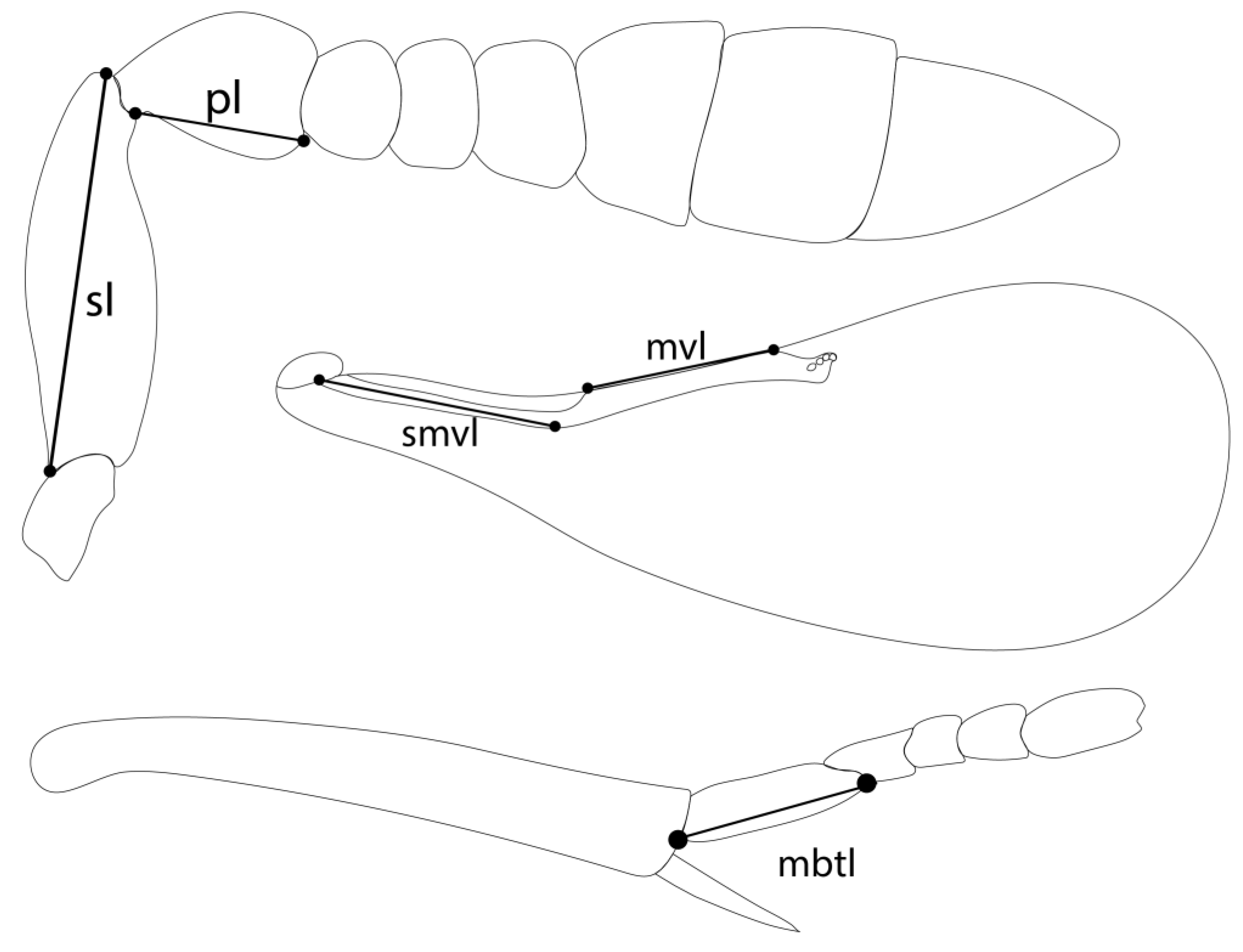
Additional measurements used in the present study: scape length (sl); pedicel length (pl); submarginal vein length (smvl); marginal vein length (mvl); mid basitarsus length (mbtl). Illustrated from type material of Encarsia aisha.
Figure 1.
Additional measurements used in the present study: scape length (sl); pedicel length (pl); submarginal vein length (smvl); marginal vein length (mvl); mid basitarsus length (mbtl). Illustrated from type material of Encarsia aisha.
2. Materials and Methods
2.1. Specimen depositories: abbreviations
Material examined as a part of this investigation are deposited at the following institutions.
- NHMUK: Natural History Museum, London, UK.
- UCRC: University of California, Riverside, USA.
- USNM: National Museum of Natural History, Smithsonian, Washington D.C., USA.
- MZUCR. Museo de Zoología Universidad de Costa Rica.
2.2. Morphological study
Populations of the Encarsia mexicana species group were studied from different localities (Table 1). Host-reared material was collected in Costa Rica, Ecuador, Mexico, and Trinidad & Tobago, as part of intensive foreign exploration efforts to search for parasitoids of whitefly pests (Hemiptera: Aleyrodidae), mostly in the subfamily Aleurodicinae. For morphological analysis, female specimens were mounted on microscope slides following Noyes [14] with some modifications as follows: no maceration in 10% KOH was needed after DNA extraction. Specimens were washed in distilled water for one hour and then dehydrated for 5 minutes in graded ethanol of the following concentrations: 35%, 70%, 85%, 100%. After clearing in clove oil and allowing alcohol evaporation, specimens were dissected in Canada balsam. The wings, antennae, head, and remaining body parts were mounted separately on a single slide.

Table 1.
Ingroup taxa (species identity, voucher IDs, accession numbers, locality, plant associate and host).
Table 1.
Ingroup taxa (species identity, voucher IDs, accession numbers, locality, plant associate and host).
| SPECIES | DNA CODE | ACCESION NUMBER | LOCALITY | PLANTASSOCIATE | HOST(S) |
| Encarsia acusa | DNA0148 | OQ683554 | Costa Rica: Heredia, Est. Biol. La Selva | ||
| DNA0212 | Peru: Loreto, Iquitos, Barillal | ||||
| Encarsia aisha | DNA0146 | OQ683562 | Costa Rica: Heredia, Est. Biol. La Selva | ||
| DNA0164 | OQ683562 | Costa Rica: Alajuela, Est. Caribe | |||
| Encarsia aphania | DNA0218 | OQ683546 | Belize: Cayo, Las Cuevas | Aleurodicuspulvinatus | |
| Belize: Cayo, Las Cuevas | Azuraleurodicus pentarthus | ||||
| Belize: Cayo, Chiquibul | Inga sp. | Nealeurodicus altissimus | |||
| DNA0213 | OQ683545 | Costa Rica: Puntarenas | |||
| Encarsia avida | DNA0143 | OQ683547 | Costa Rica: Heredia, Est. Biol. La Selva | ||
| Encarsia catula | DNA0278 | OQ683547 | Costa Rica: Limon, Hitoi-Cerere | ||
| Encarsia cylindrica | Brazil: Minas Gerais, Vicosa, | Citrus sp. | Aleurothrixus floccosus (?) | ||
| DNA0209 | Costa Rica: Puntarenas RF, Piedras Blancas | ||||
| DNA0211 | Costa Rica: San Juan, Ciudad Colon | ||||
| DNA0149 | Costa Rica: Heredia, Est. Biol. La Selva | ||||
| Jamaica: Fair Prospect | Aleurodicus jamaicensis | ||||
| Encarsia diablejo | Peru: Loreto, Iquitos | ||||
| Encarsia dichaeta | Brazil: Bahia | Aleurodicus flavus | |||
| DNA0132-0135 | OQ683550 | Costa Rica: Alajuela, P.N. Arenal, Pilon | |||
| DNA0133-0136 | OQ683552 | Costa Rica: Alajuela, P.N. Arenal, Pilon | |||
| DNA0208 | OQ683551 | Costa Rica: Guanacaste, Pitilla | |||
| DNA0151 | OQ683549 | Costa Rica: Heredia, Est. Biol. La Selva | |||
| Ecuador: Napo, Anangucocha | Aleurodicus sp. | ||||
| Encarsia encantadora | Ecuador: Napo River | ||||
| Mexico: Tabasco | Lippiamyriocephala | Nealeurodicusaltissimus | |||
| Encarsia erwini | Ecuador: Napo River | ||||
| Encarsia fredbennetti | DNA0215B | OQ683559 | Trinidad & Tobago: Trinidad, St Augustine | Theobroma cacao | Aleurodicinae |
| DNA0216 | Trinidad & Tobago: Trinidad, Mount St Benedict | ||||
| Encarsia inbioa | DNA0128 | OQ683553 | Costa Rica: Alajuela, P.N. Arenal, Pilon | ||
| Encarsia larensis | Venezuela: Cabudare, Lara | Huracrepitans | Aleurodicuspulvinatus | ||
| Encarsia marynoyesae | DNA0163 | OQ683563 | Costa Rica: Alajuela, Est. Caribe | ||
| DNA0167 | Costa Rica: Alajuela, Est. Caribe | ||||
| Encarsia mendesi | Brazil: São Paolo, Mogi-Guazu | Bauhiniaholophylla | Aleurodicusmaritimus | ||
| Encarsia mexicana | Mexico: Tabasco, San Francisco del Peal | Lippiamyriocephala | Nealeurodicusaltissimus | ||
| DNA0144 | OQ683560 | Costa Rica: Heredia, Est. Biol. La Selva | |||
| Costa Rica: Limon | |||||
| DNA0166 | Costa Rica: Alajuela, Est. Caribe R. | ||||
| DNA0129 | Costa Rica: Alajuela, P.N. Arenal, Pilon | ||||
| Encarsia napo | Ecuador: Napo River, Camp. Res. Waorani | ||||
| Encarsia noora | DNA0126 | OQ683561 | Costa Rica: Limon | ||
| Encarsia svetlana | DNA0305 | OQ683558 | Guyana: Dubulay Ranch | ||
| Encarsia venia | DNA0298 | OQ683557 | Costa Rica: Limon, Parque Nacional Cahuita | ||
| DNA0267 | Costa Rica: Limon, Hitoy-Cerere | ||||
| Costa Rica: Heredia La Selva | |||||
| E. mexicana group sp. | DNA0165 | OQ683556 | Costa Rica: CR Alajuela Est. Caribe R. Rincon Forestal | ||
| D2672 | OQ683555 | Ecuador: Orellana, Tiputini Biodiversity Sta. |
In total, 110 females and 4 males of 20 species were examined, including the extensive recording of measurements and ratios. Males are rare or unknown for most species and were not therefore included, except for Encarsia diablejo which is known only from the male. Measurements were taken with a Leitz Dialux 20EB microscope from slide-mounted material following Heraty & Polaszek [12] with the following five measurements added: scape length, pedicel length, submarginal vein length, marginal vein length and length of the mid basitarsus. (Figure 1). All measurements of antennae, fore wings, and legs refer to the maximum length of the structure in lateral view. The terminology of morphological characters follows Kim and Heraty [15] and Hayat [16].
Specimens were imaged using with a Leitz Dialux 20EB compound microscope using Nomarski Differential Interference Contrast illumination (DIC) and photographed with a MicroPublisher 5.0 RTV camera. Additional images (claval sensorial area; mandibles) were imaged with an Olympus BX63 microscope also utilizing DIC. Scanned sections were stacked and combined using Synoptics AutoMontage Pro® ver. 5.03 software (Leitz Dialux images) and Helicon Focus software (Olympus BX63). The final images were edited with Adobe Photoshop CC®.
2.3. DNA extraction, amplification, and sequencing
Genomic DNA was extracted from single, whole specimens using a non-destructive genomic DNA extraction protocol developed by Chao-Dong Zhu, John Noyes, and others at the Natural History Museum, London [17].
Specimens were softened in 70% ethanol (to reduce potential damage during subsequent steps) at room temperature for a minimum of 2 hours. 70% ethanol was removed carefully by pipette and specimens allowed to air-dry briefly. DNA was extracted using the Qiagen DNeasy Blood and Tissue Kit (250) #69506. Specimens were immersed in 180 µL of Lysis Buffer ATL, premixed with 20 µL Proteinase K and incubated at 55°C overnight (8 hours minimum) with no mixing, taking care that the specimen was submerged/floating in the buffer and not adhered to the side of the tube.
After digestion, the lysis buffer was carefully removed by pipette into a clean 1.5 mL microfuge tube. The specimen was immediately washed by adding 500 µL distilled water for a minimum of 30 mins, then replaced with 500 µL 70% ethanol for a minimum 30 mins, then finally stored in 100% ethanol until slide-mounted in Canada balsam.
DNA was extracted from the lysis buffer using the Qiagen QUIA quick PCR Purification Kit (250) #28106 following the protocol: ‘Isolation of total DNA from Animal Tissues’ (step 3 onwards). Standard PCR reactions were then carried out in a thermal cycler using 2.0 µL DNA extract, Taq buffer (1.5mM MgCl2), 1.5 U Taq polymerase (Roche), 10 nmol dNTPs (Amersham Pharmacia Biotech; APB) and 20 mol of each primer at the Natural History Museum’s DNA sequencing facility.
The D2 region of 28S rDNA was amplified using the following primers:
28SFW 5’ - AGTACCGTGAGGGAAAGTTG -3’
28SRev 5’ - TTGGTCCGTGTTTCAAGACGG -3’
PCR conditions were as follows: an initial denaturation of 94°C for 3 minutes, then 35 cycles of denaturation at 94°C for 1 minute, annealing at 50°C for 1 minute, and extension at 72°C for 2 minutes, followed by a final extension at 75°C for 10 minutes, then samples were held at 4°C until they could be analyzed. PCR product was run on a 1% agarose gel to confirm PCR success (clean bands of the expected size), then the remaining products were cleaned and sequenced. Removal of dye terminators was done by ethanol precipitation prior to sequencing.
The DNA analyzer system ABI PRISM 3730 and 377 DNA sequencer were used, the samples were loaded onto the system’s vertical polyacrylamide gel where they underwent electrophoresis, laser detection and computer analysis. Sequence editing and alignment were performed using Sequencher TM 4.8 (Genes Corp) on a Macintosh computer. The resulting molecular dataset includes eighteen sequences representing 14 species. Sequences have been deposited in the GenBank database under accession numbers OQ683545–OQ683576 (Table 1 & Table 2).
Table 2.
Outgroup taxa (species identity, voucher IDs, accession numbers, and locality).
| SPECIES | DNA CODE |
ACCESION NUMBER |
LOCALITY |
| Encarsia luteola | D0243 | AF223369 | USA: California, Brawley |
| Encarsia formosa | D0231 | AF223372 | Egypt |
| Encarsia cubensis | DNA270 | OQ683567 | Costa Rica: Limon |
| Encarsia azimi | DNA259P17 | AF254229 | Australia: Queensland |
| Encarsia inaron | D0465 | AY599399 | New Zealand |
| Encarsia lounsburyi | DNA017 | OQ683568 | Costa Rica: Puntarenas |
| Encarsia citrina | DNA376 | OQ683569 | United Kingdom: London, Barnes Common |
| Encarsia boswelli | DNAAE534 | OQ683570 | India |
| Encarsia perplexa | D0296 | AF254243 | Guatemala: Coatepeque |
| Encarsia opulenta | DNA387 | OQ683571 | Mexico: Los Tuxtlas |
| Encarsia lutea | D0235 | AF254238 | Cyprus |
| E. noyesi group sp. | DNA0091 | OQ683566 | Costa Rica |
| Encarsia tamaulipeca | DNA0123 | OQ683564 | Ecuador |
| E. noyesi group sp. | DNA0089 | OQ683565 | Ecuador: Napo |
| Encarsia sophia | D0219 | AF254198 | Find in Heraty Lab |
| Encarsia protransvena | D0136 | AF254208 | USA: California, Orange Co. |
| Coccophagus sp. | DNA010 | OQ683572 | Costa Rica |
| Coccophagus sp. | DNA0185 | OQ683573 | Costa Rica: La Selva |
| Coccophagus lycimnia | DNAA1-006A | OQ683574 | Costa Rica |
| Coccophagus semicircularis | DNAA3-023D | OQ683575 | Costa Rica: Puntarenas |
| Coccophagus sp. | DNA034 | OQ683576 | Costa Rica: Puntarenas |
| Aphytis yanonensis | D0446 | AY635336 | UCR Culture: Originally from Japan, Fukuoka |
| Aphytis melinus | D0445 | AY635342 | UCR Culture: Originally from China, Fujian, Fuzhou |
2.4. Phylogenetic Analyses
Captured sequences were combined with previously published sequence data (Table 2) and aligned using the E-INS-I algorithm in MAFFT v7.490 [18]. Ten independent iterations of maximum likelihood were reconstructed using IQ-TREE version 2.0.7 [19], implementing a General Time Reversible model with invariant sites and gamma distributed rate variation (-m GTR+I+G). Bootstrap support was estimated from 1000 bootstrap trees constructed using ultrafast bootstrapping (-b 1000) [20]. Outgroups are comprised of a broad range of Encarsia species, a diversity of recognized species-groups, as well as Coccophagus Westwood (Aphelinidae: Coccophaginae) and Aphytis Howard (Aphelinidae: Aphelininae).
2.5. Nomenclatural Acts
The electronic edition of this article conforms to the requirements of the amended International Code of Zoological Nomenclature, and hence the new names contained herein are available under that Code from the electronic edition of this article. This published work and the nomenclatural acts it contains have been registered in ZooBank, the online registration system for ICZN. The ZooBank LSIDs (Life Science Identifiers) can be resolved, and the associated information viewed through any standard web browser by appending the LSID to the prefix "http://zoobank.org/". The LSID for this publication is: urn:lsid:zoobank.org:pub:2CE58923-A39A-412A-896E-DCFD4CC01FD7. The electronic edition of this work was published in a journal with an ISSN and has been archived and is available from the following digital repositories: PubMed Central, LOCKSS.
3. Results
3.1. Phylogenetic analysis of molecular data
A maximum likelihood tree was constructed from partial sequences of 28S D2 ribosomal DNA of 13 species (from 19 specimens) and 23 out group taxa (Figure 2). The Encarsia mexicana species-group was recovered as a strongly supported clade within Encarsia. Encarsia dichaeta forms the sister clade to all remaining E. mexicana group species. The Encarsia mexicana species-group was not placed sister to the noyesi species-group; however, backbone support in the recovered phylogeny is insufficient to confidently resolve inter- and intra-species-group relationships.
3.2. Taxonomy of the Encarsia mexicana species-group
Encarsia mexicana species group
Etymology. Dirphys (Διρφυς) is a Greek feminine noun. Hence the modification by Hayat of Howard’s (1914) combination Dirphys mexicana (Howard) to Dirphys mexicanus (Howard) was an unjustified emendation [21]. Hayat attributed the new combination to Howard [1], but this is not the case.
Diagnosis. Head dorsally transverse. Frontovertex at narrowest wider than dorsal eye width. Facial lines evident, often broadly expanded; mediofrontal and transfacial lines developed. Eyes with evident setae. Mandibles usually with 2 teeth and a truncation (Figure 10A), the truncation sometimes reduced, and the teeth often strongly developed so mandibles appear to have only two teeth (Figure 22A). A bidentate upper tooth may be present in addition to the well-developed ventral tooth (Figure 4A).
Figure 2.
Maximum likelihood tree (IQ-TREE 2) based on 28S D2 ribosomal DNA (509 bp) from 42 taxa (19 ingroup, 23 outgroup); support values from 1000 Ultrafast bootstrap replicates.
Figure 2.
Maximum likelihood tree (IQ-TREE 2) based on 28S D2 ribosomal DNA (509 bp) from 42 taxa (19 ingroup, 23 outgroup); support values from 1000 Ultrafast bootstrap replicates.
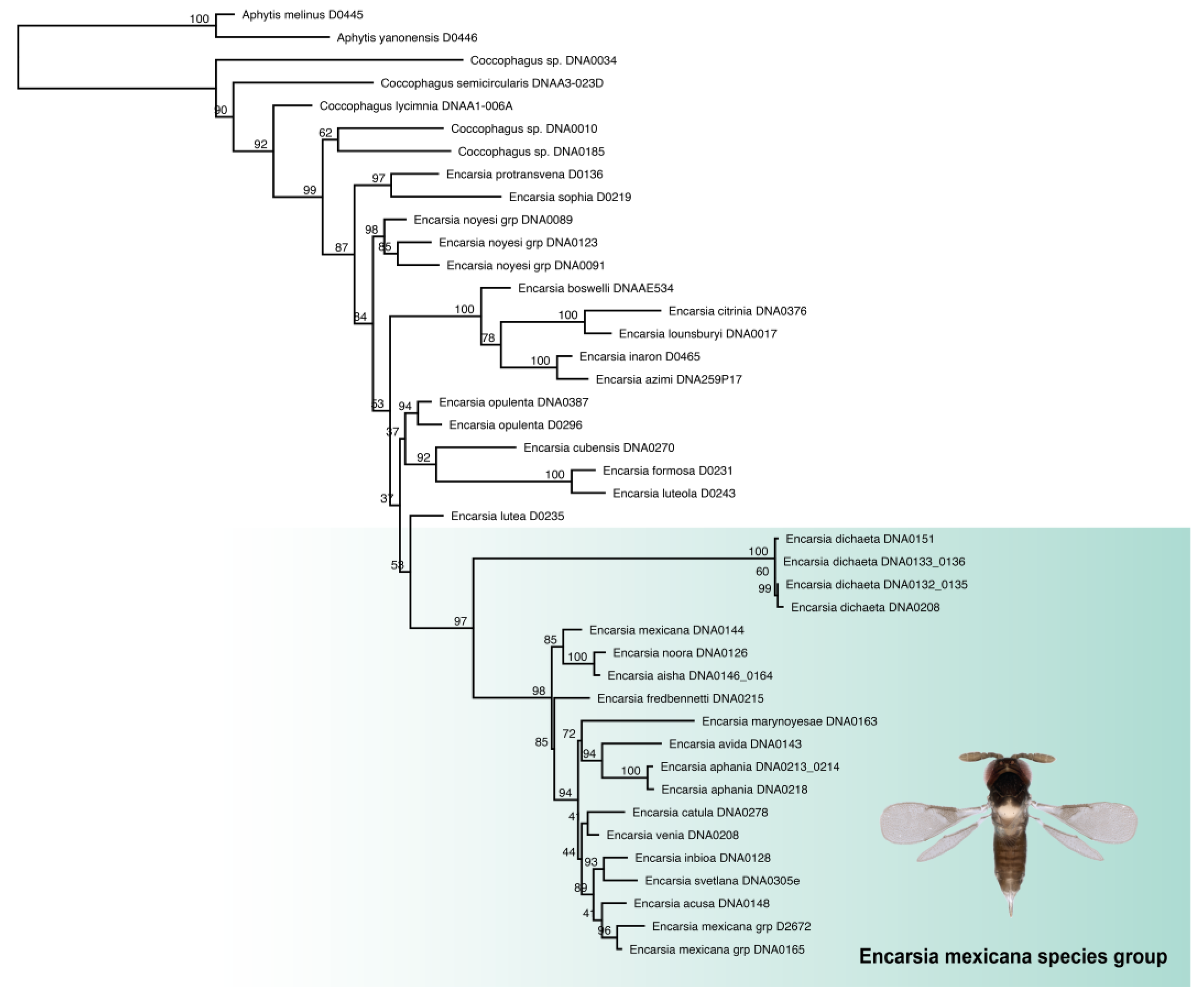
Figure 3.
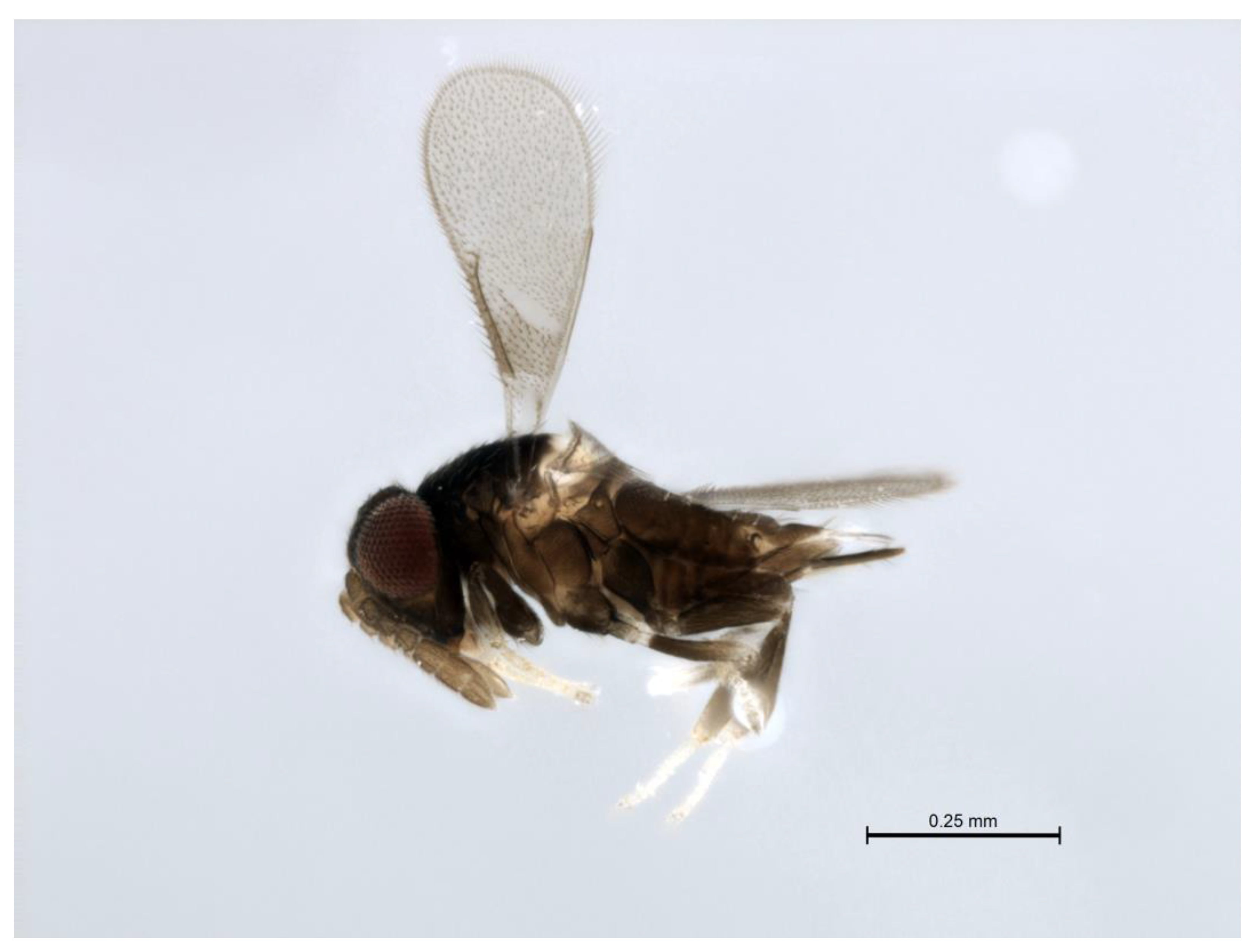
Lateral habitus of a female of the Encarsia mexicana species-group.
Maxillary palps 2-segmented. Antenna 8-segmented in both sexes, antennal formula variable (1,1,3,3 or 1,1,0,6) claval sensorial complex present (Figure 17B) or absent, suture between F5 perpendicular or oblique. Pronotum medially membranous. Mesoscutum with more than 20 setae. Side lobes divided (Figure 23E). Axillae large, strongly projecting forwards and separated medially by less than the maximum length of one axilla. Each axilla with one or two setae in E. dichaeta (Figure 11E). Thoracic sculpture aciculate, longitudinal, transverse or a combination of these types, never reticulate. Mesoscutellar sensilla close together, separated by about the width of one sensillum. Fore wings with 2 large setae on the submarginal vein, plus a variable number of smaller setae at the distal end of the submarginal vein. Linea calva present or absent. Mid basitarsi with a variable number of robust, spine-like setae, tarsi 5 segmented.
Remarks. The Encarsia mexicana species-group (Figure 3) is restricted to the Neotropical zone, with species reaching as far south as the State of Bahia (Brazil), and as far north as southern Mexico.
3.3. Species descriptions
3.3.1. Encarsia acusa Polaszek and Hernández-Suárez sp.n.
(Figure 4A–F)
urn:lsid:zoobank.org:act:F179717C-0FF0-4189-90BE-4851A8625431
Female. Color. Antennae light brown; radicle, scape, base of pedicel and F6 darker. Head dark brown, paler along the sutures and frons. Mesosoma and metasoma dark brown with posterior 80% of the mesoscutellum and sides of metanotum yellow. Legs yellow with most of mid and hind femora brown, fore femora and tibiae brown, all tarsi pale. Fore wings hyaline, slightly infuscate below marginal vein, submarginal and marginal veins dark.
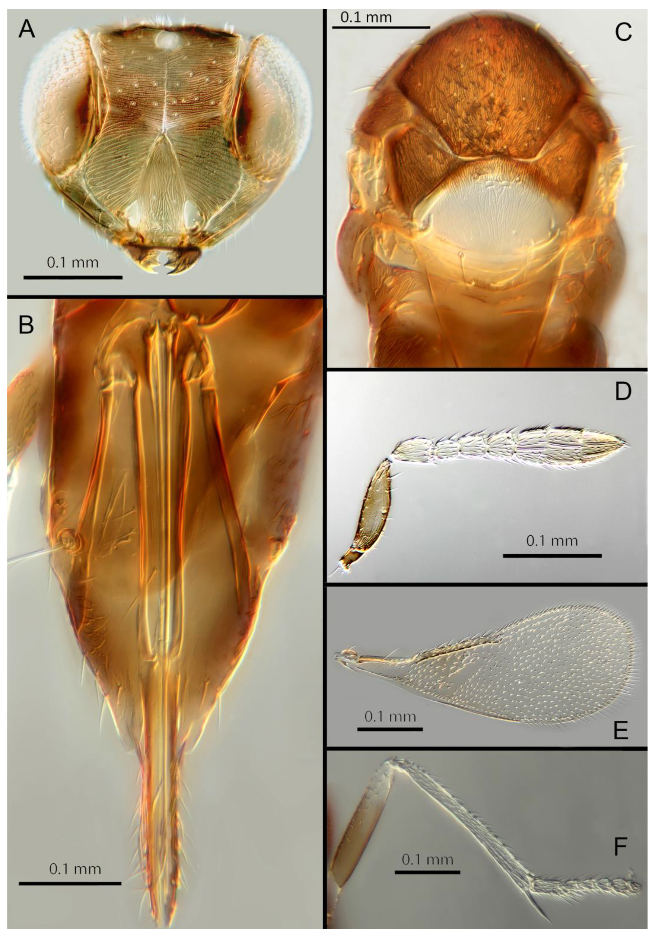
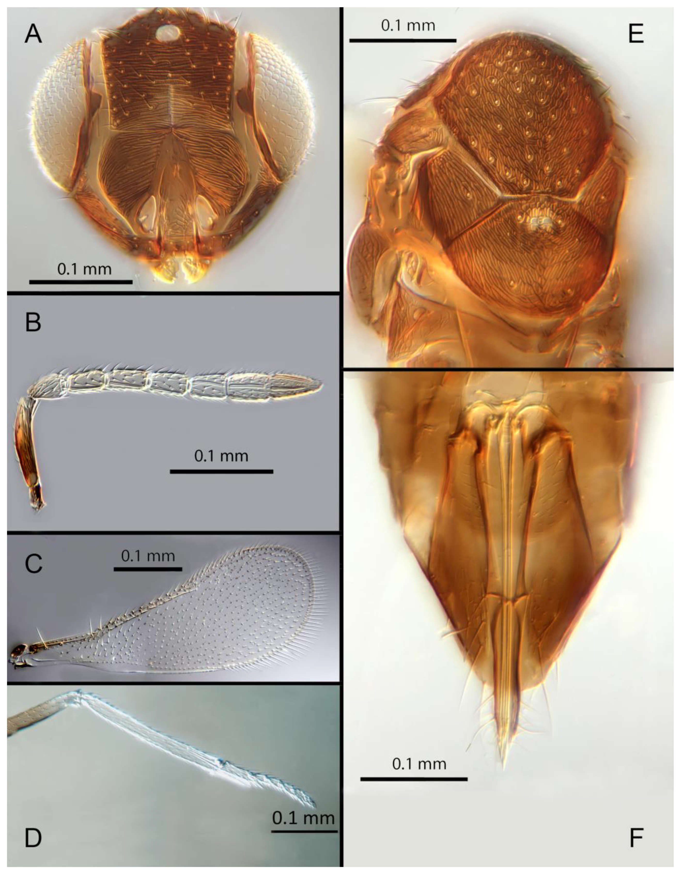
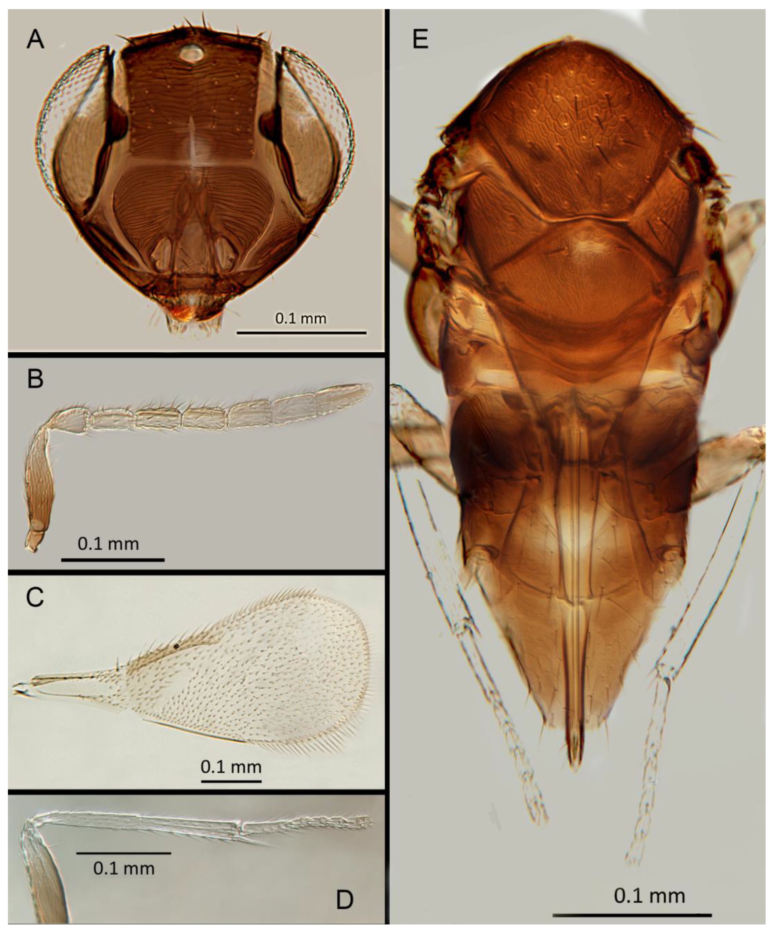
Morphology. Head (Figure 4A) with mediofrontal line complete; transfacial line obscure; facial lines narrow. Scrobes with longitudinal aciculate sculpture. Antenna (Figure 4D) with eight antennomeres; antennal formula: 1,1,3,3; scape 2.4x pedicel length; pedicel 1.9x F1; F1 0.8x F2; F2 equal to F3; funicle 0.56x clava; F6 slightly oblique, claval sensorial area present. Flagellum with the following number of longitudinal sensilla: F1: 0; F2: 1; F3: 1; F4: 3; F5: 4; F6: 3–4 (both counts present in holotype). Mandibles (Figure 4A) with 2 teeth and a broad truncation. Maxillary palps 2-segmented. Mid-lobe of mesoscutum (Figure 4C) with 34–40 setae; each lateral lobe with 2 setae; each axilla with 1 seta; scutellum with 4 setae. Sculpture of mesoscutum longitudinal; sculpture of axillae and scutellum longitudinal. Fore wing (Figure 4E) with 2 large setae and 2–3 smaller setae on submarginal vein, 3 setae in basal cell, 6–7 setae on anterior margin of marginal vein, and 1 seta at junction of the submarginal vein and parastigma. Linea calva present. Submarginal 0.75x marginal vein. Maximum length of fore wing 2.7x fore wing width, maximum width of wing 5.3x longest setae on marginal fringe. Ovipositor (Figure 4B) 1.85x mid tibial length; third valvulae 0.44x ovipositor length; second valvifer 1.3x third valvula. Mid tibial spur (Figure 4F) 0.86x corresponding basitarsus. Metasomal terga T1–T7 with 0, 2+2, 2+2, 1+1, 1+2+1, 1+2+1 and 4 setae, respectively. T7 (Figure 4B) extremely extended, almost covering ovipositor.
Distribution. COSTA RICA: Heredia, Limon; PERU: Iquitos.
Material examined. Holotype ♀ COSTA RICA, Heredia Est. Biol. La Selva, 75m, 10º26´N 84º01´W 27–28.ii.2003 (J.S. Noyes) [DNA148: OQ683554] (NHMUK). Paratypes: 1♀ COSTA RICA, Limon RB, Hitoy-Cerere 100m, 14–19.i.1991 (J.S. Noyes) (MZUCR). 1♀ PERU, Iquitos, Barillal, 10.ii.1984 (L. Huggert #BM 1984.337) [DNA 212] (NHMUK).
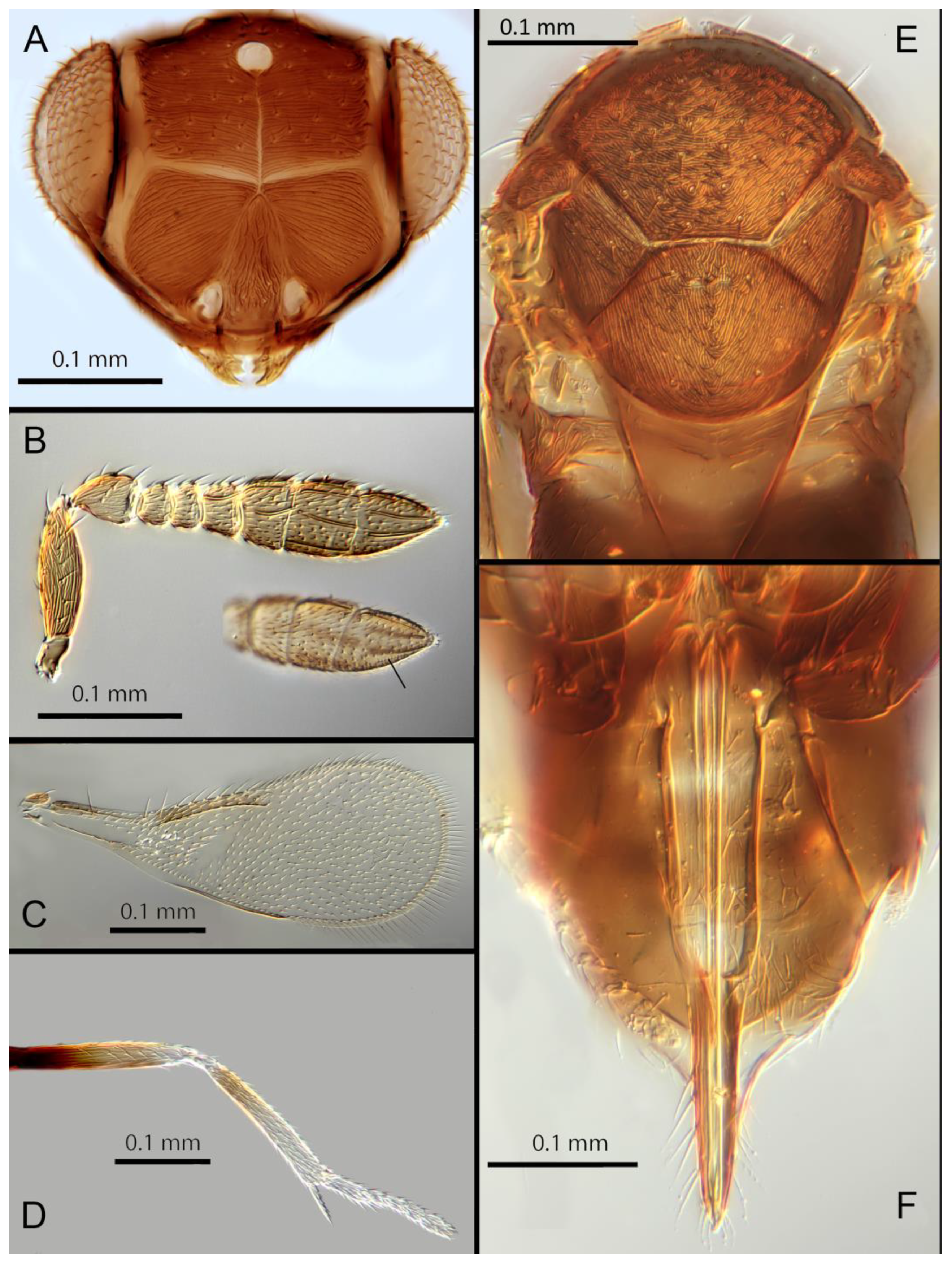
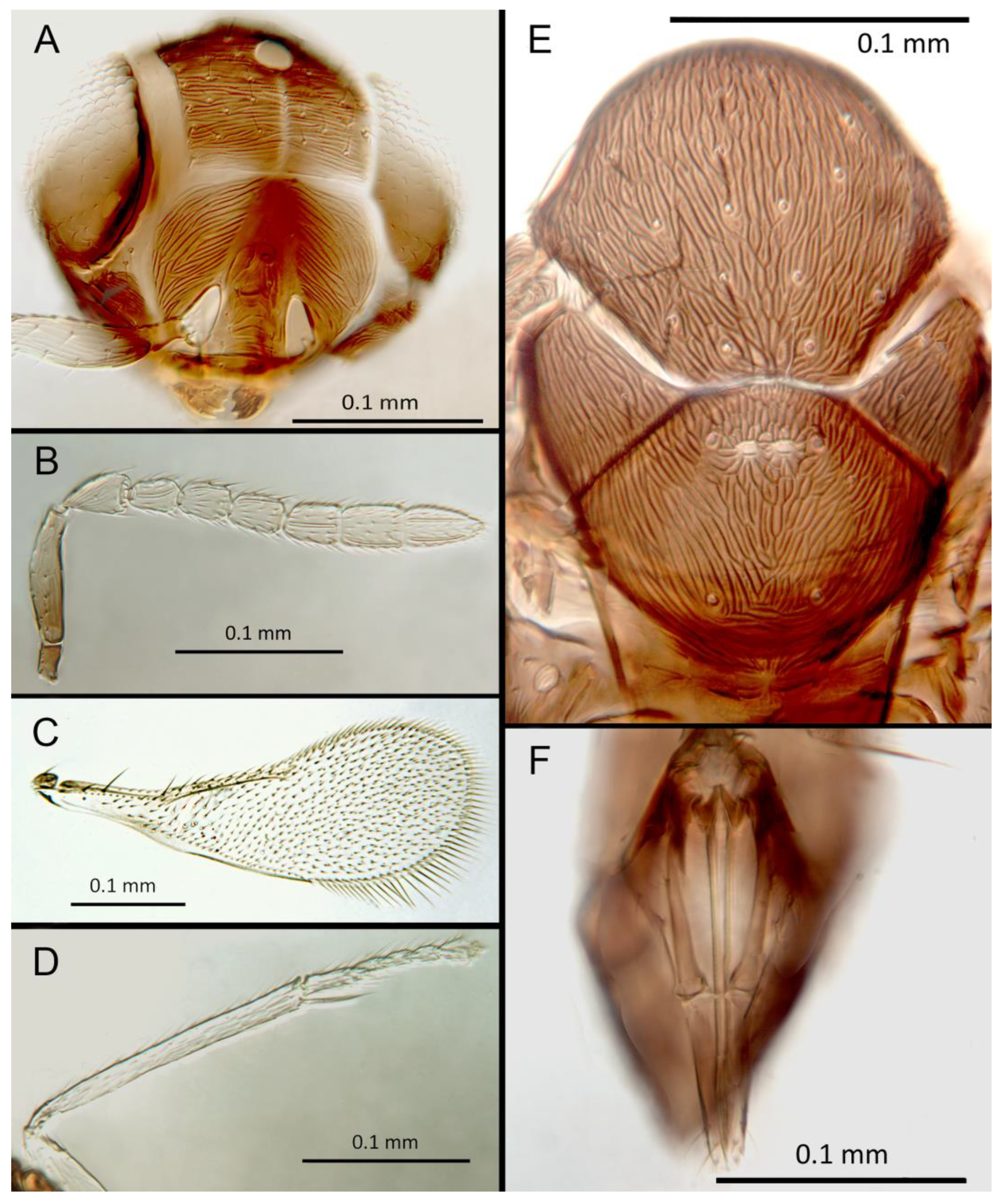
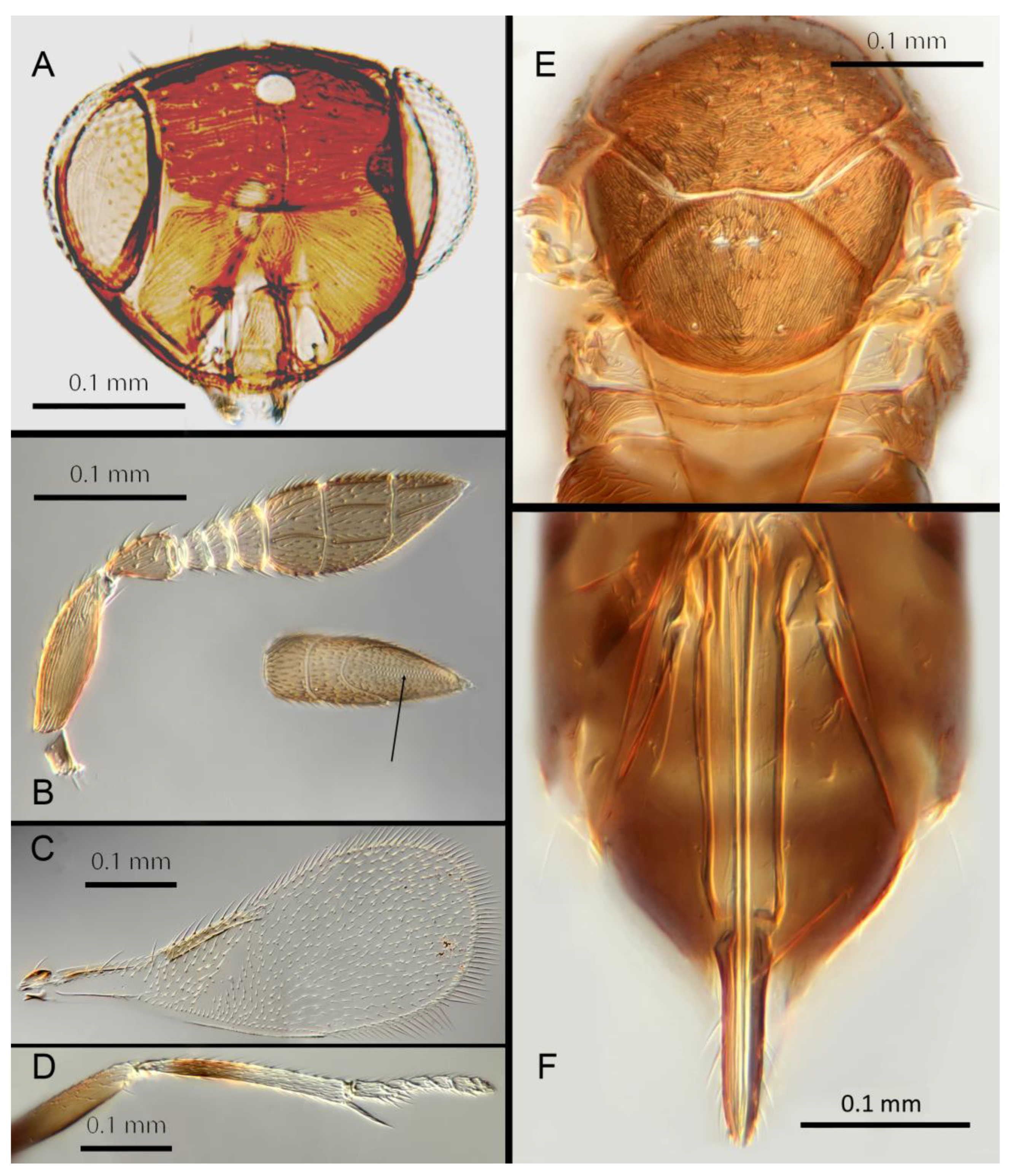
Figure 4.
Encarsia acusa: (A) head; (B) ovipositor; (C) dorsal mesosoma; (D) antenna; (E) fore wing; (F) mid leg.
Figure 4.
Encarsia acusa: (A) head; (B) ovipositor; (C) dorsal mesosoma; (D) antenna; (E) fore wing; (F) mid leg.
Remarks. T7 extremely extended, covering the ovipositor. Encarsia acusa appears to be most closely related to E. inbioa and E. svetlana but is easily distinguished from those (and all other) species by the extremely long ovipositor and T7. DNA sequences from holotype deposited under GenBank accession numbers: OQ683554.
Etymology. From “acus” Latin for needle or pin, referring to the elongated T7.
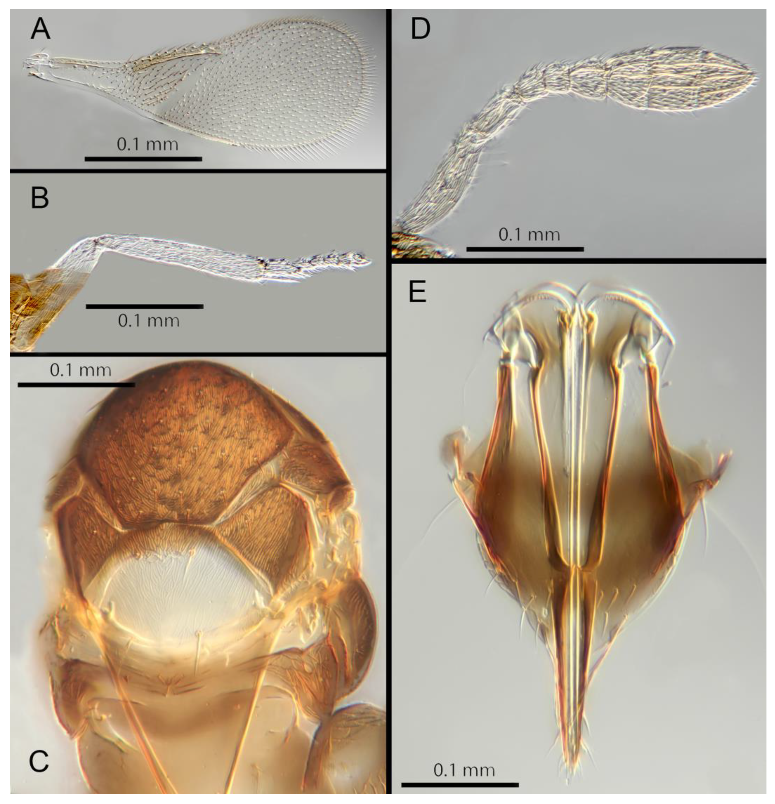
Figure 5.
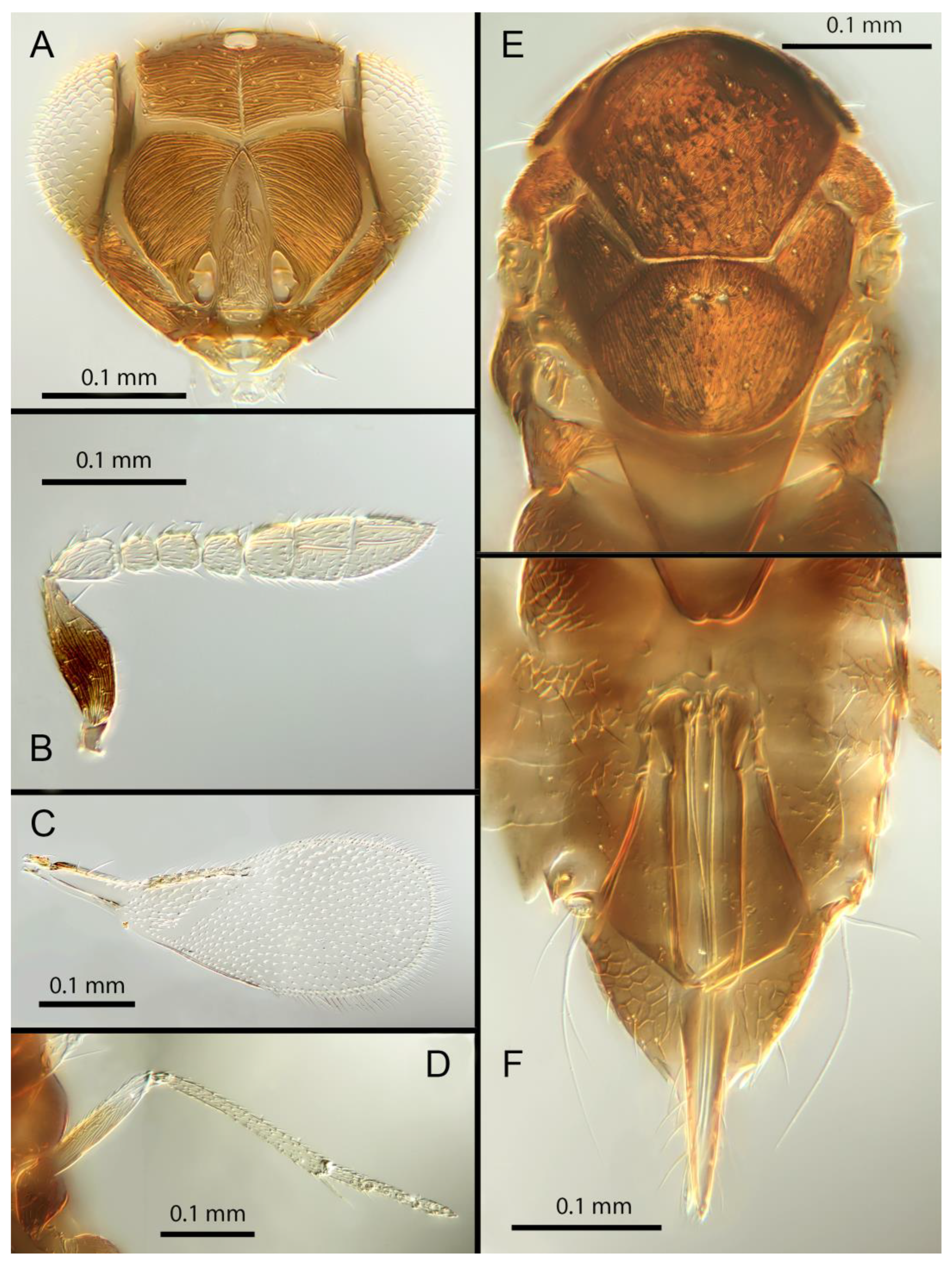
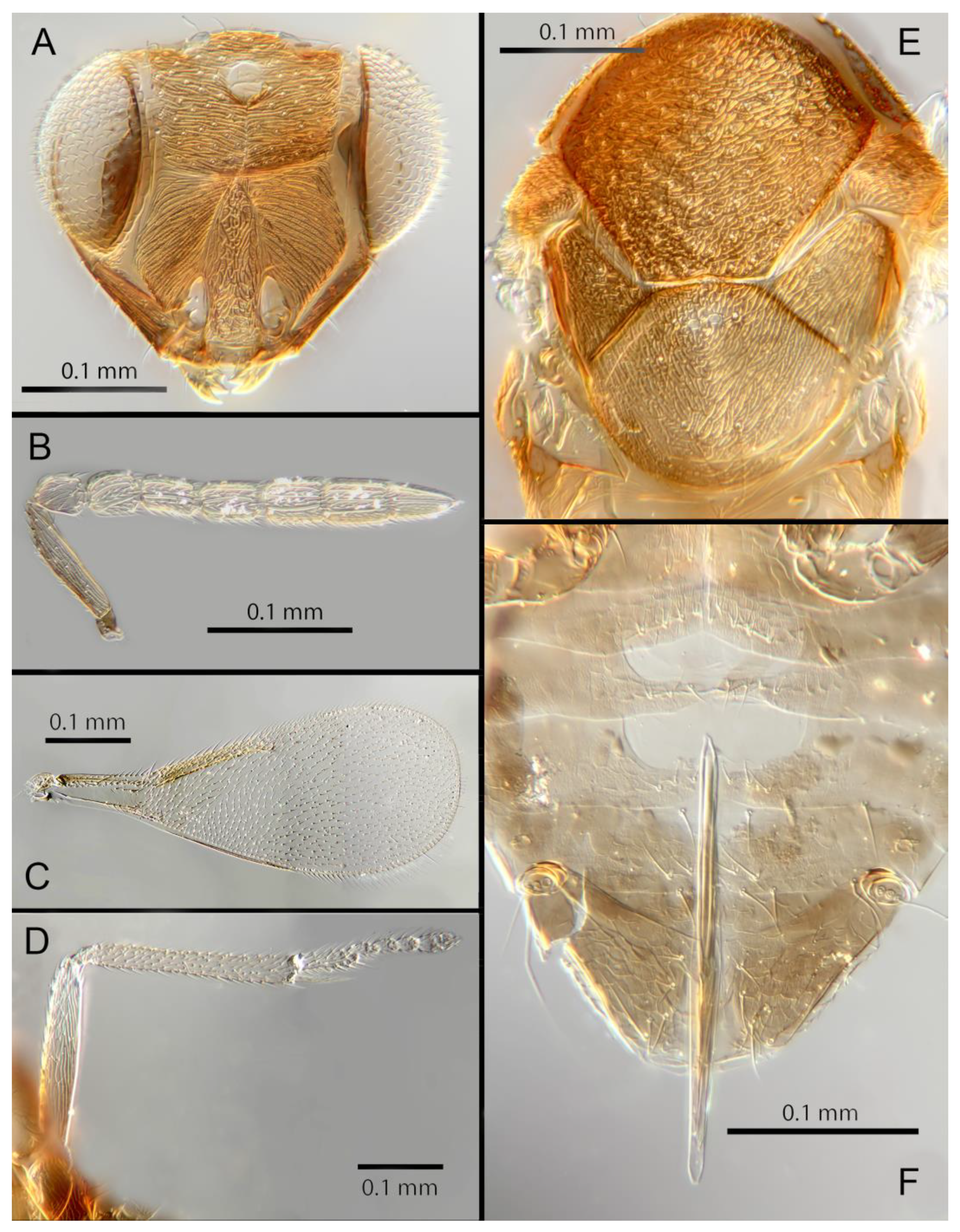
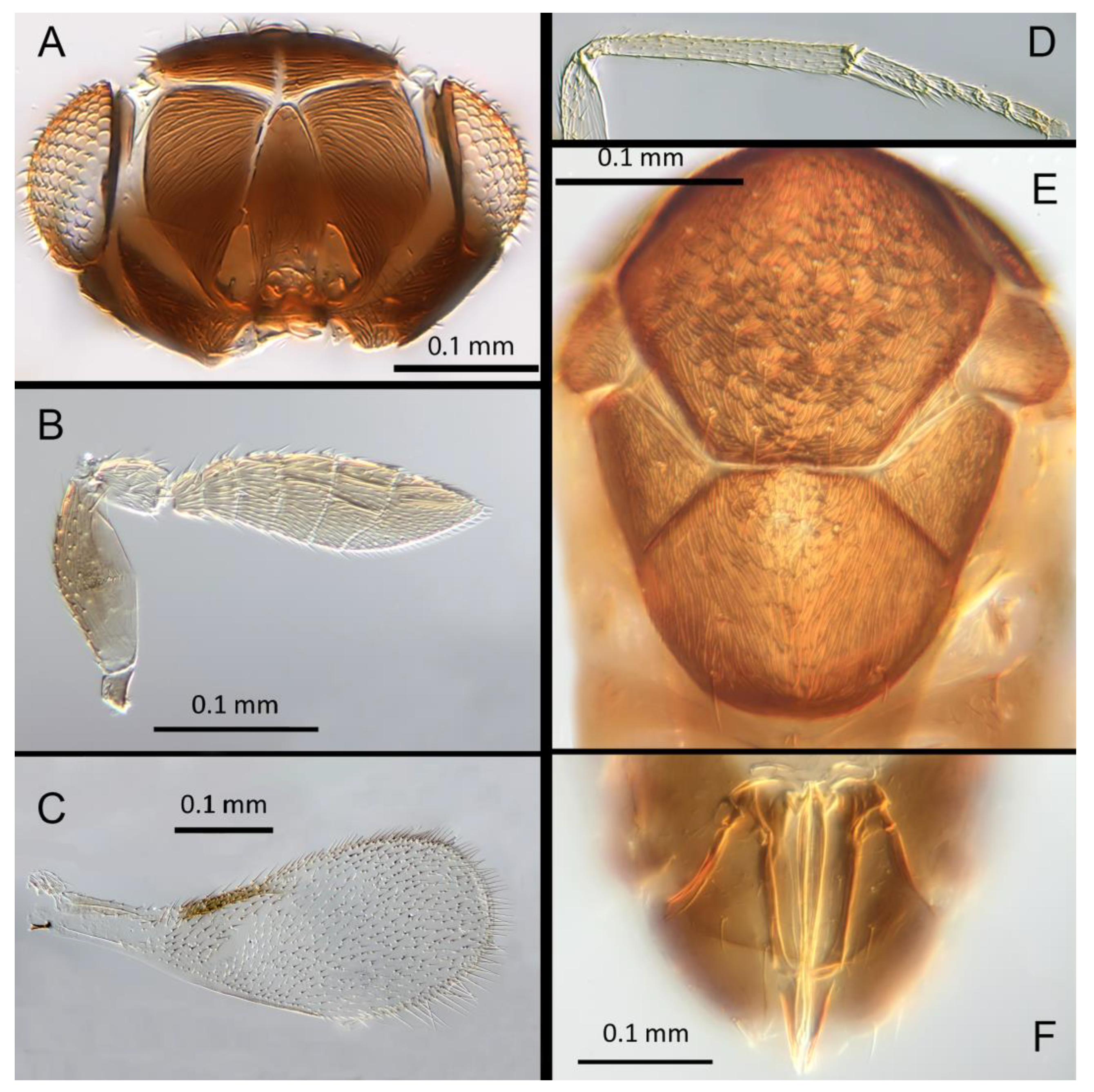
Encarsia aisha: (A) head; (B) antenna, arrow: claval sensorial area; (C) fore wing; (D) mid leg; (E) dorsal mesosoma; (F) ovipositor.
Figure 5.
Encarsia aisha: (A) head; (B) antenna, arrow: claval sensorial area; (C) fore wing; (D) mid leg; (E) dorsal mesosoma; (F) ovipositor.
3.3.2. Encarsia aisha Polaszek and Hernández-Suárez sp.n.
(Figure 5A–F)
Female. Color. Antennae light brown with F1 and F2 paler. Head dark brown, paler along the sutures. Mesosoma uniformly dark brown. Legs yellow with mid and hind femora, coxae and anterior third of mid tibiae brown, all tarsi pale. Wings hyaline, slightly
Morphology. Head (Figure 5A) with mediofrontal line complete; transfacial line evident; facial lines very broad along their entire lengths. Scrobes with longitudinally aciculate sculpture. Antenna (Figure 5B) with eight antennomeres; antennal formula 1,1,3,3; scape 2.3x pedicel length; pedicel 2x F1; F1 1.2x F2; F2 0.75x F3; funicle 0.45x clava; F6 oblique, claval sensorial area present. Flagellum with the following number of longitudinal sensilla: F1: 0; F2: 0; F3: 1; F4: 2-3; F5: 4-5; F6: 3. Mandibles (Figure 5A) with 1 large ventral tooth and a bidentate upper tooth. Maxillary palps 2-segmented. Mid-lobe of mesoscutum (Figure 5E) with about 34-40 setae; each lateral lobe with 2 setae; each axilla with 1 seta; scutellum with 4 setae and 2 apparent vestigial setal bases. Sculpture of mesoscutum aciculate; sculpture of axillae and scutellum longitudinal. Fore wing (Figure 5C) with 2 large setae and 4 smaller setae on submarginal vein, 4 setae in basal cell, 6-7 setae on anterior margin of marginal vein, and 1 seta at the junction of the submarginal vein and parastigma. Linea calva present. Submarginal 0.85x marginal vein. Maximum length of fore wing 2.47x fore wing width, maximum width of wing 5.72x longest setae on marginal fringe. Ovipositor (Figure 5F) 1.47x mid tibial length; third valvulae 0.42x ovipositor length; second valvifer 1.5x third valvula. Mid tibial spur (Figure 5D) 1.07x corresponding basitarsus. Metasomal terga T1-T7 with 0, 2+2, 2+2, 2+2, 1+2+1, 1+2+1 and 4 setae, respectively. T7 (Figure 5F) extended, as long as ovipositor.
Distribution. COSTA RICA: Alajuela, Heredia.
Material examined. Holotype ♀ COSTA RICA, Alajuela, Est. Biol. Caribe, R. Rincon Forestal 10º53´N 85º18W 400m 19-20.ii.2003 (J.S. Noyes) [DNA 164: OQ683562] (NHMUK). 1♀ COSTA RICA, Heredia, Est. Biol. La Selva, 10º26´N 84º01´W. 75m 27-28.ii.2003 (J.S. Noyes) [DNA 146: OQ683562] (ZMUCR).
Remarks. Encarsia aisha is morphologically very similar to E. marynoyesae in many respects (though distant to it based on DNA). The species can be distinguished from E. marynoyesae by the 2nd valvifers almost 2x (1.8) the 3rd valvulae; while they are 1.5x as long in E. aisha. In E. marynoyesaei the clava is well over 2x the length of the funicle; in E. aisha it is less than 2x as long. DNA sequences from holotype and paratype deposited under GenBank accession numbers: OQ683562.
Etymology. Named for Aisha, daughter of the 2nd author (EHS), and sister to Noora; see E. noora, below.
3.3.3. Encarsia aphania (Polaszek) 1999 (in Martin & Polaszek, 1999: 1556). comb. nov.
(Figure 6A–F)
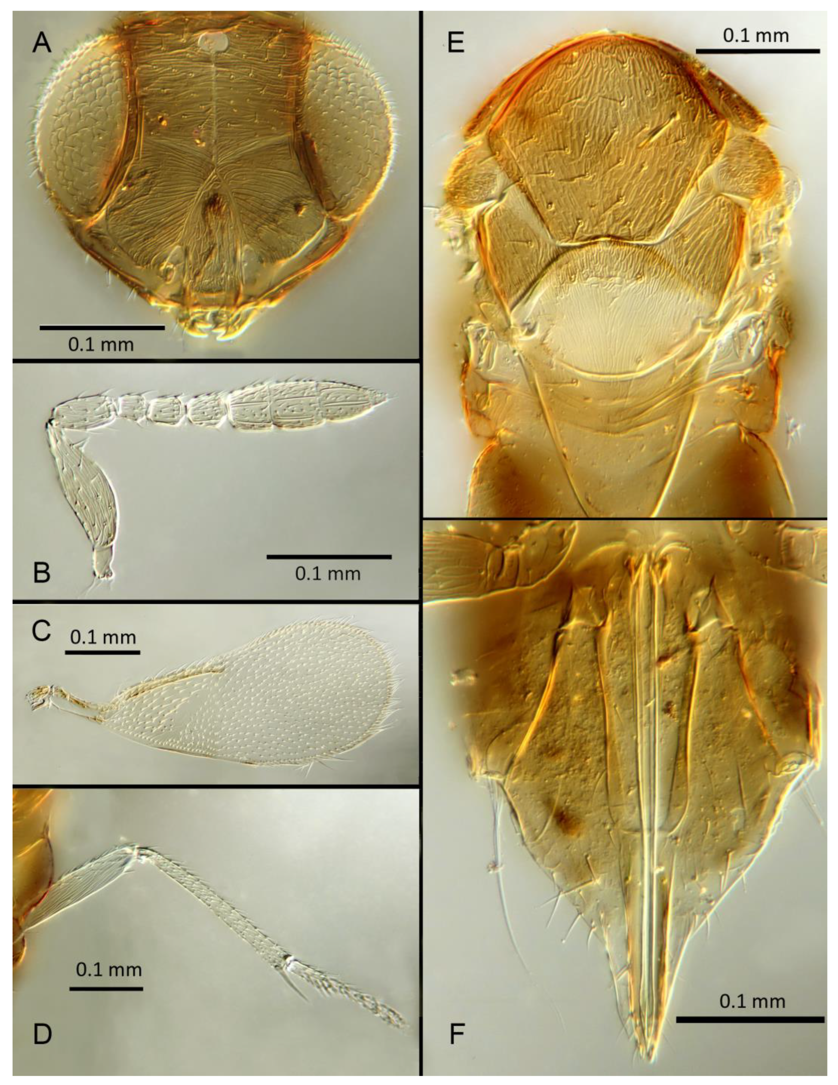
Female. Color. Antennae pale brown with scape very dark. Head dark brown with pale lines bordering the eyes and extending along the genae towards clypeus, antennal scrobes, a line from the apex of the scrobes to the median ocellus, and a transverse line midway between the antennal scrobes. Mesosoma and metasoma uniformly dark brown. Legs yellow except all coxae and mid and hind femora which are brown. Fore wings faintly infuscate along the submarginal and the marginal veins; submarginal and marginal veins darker in contrast with the stigmal vein paler.
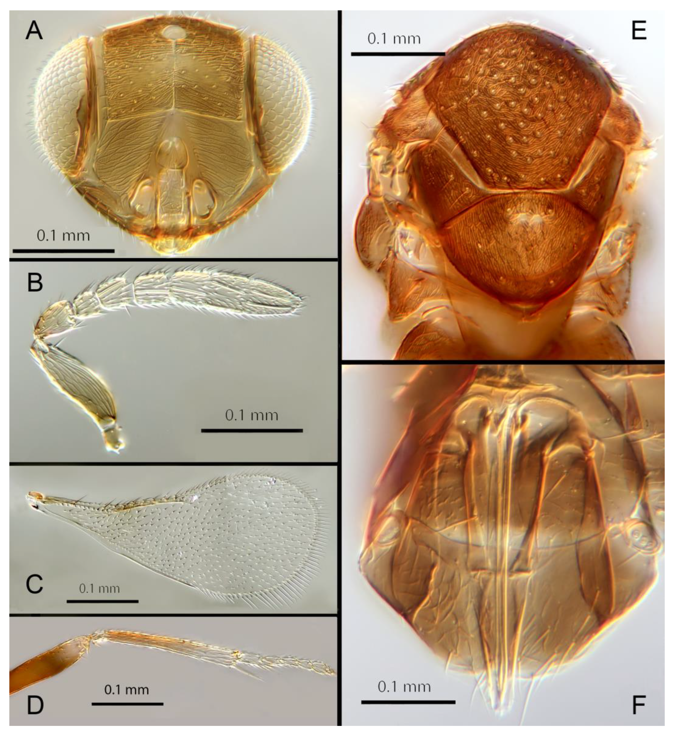
Morphology. Head (Figure 6A) with mediofrontal line complete, though fading towards anterior ocellus; transfacial and facial lines very broad along their entire lengths. Scrobes with longitudinally aciculate sculpture centrally, smooth laterally. Antenna (Figure 6B) with eight antennomeres. antennal formula: 1,1,3,3; scape 2.39x pedicel; pedicel 2x F1; F1 0.8x F2; F2 equal to F3; funicle 0.67x clava; F4 and F5 partly fused; F6 broadly oblique; Claval sensorial area present, indistinct. Flagellum with the following number of longitudinal sensilla: F1: 0; F2: 1; F3: 1; F4: 2-3; F5: 4; F6: 4. Mandibles (Figure 6A) with one large ventral
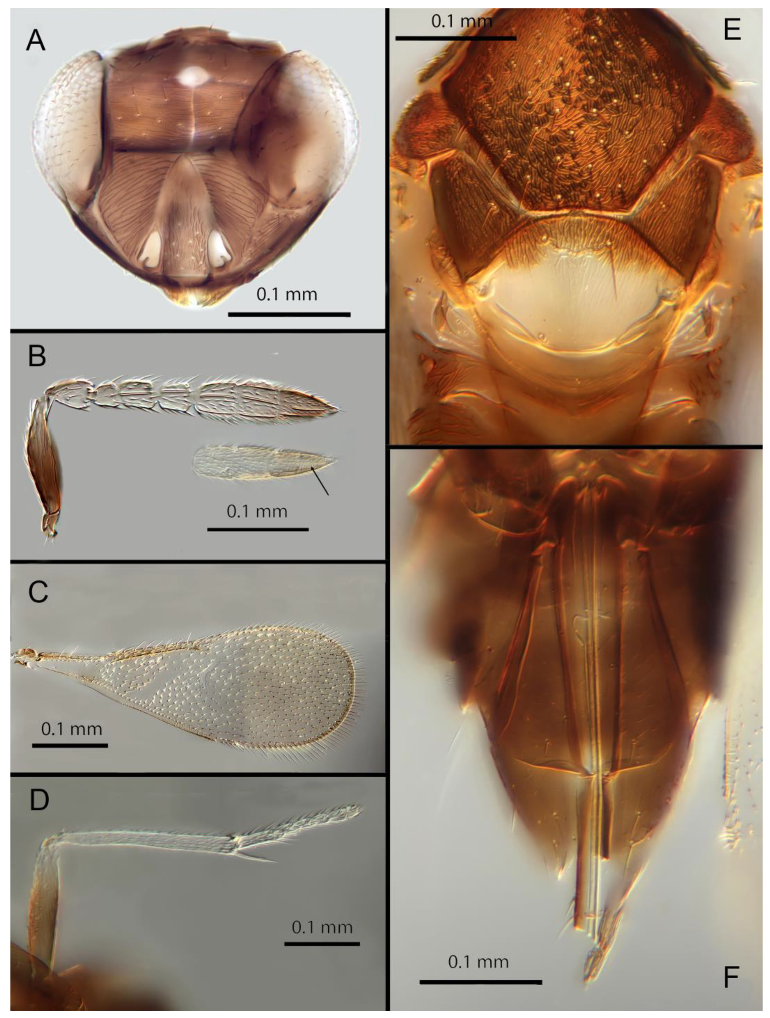
Figure 6.
Encarsia aphania: (A) head; (B) antenna; (C) fore wing; (D) mid leg; (E) dorsal mesosoma; (F) ovipositor.
Figure 6.
Encarsia aphania: (A) head; (B) antenna; (C) fore wing; (D) mid leg; (E) dorsal mesosoma; (F) ovipositor.
tooth and a broad upper truncation; Maxillary palps 2-segmented. Mid-lobe of mesoscutum (Figure 6E) with 30-40 setae; each lateral lobe with 1 seta; each axilla with 1 seta; scutellum with 4 setae. Sculpture of mesoscutum aciculate; sculpture of axillae and scutellum longitudinal. Fore wing (Figure 6C) with 2 large setae and 4-5 smaller setae on submarginal vein, 4-5 setae in basal cell, 5-7 setae on anterior margin of marginal vein, and 2-3 setae at the distal part of the base. Linea calva present. Submarginal 0.9x marginal vein. Maximum length of fore wing 2.6x fore wing width; maximum width of fore wing 5.2x longest setae on marginal fringe. Ovipositor (Figure 6F) 1.6x mid tibial length; third valvula 0.4x ovipositor length; second valvifer 1.3x third valvula. Mid tibial spur (Figure 6D) as long as corresponding basitarsus. Metasomal terga T1-T7 with 0, 2+2, 2+2, 1+1, 1+2+1, 1+2+1 and 5-6 setae, respectively. T7 (Figure 6F) with a pointed, extended apex covering ovipositor.
Distribution. BELIZE: Cayo District; COSTA RICA: Puntarenas.
Hosts. Aleurodicinae: Azuraleurodicus pentarthrus Martin; Nealeurodicus altissimus (Hempel).
Material examined. Holotype ♀ BELIZE, Cayo District, Chiquibul Forest Reserve, Las Cuevas-Monkey Tail trail, 5.iii.1996 (J.H. Martin #6747) ex Azuraleurodicus pentarthus (NHMUK). 13♀ BELIZE, Cayo Las Cuevas, monkey tail trail, 5.iii.1996 (J.H. Martin #6747) ex Azuraleurodicus pentarthus [s27, s22, DNA218: OQ683546]. 1♀ BELIZE, Cayo Chiquibul Fr., Monkey tail trail, 21.iii.2003 (J.H. Martin #7768) ex Nealeurodicus altissimus on Inga sp (all NHMUK). 1♀ COSTA RICA, Puntarenas, Est. Altamira send. Los Gigantes, 9.vii.2001 (D. Rubi #63984) [DNA213: OQ683545] 1.460m, LS 331800 572100 (MZUCR)
Remarks. Encarsia aphania presents a unique combination of characters and appears to have no very close relatives. Morphologically it is closest to E. larensis but is easily distinguishable by the much longer 3rd valvulae relative to the 2nd valvifers (compare Figures 5F & 15F). DNA sequences were obtained from 2 specimens from Belize (type locality) and Costa Rica, Puntarenas; deposited under GenBank accession numbers: OQ683546, OQ683545.
3.3.4. Encarsia avida Polaszek and Hernández-Suárez sp.n.
(Figure 7A–F)
Female. Color. Antennae pale brown, darker on F5-F6 and the scape, pedicel, and radicle. Head brown, paler along the sutures. Mesosoma dark brown with posterior three-quarters of scutellum pale. Metasoma uniformly dark brown. Legs yellow with all coxae, femora, and anterior half of fore leg tibiae brown; all tarsi pale. Wings infuscate below marginal vein; submarginal and marginal veins darker in contrast with the stigmal vein paler.
Morphology. Head (Figure 7A) with mediofrontal line complete; transfacial line obscure; facial lines present, narrow. Scrobes with longitudinally aciculate sculpture centrally, smooth laterally. Antenna (Figure 7B) with eight antennomeres; antennal formula: 1,1,3,3; scape 2.5x pedicel length; pedicel 1.95x F1; F1 0.66x
F2; F2 equal to F3; funicle 0.6x clava; F6 perpendicular. Claval sensorial area present, distinct. Flagellum with the following number of longitudinal sensilla: F1: 0; F2: 1-2; F3: 1; F4: 2; F5: 3; F6: 3. Mandibles (Figure 7A) with 2 small ventral teeth and a truncation. Maxillary palps 2-segmented. Mid-lobe of mesoscutum (Figure 7E) with 40-50 setae; each lateral lobe with 2 setae; each axilla with 1 seta; scutellum with 4 setae. Sculpture of mesoscutum aciculate; sculpture of axillae and scutellum longitudinal. Fore wing (Figure 7C) with 2 large setae and 4 smaller setae on submarginal vein, 4 setae in basal cell, 8 setae on anterior margin of marginal vein, and 1 seta at the junction of the submarginal vein and parastigma. Linea calva present. Submarginal 0.67x marginal vein. Maximum length of fore wing 2.8x fore wing width; maximum width of wing 4.7x longest setae on marginal fringe. Ovipositor (Figure 7F) 1.6x mid tibial length; third valvulae 0.4x ovipositor length; second valvifer 1.5x third valvula. Mid tibial spur (Figure 7D) 0.8x corresponding basitarsus. Metasomal terga T1-T7 with 0, 2+2, 2+2, 2+2, 1+2+1, 1+2+1 and 4 setae, respectively. T7 (Figure 7F) extended and covering ovipositor (damaged in holotype).
Distribution: COSTA RICA: Heredia.
Figure 7.
Encarsia avida: (A) head; (B) antenna, arrow: claval sensory area; (C) fore wing; (D) midleg; (E) dorsal mesosoma; (F) ovipositor.
Figure 7.
Encarsia avida: (A) head; (B) antenna, arrow: claval sensory area; (C) fore wing; (D) midleg; (E) dorsal mesosoma; (F) ovipositor.
Material examined. Holotype ♀ COSTA RICA, Heredia, Est. Biol. La Selva, 10º26´N 84º01´W 75m 27-28.ii.2003 (J.S. Noyes) [DNA143: OQ683547] (NHMUK).
Remarks. Encarsia avida appears morphologically close to E. acusa, with which it shares the color pattern (mesoscutellum anteriorly dark) and wing and antennal morphology.
The ovipositor in E. acusa is longer (1.8x mid tibia; 1.6x in E. avida). The most easily appreciated difference is in the sculpture of the frons: E. avida has scattered, shallow horizontal grooves (Figure 7A) while E. acusa has very dense horizontal grooves (Figure 4A). A similar difference in sculpture is evident on the lateral face. The two species are well-separated
Figure 8.
Encarsia catula: (A) head; (B) antenna; (C) fore wing; (D) midleg; (E) dorsal mesosoma; (F) ovipositor.
Figure 8.
Encarsia catula: (A) head; (B) antenna; (C) fore wing; (D) midleg; (E) dorsal mesosoma; (F) ovipositor.
based on DNA (Figure 1) with E. avida coming out as sister to E. aphania with high support (95%). DNA sequence from holotype deposited under GenBank accession number: OQ683547.
Etymology. From “avida -us” meaning “greedy” (Latin).
3.3.5. Encarsia catula Polaszek and Hernández-Suárez sp.n.
(Figure 8A–F)
Female. Color. Antennae brown. Head dark brown. Mesosoma uniformly dark brown. Legs yellow with mid and hind femora, coxae, and anterior half of tibiae brown, fore leg femora, coxae, and tibiae dark, all tarsi pale. Wings infuscate below marginal vein, submarginal and marginal veins dark, stigmal vein paler.
Morphology. Head (Figure 8A) with mediofrontal line complete; transfacial line broad; facial lines present, narrow. Scrobes with longitudinally aciculate sculpture centrally, irregularly aciculate basally. Antenna (Figure 8B) with eight antennomeres; antennal formula 1,1,3,3; scape expanded, 3.1x pedicel length; pedicel 2x F1; F1 0.9x F2; F2 0.8x F3; funicle 0.48x clava; F6 oblique, claval sensorial area present. Flagellum with the following number of longitudinal sensilla: F1: 0; F2: 0; F3: 1; F4: 3; F5: 3; F6: 3. Mandibles (Figure 8A) with 1 large ventral tooth and a broad truncation. Maxillary palps 2-segmented. Mid-lobe of mesoscutum (Figure 8E) with fewer than 30 setae; each lateral lobe with 2 setae; each axilla with 1 seta; scutellum with 4 setae. Sculpture of mesoscutum aciculate; sculpture of axillae and scutellum longitudinal. Fore wing (Figure 8C) with 2 large setae and 4 smaller setae on submarginal vein, 4 setae in basal cell, 6 setae on anterior margin of marginal vein, and 1 seta at the junction of the submarginal vein and parastigma. Linea calva present. Submarginal equal to marginal vein. Maximum length of fore wing 2.38x fore wing width, maximum width of wing 4.64x longest setae on marginal fringe. Ovipositor (Figure 8F) 1.48x mid tibial length; third valvulae 0.46x ovipositor length; mid tibial length; third valvulae 0.46x ovipositor length; second valvifer 1.2x third valvula. Mid tibial spur (Figure 8D) 1.1x corresponding basitarsus. Metasomal terga T1-T7 with 0, 2+2, 2+2, 2+2, 1+2+1, 1+2+1 and 4 setae, respectively. T7 (Figure 8F) extended although apparently not covering ovipositor.
Distribution. COSTA RICA: Limon.
Material examined. Holotype ♀ COSTA RICA, Limon, Hitoy-Cerere 90º40´N 83º02´W, 21-22.iii.2006 (J.S. Noyes) [DNA 278: OQ683547] (NHMUK).
Remarks. Encarsia catula shares aspects of morphology with E. marynoyesae but can be distinguished by having fewer than 30 setae on the mesoscutum, and V3 more than ½ the length of V2 (less than ½ as long in E. marynoyesae. The two species are relatively close based on DNA (Figure 1). DNA sequence from the holotype is deposited under GenBank accession number: OQ683547.
Etymology. From “catula” meaning dog/whelp (Latin).
3.3.6. Encarsia cylindrica Polaszek and Hernández-Suárez sp.n.
(Figure 9A–F)
Female. Color. Antennae pale brown, slightly darker on F6, F1 and the base of the scape, pedicel, and radicle. Head dark brown. Mesosoma uniformly dark brown. Legs yellow with femora and coxae brown, fore legs with dark tibiae, all tarsi pale. Wings infuscate below submarginal vein, marginal and stigmal veins pale.
Morphology. Head (Figure 9A) with mediofrontal line incomplete, extending halfway to anterior ocellus; transfacial line obscure; facial lines very broad along their entire lengths. Scrobes with faint longitudinal sculpture apically, irregular/lateral sculpture basally. Antenna (Figure 9B) with eight antennomeres; antennal formula: 1,1,3,3; scape 2.75x pedicel length; pedicel equal to F1; F1 0.85x F2; F2 equal to F3; funicle 0.86x clava; F6 perpendicular. Flagellum with the following number of longitudinal sensilla: F1: 2; F2: 2; F3: 2; F4: 2; F5: 3; F6: 3. Mandibles (Figure 9A) with 2 small teeth and a broad truncation. Maxillary palps 2-segmented. Mid-lobe of mesoscutum (Figure 9E) with about 40-50 setae; each lateral lobe with 2 setae; each axilla with 1 seta; scutellum with 4 setae. Sculpture of mesoscutum aciculate; sculpture of axillae and scutellum longitudinal. Fore wing (Figure 9C) with 2 large setae on submarginal vein and 11 smaller setae, 6 setae in basal cell, 11 setae on anterior
Figure 9.
Encarsia cylindrica: (A) head; (B) antenna; (C) fore wing; (D) midleg; (E) dorsal mesosoma; (F) ovipositor.
Figure 9.
Encarsia cylindrica: (A) head; (B) antenna; (C) fore wing; (D) midleg; (E) dorsal mesosoma; (F) ovipositor.
margin of marginal vein, and 1 seta at the junction of the submarginal vein and parastigma. Linea calva absent. Submarginal 0.62x times marginal vein. Maximum length of fore wing 2.7x fore wing width, maximum width of wing 5.87x longest setae on marginal fringe. Ovipositor (Figure 9F) equal to mid tibial length; third valvula 0.45x ovipositor length; second valvifer 1.3x third valvula. Mid tibial spur (Figure 9D) 1.1x corresponding basitarsus. Metasomal terga T1-T7 with 0, 1+1, 1+1, 1+1, 1+2+1, 1+2+1 and 6 setae, respectively. T7 (Figure 9F) rounded, not extended but covering ovipositor.
Distribution. BRAZIL: Minas Gerais; COSTA RICA: Puntareñas, San Juan; JAMAICA.
Host. Aleurodicinae: Aleurodicus jamaicensis Cockerell.
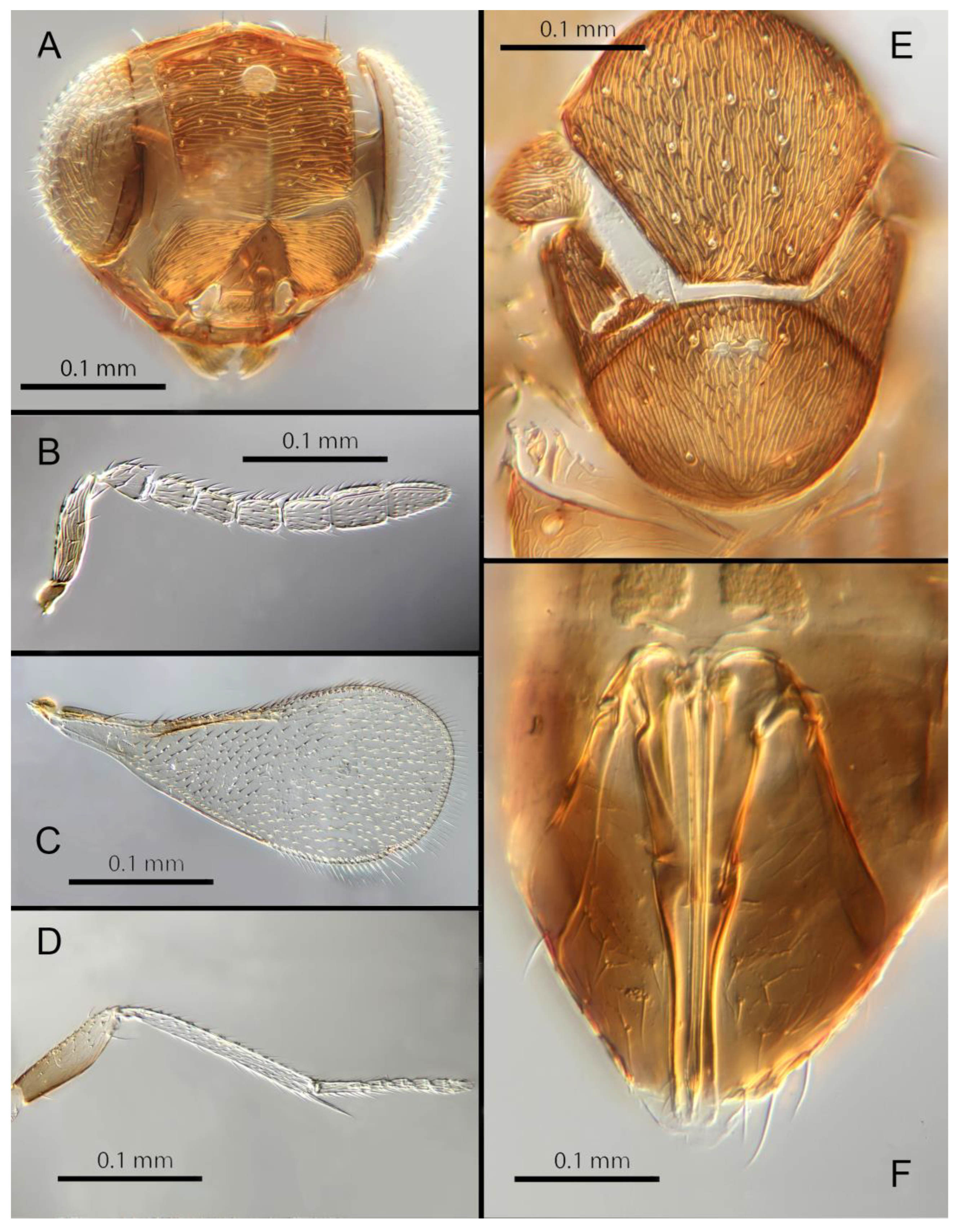
Figure 10.
Encarsia diablejo: (A) head; (B) antenna; (C) fore wing; (D) midleg; (E) dorsal mesosoma; (F) male genitalia.
Figure 10.
Encarsia diablejo: (A) head; (B) antenna; (C) fore wing; (D) midleg; (E) dorsal mesosoma; (F) male genitalia.
Material examined. Holotype ♀ COSTA RICA, San Juan, Ciudad Colon, Heredia El Rodeo coll parataxonomist 16.ii.1991 [DNA 211] (NHMUK). Paratype 1♀ COSTA RICA, Puntareñas R.F. Golfo Dulce, 24Km W. Piedras Blancas [DNA 209] (MZUCR). 4♀JAMAICA, Fair Prospect, xii.1968 (K. Heinze) ex Aleurodicus jamaicensis [s10] (on 1 slide, USNM). 8♀ BRAZIL, Vicosa, Minas Gerais, 6.xi.1935 (E.J. Hambleton) ex Aleurothirxus floccosus (?) (on 1 slide, one head missing, USNM).
Remarks. Encarsia cylindrica appears to be most closely related to E. erwini, with which it shares the elongate antenna and lack of a linea calva. It differs from E. erwini in having many more setae on the mesoscutum.
Etymology. “cylindrica” refers to the almost uniformly elongate antenna.
3.3.7. Encarsia diablejo (Polaszek & Hayat) comb. N.
(Figure 10A–F)
Dirphys diablejo Polaszek & Hayat, 1992: 189
Female. Unknown. This species is known only from the holotype.
Male. Color. Antennae uniformly light brown, slightly darker on the base of the scape, pedicel, and radicle. Head brown with paler areas bordering the eyes and extending along the genae towards the clypeus. Mesosoma and metasoma uniformly brown. Legs light brown, the mid and hind tibia pale in contrast to the dark femora; all tarsi pale. Fore wings hyaline, stigmal vein pale in contrast with a darker marginal vein.
Morphology. Head (Figure 10A) with mediofrontal line complete, though fading towards anterior ocellus; transfacial line complete, narrow; facial lines very broad along their entire lengths. Scrobes entirely with irregular aciculate sculpture. Antenna (Figure 10B) with eight antennomeres; antennal formula 1,1,3,3; scape 2.94x pedicel, F1 subequal to pedicel, F1 0.88X F2, F2 and F3 subequal; funicle 0.89x clava length. Flagellum with the following number of longitudinal sensilla: F1: 7; F2: 6; F3: 7; F4: 8; F5: 9; F6: 7. Mandibles (Figure 10A) with 2 large pointed teeth and a truncation; maxillary palps 2-segmented. Mid-lobe of mesoscutum (Figure 10E) with more than 60 setae; each lateral lobe with 1 seta; each axilla with 1 seta; scutellum with 4 setae and 2 vestigial setal bases. Sculpture of mesoscutum transverse. Fore wing (Figure 10C) with 2 large setae and 4 smaller setae on submarginal vein, 5 setae in basal cell, 11 setae on anterior margin of marginal vein. Linea calva absent. Submarginal 0.79x marginal vein. Maximum length of fore wing 2.48x fore wing width, maximum width of fore wing 7x longest setae on marginal fringe. Mid tibial spur (Figure 10D) as long as corresponding basitarsus.
Distribution. PERU: Loreto.
Host. Unknown.
Material examined. Holotype ♂ PERU, Loreto, Iquitos, Granja Unap, 9.ii.1984 (L. Huggert #BM 1984-337) [s26] (NHMUK).
Remarks. For the purposes of the identification key, we have assumed that the (unknown) female of E. diablejo shares the wing and mesosomal sculpture characters with the male; the combination of which is unique in the Encarsia mexicana species-group.
3.3.8. Encarsia dichaeta Polaszek and Hernández-Suárez sp.n.
(Figure 11A–F)
Female. Color. Antennae light brown, slightly darker on the base of the scape, pedicel, and radicle. Head dark brown with pale lines bordering the eyes and extending along the genae towards clypeus. Mesosoma and metasoma uniformly dark brown. Legs yellow with femora and coxa brown, tibia, and all tarsi pale. Wings infuscate below submarginal and marginal veins, stigmal vein pale.
Morphology. Head (Figure 11A) with mediofrontal line complete; transfacial line narrow; facial lines relatively narrow along their entire lengths. Scrobes with irregular aciculate sculpture. Antenna (Figure 11B) with eight antennomeres; antennal formula: 1,1,3,3; scape
Figure 11.
Encarsia dichaeta: (A) head; (B) antenna; (C) fore wing; (D) midleg; (E) dorsal mesosoma; (F) ovipositor.
Figure 11.
Encarsia dichaeta: (A) head; (B) antenna; (C) fore wing; (D) midleg; (E) dorsal mesosoma; (F) ovipositor.
2.59x pedicel length; pedicel 1.85x F1; F1 0.85x F2; F2 0.8x F3; funicle 0.65x clava length; F6 perpendicular. Flagellum with the following number of longitudinal sensilla: F1:0; F2:1; F3:1; F4:1-2; F5:1-2; F6:1-2. Mandibles (Figure 11A) with 2 small teeth and a truncation. Maxillary palps 2-segmented. Mid-lobe of mesoscutum (Figure 11E) with more than 50 setae; each lateral lobe with 2 setae; each axilla with 2 setae scutellum with 4 setae and 2 vestigial setal bases. Sculpture of mesoscutum aciculate; sculpture of axillae and scutellum longitudinal. Fore wing (Figure 11C) with 2 large setae and 7-8 smaller setae on submarginal vein, 14 setae
in basal cell, 8 setae on anterior margin of marginal vein, and 1 seta at the junction of the submarginal vein and parastigma. Linea calva absent. Submarginal 0.85x times marginal vein. Maximum length of fore wing 2.48x fore wing width, maximum width of wing 5.45x longest setae on marginal fringe. Ovipositor (Figure 11F) 0.93x mid tibial length; third valvulae 0.45x ovipositor; second valvifer 1.6x third valvula. Mid tibial spur (Figure 11D) equal to corresponding basitarsus. Metasomal terga T1-T7 with 0,1+1,1+1,1+1, 1+2+1, 1+2+1 and 12 setae, respectively. T7 (Figure 11F) rounded, not extended but covering ovipositor.
Distribution. BRAZIL: Bahia; COSTA RICA: Guanacaste, Alajuela, Heredia; ECUADOR: Napo River.
Host. Aleurodicinae: Aleurodicus flavus Hempel, Aleurodicus sp.
Material examined. Holotype 1♀ COSTA RICA, Alajuela, P.N. Arenal Sendero Pilon, 10º27´N 84º45´W 600m 25.ii.2003 (J.S. Noyes) [DNA 136: OQ683552] (NHMUK). Paratypes 3♀ COSTA RICA, Alajuela, P.N. Arenal, Sendero Pilon, 10º27´N 84º45´W 600m 25.ii.2003 (J.S. Noyes), [DNA 132: OQ683550, 133: OQ683552, 135: OQ683550] (2♀ NHMUK, 1♀ MZUCR). 1♀ COSTA RICA, Heredia, Est. Biol. La Selva, 10º26´N 84º01´W 75m 27-28.ii.2003 (J.S. Noyes) [DNA151: OQ683549] (UCRC); 1♀ COSTA RICA, P.N. Guanacaste,
Est. Pitilla (ACG), 11º00’N. 85º26’W. 700 m MT/YPT (J.S. Noyes) [DNA 208: OQ683551] (NHMUK). 27♀♀ BRAZIL, Bahia (Gregorio Bondar # nº65b) ex Aleurodicus flavus (on 5 slides; USNM). 5♀ ECUADOR, Napo, Camino Añangucocha, 29.iii.04 (H. Evans) ex Aleurodicus sp. (NHMUK).
Remarks. There are some color differences between the Costa Rican specimens and those from Brazil, the latter having the metasoma distally paler. Further studies on fresh material, in particular DNA sequencing, will be needed to confirm their status. DNA sequences were obtained from the holotype and 5 paratypes, deposited under GenBank accession numbers: OQ683550, OQ683552, OQ683551 and OQ683549.
Etymology. “dichaeta” refers to the 2 setae on each axilla, unique for the genus.
3.3.9. Encarsia encantadora (Polaszek & Hayat) comb. n.
(Figure 12A–F)
Dirphys encantadora Polaszek & Hayat, 1992: 191.
Female. Color. Antennae pale brown/yellow with dark scape and radicle. Head brown with paler areas bordering the eyes and extending along the genae towards the clypeus, antennal scrobes, a line from the apex of the scrobes to the median ocellus, and a transverse line midway between the antennal sockets and the median ocellus. Mesosoma brown in holotype but with posterior three-quarters of scutellum pale in Mexican specimens. Legs light brown, the mid and hind tibiae pale in contrast to the dark femora and coxa, all tarsi pale. Wings hyaline, faintly infuscate below the marginal vein, stigmal vein pale in contrast with a darker marginal vein.
Morphology. Head (Figure 12A) with mediofrontal line complete; transfacial line evident; facial lines very broad along their entire lengths. Scrobes with longitudinally aciculate sculpture. Antenna (Figure 12B) with eight antennomeres; antennal formula 1,1,3,3; scape 2.3x pedicel length; pedicel equal to F1; F1 to F3 funicle segments all subequal in length; funicle 0.75x clava length; F6 perpendicular; Flagellum with the following number of longitudinal sensilla: F1: 1-2; F2: 1; F3: 1; F4: 2-3; F5: 3; F6: 4. Mandibles (Figure 12A) with 2 small ventral teeth and a broad truncation dorsally. Maxillary palps 2-segmented. Mid-lobe of mesoscutum (Figure 12E) with fewer than 30 setae; each lateral lobe with 3 setae; each axilla with 1 seta; scutellum with 4 setae. Sculpture of mesoscutum aciculate; sculpture of axillae and scutellum longitudinal. Fore wing (Figure 12C) with 2 setae on submarginal vein, 7-9 setae in basal cell, 7-11 setae on anterior margin of marginal vein, and 1 seta at the junction of the submarginal vein and parastigma. Linea calva absent. Submarginal 0.73x times marginal vein. Maximum length of fore wing 2.52x fore wing width, maximum width of fore wing 6.9x longest setae on marginal fringe. Ovipositor (Figure 12F) 0.82x mid tibial length; third valvula 0.55x ovipositor length; second valvifer 0.79x third valvula. Mid tibial spur (Figure 12D) 0.87x corresponding basitarsus. Metasomal terga T1-T7 with 0, 1+1, 1+1, 1+1,
Figure 12.
Encarsia encantadora: (A) head; (B) antenna; (C) fore wing; (D) midleg; (E) dorsal mesosoma; (F) ovipositor.
Figure 12.
Encarsia encantadora: (A) head; (B) antenna; (C) fore wing; (D) midleg; (E) dorsal mesosoma; (F) ovipositor.
1+2+1, 1+2+1 and 4 setae, respectively. T7 (Figure 12F) rounded and covering ovipositor third valvula.
Distribution. ECUADOR: Napo; MEXICO: Tabasco.
Host. Aleurodicinae: Nealeurodicus altissimus Hempel.
Material examined. Holotype ♀ECUADOR, Napo, Sacha, 5.iii.1983 (L. Huggert) (BHMN). 1♀, fragments of a second ♀: MEXICO, Tabasco, San Francisco del Peal, 1.vii.1897 (C.H.T. Townsend) ex Nealeurodicus altissimus (Quaintance) on Lippia myriophala Schltdl. & Cham. (USNM; on slide with E. mexicana type material).
Figure 13.
Encarsia erwini: (A) head; (B) antenna; (C) fore wing; (D) midleg; (E) dorsal mesosoma; (F) ovipositor.
Figure 13.
Encarsia erwini: (A) head; (B) antenna; (C) fore wing; (D) midleg; (E) dorsal mesosoma; (F) ovipositor.
Remarks. Encarsia encantadora is morphologically closest to E. erwini, differing from that species mainly in having the 3rd valvulae longer than the second valvifers. The fore wing is also broader in E. encantadora, especially measured relative to the longest wing fringe setae (compare Figures 11C & 12C).
3.3.10. Encarsia erwini Polaszek & Hernández-Suárez sp.n.
(Figure 13A–F)
Female. Color. Antennae entirely pale, only the scape and radicle dark. Head dark brown. Mesosoma uniformly dark brown. Legs entirely pale except all coxae brown (female paratype with some infuscation on the hind femora).
Morphology. Head (Figure 13A) with mediofrontal line complete; transfacial and facial lines very broad along their entire lengths. Scrobes with irregularly aciculate sculpture centrally. Antennae (Figure 13B) with eight antennomeres; antennal formula: 1,1,3,3; scape 2.4x pedicel length; pedicel 1.2x F1 equal to F2; F2 equal to F3; funicle 0.77x clava; F6 perpendicular. Flagellum with the following number of longitudinal sensilla: F1: 1; F2: 1; F3: 1; F4: 1; F5: 2; F6: 2. Mandibles (Figure 13A) with 2 ventral teeth and a truncation dorsally. Maxillary palps 2-segmented. Mid-lobe of mesoscutum (Figure 13E) with about 18 setae; each lateral lobe with 3 setae; each axilla with 1 seta; scutellum with 4 setae. Sculpture of mesoscutum and axillae longitudinal aciculate; sculpture of scutellum longitudinal, transverse apically. Fore wing (Figure 13C) with 2 large setae on submarginal vein and 5 smaller setae above, 6 setae in basal cell, 7 setae on anterior margin of marginal vein, and 1 large seta at the junction of the submarginal vein and parastigma. Linea calva absent. Submarginal vein approximately equal in length to marginal vein. Maximum length of fore wing 2.9x fore wing width, maximum width of wing 3.75x longest seta on marginal fringe.
Ovipositor (Figure 13F) equal to mid tibial length; third valvula 0.44x ovipositor length; second valvifer 1.3x third valvula. Mid tibial spur (Figure 13D) 1.0x corresponding basitarsus. Metasomal terga T1-T7 with 0, 1+1, 1+1, 1+1, 1+2+1, 2+2 and 7 setae, respectively. T7 (Figure 13F) conical, not extended but just covering ovipositor.
Distribution. ECUADOR: Napo.
Material examined. Holotype ♀ ECUADOR, Napo, transect ent. 1 km S. Onkone gare Camp, Res. Etnica Waorani 220m 0°39’10”S 76°26’00”W TL Erwin et al fogging t.f. forest. Lot #1255 8.x.1995 (UCRC; 52715). Paratype ♀, same data as holotype except 4.x.1996 (NHMUK).
Remarks. Encarsia erwini appears to be most closely related to E. cylindrica, with which it shares the elongate antenna and lack of a linea calva. It differs from E. cylindrica in having far fewer setae on the mesoscutum. E. erwini is also morphologically close to E. encantadora, differing from that species mainly in having the 3rd valvulae shorter than the second valvifers. The fore wing is also broader in E. encantadora, especially measured relative to the longest wing fringe setae.
Etymology. Named for the late Terry Erwin (1940-2020) prolific collector of insects, especially in the rain forest canopy of Ecuador.
3.3.11. Encarsia fredbennetti Polaszek & Hernández-Suárez sp. n.
(Figure 14A–E)
Female. Color. Antennae uniformly pale brown. Head brown with pale lines bordering the eyes and extending along the genae towards clypeus. Mesosoma dark brown with most of scutellum and post-scutellum pale; metasoma uniformly brown. Legs yellow with dark coxae, femora and anterior third of hind leg tibia. Wings infuscate below marginal vein, stigmal vein pale in contrast with darker marginal vein.
Morphology. Head (not shown) with all facial lines obscure in holotype, (head absent in paratypes). Scrobes with longitudinally aciculate sculpture. Scrobes with longitudinally aciculate sculpture. Antenna (Figure 14D) with eight antennomeres; antennal formula: 1,1,3,3 (though could be interpreted as 1,1,2,4); scape 2.39x pedicel length; pedicel 1.9x F1; F1 0.8x F2; F2 0.9x F3; funicle 0.6x clava; F6 perpendicular. Flagellum with the following number of longitudinal sensilla: F1: 0; F2: 1; F3: 1; F4: 2-3; F5: 3; F6: 3. Mandibles with 1 ventral tooth and a truncation. Maxillary palps 2-segmented. Mid-lobe of mesoscutum (Figure 14C) with 30-40 setae; each lateral lobe with 2 setae; each axilla with 1 seta; scutellum with
Figure 14.
Encarsia fredbennetti: (A) fore wing; (B) mid leg; (C) dorsal mesosoma; (D) antenna; (E) ovipositor.
Figure 14.
Encarsia fredbennetti: (A) fore wing; (B) mid leg; (C) dorsal mesosoma; (D) antenna; (E) ovipositor.
4 setae. Sculpture of mesoscutum aciculate; sculpture of axillae and scutellum longitudinal. Fore wing (Figure 14A) with 2 large setae and 4 smaller setae on submarginal vein, 4-5 setae in basal cell, 7 setae on anterior margin of marginal vein. Linea calva present. Submarginal 0.93x marginal vein. Maximum length of fore wing 3.8x fore wing width, maximum width of wing 3.7x longest setae on marginal fringe. Ovipositor (Figure 14E) 1.3x mid tibial length; third valvulae 0.5x ovipositor length; second valvifer equal to third valvula. Mid tibial spur (Figure 14B) equal to corresponding basitarsus. Metasomal terga T1-T7 with 0, 2+2, 2+2, 2+2, 1+2+1, 1+2+1 and 4 setae, respectively. T7 (Figure 14E) extended, covering ovipositor.
Distribution. TRINIDAD: St Augustine.
Host. Aleurodicinae.
Material examined. Holotype ♀ TRINIDAD, [St Augustine] ICTA [Imperial College of Tropical Agriculture] xii.1953 FD Bennett ex whitefly on cocoa (NHMUK); Paratype ♀ TRINIDAD, St Augustine, ex Aleurodicinae [DNA215: OQ683559] (NHMUK); Paratype ♀ TRINIDAD, Mt St Benedict, ex whitefly Coll. Mm. Jagroep [DNA216] (NHMUK).
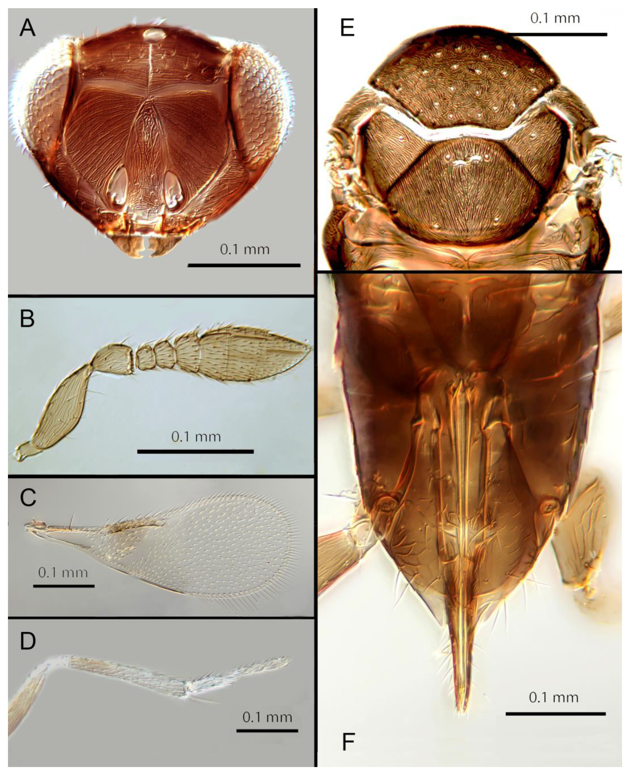
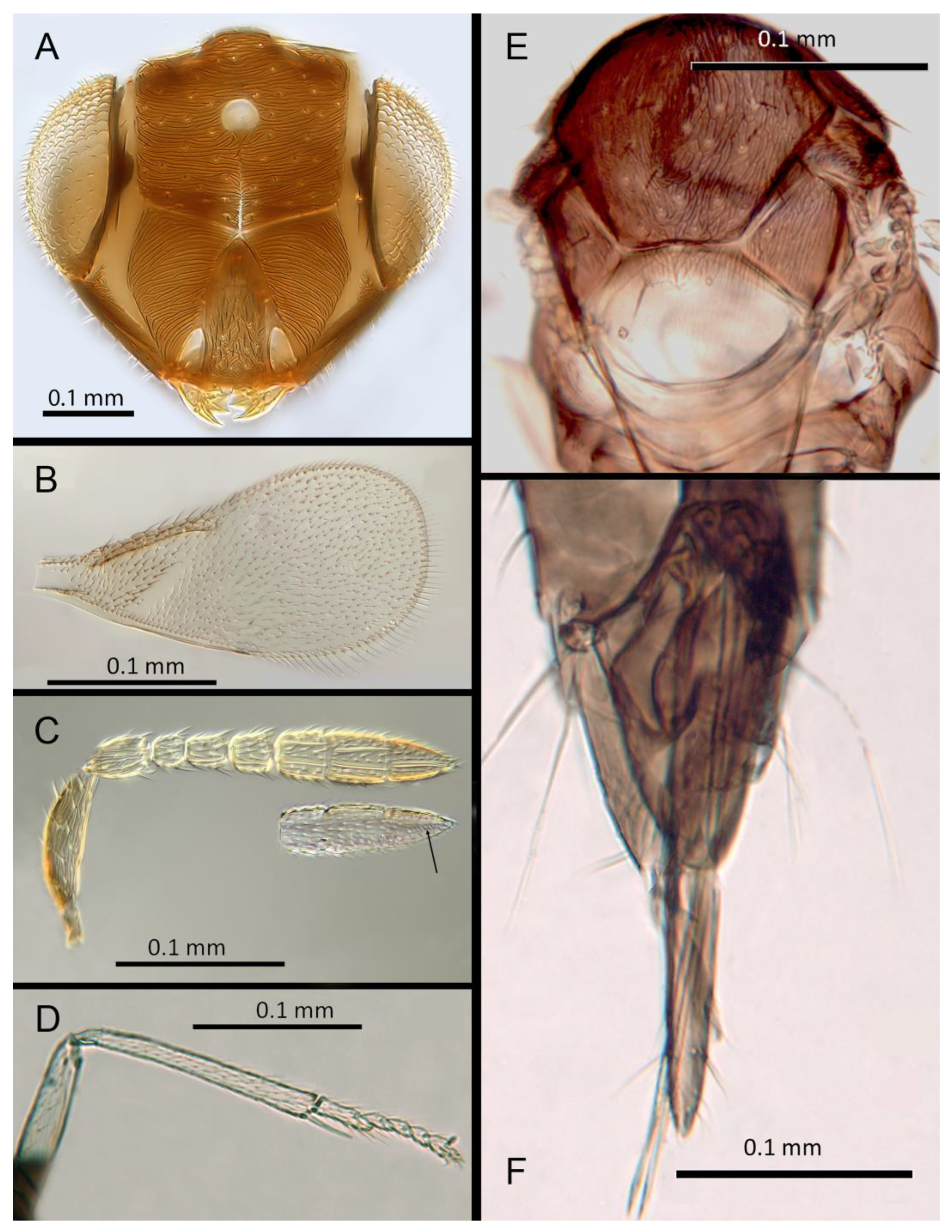
Figure 15.
Encarsia inbioa: (A) head; (B) fore wing; (C) antenna, arrow: claval sensory area; (D) mid leg; (E) dorsal mesosoma; (F) ovipositor.
Figure 15.
Encarsia inbioa: (A) head; (B) fore wing; (C) antenna, arrow: claval sensory area; (D) mid leg; (E) dorsal mesosoma; (F) ovipositor.
Remarks. Encarsia fredbennetti is morphologically closest to E. mexicana and E.inbioa from which it differs by the enlarged clava. It is also molecularly closest to E. mexicana. Deposited under GenBank accession number: OQ683559.
Etymology. Named for the late Fred D. Bennett (1925-2021). Former Director the Commonwealth Institute of Biological Control, and avid collector of parasitoids during much of his long life.
3.3.12. Encarsia inbioa Polaszek & Hernández-Suárez sp. n.
(Figure 15A –F)
Female. Color. Antennae light brown slightly darker at F5 and F6, the base of the scape, pedicel, and radicle. Head brown with pale lines bordering the eyes and extending along the genae towards clypeus. Mesosoma dark brown but with posterior quarter of scutellum pale; metasoma uniformly dark brown. Legs yellow with dark coxae, femora and anterior third of hind leg tibia. Wings infuscate below marginal vein, stigmal vein pale in contrast with the darker marginal vein.
Morphology. Head (Figure 15A) with mediofrontal line incomplete, reaching to less than half the distance to the frontal ocellus; transfacial line evident, narrow; facial lines very broad along their entire lengths, particularly at the level of the lower eye. Scrobes with aciculate/reticulate sculpture basally and centrally, smooth apically and apico-laterally. Antenna (Figure 15C) with eight antennomeres; antennal formula: 1, 1, 3, 3; scape 2.39x pedicel length; pedicel 1.9x F1; F1 0.8x F2; F2 0.9x F3; funicle 0.6x clava; F6 perpendicular. Flagellum with the following number of longitudinal sensilla: F1: 0; F2: 1; F3: 1; F4: 2-3; F5: 3; F6: 3. Mandibles (Figure 15A) with 2 large teeth and a truncation. Maxillary palps 2-segmented. Mid-lobe of mesoscutum (Figure 15E) with 30-40 setae; each lateral lobe with 2 setae; each axilla with 1 seta; scutellum with 4 setae. Sculpture of mesoscutum aciculate; sculpture of axillae and scutellum longitudinal. Fore wing (Figure 15B) with 2 large setae and 4 smaller setae on submarginal vein, 4-5 setae in basal cell, 7 setae on anterior margin of marginal vein. Linea calva present. Submarginal 0.93x marginal vein. Maximum length of fore wing 3.8x fore wing width, maximum width of wing 3.7x longest setae on marginal fringe. Ovipositor (Figure 15F) 1.3x mid tibial length; third valvulae 0.5x ovipositor length; second valvifer equal to third valvula. Mid tibial spur (Figure 15D) equal to corresponding basitarsus. Metasomal terga T1-T7 with 0, 2+2, 2+2, 2+2, 1+2+1, 1+2+1 and 4 setae, respectively. T7 (Figure 15F) extended and covering ovipositor.
Distribution. COSTA RICA: Alajuela.
Material examined. Holotype ♀ COSTA RICA, Alajuela, P.N. Arenal, Sendero Pilon, 26.ii.2003 (J.S. Noyes) [DNA 128: OQ683553] 600m 10º27´N 84º43´W (NHMUK).
Remarks. Encarsia inbioa is morphologically closest to E. fredbennetti from which it differs by the non-enlarged clava. It is, perhaps surprisingly, molecularly closest to E. svetlana. Deposited under GenBank accession number: OQ683553.
Etymology. Named for INBio (Instituto Nacional de Biodiversidad) the national institute for biodiversity and conservation in Costa Rica.
3.3.13. Encarsia larensis (Chavez) comb. n.
Dirphys larensis Chavez, 1996: 11
(Figure 16A–F)
Female. Color. Antennae pale brown with slightly darker clava and pedicel. Head dark brown with paler areas bordering the eyes and extending along the genae towards the clypeus. Mesosoma and metasoma uniformly dark brown, third valvulae dark brown contrasting with the rest of ovipositor. Legs pale, mid and hind femur, and coxae brown, anterior third of mid leg tibia brown. Wings infuscate below the submarginal and marginal vein; marginal and stigmal veins dark.
Morphology. Head (Figure 16A) with mediofrontal line complete, reaching to the frontal ocellus; other facial lines obscure in paratypes examined due to mounting method. Scrobes with coarse longitudinal aciculate sculpture becoming irregular towards clypeus. Antenna (Figure 16B) with 8 antennomeres; antennal formula 1,1,3,3; scape expanded, 2.4-2.5x pedicel length; pedicel 1.9x F1; F1 equal to F2; F2 0.8x F3; funicle 0.58x clava; F6 oblique.
Figure 16.
Encarsia larensis: (A) head; (B) antenna; (C) fore wing; (D) mid leg; (E) dorsal mesosoma; (F) ovipositor.
Figure 16.
Encarsia larensis: (A) head; (B) antenna; (C) fore wing; (D) mid leg; (E) dorsal mesosoma; (F) ovipositor.
Flagellum with the following number of longitudinal sensilla: F1: 0; F2: 0; F3: 1; F4: 4; F5: 5-6; F6: 4. Mandibles (not shown) with 1 large ventral tooth and a bidentate upper tooth.
Maxillary palps 2-segmented. Mid-lobe of mesoscutum (Figure 16E) with 46-60 setae; each lateral lobe with 2 setae; each axilla with 1 seta; scutellum with 4 setae and 2 vestigial setal bases. Sculpture of mesoscutum aciculate; sculpture of axillae and scutellum longitudinal. Fore wing (Figure 16C) with 2 large setae and 3-4 smaller setae on submarginal vein, 7-9 setae on anterior margin of marginal vein, and 1 seta at the junction of the submarginal vein and parastigma. Linea calva present. Submarginal equal to marginal vein; Maximum length of fore wing 2.54x fore wing width; maximum width of fore wing 6.7x longest setae on marginal fringe. Ovipositor (Figure 16F) 1.30x mid tibial length; third valvula 0.28x ovipositor length; second valvifer 2.4x third valvula. Mid tibial spur (Figure 16D) 1.15x corresponding basitarsus. Metasomal terga T1-T7 with 0, 2+2, 2+2, 2+2, 1+2+1, 1+2+1 and 4 setae, respectively. T7 (Figure 16F) rounded not covering ovipositor third valvula.
Male. Color. Head light brown. Mesosoma and gaster uniformly brown but posterior third of mesoscutum, anterior third of axillae and scutellum yellow. Legs brown. Fore wings hyaline.
Morphology. Similar to that of female, except antennal formula, flagellum with longitudinal sensilla on all segments and funicle segments subequal in length.
Distribution. VENEZUELA: Cabudare, Lara.
Host. Aleurodicinae: Aleurodicus pulvinatus (Maskell)
Material examined. 1♀, 1♂: VENEZUELA, Cabudare, Lara, i.1994 (A. Chavez & F. Díaz) ex Aleurodicus pulvinatus on Hura crepitans L.
Remarks. Encarsia larensis appears morphologically closest to E. marynoyesae from which it differs in having a much longer funicle, and shorter ovipositor. No molecular data were available for this species. Chavez (1996) recorded 16 E. larensis within a single whitefly host.
3.3.14. Encarsia marynoyesae Polaszek & Hernández-Suárez sp.n.
(Figure 17A–F)
Female. Color. Antennae brown with F1 and F2 paler. Head dark brown with pale lines bordering the eyes and extending along the genae towards clypeus. Mesosoma uniformly dark brown. Legs yellow with mid and hind femora, coxa and anterior third of tibia brown, all tarsi pale. Wings slightly infuscated below anterior half of submarginal vein, stigmal vein pale in contrast with a darker marginal vein.
Morphology. Head (Figure 17A) with mediofrontal line complete; transfacial line narrow; facial lines very broad along their entire lengths. Scrobes with irregular aciculate sculpture. Antenna (Figure 17B) with eight antennomeres; antennal formula 1,1,3,3; scape slightly expanded, 2.53x pedicel length; pedicel 2.85x F1; F1 equal to F2; F2 0.8x F3; funicle 0.36x clava; F5 and F6 strongly oblique, claval sensorial complex developed. Flagellum with the following number of longitudinal sensilla: F1: 0; F2: 0-1; F3: 1; F4: 3; F5: 4; F6: 4. Mandibles (Figure 17A) with 1 small ventral tooth and a bidentate upper tooth. Maxillary palps 2-segmented. Mid-lobe of mesoscutum (Figure 17E) with approximately 40 setae; each lateral lobe with 2 setae; each axilla with 1 seta; scutellum with 4 setae and 2 vestigial bases. Sculpture of mesoscutum aciculate; sculpture of axillae and scutellum longitudinal. Fore wing (Figure 17C) with 2 large setae and 4 smaller setae on submarginal vein, 4 setae in basal cell, 6-7 setae on anterior margin of marginal vein, and 1 seta at the junction of the submarginal vein and parastigma. Linea calva present. Submarginal equal to marginal vein. Maximum length of fore wing 2.57x fore wing width, maximum width of wing 6.2x longest setae on marginal fringe. Ovipositor (Figure 17F) 1.4x mid tibial length; third valvulae 0.35x ovipositor length; second valvifer 1.9x third valvula. Mid tibial spur (Figure 17D) 1.07x corresponding basitarsus. Metasomal terga T1-T7 with 0, 1+1, 1+1, 1+1, 1+2+1, 1+2+1 and 4 setae, respectively. T7 (Figure 17F) extended although apparently not covering ovipositor.
Distribution. COSTA RICA (Alajuela).
Material examined. Holotype ♀ COSTA RICA, Alajuela, Est. Caribe Reserva Rincón Forestal, 19-20.ii.2003 (J.S. Noyes) [DNA 163: OQ683563] 400m, 10º53´N 85º18´W (NHMUK). Paratype 1♀ COSTA RICA, Alajuela, Est. Caribe Reserva Rincón Forestal, 19-20.ii.2003 (J.S. Noyes) [DNA167] (NHMUK).
Remarks. Encarsia marynoyesae is morphologically very similar to E. aisha in many respects (though distant to it based on DNA). The species can be distinguished by E. marynoyesae having the 2nd valvifers almost 2x (1.8) the 3rd valvulae; while they are 1.5x
Figure 17.
Encarsia marynoyesae: (A) head; (B) antenna, arrow: claval sensory area; (C) fore wing; (D) mid leg; (E) dorsal mesosoma; (F) ovipositor.
Figure 17.
Encarsia marynoyesae: (A) head; (B) antenna, arrow: claval sensory area; (C) fore wing; (D) mid leg; (E) dorsal mesosoma; (F) ovipositor.
as long in E. aisha. In E. marynoyesae the clava is well over 2x the length of the funicle; in E. aisha it is less than 2x as long. E. marynoyesae also shares aspects of morphology with E. catula, but can be distinguished by having more than 30 setae on the mesoscutum, and V3 less than ½ the length of V2 (much more than ½ as long in E. catula). Sequences deposited under GenBank accession number: OQ683563
Etymology. Named for Mary Noyes MBE.
3.3.15. Encarsia mendesi (Polaszek & Hayat) comb. n.
(Figure 18A–F)
Dirphys mendesi, Polaszek & Hayat 1992: 191
Figure 18.
Encarsia mendesi: (A) head; (B) antenna; (C) fore wing; (D) mid leg; (E) dorsal mesosoma; (F) ovipositor.
Figure 18.
Encarsia mendesi: (A) head; (B) antenna; (C) fore wing; (D) mid leg; (E) dorsal mesosoma; (F) ovipositor.
Female. Color. Antennae brown, paler on their ventral halves. Head dark brown with pale lines bordering the eyes and extending along the genae towards clypeus, antennal scrobes, a line from the apex of the scrobes to the median ocellus, and a transverse line midway between the antennal sockets and the median ocellus centrally bordering the dorsal end of antennal scrobes. Mesosoma and metasoma uniformly dark brown. Legs pale yellow, with coxae and hind femora dark brown. Wings infuscated below the submarginal and marginal vein; stigmal vein pale in contrast with a darker marginal vein.
Morphology: Head (Figure 18A) with mediofrontal line complete check, ocellus; transfacial line evident; facial lines very broad along their entire lengths, especially at level of lower eye and adjacent to genae. Scrobes largely smooth, some irregular sculpture centrally. Antennae (Figure 18B) with eight antennomeres. Funicle apparently absent, so the entire flagellum clavate (antennal formula therefore 1,1,6); scape expanded, 2.3-2.8x pedicel length; pedicel 2.4x F1; F1 0.9x F2; F2 0.8x F3; funicle 0.5x clava; F5 and F6 broadly oblique, claval sensorial complex developed. Flagellum with the following number of longitudinal sensilla: F1: 0; F2: 1; F3: 1; F4: 3; F5: 4; F6: 3. Mandibles missing from holotype; paratype (male) apparently with 2 teeth. Maxillary palps 2-segmented. Mid-lobe of mesoscutum (Figure 18E) with fewer than 30 setae; each lateral lobe with 2 setae; each axilla with 1 seta; scutellum with 4 setae. Sculpture of mesoscutum transverse. Fore wing (Figure 18C) with 2 large setae and 2-3 smaller setae on submarginal vein, 3-5 setae in basal cell, 7-11 setae on anterior margin of marginal vein, and 1 seta at the junction of the submarginal vein and parastigma. Linea clava present. Submarginal equal to marginal vein; maximum width of fore wing 2.56x fore wing width, maximum width of wing 4.6x longest seta on marginal fringe. Ovipositor (Figure 18F) 0.8x mid tibial length; third valvulae 0.3x ovipositor length; second valvifer 2.13x third valvula. Mid tibial spur (Figure 18D) equal to corresponding basitarsus. Metasomal terga T1-T7 with 0, 1+1, 1+1, 1+1, 1+2+1, 1+2+1 and 6 setae, respectively. T7 (Figure 18F) rounded covering ovipositor third valvula.
Male. All aspects of coloration and morphology as for female, except the antennae and genitalic characters.
Distribution. BRAZIL: Sao Paulo.
Host. Aleurodicinae: Aleurodicus maritimus Hempel.
Material examined. Holotype ♀ BRAZIL, São Paulo, Mogi-Guazu, 12.v.1981 (M. Cytrynowicz) 84/8 ex Aleurodicus maritimus (NHMUK). Paratype 1♂ BRAZIL, São Paulo, Mogi-Guazu, 12.v.1981 (M. Cytrynowicz) 84/8 ex Aleurodicus maritimus. 1♂ 1♀ BRAZIL, São Paulo, E.E. Mogi-Guazu, 12.v.1981 (M. Cytrynowicz) 94 ex Aleurodicus maritimus on Bauhinia holophylla (Bong.) (Fabaceae) (all NHMUK).
Remarks. Morphologically E. mendesi appears closest to E. marynoyesae having the entire flagellum more or less clavate. It differs from that species by the very short ovipositor. No molecular data were available for E. mendesi.
3.3.16. Encarsia mexicana (Howard) comb. n.
(Figure 19A–F)
Mesidia mexicana Howard, 1907:
Dirphys mexicana (Howard, 1914)
Female. Color. Antennae pale brown, slightly darker on the base of the scape and radicle. Head dark brown with pale lines bordering the eyes and extending along the genae towards clypeus, antennal scrobes, a line from the apex of the scrobes to the median ocellus, and a transverse line midway between the antennal sockets and the median ocellus centrally bordering the dorsal end of antennal scrobes. Mesosoma and metasoma dark brown, with the posterior two-thirds of the scutellum and sides of the metanotum yellow. Legs yellow, with mid and hind coxae and femora partly brown. Wings slightly infuscated below the marginal vein; submarginal, marginal and stigmal veins dark.
Morphology. Head (Figure 19A) with mediofrontal line complete; transfacial line narrow; facial lines very broad adjacent to genae. Scrobes almost entirely with longitudinal sculpture. Antenna (Figure 19B) with eight antennomeres; antennal formula 1,1,3,3; scape slightly expanded, 2.45x pedicel length; pedicel 1.9x F1; F1 0.8X F2; F2 0.9x F3; funicle 0.57X clava length; F6 slightly oblique, claval sensorial complex developed. Flagellum with the following numbers of longitudinal sensilla: F1: 0; F2: 1; F3: 1; F4: 3; F5: 3-4; F6: 4-5. Mandibles (Figure 19A) with 2 teeth and a truncation. Maxillary palps 2-segmented. Mid-lobe of mesoscutum (Figure 19E) with 30-40 setae; each lateral lobe with 2 setae; each axilla with 1 seta; scutellum with 4 setae. Sculpture of mesoscutum aciculate; sculpture of axillae and scutellum longitudinal. Fore wing (Figure 19C) with 2 large setae and 3-4 smaller setae on submarginal vein, 3-7 setae in basal cell, 7-10 setae on anterior margin of marginal vein, and 1 seta at the junction of the submarginal vein and parastigma. Linea calva present. Submarginal 0.85x marginal vein. Maximum length of fore wing 2.7x fore wing width, maximum width of wing 4.65x longest setae on marginal fringe. Ovipositor (Figure 19F)
Figure 19.
Encarsia mexicana: (A) head; (B) antenna; (C) fore wing; (D) mid leg; (E) dorsal mesosoma; (F) ovipositor.
Figure 19.
Encarsia mexicana: (A) head; (B) antenna; (C) fore wing; (D) mid leg; (E) dorsal mesosoma; (F) ovipositor.
length 1.3x mid tibial length; third valvula 0.5x ovipositor; second valvifer 1.3x third valvula; Mid tibial spur (Figure 19D) 0.9x corresponding basitarsus. Metasomal terga T1-T7 with 0, 2+2, 2+2, 2+2, 1+2+1, 1+2+1 and 4 setae, respectively. T7 (Figure 19F) elongate covering third valvula of ovipositor.
Distribution. COSTA RICA: Limon, Heredia, Arenal, Alajuela; MEXICO: Tabasco.
Host. Nealeurodicus altissimus (Quaintance) (=Ceraleurodicus altissimus)
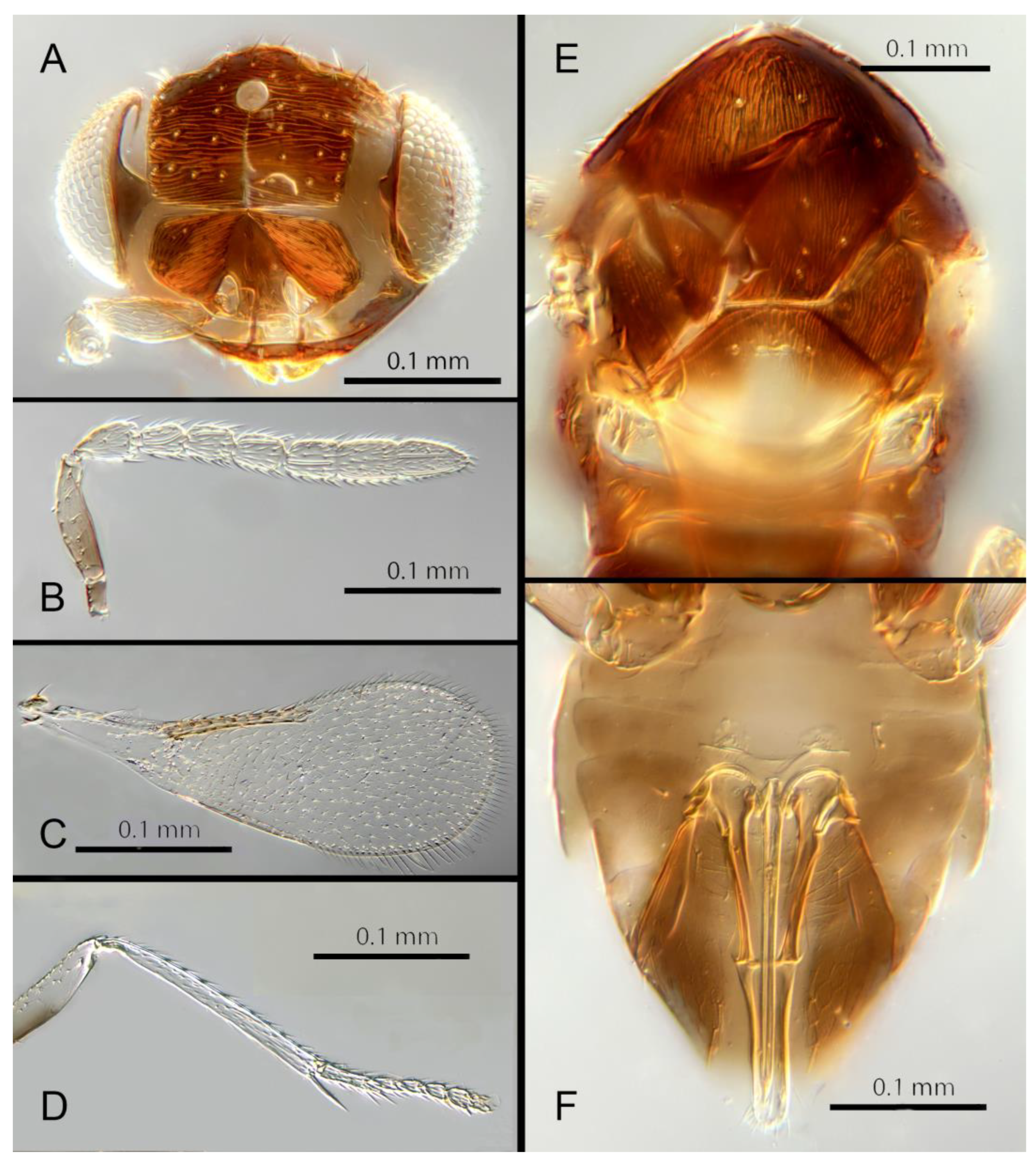
Figure 20.
Encarsia napo: (A) head; (B) antenna; (C) fore wing; (D) mid leg; (E) dorsal mesosoma; (F) ovipositor.
Figure 20.
Encarsia napo: (A) head; (B) antenna; (C) fore wing; (D) mid leg; (E) dorsal mesosoma; (F) ovipositor.
Material examined. Holotype. MEXICO, Tabasco, San Francisco del Peal. 1.vii.1887 (C.H. Townsend) (USNM). Examined. 6♀ [compared with type series AP xi.90] MEXICO, Tabasco, San Francisco del Peal, 1.vii.1897 (C.H.T. Townsend) ex Nealeurodicus altissimus [reared from Lippia myriocephala] (NHMUK, USNM). 1♀ COSTA RICA, Heredia, Est. Biol. La Selva, 27-28.ii.2003 (J.S. Noyes) 75m 10º26´N 84º01´W. [DNA 144: OQ683560] (NHMUK); 1♀ COSTA RICA, Arenal, Sen. Pilon, 26.ii.2003 (J.S. Noyes) [DNA 52] (NHMUK); 1♀ COSTA RICA, Alajuela, Est. Caribe R. Rincón Forestal, 19-20.ii.2003 (J.S. Noyes) 400m, 10º53´N 85º18´W [DNA 166] (NHMUK); 1♀ COSTA RICA, Alajuela, P.N. Arenal, send. Pilon, 26.ii.2003 (J.S. Noyes), 600m 10º27´N 84º43´W; [DNA 129] (MZUCR).
Remarks. Remarks. Encarsia mexicana is morphologically closest to E. fredbennetti from which it differs by the non-enlarged clava. It is also molecularly closest to E. fredbennetti. Sequence data deposited at GenBank accession number: OQ683560.
3.3.17. Encarsia napo Polaszek & Hernández-Suárez sp. n.
(Figure 20A–F)
Female. Color. Antennae pale brown, slightly darker on the base of the clava, scape, pedicel, and radicle. Head dark brown. Mesosoma and metasoma uniformly dark brown with the posterior two-thirds of the scutellum and sides of the metanotum yellow. Legs robust, yellow in color with hind femur, posterior half of mid femur and anterior half of fore femur light brown, all tarsi pale. Wings hyaline, submarginal vein pale in contrast with darker marginal and stigmal veins.
Morphology. Head (Figure 20A) with mediofrontal line complete; transfacial line evident, broad laterally and tapering towards the middle; facial lines extremely broad along their
entire lengths. Scrobes largely smooth, some irregular sculpture centrally. Antenna (Fig19B) with eight antennomeres; antennal formula: 1,1,3,3; scape 2.2x pedicel length; pedicel 1.3x F1; F1 0.88x F2; F2 equal to F3; funicle 0.7x clava; F6 perpendicular. Mandibles (Figure 20A) with two minute teeth and a truncation. Flagellum with the following number of longitudinal sensilla: F1: 1; F2:1; F3: 1 F4: 2 F5: 3; F6: 3. Maxillary palps 2-segmented. Mid-lobe of mesoscutum (Figure 20E) with fewer than 20 setae; each lateral lobe with 2 setae; each axilla with 1 seta; scutellum with 4 setae and 2 vestigial setal bases. Sculpture of mesoscutum, axillae and scutellum longitudinal. Fore wing with 2 large setae and 4 smaller setae on submarginal vein, 4-5 setae in basal cell, 7-8 setae on anterior margin of marginal vein, and 1 seta at the junction of the submarginal vein and parastigma. Linea calva absent. Submarginal 0.80x times marginal vein. Maximum length of fore wing 2.68x fore wing width, maximum width of wing 4.79x longest setae on marginal fringe. Ovipositor (Figure 20F) 0.8x mid tibial length; third valvulae 0.45x ovipositor length; second valvifer 1.2x third valvula. Mid tibial spur (Figure 20D) 0.9x corresponding basitarsus. Metasomal terga T1-T7 with 0, 1+1, 1+1, 1+1, 1+2+1, 1+2+1 and 6 setae, respectively. T7 (Figure 20F) rounded, not extended but covering ovipositor.
Distribution. ECUADOR: Napo River.
Material examined. Holotype ♀ ECUADOR, Napo, transect Ent. 1Km S Onkone Gare Camp, Res. Etnica Waorani, 220m 0º39´10´´S 76º26´00´´W T.L. Erwin et al. fogging tf forest lot 1193 5.v.1995 [DNA314] (NHMUK). Paratypes 2 slides ♀ ECUADOR, Napo, transect Ent. 1Km S Onkone Gare Camp, Res. Etnica Waorani, 220m 0º39´10´´S 76º26´00´´W T.L. Erwin et al. fogging tf forest lot 1193 5.v.1995 [DNA 312, 313] (NHMUK).
Remarks. Encarsia napo is morphologically closest to E. erwini from which it differs by the partly pale mesoscutellum. No molecular data were available for E. napo.
Etymology. Named for the Napo River (Rio Napo) on which the type locality is located.
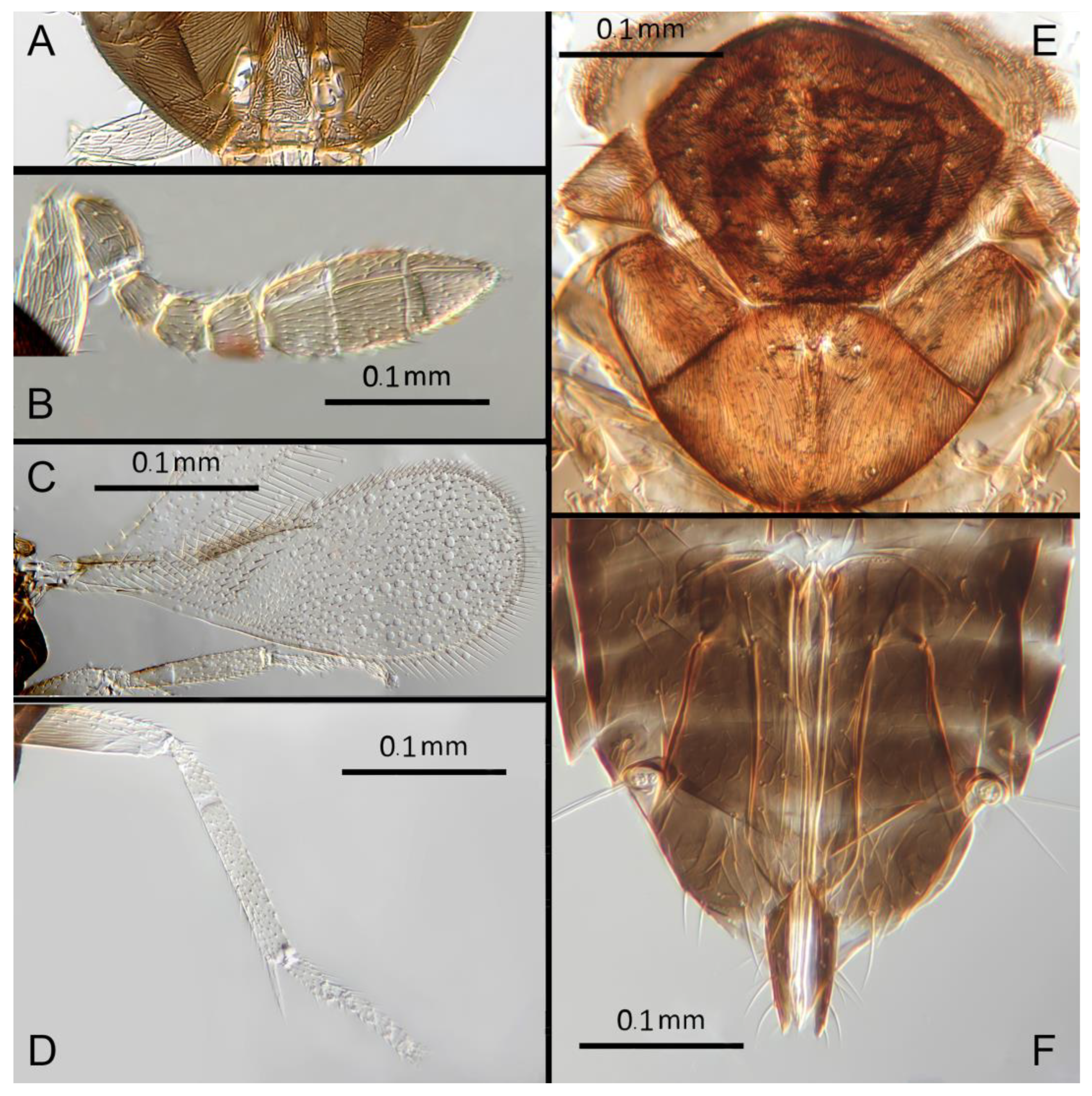
3.3.18. Encarsia noora Polaszek & Hernández-Suárez sp. n.
(Figure 21A–E)
Female. Color. Antennae pale yellow. Head brown with pale lines bordering the eyes and extending along the genae towards clypeus. Mesosoma brown but with posterior three-quarters of scutellum pale. Metasoma uniformly light brown. Legs pale yellow; hind coxae and femora brown. Wings hyaline, slightly infuscated below marginal vein; stigmal vein pale in contrast with a darker marginal vein.
Morphology. Head (Figure 21A) with mediofrontal line incomplete, reaching less than halfway to frontal ocellus, transfacial line evident; facial lines very broad along their entire lengths. Scrobes almost entirely smooth, some irregular transverse sculpture basally. Antenna (Figure 21B) with eight antennomeres; antennal formula: 1,1,3,3; scape 2x pedicel length; pedicel 2.2x F1; F1 1.2x F2; F2 0.7x F3; funicle 0.6x clava; F6 slightly oblique, claval
Figure 21.
Encarsia noora: (A) head; (B) antenna; (C) fore wing; (D) mid leg; (E) dorsal habitus.
sensorial complex apparently present. Flagellum with the following number of longitudinal sensilla: F1: 0; F2: 0; F3: 1-2; F4: 3; F5: 4; F6: 4. Mandibles (Figure 21A) with 2 minute teeth and a truncation. Maxillary palps 2-segmented. Mid-lobe of mesoscutum (Figure 21E) with fewer than 30 setae; each lateral lobe with 2 setae; each axilla with 1 seta; scutellum with 4 setae. Sculpture of mesoscutum aciculate; sculpture of axillae and scutellum longitudinal. Fore wing (Figure 21C) with 2 large setae and 4 smaller setae on submarginal vein, 8 setae in basal cell, 7-8 setae on anterior margin of marginal vein, and 1 seta at the junction of the submarginal vein and parastigma; a group of long setae below marginal vein. Linea calva present. Submarginal 0.8x marginal vein. Maximum length of fore wing 2.58x fore wing
width, maximum width of wing 6.39x longest setae on marginal fringe. Ovipositor (Figure 21E) 1.34x mid tibial length; third valvulae 0.43x ovipositor length; second valvifer 1.2x third valvula. Mid tibial spur (Figure 21D) 0.9x corresponding basitarsus. Metasomal terga T1-T7 with 0, 2+2, 2+2, 2+2, 1+2+1, 1+2+1 and 4 setae, respectively. T7 (Figure 21E) extended although apparently not covering ovipositor.
Figure 22.
Encarsia svetlana: (A) head; (B) antenna; (C) fore wing; (D) mid leg; (E) dorsal mesosoma; (F) ovipositor.
Figure 22.
Encarsia svetlana: (A) head; (B) antenna; (C) fore wing; (D) mid leg; (E) dorsal mesosoma; (F) ovipositor.
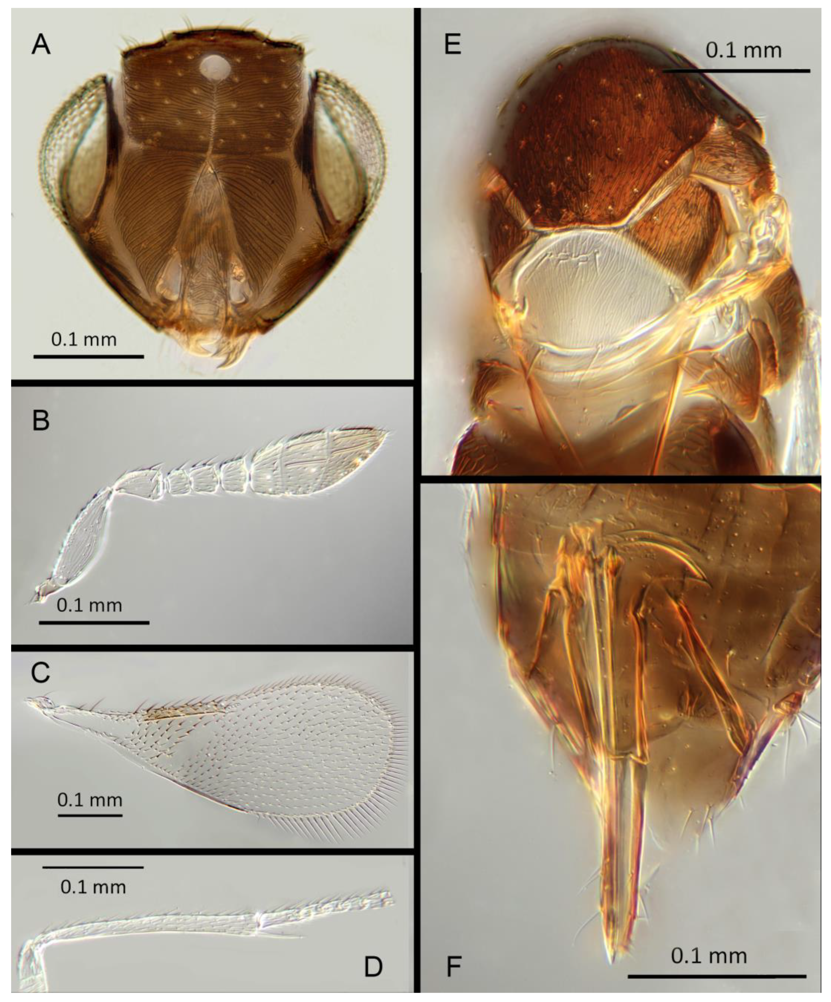
Distribution. TRINIDAD: Mt St Benedict, St Augustine.
Material examined. Holotype ♀ TRINIDAD, I.C.T.A. [Imperial College of Tropical Agriculture, St. Augustine, COORD] xii.1953 (F.D. Bennett) [s9] ex whitefly on cocoa. ?Coccophagus sp. Det. FDB [identified as Coccophagus, F.D. Bennett] Imperial Parasite Service (NHMUK). Paratypes 2♀ TRINIDAD, Mt. St. Benedict, 18.x.1997 (M. Jagroep) [DNA 126: OQ683561] ex whitefly; St. Augustine [DNA 215b] ex Aleurodicinae (NHMUK).
Remarks. Encarsia noora is morphologically closest to E. catula, differing by the longer F1 in E. noora. Molecularly sister species to E. aisha. Sequences deposited at GenBank accession number: OQ683561.
Etymology. Named for Noora, daughter of the 2nd author (EHS), and sister to Aisha; see E. aisha, above.
3.3.19. Encarsia svetlana Polaszek & Hernández-Suárez sp. n.
(Figure 22A–F)
Female. Color. Antennae light brown slightly darker on F6 and radicle. Head brown with pale lines bordering the eyes and extending along the genae towards clypeus. Mesosoma dark brown but with scutellum and sides of the metanotum yellow. Metasoma light brown. Legs uniformly pale yellow. Wings infuscated below marginal vein, stigmal vein pale in contrast with a darker marginal vein.
Morphology. Head (Figure 22A) with mediofrontal line complete; transfacial line narrow; facial lines very broad at level of lower eyes and adjacent to genae. Scrobes with longitudinal aciculate sculpture in the upper half, transverse aciculate sculpture in the lower half. Antenna (Figure 22B) with eight antennomeres; antennal formula: 1,1,3,3; scape 2.25x pedicel length; pedicel 2.36x F1; F1 0.86x F2; F2 0.9x F3; funicle 0.49x clava length; F6 broadly oblique, claval sensorial complex developed. Flagellum with the following number of longitudinal sensilla: F1: 0; F2: 1; F3: 1; F4: 1; F5: 4; F6: 4. Mandibles (Figure 22A) with 2 very large ventral teeth and a small truncation, appearing bidentate. Maxillary palps 2-segmented. Mid-lobe of mesoscutum (Figure 22E) with fewer than 30 setae; each lateral lobe with 2 setae; each axilla with 1 seta; scutellum with 4 setae. Sculpture of mesoscutum, axillae and scutellum longitudinal. Fore wing (Figure 22C) with 2 large setae and 4 smaller setae on submarginal vein, 4 setae in basal cell, 6-7 setae on anterior margin of marginal vein, and 1 seta at the junction of the submarginal vein and parastigma. Linea calva present. Submarginal 0.9x marginal vein. Maximum length of fore wing 2.5x fore wing width, maximum width of wing 3.85x longest setae on marginal fringe. Ovipositor (Figure 22F) 0.6x mid tibial length; third valvulae 0.95x ovipositor length; second valvifer 1.1x third valvula. Mid tibial spur (Figure 22D) 0.9x corresponding basitarsus. Metasomal terga T1-T7
with 0, 2+2, 2+2, 1+1, 1+1+1, 1+1 and 4 setae, respectively. T7 (Figure 22F) apex rounded not covering ovipositor.
Distribution. GUYANA: Dubulay Ranch.
Material examined. Holotype ♀ GUYANA, Dubulay Ranch, 17-22.i.1999 (M. Sharkey and B. Brown) [DNA 305: OQ683558] Univ. Calif. Riverside, Ent. Res. Museum UCR ENT 182858 5º40.954´N 57º51.524´W (UCRC).
Remarks. Encarsia svetlana is morphologically closest to E. venia from which it differs in having F3 transverse, and he scape entirely pale. Sequence data deposited under GenBank accession number: OQ683558.
Etymology. Named for Svetlana Myartseva, prolific describer of Mexican Aphelinidae, including many Encarsia species.
3.3.20. Encarsia venia Polaszek & Hernández-Suárez sp. n.
(Figure 23A–F)
Female. Color. Antennae light brown, slightly darker on scape and radicle. Head dark brown. Mesosoma and metasoma dark brown, with entire scutellum and sides of the metanotum yellow. Legs uniformly pale yellow. Wings hyaline, slightly infuscated below marginal vein.
Figure 23.
Encarsia venia: (A) head; (B) antenna; (C) fore wing; (D) mid leg; (E) dorsal mesosoma; (F) ovipositor.
Figure 23.
Encarsia venia: (A) head; (B) antenna; (C) fore wing; (D) mid leg; (E) dorsal mesosoma; (F) ovipositor.
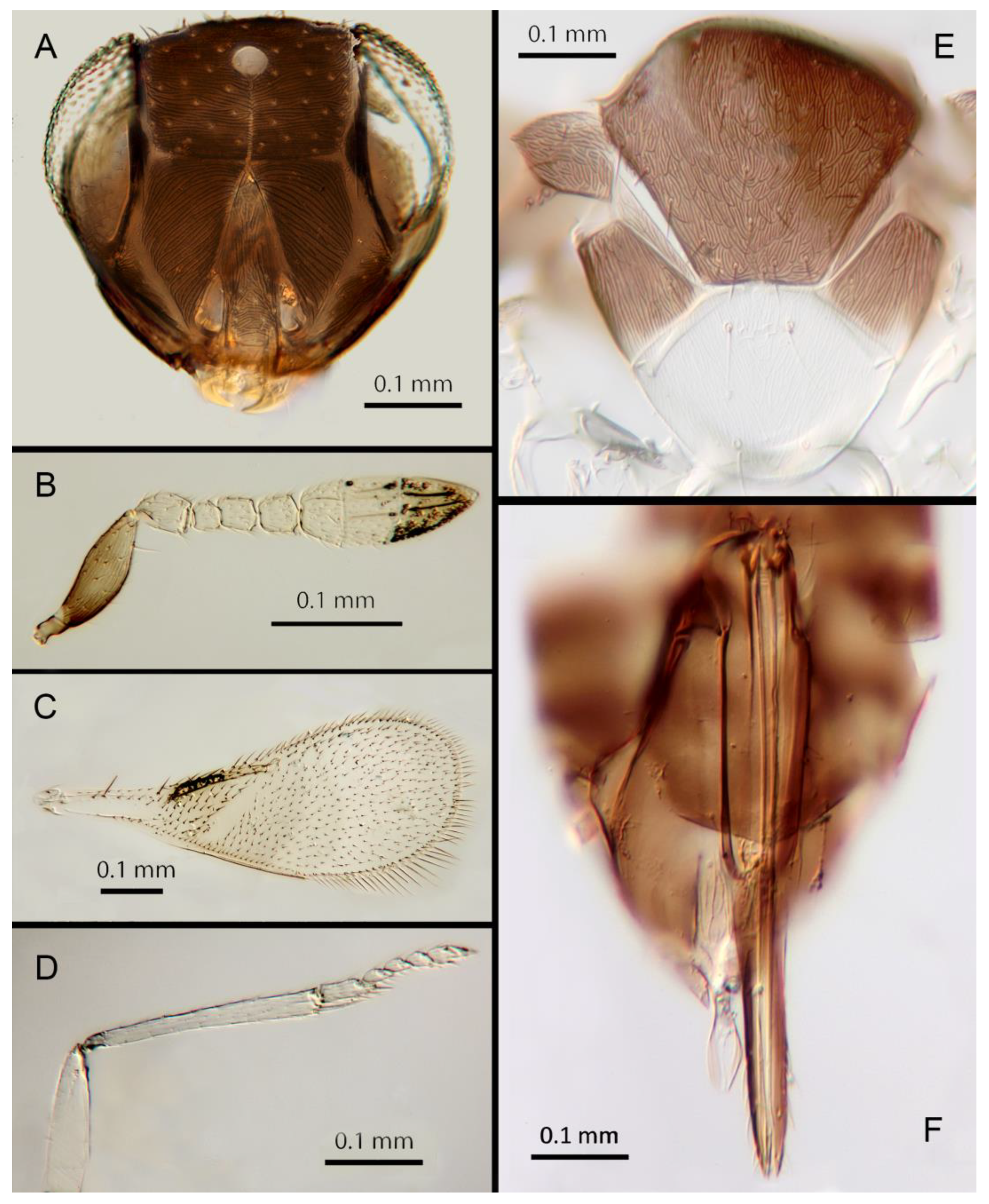
Morphology. Head (Figure 23A) with mediofrontal line complete, very narrow towards frontal ocellus; transfacial line narrow; facial lines broad along their entire lengths. Scrobes with longitudinal aciculate sculpture in the upper half, transverse aciculate sculpture in the lower half. Antenna (Figure 23B) with eight antennomeres; antennal formula: 1,1,3,3; scape 2.4x pedicel length; pedicel 2x F1; F1 0.7x F2; F2 1.1x F3; funicle 0.6x clava; F6 slightly oblique, claval sensorial complex apparently present. Flagellum with the following number of longitudinal sensilla: F1: 0; F2: 1; F3: 1; F4: 2; F5: 3; F6: 3. Mandibles (Figure 23A) with
1 very large ventral tooth and an upper bidentate tooth. Maxillary palps 2-segmented. Mid-lobe of mesoscutum (Figure 23E) with fewer than 30 setae; each lateral lobe with 2 setae; each axilla with 1 seta; scutellum with 4 setae. Sculpture of mesoscutum aciculate; sculpture of axillae and scutellum longitudinal. Fore wing with 2 large setae and 4 smaller setae on submarginal vein, 3-4 setae in basal cell, 7-8 setae on anterior margin of marginal vein, and 1 seta at the junction of the submarginal vein and parastigma. Linea calva present. Submarginal equal to marginal vein. Maximum length of fore wing 2.8x fore wing width, maximum width of wing 4.5x longest seta on marginal fringe. Ovipositor (Figure 23F) 1.4x mid tibial length; third valvulae 0.4x ovipositor length; second valvifer 1.3x third valvula. Mid tibial (Figure 23D) spur equal to corresponding basitarsus. Metasomal terga T1-T7 with 0, 1+1, 1+1, 2+2, 1+2+1, 1+2+1 and 4 setae, respectively. T7 (Figure 23F) apex extended but not covering ovipositor entirely.
Distribution. COSTA RICA: Heredia.
Material examined. Holotype ♀ COSTA RICA, Limon, Parque Nacional Cahuita 2m, 26.ii.2004 (J.S. Noyes) [DNA 298: OQ683557] 9º 43´N 82º 49´W (NHMUK). 1♀ COSTA RICA, Limon, Hitoy-Cerere, 21-22.iii.2006 (J.S. Noyes) [DNA 267] 90º40´N 83º02´W; was mexicana. 1♀ COSTA RICA, Heredia La Selva, 22.i – 2.ii. 1991 (J.S. Noyes) [s16]; ? mexicana.
Remarks. Morphologically closest to E. svetlana from which it differs in having F3 quadrate (transverse in E. svetlana), and the scape dark. Sequence deposited under GenBank accession number: OQ683557.
Etymology. From Latin venia meaning “kindness.”
3.3.21. Encarsia myartsevae Kresslein & Polaszek nom. nov.
Remarks. Myartseva (2007) originally described this species as Encarsia mexicana Myartseva. In synonymizing Dirphys and Encarsia, this species name becomes preoccupied by the new combination Encarsia mexicana (Howard, 1907). As a result, we rename this species Encarsia myartsevae nom. nov. in honor of the species’ author, Svetlana Myartseva, for her contributions to the systematics of Encarsia and in particular her dedication to providing accounts of their host associations. Encarsia myartsevae nom. nov. is not a member of the Encarsia mexicana species group, instead belonging to the opulenta species group, and is not discussed further in this revision.
3.4. Key to the species of the Encarsia mexicana species-group: females.
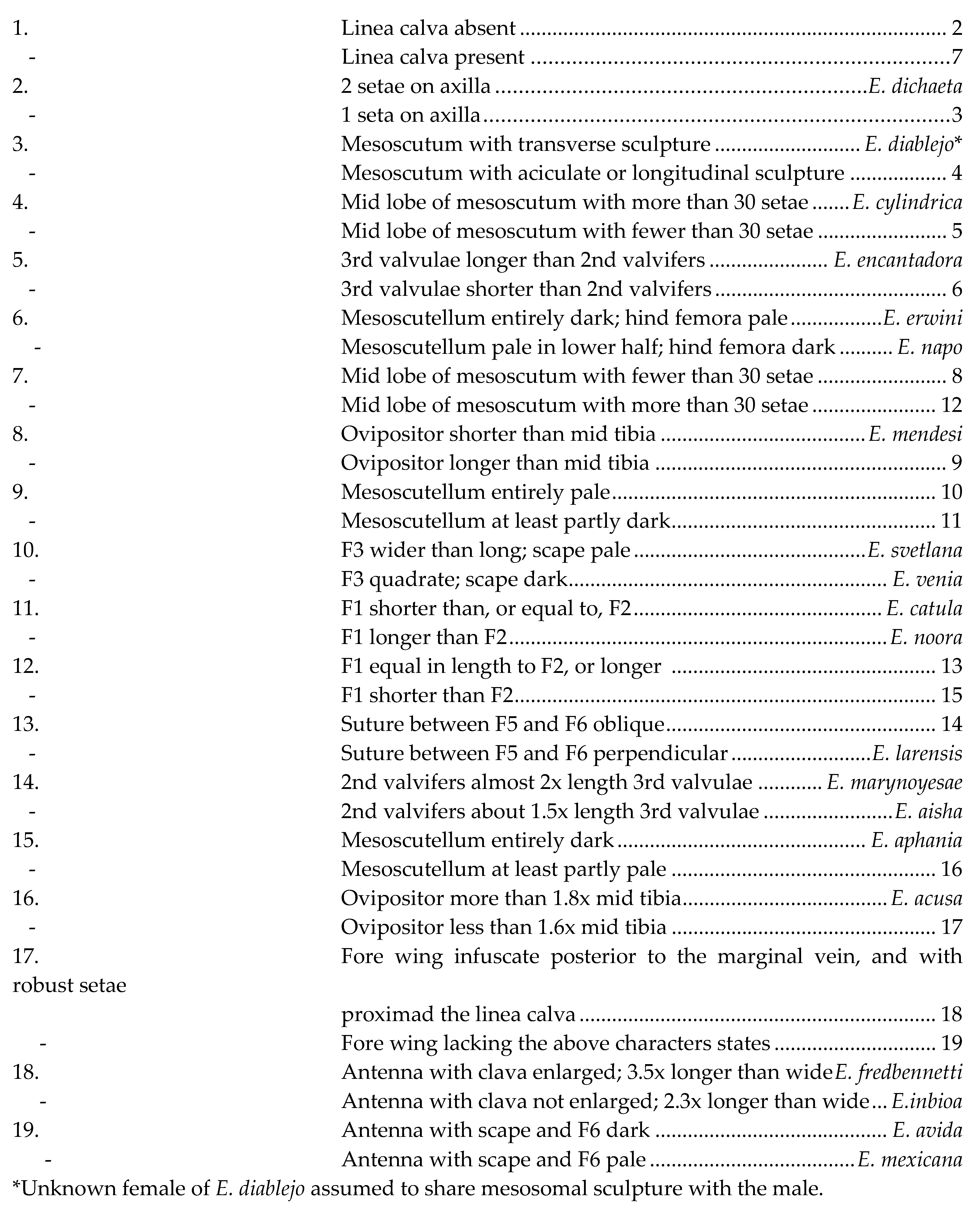
An accompanying multiple entry key can be found at: https://rkres001.github.io/rkres001.encarsiamexicanakey/.
4. Discussion
All taxa of Encarsia mexicana species group recovered sister to Encarsia dichaeta have a linea calva present on the fore wing (absent in E. dichaeta; unknown D2672, and DNA.0165). The presence or absence of a linea clava may represent a local synapomorphy for these two clades within the mexicana group. All taxa which lack a linea calva also lack the enlargement of the clava of the antenna common in this species group, though Encarsia noora possess a linea calva in the absence of an enlarged clava. Further phylogenetic analyses and sequence capture for the five other species without linea calva (E. diablejo, E. cylindrica, E. encantadora, E. erwini and E. napo) will be necessary to determine the informativeness of these characters. Other evident characters appear phylogenetically uninformative. Some surprises include E. svetlana appearing well-removed from E. venia while their morphology is extremely similar. E. aisha and E. noora despite being DNA sister species have markedly different flagellar shapes.
With the placement of Dirphys syn. nov. in the middle of Encarsia, the genus expands to 473 described species (Kresslein et al., 2023). Establishment of the Encarsia mexicana species-group further increases the difficulty with which the genus can be recognized. In particular, the genus now contains species with a linea calva on the forewing, and parasitoids with a gregarious life history, further expanding the already broad host range of the genus (Kresslein et al. 2023). The future classification of Encarsia at large will rely upon its clarification by robust phylogenetic hypotheses built upon large molecular datasets (Kresslein et al. unpublished). As the phylogeny of Encarsia is further resolved, it will be necessary to revisit the current classification of the genus and determine whether alternate generic classifications would allow for greater diagnosability of the taxa therein.
Author Contributions
Conceptualization, A.P., E.H.S.; methodology, A.P., E.H.S., J.M.D., Y.M.L., S.S.; software, S.S., R.L.K., Y.M.L.; validation, A.P., E.H.S., P.H. ; formal analysis, S.S., R.L.K.; investigation, A.P., E.H.S., S.S.; resources, A.P., J.M.D., Y.M.L., E.H.S.; data curation, A.P., J.M.D.; writing—original draft preparation, A.P., E.H.S.; writing—review and editing, A.P., E.H.S., J.M.D., P.H., Y.M.L., S.S., R.L.K.; visualization, A.P., E.H.S., R.L.K.; supervision, A.P.; project administration, A.P.; funding acquisition, E.H.S., A.P. All authors have read and agreed to the published version of the manuscript.
Funding
This work was supported in part by grants from FUNDESIMCA Fundación para el Desarrollo e Impulso de las Ciencias Agrarias en Canarias.
Data Availability Statement
All DNA sequence data that supports the results and conclusions of this study can be found at GenBank.
Acknowledgments
We would like to thank Miguel Alonso-Zarazaga and John Noyes for loaning material, and Krissy Dominguez for comments on the manuscript.
Conflicts of Interest
The authors declare no conflict of interest.
References
- Howard, L.O. Concerning some Aphelininae. Proceedings of the Entomological Society of Washington 1914, 16, 79–85. [Google Scholar]
- Howard, L.O. New genera and species of Aphelinidae with a revised table of genera. Technical Series, Bureau of Entomology, United States Department of Agriculture 1907, 12, 69–88.
- Polaszek, A.; Hayat, M. A revision of the genera Dirphys Howard and Encarsiella Hayat (Hymenoptera: Aphelinidae). Systematic Entomology 1992, 17, 181–197. [Google Scholar] [CrossRef]
- Chavez, T.A. A new species of Dirphys Howard (Hymenoptera: Aphelinidae). Boletín de Entomologia Venezolana 1996, 11, 12–16.
- Martin, J.; Polaszek, A. A new genus of Neotropical whitefly, secreting blue-iridescent wax (Sternorrhyncha, Aleyrodidae, Aleurodicinae), and its parasitoids (Hymenoptera, Aphelinidae). Journal of natural history 1999, 33, 1545–1559. [Google Scholar] [CrossRef]
- Hayat, M. The genera of Aphelinidae (Hymenoptera) of the world. Systematic Entomology 1983, 8, 63–102. [Google Scholar] [CrossRef]
- Hayat, M. Notes on some species of Coccophagoides, Dirphys and Encarsia (Hymenoptera: Aphelinidae). Oriental Insects 1989, 23, 286–290. [Google Scholar] [CrossRef]
- Hayat, M. A revision of the species of Encarsia Foerster (Hymenoptera, Aphelinidae) from India and the adjacent countries. Oriental Insects 1989, 23, 1–131. [Google Scholar] [CrossRef]
- Shafee, S.A.; Rizvi, S. Taxonomic notes on some Indian Aphelinidae (Hymenoptera: Chalcidoidea). Mitteilungen der Schweizerischen Entomologischen Gesellschaft 1984, 57, 379–381. [Google Scholar]
- Babcock, C.S.; Heraty, J.M.; De Barro, P.J.; Driver, F.; Schmidt, S. Preliminary phylogeny of Encarsia Forster (Hymenoptera : Aphelinidae) based on morphology and 28S rDNA. Molecular Phylogenetics and Evolution 2001, 18, 306–323. [Google Scholar] [CrossRef]
- Schmidt, S.; Polaszek, A. Encarsia or Encarsiella? Redefining generic limits based on morphological and molecular evidence (Hymenoptera, Aphelinidae). Systematic Entomology 2007, 32, 81–94. [Google Scholar] [CrossRef]
- Heraty, J.M.; Polaszek, A. Morphometric analysis and descriptions of selected species in the Encarsia strenua group (Hymenoptera: Aphelinidae). Journal of Hymenoptera Research 2000, 9, 142–169. [Google Scholar]
- Myartseva, S.N. Species of genus Encarsia Forster (Hymenoptera: Aphelinidae) - parasitoids of whiteflies (Hemiptera: Aleyrodidae) associated with Psidium guajava L. in Mexico, with key and description of new species. Biosystematica 2007, 1, 7–19. [Google Scholar]
- Noyes, J.S. Collecting and preserving chalcid wasps (Hymenoptera, Chalcidoidea). Journal of Natural History 1982, 16, 315–334. [Google Scholar] [CrossRef]
- Kim, J.-W.; Heraty, J.M. A phylogenetic analysis of the genera of Aphelininae (Hymenoptera: Aphelinidae), with a generic key and descriptions of new taxa. Systematic Entomology 2012, 37, 497–549. [Google Scholar] [CrossRef]
- Hayat, M. Aphelinidae of India (Hymenoptera: Chalcidoidea): a Taxonomic Revision; Associated Publishers: Gainesville, Florida, 1998; Volume 13, i-viii, 1-416. [Google Scholar]
- Polaszek, A.; Ayshford, T.; Yahya, B.E.; Fusu, L. Wallaceaphytis: an unusual new genus of parasitoid wasp (Hymenoptera: Aphelinidae) from Borneo. Journal of Natural History 2013, 48, 1111–1123. [Google Scholar] [CrossRef]
- Katoh, K.; Standley, D.M. MAFFT multiple sequence alignment software version 7: improvements in performance adn usability. Molecular Biology and Evolution 2013, 30, 772–780. [Google Scholar] [CrossRef] [PubMed]
- Minh, B.Q.; Schmidt, H.A.; Chernomor, O.; Schrempf, D.; Woodhams, M.D.; von Haeseler, A.; Lanfear, R. IQ-TREE 2: new models and efficient methods for phylogenetic inference in the genomic era. Molecular Biology and Evolution 2020, 37, 1530–1534. [Google Scholar] [CrossRef] [PubMed]
- Hoang, D.T.; Chernomor, O.; von Haeseler, A.; Minh, B.Q.; Vinh, L.S. UFBoot2: Improving the Ultrafast Bootstrap Approximation. Molecular Biology and Evolution 2017, 35, 518–522. [Google Scholar] [CrossRef] [PubMed]
- Hayat, M. On the identity and systematic position of the genus Dirphys [Hym.: Aphelinidae]. Entomophaga 1981, 26, 441-444.
Disclaimer/Publisher’s Note: The statements, opinions and data contained in all publications are solely those of the individual author(s) and contributor(s) and not of MDPI and/or the editor(s). MDPI and/or the editor(s) disclaim responsibility for any injury to people or property resulting from any ideas, methods, instructions or products referred to in the content. |
© 2023 by the authors. Licensee MDPI, Basel, Switzerland. This article is an open access article distributed under the terms and conditions of the Creative Commons Attribution (CC BY) license (http://creativecommons.org/licenses/by/4.0/).
Copyright: This open access article is published under a Creative Commons CC BY 4.0 license, which permit the free download, distribution, and reuse, provided that the author and preprint are cited in any reuse.


