
Submitted:
15 May 2023
Posted:
16 May 2023
You are already at the latest version
Abstract
Timely mitosis is critically important for the early embryo development. It is regulated by the activity of the ubiquitously conserved CDK1 kinase. The dynamics of CDK1 activation must be precisely controlled to assure physiologic and timely entry into mitosis. Recently, a known S-phase regulator CDC6 emerged as a key player in the mitotic CDK1 activation cascade in early embryonic divisions, operating together with Xic1 as a CDK1 inhibitor upstream of the Aurora A- and PLK1 kinases, both CDK1 activators. Herein we review the molecular mechanisms that underlie the control of the mitotic timing, with special emphasis on how CDC6/Xic1 function impacts CDK1 regulatory network. We focus on the presence of two independent mechanisms inhibiting the dynamics of CDK1 activation: Wee1/Myt1- and CDC6/Xic1-dependent, and how they cooperate with CDK1 activating mechanisms. As a result we propose a comprehensive model integrating CDC6/Xic1-dependent inhibition into the CDK1-activation cascade. The physiological dynamics of CDK1 activation appear to be tuned by the system of multiple inhibitors and activators and their integrated modulation ensures concomitantly both the robustness and certain flexibility of the control of this process. Identification of multiplied activators and inhibitors of CDK1 activation upon M-phase entry allows a better understanding of why cells divide at a specific time, and how the pathways involved in the timely regulation of the cell division are all integrated to precisely tune the control of mitotic events.
Keywords:
Timing of mitosis
; Cell cycle
; CDC6
; CDK1
; cyclins
; CDC25
; Xic1
; Mitotic entry
Introduction
The entry into mitosis is controlled by CDK1/Cyclin B, also known as MPF (Maturation Promoting Factor). Previous research has shown that cyclin B synthesis is the key for driving the embryonic cell cycle and determining the timing of mitosis in Xenopus leavis [1,2,3]. Apparently, it is a key, but not the only controlling factor. Cyclin B accumulation during interphase is necessary for CDK1 activation because without cyclin CDK1 cannot be active as a protein kinase. On the other hand, cyclin degradation through the ubiquitin pathway is necessary for CDK1 inactivation and mitotic exit [1,2,3]. Cyclin B is encoded by several genes and creates a family of B-type cyclins required for mitosis (e.g., cyclins B1, B2, and B3 in human; or cyclins B1-B5 in Xenopus leavis [4,5]). De novo B-type cyclin synthesis is required between meiosis I and II during Xenopus oocyte maturation [4]. However, despite a steady increase in cyclin B levels in G2 [6], CDK1 activation is biphasic characterized by a slow phase, followed by a rapid phase attributed to the positive feedback between newly activated CDK1 and its major activating phosphatase CDC25 [6]. It was intriguing for the long time how the stable increase in cyclin B level may lead to this biphasic CDK1 activation before the CDK1/CDC25 positive feedback acceleration triggering the final pic of CDK1 activity followed by its inactivation linked with cyclin B degradation (ibid.). This particularity suggested that the control of CDK1 activation at the initial stages of its mitotic activation might not solely depend on cyclin B accumulation, but might also involve an unidentified inhibitor that could counterbalance CDK1 activation assuring slow and biphasic mode of this process. Our research group performed a proteomic screen to find novel CDK1 partners in M-phase-arrested Xenopus leavis eggs vs. freshly activated ones (5 minutes post activation; [7]). We showed a rapid, taking 5 min., change in the composition of the CDK1 complex during the period between the MII arrest of oocytes and their activation for development. However, we did not find in this screen any classical CDK1 inhibitors, like INK4 or Cip/Kip, which potentially could slow down CDK1 activation [8] in a CDK1 complex. Surprisingly for us, we found a CDC6 protein associated with the active mitotic CDK1. CDC6 is an evolutionarily conserved member of the AAA + ATPase family well known as the S-phase activator [6]. Our further results showed that a CDC6-dependent mechanism inhibits CDK1 throughout the whole period of the M-phase (including the highest pic of CDK1 activity just prior its inactivation). Moreover, CDC6 associated with CDK1 during the M-phase rapidly dissociates from this kinase immediately after CDK1 inactivation suggesting that once CDK1 fully inactivated by separation from cyclin B, the association with CDC6 becomes unnecessary. It was a surprising observation in the light of the knowledge at that time suggesting a potential role of CDC6 only in the mitotic exit, and not entry and progression [9]. Therefore, we became particularly intrigued by the role of CDC6 in the M-phase regulation and especially in the control of the dynamics of CDK1 mitotic activation and pursuit this research. The important hint to CDC6 role in CDK1 activation process was its mentioned above function in the M-phase exit described in yeast and human cells [10,11,12]. Through biochemical analysis of Xenopus embryo cell-free extracts we showed that a recombinant CDC6 protein acts as an inhibitor of CDK1 during the first embryonic mitosis in Xenopus leavis [9,13,14,15,16,17]. Importantly, when endogenous CDC6 is depleted, the first, initial and slow phase of CDK1 activation is removed, and the kinase activation ceases to be biphasic. This, in turn, accelerates the timing of mitosis and the pic of CDK1 activity induced by the CDC25/CDK1 activation loop [9,13,14,15,16,17]. Moreover, we confirmed that it is also the case in the mouse embryos, which suggested a universal inhibitory role of CDC6 in CDK1 activation (ibid.). Furthermore, we showed that two proteins: CDC6 and Xic1 (the last one is a bona fide CDK1 inhibitor identified for its role in later stages of Xenopus embryo development) interact within the mitotic CDK1 protein complex. Upon arriving at the peak of the CDK1 activity (measured by histone H1 kinase activity), the Xic1 momentarily dissociates from the mitotic CDK1 allowing maximum activation of CDK1 during the M-phase [9]. The identification of this new inhibitory mechanism towards CDK1 was surprising because it has been well known already for years that CDK1 is inhibited by the posttranslational modification by Wee1/Myt1 kinases. Thus, our data showed that there are two independent mechanisms of CDK1 activity in the game during the mitotic entry.
In parallel to our findings, other researchers continued exploring the molecular mechanisms that accurately regulate the timing of mitosis in Xenopus laevis as well as in C. elegans and human cells [18,19,20,21,22,23,24,25,26,27,28]. Importantly, it was shown that CDK1/Cyclin A/B dimers phosphorylate Aurora-A kinase co-factor Bora, and this triggers the activation of the kinase, in turn activating PLK1 kinase and the CDK1's amplification loop, which helps the cell to enter and progress into M-phase in a timely manner [21,22,22,24,28]. All these data suggested an interplay between inhibitor and activator molecular mechanisms governing mitotic CDK1 activation process and required a precise definition of their interrelationship.
For this reason, we provide here an overview of the molecular processes underlying timely mitotic entry and progression. We focus on the CDK1 regulatory network and especially on the function of CDC6/Xic1 and Wee1/Myt1 acting as CDK1 inhibitors. The multiplied system of CDK1 activators and inhibitors explains how the mitotic activation of CDK1 is tuned and why cells divide in the strictly defined time frame. We also explain how the various pathways regulating this process are integrated.
1. Activation of CDK1 by association with cyclins A and B and its concomitant inhibition by Wee1/Myt1 kinases posttranslational modifications
During the G2/M transition, the increased stability of cyclin B mRNA allows a significant and continuous accumulation of cyclin B protein [29]. This newly synthesized cyclin B associates with CDK1, but due to the inhibitory phosphorylation on CDK1 Thr14 and Tyr15 sites, by Wee1 and Myt1 kinases, respectively, the newly formed CDK1/cyclin B complex is catalytically inactive in the cytoplasm [30,31]. Cyclin B will continue to be produced during the G2 phase and increase the amount of CDK1/cyclin B complexes until a critical quantity is reached just before mitotic entry. At this moment, CDK1 kinase is activated by dephosphorylation of Tyr14 and Thr15 by the CDC25 phosphatase family members (CDC25 A, B, and C) which counterbalance the Wee1/Myt1-dependent inhibition. CDK1 activation requires also the continuous phosphorylation of Thr161 by CAK [32,33,34,35,36,37,38]. During the dephosphorylation process of CDK1 on Tyr14 and Thr15, CDC25B acts as a starter phosphatase for the final pic of the activation of CDK1 [39,40]. Importantly, CDK1/cyclin B phosphorylates and activates CDC25C. This triggers the positive feedback between CDK1 and CDC25 called the MPF activating loop, which is responsible for the final, explosive CDK1 activation phase [41]. Moreover, active CDK1 phosphorylates and inhibits Wee1/Myt1, thereby eliminating the inhibitory effect of these kinases on CDK1 itself [42]. Thus, the amplification loop of CDK1 activation guarantees the extremely rapid mitotic progression following the slow and progressive entry into the M phase.
The beginning of the mitotic CDK1 activation is preceded by the accumulation of CDK1/Cyclin B in the cell nucleus. If cyclin B is not imported to the nucleus, the mitotic entry is halted, which underlies the importance of nuclear localization of the CDK1/cyclin B complex for the mitotic entry [43]. However, the association of CDK1 with cyclin B did not explain how the initiation of CDK1 activation can be triggered. The main problem was that CDK1/cyclin B is efficiently inactivated by Wee1/Myt1 kinases, which did not allow initiation of the kinase activation. The study by Gheghiani et al. (2017) provided the first indications that CDK1/cyclin A may act as a trigger for CDK1 activation [44] while PLK1 activation in late G2 sets up commitment to mitosis. Another observation suggesting an important role of cyclin A in the initiation of the mitotic activation of CDK1, and especially in the control of its timing, was that cyclin A depletion from the Xenopus cell-free extract enormously prolonged the time of CDK1 activation [9,24]. The final proof of such a role of cyclin A was shown by Vigneron et al. (2018) who demonstrated that contrary to CDK1/cyclin B, CDK1/cyclin A is not inhibited by Wee1 during interphase. This opened the possibility that accumulation of CDK1/cyclin A may easily attain a critical activity to start the whole pool of cyclin A and B-associated CDK1 activation. The escape of CDK1/cyclin A from the inhibitory phosphorylation by Wee1/Myt1 was indeed shown to break the posttranslational inhibition of CDK1 (ibid.). Also, our results showing that cyclin A and cyclin B overexpression in cell-free extracts have different effects on the timing and amplitude of CDK1 activation pointed out that an interplay between CDK1/cyclin A and CDK1/cyclin B is important in the regulation of the timing of CDK1 activation [9]. Indeed, the increase in cyclin A in cell-free extracts modifies only the level of CDK1 activity, while the increase in cyclin B modifies both the level of CDK1 activity and the timing of its full activation, which is accelerated in the latter case (ibid.). However, it must be noted that in this experimental system, the important increase in A or B-type cyclins may install a competition between the two types of cyclins for the binding with free CDK1 (ibid.).
2. Role of PLK1 in CDK1 activation upon mitotic entry
The kinase PLK1 is involved in the MPF amplification loop (being a part of the CDK1 activation process upon M-phase entry) through phosphorylating, and thus activating CDC25C [45,46,47]. PLK1 depletion delays CDK1 activation and stops CDC25C hyperphosphorylation (ibid.). PLK1 docking to CDC25C requires priming, which depends on CDK1-mediated CDC25C phosphorylation [48]. PLK1 phosphorylation of CDC25C triggers its nuclear translocation [49].
As they contain CDK1 kinase in G2 phase, centrosomes are crucial CDK1/Cyclin B regulators [50,51,52]. However, that pool is inactive because CDK1 inhibitors CHK1 and CHK2 kinases are present and/or active [53,54], preventing the pool's premature activation. The centrosomal pool of PLK1 contributes to CDK1 activation by counteracting the inhibition of the kinase by the CHK1 and 2 [52,55]. Injecting anti-PLK1 antibodies into primary human cells or depleting PLK1 from Xenopus eggs extracts induces G2 arrest [47,56,57]. However, PLK1-depleted cells enter mitosis in the presence of partially active CDK1, suggesting that partial CDC25C phosphorylation is sufficient for mitotic entry [58]. Additionally, PLK1 regulates the CDK1 activation via phosphorylation of CDK1-inactivating kinase Wee1, which, in turn, promotes its degradation, and thus decreases the inhibitory phosphorylation of CDK1 [42]. However, the discovery that the active CDK1/cyclin A complex promotes Plx1 activation via Bora phosphorylation allowed to understand better the role of Plk1 in CDK1 activation [24], see below. All these findings indicate the function of PLK1 as a key factor for activating CDK1, its downstream activation pathway and point to the role of CDK1/cyclin A and Bora in this process.
3. Role of Aurora A/Bora complex in CDK1 activation through PLK1
The serine/threonine kinase Aurora A plays a crucial role in the regulation of mitotic entry [19,20,59,60,61]. One of the best-known substrates of Aurora A is kinesin Eg5, whose activity is implicated in centrosome separation and maturation [62,63]. Aurora A induces CDC25B phosphorylation leading to CDK1/cyclin B activation [64].
It has been shown that Bora is necessary for Aurora A to phosphorylate PLK1 in human cells [65,66]. CDK1-mediated phosphorylation of Bora significantly increases Aurora A's ability to phosphorylate PLK1 [20,59,60,67], showing the importance of an upstream CDK1 complex for the activation of PLK1. This CDK1 complex has been identified as CDK1/Cyclin A complex [24]. Recently, it has been demonstrated that in G2/M transition, both CDK1/cyclin A and CDK1/cyclin B phosphorylate Bora to induce Aurora A's phosphorylation of PLK1 and the full activation of the CDK1/cyclin B feedback loop [22,23,24,28]. More precisely, Aurora A-mediated PLK1 phosphorylation in G2 is enabled by Bora-induced conformational shift in PLK1 kinase. Consequently, PLK1, CDC25, and CDK1/cyclin B complexes form a positive feedback loop promoting entry into mitosis. PLK1 causes the degradation of Bora by the ubiquitin/proteasome pathway [66].
All these results are consistent with the idea that the Aurora-Bora complex is a crucial component of the CDK1/Cyclin B amplification mechanism and, consequently, of the timely decision-making for mitosis.
4. Role of nuclear transport in CDK1 activation and function
The nuclear translocation is another essential element that controls CDK1 activation. Cyclins A and B1 are differentially located in the cell and undergo cell cycle-dependent nuclear transport in humans [29]. Cyclin A is primarily nuclear from S phase onwards in both primary human fibroblasts and epithelial carcinoma cells. Cyclin A is associated with condensing chromosomes during prophase but not with condensed chromosomes during metaphase. Cyclin B1, on the other hand, accumulates in the cytoplasm of interphase cells and enters the nucleus only at the start of mitosis before the nuclear lamina breaks down. In human cells, cyclin B1 binds with condensed chromosomes in prophase and metaphase, as well as the mitotic apparatus [29]. In Xenopus, cyclin B1 nuclear localization is regulated by phosphorylation [43,68]. Five phosphorylation sites have been identified: Ser2, Ser94, Ser96, Ser101, and Ser113 [68,69]. Four of these sites are inside the cytoplasmic retention signal (CRS) domain, a region of 78-127 residues in Xenopus cyclin B1 that was previously assumed to be required for cytoplasmic retention [70]. When these Ser residues are changed to Ala, cyclin B1 loses its capacity to translocate to the nucleus causing lack in oocyte maturation. Incorporation of a nuclear localization signal (NLS) into the Ala mutant restores the lack maturation, implying that cyclin B1 phosphorylation within the CRS is required for nuclear translocation [43]. This is supported by the fact that changing these Ser residues to Glu causes cyclin B1 accumulation in the nucleus and accelerates oocyte maturation (ibid.). In Xenopus, cyclin B1 translocation to the nucleus appears to also be essential for mitotic entry. Mutations that inhibit cyclin B1's nuclear localization block its mitotic functions, but mutations that result in constitutive nuclear localization of cyclin B1 are sufficient to trigger premature mitotic processes under certain conditions [43,71,72].
Changes in subcellular localization of cyclin B1 are known to impact/drive nuclear translocation and activation of CDK1. Cyclin B1 has two domains that ensure its cytoplasmic localisation in G2 phase: a nuclear export domain NES (Nuclear Export Signal) and a cytoplasmic retention domain (CRS) [72]. These cyclin B1 domains are phosphorylated by CDK1 and PLK1 in mitosis [73,74]. Disruption of cyclin B1 NES phosphorylation prevents the cyclin from being retained in the nucleus [52,75]. Its nuclear import is also triggered by associating with cyclin F [76] and β-importin [77,78]. Once in the nucleus, CDK1 binds the protein CRM1 (Chromosome Region Maintenance 1), which, in turn, prevents the export of cyclin B from the nucleus [79]. Once in the nucleus, CDK1/cyclin B1 phosphorylates a wide range of substrates, including the retinoblastoma protein (pRb), an important tumor suppressor gene. pRb disassembles the histone deacetylase complex (HDAC) and releases transcription factors E2F-1 and DP-1, triggering transcription of genes whose products are required for S-phase progression, including cyclin A, cyclin E and CDC25 [80,81,82]. Additionally, vimentin, nuclear lamins, microtubules and other cytoskeleton proteins are among key CDK1 targets that govern mitotic progression [41,83,84,85].
5. CDC6 as an upstream regulator of CDK1 through its inhibition
In eukaryotes, CDC6 is necessary for the assembly of pre-replicative complexes (pre-RCs) located on the origins of chromosomal replication sites [10]. CDC6 initiates DNA replication at each origin once per cell cycle to maintain genomic stability. CDC6 is regulated during the S-phase by another member of the CDK family, namely CDK2. The CDK2/cyclin A-mediated phosphorylation of CDC6 causes its translocation from the nucleus to the cytoplasm, blocking DNA replication [86]. CDK1 can additionally phosphorylate CDC6 to maintain it in the cytoplasm (ibid.) These findings collectively show that CDC6 interacts with various CDK complexes during the interphase and that this interaction is essential for proper DNA replication.
CDC6 is also essential for the folding, unfolding, and degradation of proteins. Its 200–250 amino acid ATPase region is necessary for the assembly of pre–RCs. More specifically, CDC6 attaches to ORI (ORC-attached Replication Origins) on chromosomes to generate an ORI-CDC6-ORC complex, which then binds Cdt1 to recruit several MCM2-7 helicase subunits to ORI. This is powered by its ATP-hydrolase activity [87,88,89,90].
Furthermore, CDC6 has a phosphorylation site for PLK1 in its N-terminal side, and three consensus sites of phosphorylation for CDK1 and CDK2 [91]. CDC6 has a leucine zipper domain for protein-protein interaction, and Cy-motif for cyclin interaction (ibid.). It also has amino-acid sequences of D- and Ken- boxes, which are necessary for ubiquitination by the Anaphase-Promoting Complex/Cyclosome APC/C, the main mitotic ubiquitin ligase, which participates in CDC6 degradation by the proteasome in the G1/G0 phase (ibid.).
It was shown that in yeast CDC6 acts as CDK1 inhibitor during the mitotic exit and that it inhibits CDK1-dependent histone H1 phosphorylation in vitro [11]. Moreover, together with SIC1 (a stoichiometric inhibitor of CDK1) and CDH1, an activator of the APC (Anaphase Promoting Complex), CDC6 participates in activation of APC necessary for cyclin B degradation and the mitotic exit [92]. Both SIC1 and CDC6 have been shown to associate with CDK1/cyclin B complexes with low nanomolar affinity, which is partially facilitated by the docking of the phospho-adaptor CKS1 [93,94]. Deletion of the CDC6 N-terminal CDK1-binding site increases CDK1 activity in mitosis, indicating that the CDC6 N-terminus is critical for the regulation of CDK1 activity [92]. Depletion of CDC6 causes a delay in mitotic exit. This finding was further supported by an in vitro assay showing the CDC6-dependent inhibition of CDK1 kinase activity mediated by a CDK-specific cyclin docking motif, LxF, in CDC6 and the phospho-adaptor Cks1, leading to shielding of the degron and CDC6 sequestration by CDK during mitotic exit [93].
In human cells, CDC6 also associates with and inhibits CDK1 at mitotic exit [95]. In mitosis, CDC6 is hyperphosphorylated in correlation with an increase in the level of PLK1. However, CDC6 is hypophosphorylated in PLK1-depleted cells, even when cyclins A and B are present at high levels. PLK1 phosphorylates CDC6 during mitosis, and this promotes its binding to CDK1, which in turn downregulates CDK1 activity. This results in the activation of separase, an enzyme that resolves sister chromatid cohesion during the metaphase-to-anaphase transition, leading to the mitotic exit. CDC6 depletion results in defects in chromosome segregation and cytokinesis, leading to aneuploid cells with increased CDK1 activity [95].
In addition to its role in mitotic exit in yeast and human cells, our group has also showed the role of CDC6 in regulating M-phase entry by inhibiting CDK1 activity. In Xenopus laevis premitotic cell-free extracts, the depletion of CDC6 accelerates M-phase entry eliminating the initial slow, progressive, and biphasic mode of CDK1 histone H1 kinase activity and prematurely increasing the level of CDK1 activity. Adding a recombinant CDC6 completely reverses all these effects [14,16, 95]. Additionally, exogenous cyclin A or cyclin B added to Xenopus laevis cycling extracts leads to distinct regulation of the kinetics of this mitotic kinase activity [9]. We found that while adding cyclin A increases only the level of CDK1 activity, adding cyclin B increases the level of CDK1 activity and prolongs the timing of its activation. On the other hand, the absence of cyclin A causes an important delay in CDK1 activation. While adding recombinant CDC6 to cyclin A-depleted extract does not affect the timing of the delayed mitosis, it does inhibit the CDK1 activity. Thus, it demonstrates that the CDC6-dependent mechanism inhibits both CDK1/cyclin A and CDK1/cyclin B (ibid). This implies in turn that a CDC6-dependent mechanism of CDK1 inhibition plays a role in also delaying the initial activation of CDK1/cyclin A.
In addition to this inhibitory function of CDC6 regarding CDK1 activity, the switch from cyclin A- to cyclin B-dependent CDK1 activity at the beginning of the M-phase may also contribute to the biphasic CDK1 activation pattern during the M-phase entry. The diauxic, two-step mode of CDK1 activation in Xenopus laevis cell-free extract resembles the dynamics of the diauxic growth of bacterial or yeast population upon the switch from one sugar to another one when the first becomes exhausted in the culture medium [16,96]. It suggested an analogic change concerning CDK1 activity, and the switch between cyclin A and cyclin B as a partner of CDK1 during the initial phases of CDK1 activation fits perfectly with this view. Based on these observations, we developed a mathematical model describing the role of CDC6 in the diauxic character of CDK1 activation [16].
In mouse zygotes, depletion of CDC6 also causes acceleration of the entry into mitosis similarly to the Xenopus cell-free extract. It showed the physiological relevance of CDC6 not only in the cell-free system, but also in intact cells [14,17]. In addition, CDC6 regulates both meiotic entry and the metaphase-to-anaphase transition during the first meiotic division in mouse oocytes [97].
Taken together, these results show that CDC6 is involved in a CDK1 inhibitory mechanism both in vitro and in vivo during the entry, progression and exit of mitosis (and meiosis).
6. Regulation of CDC6 by PP2A
The serine/threonine phosphatase PP2A is the major protein phosphatase involved in dephosphorylating CDK substrates [98]. This PP2A function is well-conserved across eukaryotes, including yeast and humans [99,100]. The majority of CDK1 substrates are dephosphorylated by PP2A [100,101,102,103,104]. Inhibiting PP2A using okadaic acid triggers premature CDK1 activation in human cells, Xenopus, mouse, starfish, and other organisms [105,106,107,108,109,110,111].
In yeast, the PP2A heterotrimeric complex is composed of two catalytic C subunits (Pph21 and Pph22), a regulatory B subunit (Cdc55, Rts1, or Rts3), and a scaffold A subunit (Tpd3) [112,113,114]. Among these subunits, the regulatory B subunit is responsible both for the cellular localization and PP2A substrate specificity [115]. Cdc55 and Rts1 have both been involved in mitotic progression. Cytoplasmic PP2A/Cdc55 dephosphorylates and inhibits the CDK1 inhibitor Swe1 [116,117], while nuclear PP2A/Cdc55 prevents the anaphase onset by dephosphorylating Cdc20, a co-factor of APC/C [118,119,120,121]. Furthermore, PP2A/Cdc55 also dephosphorylates Net1, an inhibitor of CDC14 phosphatase [122]. CDC14 release from the nucleolus during early anaphase is mediated by the FEAR and MEN networks and results in exit from mitosis through Net1 dephosphorylation [123,124,125,126].
Recently, it has been demonstrated that CDC6 is dephosphorylated by PP2A/Cdc14phosphatases in yeast, which triggers the exit from mitosis. PP2A/Cdc55 and CDC14 directly dephosphorylate CDC6 at different CDK1 sites, causing CDC6 stabilization [127,128]. These findings support a scenario in which CDC6 dephosphorylation is required to abolish the inhibition of CDK1 at the mitotic exit.
7. Role of PP2A-Greatwall pathway in potential CDC6 regulation
Greatwall kinase is a conserved regulator of mitotic entry through PP2A phosphatase regulation [129]. There was no evidence from previous studies that PP2A-Greatwall was part of a regulatory network during the initial stages of mitotic entry. However, it is well established now that Greatwall kinase triggers the activation of CDK1 and other cell cycle regulators preparing the cells for mitosis [129,130,131,132,133,134,135]. Upon activation, Greatwall takes part in the amplification cycle downstream of CDK1/Cyclin [136]. It has been shown that Greatwall controls PP2A activity in Xenopus egg extracts [100,101,131]. Greatwall phosphorylates the thermostable protein Arpp-19 (cAMP-regulated phosphoprotein-19) and Endosulfine protein (Ensa) to inhibit the activity of PP2A-B55 and triggers mitotic entry [100,129,135].
These findings suggest an interaction between CDC6-dependent machinery and Greatwall kinase activity. Does CDC6 inhibit Greatwall and its substrates to delay mitosis? Or does CDC6 is involved in the activation of PP2A directly? Addressing these questions must be relevant to highlight the CDC6 regulatory mechanism necessary for mitotic regulation.
8. A model of CDC6 involvement in CDK1 activation
Beyond a simple mechanism of accumulation and degradation of cyclins A and B, the control of the cell division is orchestrated by a complex network of regulators that include kinases and phosphatases, as well as inhibitory partners. They are required to precisely regulate CDK1 levels determining the timing of mitotic events and the timely progression of mitosis. CDC6 participates in the inhibitory mechanisms controlling the CDK1/cyclin A and CDK1/cyclin B activities and regulates the time of mitosis in Xenopus laevis and mouse embryos [9,14]. It corroborates with the data obtained in yeast and human cells [11,92,93,94,95]. This regulatory mechanism is particularly important for the coordinated activation of mitotic CDK1 and the control of both the timing and amplitude of CDK1 kinase activity during early embryo cleavage divisions which are highly synchronous in Xenopus. Therefore, this regulation is required for coordination between cell divisions and the embryo genetic developmental program.
According to recent research [22,23,24,28], CDK1/cyclin A-B phosphorylates Bora to induce Aurora A's phosphorylation of PLK1 to fully activate the CDK1/cyclin B amplification loop. This shows that both CDC6 and Aurora-Bora networks are crucial components of the CDK1/Cyclin B amplification mechanism and, consequently, of the control of the timing of mitosis. Here, we present a new model for the entry into mitosis, integrating CDK1, CDC6/Xic1, Wee1/Myt1 and Aurora A-Bora-PLK1 network that together regulate the activation of the CDK1 amplification loop (Figure 1).
During interphase, cyclin A accumulates and binds to CDK1. Judging by our experiments [9], we postulate that the initial pool of CDK1/cyclin A, which escapes from Wee1/Myt1 inhibition, is under an inhibitory control of the CDC6-dependent mechanism. Thus, CDK1/cyclin A complex is inhibited only by CDC6, and not by Wee1/Myt1 kinases in contrast to CDK1/cyclin B complex. Thus, the first active CDK1/cyclin A molecules appear when they escape from CDC6-dependent inhibition, while CDK1/cyclin B complexes are inhibited by both CDC6/Xic1 and Wee1/Myt1. This single inhibition of CDK1/cyclin A pool and the double inhibition of CDK1/cyclin B pool shows the major role of CDC6 in the control of the timing of this event. The levels of cyclin A and B continue to rise throughout time and newly synthesized cyclin molecules become progressively associated with CDK1. The active CDK1/Cyclin A and then CDK1/Cyclin B become more numerous, and they can phosphorylate their substrates such as Bora and Gw1. During this phase the CDK1 activity is in its slow phase of increase. Bora interacts with PLK1 and Aurora kinase A, causing PLK1 to be activated. The process is more effective since PLK1 is activated via these two separate mechanisms. While part of the amplification loop, PLK1 also allows controlled CDK1 activation by promoting CDC6-mediated inhibition of CDK1 leading to gradual CDK1 activity increase [14,95]. Gw1 phosphorylation keeps CDC25 from dephosphorylating, allowing it to efficiently activate CDK1. During that period, cyclins accumulate concurrently and associate with CDK1 to produce new active CDK1 molecules. This results in a constant increase in CDK1 activity level overcoming the CDC6/Xic1-inhibition mechanism. At that time the CDK1 activation enters the rapid phase of activation. When this occurs, CDK1 action becomes efficient to activate CDC25 and the CDC25 phosphorylation continues to be further activated over time. At that point, the CDC25 and CDK1 amplification loop starts functioning efficiently, which causes the extremely rapid and massive CDK1 activation. Therefore, the full activation of the CDK1 amplification loop occurs, and the mitosis progresses.
It is essential to fully investigate the molecular mechanisms by which CDC6/Xic1 exerts its mitotic function. In yeast, the CDC6 inhibition is mediated by a M-Cyclin-Binding Motif [93]. As a result, it is critical to investigate whether CDC6 inhibits CDK1 in all species, including mammals, using the same inhibition mechanism. A deeper understanding of CDC6 interactions with other mitotic factors will clarify its role in mitosis and in the regulation of the mitotic timing. It may also offer further evidence for multiple roles that CDC6 plays in regulating the cell cycle and embryonic developmental program.
Acknowledgments
JZK acknowledges here the financial support during writing this article by the internal grant nr. 613 from Wojskowy Instytut Medyczny-Panstwowy Instytut Badawczy (WIM-PIB).
References
- Murray, A.W.; Kirschner, M.W. Cyclin Synthesis Drives the Early Embryonic Cell Cycle. Nature 1989, 339, 275–280. [Google Scholar] [CrossRef] [PubMed]
- Murray, A.W.; Solomon, M.J.; Kirschner, M.W. The Role of Cyclin Synthesis and Degradation in the Control of Maturation Promoting Factor Activity. Nature 1989, 339, 280–286. [Google Scholar] [CrossRef] [PubMed]
- Glotzer, M.; Murray, A.W.; Kirschner, M.W. Cyclin Is Degraded by the Ubiquitin Pathway. Nature 1991, 349, 132–138. [Google Scholar] [CrossRef]
- Hochegger, H.; Klotzbücher, A.; Kirk, J.; Howell, M.; le Guellec, K.; Fletcher, K.; Duncan, T.; Sohail, M.; Hunt, T. New B-Type Cyclin Synthesis Is Required between Meiosis I and II during Xenopus Oocyte Maturation. Development 2001, 128, 3795–3807. [Google Scholar] [CrossRef]
- Gong, D.; Ferrell, J.E. The Roles of Cyclin A2, B1, and B2 in Early and Late Mitotic Events. Molecular Biology of the Cell 2010, 21, 3149–3161. [Google Scholar] [CrossRef] [PubMed]
- Pomerening, J.R.; Kim, S.Y.; Ferrell, J.E. Systems-Level Dissection of the Cell-Cycle Oscillator: Bypassing Positive Feedback Produces Damped Oscillations. Cell 2005, 122, 565–578. [Google Scholar] [CrossRef]
- Marteil, G.; Gagné, J.P.; Borsuk, E.; Richard-Parpaillon, L.; Poirier, G.G.; Kubiak, J.Z. Proteomics Reveals a Switch in CDK1-Associated Proteins upon M-Phase Exit during the Xenopus Laevis Oocyte to Embryo Transition. International Journal of Biochemistry and Cell Biology 2012, 44, 53–64. [Google Scholar] [CrossRef] [PubMed]
- Sherr, C.J.; Roberts, J.M. CDK Inhibitors: Positive and Negative Regulators of G1-Phase Progression. Genes and Development 1999, 13, 1501–1512. [Google Scholar] [CrossRef]
- El Dika, M.; Wechselberger, L.; Djeghout, B.; Benouareth, D.E.; Jęderka, K.; Lewicki, S.; Zdanowski, R.; Prigent, C.; Kloc, M.; Kubiak, J.Z. Mitotic Timing Is Differentially Controlled by A-and B-Type Cyclins and by CDC6 Associated with a Bona Fide CDK Inhibitor Xic1 in Xenopus Laevis Cell-Free Extract. International Journal of Developmental Biology 2021, 65, 487–496. [Google Scholar] [CrossRef]
- Duderstadt, K.E.; Berger, J.M. AAA+ ATPases in the Initiation of DNA Replication. Critical Reviews in Biochemistry and Molecular Biology 2008, 43, 163–187. [Google Scholar] [CrossRef]
- Elsasser, S.; Lou, F.; Wang, B.; Campbell, J.L.; Jong, A. Interaction between Yeast Cdc6 Protein and B-Type Cyclin/Cdc28 Kinases. Molecular Biology of the Cell 1996, 7, 1723–1735. [Google Scholar] [CrossRef] [PubMed]
- Archambault, V.; Li, C.X.; Tackett, A.J.; Wasch, R.; Chait, B.T.; Rout, M.P.; Cross, F.R. Genetic and Biochemical Evaluation of the Importance of Cdc6 in Regulating Mitotic Exit. Molecular biology of the cell 2003, 14, 4592–4604. [Google Scholar] [CrossRef] [PubMed]
- El Dika, M. Régulation de La Phase M Du Cycle Cellulaire Par CDK1, PP2A et CDC6 2013.
- El Dika, M.; Laskowska-Kaszub, K.; Koryto, M.; Dudka, D.; Prigent, C.; Tassan, J.P.; Kloc, M.; Polanski, Z.; Borsuk, E.; Kubiak, J.Z. CDC6 Controls Dynamics of the First Embryonic M-Phase Entry and Progression via CDK1 Inhibition. Developmental Biology 2014, 396, 67–80. [Google Scholar] [CrossRef] [PubMed]
- Debowski, M.; El Dika, M.; Malejczyk, J.; Zdanowski, R.; Prigent, C.; Tassan, J.P.; Kloc, M.; Lachowicz, M.; Kubiak, J.Z. Flexibility vs. Robustness in Cell Cycle Regulation of Timing of M-Phase Entry in Xenopus Laevis Embryo Cell-Free Extract. International Journal of Developmental Biology 2016, 60, 305–314. [Google Scholar] [CrossRef] [PubMed]
- Dębowski, M.; Szymańska, Z.; Kubiak, J.Z.; Lachowicz, M. Mathematical Model Explaining the Role of CDC6 in the Diauxic Growth of CDK1 Activity during the M-Phase of the Cell Cycle. Cells 2019, 8, 1537. [Google Scholar] [CrossRef] [PubMed]
- Borsuk, E.; Jachowicz, J.; Kloc, M.; Tassan, J.P.; Kubiak, J.Z. Role of Cdc6 during Oogenesis and Early Embryo Development in Mouse and Xenopus Laevis. In Results and Problems in Cell Differentiation; Springer Verlag, 2017; Vol. 59, pp. 201–211.
- Parrilla, A.; Barber, M.; Majem, B.; Castellví, J.; Morote, J.; Sánchez, J.L.; Pérez-Benavente, A.; Segura, M.F.; Gil-Moreno, A.; Santamaria, A. Aurora Borealis (Bora), Which Promotes Plk1 Activation by Aurora A, Has an Oncogenic Role in Ovarian Cancer. Cancers 2020, 12, 886. [Google Scholar] [CrossRef]
- Parrilla, A.; Cirillo, L.; Thomas, Y.; Gotta, M.; Pintard, L.; Santamaria, A. Mitotic Entry: The Interplay between Cdk1, Plk1 and Bora. Cell Cycle 2016, 15, 3177–3182. [Google Scholar] [CrossRef]
- Thomas, Y.; Cirillo, L.; Panbianco, C.; Martino, L.; Tavernier, N.; Schwager, F.; Van Hove, L.; Joly, N.; Santamaria, A.; Pintard, L. Cdk1 Phosphorylates SPAT-1/Bora to Promote Plk1 Activation in C. Elegans and Human Cells. Cell reports 2016, 15, 510–518. [Google Scholar] [CrossRef]
- Tavernier, N.; Sicheri, F.; Pintard, L. Aurora A Kinase Activation: Different Means to Different Ends. The Journal of Cell Biology 2021, 220. [Google Scholar] [CrossRef]
- Tavernier, N.; Thomas, Y.; Vigneron, S.; Maisonneuve, P.; Orlicky, S.; Mader, P.; Regmi, S.; Van Hove, L.; Levinson, N.; Gasmi-Seabrook, G. Bora Phosphorylation Substitutes in Trans for T-Loop Phosphorylation in Aurora A to Promote Mitotic Entry. Nature communications 2021, 12, 1899. [Google Scholar] [CrossRef]
- Tavernier, N.; Sicheri, F.; Pintard, L. Aurora A Kinase Activation: Different Means to Different Ends. The Journal of Cell Biology 2021, 220. [Google Scholar] [CrossRef] [PubMed]
- Vigneron, S.; Sundermann, L.; Labbé, J.-C.; Pintard, L.; Radulescu, O.; Castro, A.; Lorca, T. Cyclin A-Cdk1-Dependent Phosphorylation of Bora Is the Triggering Factor Promoting Mitotic Entry. Developmental cell 2018, 45, 637–650. [Google Scholar] [CrossRef] [PubMed]
- Bruinsma, W.; Aprelia, M.; García-Santisteban, I.; Kool, J.; Xu, Y.; Medema, R. Inhibition of Polo-like Kinase 1 during the DNA Damage Response Is Mediated through Loss of Aurora A Recruitment by Bora. Oncogene 2017, 36, 1840–1848. [Google Scholar] [CrossRef]
- Joukov, V.; De Nicolo, A. Aurora-PLK1 Cascades as Key Signaling Modules in the Regulation of Mitosis. Science signaling 2018, 11, eaar4195. [Google Scholar] [CrossRef] [PubMed]
- Larouche, M.; Kachaner, D.; Wang, P.; Normandin, K.; Garrido, D.; Yao, C.; Cormier, M.; Johansen, K.M.; Johansen, J.; Archambault, V. Spatiotemporal Coordination of Greatwall-Endos-PP2A Promotes Mitotic Progression. Journal of Cell Biology 2021, 220, e202008145. [Google Scholar] [CrossRef] [PubMed]
- Pillan, A.; Tavernier, N.; Pintard, L. [The kiss of life: Aurora A embraces the phosphate of its cofactor Bora to trigger mitotic entry]. Med Sci (Paris) 2022, 38, 345–347. [Google Scholar] [CrossRef]
- Pines, J.; Hunter, T. Human Cyclins A and B1 Are Differentially Located in the Cell and Undergo Cell Cycle-Dependent Nuclear Transport. Journal of Cell Biology 1991, 115, 1–17. [Google Scholar] [CrossRef]
- Atherton-Fessler, S.; Parker, L.L.; Geahlen, R.L.; Piwnica-Worms, H. Mechanisms of P34cdc2 Regulation. Mol Cell Biol 1993, 13, 1675–1685. [Google Scholar] [CrossRef]
- Booher, R.N.; Holman, P.S.; Fattaey, A. Human Myt1 Is a Cell Cycle-Regulated Kinase That Inhibits Cdc2 but Not Cdk2 Activity. J Biol Chem 1997, 272, 22300–22306. [Google Scholar] [CrossRef]
- Fesquet, D.; Labbé, J.C.; Derancourt, J.; Capony, J.P.; Galas, S.; Girard, F.; Lorca, T.; Shuttleworth, J.; Dorée, M.; Cavadore, J.C. The MO15 Gene Encodes the Catalytic Subunit of a Protein Kinase That Activates Cdc2 and Other Cyclin-Dependent Kinases (CDKs) through Phosphorylation of Thr161 and Its Homologues. EMBO J 1993, 12, 3111–3121. [Google Scholar] [CrossRef]
- Poon, R.Y.; Yamashita, K.; Adamczewski, J.P.; Hunt, T.; Shuttleworth, J. The Cdc2-Related Protein P40MO15 Is the Catalytic Subunit of a Protein Kinase That Can Activate P33cdk2 and P34cdc2. EMBO J 1993, 12, 3123–3132. [Google Scholar] [CrossRef] [PubMed]
- Solomon, M.J.; Harper, J.W.; Shuttleworth, J. CAK, the P34cdc2 Activating Kinase, Contains a Protein Identical or Closely Related to P40MO15. EMBO J 1993, 12, 3133–3142. [Google Scholar] [CrossRef] [PubMed]
- Fisher, R.P.; Morgan, D.O. A Novel Cyclin Associates with MO15/CDK7 to Form the CDK-Activating Kinase. Cell 1994, 78, 713–724. [Google Scholar] [CrossRef] [PubMed]
- Sadhu, K.; Reed, S.I.; Richardson, H.; Russell, P. Human Homolog of Fission Yeast Cdc25 Mitotic Inducer Is Predominantly Expressed in G2. Proc Natl Acad Sci U S A 1990, 87, 5139–5143. [Google Scholar] [CrossRef]
- Galaktionov, K.; Beach, D. Specific Activation of Cdc25 Tyrosine Phosphatases by B-Type Cyclins: Evidence for Multiple Roles of Mitotic Cyclins. Cell 1991, 67, 1181–1194. [Google Scholar] [CrossRef] [PubMed]
- Nagata, A.; Igarashi, M.; Jinno, S.; Suto, K.; Okayama, H. An Additional Homolog of the Fission Yeast Cdc25+ Gene Occurs in Humans and Is Highly Expressed in Some Cancer Cells. New Biol 1991, 3, 959–968. [Google Scholar]
- Gabrielli, B.G.; De Souza, C.P.; Tonks, I.D.; Clark, J.M.; Hayward, N.K.; Ellem, K.A. Cytoplasmic Accumulation of Cdc25B Phosphatase in Mitosis Triggers Centrosomal Microtubule Nucleation in HeLa Cells. J Cell Sci 1996, 109 Pt 5, 1081–1093. [Google Scholar] [CrossRef]
- Lammer, C.; Wagerer, S.; Saffrich, R.; Mertens, D.; Ansorge, W.; Hoffmann, I. The Cdc25B Phosphatase Is Essential for the G2/M Phase Transition in Human Cells. J Cell Sci 1998, 111 Pt 16, 2445–2453. [Google Scholar] [CrossRef]
- Hoffmann, I.; Clarke, P.R.; Marcote, M.J.; Karsenti, E.; Draetta, G. Phosphorylation and Activation of Human Cdc25-C by Cdc2--Cyclin B and Its Involvement in the Self-Amplification of MPF at Mitosis. EMBO J 1993, 12, 53–63. [Google Scholar] [CrossRef]
- Watanabe, N.; Arai, H.; Nishihara, Y.; Taniguchi, M.; Watanabe, N.; Hunter, T.; Osada, H. M-Phase Kinases Induce Phospho-Dependent Ubiquitination of Somatic Wee1 by SCFbeta-TrCP. Proc Natl Acad Sci U S A 2004, 101, 4419–4424. [Google Scholar] [CrossRef]
- Li, J.; Meyer, A.N.; Donoghue, D.J. Nuclear Localization of Cyclin B1 Mediates Its Biological Activity and Is Regulated by Phosphorylation. Proc Natl Acad Sci U S A 1997, 94, 502–507. [Google Scholar] [CrossRef] [PubMed]
- Gheghiani, L.; Loew, D.; Lombard, B.; Mansfeld, J.; Gavet, O. PLK1 Activation in Late G2 Sets Up Commitment to Mitosis. Cell Rep 2017, 19, 2060–2073. [Google Scholar] [CrossRef] [PubMed]
- Kumagai, A.; Dunphy, W.G. Purification and Molecular Cloning of Plx1, a Cdc25-Regulatory Kinase from Xenopus Egg Extracts. Science 1996, 273, 1377–1380. [Google Scholar] [CrossRef] [PubMed]
- Abrieu, A.; Brassac, T.; Galas, S.; Fisher, D.; Labbé, J.C.; Dorée, M. The Polo-like Kinase Plx1 Is a Component of the MPF Amplification Loop at the G2/M-Phase Transition of the Cell Cycle in Xenopus Eggs. J Cell Sci 1998, 111 Pt 12, 1751–1757. [Google Scholar] [CrossRef]
- Qian, Y.W.; Erikson, E.; Li, C.; Maller, J.L. Activated Polo-like Kinase Plx1 Is Required at Multiple Points during Mitosis in Xenopus Laevis. Mol Cell Biol 1998, 18, 4262–4271. [Google Scholar] [CrossRef] [PubMed]
- Elia, A.E.H.; Cantley, L.C.; Yaffe, M.B. Proteomic Screen Finds PSer/PThr-Binding Domain Localizing Plk1 to Mitotic Substrates. Science 2003, 299, 1228–1231. [Google Scholar] [CrossRef] [PubMed]
- Toyoshima-Morimoto, F.; Taniguchi, E.; Nishida, E. Plk1 Promotes Nuclear Translocation of Human Cdc25C during Prophase. EMBO Rep 2002, 3, 341–348. [Google Scholar] [CrossRef] [PubMed]
- Bailly, E.; Bornens, M. Cell Biology. Centrosome and Cell Division. Nature 1992, 355, 300–301. [Google Scholar] [CrossRef]
- Bailly, E.; Dorée, M.; Nurse, P.; Bornens, M. P34cdc2 Is Located in Both Nucleus and Cytoplasm; Part Is Centrosomally Associated at G2/M and Enters Vesicles at Anaphase. EMBO J 1989, 8, 3985–3995. [Google Scholar] [CrossRef]
- Jackman, M.; Lindon, C.; Nigg, E.A.; Pines, J. Active Cyclin B1-Cdk1 First Appears on Centrosomes in Prophase. Nat Cell Biol 2003, 5, 143–148. [Google Scholar] [CrossRef]
- Tsvetkov, L.M.; Tsekova, R.T.; Xu, X.; Stern, D.F. The Plk1 Polo Box Domain Mediates a Cell Cycle and DNA Damage Regulated Interaction with Chk2. Cell Cycle 2005, 4, 609–617. [Google Scholar] [CrossRef] [PubMed]
- Krämer, A.; Mailand, N.; Lukas, C.; Syljuåsen, R.G.; Wilkinson, C.J.; Nigg, E.A.; Bartek, J.; Lukas, J. Centrosome-Associated Chk1 Prevents Premature Activation of Cyclin-B-Cdk1 Kinase. Nat Cell Biol 2004, 6, 884–891. [Google Scholar] [CrossRef] [PubMed]
- Golsteyn, R.M.; Mundt, K.E.; Fry, A.M.; Nigg, E.A. Cell Cycle Regulation of the Activity and Subcellular Localization of Plk1, a Human Protein Kinase Implicated in Mitotic Spindle Function. J Cell Biol 1995, 129, 1617–1628. [Google Scholar] [CrossRef] [PubMed]
- Lane, H.A.; Nigg, E.A. Antibody Microinjection Reveals an Essential Role for Human Polo-like Kinase 1 (Plk1) in the Functional Maturation of Mitotic Centrosomes. The Journal of cell biology 1996, 135, 1701–1713. [Google Scholar] [CrossRef]
- Qian, Y.W.; Erikson, E.; Taieb, F.E.; Maller, J.L. The Polo-like Kinase Plx1 Is Required for Activation of the Phosphatase Cdc25C and Cyclin B-Cdc2 in Xenopus Oocytes. Mol Biol Cell 2001, 12, 1791–1799. [Google Scholar] [CrossRef] [PubMed]
- van Vugt, M.A.T.M.; Brás, A.; Medema, R.H. Polo-like Kinase-1 Controls Recovery from a G2 DNA Damage-Induced Arrest in Mammalian Cells. Mol Cell 2004, 15, 799–811. [Google Scholar] [CrossRef] [PubMed]
- Tavernier, N.; Noatynska, A.; Panbianco, C.; Martino, L.; Van Hove, L.; Schwager, F.; Léger, T.; Gotta, M.; Pintard, L. Cdk1 Phosphorylates SPAT-1/Bora to Trigger PLK-1 Activation and Drive Mitotic Entry in C. Elegans Embryos. J Cell Biol 2015, 208, 661–669. [Google Scholar] [CrossRef]
- Tavernier, N.; Panbianco, C.; Gotta, M.; Pintard, L. Cdk1 Plays Matchmaker for the Polo-like Kinase and Its Activator SPAT-1/Bora. Cell Cycle 2015, 14, 2394–2398. [Google Scholar] [CrossRef]
- Zheng, G.; Yu, H. Cyclin A Turns on Bora to Light the Path to Mitosis. Dev Cell 2018, 45, 542–543. [Google Scholar] [CrossRef]
- Giet, R.; Uzbekov, R.; Cubizolles, F.; Le Guellec, K.; Prigent, C. The Xenopus Laevis Aurora-Related Protein Kinase PEg2 Associates with and Phosphorylates the Kinesin-Related Protein XlEg5. J Biol Chem 1999, 274, 15005–15013. [Google Scholar] [CrossRef]
- Berdnik, D.; Knoblich, J.A. Drosophila Aurora-A Is Required for Centrosome Maturation and Actin-Dependent Asymmetric Protein Localization during Mitosis. Curr Biol 2002, 12, 640–647. [Google Scholar] [CrossRef] [PubMed]
- Dutertre, S.; Cazales, M.; Quaranta, M.; Froment, C.; Trabut, V.; Dozier, C.; Mirey, G.; Bouché, J.-P.; Theis-Febvre, N.; Schmitt, E.; et al. Phosphorylation of CDC25B by Aurora-A at the Centrosome Contributes to the G2-M Transition. J Cell Sci 2004, 117, 2523–2531. [Google Scholar] [CrossRef] [PubMed]
- Macůrek, L.; Lindqvist, A.; Lim, D.; Lampson, M.A.; Klompmaker, R.; Freire, R.; Clouin, C.; Taylor, S.S.; Yaffe, M.B.; Medema, R.H. Polo-like Kinase-1 Is Activated by Aurora A to Promote Checkpoint Recovery. Nature 2008, 455, 119–123. [Google Scholar] [CrossRef] [PubMed]
- Seki, A.; Coppinger, J.A.; Jang, C.-Y.; Yates, J.R.; Fang, G. Bora and the Kinase Aurora a Cooperatively Activate the Kinase Plk1 and Control Mitotic Entry. Science 2008, 320, 1655–1658. [Google Scholar] [CrossRef] [PubMed]
- Chan, E.H.Y.; Santamaria, A.; Silljé, H.H.W.; Nigg, E.A. Plk1 Regulates Mitotic Aurora A Function through BetaTrCP-Dependent Degradation of HBora. Chromosoma 2008, 117, 457–469. [Google Scholar] [CrossRef] [PubMed]
- Li, J.; Meyer, A.N.; Donoghue, D.J. Requirement for Phosphorylation of Cyclin B1 for Xenopus Oocyte Maturation. Mol Biol Cell 1995, 6, 1111–1124. [Google Scholar] [CrossRef] [PubMed]
- Izumi, T.; Maller, J.L. Phosphorylation of Xenopus Cyclins B1 and B2 Is Not Required for Cell Cycle Transitions. Mol Cell Biol 1991, 11, 3860–3867. [Google Scholar] [CrossRef]
- Pines, J.; Hunter, T. The Differential Localization of Human Cyclins A and B Is Due to a Cytoplasmic Retention Signal in Cyclin B. EMBO J 1994, 13, 3772–3781. [Google Scholar] [CrossRef]
- Jin, P.; Hardy, S.; Morgan, D.O. Nuclear Localization of Cyclin B1 Controls Mitotic Entry after DNA Damage. J Cell Biol 1998, 141, 875–885. [Google Scholar] [CrossRef]
- Toyoshima, F.; Moriguchi, T.; Wada, A.; Fukuda, M.; Nishida, E. Nuclear Export of Cyclin B1 and Its Possible Role in the DNA Damage-Induced G2 Checkpoint. EMBO J 1998, 17, 2728–2735. [Google Scholar] [CrossRef]
- Yang, J.; Song, H.; Walsh, S.; Bardes, E.S.; Kornbluth, S. Combinatorial Control of Cyclin B1 Nuclear Trafficking through Phosphorylation at Multiple Sites. J Biol Chem 2001, 276, 3604–3609. [Google Scholar] [CrossRef] [PubMed]
- Toyoshima-Morimoto, F.; Taniguchi, E.; Shinya, N.; Iwamatsu, A.; Nishida, E. Polo-like Kinase 1 Phosphorylates Cyclin B1 and Targets It to the Nucleus during Prophase. Nature 2001, 410, 215–220. [Google Scholar] [CrossRef] [PubMed]
- Yuan, J.; Eckerdt, F.; Bereiter-Hahn, J.; Kurunci-Csacsko, E.; Kaufmann, M.; Strebhardt, K. Cooperative Phosphorylation Including the Activity of Polo-like Kinase 1 Regulates the Subcellular Localization of Cyclin B1. Oncogene 2002, 21, 8282–8292. [Google Scholar] [CrossRef] [PubMed]
- Kong, M.; Barnes, E.A.; Ollendorff, V.; Donoghue, D.J. Cyclin F Regulates the Nuclear Localization of Cyclin B1 through a Cyclin-Cyclin Interaction. EMBO J 2000, 19, 1378–1388. [Google Scholar] [CrossRef] [PubMed]
- Hagting, A.; Jackman, M.; Simpson, K.; Pines, J. Translocation of Cyclin B1 to the Nucleus at Prophase Requires a Phosphorylation-Dependent Nuclear Import Signal. Curr Biol 1999, 9, 680–689. [Google Scholar] [CrossRef]
- Moore, J.D.; Yang, J.; Truant, R.; Kornbluth, S. Nuclear Import of Cdk/Cyclin Complexes: Identification of Distinct Mechanisms for Import of Cdk2/Cyclin E and Cdc2/Cyclin B1. J Cell Biol 1999, 144, 213–224. [Google Scholar] [CrossRef]
- Yang, J.; Bardes, E.S.; Moore, J.D.; Brennan, J.; Powers, M.A.; Kornbluth, S. Control of Cyclin B1 Localization through Regulated Binding of the Nuclear Export Factor CRM1. Genes Dev 1998, 12, 2131–2143. [Google Scholar] [CrossRef]
- Buchkovich, K.; Duffy, L.A.; Harlow, E. The Retinoblastoma Protein Is Phosphorylated during Specific Phases of the Cell Cycle. Cell 1989, 58, 1097–1105. [Google Scholar] [CrossRef]
- Kato, J.; Matsushime, H.; Hiebert, S.W.; Ewen, M.E.; Sherr, C.J. Direct Binding of Cyclin D to the Retinoblastoma Gene Product (PRb) and PRb Phosphorylation by the Cyclin D-Dependent Kinase CDK4. Genes Dev 1993, 7, 331–342. [Google Scholar] [CrossRef]
- Brehm, A.; Miska, E.A.; McCance, D.J.; Reid, J.L.; Bannister, A.J.; Kouzarides, T. Retinoblastoma Protein Recruits Histone Deacetylase to Repress Transcription. Nature 1998, 391, 597–601. [Google Scholar] [CrossRef]
- Heald, R.; McKeon, F. Mutations of Phosphorylation Sites in Lamin A That Prevent Nuclear Lamina Disassembly in Mitosis. Cell 1990, 61, 579–589. [Google Scholar] [CrossRef] [PubMed]
- Courvalin, J.; Segil, N.; Blobel, G.; Worman, H. The Lamin B Receptor of the Inner Nuclear Membrane Undergoes Mitosis-Specific Phosphorylation and Is a Substrate for P34cdc2-Type Protein Kinase. Journal of Biological Chemistry 1992, 267, 19035–19038. [Google Scholar] [CrossRef] [PubMed]
- Blangy, A.; Lane, H.A.; d’Hérin, P.; Harper, M.; Kress, M.; Nigg, E.A. Phosphorylation by P34cdc2 Regulates Spindle Association of Human Eg5, a Kinesin-Related Motor Essential for Bipolar Spindle Formation in Vivo. Cell 1995, 83, 1159–1169. [Google Scholar] [CrossRef] [PubMed]
- Delmolino, L.M.; Saha, P.; Dutta, A. Multiple Mechanisms Regulate Subcellular Localization of Human CDC6. J Biol Chem 2001, 276, 26947–26954. [Google Scholar] [CrossRef] [PubMed]
- Edwards, M.C.; Tutter, A.V.; Cvetic, C.; Gilbert, C.H.; Prokhorova, T.A.; Walter, J.C. MCM2-7 Complexes Bind Chromatin in a Distributed Pattern Surrounding the Origin Recognition Complex in Xenopus Egg Extracts. J Biol Chem 2002, 277, 33049–33057. [Google Scholar] [CrossRef]
- Randell, J.C.W.; Bowers, J.L.; Rodríguez, H.K.; Bell, S.P. Sequential ATP Hydrolysis by Cdc6 and ORC Directs Loading of the Mcm2-7 Helicase. Mol Cell 2006, 21, 29–39. [Google Scholar] [CrossRef]
- Speck, C.; Chen, Z.; Li, H.; Stillman, B. ATPase-Dependent Cooperative Binding of ORC and Cdc6 to Origin DNA. Nat Struct Mol Biol 2005, 12, 965–971. [Google Scholar] [CrossRef]
- Weinreich, M.; Liang, C.; Stillman, B. The Cdc6p Nucleotide-Binding Motif Is Required for Loading Mcm Proteins onto Chromatin. Proc Natl Acad Sci U S A 1999, 96, 441–446. [Google Scholar] [CrossRef]
- Petersen, B.O.; Lukas, J.; Sørensen, C.S.; Bartek, J.; Helin, K. Phosphorylation of Mammalian CDC6 by Cyclin A/CDK2 Regulates Its Subcellular Localization. EMBO J 1999, 18, 396–410. [Google Scholar] [CrossRef]
- Calzada, A.; Sacristán, M.; Sánchez, E.; Bueno, A. Cdc6 Cooperates with Sic1 and Hct1 to Inactivate Mitotic Cyclin-Dependent Kinases. Nature 2001, 412, 355–358. [Google Scholar] [CrossRef]
- Örd, M.; Venta, R.; Möll, K.; Valk, E.; Loog, M. Cyclin-Specific Docking Mechanisms Reveal the Complexity of M-CDK Function in the Cell Cycle. Molecular cell 2019, 75, 76–89.e3. [Google Scholar] [CrossRef]
- Venta, R.; Valk, E.; Örd, M.; Košik, O.; Pääbo, K.; Maljavin, A.; Kivi, R.; Faustova, I.; Shtaida, N.; Lepiku, M.; et al. A Processive Phosphorylation Circuit with Multiple Kinase Inputs and Mutually Diversional Routes Controls G1/S Decision. Nat Commun 2020, 11, 1836. [Google Scholar] [CrossRef] [PubMed]
- Yim, H.; Erikson, R.L. Cell Division Cycle 6, a Mitotic Substrate of Polo-like Kinase 1, Regulates Chromosomal Segregation Mediated by Cyclin-Dependent Kinase 1 and Separase. Proceedings of the National Academy of Sciences of the United States of America 2010, 107, 19742–19747. [Google Scholar] [CrossRef] [PubMed]
- Monod, J. The Growth of Bacterial Cultures. Annual Review of Microbiology 1949, 3, 371–394. [Google Scholar] [CrossRef]
- Yi, Z.; Meng, T.; Ma, X.; Li, J.; Zhang, C.; Ouyang, Y.; Schatten, H.; Qiao, J.; Sun, Q.; Qian, W. CDC6 Regulates Both G2/M Transition and Metaphase-to-anaphase Transition during the First Meiosis of Mouse Oocytes. Journal of cellular physiology 2020, 235, 5541–5554. [Google Scholar] [CrossRef]
- Virshup, D.M.; Shenolikar, S. From Promiscuity to Precision: Protein Phosphatases Get a Makeover. Mol Cell 2009, 33, 537–545. [Google Scholar] [CrossRef] [PubMed]
- Janssens, V.; Longin, S.; Goris, J. PP2A Holoenzyme Assembly: In Cauda Venenum (the Sting Is in the Tail). Trends Biochem Sci 2008, 33, 113–121. [Google Scholar] [CrossRef]
- Mochida, S.; Ikeo, S.; Gannon, J.; Hunt, T. Regulated Activity of PP2A-B55 Delta Is Crucial for Controlling Entry into and Exit from Mitosis in Xenopus Egg Extracts. EMBO J 2009, 28, 2777–2785. [Google Scholar] [CrossRef]
- Castilho, P.V.; Williams, B.C.; Mochida, S.; Zhao, Y.; Goldberg, M.L. The M Phase Kinase Greatwall (Gwl) Promotes Inactivation of PP2A/B55delta, a Phosphatase Directed against CDK Phosphosites. Mol Biol Cell 2009, 20, 4777–4789. [Google Scholar] [CrossRef]
- Ferrigno, P.; Langan, T.A.; Cohen, P. Protein Phosphatase 2A1 Is the Major Enzyme in Vertebrate Cell Extracts That Dephosphorylates Several Physiological Substrates for Cyclin-Dependent Protein Kinases. Mol Biol Cell 1993, 4, 669–677. [Google Scholar] [CrossRef]
- Mayer-Jaekel, R.E.; Ohkura, H.; Gomes, R.; Sunkel, C.E.; Baumgartner, S.; Hemmings, B.A.; Glover, D.M. The 55 Kd Regulatory Subunit of Drosophila Protein Phosphatase 2A Is Required for Anaphase. Cell 1993, 72, 621–633. [Google Scholar] [CrossRef] [PubMed]
- Mayer-Jaekel, R.E.; Ohkura, H.; Ferrigno, P.; Andjelkovic, N.; Shiomi, K.; Uemura, T.; Glover, D.M.; Hemmings, B.A. Drosophila Mutants in the 55 KDa Regulatory Subunit of Protein Phosphatase 2A Show Strongly Reduced Ability to Dephosphorylate Substrates of P34cdc2. J Cell Sci 1994, 107 Pt 9, 2609–2616. [Google Scholar] [CrossRef]
- Goris, J.; Hermann, J.; Hendrix, P.; Ozon, R.; Merlevede, W. Okadaic Acid, a Specific Protein Phosphatase Inhibitor, Induces Maturation and MPF Formation in Xenopus Laevis Oocytes. Febs Letters 1989, 245, 91–94. [Google Scholar] [CrossRef] [PubMed]
- Karaïskou, A.; Cayla, X.; Haccard, O.; Jessus, C.; Ozon, R. MPF Amplification in Xenopus Oocyte Extracts Depends on a Two-Step Activation of Cdc25 Phosphatase. Exp Cell Res 1998, 244, 491–500. [Google Scholar] [CrossRef] [PubMed]
- Rime, H.; Jessus, C.; Ozon, R. Estramustine Phosphate Inhibits Germinal Vesicle Breakdown and Induces Depolymerization of Microtubules in Mouse Oocyte. Reprod Nutr Dev (1980) 1988, 28, 319–334. [Google Scholar] [CrossRef] [PubMed]
- Picard, A.; Labbé, J.C.; Barakat, H.; Cavadore, J.C.; Dorée, M. Okadaic Acid Mimics a Nuclear Component Required for Cyclin B-Cdc2 Kinase Microinjection to Drive Starfish Oocytes into M Phase. J Cell Biol 1991, 115, 337–344. [Google Scholar] [CrossRef]
- Clarke, P.R.; Hoffmann, I.; Draetta, G.; Karsenti, E. Dephosphorylation of Cdc25-C by a Type-2A Protein Phosphatase: Specific Regulation during the Cell Cycle in Xenopus Egg Extracts. Mol Biol Cell 1993, 4, 397–411. [Google Scholar] [CrossRef]
- Maton, G.; Lorca, T.; Girault, J.-A.; Ozon, R.; Jessus, C. Differential Regulation of Cdc2 and Aurora-A in Xenopus Oocytes: A Crucial Role of Phosphatase 2A. J Cell Sci 2005, 118, 2485–2494. [Google Scholar] [CrossRef]
- Félix, M.A.; Cohen, P.; Karsenti, E. Cdc2 H1 Kinase Is Negatively Regulated by a Type 2A Phosphatase in the Xenopus Early Embryonic Cell Cycle: Evidence from the Effects of Okadaic Acid. EMBO J 1990, 9, 675–683. [Google Scholar] [CrossRef]
- Healy, A.M.; Zolnierowicz, S.; Stapleton, A.E.; Goebl, M.; DePaoli-Roach, A.A.; Pringle, J.R. CDC55, a Saccharomyces Cerevisiae Gene Involved in Cellular Morphogenesis: Identification, Characterization, and Homology to the B Subunit of Mammalian Type 2A Protein Phosphatase. Mol Cell Biol 1991, 11, 5767–5780. [Google Scholar] [CrossRef]
- van Zyl, W.; Huang, W.; Sneddon, A.A.; Stark, M.; Camier, S.; Werner, M.; Marck, C.; Sentenac, A.; Broach, J.R. Inactivation of the Protein Phosphatase 2A Regulatory Subunit A Results in Morphological and Transcriptional Defects in Saccharomyces Cerevisiae. Mol Cell Biol 1992, 12, 4946–4959. [Google Scholar] [CrossRef] [PubMed]
- Shu, Y.; Yang, H.; Hallberg, E.; Hallberg, R. Molecular Genetic Analysis of Rts1p, a B’ Regulatory Subunit of Saccharomyces Cerevisiae Protein Phosphatase 2A. Mol Cell Biol 1997, 17, 3242–3253. [Google Scholar] [CrossRef] [PubMed]
- Rossio, V.; Yoshida, S. Spatial Regulation of Cdc55-PP2A by Zds1/Zds2 Controls Mitotic Entry and Mitotic Exit in Budding Yeast. J Cell Biol 2011, 193, 445–454. [Google Scholar] [CrossRef] [PubMed]
- Lin, F.; Arndt, K.T. The Role of Saccharomyces Cerevisiae Type 2A Phosphatase in the Actin Cytoskeleton and in Entry into Mitosis. The EMBO journal 1995, 14, 2745–2759. [Google Scholar] [CrossRef] [PubMed]
- Yang, H.; Jiang, W.; Gentry, M.; Hallberg, R.L. Loss of a Protein Phosphatase 2A Regulatory Subunit (Cdc55p) Elicits Improper Regulation of Swe1p Degradation. Mol Cell Biol 2000, 20, 8143–8156. [Google Scholar] [CrossRef] [PubMed]
- Rossio, V.; Michimoto, T.; Sasaki, T.; Ohbayashi, I.; Kikuchi, Y.; Yoshida, S. Nuclear PP2A-Cdc55 Prevents APC-Cdc20 Activation during the Spindle Assembly Checkpoint. J Cell Sci 2013, 126, 4396–4405. [Google Scholar] [CrossRef] [PubMed]
- Minshull, J.; Straight, A.; Rudner, A.D.; Dernburg, A.F.; Belmont, A.; Murray, A.W. Protein Phosphatase 2A Regulates MPF Activity and Sister Chromatid Cohesion in Budding Yeast. Curr Biol 1996, 6, 1609–1620. [Google Scholar] [CrossRef] [PubMed]
- Wang, Y.; Burke, D.J. Cdc55p, the B-Type Regulatory Subunit of Protein Phosphatase 2A, Has Multiple Functions in Mitosis and Is Required for the Kinetochore/Spindle Checkpoint in Saccharomyces Cerevisiae. Mol Cell Biol 1997, 17, 620–626. [Google Scholar] [CrossRef]
- Lianga, N.; Williams, E.C.; Kennedy, E.K.; Doré, C.; Pilon, S.; Girard, S.L.; Deneault, J.-S.; Rudner, A.D. A Wee1 Checkpoint Inhibits Anaphase Onset. J Cell Biol 2013, 201, 843–862. [Google Scholar] [CrossRef]
- Queralt, E.; Lehane, C.; Novak, B.; Uhlmann, F. Downregulation of PP2A(Cdc55) Phosphatase by Separase Initiates Mitotic Exit in Budding Yeast. Cell 2006, 125, 719–732. [Google Scholar] [CrossRef]
- Visintin, R.; Hwang, E.S.; Amon, A. Cfi1 Prevents Premature Exit from Mitosis by Anchoring Cdc14 Phosphatase in the Nucleolus. Nature 1999, 398, 818–823. [Google Scholar] [CrossRef] [PubMed]
- Azzam, R.; Chen, S.L.; Shou, W.; Mah, A.S.; Alexandru, G.; Nasmyth, K.; Annan, R.S.; Carr, S.A.; Deshaies, R.J. Phosphorylation by Cyclin B-Cdk Underlies Release of Mitotic Exit Activator Cdc14 from the Nucleolus. Science 2004, 305, 516–519. [Google Scholar] [CrossRef] [PubMed]
- Shou, W.; Seol, J.H.; Shevchenko, A.; Baskerville, C.; Moazed, D.; Chen, Z.W.; Jang, J.; Shevchenko, A.; Charbonneau, H.; Deshaies, R.J. Exit from Mitosis Is Triggered by Tem1-Dependent Release of the Protein Phosphatase Cdc14 from Nucleolar RENT Complex. Cell 1999, 97, 233–244. [Google Scholar] [CrossRef] [PubMed]
- Tomson, B.N.; Rahal, R.; Reiser, V.; Monje-Casas, F.; Mekhail, K.; Moazed, D.; Amon, A. Regulation of Spo12 Phosphorylation and Its Essential Role in the FEAR Network. Curr Biol 2009, 19, 449–460. [Google Scholar] [CrossRef] [PubMed]
- Zhai, Y.; Yung, P.Y.K.; Huo, L.; Liang, C. Cdc14p Resets the Competency of Replication Licensing by Dephosphorylating Multiple Initiation Proteins during Mitotic Exit in Budding Yeast. J Cell Sci 2010, 123, 3933–3943. [Google Scholar] [CrossRef] [PubMed]
- Philip, J.; Örd, M.; Silva, A.; Singh, S.; Diffley, J.F.; Remus, D.; Loog, M.; Ikui, A.E. Cdc6 Is Sequentially Regulated by PP2A-Cdc55, Cdc14, and Sic1 for Origin Licensing in S. Cerevisiae. Elife 2022, 11, e74437. [Google Scholar] [CrossRef] [PubMed]
- Gharbi-Ayachi, A.; Labbe, J.-C.; Burgess, A.; Vigneron, S.; Strub, J.-M.; Brioudes, E.; Van-Dorsselaer, A.; Castro, A.; Lorca, T. The Substrate of Greatwall Kinase, Arpp19, Controls Mitosis by Inhibiting Protein Phosphatase 2A. Science 2010, 330, 1673–1677. [Google Scholar] [CrossRef]
- Burgess, A.; Vigneron, S.; Brioudes, E.; Labbé, J.-C.; Lorca, T.; Castro, A. Loss of Human Greatwall Results in G2 Arrest and Multiple Mitotic Defects Due to Deregulation of the Cyclin B-Cdc2/PP2A Balance. Proceedings of the National Academy of Sciences 2010, 107, 12564–12569. [Google Scholar] [CrossRef]
- Vigneron, S.; Brioudes, E.; Burgess, A.; Labbé, J.-C.; Lorca, T.; Castro, A. Greatwall Maintains Mitosis through Regulation of PP2A. EMBO J 2009, 28, 2786–2793. [Google Scholar] [CrossRef]
- Vigneron, S.; Robert, P.; Hached, K.; Sundermann, L.; Charrasse, S.; Labbé, J.-C.; Castro, A.; Lorca, T. The Master Greatwall Kinase, a Critical Regulator of Mitosis and Meiosis. Int J Dev Biol 2016, 60, 245–254. [Google Scholar] [CrossRef]
- Dupré, A.-I.; Haccard, O.; Jessus, C. The Greatwall Kinase Is Dominant over PKA in Controlling the Antagonistic Function of ARPP19 in Xenopus Oocytes. Cell Cycle 2017, 16, 1440–1452. [Google Scholar] [CrossRef] [PubMed]
- Ma, S.; Vigneron, S.; Robert, P.; Strub, J.M.; Cianferani, S.; Castro, A.; Lorca, T. Greatwall Dephosphorylation and Inactivation upon Mitotic Exit Is Triggered by PP1. J Cell Sci 2016, 129, 1329–1339. [Google Scholar] [CrossRef] [PubMed]
- Hached, K.; Goguet, P.; Charrasse, S.; Vigneron, S.; Sacristan, M.P.; Lorca, T.; Castro, A. ENSA and ARPP19 Differentially Control Cell Cycle Progression and Development. J Cell Biol 2019, 218, 541–558. [Google Scholar] [CrossRef] [PubMed]
- Yu, J.; Zhao, Y.; Li, Z.; Galas, S.; Goldberg, M.L. Greatwall Kinase Participates in the Cdc2 Autoregulatory Loop in Xenopus Egg Extracts. Molecular cell 2006, 22, 83–91. [Google Scholar] [CrossRef]
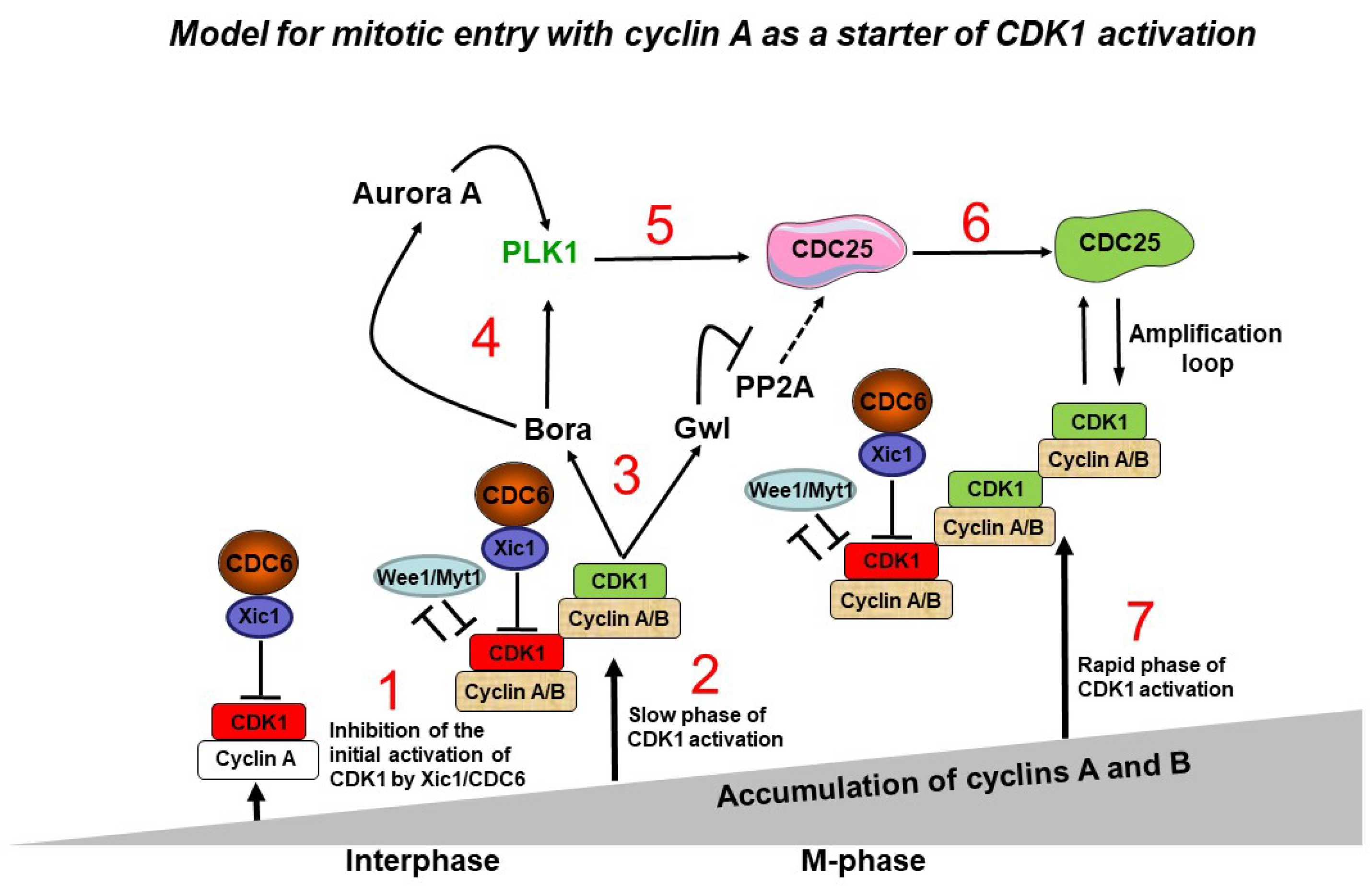
Figure 1.
A model of the key role of CDC6 in the control of the M-phase entry. When the CDK1 active molecules start appearing after cyclin A levels reach a predetermined threshold, they are inhibited by CDC6/Xic1. The first active CDK1/cyclin A molecules appear when they escape from CDC6-dependent inhibition, while CDK1/cyclin B complexes are inhibited by both CDC6/Xic1 and Wee1/Myt1. This is the step of the initial activation of CDK1. In parallel, both cyclin A and B level continue to increase with time and keep associating with CDK1. The active CDK1/cyclin A and CDK1/cyclin B complexes become more abundant, and they, or only CDK1/cyclin B, becomes able to phosphorylate its substrates Bora and Gw1. Bora interacts with PLK1 and Aurora kinase A, activating PLK1. Due to this activation of PLK1 via these two distinct pathways, the procedure is highly effective. Then, PLK1 phosphorylation of CDC6 enhances CDK1 inhibition effectiveness. PLK1 concurrently stimulates CDC25, which triggers the rapid CDK1/cyclin B activation. Gw1 phosphorylation also prevents CDC25 dephosphorylation, allowing it to function even more efficiently. Continuous cyclin B accumulation causes an increase in active CDK1/cyclin B complexes, which in turn promotes CDC25 activity acting as the CDK1 amplification loop.
Disclaimer/Publisher’s Note: The statements, opinions and data contained in all publications are solely those of the individual author(s) and contributor(s) and not of MDPI and/or the editor(s). MDPI and/or the editor(s) disclaim responsibility for any injury to people or property resulting from any ideas, methods, instructions or products referred to in the content. |
© 2023 by the authors. Licensee MDPI, Basel, Switzerland. This article is an open access article distributed under the terms and conditions of the Creative Commons Attribution (CC BY) license (http://creativecommons.org/licenses/by/4.0/).
Copyright: This open access article is published under a Creative Commons CC BY 4.0 license, which permit the free download, distribution, and reuse, provided that the author and preprint are cited in any reuse.


