1. Introduction
An approach based on epitaxial synthesis of graphene as a result of thermal decomposition of silicon carbide SiC holds a valuable place among the various techniques for obtaining graphene developed with the aim of using graphene in nanoelectronic devices. It looks particularly important to make a single-layer graphene with a very perfect crystal structure using the Si-face of 4H-SiC and 6H-SiC carbides.
During thermal decomposition of silicon carbide in a high vacuum and in an argon atmosphere with an increase in the annealing temperature, the sublimation of Si atoms from the surface of silicon carbide occurs with the formation of several reconstructions enriched in carbon atoms on the Si-face at certain stages of annealing. As a result, at the last stage, the Si-face is covered with an exclusively carbon layer, a hexagonal lattice of which has the form of the 6(√3×√3)R30⁰ reconstruction (hereinafter abbreviated as 6√3). The epitaxial relation for the lattice plain given in [
1] expresses the connection of this reconstruction with the substrate. A further increase in the annealing temperature leads to the growth on the surface of the 6√3 reconstruction, first of a single-layer epitaxial graphene film, and then of a multilayer one. Thus, the 6√3 reconstruction is an intermediate buffer layer (BL) between the SiC surface and the graphene layer; however, due to the strong covalent bond between the BL and the Si atoms, the transport properties and other characteristics of the graphene layer deteriorate in the upper layer of the substrate adjacent to the BL [
2,
3]. As established in [
4], covalent bonds can be eliminated by hydrogen intercalation. During hydrogen intercalation, the covalent bond of the buffer layer with the uppermost Si atoms of the substrate is broken and the Si atoms are saturated with hydrogen bonds, and the BL now becomes the first layer of epitaxial graphene, which is called quasi-free single-layer epitaxial graphene. Later, some studies were reported using various techniques of the formation of quasi-free epitaxial graphene from the BL grown on SiC both in a high vacuum and in an Ar medium with the subsequent intercalation of the BL with hydrogen (see, for example, [
5,
6,
7]).
This contribution is dedicated to the structural study of the conversion of the 6√3 reconstruction, which occurs at a certain stage of the sublimation of Si atoms from the 4H-SiC surface during short-term annealing in an Ar atmosphere, into epitaxial quasi-free single-layer graphene as a result of hydrogen intercalation of the reconstructed substrate layer. This structural study was carried out using the technique of reflection high-energy electron diffraction (RHEED) in order to obtain any additional information regarding the degree of perfection of the structure and uniformity of the formed carbon layers, initially revealed using the following research methods: the Raman spectroscopy, the slow electron diffraction, the atomic force microscopy, the Kelvin-probe force microscopy, and the X-ray photoelectron spectroscopy [
8,
9]. The last method was applied only to the intercalated sample.
3. Results and discussion
Figure 1(a-c) show diffraction patterns (DPs) taken in the azimuth direction of [
2
0]
SiC from the reconstructed surface of the single-crystal SiC substrate at an angle of incidence of an electron beam on the sample of about 1° (
Figure 1a) and at angles of about 2.5° (
Figure 1b,c).
In the case of beam incidence at an angle of 1°, the RHEED pattern contains information from the uppermost monolayer of the sample surface, and at angles above 2° it contains information from several subsurface monolayers.
In
Figure 1a, the DP consists of a very limited pattern in the zero Laue zone L0 and the fractional Laue zone of the first order L1/6 corresponding to the 6√3 reconstruction. The fully formed RHEED pattern 6√3 in the zero Laue zone in the azimuth direction of [
2
0]
SiC contains the rod-like reflections (under the condition of surface smoothness of the substrate single crystal) corresponding to
(10)SiC and
(20)SiC accompanied by satellites, as well as with satellites localized near the central reflection [
10]. However, only reflections corresponding to
(10)SiC and satellite reflections near the central reflection are revealed in the DP in
Figure 1a.
In addition to the L1/6 fractional zone from 6√3, the DP in
Figure 1a shows an inconspicuous fragment of the Laue fractional zone superfluous for the 6√3 reconstruction, which can be attributed to the fractional zone of the first order of the 6x6 reconstruction, which is formed at a lower SiC annealing temperature (according to [
10,
11]).
A full-fledged DP in the zero Laue zone corresponding to the 6√3 reconstruction was registered only at angles of incidence of the electron beam of about 2.5° (
Figure 1b,c).
The reflections corresponding to the 6√3 reconstruction in the DP in
Figure 1b are marked with arrows. Comparison of the RHEED patterns in
Figure 1(b,c) with the RHEED patterns from the 6√3 reconstruction on a single-crystal substrate with the 6H-SiC(0001) structure, formed by high-vacuum annealing under strict control, carried out using Auger methods and photoelectron spectroscopy [
10,
11] showed an overestimated intensity of satellite reflections with an interplanar spacing corresponding to
(11)G relative to the intensity of
(20)SiC reflections in
Figure 1b and 1c. In
Figure 1b, the satellite corresponding to
(11)G is marked with a red arrow.
Such an increase in the intensity of the satellite in the DP indicates the formation of a certain fraction of epitaxial single-layer graphene along with the 6√3 reconstruction, apparently, in the area of the terrace steps of the SiC substrate, since the sublimation of Si atoms begins precisely at the ledges of the terraces [
12].
A slight violation of the uniformity of the 6√3 reconstruction formation also generates very short low intensive reflections that are not related to 6√3 on the DP (
Figure 1a,b) in the zero Laue zone. The short extra reflections not marked with arrows in
Figure 1b can be interpreted as belonging to the 6x6 reconstruction. These reflections are revealed only in the left part of the DP due to a small deviation from the [
2
0]
SiC azimuth during the survey.
Attention is drawn to DP, where, in addition to the RHEED reflection pattern, there is a polycrystalline pattern with amorphous halos superimposed on it around the trace from the primary beam. The partially polycrystalline pattern corresponds to the structure of tridymite SiO2. The DP of a ring character is interpreted as a DP for the passage from a thin oxide inclusion protruding above the reconstructed surface.
Figure 1.
Diffraction patterns recorded in the [ 2 0]SiC azimuth with varying angles of incidence on the reconstructed SiC surface: (a) at an angle of about 1° from the 6√3 reconstruction with the first fractional Laue zone and a fragment of the SiC fractional reconstruction zone (6 × 6); (b) at an angle of about 2.5° from the 6√3 reconstruction in the zero Laue zone with reflections indicated by arrows; (c) at an angle of incidence of about 2° from the 6√3 reconstruction and from the occasional inclusion of Si oxide in the formed reconstructed layer.
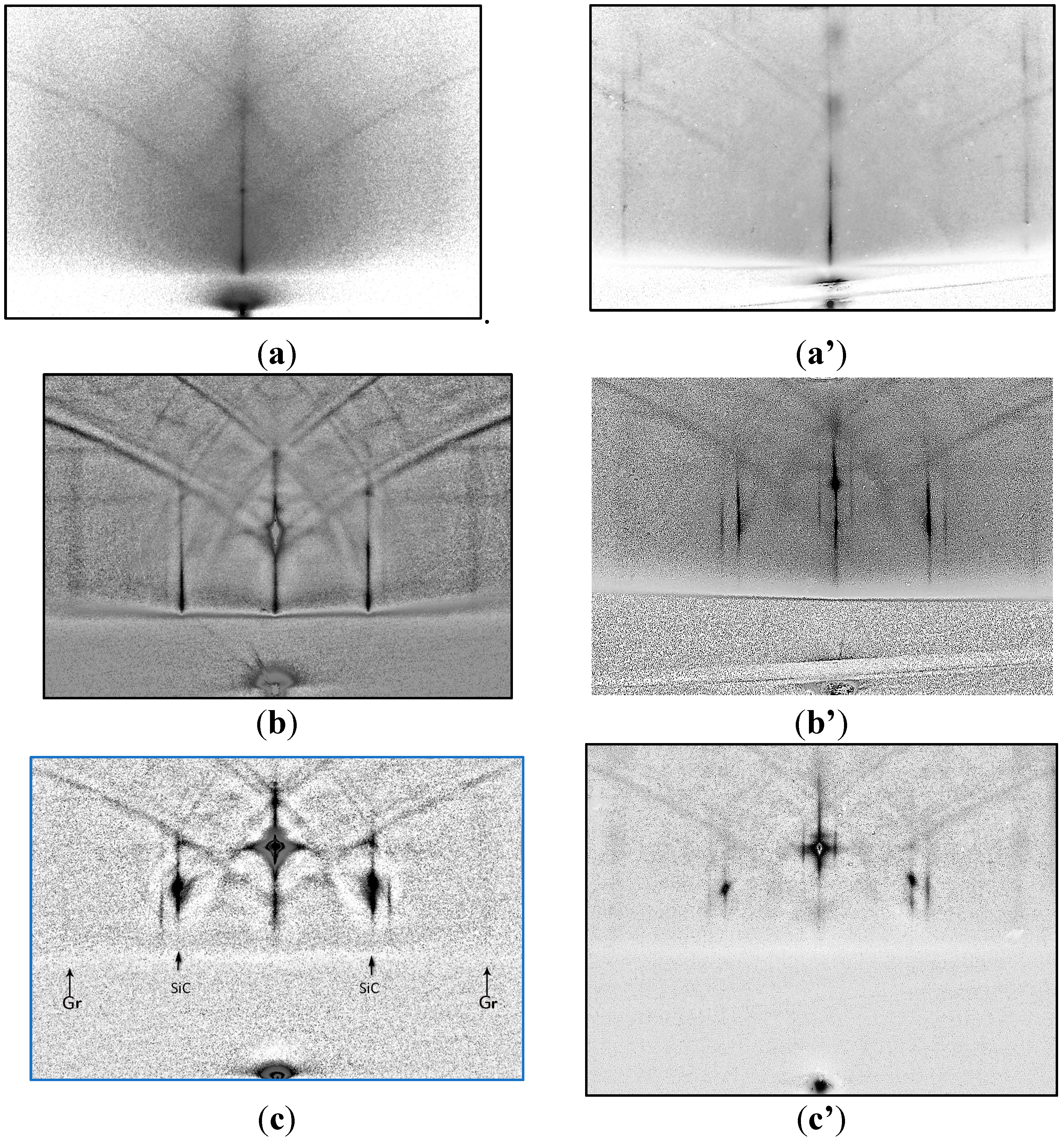
Figure 2 shows the DP obtained using registration at angles of incidence of the beam on the surface of the samples from 1 to 4°. The left column in
Figure 2 shows DPs from the reconstructed SiC surface annealed in an H
2 atmosphere. The right column shows DPs from the surface of an epitaxial graphene monolayer sample grown on a 4H-SiC(0001) substrate with a similar pre-epitaxial preparation of the 4H-SiC(0001) substrate in argon for 15 min at a temperature of 1855°C without intercalation [
13]. The DP in
Figure 2a with a registration of about 1° demonstrates continuous rod-like reflections (
11) of graphene, which is evidence of the 6√3 reconstruction conversion into epitaxial graphene. However, the observed smearing of reflections (
11) (especially noticeable when compared with DP in
Figure 2a′) against the diffuse background of electron scattering indicates that buffer layer conversion is accompanied by the appearance of structural disturbances in the formed epitaxial graphene.
In the case of gliding of the electron beam close to 1° (
Figure 2a) in the DP from the intercalated sample, barely visible fractional streaks of the reconstruction 6√3 over the reflections (11)G in the zero of Laue zone are fixed, which in the fractional zone on the DP from graphene monolayer with a buffer layer in
Figure 2a′ are clearly identified.
Thus, it has been proved that the layer of epitaxial graphene is almost completely quasi-free.
Further, in
Figure 3b, 3b′, 3c, and 3c′, frames with DP from several near-surface monolayers of samples are shown when the angle of incidence of electrons was approximately 2° and 4°, respectively, for DP in
Figure 2b, 2b′ and 2c, 2c′.
In
Figure 2b, and 2c, DPs from the intercalated sample consist of reflections
(11)G and
(10)SiC accompanied by a satellite on the outer side, whereas localized satellites around the central reflection of the reconstruction 6√3 have disappeared. On DP from a sample with a barrier layer (
Figure 2b,c') they are clearly revealed.
A possible reason for the appearance of a satellite near
(10)SiC in a DP in an intercalated sample may be nanostructuring of the SiC surface in contact with quasi-free single-layer graphene [
14]. As was established in [
14], by studying the process of hydrogenation by reverse photoelectron spectroscopy and Auger spectroscopy, hydrogen intercalation of the buffer layer above 700°C leads to nanostructuring of the SiC surface in contact with quasi-free single-layer graphene. The authors of [
14] showed the formation of a SiC subsurface with a reconstruction close to the Н-(√3×√3)R30° one under single-layer graphene. We have compared the DP in
Figure 2b,c with the RHEED pattern (in the zero Laue zone) with a survey in the <11
0>
SiC azimuth from the (√3x√3)R30° reconstruction given in ([
15], p.126), and found similarity of (√3x√3)R30° and H-(√3x√3)R30° ones.
Figure 2.
Comparison of the RHEED patterns of the surface of the intercalated sample with the sample of single-layer graphene on the barrier layer: (a) in the [20]SiC azimuth at an electron beam incidence angle of about 1° on the sample after annealing in Ar; (a') under similar conditions of registration of a monolayer sample; (b) at an incidence angle of about 2° in the [110]SiC azimuth of the intercalated sample; (b′) with an incidence angle of about 2.5° in the azimuth of the sample without intercalation; (c) in the [20]SiC azimuth with an incidence angle of about 3.5° onto the intercalated sample; and (c') with an incidence angle of about 4° onto a graphene monolayer with a buffer layer.
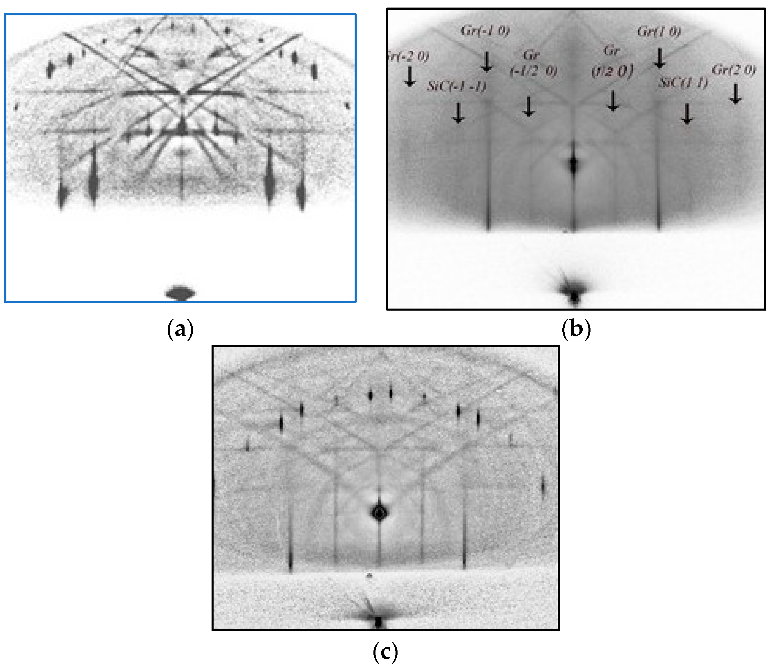
Figure 3 provides the RHEED patterns recorded in the [1
00]SiC azimuth.
Figure 3a presents the diffraction pattern from the SiC surface after short-term annealing corresponds to the 6√3 reconstruction in the zero Laue zone with a first-order fractional zone.
Figure 3b shows DP from the SiC surface after short annealing followed by intercalation. DP consists of graphene reflections with no satellite reflections and fractional 6√3 reconstruction zone.
DP in
Figure 3c, recorded from the surface of a substrate with single-layer epitaxial graphene grown on a buffer layer without intercalation, is a diffraction pattern from graphene in the zero Laue zone and the first fractional reconstruction zone 6√3.
Thus, comparison of DP in
Figure 3a–c confirms the conclusion about the b layer conversion into epitaxial quasi-free graphene.
Figure 3.
DPs recorded in the [1100]SiC azimuth: (a) from the SiC surface with annealing in argon; (b) from the SiC surface after annealing followed by intercalation; (c) from the SiC surface with single-layer epitaxial graphene grown on the buffer layer.
Figure 3.
DPs recorded in the [1100]SiC azimuth: (a) from the SiC surface with annealing in argon; (b) from the SiC surface after annealing followed by intercalation; (c) from the SiC surface with single-layer epitaxial graphene grown on the buffer layer.


