
Submitted:
07 June 2023
Posted:
07 June 2023
You are already at the latest version
Abstract
The phenomenon of pathogens co-infection detected in the half fed on humans I. persulcatus tick in the south of the Far East was studied. Researchs were carried out on PEK, Vero, Vero-E6 cell lines, outbred mice, chicken embryos, using ELISA, PCR, IMFA, plaque formation, and electron microscopy. The tick contained an antigen and a genetic marker of the tick-borne encephalitis virus (TBEV). The patient had post-vaccination antibodies in a titer of 1:200, as a result of which, obviously, an antibody-dependent elimination of TBEV occurred. The tick-borne co-isolate also contained an unknown pathogen (Kiparisovo-144 virus), which, in our opinion, was a trigger for the activation of chronic infection in suckling white mice. In the laboratory coisolate, ectromelia virus was present, as evidenced by paw edema during intradermal infection of mice, characteristic rashes on the chorion-allantoic envilope of chicken embryos, and typical plaques on Vero-E6. The Kiparisovo-144 virus was not pathogenic for white mice and chicken embryos, but successfully multiplied in the PEK, Vero, and Vero-E6 lines. Viral co-infection was confirmed by electron microscopy. Passaging on mice contributed to an increase in the virulence of the co-isolate, whose titer increased by 10,000 times by the 5th passage, which poses a serious epidemiological danger.
Keywords:
coinfection
; tick Ixodes persulcatus
; virus isolate
; tick-borne encephalitis virus (TBEV)
; ectromelia virus
; biological properties of co-isolate
; electron microscopy
; south of the Russian Far East
1. Introduction
In recent years, in the study of the tick virome, modern research approaches (metagenomic and metatranscriptomic) have been used, which reveal a enormous number of putative new pathogens [1]. Such information greatly enriches knowledge of the natural diversity of possible pathogens in different tick species [2,3]. To be aware of potentially dangerous pathogens capable of causing infection in humans and animals, it is necessary to have isolates of pathogens with disclosed molecular structure sequences and studied biological characteristics [4].
Thus, since 2009, the severe fever with thrombocytopenia syndrome (SFTS) began to be registered in six provinces of China, the cause of which was not known [5,6,7,8,9]. Genome sequencing of the isolated pathogen has established that the SFTS virus belongs to a new (third) group of the Phlebovirus genus of the Bunyaviridae family. This virus has aroused great interest among researchers. It turned out that the SFTS virus is found not only in China, but also in the territories adjacent to the Far East - in South Korea and Japan. In addition, its wide distribution has become known in many countries of the world [2,6,10,11].
Earlier, in 1971, we isolated the Khasan virus (KHAV) from ticks Haemophysalis longicornis Neumann, 1901, collected from spotted deer Cervus nippon Temmink, 1838, on the territory of the Khasansky region (south of the Primorsky Territory of the Far East). Based on biological properties, morphology [12,13], and molecular genetic characteristics, the KHAV virus was assigned to the genus Phlebovirus of the Bunyaviridae family [14]. In this regard, our attention was directed to the identification of similar and other new pathogens in natural foci in the south of the Far East.
In the last decade, great attention has been drawn to reports on the isolation of especially dangerous pathogens of viral infections. Numerous publications are known about new variants of the smallpox virus with non-traditional sources of isolation [15,16,17]. This applies to recently discovered pathogens of the smallpox virus, which were isolated from relatively poorly studied hosts (fish, bats, porcupines, mosquitoes, birds and aquatic mammals) or from humans [18,19]. Noteworthy is the history of one strain of ectromelia virus isolation during intracerebral infection of laboratory white mice with homogenates of ixodid ticks [20]. Initially, the authors identified co-infection of two viruses (mouse ectromelia virus and lactate dehydrogenase-elevating virus (LDV) belonging to the family Arteriviridae) in ticks, but they failed to pass LDV. Therefore, the authors of the article characterized only a new strain of ectromelia virus ECTV (ECTV-WH) [20].
A similar situation arose in our study of a viral isolate from the tick Ixoodes persulcatus. During the isolation of the putative virus from a half-fed I. persulcatus tick taken from a person in 2016, it was possible to identify the manifestation of co-infection of viral pathogens. It was necessary to verify it, and to give a comprehensive biological characterization of viruses.
2. Materials and Methods
2.1. Virus
An unknown for us pathogen was isolated from a half-fed Ixodes persulcatus tick taken from a patient on May 21, 2016 in the south of the Russian Far East (Nadezhda region of Primorsky Krai). In case of primary simultaneous infection in the brain and subcutaneously of 2-day-old outbred white mice of the same litter with a 10% suspension of ticks, on the 6th day one animal with an unclear clinic of the disease was euthanized, from which the brain was taken for further virological examination. The virus isolate from the 2nd to the 5th passages was used in the work.
2.2. Enzyme Immunoassay (ELISA)
Detection of the antigen of the tick-borne encephalitis virus (TBEV) in the tick homogenate was carried out by ELISA using the “VectoTBEV-antigen” kit (ZAO “Vector-Best”, Novosibirsk) according to the instructions of the test system manufacturer.
2.3. Real-Time PCR (RT-PCR)
Tick was examined for the presence of genetic markers: tick-borne encephalitis virus (TBE), Borrelia burgdorferi sensu lato, Anaplasma phagocytophilum, Ehrlichia muris/Ehrlichia chaffeensis by real-time polymerase chain reaction (RT-PCR) using the kit "AmpliSense TBEV, B. burgdorferi s.l., A. phagocytophilum, E. chaffeensis / E. muris-FL" (Central Research Institute of Epidemiology, Moscow) according to the manufacturer’s instructions on a cycler with fluorescent detection "ROTOR-GENE Q" (QIAGEN, Germany).
2.4. Indirect Immunofluorescent Antibody (IFA) Method
The indirect MFA method was used to detect the antigen in Pig embryo kidney (PEK), Vero, and Vero-E6 culture cells contaminated with test samples in 3 tubes. Cells from these tubes were collected at certain times of the experiments, then slides were prepared from the mixture of cells on objective glasses. The antigen of the virus was detected in the cells by applying specific immune serum to the slides and, subsequently, fluorescent immunoglobulins (FITS) in the working dilution specified in the manufacturer’s instructions (Branch "MEDGAMAL", N.F. Gamaleya NIIEM). Slides were viewed in 3 fields on a fluorescent microscope MC-200 TF (Austria). The average percentage of cells with the content of the fluorescent virus antigen in relation to the total number of cells in the field of view was considered.
2.5. Plaque Method
The plaque method was used to study the biological characterization of the virus in the pig embryonic kidney (PEK), Vero and Vero-E6 cell lines. The investigation was carried out on 24-well plates using a coating of carboxymethyl cellulose (ICN Biomedical). The titer of virus samples was calculated in plaque forming units (PFU).
2.6. Methods for Studying the Biological Properties of a Viral Isolate
2.6.1. Virulence
The studies were carried out on models of 2-day-old and 4-week-old outbred white mice, which were infected in the brain, subcutaneously and intraperitoneally with a 10% suspension of the sick mice brain of the 2nd and 5th bioisolate passages and its titer was determined in lg LD50/ml.
2.6.2. Sensitivity of Chick Embryos
The study was carried out by infecting 9-day-old chicken embryos with a 0.1 ml virus suspension containing 3 lg PFU on the chorioallantoic envelope (CAE). The results were determined on days 1, 2, and 3 postinfection (pi) by detecting rashes on CAE, as well as by the accumulation of the virus in CAE using the indirect MFA method on the PEK cell line and by detecting the virus titer (PFU) on the Vero-E6 cell line.
2.6.3. Influence of Physical Factors on the ell Line. Inactivation of a Viral Isolate
Influence of physical factors on the inactivation of a viral isolate. The influence of physical factors (temperature 600, 1000, ultraviolet irradiation) on the virus inactivation was carried out when exposed to them for 1, 2, 5, and 10 min pi. To verify the results on PEK cells, the indirect MFA method was used, and on the Vero-E6 cell line, the plaque method was used. To assess the effect of ultraviolet radiation on the infectious activity of the pathogen, a small Petri dish with a virus-containing liquid (3 lg PFU) 3 mm thick was used. This dish was installed at a distance of 24 cm from a source of short-wave ultraviolet rays with a maximum wavelength of 253.7 nm.
2.6.4. The Study of the Infectious Activity of Viral Particles after Filtration through a 0.22 µm Millipore Filter (Biofil) was Carried Out using the Virological Methods described Above.2.6.5. Electron Microscopic Examination of the Viral Isolate was Carried Out by Method of Negative Staining
Negative contrast electron microscopy was performed on two samples of the isolate. The first sample is the original isolate and the second sample is the isolate after filtration (filter 0.22 nm). The PEK cell line was infected with these samples, the supernatant was collected on days 1-3. Virus particles in the supernatants were visualized by negative staining EM analysis. Supernatants from each sample (1 mL) were centrifuged at 3000 × g for 20 min at 4 °C to remove cell debris. The clarified supernatants were then centrifuged at 13000 × g for 40 min at 4 °C. The pellets were resuspended in 10 μL PBS. Formvar carbon-coated copper grids were floated in droplets of virus suspension for 10 min and stained with 2% phosphotungstic acid for 1 min at room temperature. Subsequently, the grids were examined by transmission electronic microscopy [7]. The samples were viewed using a JEM-100S transmission electron microscope (JEOL, Japan) at an accelerating voltage of 80 kV.
3. Results
3.1. History of Virus Isolation
In May 2016, a 75-year-old woman with a half-fed tick Ixodes persulcatus applied to the laboratory for testing on tick-borne infections. The tick was sucked on the territory of the Nadezhdinsky region of Primorsky Krai. The tick was removed 5 days after feeding on the body of the victim. Due to the fact that tick-borne encephalitis virus (TBEV), the first representative of the Flaviviridae family, dominates and is widespread in the Primorsky Territory of the Russian Far East [4], a tick was studied for the detection of tick-borne encephalitis virus (TBEV) antigen in ELISA, as well as in RT-PCR-RT for the genetic marker of tick-borne infections (TBEV, Borrelia burgdorferi sensu lato, Anaplasma phagocytophilum, Ehrlichia muris/Ehrlichia chaffeensis). At the time of the study, in the bioassay of a homogenized tick, an antigen of the TBE virus was detected with a low positivity coefficient (K=1.7); in real-time PCR, a genetic marker was determined only for the TBE virus and also with a low indicator (Ct = 34), which indicated a low degree of viral load of this pathogen.
A suspension of a homogenized tick was used to infect into the brain of outbred white mice of 2 days of age. The brain of one sick suckling mouse with an unclear clinic was taken on the 6th day pi and passaged also by intracerebral infection of suckling mice of another family. At the first passage, mice began to get sick for 6 to 10 days, at the second passage - for 5-7 days, with the maximum activity of clinical manifestations on the 5th day. At the third passage, all infected suckling mice fell ill on the 4th day, at the fourth and fifth passages, on the 3rd day. It was suggested that the studied isolate could be a TBE virus. However, the study of the sick mice brain of all passages did not show, either in ELISA or in PCR-RT, the antigen and genetic marker of TBEV. It was necessary to understand the reason for the elimination of TBEV from a bioassay of a half-fed tick, in which, as shown above, low rates of infection with this virus were determined.
From the anamnesis it was found out that the patient had previously been vaccinated against TBE. An ELISA study of her blood serum showed the presence of IgG antibodies to the TBE virus in a titer of 1:200. It has been suggested that in the process of bloodsucking the tick for 5 days on a human with antibodies to the TBE virus in a titer of 1:200, neutralization and elimination of the TBE virus occurred in the fed tick. This means that in the present case we have not TBE, but some other isolate unknown to us.
Along with this, the study of the blood serum of this woman by the indirect MFA method for the detection of antibodies to this isolate showed the presence of those with a low titer of 1:8 (Figure 1). No clinical manifestations of the disease were observed in the patient.
3.2. Characterization of the Virus In Vivo
In the study of a new unknown pathogen, an important feature is the determination of the sensitivity of various laboratory models to this pathogen. First of all, this refers to the determination of the susceptibility of laboratory animals, in particular, white mice of different age groups with different methods of pathogen inoculation. On Figure 2 shows 2 days white mice infected into the brain with a 10% viral suspension of the 2nd passage with signs of encephalitis (convulsions, paresis of the limbs).
On the model of white mice of 2 days of age, the titer of the 2nd passage virus was determined, which was 7.2 lg LD50/ml with intracerebral (i/c) infection, and 5.6 lg LD50/ml with subcutaneous (s/c) infection. On the model of white mice of 4 weeks of age weighing 8-10 g, the virus titer upon i/c infection was 6.0 lg LD50/ml, mice did not get sick with s/c infection (0 lg LD50/ml). After intradermal infection with a viral isolate in the paw of the hind limb, swelling of all paws was observed on days 7–10 due to pronounced diffuse edema and exudation, skin rashes and necrotic changes were absent. However, when the viral isolate was passaged up to passage 5, its activity increased sharply, the incubation period was reduced to 3 days, the virus titer increased significantly to 10.0 lg LD50/ml, i.e. 10000 times. The virulence of the virus isolate also notably increased, it became pathogenic in all methods of inoculation (Figure 3).
3.3. Characteristics of the Virus in Experiments In Vitro
To obtain data on the characterization of a new pathogen, it was necessary to study the comparative sensitivity of some cell cultures to it, for which the PEK, Vero, and Vero-E6 cell lines were used. Figure 4 shows that a 10% brain suspension of the 2nd passage of suckling mice viral isolate does not form plaques on the PEK cell line. On cell lines Vero and Vero-E6, the plaques were small, dotted, like a needle prick. At the same time, on the 5th day pi of the monolayer of cell lines, the virus titer on Vero cells was 2 lg PFU lower compared to the virus titer on Vero-E6 cells.
The infectivity of the viral isolate was shown in MFA starting from the early stages (3 h and 24 h) pi of PEK, Vero and Vero-E6 cells (Figure 5).
This figure shows a bright fluorescence of the antigen in infected cells of all lines. Despite the fact that the viral isolate did not cause a cytopathic effect and the formation of plaques on the PEK culture, in Indirect MFA method the viral antigen formed a thin fluorescent layer on the cell surface as early as 3 h pi, and a more distinct fluorescence of the cytoplasmic membrane of these cells was observed 24 h later. Less bright fluorescence was obtained on Vero cells. Vero-E6 cells highly sensitive to the virus isolate showed the brightest fluorescence and especially in their cytoplasm. In the infected culture of these cells, after 3 h one could observe a bright fluorescent of the cytoplasmic membrane, and after 24 h, not only the membrane, but also the cytoplasm, indicating active reproduction of the viral isolate in Vero-E6 cells.
In addition, 5 days pi of PEK cells with the 2nd passage viral isolate, it was detected in the supernatant, as well as in the cell sediment during i/c infection of mice 4 weeks old, its titer was low and amounted to 2 lg LD50/0.03 ml and 1.2 lg LD50/0.03 ml, respectively. On this basis, relative to the viral isolate, the PEK line can be classified as a permissive cell culture, but with a low level of productive viral infection.
The results obtained above allowed us to continue studying the biological properties of the isolate on PEK cells using the indirect method of fluorescent antibodies (MFA). The results were obtained on the accumulation of the pathogen in the supernatant of the infected culture, collected in the period from 3 hours to 9 days pi. with which the cells of the PEK line were infected. Figure 6 shows that when the monolayer of the PEK line was infected with samples of supernatants, fluorescence of cells was detected already from the first day pi. The active replication of the isolate and its release into the supernatant were confirmed by positive results in MFA, especially from days 5 to 8, when there was a gradual increase in the number of fluorescent cells (47%, 83, 88% and 95%) of the infected PEK line.
3.4. The Effect of Physical Factors on the Isolate
To conduct experiments on the action of physical factors, the dynamics of isolate replication in a highly permissive Vero-E6 cell line 2, 3, 5, and 7 days pi was previously studied (Table 1). Solitary plaques appeared on the monolayer of this cell culture already on the second day pi. On day 3, a large number of plaques were observed at a 10-1 dilution. On the 5th day, in the cells of the Vero-E6 line, the isolate actively multiplied to a dilution of 10-4. Finally, on the 7th day, we observed its maximum accumulation up to a dilution of 10-5 in this permissive medium (Table 1).
Our further studies were aimed at studying the effect of physical factors on the viral isolate. Studies were carried out by inactivating it at a temperature of 60o and 100o, as well as by ultraviolet irradiation (UVR) when exposed to these factors from 1 to 10 minutes. Observations were made in parallel on two cell lines Vero-E6 and PEK, differing in their susceptibility to the studied viral isolate (Table 2 and Table 3).
Reproduction of the virus in Vero-E6 cells was observed during infection with samples after exposure to a temperature of 60°C for 1 and 2 minutes (7x10³ PFU and 4x101 PFU). With further heating of the viral isolate, the ability to form plaques was lost (Table 2). Complete inactivation of the viral isolate was noted after exposure to a temperature of 100°C. At the same time, the detection of plaques of the viral isolate was observed 1, 3 and even 5 minutes after exposure to UVI (6x10³, 11x101 and 8x101, respectively).
As can be seen in Table 3, heating the viral isolate at 60°C contributed to a decrease in its activity by 13% at 1 min, by 33% at 2 min, by 37% at 5 min, and by 55% at 10 min of exposure. Heating of the virus isolate at 100°C, as well as exposure to UVI, contributed to a significant decrease in its activity.
3.5. Determining the Size of Viral Particles
A preliminary determination of viral particles size was carried out by filtering the bioisolate through a millipore filter (0.22 µm). A 10% brain suspension of the sick mice 2 days of age of the 5th passage, taken on the 3rd day pi was used. At the same time, suckling mice infected in the brain by the resulting filtrate remained healthy for 21 days of observation. Two assumptions were made: mice could not get sick if the virus was larger than 220 nm and it remained on the filter, and also if the virus was smaller than 220 nm and it was not pathogenic for outbred suckling mice.
To test this hypothesis, the amount of virus in the bioisolate and in its filtrate was determined. For this purpose, the Vero-E6 cell line was used (Figure 7). The obtained results indicated that small plaques were detected in the virus isolate before and after filtration, the size of a needle prick, i.e. the same as in Figure 4. The number of plaques before filtration was 15x10-4, and after filtration - 6x10⁻³. It was concluded that the number of viral particles after filtration significantly decreased, but not completely.
Since the studied isolate was received on the model of outbred white mice, and if the new pathogen has a large size of more than 220 nm, then, first of all, it was necessary to exclude the relationship of the isolate with the mouse ectromelia virus. For this purpose, chicken embryos of 9-10 days of age were infected by introducing a virus-containing material, as well as its filtrate at a dilution of 10-1 by 0.1 ml, onto the surface of the allantoic envelope (CAE) of a chicken embryo.
Figure 8A shows the chorion-allantoic envelope of chick embryos infected with a viral isolate. Here, on the 1st day, we did not observe any rashes; on the 3rd-7th day, typical rashes characteristic of the smallpox virus - white pockmarks (plaques) were detected. During these studies, we did not observe hemorrhagic pockmarks. The chorion-allantoic envelope of chicken embryos contaminated with filtrate remained without rashes (Figure 8B).
To prove the specific effect of the virus on chicken embryos, a monolayer of the SPEV line was infected with a suspension of samples of the chorion-allantoic envelope; 3 days pi, bright fluorescence was detected on PEK cells in IMPA, indicating the presence of antigen in samples of both the initial isolate and its filtrate. In addition, the virus titer was determined on the Vero-E6 cell line. Virus isolate 1 day pi was not detected, and after 3 and 7 days its titer was 3 lg and 5 lg PFU. In samples of CAE chicken embryos contaminated with filtrate, the virus was not verified during all periods of observation.
Based on the obtained results of a comprehensive study, it was suggested that the isolate from the tick I. persulcatus contains at least two viruses. To test this hypothesis, it was necessary to receive the direct evidences using the method of electron microscopy.
3.6. Electron Microscopy of Viral Co-Isolate
To confirm the above assumption, electron microscopic studies of the co-isolate were carried out using the negative staining method. In the cells of the PEK line infected with the co-isolate, on the 3rd day pi, two types of viral particles were verified, which differed from each other. Two viruses were detected in the sample: one large smallpox-like up to 500 nm, the other much smaller, from 60 to 100 nm (Figure 9). In addition, conglomerates consisting of several viral particles can be observed here, on the surface of which adherent particles of small sizes were sometimes observed.
For better visualization of both co-infection viruses, the samples were prepared from the supernatant of the SPEV cell line, taken after 1 day pi that is in the early period of viral replication, as shown in the IMFA in Figure 5 and Figure 6.
Figure 10A shows a significant predominance of large particles up to 1000 nm, which, in our opinion, consists of 2-3 confluent smallpox-like virus particles. There was a fragmented rim along the periphery of such an electron-dense conglomerate. Figure 10B, C, D shows that the conglomerates consist of merged, apparently pox-viral particles, and the rim was adhered particles of a small virus, up to 100 nm in size. In our opinion, such a morphological picture demonstrated the phenomenon of cocultivation of two viruses. At the same time, free-lying particles of a small virus, up to 100 nm in size, were also found.
Thus, based on biological characterization results and using electron microscopy, it was shown that the viral isolate is presented as a coinfection of two pathogens: a representative of the smallpox virus family and a pathogen unknown to us.
4. Discussion
Currently, researchers are paying close attention to the study of arthropods infection with known and unknown pathogens that represent a danger not only to the human population, but also to wild and domestic animals. Modern publications on the verification of pathogens from different members of natural foci of infection, as a rule, are based on the results of the genetic markers detection in PCR and other molecular biological studies [2,3,21]. Widely conducted studies on the prevalence of tick-borne pathogens in various focal areas have significantly enriched our understanding of the biodiversity of the ixodid tick virome [22]. In addition, there are often cases of mixed infection of ticks simultaneously with various pathogens [23]. Probably, in such cases, favorable conditions arise for the equilibrium co-cultivation of several pathogens in in the same biological object [24]
In this work, we also had to solve the issues of interaction of several pathogens in the microbiome of the tick I. persulcatus. In the process of studying the bioisolate, we were able to verify three different viruses. Initially, in the test tick after 5 days of bloodsucking on a human, infection with tick-borne encephalitis virus (TBEV) was established at low rates in ELISA and PCR. As it turned out, specific post-vaccination antibodies to TBEV were present in the blood of the examined patient, which probably could contribute to the neutralization and elimination of this pathogen in the tick during bloodsucking. This outcome could be supported by the results of our long-term studies in vitro, ex vivo and in vivo, aimed at studying the effectiveness of specific protection against the TBE virus [25]. Using a clear biological model - the blood of vaccinated individuals with different titers of antibodies to TBEV: 1:100; 1:200; 1:400; 1:800; 1:1600; 1:3200, we showed that in samples with specific antibodies in titers of more than 1:400, the neutralization of the virus occurred quickly (after 24 hours). Under the action of antibodies in titers of 1:100 and 1:200, the elimination of the pathogen also occurred, but at a later date, on days 3–4 pi of these samples with the TBE virus [26]. This allowed us to assume that specific antibodies circulating in the patient’s blood in a titer of 1:200 during tick bloodsucking could affect the suppression of the viability of the TBE virus, as a result of which it was not isolated during virological studies, and the patient did not get sick with TBE.
Such a classical concept of antibody-dependent elimination of the TBE virus is quite understandable. But there is an assumption that in the case of co-infection of the TBE virus and a new virus, the possibility of TBEV elimination can be assumed if the new virus acts as a suppressor during its replication in the tick body. In this regard, it should be noted that for almost two decades we have been observing a gradual decrease in the infection rates of TBEV ticks collected from vegetation in natural foci, as well as ticks attached to people. This downward trend in TBE virus infection of ticks was reflected in TBE incidence rates in the south of the Far East, where isolated cases of this infection have been recorded in the population in the last decade [27].
In the case under study, probably, the previously unknown pathogen of the coinfection we identified in the tick microbiome got the opportunity for its replication and the appearance of specific antibodies in the patient, although its titer was low (Figure 1). Earlier, for 2 years, we noted in one patient the clinical and diagnostic features of a mixed infection caused by TBEV, Borrelia, and SARS-Cov2 [28]. Using the indicators of antigen detection in ELISA, genetic markers in PCR, antibodies, as well as the degree of their avidity to these pathogens, we found that in the case of detection of three infections simultaneously, the bacterial pathogen B. burgdorferi dominates in the human body, causing him a long-term chronic course of the disease [27]. Such dominance of Borrelia in the body of a tick infected with the TBEV and B. burgdorferi was previously proved in the experiment [29].
This means that in cases of co-infection of different pathogens, any pathogen can dominate. In the described case, when studying the biological characteristics of the viral isolate, we observed an increase in the activity of the mouse pox virus. The isolation of a new virus in ticks can be considered an accident, because. as it was found that this virus is not pathogenic for white mice. But this chance was predetermined by the fact that the isolation of the virus was performed on a model of outbred mice of 2 days of age, which received the ectromelia virus vertically from the mother - the carrier of this chronic infection. We failed to isolate the TBE virus, but an unknown pathogen was identified, which, in our opinion, was a trigger for the activation of a latent chronic infection in sucklings of outbred white mice. A similar case of coinfection of two viruses (murine ectromelia virus and lactate dehydrogenase-elevating virus) in ticks was described in 2021 [20]. However, the authors failed to pass the second LDV virus.
Using various virological research methods, we focused our efforts on identifying the features of the biological characteristics of the isolated viral co-isolate. In favor of the presence of the ectromelia virus in it, the following manifestations testified: paw edema during intradermal infection of adult mice, characteristic rashes on the CAE of infected chicken embryos, typical plaques in the form of an injection on the infected Vero-E6 culture, instability to warming and resistance to ultraviolet irradiation. However, an electron microscopic examination showed that in addition to the smallpox-like virus, the co-isolate contained another virus that notably differed from the smallpox virus.
And although the new virus we identified turned out to be non-pathogenic for white mice of all age groups and chicken embryos, did not cause cytopathogenic effects and plaque formation on PEK cells, but it successfully multiplied in PEK, Vero and Vero-E6 cell lines. Its presence in the viral co-isolate is shown in the electron microscopy images.
Based on the obtained results, this viral co-isolate can be characterized as a stable form of co-cultivation of murine ectromelia virus and a new tick-borne virus, named "Kiparis-144". Moreover, the dominant mouse ectromelia virus served as an indicator for the verification of the second virus, Kiparis-144. This is probably why, without such an indicator and without a permissive model for virus isolation, researchers [2,3,20] failed to isolate new pathogens in molecular genetic studies of the ixodid tick virome.
The interaction of these two viruses contributed to a significant increase in the virulence of the viral coisolate from passages 2 to 5 in outbred white mice with all methods of inoculation. The incubation period in suckling mice infected in the brain was reduced from 5-7 days to 2.5-3 days. The virus titer after intracerebral infection of 3-week-old mice increased by 4 lg LD50, i.e. 10000 times. This indicated that when two viruses were co-cultivated in warm-blooded animals, the new virus acts as an amplifier or "accelerator" of mouse pox virus replication. Their combined interaction was clearly shown by us using electron microscopy (Figure 9).
In epidemiological and epizootological terms this property of the new virus is a potential danger and naturally causes concern. Moreover, in recent years, natural populations of various representatives of the smallpox virus have become more active in the world [30,31,32]. We admit the possibility of expanding the ecological and virological competencies of ticks containing pathogens like the Kiparis-144 virus. When bloodsucking ticks with such viruses on cows, monkeys, donkeys, and other animals in cases of chronically occurring smallpox virus infection, its activation can occur with the involvement of a human in this process.
5. Conclusion
Thus, the obtained results allow us to conclude that it is necessary to conduct further scientific research to study the molecular genetic characteristics of not only the ectromelia virus, but also the new Kiparis-144 virus detected in the half-fed tick I. persulcatus in the south of the Far East, as well as to determine the degree its pathogenicity for humans and animals.
Author Contributions
Conceptualization, G.L.; validation, writing and editing G.L., editing L.S.; Formal analysis, resources investigation E.P. and S.A. All authors have read and agreed to the published version of the manuscript.
Funding
This study was funded by funds received for the implementation of a scientific topic. The work was carried out within the framework of State Assignment to Somov Research Institute of Epidemiology and Microbiology.
Data Availability Statement
This article does not contain any studies involving humans as primary objects of research.
Acknowledgments
The authors express their gratitude to the laboratory colleagues, who provided advice, support, and assistance in the work.
Conflicts of Interest
The authors declare no conflict of interest. The protocols for animal studies were approved by the Committee on the Ethics of Animal Experiments of the Somov Research Institute of Epidemiology and Microbiology (Rospotrebnadzor), Vladivostok, Russia.
References
- Bratuleanu, B.E.; Raileanu, C.; Chrétien, D.; Guardado-Calvo, P.; Bigot, T.; Savuta, G.; Temmam, S.; Eloit, M. A Search for Tick-Associated, Bronnoya-like Virus Spillover into Sheep. Microorganisms 2023, 11, 209. [Google Scholar] [CrossRef]
- Pettersson, J.H.-O.; Shi, M.; Bohlin, J.; Eldholm, V.; Brynildsrud, O.B.; Paulsen, K.M.; Andreassen. ; Holmes, E.C. Characterizing the virome of Ixodes ricinus ticks from northern Europe. Sci. Rep. 2017, 7, 10870. [Google Scholar] [CrossRef]
- Kong, Y.; Zhang, G.; Jiang, L.; Wang, P.; Zhang, S.; Zheng, X.; Li, Y. Metatranscriptomics Reveals the Diversity of the Tick Virome in Northwest China. Microbiol. Spectr. 2022, 10, e0111522. [Google Scholar] [CrossRef] [PubMed]
- Leonova, G.N.; Belikov, S.I. Phylogenetic analysis and distribution of far eastern tick-borne encephalitis virus subtype (Flaviridae, Flavirus, TBEV-FE) from Asia. Probl. Virol. 2019, 64, 250–256. [Google Scholar] [CrossRef] [PubMed]
- Yu, X.J.; Liang, M.F.; Zhang, S. Y.; Liu, Y.; Li, J. D.; Sun, Y. L.; Zhang, L.; Zhang, Q. F.; Popov, V. L.; Li, C.; et al. Fever with Thrombocytopenia Associated with a Novel Bunyavirus in China. N. Engl. J. Med. 2011, 364, 1523–1532. [Google Scholar] [CrossRef] [PubMed]
- Zhang, Y.-Z.; He, Y.-W.; Dai, Y.-A.; Xiong, Y.; Zheng, H.; Zhou, D.-J.; Li, J.; Sun, Q.; Luo, X.-L.; Cheng, Y.-L.; et al. Hemorrhagic Fever Caused by a Novel Bunyavirus in China: Pathogenesis and Correlates of Fatal Outcome. Clin. Infect. Dis. 2011, 54, 527–533. [Google Scholar] [CrossRef] [PubMed]
- Zhang, Y.; Shen, S.; Shi, J.; Su, Z.; Li, M.; Zhang, W.; Li, M.; Hu, Z.; Peng, C.; Zheng, X.; et al. Isolation, characterization, and phylogenic analysis of three new severe fever with thrombocytopenia syndrome bunyavirus strains derived from Hubei Province, China. Virol. Sin. 2017, 32, 89–96. [Google Scholar] [CrossRef]
- Cui, F.; Cao, H.-X.; Wang, L.; Zhang, S.-F.; Ding, S.-J.; Yu, X.-J.; Yu, H. Clinical and Epidemiological Study on Severe Fever with Thrombocytopenia Syndrome in Yiyuan County, Shandong Province, China. Am. J. Trop. Med. Hyg. 2013, 88, 510–512. [Google Scholar] [CrossRef] [PubMed]
- Sun, Y.; Guo, B.; Yan, H.; Wu, A.L.; Yao, W.W.; Chen, K.; Pan, J.H.; Li, Z.X.; Mao, H.Y.; Zhang, Y.J. Patient with severe fever with thrombocytopenia syndrome virus infection and central nervous system disturbance in Dongyang, Zhejiang Province, China, 2017. Virol. J. 2019, 16, 1–6. [Google Scholar] [CrossRef]
- Kim, K.-H.; Yi, J.; Kim, G.; Choi, S.J.; Jun, K., II; Kim, N.-H.; Choe, P.G.; Kim, N.-J.; Lee, J.-K.; Oh, M. Severe Fever with Thrombocytopenia Syndrome, South Korea, 2012. Emerg. Infect. Dis. 2013, 19, 1892–1894. [Google Scholar] [CrossRef]
- Takahashi, T.; Maeda, K.; Suzuki, T.; Ishido, A.; Shigeoka, T.; Tominaga, T.; Kamei, T.; Honda, M.; Ninomiya, D.; Sakai, T.; et al. The First Identification and Retrospective Study of Severe Fever With Thrombocytopenia Syndrome in Japan. J. Infect. Dis. 2014, 209, 816–827. [Google Scholar] [CrossRef]
- Lvov, D.K.; Leonova, G.N.; Gromashevsky, V.L.; Skvortsova, T.M.; I Shestakov, V.; Belikova, N.P.; Berezina, L.K.; Gofman, Y.P.; Klimenko, S.M.; Safonov, A.V.; et al. Khasan virus, a new ungrouped bunyavirus isolated from Haemaphysalis longicornis ticks in the Primorie region. . 1978, 22, 249–52. [Google Scholar] [PubMed]
- KHASAN. In: Karabatsos, N. (Ed.) KHASAN. In: Karabatsos N., ed. International catalogue of arboviruses including certain other viruses of vertebrates. San Antonio, Texas: Am. Soc. Trop. Med. Hyg.; 1985: 567–8.
- Alkhovsky, S.V.; Lvov, D.K.; Shchelkanov, M.Yu.; Shchetinin, A.M.; Deryabin, P.G.; Samokhvalov, E.I.; Gitelman, A.K.; Botikov, A.G. Taxonomy of the Khasan virus (Khasan, KHAV), a new virus of the genus Phlebovirus (Family Bunyaviridae), isolated from ticks Haemaphysalis longicornis (Neumann, 1901) in Primorsky Krai (Russia). Voprosy virusologii 2013, 58, 5:15–18 (In Russian). (In Russian) [Google Scholar]
- Zheng, Z.-M.; Specter, S.; Zhang, J.-H.; Friedman, H.; Zhu, W.-P. Further characterization of the biological and pathogenic properties of erythromelalgia-related poxviruses. J. Gen. Virol. 1992, 73, 2011–2019. [Google Scholar] [CrossRef] [PubMed]
- Priscila P. Afonso; Patrícia M. Silva; Laila C. Schnellrath; Desyreé M. Jesus; Jianhong Hu; Yajie Yang; Rolf Renne; Marcia Attias; Richard C. Condit; Nissin Moussatché; Clarissa R. Damaso Biological Characterization and Next-Generation Genome Sequencingof the Unclassified Cotia Virus SPAn232 (Poxviridae). jvi.asm.org 2012, 86 9: 5039–5054.
- Singh, R.K.; Balamurugan, V.; Bhanuprakash, V.; Venkatesan, G.; Hosamani, M. Emergence and Reemergence of Vaccinia-Like Viruses: Global Scenario and Perspectives. Indian J. Virol. 2012, 23, 1–11. [Google Scholar] [CrossRef] [PubMed]
- Smithson, C.; Meyer, H.; Gigante, C.M.; Gao, J.; Zhao, H.; Batra, D.; Damon, I.; Upton, C.; Li, Y. Two novel poxviruses with unusual genome rearrangements: NY_014 and Murmansk. Virus Genes 2017, 53, 883–897. [Google Scholar] [CrossRef] [PubMed]
- Hora, A.S.; Taniwaki, S.A.; Martins, N.B.; Pinto, N.N.; Schlemper, A.E.; Santos, A.L.; Szabó, M.P.; Brandão, P.E. Genomic Analysis of Novel Poxvirus Brazilian Porcupinepox Virus, Brazil, 2019. Emerg. Infect. Dis. 2021, 27, 1177–1180. [Google Scholar] [CrossRef]
- Wang, J.; Liu, X.; Zhu, Q.; Wu, Q.; Tang, S.; Zhang, L.; Fan, Z.; Hu, Z.; Wang, H.; Deng, F.; et al. Identification, Isolation, and Characterization of an Ectromelia Virus New Strain from an Experimental Mouse. Virol. Sin. 2020, 36, 155–158. [Google Scholar] [CrossRef]
- Rudakova, S.A.; Rudakov, N.V.; Petrova, Yu.A.; Berezkina, G.V.; Okolelova, N.A.; Kolomeetz, A.N. The molecular biological methods of express-detection of pathogens in ticks taken way from patients as a basis of preventive therapy of tick transmissible infections. Klinicheskaya laboratornaya diagnostika. 2017;62(10): 615-618. (In Russian). [CrossRef]
- Yang, Z.; Wang, H.; Yang, S.; Wang, X.; Shen, Q.; Ji, L.; Zeng, J.; Zhang, W.; Gong, H.; Shan, T. Virome diversity of ticks feeding on domestic mammals in China. Virol. Sin. 2023, 38, 208–221. [Google Scholar] [CrossRef]
- Stańczak, J.; Gabre, R.M.; Kruminis-Łozowska, W.; Racewicz, M.; Biernat, B. Ixodes ricinus as a vector of Borrelia burgdorferi sensu lato, Anaplasma phagocytophilum and Babesia microti in urban and suburban forests. Ann. Agric. Environ. Med. 2004, 11, 109–14. [Google Scholar]
- I Tsao, J.; A Hamer, S.; Han, S.; Sidge, J.L.; Hickling, G.J. The Contribution of Wildlife Hosts to the Rise of Ticks and Tick-Borne Diseases in North America. J. Med Èntomol. 2021, 58, 1565–1587. [Google Scholar] [CrossRef]
- Leonova, G.N. Influence of specific prevention on the infectious activity of the virus tick-borne encephalitis (review of own research) Zhurnal infektologii 2022, 14, 1: 43-52. [CrossRef]
- Leonova, G.N.; Majstrovskaya, O.S.; Lubova, V.A.; Sanina, N.B. Comprehensive assessment of specific antibodies on infectious activity of tick-borne encephalitis virus. Russ. J. Infect. Immun. 2019, 9, 559–567. [Google Scholar] [CrossRef]
- Leonova, G.N.; Shutikova, A.L.; Popov, A.F.; Shchelkanov, M.Y. Verification of a case of mixed infection with Lyme disease, tick-borne encephalitis and COVID-19. J. Microbiol. epidemiology Immunobiol. 2020, 97, 150–158. [Google Scholar] [CrossRef]
- Leonova, G.N.; Shutikova, A.L.; Popov, A.F.; Shchelkanov, M.Y. Verification of a case of mixed infection with Lyme disease, tick-borne encephalitis and COVID-19. Acta Biomed. Sci. (East Sib. Biomed. Journal) 2022, 7, 67–73. [Google Scholar] [CrossRef]
- Alekseev AN, Dubinina EV, Vashukova MA, Volkova LI. Borrelia as probable antagonists of tick-borne encephalitis virus: Parasitological and clinical aspects of the problem. Medical Parasitology and Parasitic Diseases. 2001; 3: 3-11 (In Russian). Medical Parasitology and Parasitic Diseases (In Russian). 2001, 3, 3–11.
- Gigante, C.M.; Gao, J.; Tang, S.; McCollum, A.M.; Wilkins, K.; Reynolds, M.G.; Davidson, W.; McLaughlin, J.; Olson, V.A.; Li, Y. Genome of Alaskapox Virus, a Novel Orthopoxvirus Isolated from Alaska. Viruses 2019, 11, 708. [Google Scholar] [CrossRef] [PubMed]
- Gushchin, V.A.; Ogarkova, D.A.; Dolzhikova, I.V.; Zubkova, O.V.; Grigoriev, I.V.; Pochtovyi, A.A.; Iliukhina, A.A.; Ozharovskaia, T.A.; Kuznetsova, N.A.; Kustova, D.D.; et al. Estimation of anti-orthopoxvirus immunity in Moscow residents and potential risks of spreading Monkeypox virus. Front. Immunol. 2022, 13, 1023164. [Google Scholar] [CrossRef]
- Hatakeyama, S.; Moriya, K.; Saijo, M.; Morisawa, Y.; Kurane, I.; Koike, K.; Kimura, S.; Morikawa, S. Persisting Humoral Antiviral Immunity within the Japanese Population after the Discontinuation in 1976 of Routine Smallpox Vaccinations. Clin. Vaccine Immunol. 2005, 12, 520–524. [Google Scholar] [CrossRef]
Figure 1.
Verification of antibodies to a new isolate in the blood serum of a patient who removed a tick from himself on the 5th day of bloodsucking. Indirect MFA method on the PEK cell line model. Note: A - control of uninfected cells; B - original serum; C - serum diluted 1:2; D - serum diluted 1:4; E - serum diluted 1:8; F - serum diluted 1:16.
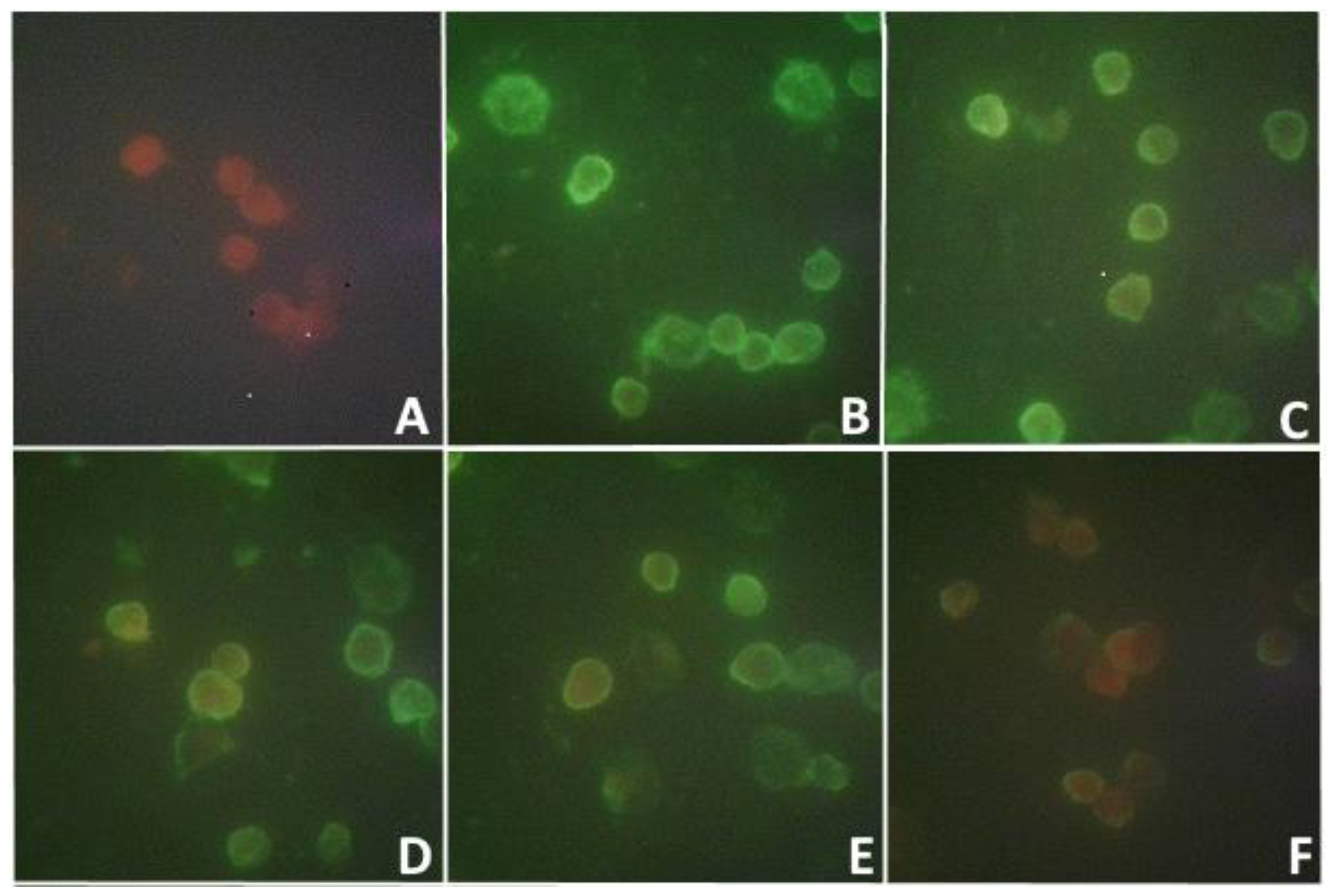
Figure 2.
Clinical manifestations in mice of 2 days of age on the 5th day after intracerebral infection with the isolate (2nd passage).
Figure 2.
Clinical manifestations in mice of 2 days of age on the 5th day after intracerebral infection with the isolate (2nd passage).
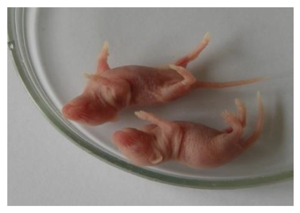
Figure 3.
The death of white mice during i/c, s/c and i/p infection with the viral isolate of the 5th passage (dilution 10-1, which amounted to 3 lg PFU).
Figure 3.
The death of white mice during i/c, s/c and i/p infection with the viral isolate of the 5th passage (dilution 10-1, which amounted to 3 lg PFU).
Figure 4.
Plaque-forming ability of the bioisolate on cell lines PEK, Vero, Vero-E6 (accounting on the 5th day after infection).
Figure 4.
Plaque-forming ability of the bioisolate on cell lines PEK, Vero, Vero-E6 (accounting on the 5th day after infection).
Figure 5.
Specific antigen fluorescence in PEK, Vero and Vero-E6 cells infected with a viral isolate 3 and 24 hours’ pi. MFA method: A–PEK cells; B, Vero cells; C-Vero-E6 cells.
Figure 5.
Specific antigen fluorescence in PEK, Vero and Vero-E6 cells infected with a viral isolate 3 and 24 hours’ pi. MFA method: A–PEK cells; B, Vero cells; C-Vero-E6 cells.
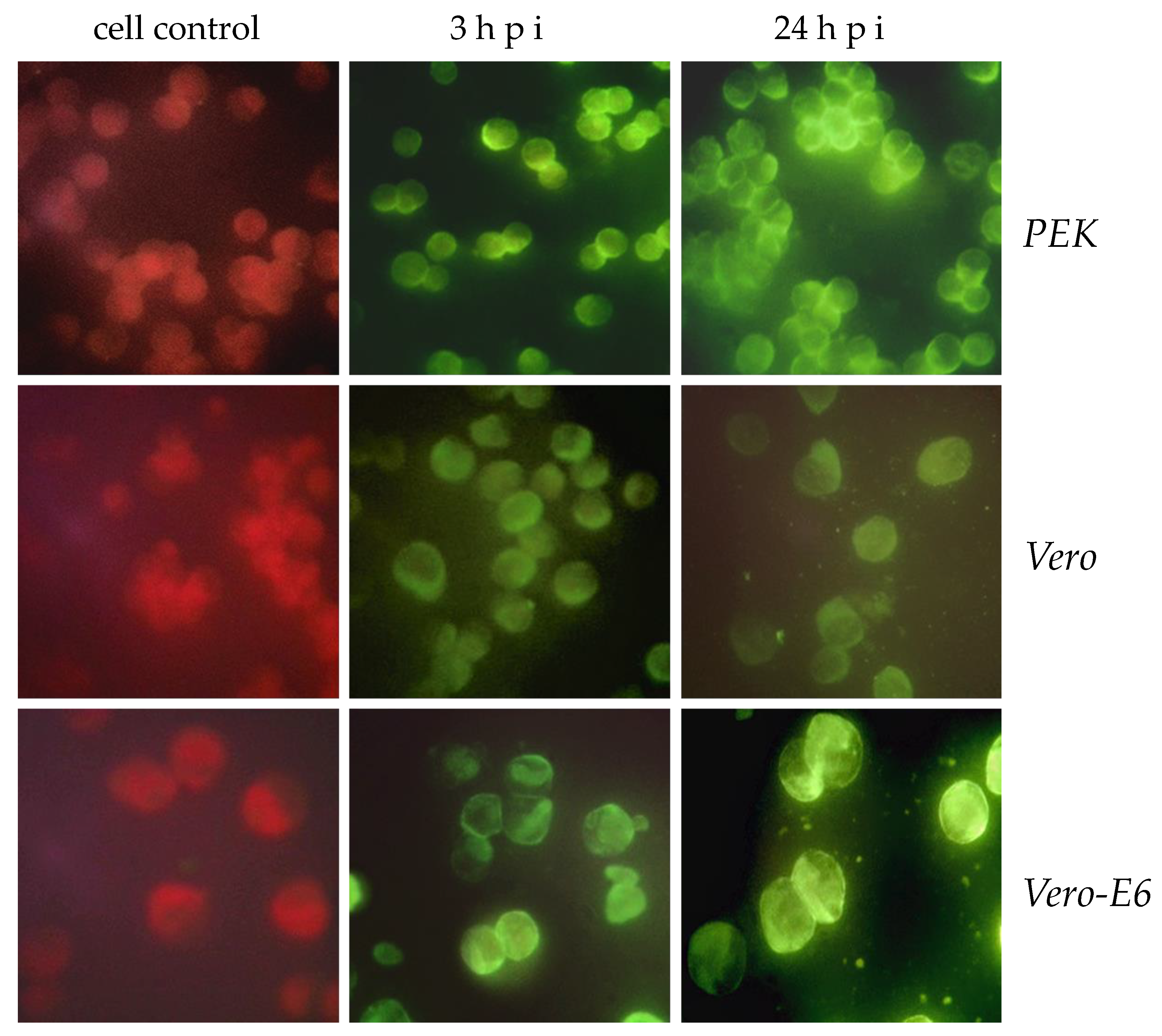
Figure 6.
Accumulation of a viral isolate in the supernatant in the period from 3 hours to 9 days pi of the PEK cell line. Visualization of the antigen on cells using the method of fluorescent antibodies; shows the average level of antigen-positive cells (%).
Figure 6.
Accumulation of a viral isolate in the supernatant in the period from 3 hours to 9 days pi of the PEK cell line. Visualization of the antigen on cells using the method of fluorescent antibodies; shows the average level of antigen-positive cells (%).
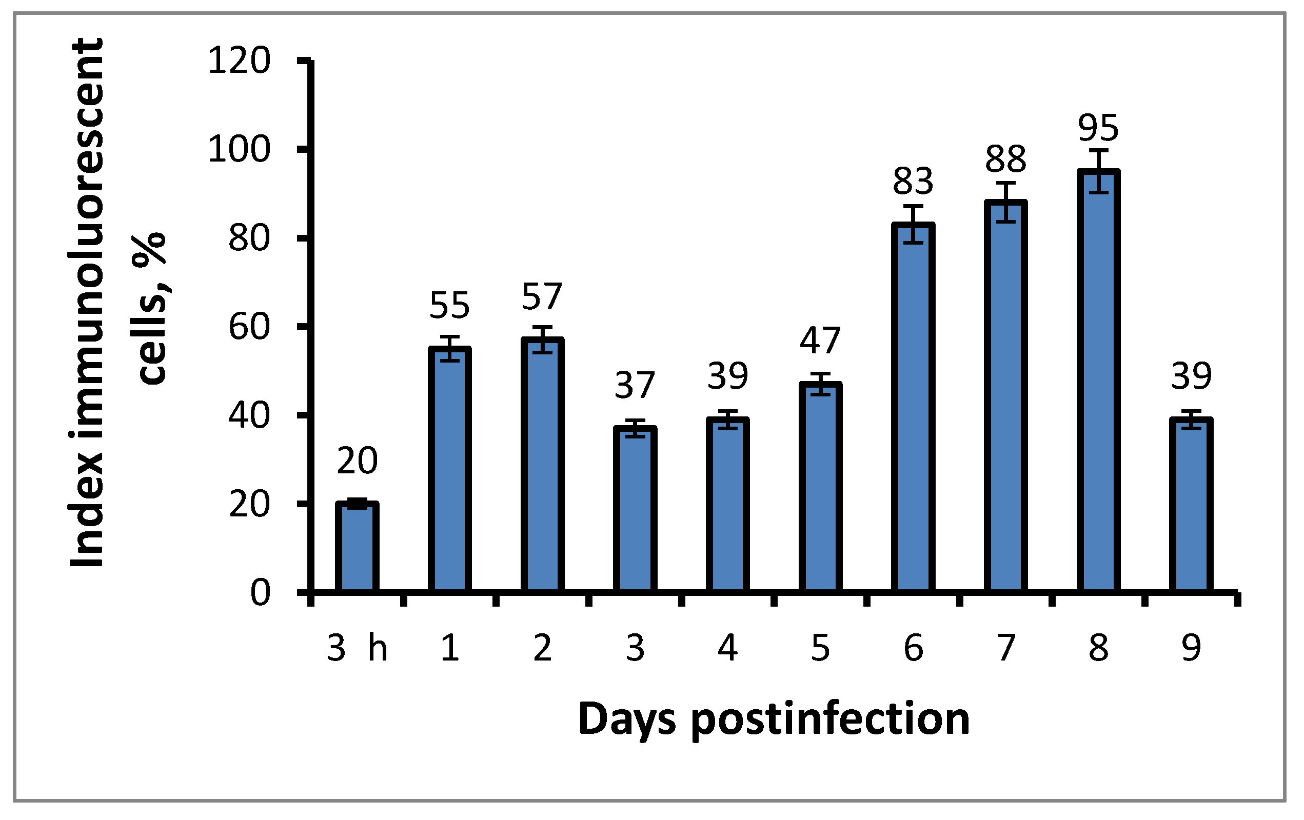
Figure 7.
Verification of the viral isolate in Vero-E6 cell culture 5 days pi. Titer of virus isolate before filtration (1st row) and after filtration (2nd row).
Figure 7.
Verification of the viral isolate in Vero-E6 cell culture 5 days pi. Titer of virus isolate before filtration (1st row) and after filtration (2nd row).
Figure 8.
Chorion-allantoic envelope of chicken embryos 9-10 days old, infected with a suspension of the original viral isolate (A) and its filtrate (B) on days 1, 3 and 7-9 pi.
Figure 8.
Chorion-allantoic envelope of chicken embryos 9-10 days old, infected with a suspension of the original viral isolate (A) and its filtrate (B) on days 1, 3 and 7-9 pi.
Figure 9.
Virus co-isolate from the tick I. persulcatus half fed on a human. Two viruses of different sizes are visible: one large smallpox-like up to 500 nm (double arrow), the other is much smaller, from 60 to 100 nm (arrow). Electron microscopy with negative contrast.
Figure 9.
Virus co-isolate from the tick I. persulcatus half fed on a human. Two viruses of different sizes are visible: one large smallpox-like up to 500 nm (double arrow), the other is much smaller, from 60 to 100 nm (arrow). Electron microscopy with negative contrast.
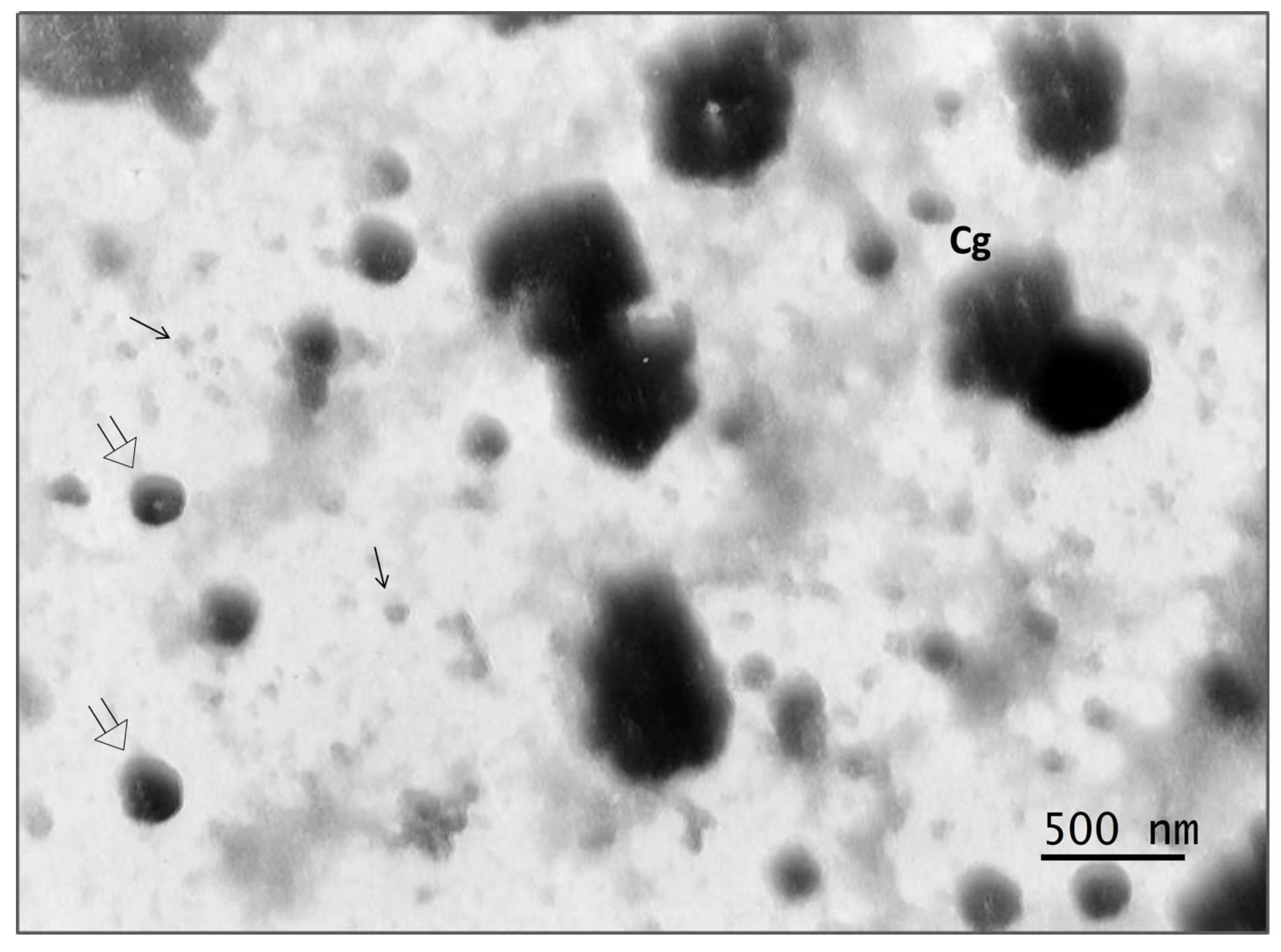
Figure 10.
Virus co-isolate from the tick I. persulcatus half fed on a human. A - a significant predominance of large viral particles up to 500 nm in size; B, C, D - electron-dense conglomerates, which consist of fused pox-like viral particles with a rim around the periphery, representing adherent particles of a small virus, up to 100 nm in size.

Table 1.
Dynamics of plaque-forming ability of the isolate at its tenfold dilutions from 10-1 to 10-5 in Vero-E6 cells on the 2nd, 3rd, 5th and 7th days pi.
Table 1.
Dynamics of plaque-forming ability of the isolate at its tenfold dilutions from 10-1 to 10-5 in Vero-E6 cells on the 2nd, 3rd, 5th and 7th days pi.
| Observation Day | Number of plaques, PFU | ||||
|---|---|---|---|---|---|
| 10-1 | 10-2 | 10-3 | 10-4 | 10-5 | |
| 2 | single | 0 | 0 | 0 | 0 |
| 3 | multiple | 0 | 0 | 0 | 0 |
| 5 | confluent | multiple | 26 | 13 | 0 |
| 7 | confluent | confluent | multiple | 15-17 | 1-3 |
Table 2.
Influence of physical factors (temperature and UVR) on the viral isolate during exposure from 1 to 10 minutes. Determination of the plaque-forming ability of the virus in the permissive cell line Vero-E6.
Table 2.
Influence of physical factors (temperature and UVR) on the viral isolate during exposure from 1 to 10 minutes. Determination of the plaque-forming ability of the virus in the permissive cell line Vero-E6.
| № № | Physical factors | Exposure time | |||
|---|---|---|---|---|---|
| 1 min | 2 min | 5 min | 10 min | ||
| 1 | Heat inactivation of the virus, 60° (lg PFU) | 7х103 | 4х101 | 0 | 0 |
| 2 | Heat inactivation of the virus, 100° (lg PFU) | 0 | 0 | 0 | 0 |
| 3 | UVI virus inactivation (lg PFU) | 6х103 | 11х101 | 8х101 | 0 |
| 4 | Virus Control (lg PFU) | 6х104 | |||
Table 3.
Influence of physical factors (temperature and UVI) on the viral isolate during exposure from 1 to 10 minutes. Visualization of the pathogen by detecting fluorescence of PEK cells in IMFA.
Table 3.
Influence of physical factors (temperature and UVI) on the viral isolate during exposure from 1 to 10 minutes. Visualization of the pathogen by detecting fluorescence of PEK cells in IMFA.
| № | physical impact | Exposure time | |||
|---|---|---|---|---|---|
| 1 min | 2 min | 5 min | 10 min | ||
| 1 | Heat inactivation of the virus, 60o (%) | 63 | 43 | 39 | 21 |
| 2 | Heat inactivation of the virus, 100o (%) | 36 | 23 | 17 | 4 |
| 3 | UVI virus inactivation (%) | 30 | 25 | 17 | 7 |
| 4 | Virus control | 76% of cells with antigen (IMPA+) | |||
Disclaimer/Publisher’s Note: The statements, opinions and data contained in all publications are solely those of the individual author(s) and contributor(s) and not of MDPI and/or the editor(s). MDPI and/or the editor(s) disclaim responsibility for any injury to people or property resulting from any ideas, methods, instructions or products referred to in the content. |
© 2024 by the authors. Licensee MDPI, Basel, Switzerland. This article is an open access article distributed under the terms and conditions of the Creative Commons Attribution (CC BY) license (https://creativecommons.org/licenses/by/4.0/).
Copyright: This open access article is published under a Creative Commons CC BY 4.0 license, which permit the free download, distribution, and reuse, provided that the author and preprint are cited in any reuse.


