Preprint
Article

In Vitro and In Silico Analysis of Alpha-Amylase Inhibitory Activity of Ethanolic Extract of Adhatoda vasica Leaves

Altmetrics

Downloads

260

Views

135

Comments

0

Submitted:

19 July 2023

Posted:

21 July 2023

You are already at the latest version

Alerts
Abstract
Background: Diabetic patients are more likely to experience morbidity and mortality as a result of microvascular complications such as retinopathy, neuropathy, nephropathy, and stroke. There are many synthetic anti-diabetic agents available which are expensive and have undesirable pathological effects. Thus, it is essential to look for cost-effective, natural, and safe antidiabetic agents. The aim of this study was to screen phytoconstituent and evaluate the in-vitro and in-silico α-amylase inhibitory activity of ethanolic extract of Adhatoda vasica leaves. Method: The extraction of Adhatoda vasica leaves was performed with ethanol through Soxhlet extraction process. Different concentrations (0.1 mg/ml to 1 mg/ml) of ethanolic extract, Acarbose, and Sitagliptin, were prepared and all concentrations were evaluated for α-amylase inhibitory activity through the spectrophotometric method. Molecular docking (AutodockVina 1.2.0) and toxicity profiling (SToPToX web server) studies were performed. Results: The plant extract showed highest inhibition of α-amylase (56.763±0.0035) at a concentration of 1 mg/ml. This inhibitory activity was supported by the in-silico study. Vasicoline (C5) and quercetin (C9), active constitute of plant Adhatoda vasica, showed the best binding energy of 8.3 and 8.0 Kcal/mol respectively with α-amylase enzyme (PDBID: 4W93). Toxicity study revealed the safety profile of plant extract. Conclusion: It was concluded that Adhatoda vasica leaves possess some bioactive compounds which are responsible for controlling blood glucose levels and its identification, purification, and isolation may lead to the development of newer therapeutic agent with lesser side effects.
Keywords: 
Subject: Chemistry and Materials Science  -   Medicinal Chemistry

1. Introduction

Diabetes mellitus (DM) is a common metabolic endocrine disorder diagnosed by hyperglycemia. This disorder is associated with many complications which may lead to morbidity and death of a patient [1]. The burden of diabetes mellitus (DM) has increased globally, particularly in low-income and middle-income countries, including Nepal (8.5%, 2020) [2]. The main clinical treatment strategy for DM is to control the blood glucose level using therapeutic agents (drugs). The most prevalence reason for increase in blood glucose level is due to the catalytic hydrolysis of carbohydrates by an enzyme such as alpha-glucosidase and alpha-amylase [3,4]. α-amylase (hydrolase) is prominent enzyme secreted by pancreas & salivary gland responsible for catalytic hydrolysis of starch to maltose & glucose by cleaving α-1,4-glucosidic bonds. The inhibition of activity of α-amylase delays the hydrolysis of polysaccharides, results reduction of the postprandial blood glucose level which is believed as an effective approach for the treatment of DM [5,6]. The clinically approved inhibitor, i.e., Acarbose, Voglibose, and Miglitol has several side effects associated with gastrointestinal problems such as flatulence and diarrhea. Moreover, most of the approved antidiabetic drugs are expensive and have undesirable pathological effects like weight gain, diabetic ketoacidosis, fluid retention, and hypoglycemia [7].
Various traditional medicines and phytochemicals having α-amylase inhibitory activity are well known for their role in the prevention and treatment of diabetes till date. Some plant-derived constituents with antidiabetic properties have been isolated and shown to have high potential and lower side effects than clinically approved synthetic drugs [8]– [10]. Therefore, it is still worth further investigation for the development of more effective inhibitors towards alpha-amylase. Adhatoda vasica is one of the traditionally used medicinal plant. Furthermore, various literatures explained the anti-ulcer, antidiabetic, antioxidant, muscle relaxant, anti-allergic, hepatoprotective, and cardioprotective activity of different parts of Adhatoda vasica [11]. Although the in-vivo anti-diabetic activity of Adhatoda vasica leaves, roots had been thoroughly investigated but the in-vitro and in-silico investigation is not studied yet as per our knowledge. The present study was designed to perform the phytochemical screening, and investigation of in-vitro, in-silico alpha-amylase inhibitory effect of Adhatoda vasica leaves.

2. Materials and Methods

All the chemical used were purchase from commercial source and were analytical grade.

2.1. Plant Materials Collection and Authentication
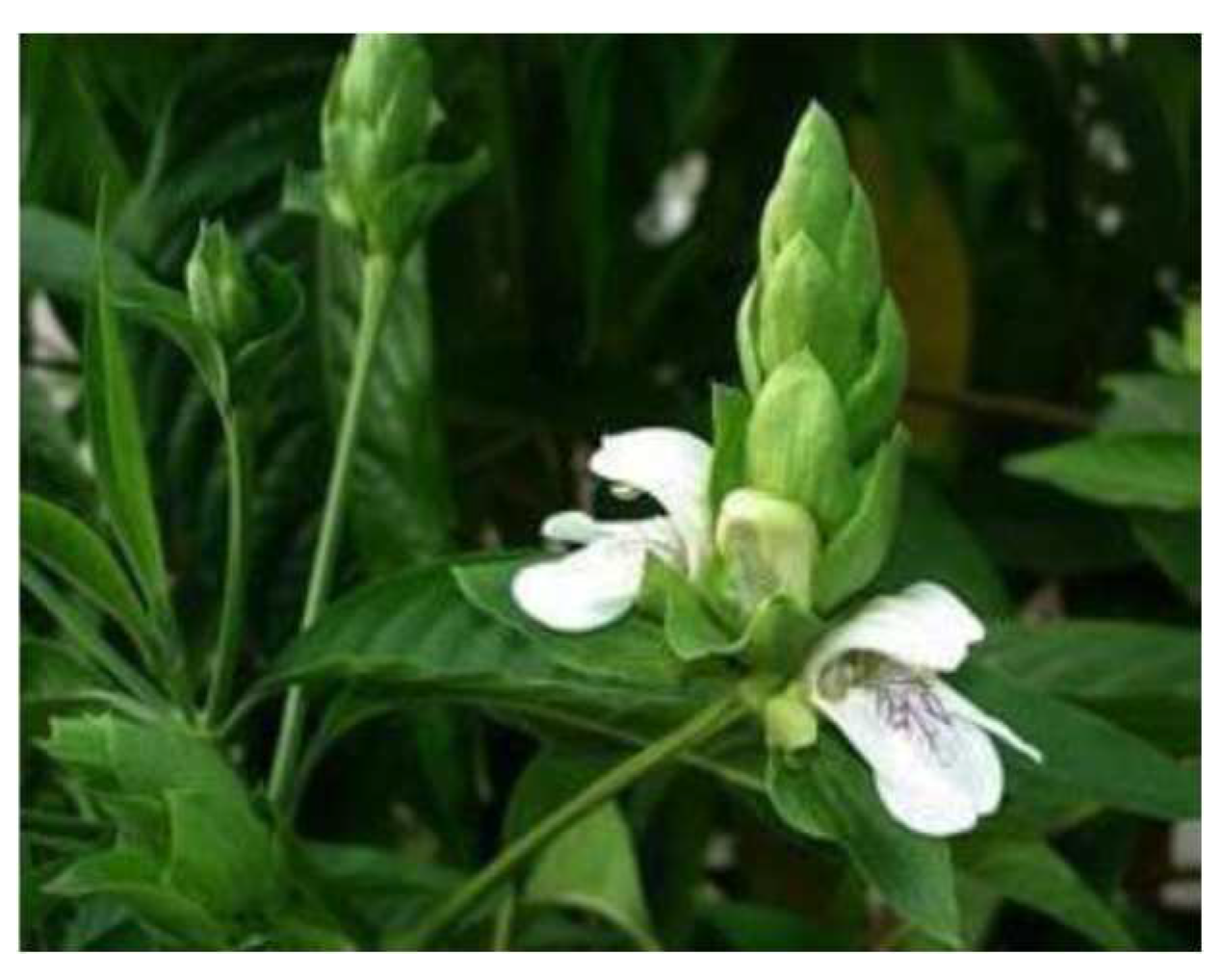
This study was carried out within 4 months after approval of proposal from the Institutional Review Committee (UCMS/IRC/146/22) of Universal College of Medical Science, Bhairahawa, Nepal at the department of Pharmacy and Pharmacology. Leaves of Adhatoda vasica were collected from different areas of the Arghakhanchi district, Nepal. The herbarium was made and sent for identification to Institute of Agriculture and Animal Science, Paklihawa campus, Rupandehi, Nepal.
Figure 1. Adhatoda vasica and its leaves.
Preprints 80019 g001

2.2. Extraction

The fresh leaves of Adhatoda vasica were collected and washed properly with distilled water to remove dust and foreign materials. The leaves were air-dried with proper ventilation in a laboratory at room temperature. Then further, dried in a hot air oven to remove the remaining moisture at 40℃ for 24 hours. The completely dried leaves were crushed in a grinder to obtain a coarse powder and kept in an airtight container. Aliquot quantity of 50gm of powdered was extracted with solvents methanol using Soxhlet apparatus (40-45 hours each) at a temperature below 50℃. After successful completion of extraction, the liquid extract was concentrated separately under vacuum, and the resulting dried extract was preserved in a desiccator until further use [12,13]. The dried extract was weighed and the percent yield was calculated by the formula mentioned below:
% y i e l d   o f   e x t r a c t = w e i g h t   o f   e x t r a c t   o b t a i n e d w e i g h t   o f   c o a r s   p o w d e r   t a k e n × 100 %

2.3. Phytochemical Screening

Phytochemical screening of leaves extract of Adhatoda vasica was performed for the presence of alkaloids, flavonoids, phenol, saponins, tannins, amino acids, cardiac glycoside, reducing sugar, carbohydrates, steroids, and terpenoid by using published methodology [14,15,16].

2.4. Determination of α- Amylase Inhibition Activity

The alpha-amylase inhibition assay was performed using published method with slight modification. About 250 μL of test samples and standard drug (100-1000μg/ml) was added to 250 μL of 0.20 mM phosphate buffer (pH 6.9) containing α-amylase (1U/ml) solution and was incubated at 25℃ for 10 min. The reaction was stopped with 1.0 ml of 3,5 di-nitro salicylic acid color reagent. The test tubes were then incubated in a boiling water bath for 5 min, and cooled to room temperature. The reaction mixture was then diluted after adding 10ml distilled water and absorbance (abs.) was measured at 545 nm using a UV-Vis spectroscopy. The absorbance of the control sample was measured accordingly without plant extract and acted as a negative control. The experimental extract and acarbose, and sitagliptin were tested with varying concentrations [17,18]. The results were expressed as % inhibition of alpha-amylase activity and calculated according to the following equation:
%   I n h i b i t i o n   o f   a l p h a a m y l a s e = A b s .   o f   c o n t r o l A b s . o f   e x t r a c t A b s .   o f   c o n t r o l × 100 %

2.5. Statistical Analysis

The in-vitro experiment was performed in duplet and the results were expressed in the mean± SD. Statistical analysis between the control and treatments group were determined using Microsoft excel.

2.6. Molecular Docking

Molecular docking study provides the mechanistic view of ligand-protein complex and determine the binding energy to explore the different pose of ligand-receptor interaction. Bioactive compounds from Adhatoda Vasaka plant that had demonstrated inhibitory effects on alpha-amylase enzyme activity in previous studies were identified and around 12 ligands (Figure 2) were selected through an extensive literature review [19]–[21]. The 3 dimensional (3D) molecular coordinates were downloaded from PubChem database (https://pubchem.ncbi.nlm.nih.gov/) in sdf format. Next, energy minimization was performed using the Molecular Mechanics Force Field (MMFF94). This process optimized the ligand structures by minimizing their potential energy, resulting in stable conformations suitable for docking analysis. The ligand structures were then converted to pdbqt format using OpenBabel 3.1.1 for compatibility with AutoDock Vina 1.2.0, a widely used molecular docking software [22].
Simultaneously, the 3D crystal structure of the α-amylase enzyme in complex with co-crystal Acarbose (PDBID: 3BAJ, X-ray crystallography resolution = 2.10 Å) [23] was downloaded from the Protein Data Bank (PDB, https://www.rcsb.org/). The protein structure underwent preparation steps, including the removal of water molecules, addition of missing hydrogen atoms, and assignment of proper charges. The prepared protein structure was visualized using BIOVIA discovery visualizer studio 2021, allowing for a better understanding of its active site and potential binding pockets [24]. The binding site amino acids were determined by BIOVIA discovery visualizer studio 2021 and validated with previously published original article about the protein (PDBID: 3BAJ) [25]. Similarly, the grid parameters, to cover the active site, were determined as -9.632, 4.340, and -23.107 for x, y, and z-axis respectively.
Molecular docking was performed in Linux (Ubuntu 20.04.6 LTS-GNU/Linux 4.4.0-19041-Microsoft x86_64) environment using AutoDock Vina 1.2.0 software (https://vina.scripps.edu/) [26], which predicted the binding modes and affinities of the selected ligands with the alpha-amylase enzyme. This analysis explored various orientations and conformations of the ligands within the enzyme's active site, providing insights into potential interactions and binding affinities. The ligand-target interaction and the nature of interaction was visualized by PyMol 2.5 software (https://pymol.org/2/) [27].
To validate the docking results and assess their reliability, the Root-Mean-Square Deviation (RMSD) value was calculated by PyMol 2.5 software. This measure quantified the differences between the docked ligand conformations and the co-crystal structure of the native ligand-protein complex [28].

2.7. Toxicity Profiling

The molecular structure and their relationship with biological activity and toxicity is very crucial step in drug design and development. Presence of certain functional group, rigidity of the structure, atomic composition and radii contribute to the development and occurrence of toxic properties in the molecule. SToPToX (https://stoptox.mml.unc.edu/), an atomic algorithmic online web server, was used to predict the acute inhalation toxicity and acuate oral toxicity [29].

3. Results

3.1. Percentage Yield

The extract was dried and then percentage yield of plant extract was calculated which was found to be 6.69%.
Weight of coarse powder of leaves was taken = 50grams
Weight of extract obtained after extraction=3.345grams
%   y i e l d   o f   e x t r a c t = w e i g h t   o f   e x t r a c t   o b t a i n e d w e i g h t   o f   c o a r s   p o w d e r   t a k e n × 100 % = 3.345 50.00 × 100 % = 6.69 %

3.2. Phytochemical Screening

In the present study phytochemical screening of ethanolic leaves extract detected the presence of alkaloids, gylcosides, phenol, flavonoids, steroids and terpenoids, saponins and tannins while absence of carbohydrates, reducing sugar and amino acids.
Table 1. Results of phytochemical screening of ethanolic leaves extract of Adhatoda vasica.
S.N. Test Result
1. Alkaloids Mayer’s test +
Wagner’s test +
Hager’s test +
2. Glycosides Brontrager’s test +
3. Carbohydrates Molish’s test _
4. Reducing sugar Benedicts test _
5. Tannins Potassium dichromate test +
6. Phenol Ferric chloride test +
7. Flavonoids Ferric chloride test +
8. Saponins Foam test +
9. Amino acids Ninhydrine test _
10. Steroids and terpenoids Salkoski test +
‘+’ for presence, ‘-’ for absence.

3.3. Alpha-Amylase Inhibitory Activity

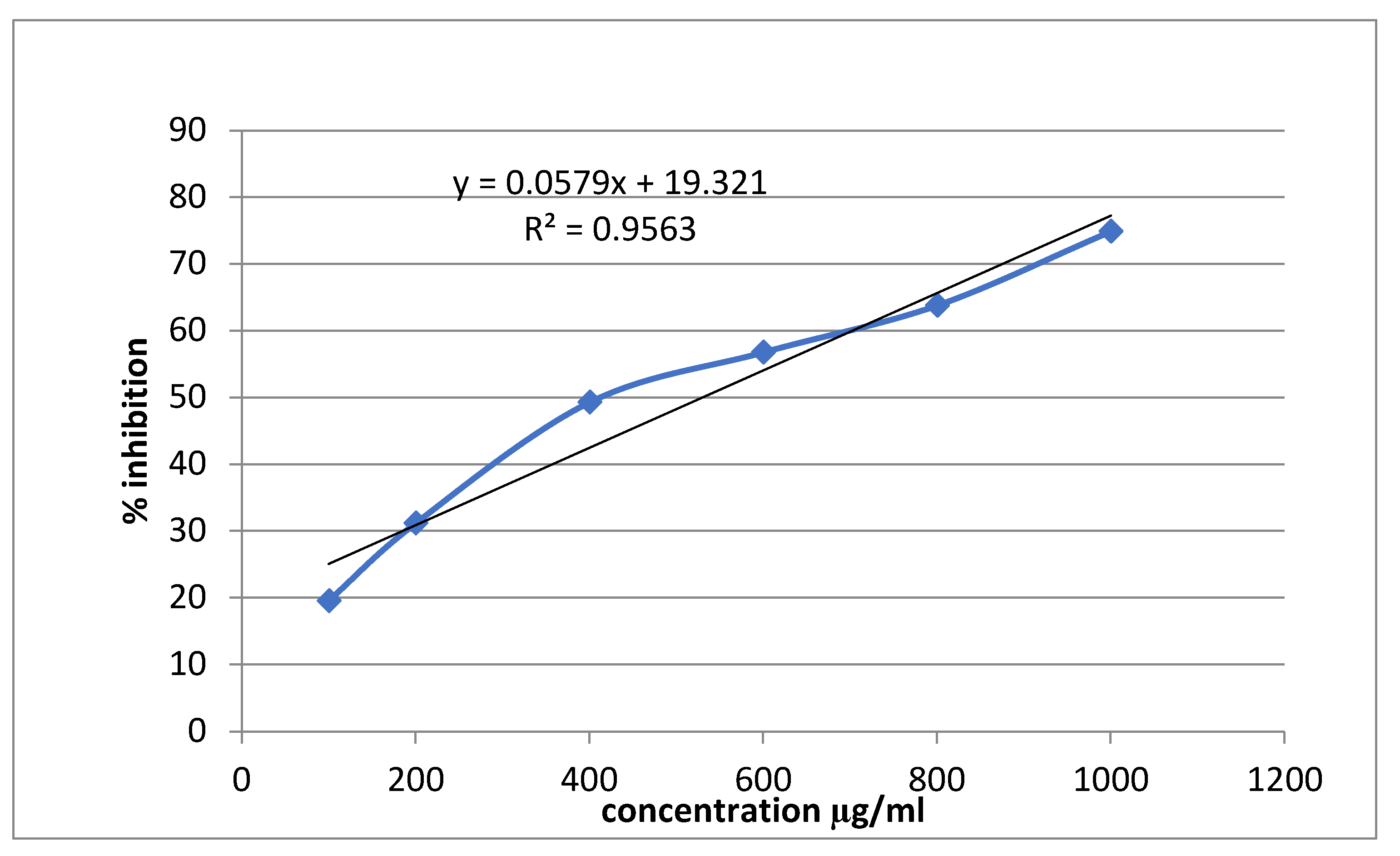
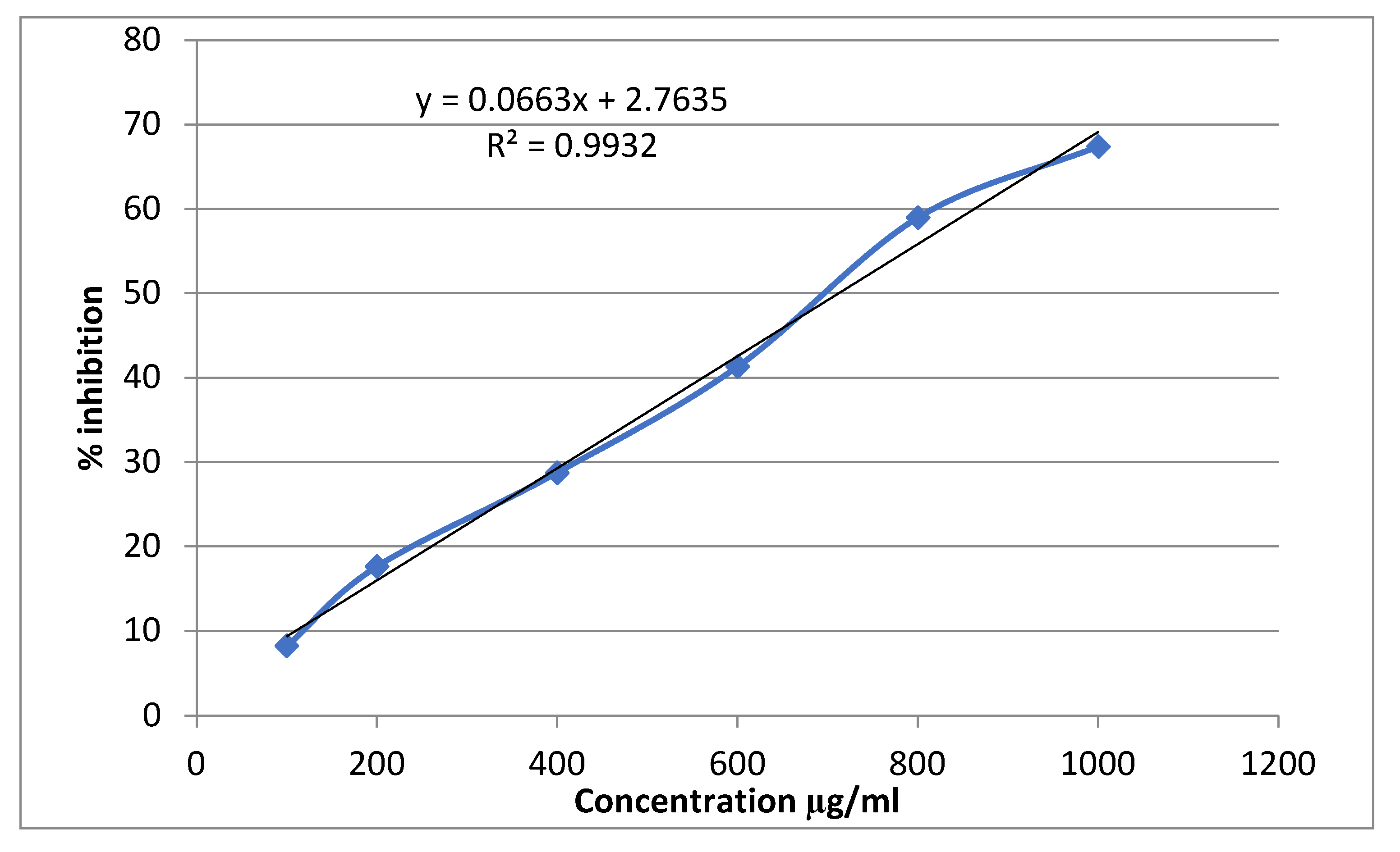
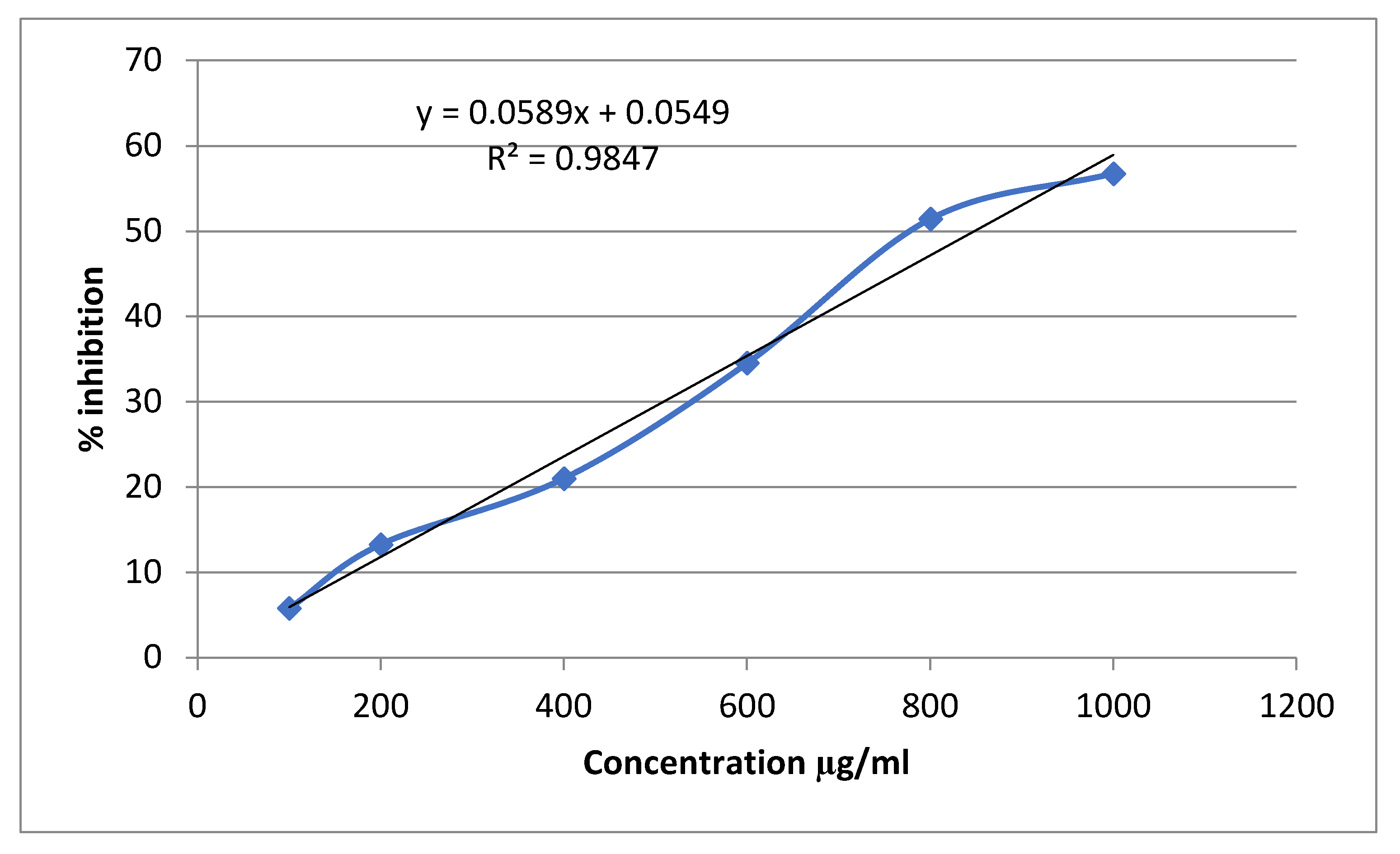
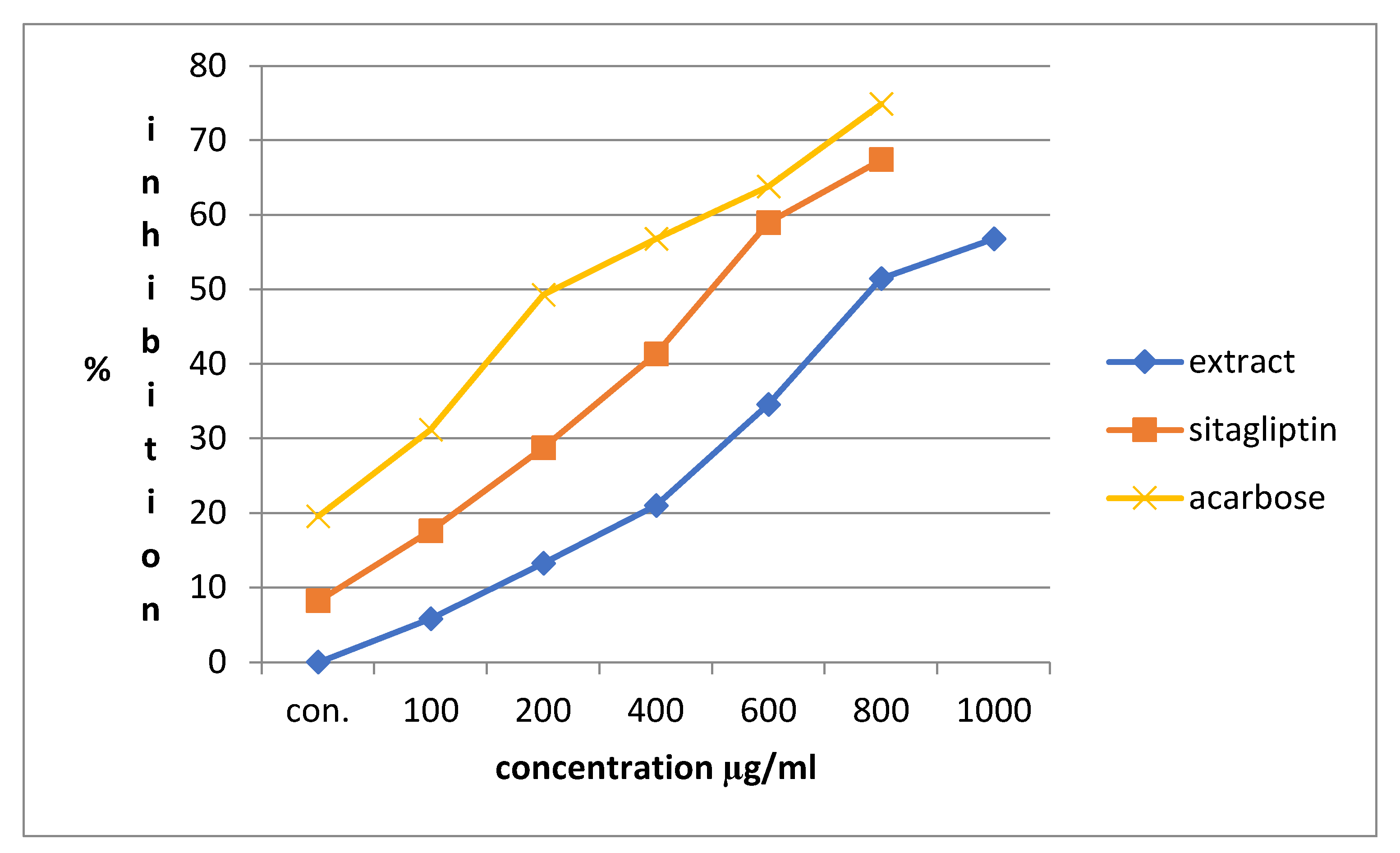
The percentage inhibition of plant extract and reference drugs at a concentration range of 0.1-1 mg/ml was evaluated. The results were expressed in mean±SD. The plant extract at a concentration of 1 mg/ml showed the highest alpha-amylase inhibitory activity with percentage inhibition value of 56.76329±0.0035. The % inhibition of alpha-amylase at a concentration 1mg/ml by acarbose, sitagliptin and plant extract was found to be 69.565±0.0084, 67.391±0.0049 and 56.763±0.0035 respectively (Table 2). Following graph explain the percentage inhibition of Acarbose (Figure 3), Sitagliptin (Figure 4), and plant extract (Figure 5).

3.4. Molecular Docking

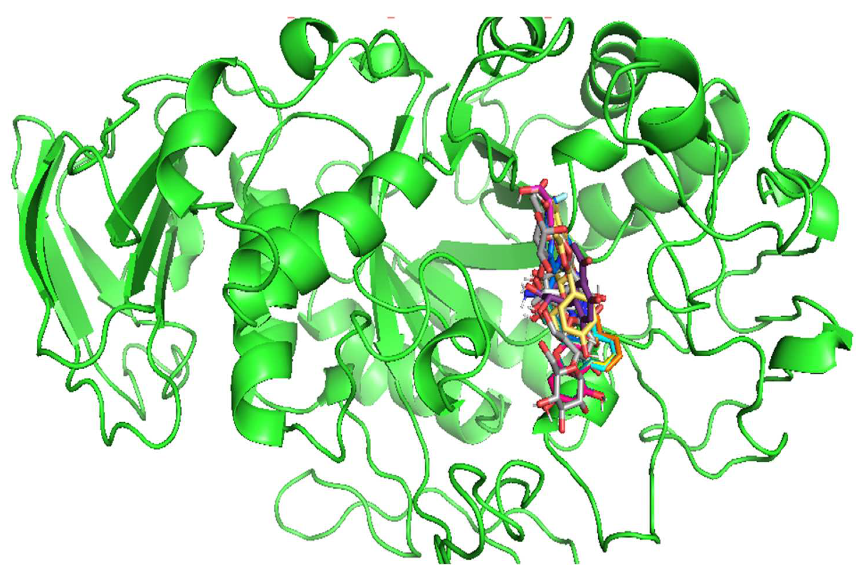
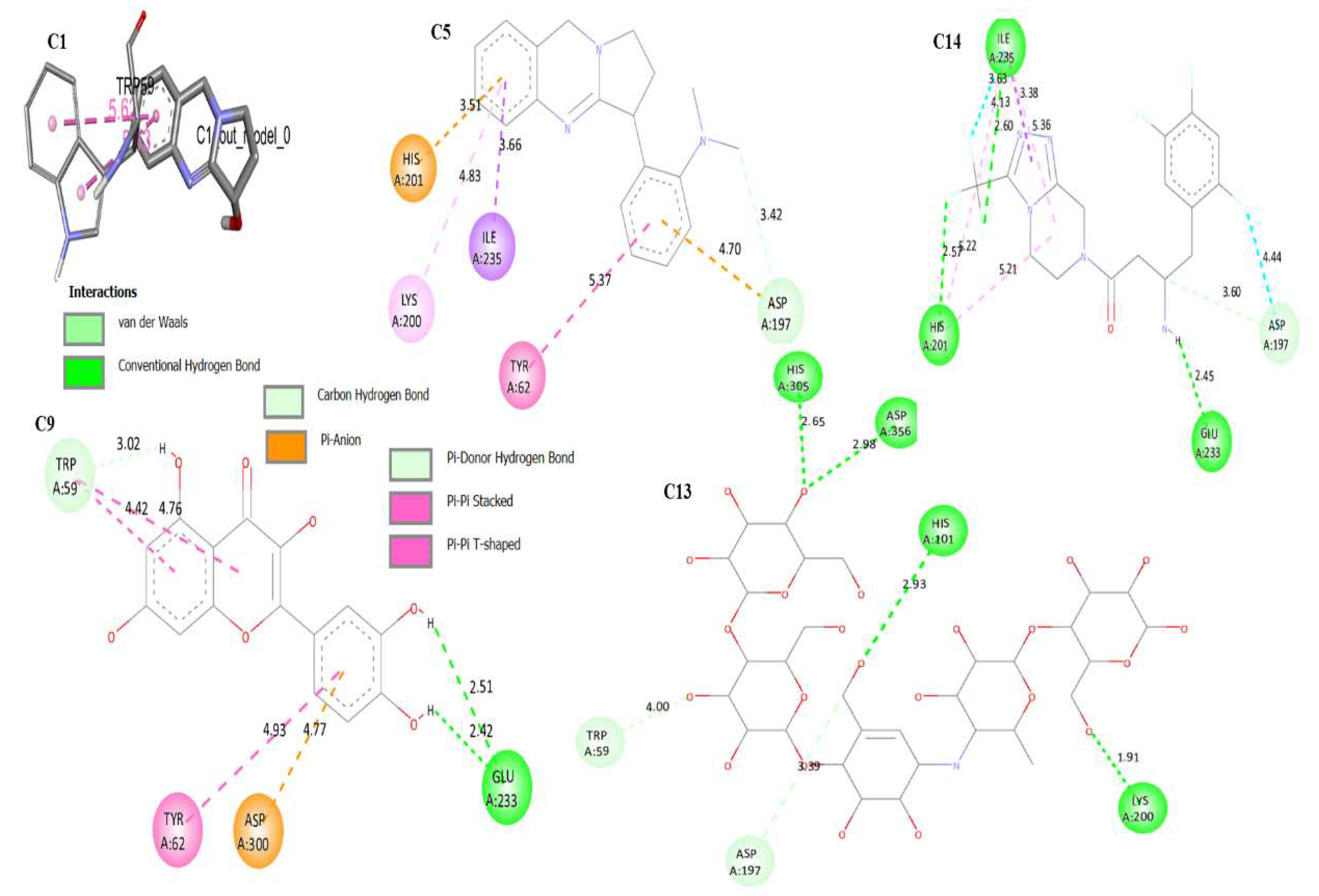
In order to generate binding energy (ΔG, Kcal/mol), the phytoconstituents (C1 to C12) were docked into the active sites of α -amylase. A more negative binding energy (least binding energy) denotes a spontaneous connection and a stronger affinity between the ligand and protein, and consequently, a more powerful inhibition. All phytoconstituents showed binding energy in the range of -5.9 kcal/mol to -8.3 kcal/mol. All the phytochemical showed the slightly higher binding energy (weak affinity) than native co-crystalized ligand (Acarbose, C13, ΔG = -8.6 kcal/mol) and reference drug Sitagliptin (A14, ΔG = 8.5 kcal/mol). The binding energy, which measures the strength of the interaction between the compounds and the protein, was found to be -8.3 kcal/mol for Vasicoline (C5) (Figure 8) and -8.0 kcal/mol for Quercetin (C9). All the ligand showed the RMSD value less than 2 Å, which validate the docking results (Figure 7). Table 3 provide the detail binding energy, amino acid required for interaction, and RMSD values.

3.5. Toxicity Profiling Analysis

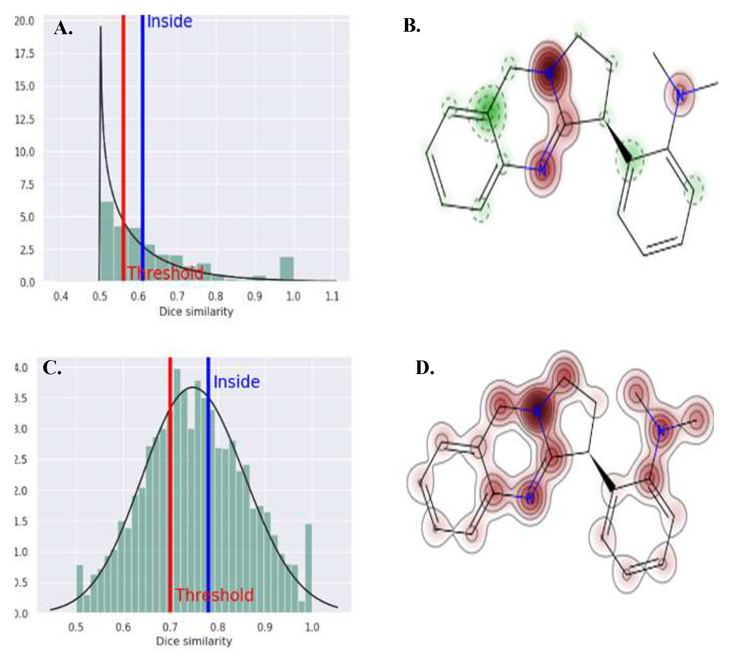
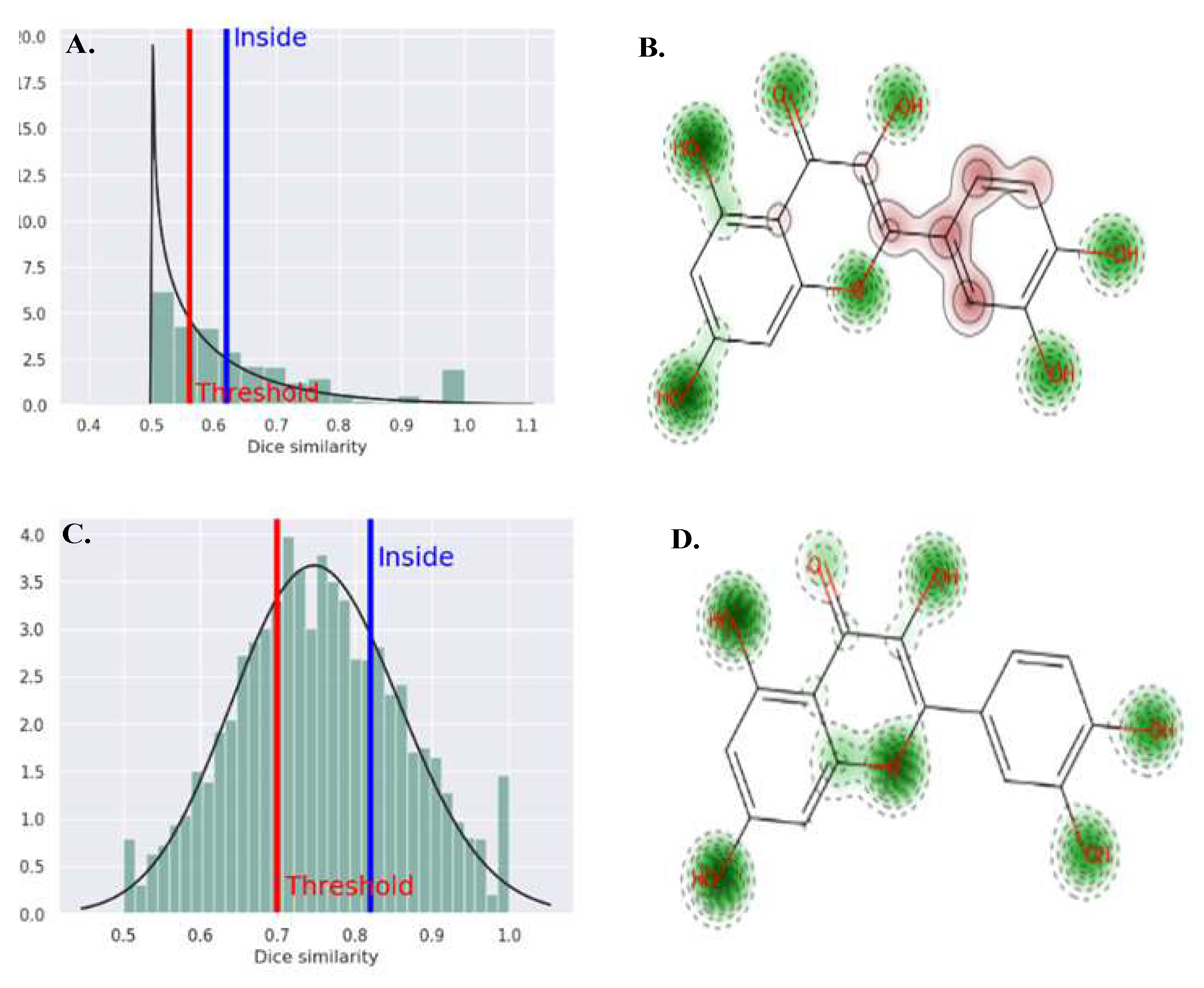
Acute inhalation toxicity and acute oral toxicity of active constitutes were investigated by SToPToX server. The predicted toxicities were indicated in negative sign (-) for non-toxic compound and positive sign (+) for toxic compound. The contributing functional group in the toxicity properties also predicted. Green color in the structure represents the functional group to contribute for non-toxic properties and brown color represent the functional group that contribute to enhance the toxic character. The Vasicoline (C5) was predicted toxic compound for inhalation. Quercetin (C9) showed negative sign for both acute inhalation toxicity and acute oral toxicity resulting as nontoxic compound. Detail toxicity study is given in Table 4.
Figure 9. ‘A’ and ‘C’ applicability domain (AD) for acute inhalation and acute real toxicity of compound C5 respectively. ‘B’ and ‘C’ predicted fragment contribution for acute inhalation and acute real toxicity of compound C5 respectively. Green color in the structure represents the functional group to contribute for non-toxic properties and brown color represent the functional group that contribute to enhance the toxic character.
Preprints 80019 g009

4. Discussion

The present study was carried out to investigate the potential alpha amylase inhibitory activity of ethanolic extract of Adhatoda vasica leaves from in-vitro and in-silico experimental process. We also performed the phytochemical screening of Adhatoda vasica leaves. In diabetes, the critical mechanism involves the impairment of glucose regulation and metabolism. The excessive glucose release contributes to elevate the blood glucose levels, and observed characteristic hyperglycemia. Understanding the regulation and inhibition of α-amylase activity is important for developing strategies to control postprandial glucose levels and manage diabetes effectively. The amylase inhibitors are considered as starch blockers as they prevent the body from absorbing dietary starch and slow down the digestion. This decreases blood glucose level has positive effects on insulin resistance and glycemic index control in diabetes [30,31].
We conducted an investigation where we measured the percentage inhibition of alpha-amylase activity using different concentrations (0.1-1 mg/ml) of ethanolic leaves extract of Adhatoda vasica. The inhibiting activity of ethanolic extract at a concentration of 1 mg/ml was found to be 56.76%. It is worth to mention that the phytochemical screening of the ethanolic extract of Adhatoda vasica leaves showed the presence of alkaloids, glycosides, phenol, flavonoids, saponnin, tannin, steroids, and terpenoids. Extract did not contain the reducing sugar. The present study showed that the % inhibition less than 50% at a dose of 0.8mg/ml. A similar study was conducted by Khadayat K. et al. and evaluated the % inhibition of alpha-amylase activity of Nepalese medicinal plants used in the treatment of diabetes mellitus [32]. The findings of their studies (IC50 value 413.5μg/ml) are in align with the results of our study.
Our study revealed the presence of alkaloids in the ethanolic extract of Adhatoda vasica leaves showed the anti-diabetic activity. A previous study conducted by Gao H et al. explained that alkaloids like Vasicine, Vasicinol, and Vasicinone are responsible for anti-diabetic activity by inhibiting α-amylase enzyme more than 50% (the IC50 values were 125μM to 250μM) [33]. This comparison highlights the consistency and agreement between our findings and the previous study regarding the presence and potential therapeutic effects of alkaloids in the plant extract. Graphical calculation revealed the IC50 value of ethanolic extract of Adhatoda vasica was 847.96μg/ml, which indicate inhibitory effect against alpha-amylase. However, an earlier study conducted by Rudrapal M et al. [34] reported a significantly lower IC50 value of 47.6μg/ml for the Adhatoda vasica leaves extract. Their study involved the purification and isolation of specific constituents, particularly vasicine, using chromatographic techniques. These contrasting results suggest that the reported antidiabetic effect of Adhatoda vasica may not solely rely on the presence of alkaloids in general but rather on the specific constituents present, such as vasicine. The lower IC50 value observed in the study could indicate that vasicine, among other constituents plays a significant role in the anti-diabetic properties of Adhatoda vasica. Therefore, the effectiveness of the plant extract in managing diabetes may be attributed to a combination of alkaloids and specific constituents, emphasizing the importance of studying and isolating individual compounds for a comprehensive understanding of their therapeutic potential.
The results demonstrated a dose-dependent relationship, as higher concentrations of the extract led to greater percentage inhibition. At the highest concentration tested (1 mg/ml), the extract showed a significant percentage inhibition of 56.763%. Interestingly, our findings align with a previous in vivo study that explored the anti-diabetic activity of the ethanolic extract of leaves and roots of Justicia adhatoda. This study examined the extract's effect on blood glucose levels in alloxan-induced diabetic rats. The results indicated that the ethanolic extract of Justicia adhatoda also demonstrated a dose-dependent reduction in blood glucose levels, meaning higher doses led to greater reductions in glucose levels [35]. The parallel dose-dependent patterns observed in both studies suggest a potential correlation between the inhibitory effect on alpha amylase activity and the ability to lower blood glucose levels. These findings support the notion that the ethanolic extracts of Adhatoda vasica and Justicia adhatoda hold promise for their anti-diabetic properties. Alkaloids may bind to the competitive or non-competitive sites of digestive enzymes, preventing the formation of an enzyme substrate complex and ultimately reducing the activity of the enzymes.
To investigate the binding mode and binding energy of the new compounds and to elucidate the ligand–protein interactions underlying the observed α-amylase selectivity, molecular docking studies have been performed [36]. Molecular docking method is virtual screen technique which help to eliminate the unsuits ligand from the library. The docking result was validated by calculating RMSD value superimposed with co-crystal native ligand [37,38].
At locations close to co-crystallized ligand (Acarbose), which is made up of the amino acids Trp59, Lys200, Asp356, His101, Ala169, His305, and Tyr159, it was discovered that all of the phytoconstituents (C1-C12) fit neatly into the active region of the enzyme. AutoDock Vina 1.2.0, molecular docking software, was used to simulate the binding modes of alpha amylase with the topmost active compounds, C1 to C12 to further understand the molecular inhibition mechanism. Docking interaction revealed Vasicoline (C5, Figure 8) and Quercetin (C9, Figure 8) were well nested into the active site of α-amylase and presented similar coordination with the active site of enzyme. The interactions mode obtained by molecular docking for Vasicoline, Quercetin, and the standard drug sitagliptin (ΔG = 8.5 kcal/mol) were illustrated in Figure 8. Quercetin (ΔG = −8.0 kcal/mol) showed two hydrogen bonds with the amino acids Glu233 with bond distances of 2.51 and 2.42 Å. Additionally, Quercetin has shorted bond length specially for hydrogen bond which justifies better stability. It is worth to mention that Kaempferol (C8), Apigenin (C10), and Vitexin (C12) were among the top-scored compounds with α-amylase (Table 3).
Rudrapal M et al. [34] conducted a molecular docking for vasicine with α-amylase enzyme and reported binding energy was -6.7 Kcal/mol. But we observed slightly higher binding energy for Vasicine (C1) with α-amylase enzyme (Figure 8, ΔG = -5.9 kcal/mol). Additionally, Vasicine showed higher binding energy than the co-crystal ligand Acarbose (ΔG -8.6 kcal/mol). Intriguingly, Vasicine (Figure 8) has lost its hydrophobic interaction with Trp59, Lys200, Asp356, His101, and His305. In contrary, these amino acids were found to interact with native co-crystal (Acarbose). The absence of these amino acids interacting with Vasicine could be the reason for its weak binding affinity with alpha-amylase enzyme. The findings of the docking study revealed that the binding energies of Vasicoline and Quercetin were comparable to those of the reference drug (Table 3). Jhong C et al. [39] reported the in-silico inhibitory activity of Quercetin. They also suggested the comparable binding energy of Quercetin (Docking score = -135.75) with commercial α-amylase inhibitor Acarbose (Docking score = -166.28). The presence of five hydroxy group (-OH) with Quercetin could be the reason for relatable binding energy, because they formed hydrophobic interaction with active site amino acid which could contributed in binding energy. A similar result was concluded by Nabil A et al. [40] and they explained Quercetin as best α-amylase inhibitor with minimum binding energy of -8.8 kcal/mol. They also revealed that the hydroxy group contributed to form conventional hydrogen interaction. Interestingly, hydroxyl group acted hydrogen donor as similar to our findings. In Figure 8, Trp59 formed a carbon hydrogen bond with the hydroxyl group of Quercetin at a bond distance of 3.02Å. Meanwhile, in the interaction of Acarbose, a similar amino acid was seen, but the bond distance of the pi-donor hydrogen bond was 4Å. The better stability of contact was explained by quercetin's shorter binding distance. Since the Acarbose and Quercetin interact with the same amino acid, it can be said that Quercetin inhibits α-amylase by competitive inhibition [41].
Acute toxicity is the term used to describe the negative consequences that accompany the administration of a single dose of a chemical or numerous doses within a 24 hours period, or a 4-hour inhalation exposure [42]. Immediately Inhaled When a group of 10 or more laboratory white rats inhales a substance or mixture of substances continuously for eight hours, it results in 50% of the group dying within 14 days, and this is referred to as toxicity [43]. Most of the active constitute of the plant extract showed the non-toxic characteristic when taken orally. For Vasicoline, the 3-nitrogen atom and the rigid benzene ring contribute to increase the toxicity. The presence of hydroxy, carbonyl, and oxy group with saturated structure of the compound (C9, Figure 10) increases the non-toxic characteristic. This indicates the safety profile of the extract. It can be taken orally for the treatment of DM when the allopathic medication is not effective or unavailable [44].

5. Conclusions

Our study revealed that ethanolic extract of Adhatoda vasica leaves to be effective in inhibiting alpha-amylase activity which regulates the glucose level. The in-silico analysis supports the invitro results. Molecular docking studies concluded that the presence of alkaloid and flavonoid supports the alpha inhibitory activity. It was concluded that leaves possessed certain α-amylase inhibitory activity which can be used as an alternative medicine to treat DM.

Author Contributions

Conceptualization, Chandrajeet Kumar Yadav, Sandhya KC, Shankar Thapa.; methodology, Chandrajeet Kumar Yadav, Sandhya KC, and Shankar Thapa.; software, Shankar Thapa and Sandhya KC.; validation, Chandrajeet Kumar Yadav, Laxmi Shrestha, and Anjan Palikhey.; formal analysis, Chandrajeet Kumar Yadav and Kamal Kandel.; investigation, Ashish Lamsal.; data curation, Chandrajeet Kumar Yadav and Sandhya KC.; writing—original draft preparation, Sandhya KC and Shankar Thapa.; writing—review and editing, Chandrajeet Kumar Yadav, Laxmi Shrestha, Anjan Palikhey, Kamal Kandel, and Ashish Lamsal.; visualization, Shankar Thapa.; supervision, Chandrajeet Kumar Yadav and Shankar Thapa. All authors have read and agreed to the published version of the manuscript.

Funding

This research received no external funding.

Data Availability Statement

None.

Acknowledgments

Authors are thankful to the administration department of Universal College of Medical science, Bhairahawa, Nepal.

Conflicts of Interest

The authors declared no conflict of interest.

References

  1. Kharroubi, AT. Diabetes mellitus: The epidemic of the century. World J Diabetes. 2015, 6, 850. [Google Scholar] [CrossRef] [PubMed]
  2. Shrestha DB, Budhathoki P, Sedhai YR, et al. Type 2 Diabetes Mellitus in Nepal from 2000 to 2020: A systematic review and meta-analysis. F1000Research. 2021, 10, 543. [Google Scholar] [CrossRef] [PubMed]
  3. Padhi S, Nayak AK, Behera A. Type II diabetes mellitus: a review on recent drug-based therapeutics. Biomed Pharmacother. 2020, 131, 110708. [Google Scholar] [CrossRef] [PubMed]
  4. Dahlén AD, Dashi G, Maslov I, et al. Trends in Antidiabetic Drug Discovery: FDA Approved Drugs, New Drugs in Clinical Trials and Global Sales. Front Pharmacol. 2022, 12, 1–16. [Google Scholar] [CrossRef]
  5. Oyedemi SO, Oyedemi BO, Ijeh II, Ohanyerem PE, Coopoosamy RM, Aiyegoro OA. Alpha-Amylase Inhibition and Antioxidative Capacity of Some Antidiabetic Plants Used by the Traditional Healers in Southeastern Nigeria. Sci World J. 2017, 2017, 1–11. [Google Scholar] [CrossRef]
  6. Skalli S, Hassikou R, Arahou M. An ethnobotanical survey of medicinal plants used for diabetes treatment in Rabat, Morocco. Heliyon. 2019, 5, e01421. [Google Scholar] [CrossRef] [PubMed]
  7. Chaudhury A, Duvoor C, Reddy Dendi VS. Clinical Review of Antidiabetic Drugs: Implications for Type 2 Diabetes Mellitus Management. Front Endocrinol. 2017, 8, 1–12. [Google Scholar] [CrossRef]
  8. Bindu J, Narendhirakannan RT. Role of medicinal plants in the management of diabetes mellitus: a review. 3 Biotech. 2019, 9, 1–17. [CrossRef]
  9. Salleh NH, Zulkipli IN, Mohd Yasin H. Systematic Review of Medicinal Plants Used for Treatment of Diabetes in Human Clinical Trials: An ASEAN Perspective. Zarrelli A, ed. Evidence-Based Complement Altern Med. 2021, 2021, 1–10. [Google Scholar] [CrossRef]
  10. Yin Z, Zhang W, Feng F, Zhang Y, Kang W. α-Glucosidase inhibitors isolated from medicinal plants. Food Sci Hum Wellness. 2014, 3, 136–174. [Google Scholar] [CrossRef]
  11. Choudhary S, Kaurav H, Chaudhary G. Adhatoda Vasica (Vasapatra): A Review Based Upon Its Medicinal Properties. Int J Res Ayurveda Pharm. 2021, 12, 79–87. [Google Scholar] [CrossRef]
  12. Shukla S, Ahirwal L, Bajpai VK, Huh YS, Han Y-K. Growth Inhibitory Effects of Adhatoda vasica and Its Potential at Reducing Listeria monocytogenes in Chicken Meat. Front Microbiol. 2017, 8, 1–13. [Google Scholar] [CrossRef]
  13. Nikhitha JN, Swathy KS, Chandran RP. In vitro anticancer activity of ethanol extract of Adhatoda vasica Nees on human ovarian cancer cell lines. J Genet Eng Biotechnol. 2021, 19, 1–9. [Google Scholar] [CrossRef]
  14. Mogole L, Omwoyo W, Mtunzi F. Phytochemical screening, anti-oxidant activity and α-amylase inhibition study using different extracts of loquat (Eriobotrya japonica) leaves. Heliyon. 2020, 6, e04736. [Google Scholar] [CrossRef] [PubMed]
  15. Gul R, Jan SU, Faridullah S, Sherani S, Jahan N. Preliminary Phytochemical Screening, Quantitative Analysis of Alkaloids, and Antioxidant Activity of Crude Plant Extracts from Ephedra intermedia Indigenous to Balochistan. Sci World J. 2017, 2017, 1–7. [Google Scholar] [CrossRef]
  16. Dubale S, Kebebe D, Zeynudin A, Abdissa N, Suleman S. Phytochemical Screening and Antimicrobial Activity Evaluation of Selected Medicinal Plants in Ethiopia. J Exp Pharmacol. 2023, 15, 51–62. [Google Scholar] [CrossRef] [PubMed]
  17. Kazeem MI, Mayaki AM, Ogungbe BF, Ojekale AB. In-vitro studies on Calotropis procera leaf extracts as inhibitors of key enzymes linked to diabetes mellitus. Iran J Pharm Res. 2016, 15, 37–44. [Google Scholar]
  18. Amin Mir M, Sawhney SS, S Jassal MM. In-vitro antidiabetic studies of various extracts of Taraxacum officinale. Pharma Innov J. 2015, 4, 61–66. [Google Scholar]
  19. Kumar Singh Scholar S, Ram Patel J, Dangi A, et al. A complete over review on Adhatoda vasica a traditional medicinal plant. J Med Plants Stud. 2017, 017, 175–180.
  20. Kumar M, Dandapat S, Kumar A, Sinha MP. Pharmacological Screening of Leaf Extract of Adhatoda vasica for Therapeutic Efficacy. Glob J Pharmacol. 2014, 8, 494–500. [Google Scholar] [CrossRef]
  21. Shahzad Q, Sammi S, Mehmood A, Naveed K, Azeem K, Ayub A. Phytochemical analysis and antimicrobial activity of adhatoda vasica leaves. Pure Appl Biol. 2020, 9, 1654–1661. [Google Scholar] [CrossRef]
  22. Kumar BS, Anuragh S, Kammala AK, Ilango K. Computer Aided Drug Design Approach to Screen Phytoconstituents of Adhatoda vasica as Potential Inhibitors of SARS-CoV-2 Main Protease Enzyme. Life. 2022, 12, 1–24. [Google Scholar] [CrossRef]
  23. Williams LK, Zhang X, Caner S. The amylase inhibitor montbretin A reveals a new glycosidase inhibition motif. Nat Chem Biol. 2015, 11, 691–696. [Google Scholar] [CrossRef]
  24. Thapa S, Nargund SL, Biradar MS. Molecular Design and In-Silico Analysis of Trisubstituted Benzimidazole Derivatives as Ftsz Inhibitor. J Chem. 2023, 2023, 1–9. [Google Scholar] [CrossRef]
  25. Maurus R, Begum A, Williams LK. Alternative Catalytic Anions Differentially Modulate Human alpha-Amylase Activity and Specificity. Biochemistry. 2008, 47, 3332–3344. [Google Scholar] [CrossRef] [PubMed]
  26. Trott O, Olson AJ. AutoDock Vina: Improving the speed and accuracy of docking with a new scoring function, efficient optimization, and multithreading. J Comput Chem. 2009, 31, 21334. [Google Scholar] [CrossRef]
  27. Seeliger D, de Groot BL. Ligand docking and binding site analysis with PyMOL and Autodock/Vina. J Comput Aided Mol Des. 2010, 24, 417–422. [Google Scholar] [CrossRef] [PubMed]
  28. Thapa S, Nargund SL, Biradar MS, Banerjee J, Karati D. In-silico investigation and drug likeliness studies of benzimidazole congeners: The new face of innovation. Informatics Med Unlocked. 2023, 38, 101213. [Google Scholar] [CrossRef]
  29. Biradar MS, Nargund SL, Thapa S. Synthesis and Cytotoxicity Assay of Aniline Substituted Thienopyrimidines for Anti-Colorectal Cancer Activity. Results Chem. 2023, 5, 100926. [Google Scholar] [CrossRef]
  30. Zadhoush F, Sadeghi M, Pourfarzam M. Biochemical changes in blood of type 2 diabetes with and without metabolic syndrome and their association with metabolic syndrome components. J Res Med Sci. 2015, 20, 763. [Google Scholar] [CrossRef]
  31. Mariadoss AVA, Sivakumar AS, Lee C-H, Kim SJ. Diabetes mellitus and diabetic foot ulcer: Etiology, biochemical and molecular based treatment strategies via gene and nanotherapy. Biomed Pharmacother. 2022, 151, 113134. [Google Scholar] [CrossRef]
  32. Khadayat K, Marasini BP, Gautam H, Ghaju S, Parajuli N. Evaluation of the alpha-amylase inhibitory activity of Nepalese medicinal plants used in the treatment of diabetes mellitus. Clin Phytoscience. 2020, 6, 34. [Google Scholar] [CrossRef]
  33. Gao H, Huang Y-N, Gao B, Li P, Inagaki C, Kawabata J. Inhibitory effect on α-glucosidase by Adhatoda vasica Nees. Food Chem. 2008, 108, 965–972. [CrossRef]
  34. Rudrapal M, Vallinayagam S, Aldosari S. Valorization of Adhatoda vasica leaves: Extraction, in vitro analyses and in silico approaches. Front Nutr. 2023, 10, 1161471. [Google Scholar] [CrossRef] [PubMed]
  35. Gulfraz M, Ahmad A, Asad MJ. Antidiabetic activities of leaves and root extracts of Justicia adhatoda linn against alloxan induced diabetes in rats. African J Biotechnol. 2011, 10, 6101–6106. [Google Scholar] [CrossRef]
  36. Abdulfatai U, Uba S, Abdullahi B, Muhammad U, Ibrahim T. Molecular design and docking analysis of the inhibitory activities of some α _ substituted acetamido - N - benzylacetamide as anticonvulsant agents. SN Appl Sci. 2019. [CrossRef]
  37. Pham TNH, Nguyen TH, Tam NM. Improving Ligand-Ranking of AutoDock Vina by Changing the Empirical Parameters. J Comput Chem. 2022, 43, 160–169. [Google Scholar] [CrossRef]
  38. Nguyen NT, Nguyen TH, Pham TNH. Autodock Vina Adopts More Accurate Binding Poses but Autodock4 Forms Better Binding Affinity. J Chem Inf Model. 2020, 60, 204–211. [Google Scholar] [CrossRef]
  39. Jhong CH, Riyaphan J, Lin SH, Chia YC, Weng CF. Screening alpha-glucosidase and alpha-amylase inhibitors from natural compounds by molecular docking in silico. BioFactors. 2015, 41, 242–251. [Google Scholar] [CrossRef]
  40. Nabil-Adam A, Ashour ML, Tamer TM, Shreadah MA, Hassan MA. Interaction of Jania rubens Polyphenolic Extract as an Antidiabetic Agent with α-Amylase, Lipase, and Trypsin: In Vitro Evaluations and In Silico Studies. Catalysts 2023, 13, 443. [Google Scholar] [CrossRef]
  41. Salim AM, Mohammed N, Ghazi R. In vitro and In silico Analysis of Pomegranate (Punica granatum L.) Fruit Powder as Pancreatic Lipase and α-Amylase Inhibitor. J Phys Conf Ser. 2020, 1665, 1–9. [Google Scholar] [CrossRef]
  42. Colerangle, JB. Preclinical Development of Nononcogenic Drugs (Small and Large Molecules). In: A Comprehensive Guide to Toxicology in Nonclinical Drug Development. Elsevier 2017, 1, 659–683. [Google Scholar] [CrossRef]
  43. Guimarães D, Noro J, Loureiro A. Increased Encapsulation Efficiency of Methotrexate in Liposomes for Rheumatoid Arthritis Therapy. Biomedicines. 2020, 8, 630. [Google Scholar] [CrossRef] [PubMed]
  44. Schreck K, Melzig MF. Traditionally Used Plants in the Treatment of Diabetes Mellitus: Screening for Uptake Inhibition of Glucose and Fructose in the Caco2-Cell Model. Front Pharmacol. 2021, 12, 1–12. [Google Scholar] [CrossRef]
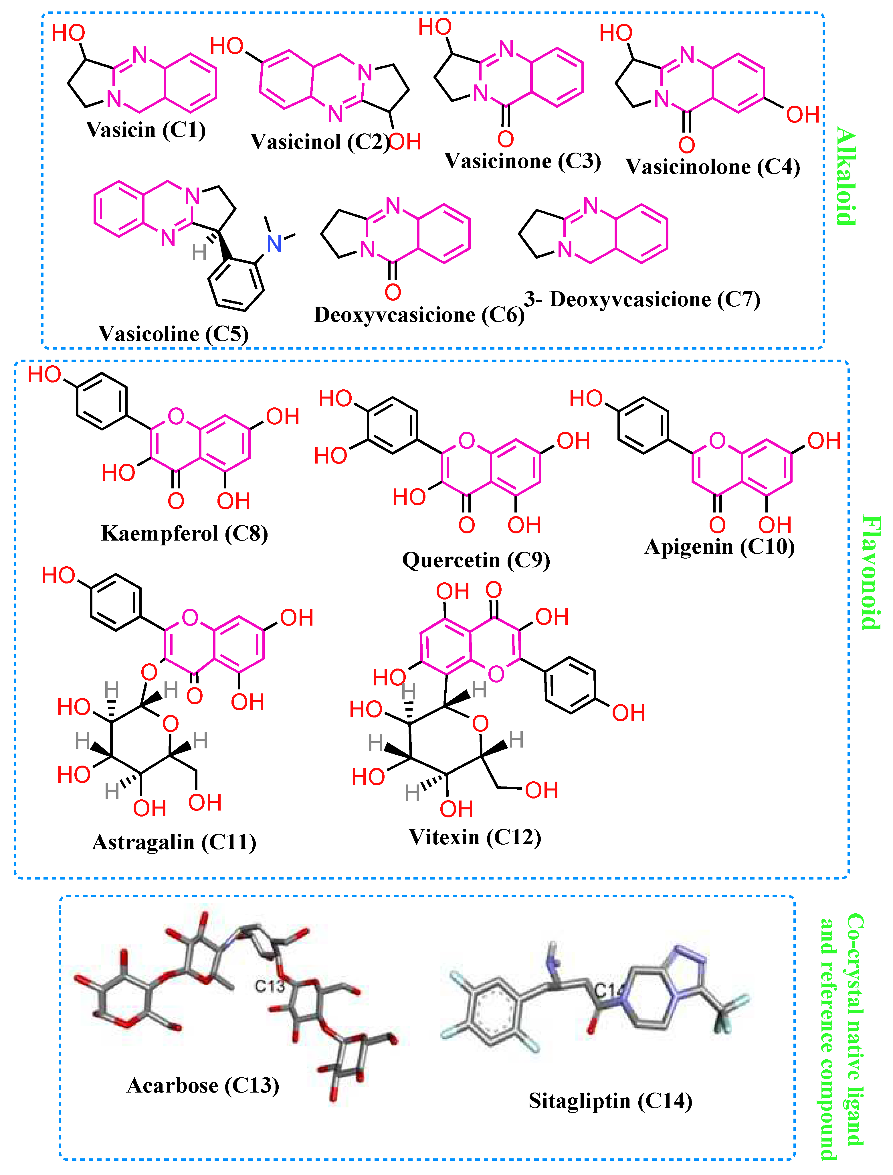
Figure 2. Chemical constitutes (C1-C7 alkaloid and C8-C12 flavonoids) of plant Adhatoda vasica.
Preprints 80019 g002
Figure 3. Linear calibration curve of Acarbose for IC50 value, % inhibition at different concentration.
Preprints 80019 g003
Figure 4. Linear calibration curve of Sitagliptin for IC50 value, % inhibition at different concentration.
Preprints 80019 g004
Figure 5. Linear calibration curve of plant extract for IC50 value, % inhibition at different concentration.
Preprints 80019 g005
Figure 6. Percentage inhibition of α-amylase activity at different concentration of plant extract and standard drugs used.
Preprints 80019 g006
Figure 7. 3D structure of superimposed ligands with co-crystal native ligand.
Preprints 80019 g007
Figure 8. 2D molecular interaction of Vasicoline (C5), Quercetin (C9), co-crystal native ligand (Acarbose C13), reference drug Sitagliptin (C14), and Vasicin (Q1, 3D) with α-amyla.
Preprints 80019 g008
Figure 10. ‘A’ and ‘C’ applicability domain (AD) for acute inhalation and acute real toxicity of compound C9 respectively. ‘B’ and ‘D’ predicted fragment contribution for acute inhalation and acute real toxicity of compound C9 respectively. Green color in the structure represents the functional group to contribute for non-toxic properties and brown color represent the functional group that contribute to enhance the toxic character.
Preprints 80019 g010
Table 2. Percentage inhibition of alpha amylase activity of different concentration of standard drugs and plant extract and IC50 value of standard drugs and plant extract.
Conc (mg/ml) Alpha amylase inhibition
Percentage inhibition of Acarbose IC50 (μg/ml) Percentage inhibition of Sitagliptin IC50 (μg/ml Percentage inhibition of Extract IC50 (μg/ml
0.1 19.56522±0.0063 529.861 8.21256±0.0028 712.466 5.797101±0.0028 847.964
0.2 31.15942±0.0035 17.63285±0.0021 13.28502±0.0021
0.4 49.27536±0.0028 28.74396±0.0007 21.01449±0.0070
0.6 56.76329±0.0049 41.30435±0.0035 34.54106±0.0021
0.8 63.76812±0.0028 58.9372±0.0028 51.44928±0.0049
1 74.87923±0.0056 67.3913±0.0049 56.76329±0.0035
Table 3. Binding energy and interactive amino acid.
Compound Code 4W93
Binding energy (Kcal/mol) Amino acid RMSD value (Å)
C1 -5.9 Ala169, Lys178, Trp59 1.386
C2 -6.2 Tyr151, Lys200, Ala198, Ile235, His201 1.334
C3 -6.3 Trp59, Tyr62 1.271
C4 -6.3 Ala198, Lys200, Ile235, His201, Tyr151 1.302
C5 -8.3 Asp197, Tyr62, Ile235, Lys200, His201 0.925
C6 -6.4 Tyr62, His299, Trp58 1.254
C7 -5.9 Trp59, Tyr62 1.223
C8 -7.7 Asp197, 300, 356, Trp59, Tyr 62 1.497
C9 -8.0 Trp59, Tyr62, Asp300, Glu233 1.425
C10 -7.9 Tyr62, Asp356, His299, Glu233 1.595
C11 -7.4 Tyr151, His299, Ile151, Glu233 1.896
C12 -7.8 Glu233, Ile235, Lys200, His201, Leu162 1.338
C13 -8.6 Trp59, Lys200, Asp356, His101,305 0.856
C14 -8.5 Ile235, His201, Glu233, Asp197 1.392
Table 4. Acute inhalation toxicity and acute oral toxicity of phytoconstituents.
Compound code Acute Inhalation Toxicity Acute Oral Toxicity
Prediction Confidence Prediction Confidence
C1 + 55.0 % + 81.0%
C2 - 54.0% + 68.0%
C3 - 60.0% + 75.0%
C4 - 75.0% + 56.0%
C5 + 69.0% + 91.0%
C6 - 58.0% + 72.0%
C7 + 75.0% + 90.0%
C8 - 68.0% - 70.0%
C9 - 69.0% - 72.0%
C10 - 66.0% - 73.0%
C11 - 77.0% - 66.0%
C12 - 76.0% - 75.0%
C13 - 78.0% - 96.0%
C14 - 70.0% - 69.0%
‘+’ = toxic, ‘-’ = Non-toxic.
Disclaimer/Publisher’s Note: The statements, opinions and data contained in all publications are solely those of the individual author(s) and contributor(s) and not of MDPI and/or the editor(s). MDPI and/or the editor(s) disclaim responsibility for any injury to people or property resulting from any ideas, methods, instructions or products referred to in the content.
Copyright: This open access article is published under a Creative Commons CC BY 4.0 license, which permit the free download, distribution, and reuse, provided that the author and preprint are cited in any reuse.
Prerpints.org logo

Preprints.org is a free preprint server supported by MDPI in Basel, Switzerland.

Subscribe

© 2024 MDPI (Basel, Switzerland) unless otherwise stated