Preprint
Article

Effects of Single Nucleotide Polymorphism BsmI of the Vitamin D Receptor (VDR) Gene on VDR, SOD-2, and CYP24A1 Gene Expression in Subjects with Low Serum Vitamin D Levels

Altmetrics

Downloads

99

Views

51

Comments

0

A peer-reviewed article of this preprint also exists.

Submitted:

20 July 2023

Posted:

21 July 2023

You are already at the latest version

Alerts
Abstract
Background: Hypovitaminosis D is a public health problem due to its implications for various diseases. Vitamin D has numerous functions, such as modulating the metabolism of some cellular tissues, and is expressed through the VDR gene that may influence gene expression modulation, which plays an important role in vitamin D metabolism. Objective: To evaluate the effect of the genotypes of single nucleotide polymorphism (SNP) BsmI of the vitamin D receptor (VDR) gene on VDR, SOD2, and CYP24A1 gene expression in individuals with low serum vitamin D levels. Methods: This was a cross-sectional analytical study. Individuals were invited to participate and, after signing the informed consent form, answered a structured questionnaire with identification data. Blood was collected for biochemical analysis, and vitamin D was measured by chemiluminescence; BsmI polymorphism was determined using real-time polymerase chain reaction (PCR) assays with TaqMan allelic discrimination, and gene expression was conducted by qRT-PCR using QuantiFast SYBR® Green PCR Master Mix. Data were analyzed using the SPSS 20.0 software, and differences were considered significant at p<0.05. Results: 98 individuals with vitamin D ≤20 ng/dL were evaluated, and the SNP BsmI of the VDR gene showed CYP24A1 overexpression and low SOD2 expression. Conclusion: SNP Bsm I of the VDR gene can modulate the expression of the genes evaluated without interfering with serum levels.
Keywords: 
Subject: Public Health and Healthcare  -   Other

1. Introduction

Vitamin D is a steroid hormone of the fat-soluble vitamin class involved in various biological processes, including cell proliferation, bone metabolism, and cell differentiation [1]; it is also responsible for a complex multi-step metabolism and acts as a hormone in numerous extra-skeletal targets [2]. Vitamin D deficiency, which occurs when vitamin D levels in the blood are below 20 ng/mL, is prevalent around the world [3], mainly affecting countries with little sun exposure due to climatic conditions, high latitudes, and winter regimes, in addition to other conditions (e.g., skin hyperpigmentation and chronic diseases) and affecting eating habits, pregnancy, breastfeeding [4]. This condition is considered a global health problem, as low vitamin D levels are associated with increased risk for various diseases and metabolic changes [5]. In Brazil, the Brazilian Society of Endocrinology and Metabology recommends serum levels above 20 ng/mL for the general healthy population and 30–60 ng/mL for risk groups such as the elderly, pregnant women, patients with osteomalacia, rickets, osteoporosis, secondary hyperparathyroidism, pre-bariatric patients, and inflammatory, autoimmune, and chronic kidney diseases [6].
The circulating effects of 1,25(OH)2D are mediated by the vitamin D receptor (VDR), which is a member of the superfamily of intracellular nuclear receptors [7]. Vitamin D also has some genomic effects, including cell apoptosis regulation, differentiation, proliferation, DNA repair, oxidative stress, and cell metabolism, which are driven by transcription factors [1].
Hence, VDR and vitamin D metabolic enzymes are expressed in all innate and adaptive arms of the immune system, and genomic approaches for gene expression profiling have identified various VDR-regulated genes implicated in regulating innate and adaptive immunity, including CYP and manganese-dependent superoxide dismutase (SOD2) genes [8]. Genetic variations called single nucleotide polymorphisms (SNP) can reach the human genome and alter different genes’ transcription and translation steps. The VDR gene is located on chromosome 12q13.1 and has several SNPs, more notably BsmI (rs 1544410), which is located in intron 8 and the result of substituting an adenine-guanine (A-G) [9]. Researchers have associated BsmI with osteoarthritis [10], breast cancer [11], melanoma [12], and system lupus erythematosus [13], among others, although data on its association with serum vitamin D levels are still conflicting [13,14].
Other genes are also known to modulate the expression of the VDR gene. For instance, one study found that the CYP gene, whose expression was constant, is regulated by fasting in the liver, while adipose tissue and the brain are the organs where vitamin D seems to play an important, albeit not fully known, role [3]. Additionally, the SOD2 gene has an antioxidant function, which may be relevant at low serum vitamin D levels [15]. Therefore, it is important to shed more light on how the different genotypes of the SNP BsmI of the VDR gene can modulate the expression of the genes of the VDR itself and others that may be in its pathway.
Given the above, this study sought to evaluate the effect of the genotypes of SNP BsmI of the VDR gene on the expression of different genes (VDR, SOD2, and CYP24A1) in individuals with low serum vitamin D levels.

2. Materials and Methods

2.1. Experimental Design

This was an epidemiological, analytical, observational cross-sectional study in which inflammatory, biochemical, and genetic marker levels were assessed in individuals with hypovitaminosis D.

2.2. Study Population and Design

The study was conducted with adults (18–59 years of age) living in the municipalities of Uruguaiana and São Borja (Rio Grande do Sul State, southern Brazil) from August to November (i.e., winter and spring). The volunteers were recruited from an extension program of the Federal University of Pampa (UNIPAMPA), signed an informed consent form, and answered a questionnaire with their data, self-declaration of color, socioeconomic, lifestyle, and nutritional data, including a 24 h food report, and eating frequency questionnaire. After fasting for 12 h, venous blood was collected for analysis. After obtaining the serum vitamin D values results, the volunteers with measurements of up to 20 ng/mL continued in the study [16], and the others were excluded (Figure 1). Genotyping for SNP BsmI was performed, and afterward, participants were divided into three groups according to genotypes: group GG (n = 40), group GA (n = 44), and group AA (n = 14).

2.3. Genetic-Molecular Analysis

2.3.1. DNA Extraction

Genomic DNA was isolated from peripheral blood leukocytes using the GFX Genomic Blood DNA Purification Extraction Kit (Amersham Biosciences Inc, Co.).

2.3.2. Gene Amplification (04121019085)

BsmI polymorphism was determined using real-time PCR assays with TaqMan allelic discrimination (Applied Biosystems, Foster City, CA, USA). A hot-start PCR consists of maintaining the temperature at 95 °C for 10 min, followed by 40 cycles of 94 °C for 15 s and then 60 °C for 1 min. Fluorescence detection takes place at 60 °C. All assays were performed in 10 µL reactions using TaqMan Genotyping Master Mix in 48-well plates on a StepOne® real-time thermocycler (Applied Biosystems, Foster City, CA, USA). Control samples representing all possible genotypes and negative control were included in each reaction.

2.4. Gene Expression by qRT-PCR

Gene expression modulation was conducted by qRT-PCR analysis using a similar approach described in the literature [17]. In summary, the total RNA obtained from each treatment was isolated using TRIzol® reagent and quantified using a NanoDrop™ 1000 Spectrophotometer System® (Thermo Fisher Scientific, Wilmington, DE, USA). Next, a cDNA was obtained using Script™ cDNA Synthesis Kit (Bio-Rad Laboratories, Hercules, CA, USA) according to the manufacturer’s instructions. The qRT-PCR was performed in a Rotor-Gene Q 5plex HRM System (QIAGEN biotechnology, Germany). A melt curve was generated from 60 to 90 °C in 0.5 °C increments for 5 s at each temperature. All reactions were performed in triplicate, with 1 µM of each primer and 2× QuantiFast SYBR® Green PCR Master Mix; the final reaction volume was 20 µL. The specific forward and reverse primer sequences are listed in Table 1.
The β-actin housekeeping gene was used as an internal control of gene expression analysis. Relative gene expression was calculated using the comparative Ct method and expressed as fold expression relative to the control.

2.4.2. Anthropometric and Physiological Assays

The participants’ weight (kg), height (cm), waist circumference (cm), and blood pressure (mmHg) were measured. The nutritional status of the participants was assessed by body mass index (BMI) as instructed by WHO [18]. Waist circumference measurements were used to assess cardiovascular risk and metabolic complications associated with obesity, and the cut-off values were chosen according to the Brazilian Guidelines on Obesity [19].

2.5. Biochemical Assays

Peripheral blood samples were collected after 12 h of fasting. The samples were centrifuged for 15 min at 3000 rpm, and aliquots of serum and plasma were stored at -20 °C for further analyses. The total cholesterol, triglyceride, and glucose levels were measured with colorimetric reagent kits/standard (Labtest®, Lagoa Santa, MG, Brazil). The HDL cholesterol levels were determined with an enzymatic kit (Bioclin®, Belo Horizonte, MG, Brazil). All determinations were performed in semi-automated biochemical apparatus (Chemwell T Labtest®, Lagoa Santa, MG, Brazil). LDL-cholesterol levels were determined using the Friedewald equation for triglyceride values below 400 mg/dL [12]. Samples with triglyceride values above 400 mg/dL were excluded.

2.6. Vitamin D Analysis

Serum vitamin D 25OHD3 was analyzed by high-performance chromatography (HPLC). The deficiency, insufficiency, and adequacy of 25OHD3 were classified according to the cut-off points of the Brazilian Society of Endocrinology and Metabology (2017).

2.7. Statistical Analysis

Data were plotted in an Excel spreadsheet, transferred, and analyzed by the SPSS statistical software (version 20.0). Quantitative variables were analyzed using the Student’s t-test or one-way analysis of variance, followed by Bonferroni’s post hoc test. Categorical variables were analyzed by the chi-squared test, which was also used to test genetic distributions for Hardy-Weinberg equilibrium. Comparisons with p<0.05 were considered significant.

2.8. Ethics

This study complied with the ethical principles for research involving humans according to the Declaration of Helsinki and was approved by the Ethics Committee of UNIPAMPA (protocol number 977827). The participants’ privacy rights were respected, and all individuals signed an informed consent form to participate.

3. Results

Nine-eight individuals with hypovitaminosis D (≤20 ng/dL), with a mean age of 30.5 ± 11 years, 54.1% female, and self-declared white (54.1%), participated in the study. Table 2 lists the baseline characteristics of the total sample.
Table 3 lists the descriptive analyses of anthropometric and biochemical measurements.
The genotypic and allelic frequencies of the BsmI polymorphism of the VDR gene in the total sample are described in Table 4. The studied sample presented Hardy-Weinberg equilibrium (χ2 =2.01).
Comparisons between genotype groups are listed in Table 5, and no differences were found between the markers analyzed among the different BsmI genotypes.
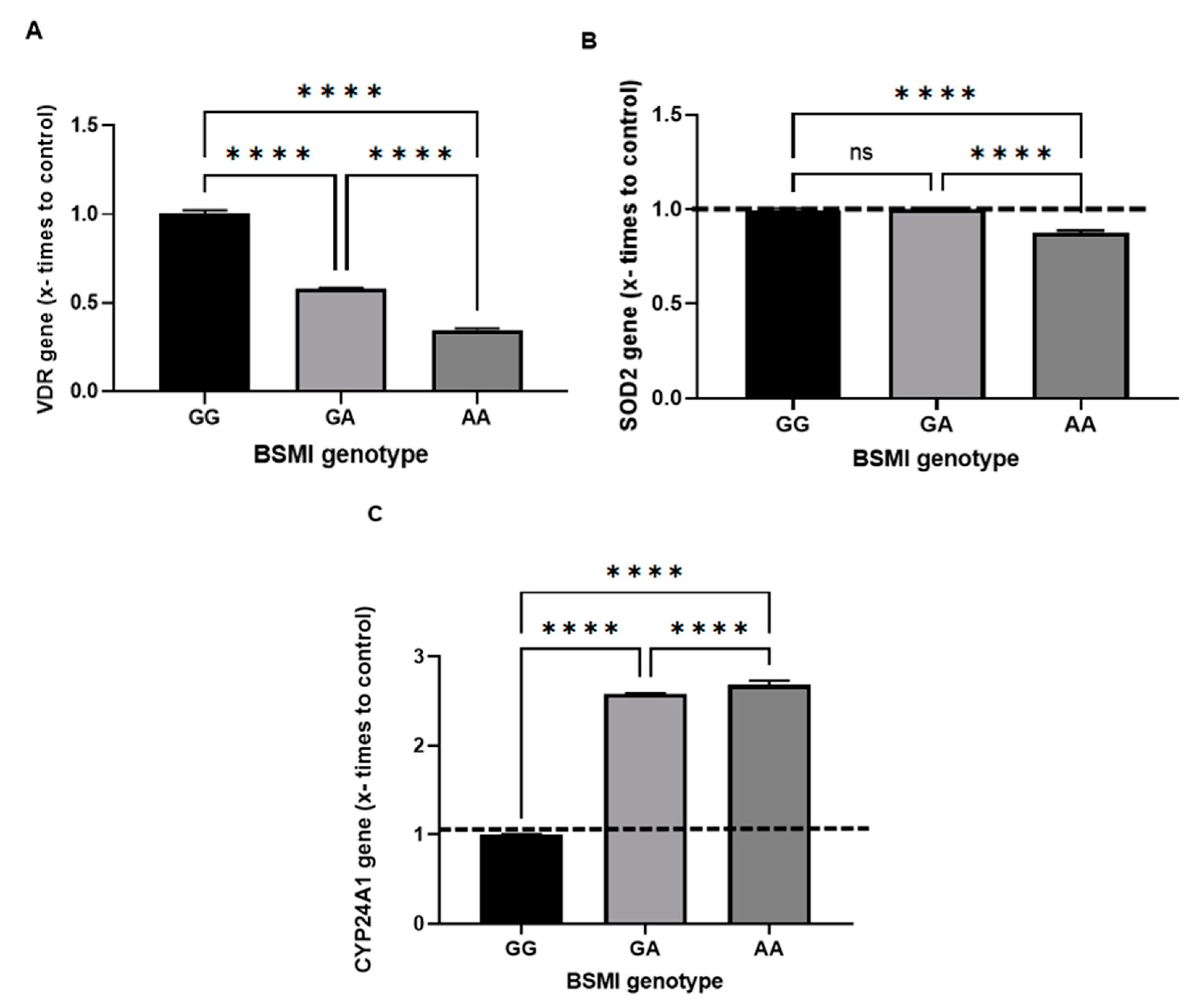
The VDR, SOD2, and CYP24A1 expressions were performed in each genotype group for the VDR SNP BsmI, and the results are illustrated in Figure 2; VDR gene expression was significantly lower in the GA and AA genotypes than in the wild-type GG genotype, and AA showed the lowest expression (Figure 2A). The SOD2 gene was also significantly less expressed in the AA group than GG and GA, which showed no differences (Figure 2B). In contrast, CYP24A1 overexpression was observed in the GA and AA genotypes (Figure 2C).

4. Discussion

This study demonstrated, for the first time, the effects of different BsmI genotypes of the VDR gene on the expression of different genes (besides VDR itself) in patients with hypovitaminosis D. BsmI downregulated the VDR gene and SOD2 and overregulated CYP24A1, thus showing the modulating influence of BsmI on other genes in hypovitaminosis D.
The sample represents general characteristics observed in young and active individuals, most of whom were women, as observed in other studies (Table 2). The higher self-declaration of color (white) is due to southern Brazil (i.e., the study region) presenting a higher prevalence of self-declared white individuals [20]. As for the level of education, most individuals had incomplete higher education, implying that they could understand the questions of the questionnaire, different from the data found in the rest of the country where the proportion of people aged 25 years or older are those who have completed compulsory basic education (i.e., high school) [21]. Indeed, another study carried out with individuals with vitamin D insufficiency and assisted by primary health care services reported serum vitamin D levels not being associated with the level of education, age, marital status, and income [22].
Another important finding is regarding the participants’ being overweight (Table 3), as the average BMI was within 27.1 kg/m2. A recent study evaluated serum vitamin D levels in healthy adult women and also showed that the nutritional status of the group evaluated was classified as overweight/obese, although without showing a significant association [23]. In this study, the distribution of genotypes and alleles of SNP BsmI were in Hardy-Weinberg equilibrium, and there were no significant associations between BsmI genotypes and serum vitamin D levels or the other markers (Table 5), as previously demonstrated by Retamoso and collaborators [15]. In other reports, no significant differences between vitamin D levels or genotypic and allelic frequencies of polymorphisms in the VDR gene were found, even though cross-sectional studies have shown that VDR gene polymorphisms can reduce the affinity of the VDR for serum vitamin D levels [24]. Despite VDR genetic polymorphisms being determinants of vitamin D levels, they have other genetic and environmental factors that are influenced by sun exposure, diet, and even skin pigmentation [25].
In order to clarify whether the BsmI SNP may influence the expression of other genes related to the physiological role of vitamin D, VDR, CYP24A1, and SOD-2 gene expression was studied in the groups of the three genotypes. Thus, we know that the VDR can modulate the expression of various genes, and its inactivation occurs due to the lack or excess of vitamin D since the almost ubiquitous expression of the VDR gene supports corroborates data from the last 30 years showing that vitamin D not only regulates calcium homeostasis but also promotes immunity, growth, and cell differentiation [26].
The VDR gene was under-expressed in the GA and AA genotypes compared to GG and AA less than the others (Figure 2A). In another study that compared the frequency of GG versus AA and AG genotypes, the association with insufficient 25(OH)D concentrations was maintained, suggesting that BsmI, which regulates VDR expression, can modulate vitamin D levels in patients with cognitive disorders [24]. It is important to emphasize that this study did not evaluate specific pathological conditions but individuals with hypovitaminosis D, which may explain these findings since they were different groups (people with hypovitaminosis D against a population with cognitive impairment) or the degree of genetic mixture of the population studied considering that they are of different ethnic origins and have a high degree of miscegenation among the populations investigated.
Moreover, the SOD-2 gene was significantly less expressed in AA genotype carriers compared to GG and GA (Figure 2B). This result can be explained by the study of Dauletbaev and collaborators [28], who investigated the impact of the genome on transcription by 1,25(OH)2D in carcinogenic cells, in which 1,25(OH)2D induced genes such as SOD2, IRS2, BIRC3, and DUSP1/5, which are cytoplasmic or mitochondrial signaling molecules that mediate the effects of growth factors and/or cytokine interactions with known anticancer properties. In other words, 1,25(OH)2D significantly induced mitochondrial expression but not cytosolic SOD2 that converts the free radical O2•− (superoxide) into H2 O2 to defend against free radicals [8].
Lastly, we evaluated whether the BsmI SNP of the VDR gene would modulate the expression of the CYP24A1 gene, which showed overexpression in carriers of the GA and AA genotypes compared to GG (Figure 2C), with AA being significantly more expressed than GG and GA, in which the mutated allele “A” possibly increases CYP24A1 expression. This is an unprecedented analysis in the literature, and the expression of most vitamin D target genes is 5x up- or down-regulated, meaning only a few genes respond with significant changes in vitamin D expression [27], as is the case of CYP24A1, which showed higher expression against BsmI SNP of the VDR gene.
Despite this study not evaluating the individuals by racial group, which was a limiting factor, it was possible to achieve unpublished results in which the SNP BsmI of the VDR gene can modulate CYP24A1 and SOD2 expression, even if it did not influence serum vitamin D levels. Given this context, future studies can seek to shed more light according to skin color.

Author Contributions

-Vanessa Retamoso: designed research (project conception, development of overall research plan, and study oversight); conducted research; analyzed data or performed statistical analysis; wrote paper; had primary responsibility for final content. -Fernanda Barbisan: conducted gene expression analyses; wrote paper. - Graziele Moro Meira: conducted gene expression analyses; wrote paper. -Patricia Maurer: conducted gene expression analyses; wrote paper. - Debora Rubio: conducted research (hands-on conduct of the experiments and data collection); wrote paper. - Lauren Flores: conducted research (hands-on conduct of the experiments and data collection); wrote paper. - Lyana Berro: Conducted research (hands-on conduct of the experiments and data collection); wrote paper. -Matias Nunes Frizzo: biochemical analysis; wrote paper. - Ivana Beatrice Mânica da Cruz: conducted gene expression analyses; wrote paper. -Vanusa Manfredini: biochemical analysis; wrote paper. -Ana Leticia Barcelos: Coordinator; designed research; analyzed data or performed statistical analysis; wrote paper. -Jacqueline da Costa Escobar Piccoli: Supervisor; designed research; analyzed data or performed statistical analysis; wrote paper; had primary responsibility for final content. All authors read and approved the final manuscript.

Funding

This research was funded in part by the Coordenação de Aperfeiçoamento de Pessoal de Nível Superior - Brasil (CAPES) - Finance Code 001".

Institutional Review Board Statement

This study complied with the ethical principles for research involving human subjects contained in the Declaration of Helsinki. The research project was submitted for consideration by the Ethics Committee of the UNIPAMPA and was approved under protocol number 977827.

Informed Consent Statement

All study subjects had their privacy rights observed. They also signed an informed consent form in order to participate and took a copy of it with them.
Conflicts of interest: The authors declare no conflict of interest.

References

  1. Durak S, Gheybi A, Demirkol S, Arıkan S, et al., The effects of serum levels, and alterations in the genes of binding protein and receptor of vitamin D on gastric câncer. Molecular Biology Reports. Springer Nature B.V. 2019. [CrossRef]
  2. Saponaro F, Saba A, Zucchi R. An Update on Vitamin D Metabolism. Int. J. Mol. Sci. 2020, 21, 6573. [CrossRef]
  3. Oczkowicz M, Szymczyk B, Swiątkiewicz M, Furgał-Dzier˙ zuk I, et al., Analysis of the effect of vitamin D supplementation and sex on Vdr, Cyp2r1 and Cyp27b1 gene expression in Wistar rats’ tissues. Journal of Steroid Biochemistry and Molecular Biology 212 (2021) 105918. [CrossRef]
  4. Ferreira, Carlos ES; Maeda, Sergio; Batista, Marcelo; et al., Consensus - Reference ranges of vitamin D [25(OH)D] from the Brazilian medical societies. Brazilian Society of Clinical Pathology/Laboratory Medicine (SBPC/ML) and Brazilian Society of Endocrinology and Metabolism (SBEM). Laboratory Medicine Journal Bras. Patol. Med. Lab. 53 (6) nov-dec 2017. [CrossRef]
  5. Zhumina A, Konstantin L, Konovalova A, Li Y et al., Plasma 25-Hydroxyvitamin D Levels and VDR Gene Expression in Peripheral Blood Mononuclear Cells of Leukemia Patients and Healthy Subjects in Central Kazakhstan. Nutrients. 2020, 12, 1229. [CrossRef]
  6. Maeda SS, Borba VZ, Camargo MB, Silva DM, Borges JL, Bandeira F, Lazaretti-Castro M; Brazilian Society of Endocrinology and Metabology (SBEM). Recommendations of the Brazilian Society of Endocrinology and Metabology (SBEM) for the diagnosis and treatment of hypovitaminosis D. Arq Bras Endocrinol Metabol. 2014 Jul;58(5):411-33. [CrossRef]
  7. Quigley M, Rieger S, Capobianco E, Wang Z et al. Vitamin D Modulation of Mitochondrial Oxidative Metabolism and mTOR Enforces Stress Adaptations and Anticancer Responses. JBMR Plus 2021 Dec 1;6(1): e10572. [CrossRef]
  8. Mailhot G, White J. Vitamin D and Immunity in Infants and Children. Nutrients. 2020, 12, 1233. [CrossRef]
  9. Schuch, Natielen Jacques. Relação entre concentração sérica de vitamina D, polimorfismos do gene VDR e Síndrome Metabólica em indivíduos adultos. Tese de doutorado. Universidade de São Paulo. Faculdade de Saúde Pública. São Paulo. 2011. [CrossRef]
  10. Li HM, Liu Y, Zhang RJ, Ding JY, Shen CL. Vitamin D receptor gene polymorphisms and osteoarthritis: a meta-analysis. Rheumatology (Oxford). 2021 Feb 1;60(2):538-548. [CrossRef]
  11. Shahbazi S, Alavi S, Majidzadeh-A K, Ghaffarpour M, Soleimani A, Mahdian R. BsmI but not FokI polymorphism of VDR gene is contributed in breast cancer. Med Oncol. 2013 Mar;30(1):393. [CrossRef]
  12. Lee YH, Gyu Song G. Vitamin D receptor FokI, BsmI, TaqI, ApaI, and EcoRV polymorphisms and susceptibility to melanoma: a meta-analysis. J BUON. 2015 Jan-Feb;20(1):235-43.
  13. Issa CT, Silva AS, Toscano LT, Medeiros MS, Persuhn DC, da Silva Diniz A, de Carvalho Costa MJ, Rodrigues Gonçalves Mda C. Relationship between cardiometabolic profile, vitamin D status and BsmI polymorphism of the VDR gene in non-institutionalized elderly subjects: Cardiometabolic profile, vitamin D status and BsmI polymorphism of the VDR gene in non-institutionalized elderly subjects. Exp Gerontol. 2016 Aug;81: 56-64. [CrossRef]
  14. Retamoso V, Feijoo L, Rubio D, Santos L et al. Black skin color but not VDR gene represent a risk factor for low serum levels of vitamin D in self-declared black individuals. Clinical Nutrition ESPEN 55 (2023) 230e237. [CrossRef]
  15. Khalid N, Mahjabeen I, Kayani MA, Akram Z. Association of arsenic-related AS3MT gene and antioxidant SOD2 gene expression in industrial workers occupationally exposed to arsenic. Toxicol Ind Health. 2020 Mar;36(3):161-169. [CrossRef]
  16. Maeda SS, Borba VZ, Camargo MB, Silva DM, Borges JL, Bandeira F, Lazaretti-Castro M; Brazilian Society of Endocrinology and Metabology (SBEM). Recommendations of the Brazilian Society of Endocrinology and Metabology (SBEM) for the diagnosis and treatment of hypovitaminosis D. Arq Bras Endocrinol Metabol. 2014 Jul;58(5):411-33. [CrossRef]
  17. Duarte T., Barbisan F., Prado-Lima P.A.S. do, V.F. Azzolin,et al., Ziprasidone, a second-generation antipsychotic drug, triggers a macrophage inflammatory response in vitro, Cytokine 106 (2018) 101–107. https://doi.org/10.1016/j.cyto. 2017.10.017.World Health Organization. Obesity: preventing and managing the global epidemic. Report of a World Health Organization Consultation. Geneva: World Health Organization, 2000. p. 256. WHO Obesity Technical Report Series, n. 284.
  18. Brazilian Obesity Guidelines. Brazilian Association for the Study of Obesity and Metabolic Syndrome. Brazilian Obesity Guidelines 2016 ABESO-Brazilian Association for the Study of Obesity and Metabolic Syndrome. – 4 ed. - Itapevi, SP: AC Farmacêutica, ISBN 978-85-60549-15-3. 2016.
  19. Brazilian Institute of Geography and Statistics (IBGE). Brazilian Census 2022. Rio de Janeiro: IBGE, 2023.
  20. Brazilian Institute of Geography and Statistics (IBGE). Household budget survey (POF). Ministry of Economy. Rio de Janeiro. 2019.
  21. Rolizola P, Freiria C, Silva G, Brito T et al. Insuficiência de vitamina D e fatores associados: um estudo com idosos assistidos por serviços de atenção básica à saúde. Ciênc. saúde coletiva 27 (02) 02 Fev 2022. [CrossRef]
  22. Cheong W, Ji S, Gassiot A, Thu W et al. Predictors of circulating vitamin D levels in healthy mid-life Singaporean women. Archives of Osteoporosis. Springer p.1-9. 2021. [CrossRef]
  23. Oliveira AC, Magalhaes C, Loures C, Fraga V et al. BsmI polymorphism in the vitamin D receptor gene is associated with 25-hydroxy vitamin D levels in individuals with cognitive decline. Arq. Neuro-Psiquiatr. v.76p.11 Nov 2018. [CrossRef]
  24. Monteiro Junior FC, Mandarino N, Santos E, Santos A et al. Correlation between serum 25-hydroxyvitamin D levels and carotid intima-media thickness in a Brazilian population descended from African slaves. Brazilian Journal of Medical and Biological Research (2018) 51(4): e7185. [CrossRef]
  25. Awasthi R, Manger P, Khare R. Fok I and Bsm I gene polymorphism of vitamin D receptor and essential hypertension: a mechanistic link. Clinical Hypertensio. V.29 n.5 p.2-12. 2023. [CrossRef]
  26. Dauletbaev N, Herscovitch K, Das M, Chen H, Bernier J, Matouk E, Bérubé J, Rousseau S, Lands LC. Down-regulation of IL-8 by high-dose vitamin D is specific to hyperinflammatory macrophages and involves mechanisms beyond up-regulation of DUSP1. Br J Pharmacol. 2015 Oct;172(19):4757-71. [CrossRef]
  27. Carlberg, C. Vitamin D and Its Target Genes. Nutrients 2022, 14, 1354. [Google Scholar] [CrossRef] [PubMed]
Figure 1. Participant flowchart.
Preprints 80110 g001
Figure 2. Comparison of the effects of BsmI on VDR, SOD2, and CYP24A1 gene expression. VDR (A), where p**** (0.001) among all BsmI genotypes: GGxGA, GGxAA, GAxAA. SOD2(B) p**** (0.001) was among the BsmI genotypes: GGxAA GAxGG. CYP24A1 (C) p**** (0.001) among all BsmI genotypes: GGxGA, GGxAA, GAxAA SOD2 (B) and CYP24A1 (C) gene expression among PBMCs carrying different BsmI genotypes of the VDR gene. Data are presented as mean ± SD. Analysis was performed and compared by one-way analysis of variance followed by the Bonferroni post hoc test. VDR gene = vitamin D receptor; SOD-2 gene = superoxide dismutase 2; CYP24A1 gene = cytochrome P450 family 24 subfamily A member 1.
Preprints 80110 g002
Table 1. List of primers used and gene information.
Gene and gene ID NCBI reference sequence Location Size (pb) Primers
VDR - 7421 NG_008731.1 12q13.11 4.738 F- 5′CCTTCACCATGGACGACATG3’
R-5′CGGCTTTGGTCACGTCACT3′
CYP24A1 - 1591 NG_008334 20q12 37449 F-5′CTCATGCTAAATACCCAGGTG-3′
R-5′TCGCTGGCAAAACGCGATGGG3′
SOD-2 - 6648 NG_008729 6q11 100213 F-5’GCCCTGGAACCTCACATCAA-3’
R-5’GGTACTTCTCCTCGGTGACGTT3’
Table 2. Sociodemographic and lifestyle characteristics of the sample.
Characteristic N (98) (%)
Sex
Female 53 54.1
Male 45 45.9
Self-declaration of color
Black 20 20.4
Brown 25 25.5
White 53 54.1
Education
Primary education (i) 5 5.1
Primary education (c) 1 1.0
Secondary education (i) 1 1.0
Secondary education (c) 16 16.3
Higher education (i) 53 54.1
Higher education (c) 20 20.4
Other (graduate degree) 2 2.0
Family income
5–15 minimum wages 24 24.5
3–5 minimum wages 34 34.7
1–3 minimum wages 39 38.8
Up to 1 minimum wage 1 1.0
Marital status
Single 66 67.3
Married 25 25.5
Common-law marriage 6 6.1
Widowed 1 1.0
Performs physical activity
Yes 48 49
No 50 51
Smokes
Yes 10 10.2
No 87 88.8
Ex-smoker 1 1.0
Drinks alcohol
Yes 63 63.3
No 35 35.7
C = complete, I = incomplete.
Table 3. Biochemical and anthropometric analysis of the sample.
Marker Mean ±SD
Weight (kg) 77.8 19.0
Height (m) 1.8 0.16
BMI (kg/m)2 27.1 5.4
CC (cm) 88.2 14.7
QC (cm) 103.6 9.8
% fat 12.6 10.4
Total col (mg/dL) 177 47
HDL-c (mg/dL) 55.2 22.2
LDL-c (mg/dL) 94.9 41.2
Triglycerides (mg/dL) 146.1 142.2
Vitamin D (ng/mL) 17.03 4.04
BMI = body mass index, HDL-C = high-density, LDL = lipoprotein cholesterol, WC = waist circumference, HC= hip circumference.
Table 4. Allelic and genotypic frequency of SNP BsmI of the VDR gene.
SNP BsmI Participants (n = 98) (%) *p-value
Genotypic frequency
GG 40 40.8 0.4
GA 44 44.9 0.46
AA 14 14.3 0.13
Allelic frequency
Allele G 102 67.1
Allele A 50 32.9
Model
AA+GA 54 55.1
GG 40 44.9
*Chi-square test.
Table 5. Comparison between the SNP BsmI genotype groups of the VDR gene and the variables studied.
Genotype group BsmI VDR
GG (40) GA (44) AA (14) p
Age 29.8 ± 10.2 32.3 ± 11.4 29.8 ± 11.3 0.80
BMI (kg/m2) 26.8 ± 5.2 26.8 ± 5.3 28.6 ± 6.2 0.51
CC (cm) 87.46 ± 15.3 87.5 ± 12.7 92.6 ± 18.7 0.49
QC (cm) 103.36 ± 9 102.7 ± 9.6 107.36 ± 12.2 0.29
%fat 12.26 ± 10.9 12.6 ± 9.96 13.7 ± 11.0 0.90
Glucose (mg/dL) 91.24 ± 21.9 84.07 ± 22.1 92.6 ± 24.12 0.25
Total cholesterol (mg/dL) 182.5 ± 51.6 174.4 ± 47.3 169.8 ± 47.2 0.63
HDL (mg/dL) 58.16 ± 22.7 55.1 ± 22.6 45.34 ± 17.7 0.17
LDL (mg/dL) 95 ± 40.8 94.4 ± 44.8 96.17 ± 41.6 0.99
Triglycerides (mg/dL) 151.03 ± 149 135.91 ± 148.4 164.3 ± 104.8 0.78
Vitamin D (ng/mL) 17.45 ± 2.2 16.88 ± 4.3 16.34 ± 2.4 0.64
BMI = body mass index, HDL-C = high-density, LDL = lipoprotein cholesterol, WC = waist circumference, HC= hip circumference.
Disclaimer/Publisher’s Note: The statements, opinions and data contained in all publications are solely those of the individual author(s) and contributor(s) and not of MDPI and/or the editor(s). MDPI and/or the editor(s) disclaim responsibility for any injury to people or property resulting from any ideas, methods, instructions or products referred to in the content.
Copyright: This open access article is published under a Creative Commons CC BY 4.0 license, which permit the free download, distribution, and reuse, provided that the author and preprint are cited in any reuse.
Prerpints.org logo

Preprints.org is a free preprint server supported by MDPI in Basel, Switzerland.

Subscribe

© 2024 MDPI (Basel, Switzerland) unless otherwise stated