
Submitted:
31 July 2023
Posted:
02 August 2023
You are already at the latest version
Abstract
Helicobacter pylori, a gram-negative bacteria usually found in stomach and first part of small intestine, is known to cause infections and ulcers and may have association with cancer. Normally, the treatment options include combinations of antibiotics with proton pump inhibitors and antiinflammatory drugs. However, all of them have high systemic exposure, and hence, unfavourable side effects, whereas their exposure in stomach mucus, the predominant location of the bacteria, is very limited. We have attempted to develop a predominantly gut-restricted formulations of antibiotic + adjuvant combinations as a basis for potentially more safe and efficacious treatment of gastrointestinal tract infections. We have considered E. coli (as a model of H. pylori) and Lactobacilli to study the antibacterial activity of nanogel with combined drugs (classical antibiotics and individual components of essential oils). It has been shown that levofloxacin (LF) in combination with zephirol demonstrates synergy effect against E. coli (cell viability has decreased by about 50%) and weakens the effect against Lactobacilli. A number of other combinations of antibiotic + adjuvant is also shown to be effective. Using FTIR and UV spectroscopy, it has been proved that chitosan nanogels with the drug are well adsorbed on the mucosal model, thereby increasing the bioavailability of drugs. Using ABTS assay, the antioxidant properties of flavonoids and other drugs are shown, which is potentially necessary to minimize the harmful effects of toxins and radicals produced by pathogens. Thus, chitosan nanogels loaded with a combination of drugs can be considered as a new strategy for the treatment of infectious diseases of the gastrointestinal tract, and applicable to difficult-to-treat bacteria.
Keywords:
gastrointestinal diseases
; H. pylori model
; chitosan nanogel
; combined formulation adjuvant+antibiotic
; antiadhesive agent
1. Introduction
Diseases of the gastrointestinal tract are a common medical problem in modern society. A number of mechanism-different diseases of the gastrointestinal tract are as follows: typical feed or chemical poisoning [1,2], chronical [3] or situational digestive disorders, old age related disorders [4], bacterial infections [5,6,7,8] and rotavirus infections that lead to gastritis gastroduodenitis, peptic ulcer of the stomach and duodenum, gastroesophageal reflux disease, pancreatitis, functional diseases of the gastrointestinal tract [9]. These infections lead to inflammation and disruption of the gastrointestinal tract with more or less pronounced symptoms.
Helicobacter pylori is the leading etiological factor of many diseases of the stomach and duodenum. H. pylori can cause gastroesophageal reflux disease and chronic atrophic gastritis [10]. The European Group for the study of Helicobacter pylori has adopted a number of treatment regimens for the disease related to this bacterial infection (https://www.ehmsg.org/publications). The treatment strategy is that it is necessary to use a combination of 2 antibiotics with different mechanism that prevents the development of the resistance. Thus, the first line: 1) proton pump inhibitor (omeprazole or analogues), 2) clarithromycin, 3) amoxicillin or metronidazole (MN) – at least 7-10 days. Second line: 1) proton pump inhibitor (omeprazole or analogues), 2) bismuth-based drug, 3) MN and 4) tetracycline. However, these drugs have an adverse effect on the organism due to the high dosage. In particular, MN is well absorbed (up to 80%) and metabolized in liver, causing hepatotoxicity, and in the case of gastrointestinal diseases caused by H. pylori, it would be better if it remained longer and acted on the mucous shell.
Currently, the direction of bio-friendly medicine is actively developing, in other words, the use of safe natural extracts and essential oils [11,12,13,14,15,16,17,18,19,20,21,22,23,24,25,26], which have antiinflammatory, antitumor, antioxidant and regenerating properties. Earlier we showed that adjuvants (allylmethoxybenzenes, terpenoids, etc.) have an enhancing effect on antibacterial drugs, including LF and MF [15,17,18,27].
Plant extracts exhibiting an anti-H.pylori effect are the main source of the most important biologically active compounds, in particular such as polyphenols, flavonoids, terpenoids, etc. Flavonoids (for example, baicalein) have multiple biological effects, including antiviral, antithrombotic, anti-ischemic, anti-inflammatory, antihistamine, antioxidant, and block the activity of free radicals [28,29]. The gastroprotective effect of flavonoids is associated with an increase in endogenous prostaglandins, a decrease in histamine secretion, absorption of free radicals, oxygen derivatives, and even stimulation of mucus formation by stomach cells. Catechins – the main component of green tea, inhibit H. pylori urease [30].
For a number of biologically active components, anti-H.pylori activity was shown: Eugenol (EG) and cinnamaldehyde inhibited the growth of H. pylori strains tested, at a concentration of 2 µg/ml, while in an acidic environment (gastric juice model) activity increases, and bacterial resistance does not develop. These data indicate the potential use of adjuvants in combination with the main components (antibiotics LF or MN) for the treatment of gastrointestinal diseases, in particular H.pylori- or E.coli-associated infections.
To improve the effectiveness of drugs, it is necessary to increase solubility, bioavailability and prolongation, as well as to give the composition mucoadhesive, enveloping and wound healing properties which will promote treatment. It is very appropriate to use chitosan (a natural polymer of D-glucosamine) for this purpose [10,31,32]: since it is biocompatible, biodegradable, mucoadhesive and capable of enhancing the effect of drugs and accelerating tissue regeneration [33,34,35,36]. Since there is a pH gradient in the gastrointestinal tract, medicinal formulations based on chitosan will “intelligently” release the drug, as well as adhere to the stomach wall and cause local healing of ulcers [36]. Chitosan and its derivatives enhance the antibacterial effect on Gram-negative bacteria and gram-positive bacteria of various drugs due to adsorption on the cell surface and increased influx [10,37]. It is known that one of the important mechanisms of H. pylori infection is the interaction of bacterial oligosaccharides located on the outer cell wall with glycoproteins and epithelial mucins of the mucous membrane [38,39]. In this regard, an important role in suppressing this activity of adhesives belongs to natural polysaccharides, such as chitosan. Polysaccharides can interact with bacterial patterns and prevent infection of the gastrointestinal mucosa with H. pylori [40].
Inflammatory diseases of the gastrointestinal tract can also be caused by other bacteria: Clostridioides difficile in ulcerative colitis and invasive adhesive E. coli in Crohn's disease [41]. E. coli can be used as a model of H. pylori available for research – as a primary screening of the effectiveness of the formulations being developed. E. coli is a gram-negative bacterium, a common inhabitant of the human gastrointestinal tract and pathogenic E. coli can cause gastrointestinal diseases.
Here we have attempted to develop a gut-restricted drug formulation for potentially more safe and efficacious treatment of gastrointestinal tract infections: as a combination of levofloxacin (LF) [42,43], a powerful fluoroquinolone, less toxic and less prone to the development of bacterial resistance, in the complex with adjuvants and included in chitosan-based systems for mucoadhesiveness and prolongation of action on the gastral- (gut-)mucosa. Этo пoзвoлит избежать всасывания и high systemic exposure, and hence, unfavourable side effects, whereas their exposure in stomach mucus, the predominant location of the bacteria, будет усилена.
Thus, this paper presents experimental bases for creating a combined formulation for the treatment of gastrointestinal diseases based on several components: main drug LF/MN or miramistin (MM) enhanced with salicylhydroxamic acid (SHA, a powerful and irreversible inhibitor of the urease enzyme of various bacteria) and plant extracts (EG, menthol, baicalein, limonene, linalool as efflux inhibitors, membrane-penetrating enhancing agent, antioxidant, tissue regenerating agent).
2. Materials and Methods
2.1. Reagents
Chitosan oligosaccharide lactate 5 kDa (Chit5), methyl-β-cyclodextrin (MCD), baicalein, (+)-limonene were purchased from Sigma Aldrich (St. Louis, MI, USA). Menthol and linalool were purchased from Rotichrom GC (Germany). Eugenol (EG) and menthol at the highest commercial quality were purchased from Acros Organics (Belgium). Levofloxacin (LF) from Zhejiang Kangyu Pharm Co Ltd. (China). The rest of the reagents were kindly provided by the Institute of Organic Chemistry of the Russian Academy of Sciences (Krylov S.S.). Organic solvents, salts and acids – production Reakhim (Russia). Components for LB medium were bactotrypton, agarose and yeast extract (Helicon, Russia), NaCl (Sigma Aldrich, USA).
2.2. Preparation of β-Cyclodextrin Inclusion Complexes, Chitosan Nanogels
Preparation of methyl-β-cyclodextrin (MCD) inclusion complexes was performed as earlier described [18].
Using CD spectroscopy (Jasco J-815 CD Spectrometer, Japan), the degree of deacylation of Chit5 was determined by the peak at 215 nm corresponding to the absorption of the amide bond and it was 92–95%.
2.3. Nanoparticles Obtaining and Characterization
Chit5 nanoparticles was obtained after 1 h incubation of 5 mg of Chit5 and drug-MCD inclusion complex (5 mg on drug for 20-35% of mass content in final formulation) followed by extrusion (200 or 400 nm membrane, Avanti Polar Lipids)
Chit5-genipin (Chit5-gen) nanoparticles was obtained after 24 h incubation of Chit5 nanoparticles with 0.1 mg of genipin (dissolved in 10 µL of EtOH).
Particles hydrodynamic diameter sizes and ζ-potentials were measured using Zetasizer Nano S «Malvern» (Malvern, UK) as earlier described [44]. Topography, phase and magnitude signal images of the nanogels deposited onto freshly cleaved surface of mica were obtained by atomic force microscopy (AFM) using a scanning probe microscope NTEGRA Prima (NT-MDT, Russia) [44].
2.4. FTIR Spectroscopy
2.5. UV Spectroscopy
UV spectra were recorded on the AmerSham Biosciences UltraSpec 2100 pro device (USA) three times in the range of 200–700 nm in a quartz cell Hellma 100–QS with an optical path of 1 cm.
2.6. Drug Capacity of Nanogels
Drug capacity of nanogels was determined using UV and FTIR spectroscopy using calibration spectra. Mass content of drug was varied from 20 to 35%.
2.7. Study of the Mucoadhesive Properties of Nanogels
To study the mucoadhesive properties, a film of mucin of 1 mg per cell of a culture tablet was used, then 200 µl of nanogel (0.5 mg/mL) was added. UV and FTIR spectra of the solution over the mucin film were recorded at certain intervals and the amount of adsorbed substance was determined.
2.8. Antioxidant Activity Using ABTS Assay
2.9. Antibacterial Activity Studies: FTIR Spectroscopy, Microbiology
The strains used in this study were Escherichia coli (ATCC 25922) from National Resource Center Russian collection of industrial microorganisms SIC "Kurchatov Institute") and Lactobacillus (Lactobacillus plantarum 8P-АЗ, Lactobacillus fermentum 90TC-4, Lactobacillus casei) from commercially available drug Lactobacterin. The culture was cultivated for 18-20 h at 37 °C to CFU/mL ≈ 4×10⁷ (determined by A600) in liquid nutrient medium Luria-Bertani (pH 7.2) with stirring 100 rpm.
FTIR spectra of cells samples suspension were recorded using followed procedure: overnight cell suspensions were washed twice with sterile PBS (pH 7.4) from the culture medium by centrifuging (Eppendorf centrifuge 5415C, 5 min, 5,000×g). The cells are precipitated by centrifugation and separated from the supernatant, washed twice and resuspended in PBS (2×10⁹ CFU/mL) to register FTIR spectra. Cell suspensions were incubated with drug samples and FTIR spectra were registered at 37 °C online.
Microbiologic studies. The culture was cultivated for 18–20 h at 37 °C to CFU ≈ 0.3 × 10⁸ (colony-forming unit) in liquid nutrient medium Luria–Bertani (pH 7.2). The experiments in liquid media were conducted by adding 50 μL of the samples to the 5000 μL of cell culture. The specimens were incubated at 37 °C for 8 days. At the specific time, 300 μL of each sample was taken, diluted with PBS, and the absorbance was measured at 600 nm (with CFU control on Petri dishes). For quantitative analysis of the dependences of CFU (cell viability) on the concentration of MF 50 μL of each sample was diluted 106–108 times and seeded on the Petri dish. Dishes were placed in the incubator at 37 °C for 24 h. Then the number of the colonies (CFU) was counted. The number of living cells was additionally determined by the fluorescence of the DAPI dye relative to obviously dead cells and living ones after 10 minutes incubation of 200 µL of a cell suspension sample with 1 µg/mL of DAPI.
3. Results and Discussion
The drug formulation for the fight against gastrointestinal infections implies the main drug (LF – levofloxacin, MM – miramistin, or MN – metronidazole), enhanced with adjuvants (allylbenzenes, terpenes, terpenoids, flavonoids, etc.) in the form of inclusion complexes with MCD (to increase solubility) in the composition of chitosan-based nanogels (based on Chit5). The experimental points necessary for the development of such a composition are given below: 1) Obtaining complexes of inclusion of antibiotics and adjuvants with MCD, as well as the inclusion of these complexes in nanogels of chitosan, cross-linked or not with genipin; 2) Primary screening of antibacterial activity of formulations against "bad" E. coli and "good" Lactobacilli to optimize the composition of the drug; 3) Studying the effect of the most promising preparations in a long-term experiment; 4) The study of the mechanisms of antibacterial action of nanogels using FTIR spectroscopy, which provides information about the structural components of cells, as well as the study of the physico-chemical bases of the nanogels formation; 5) Study of antioxidant and mucoadhesive properties of chitosan nanogels, which are important for preventing H. pylori infection and stimulating early healing; 6) Summarizing the activity of drug formulations and discussing the potential applicability for the treatment of infectious diseases of the gastrointestinal tract.
3.1. Inclusion of Drugs in MCD and Chitosan Nanogels
Antibacterial drugs and their adjuvants are compounds that are poorly soluble in water, therefore, their use for biomedical purposes requires the obtaining of soluble forms. Cyclodextrins (CDs) are often used as molecular containers for drugs because they have a hydrophobic inner cavity and a hydrophilic outer shell [18,49,50,51,52,53,54,55,56,57,58,59]. However, the inclusion complexes formed are not very strong (Kd 10–2-10–4 M). Therefore, in this work, drugs were used not just as an inclusion complex with MCD, but were additionally packaged in chitosan-based polymer nanoparticles, which further increased the degree of loading and stability of the formulation, and also made it possible to use several drug molecules simultaneously. The formation of inclusion complexes and nanogels was monitored using FTIR spectroscopy, which provides information about the microenvironment of functional groups. Figure 1 shows the FTIR spectra of the main components of the drug formulations, as well as their adjuvants. Characteristic peaks of drug molecule bond oscillations (ν(C=C – alone double bond or aromatic system) 1450-1650 cm–1 depending on the structure, ν(C=O) 1640-1720 cm–1, ν(O–H) 3300-3500 cm–1) and MCD or Chit5 bonds oscillations (ν(C-O-C) 1000-1100 cm–1) are observed in the FTIR spectra of nanoparticles. When drug is included in the MCD cavity, the hydrophobicity of the molecule increases, which is reflected in the shifts of peaks in the FTIR spectra. For example, when MM is included in the MCD cavity, the band of valence oscillations C=C shifts from 1483 and 1467 cm–1 to 1460 cm–1. After the formation of chitosan nanoparticles, there is practically no additional shift (regarding the complex with MCD), which indicates the inclusion of drug-MCD complexes in the nanogel. After crosslinking of chitosan chains with genipin, particles are compacted, as we have recently shown [44], which is reflected in a low-frequency shift of the specified band up to 1456 cm–1.
3.2. Molecular Mechanism of Formation of Chitosan-Genipin Based Nanogels
The molecular mechanism of formation of chitosan-based nanoparticles can be studied in detail using FTIR spectroscopy (Figure 2a) [60]. With increasing temperatures, the mobility of polymer chains increases, and the structure of polymer particles changes, which is reflected in the position of the characteristic peaks (Figure 2b). The peak corresponding to the N-H valence oscillations shifts to the high-frequency region from 3420 to 3580 cm–1 with the brightest character in the temperature range of 25-30 °C, which indicates the rupture of hydrogen bonds. The peak corresponding to the C=O valence oscillations gradually shifts to the low-frequency region from 1641 to 1633 cm–1, which indicates an increase in the degree of hydration of carbonyl groups, probably due to their exposure to an external solution while laying hydrophobic sections of chitosan polymer chains inside nanoparticles (into core). The peak corresponding to the C-O-C valence oscillations shifts to the high-frequency region from 1080 to 1100 cm–1 with a sharp transition in the temperature range of 40-47 °C following the compaction of particles at elevated temperature. Thus, the general mechanism of the formation of nanoparticles is as follows: the breaking of hydrogen bonds at 25-30 °C, a gradual increase in hydration, exposure of hydrophilic areas to the outside, followed by compaction of the nanoparticle core. The study of the behavior of chitosan nanoparticles with a drug is important in the terms of their physiological and chemical properties (zeta potential, size, mucoadhesiveness, antibacterial properties) differ significantly from simple polymers.
The formation of a gel nanoparticle is confirmed by flow cytometry data for chitosan particles, crosslinked with genipin, loaded with FITC (Figure 2cd). We have previously proposed the flow cytometry method as a way to characterize nanoparticles and their effects on cells [61]. Side scattering is approximately equal to front scattering (Figure 2cd) for most particles in the population, this really indicates the formation of large nanoparticles, but not debris.
3.3. Antibacterial Activity of Drugs in a Simple Form and in the Form of a Nanogel
3.3.1. Broad Activity Screening of Individual Components of Drug Formulations
Consider the data on the antibacterial activity of individual compounds. Table 1 presents data on the bacterial growth inhibiting ability for various forms of drugs, and also shows the selectivity coefficients of the action against E. coli vs Lactobacillus, meaning the purposefulness (intelligence) of the formulations. Among the candidates for the main components of the formulation: LF is the most active, MM and SHA are of medium activity, and MN is weakly active. At the same time, it is worth noting the high selectivity coefficients against E. coli, especially for LF and MM in the form of nanogels in comparison with other drugs. Of the adjuvants, the most active are zephirol, baicalein, medium-active linalool and EG in the form of nanogels. EG and zephirol are characterized by selectivity coefficients greater than 1, which means their use is justified. Myristicin and limonene show good results both in free form and in nanogel. Thus, the most effective drug formulation has the following variable composition: main component (LF/MN/MM/SHA) + adjuvant (EG/baicalein/zephirol/quercetin)) in Chit5-genipin nanogel. Genipin crosslinking of chitosan chains allows to obtain a nanogel from a volumetric gel with simultaneous compaction of particles and an increase in the effectiveness and selectivity of antibacterial drugs.
3.3.2. Combined Formulations LF + Second Drug in Nanogel
Table 2 shows the coefficients of synergy of antibacterial activity, levofloxacin and the second component in the composition of chitosan nanogels in comparison with single substances. Menthol, quercetin, dihydroquercetin, SHA in combination with LF enhance each other's action against Escherichia coli by 1.3-1.7 times. Linalool, zephirol, EG, baicalein, azaron, MN and SHA weaken the effect of LF against “good” Lactobacilli. Thus, LF formulations with linalool, zephirol, menthol, EG, baicalein and SHA are highly specific against E. coli and do not destroy “good” bacteria.
3.3.3. Prolonged Effect of Selected Medicinal Formulations
It is advisable to study the effectiveness of the drug in a prolonged experiment. Figure 3 shows the growth curves of E. coli and Lactobacillus bacteria in a liquid medium. MN has a weak effect against E. coli and is practically not adjuvated by additional components. On the contrary, LF in the form of a nanogel is more active against E. coli and less active against Lactobacilli than simple LF. In addition, LF in combination with zephirol in nanogel (Chit5-genipin) increases the effect against E. coli by about 50% and weakens against Lactobacilli. Drugs in the form of nanogels are active for more than a week, which is promising for use in the treatment of difficult bacterial infections, including H. pylori. A simple MCD is not enough to significantly enhance the effect of the drug, but nanogel has a powerful effect. It is expected that in the acidic environment of the stomach, a fairly rapid destruction of complexes with MCD will occur, but the nanogel will be active for a long time (at least a week) due to adsorption on the mucous membrane. Prolongation of the drug's action in the form of nanogels based on chitosan cross-linked with biocompatible (and also biologically active agent) genipin is necessary for targeted drug action in the focus of infection and constant maintenance of drug concentration to minimize the development of infection and resistance.
3.3.4. FTIR Spectroscopy and Flow Cytometry for Studying of Mechanism of Drug Action on Cells
FTIR spectroscopy is applicable both for quantitative measurement of the activity of anti-bacterial formulations (Figure 4a) and for studying the mechanism of action of the drug and polymer on cells (Figure 4b) [45,46,61,62]. Figure 3a shows the FTIR spectra of E. coli cells after 24h incubation with drug formulations: the intensity of peaks of amide I and amide II correlate with the number of living cells. The FTIR spectroscopy data on cell viability are in good agreement with the above data of the microbiological experiment. To determine the mechanism of action of nanoparticles on bacterial cells, the technique of online incubation of a bacteria suspension with drug- nanogel was used (Figure 4b). An increase in all characteristic peaks is observed: amide I and amide II peaks indicate the interaction of the drug and polymer with cell proteins; the peak at 1000-1100 cm–1 demonstrates the adsorption of the polymer on the cell surface; peaks of C–H valence oscillations (2850 and 2920 cm–1) indicate the state of lipid bilayer of the cell. An additional control that the polymer is actually adsorbed on the surface of E. coli cells was carried out using flow cytometry (Figure 4 c-f): histograms show the appearance of a green fraction in the FITC (loaded into Chit5-genipin nanoparticles) fluorescence intensity distribution, which indicates the adsorption of nanoparticles on the surface of bacteria. Thus, it is shown that the drug in the form of a nanogel is effectively adsorbed on the cell wall and then penetrates inside, which affects the enhancement of the antibacterial effect demonstrated above.
3.4. Antioxidant Activity of Components of Natural Extracts and Drugs in Chitosan Nanogel
One of the requirements for the antibacterial formulation is antioxidant activity, since it is necessary to neutralize free radicals and toxins produced by pathogens. There are several methods for studying antioxidant activity: ABTS, DPPH, beta-carotene assay [29,48,63]. Here we use ABTS assay because it provides relevant data on antioxidant activity consistent with other methods. Figure 5 shows the curves of free radical scavenging activity of adjuvants and main drugs (LF, MM, MN, SHA). Table 3 shows the values of IC50 free radical scavenging of the studied substances in nanogels. Quercetin, dihydroquercetin, EG, baicalein, SHA demonstrate powerful antioxidant activity at concentrations ~0.01 mg/mL: almost instantaneous elimination of free radicals is observed. LF, MN, MM, myristicin and azaron demonstrate average antioxidant activity (0.1 mg/ml concentration is required). Linalool, zephyrol, limonene were weakly active, and menthol is not active at all. In comparison with the literature data [47], we obtained 1-2 orders of magnitude increased antioxidant capacity of adjuvants due to their use in the form of nanogels. Thus, the drug formulation for the treatment of gastrointestinal diseases will specifically neutralize the actions of pathogens and protect the body.
3.5. Mucoadhesive Properties of Chitosan Nanoparticles
The study of the mucoadhesive properties of chitosan nanoparticles is important from the point of view of reducing the interaction of pathogens with mucous shells, which potentially minimizes infection of the body and prevents disease. In other words, chitosan acts as an antiadhesive agent, since it adheres to the mucous membranes itself. Mucoadhesiveness was determined by the amount of sorbed LF or baicalein in the composition of nanoparticles on a mucin coating (Figure 6). Figure 6a shows the FTIR spectra of mucin with adsorbed limonene-MCD in a simple form and in the form of a nanogel. By changing the intensity of the amide I peak (1620-1680 cm–1), we can judge the degree of binding of mucin to the drug and/or polymer, which we showed earlier on the concanavalin A [15,17,27,64]. Based on the visual analysis of the amide I peak, it follows that limonene-MCD and Chit5 without drug are poorly adsorbed on mucin (a slight change in intensity, about 10% is observed). On the contrary, limonene in the composition of nanogels based on Chit5 and Chit5-stitched with genipin are well adsorbed on the surface of the mucosal model (significant changes in the intensity of mucin’ amide I peak by 2-3 times). Nanoparticles with the drug have physicochemical properties different from those of a simple polymer. Quantitatively the adsorption characteristics of nanoparticles on mucin was studied using UV spectroscopy (Figure 6b). The content of LF or baicalein in the solution above the mucin substrate was determined. Both in an acidic medium (pH 2) and in a neutral one (pH 7.4), effective adsorption of the drug in the composition of polymer particles on the mucin surface is observed, while free LF or LF-MCD is approximately 5-6 times worse adsorbed. Thus, we have demonstrated the mucoadhesive properties based on chitosan, which has 3 positive effects: 1) targeted delivery of the drug to the affected areas of the mucous membrane, 2) antiadhesive effect for pathogenic bacteria, 3) wound healing due to the properties of chitosan. So, there are prerequisites to develop a predominantly gastro-gut-restricted formulations of antibiotic + adjuvant, with preferential exposure in stomach mucus, the predominant location of the bacteria.
4. Conclusions
Gastrointestinal diseases are a serious problem in the modern world, pathogenic microorganisms resistant to antibiotics pose a particular threat. An actual direction in modern science is the use of safe, biocompatible components of essential oils (such as terpenoids, flavonoids, allylbenzenes, etc.), which have a number of important biological properties: antibacterial, anti-inflammatory, antioxidant, regenerating activity. The second promising direction in the field of drug development is the use of polymers of natural origin (chitosan, mannan, heparin, pectin, etc.) as drug delivery systems in form of nanogels. In this paper, we have combined these two approaches to achieve a powerful effect in the aspects of creating medicinal formulations for the treatment of infectious diseases of the gastrointestinal tract. In this work, we have developed a promising drug formulation based on following components: the main drug (antibiotic), its adjuvant in the complex with cyclodextrin (MCD) (for solubility) and chitosan for the formation of nanogels that demonstrate improved properties compared to a simple polymer. E. coli cells (H. pylori model) as target and Lactobacilli (“good” cells) were considered to study the selectivity of the combined formulation against target bacteria. We selected 4 candidates for the main component and 10 as an additional one and optimized the composition of the drug formulation in terms of strength and selectivity to antibacterial action. The use of natural extracts and essential oils is a promising direction for the creation of non-toxic drugs, besides having antibacterial, antioxidant, wound-healing activity. Adjuvants are able to inhibit efflux in bacteria due to which it is possible to overcome drug resistance. The genipin stitched chitosan-based nanogels used in the present work are well sorbed on the surface of the mucous membrane, thereby increasing the bioavailability of the drug and at the same time acting as an antiadhesive agent for pathogenic bacteria, preventing infection of the body. Free drugs are absorbed into the gastrointestinal tract, and this should be avoided at the expense of chitosan nanogels, if we want it to act specifically on infection in the gastrointestinal tract, and not get into the system bloodstream and metabolized in the liver. Thus, the presented combined formulation is due to the joint action of each component by different mechanisms, which potentially significantly strengthens existing methods of treating infectious diseases of the gastrointestinal tract and, in principle, claims the right to be an independent therapy strategy.
Author Contributions
Conceptualization, E.V.K. and I.D.Z.; methodology, I.D.Z., E.V.K.; formal analysis, I.D.Z.; investigation, I.D.Z., E.V.K.; data curation, I.D.Z.; writing—original draft preparation, I.D.Z.; writing—review and editing, E.V.K.; project supervision, E.V.K.; funding acquisition, E.V.K. All authors have read and agreed to the published version of the manuscript.
Funding
This research was funded by Russian Science Foundation, grant number 22-24-00604.
Institutional Review Board Statement
Not applicable.
Informed Consent Statement
Not applicable.
Data Availability Statement
The data presented in this study are available in the main text.
Acknowledgments
The work was performed using equipment (FTIR spectrometer Bruker Tensor 27, Jasco J-815 CD Spectrometer, AFM microscope NTEGRA II) of the program for the development of Moscow State University.
Conflicts of Interest
The authors declare no conflict of interest.
Abbreviations
| CD | cyclodextrin |
| EG | eugenol |
| FITC | Fluorescein isothiocyanate |
| FTIR | Fourier-transform infrared spectroscopy |
| LF | levofloxacin |
| MCD | methyl-cyclodextrin |
| MM | miramistin |
| MN | metronidazole |
| SEC | selectivity coefficient |
| SHA | salicylhydroxamic acid |
| SYC | synergy coefficient |
References
- Parnmen, S.; Nooron, N.; Leudang, S.; Sikaphan, S.; Polputpisatkul, D.; Rangsiruji, A. Phylogenetic evidence revealed Cantharocybe virosa (Agaricales, Hygrophoraceae) as a new clinical record for gastrointestinal mushroom poisoning in Thailand. Toxicol. Res. 2020, 36, 239–248. [Google Scholar] [CrossRef] [PubMed]
- Hoegberg, L.C.G.; Shepherd, G.; Wood, D.M.; Johnson, J.; Hoffman, R.S.; Caravati, E.M.; Chan, W.L.; Smith, S.W.; Olson, K.R.; Gosselin, S. Systematic review on the use of activated charcoal for gastrointestinal decontamination following acute oral overdose. Clin. Toxicol. 2021, 59, 1196–1227. [Google Scholar] [CrossRef]
- Keefer, L.; Palsson, O.S.; Pandolfino, J.E. Best Practice Update: Incorporating Psychogastroenterology Into Management of Digestive Disorders. Gastroenterology 2018, 154, 1249–1257. [Google Scholar] [CrossRef] [PubMed]
- Grassi, M.; Petraccia, L.; Mennuni, G.; Fontana, M.; Scarno, A.; Sabetta, S.; Fraioli, A. Cambios, dolencias funcionales y enfermedades en el sistema gastrointestinal en personas mayores. Nutr. Hosp. 2011, 26, 659–668. [Google Scholar] [CrossRef] [PubMed]
- Kalelkar, P.P.; Riddick, M.; García, A.J. Biomaterial-based antimicrobial therapies for the treatment of bacterial infections. Nat. Rev. Mater. 2022, 7, 39–54. [Google Scholar] [CrossRef] [PubMed]
- Liang, D.; Ma, J.; Wei, B. Oral absorption and drug interaction kinetics of moxifloxacin in an animal model of weightlessness. Sci. Rep. 2021, 11, 1–11. [Google Scholar] [CrossRef]
- Tee, W.; Lambert, J.R.; Dwyer, B. Cytotoxin production by Helicobacter pylori from patients with upper gastrointestinal tract diseases. J. Clin. Microbiol. 1995, 33, 1203–1205. [Google Scholar] [CrossRef]
- Walker, M.M.; Talley, N.J. Review article: Bacteria and pathogenesis of disease in the upper gastrointestinal tract - Beyond the era of Helicobacter pylori. Aliment. Pharmacol. Ther. 2014, 39, 767–779. [Google Scholar] [CrossRef]
- Pilat, T.L.; Kuzmina, L.P.; Bezrukavnikova, L.M.; Kolyaskina, M.M.; Korosteleva, M.M.; Ismatullaeva, S.S.; Khanferyan, R.A. Dietary therapeutic and preventive food products in complex therapy of gastroinal tract diseases associated with helicobacter pylori. Meditsinskiy Sov. 2021, 2021, 176–183. [Google Scholar] [CrossRef]
- Meng, Q.; Sun, Y.; Cong, H.; Hu, H.; Xu, F.J. An overview of chitosan and its application in infectious diseases. Drug Deliv. Transl. Res. 2021, 11, 1340–1351. [Google Scholar] [CrossRef]
- Hill, L.E.; Gomes, C.; Taylor, T.M. Characterization of beta-cyclodextrin inclusion complexes containing essential oils (trans-cinnamaldehyde, eugenol, cinnamon bark, and clove bud extracts) for antimicrobial delivery applications. LWT - Food Sci. Technol. 2013, 51, 86–93. [Google Scholar] [CrossRef]
- Cardoso, N.N.R.; Alviano, C.S.; Blank, A.F.; Romanos, M.T. V.; Fonseca, B.B.; Rozental, S.; Rodrigues, I.A.; Alviano, D.S. Synergism Effect of the Essential Oil from Ocimum basilicum var. Maria Bonita and Its Major Components with Fluconazole and Its Influence on Ergosterol Biosynthesis. Evidence-based Complement. Altern. Med. 2016, 2016. [Google Scholar] [CrossRef]
- Razzaghi-Abyaneh, M.; Yoshinari, T.; Shams-Ghahfarokhi, M.; Rezaee, M.B.; Nagasawa, H.; Sakuda, S. Dillapiol and apiol as specific inhibitors of the biosynthesis of aflatoxin G1 in Aspergillus parasiticus. Biosci. Biotechnol. Biochem. 2007, 71, 2329–2332. [Google Scholar] [CrossRef] [PubMed]
- Semenov, V. V.; Rusak, V. V.; Chartov, E.M.; Zaretskii, M.I.; Konyushkin, L.D.; Firgang, S.I.; Chizhov, A.O.; Elkin, V. V.; Latin, N.N.; Bonashek, V.M.; и др. Polyalkoxybenzenes from plant raw materials 1. Isolation of polyalkoxybenzenes from CO2 extracts of Umbelliferae plant seeds. Russ. Chem. Bull. 2007, 56, 2448–2455. [Google Scholar] [CrossRef]
- Zlotnikov, I.D.; Kudryashova, E. V Spectroscopy Approach for Highly - Efficient Screening of Lectin - Ligand Interactions in Application for Mannose Receptor and Molecular Containers for Antibacterial Drugs. 2022.
- Leite, A.M.; Lima, E.D.O.; De Souza, E.L.; Diniz, M.D.F.F.M.; Trajano, V.N.; De Medeiros, I.A. Inhibitory effect of β-pinene, α-pinene and eugenol on the growth of potential infectious endocarditis causing Gram-positive bacteria. Rev. Bras. Ciencias Farm. J. Pharm. Sci. 2007, 43, 121–126. [Google Scholar] [CrossRef]
- Zlotnikov, I.D.; Ezhov, A.A.; Petrov, R.A.; Vigovskiy, M.A.; Grigorieva, O.A.; Belogurova, N.G.; Kudryashova, E. V. Mannosylated Polymeric Ligands for Targeted Delivery of Antibacterials and Their Adjuvants to Macrophages for the Enhancement of the Drug Efficiency. Pharmaceuticals 2022, 15, 1172. [Google Scholar] [CrossRef] [PubMed]
- Zlotnikov, I.D.; Belogurova, N.G.; Krylov, S.S.; Semenova, M.N.; Semenov, V. V; Kudryashova, E. V Plant Alkylbenzenes and Terpenoids in the Form of Cyclodextrin Inclusion Complexes as Antibacterial Agents and Levofloxacin Synergists. 2022.
- Valdivieso-Ugarte, M.; Gomez-Llorente, C.; Plaza-Díaz, J.; Gil, Á. Antimicrobial, antioxidant, and immunomodulatory properties of essential oils: A systematic review. Nutrients 2019, 11, 1–29. [Google Scholar] [CrossRef]
- Boire, N.A.; Riedel, S.; Parrish, N.M. Essential Oils and Future Antibiotics: New Weapons against Emerging’Superbugs’? Journal of Ancient Diseases & Preventive Remedies. J Anc Dis Prev Rem 2013, 1, 1–5. [Google Scholar] [CrossRef]
- Samet, A. V.; Shevchenko, O.G.; Rusak, V. V.; Chartov, E.M.; Myshlyavtsev, A.B.; Rusanov, D.A.; Semenova, M.N.; Semenov, V. V. Antioxidant Activity of Natural Allylpolyalkoxybenzene Plant Essential Oil Constituents. J. Nat. Prod. 2019, 82, 1451–1458. [Google Scholar] [CrossRef] [PubMed]
- Yoo, C. Bin; Han, K.T.; Cho, K.S.; Ha, J.; Park, H.J.; Nam, J.H.; Kil, U.H.; Lee, K.T. Eugenol isolated from the essential oil of Eugenia caryophyllata induces a reactive oxygen species-mediated apoptosis in HL-60 human promyelocytic leukemia cells. Cancer Lett. 2005, 225, 41–52. [Google Scholar] [CrossRef] [PubMed]
- Tadtong, S.; Watthanachaiyingcharoen, R.; Kamkaen, N. Antimicrobial constituents and synergism effect of the essential oils from Cymbopogon citratus and Alpinia galanga. Nat. Prod. Commun. 2014, 9, 277–280. [Google Scholar] [CrossRef]
- Teles, A.M.; Silva-Silva, J.V.; Fernandes, J.M.P.; Abreu-Silva, A.L.; Calabrese, K.D.S.; Mendes Filho, N.E.; Mouchrek, A.N.; Almeida-Souza, F. GC-MS Characterization of Antibacterial, Antioxidant, and Antitrypanosomal Activity of Syzygium aromaticum Essential Oil and Eugenol. Evidence-based Complement. Altern. Med. 2021, 2021. [Google Scholar] [CrossRef] [PubMed]
- Arana-Sánchez, A.; Estarrón-Espinosa, M.; Obledo-Vázquez, E.N.; Padilla-Camberos, E.; Silva-Vázquez, R.; Lugo-Cervantes, E. Antimicrobial and antioxidant activities of Mexican oregano essential oils (Lippia graveolens H. B. K.) with different composition when microencapsulated inβ-cyclodextrin. Lett. Appl. Microbiol. 2010, 50, 585–590. [Google Scholar] [CrossRef] [PubMed]
- Herman, A.; Tambor, K.; Herman, A. Linalool Affects the Antimicrobial Efficacy of Essential Oils. Curr. Microbiol. 2016, 72, 165–172. [Google Scholar] [CrossRef] [PubMed]
- Zlotnikov, I.D.; Vigovskiy, M.A.; Davydova, M.P.; Danilov, M.R.; Dyachkova, U.D.; Grigorieva, O.A.; Kudryashova, E. V Mannosylated Systems for Targeted Delivery of Antibacterial Drugs to Activated Macrophages. 2022, 1–29.
- Panche, A.N.; Diwan, A.D.; Chandra, S.R. Flavonoids: An overview. J. Nutr. Sci. 2016, 5. [Google Scholar] [CrossRef] [PubMed]
- Ilyasov, I.R.; Beloborodov, V.L.; Selivanova, I.A.; Terekhov, R.P. ABTS/PP decolorization assay of antioxidant capacity reaction pathways. Int. J. Mol. Sci. 2020, 21. [Google Scholar] [CrossRef]
- Białas, N.; Sokolova, V.; van der Meer, S.B.; Knuschke, T.; Ruks, T.; Klein, K.; Westendorf, A.M.; Epple, M. Bacteria ( E. coli ) take up ultrasmall gold nanoparticles (2 nm) as shown by different optical microscopic techniques (CLSM, SIM, STORM). Nano Sel. 2022, 3, 1407–1420. [Google Scholar] [CrossRef]
- Li, S.; Zhang, H.; Chen, K.; Jin, M.; Vu, S.H.; Jung, S.; He, N.; Zheng, Z.; Lee, M.S. Application of chitosan/alginate nanoparticle in oral drug delivery systems: prospects and challenges. Drug Deliv. 2022, 29, 1142–1149. [Google Scholar] [CrossRef] [PubMed]
- Kumari, R.; Gupta, S.; Singh, A.R.; Ferosekhan, S.; Kothari, D.C.; Pal, A.K.; Jadhao, S.B. Chitosan Nanoencapsulated Exogenous Trypsin Biomimics Zymogen-Like Enzyme in Fish Gastrointestinal Tract. PLoS One 2013, 8, 1–12. [Google Scholar] [CrossRef] [PubMed]
- Kumar, R.; Sirvi, A.; Kaur, S.; Samal, S.K.; Roy, S.; Sangamwar, A.T. Polymeric micelles based on amphiphilic oleic acid modified carboxymethyl chitosan for oral drug delivery of bcs class iv compound: Intestinal permeability and pharmacokinetic evaluation. Eur. J. Pharm. Sci. 2020, 153, 105466. [Google Scholar] [CrossRef]
- Almeida, A.; Araújo, M.; Novoa-Carballal, R.; Andrade, F.; Gonçalves, H.; Reis, R.L.; Lúcio, M.; Schwartz, S.; Sarmento, B. Novel amphiphilic chitosan micelles as carriers for hydrophobic anticancer drugs. Mater. Sci. Eng. C 2020, 112, 110920. [Google Scholar] [CrossRef]
- Buranachai, T.; Praphairaksit, N.; Muangsin, N. Chitosan/polyethylene glycol beads crosslinked with tripolyphosphate and glutaraldehyde for gastrointestinal drug delivery. AAPS PharmSciTech 2010, 11, 1128–1137. [Google Scholar] [CrossRef]
- Du, H.; Liu, M.; Yang, X.; Zhai, G. The design of pH-sensitive chitosan-based formulations for gastrointestinal delivery. Drug Discov. Today 2015, 20, 1004–1011. [Google Scholar] [CrossRef] [PubMed]
- Zlotnikov, I.D.; Streltsov, D.A.; Belogurova, N.G.; Kudryashova, E. V. Chitosan or Cyclodextrin Grafted with Oleic Acid Self-Assemble into Stabilized Polymeric Micelles with Potential of Drug Carriers. Life 2023, 13. [Google Scholar] [CrossRef] [PubMed]
- Lindén, S.; Mahdavi, J.; Hedenbro, J.; Borén, T.; Carlstedt, I. Effects of pH on Helicobacter pylori binding to human gastric mucins: Identification of binding to non-MUC5AC mucins. Biochem. J. 2004, 384, 263–270. [Google Scholar] [CrossRef] [PubMed]
- Andersen, L.P. Colonization and infection by Helicobacter pylori in humans. Helicobacter 2007, 12, 12–15. [Google Scholar] [CrossRef]
- Menchicchi, B.; Hensel, A.; Goycoolea, F. Polysaccharides as Bacterial Antiadhesive Agents and “Smart” Constituents for Improved Drug Delivery Systems Against Helicobacter pylori Infection. Curr. Pharm. Des. 2015, 21, 4888–4906. [Google Scholar] [CrossRef] [PubMed]
- Kuo, C.J.; Guo, R.T.; Lu, I.L.; Liu, H.G.; Wu, S.Y.; Ko, T.P.; Wang, A.H.J.; Liang, P.H. Structure-based inhibitors exhibit differential activities against Helicobacter pylori and Escherichia coli undecaprenyl pyrophosphate synthases. J. Biomed. Biotechnol. 2008, 2008, 1–7. [Google Scholar] [CrossRef] [PubMed]
- Di Caro, S.; Zocco, M.A.; Cremonini, F.; Candelli, M.; Nista, E.C.; Bartolozzi, F.; Armuzzi, A.; Cammarota, G.; Santarelli, L.; Gasbarrini, A. Levofloxacin based regimens for the eradication of Helicobacter pylori. Eur. J. Gastroenterol. Hepatol. 2002, 14, 1309–1312. [Google Scholar] [CrossRef] [PubMed]
- Di Caro, S.; Fini, L.; Daoud, Y.; Grizzi, F.; Gasbarrini, A.; De Lorenzo, A.; Di Renzo, L.; McCartney, S.; Bloom, S. Levofloxacin/amoxicillin-based schemes vs quadruple therapy for helicobacter pylori eradication in second-line. World J. Gastroenterol. 2012, 18, 5669–5678. [Google Scholar] [CrossRef] [PubMed]
- Zlotnikov, I.D.; Savchenko, I. V; Kudryashova, E. V Fluorescent Probes with Förster Resonance Energy Transfer Function for Monitoring the Gelation and Formation of Nanoparticles Based on Chitosan Copolymers. 2023.
- Zlotnikov, I.D.; Streltsov, D.A.; Ezhov, A.A. Smart pH- and Temperature-Sensitive Micelles Based on Chitosan Grafted with Fatty Acids to Increase the Efficiency and Selectivity of Doxorubicin and Its Adjuvant Regarding the Tumor Cells. 2023.
- Zlotnikov, I.D.; Ezhov, A.A.; Vigovskiy, M.A.; Grigorieva, O.A.; Dyachkova, U.D.; Belogurova, N.G.; Kudryashova, E. V Application Prospects of FTIR Spectroscopy and CLSM to Monitor the Drugs Interaction with Bacteria Cells Localized in Macrophages for Diagnosis and Treatment Control of Respiratory Diseases. 2023, 1–23.
- Dawidowicz, A.L.; Olszowy, M. Does antioxidant properties of the main component of essential oil reflect its antioxidant properties? The comparison of antioxidant properties of essential oils and their main components. Nat. Prod. Res. 2014, 28, 1952–1963. [Google Scholar] [CrossRef] [PubMed]
- Zheng, L.; Zhao, M.; Xiao, C.; Zhao, Q.; Su, G. Practical problems when using ABTS assay to assess the radical-scavenging activity of peptides: Importance of controlling reaction pH and time. Food Chem. 2016, 192, 288–294. [Google Scholar] [CrossRef] [PubMed]
- Kayaci, F.; Ertas, Y.; Uyar, T. Enhanced thermal stability of eugenol by cyclodextrin inclusion complex encapsulated in electrospun polymeric nanofibers. J. Agric. Food Chem. 2013, 61, 8156–8165. [Google Scholar] [CrossRef] [PubMed]
- Jelić, R.; Tomović, M.; Stojanović, S.; Joksović, L.; Jakovljević, I.; Djurdjević, P. Study of inclusion complex of β-cyclodextrin and levofloxacin and its effect on the solution equilibria between gadolinium(III) ion and levofloxacin. Monatshefte fur Chemie 2015, 146, 1621–1630. [Google Scholar] [CrossRef]
- Gong, L.; Li, T.; Chen, F.; Duan, X.; Yuan, Y.; Zhang, D.; Jiang, Y. An inclusion complex of eugenol into β-cyclodextrin: Preparation, and physicochemical and antifungal characterization. Food Chem. 2016, 196, 324–330. [Google Scholar] [CrossRef] [PubMed]
- Zhang, W.; Chen, M.; Zha, B.; Diao, G. Correlation of polymer-like solution behaviors with electrospun fiber formation of hydroxypropyl-β-cyclodextrin and the adsorption study on the fiber. Phys. Chem. Chem. Phys. 2012, 14, 9729–9737. [Google Scholar] [CrossRef] [PubMed]
- de Miranda, J.C.; Martins, T.E.A.; Veiga, F.; Ferraz, H.G. Cyclodextrins and ternary complexes: Technology to improve solubility of poorly soluble drugs. Brazilian J. Pharm. Sci. 2011, 47, 665–681. [Google Scholar] [CrossRef]
- Angelova, A.; Ringard-lefebvre, C.; Baszkin, A. Drug – Cyclodextrin Association Constants Determined by Surface Tension and Surface Pressure Measurements. 1999, 285, 280–285.
- Szabó, Z.I.; Deme, R.; Mucsi, Z.; Rusu, A.; Mare, A.D.; Fiser, B.; Toma, F.; Sipos, E.; Tóth, G. Equilibrium, structural and antibacterial characterization of moxifloxacin-β-cyclodextrin complex. J. Mol. Struct. 2018, 1166, 228–236. [Google Scholar] [CrossRef]
- El-Kemary, M.; Sobhy, S.; El-Daly, S.; Abdel-Shafi, A. Inclusion of Paracetamol into β-cyclodextrin nanocavities in solution and in the solid state. Spectrochim. Acta - Part A Mol. Biomol. Spectrosc. 2011, 79, 1904–1908. [Google Scholar] [CrossRef] [PubMed]
- Kfoury, M.; Landy, D.; Auezova, L.; Greige-Gerges, H.; Fourmentin, S. Effect of cyclodextrin complexation on phenylpropanoids’ solubility and antioxidant activity. Beilstein J. Org. Chem. 2014, 10, 2322–2331. [Google Scholar] [CrossRef] [PubMed]
- Szente, L.; Singhal, A.; Domokos, A.; Song, B. Cyclodextrins: Assessing the impact of cavity size, occupancy, and substitutions on cytotoxicity and cholesterol homeostasis. Molecules 2018, 23, 1–15. [Google Scholar] [CrossRef] [PubMed]
- Zarzycki, P.K.; Lamparczyk, H. The equilibrium constant of β-cyclodextrin-phenolphtalein complex; Influence of temperature and tetrahydrofuran addition. J. Pharm. Biomed. Anal. 1998, 18, 165–170. [Google Scholar] [CrossRef] [PubMed]
- Zlotnikov, I.D.; Malashkeevich, S.M.; Belogurova, N.G.; Kudryashova, E. V. Thermoreversible Gels Based on Chitosan Copolymers as “Intelligent” Drug Delivery System with Prolonged Action for Intramuscular Injection. Pharmaceutics 2023, 15. [Google Scholar] [CrossRef] [PubMed]
- Zlotnikov, I.D.; Ezhov, A.A.; Ferberg, A.S.; Krylov, S.S.; Semenova, M.N.; Semenov, V. V; Kudryashova, E. V Polymeric Micelles Formulation of Combretastatin Derivatives with Enhanced Solubility, Cytostatic Activity and Selectivity against Cancer Cells. 2023.
- Zlotnikov, I.D.; Dobryakova, N. V; Ezhov, A.A.; Kudryashova, E. V Achievement of the selectivity of cytotoxic agents against can- cer cells by creation of combined formulation with terpenoid adjuvants as prospects to overcome multidrug resistance. 2022, 1–34.
- Elshafie, H.S.; Sakr, S.H.; Sadeek, S.A.; Camele, I. Biological Investigations and Spectroscopic Studies of New Moxifloxacin/Glycine-Metal Complexes. Chem. Biodivers. 2019, 16. [Google Scholar] [CrossRef] [PubMed]
- Zlotnikov, I.D.; Kudryashova, E. V. Mannose Receptors of Alveolar Macrophages as a Target for the Addressed Delivery of Medicines to the Lungs. Russ. J. Bioorganic Chem. 2022, 48, 46–75. [Google Scholar] [CrossRef]
Figure 1.
FTIR spectra of drugs in free form, in form of MCD-inclusion complexes, including wrapped in a Chit5 polymer globule or cross-linked Chit5 with genipin: (a) linalool, (b) zephirol, (c) quercetin, (d) dihydroquercetin, (e) EG, (f) baicalein, (g) myristicin, (h) limonene, (i) azaron, (j) LF, (k) MN, (l) MM, (m) SHA. PBS (0.01 M, pH 7.4). T = 22 °C.
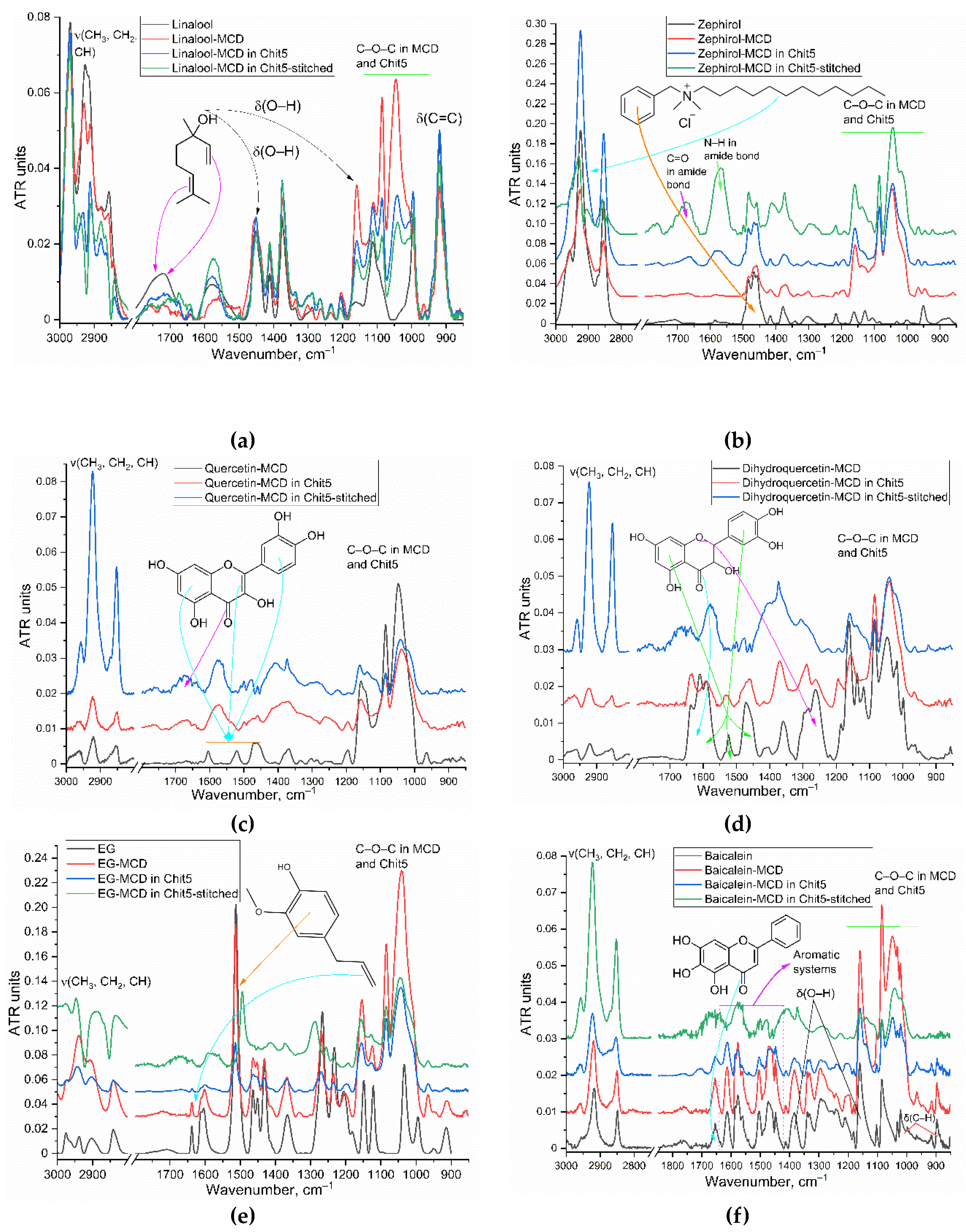
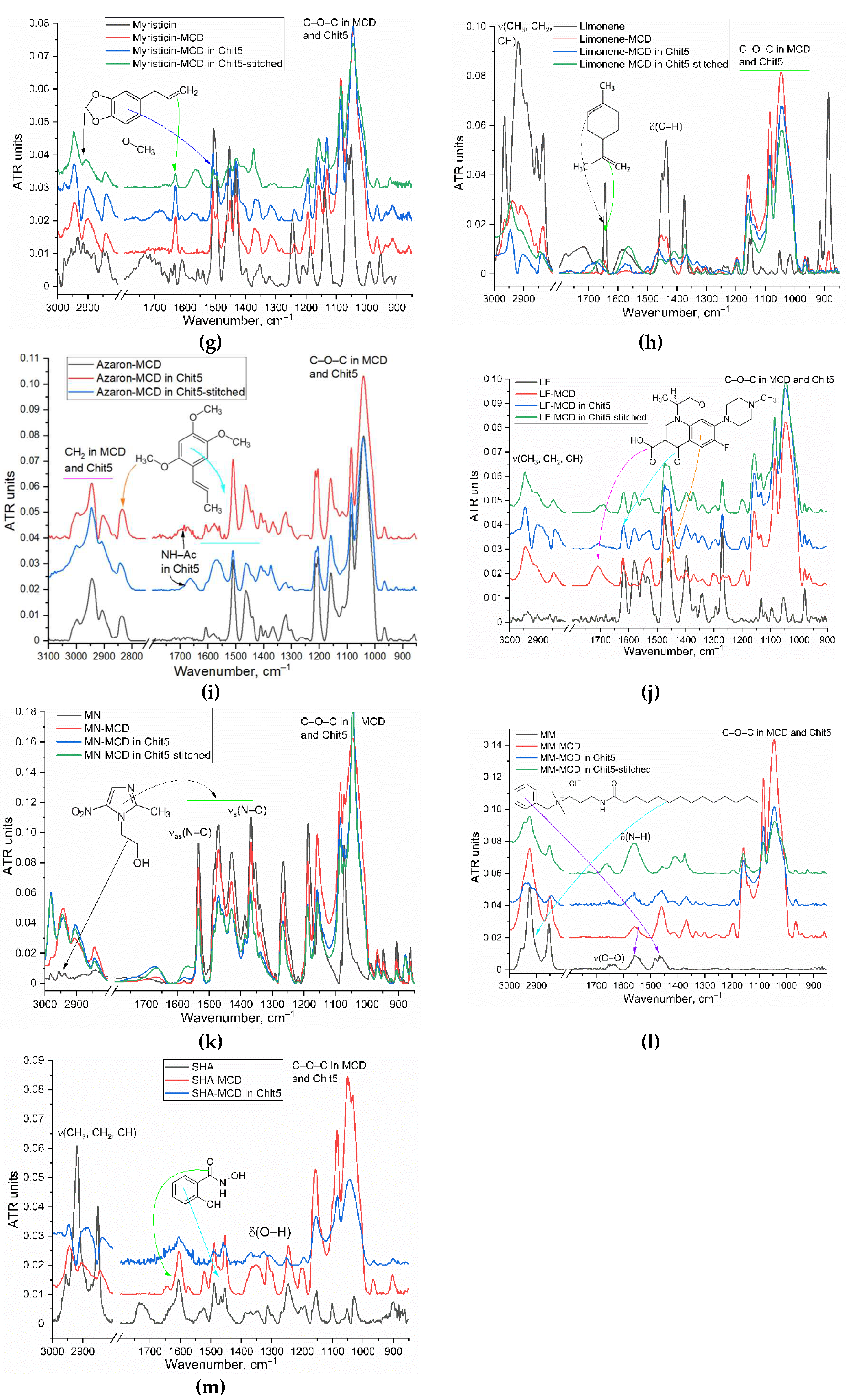
Figure 2.
(a) FTIR spectra of limonene-MCD in Chit5-genipin particles depending on temperature – phase transition of chitosan nanogels. (b) Corresponding dependences of the position of the characteristic peaks on temperature. (c), (d) Flow cytometry diagrams of Chit5-genipin nanogel with loaded FITC (CFITC = 1 µg/mL). SSC – side scattering, FSC – front scattering, FITC – fluorescence channel.
Figure 3.
The dependences of E. coli and Lactobacillus colony-forming units on the incubation time of cells with antibacterial drugs. For E. coli: С(LF) = 1 μg/mL, C(MN) = 0.1 mg/mL, C(zephirol) = 0.1 mg/mL, C(EG) = 0.1 mg/mL. For Lactobacillus: С(LF) = 10 μg/mL, C(MN) = 1 mg/mL, C(zephirol) = 0.1 mg/mL, C(EG) = 1 mg/mL. LB medium (pH 7.2). 37 °C.

Figure 4.
(a) FTIR spectra of suspension of E. coli cells (109 CFU) after a day of incubation with drug formulations in Chit5 particles. С(LF) = 1 μg/mL, C(MN, MM, SHA) = 0.1 mg/mL, C(other substances) = 1 mg/mL. LB medium (pH 7.2). (b) FTIR spectra of suspension of E. coli cells (109 CFU) during incubation (online) with linalool-MCD in Chit5 particles. 37 °C. (c), (d) Flow cytometry diagrams of E. coli cells incubated with FITC-labelled Chit5-genipin nanogel 15 min (CFITC = 1 µg/mL). (e), (f) Flow cytometry diagrams of E. coli cells (control). Green indicates a population with a high intensity of FITC fluorescence. SSC – side scattering, FSC – front scattering, FITC – fluorescence channel.
Figure 5.
Free radical-scavenging activity of adjuvants and main drugs (LF, MM, MN, SHA) examined by using ABTS assay.
Figure 5.
Free radical-scavenging activity of adjuvants and main drugs (LF, MM, MN, SHA) examined by using ABTS assay.
Figure 6.
(a) FTIR spectra of pre-incubated mucin with limonene-containing formulations and chitosan. pH 2. T = 37 °C. (b) Adsorption curves of LF (0.1 mg/mL), baicalein (0.1 mg/mL) and zephirol (0.1 mg/mL) in polymeric particles on a mucin (1 mg) substrate. 0.01M HCl or 0.01M Na-phosphate buffer (pH 7.4).
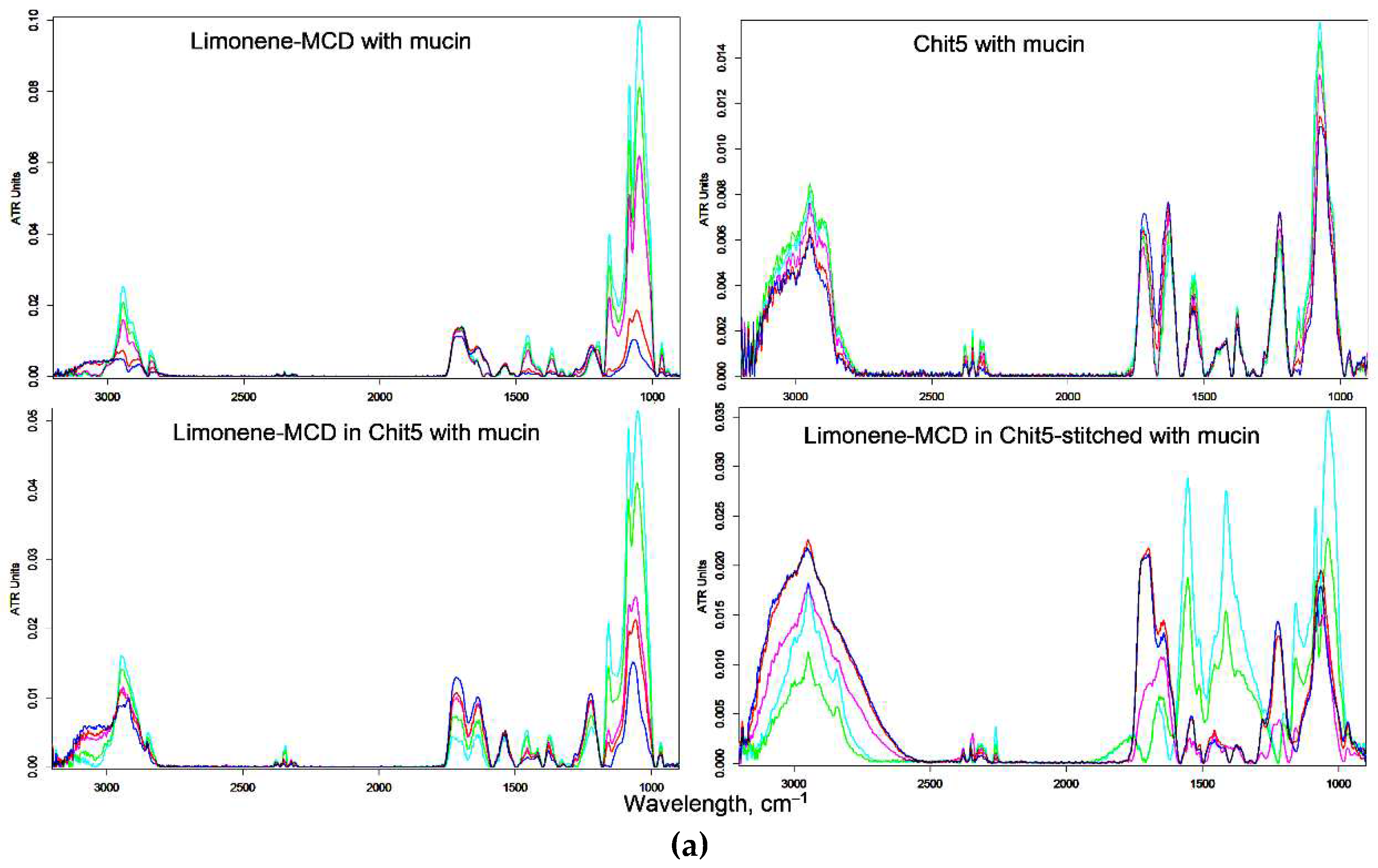
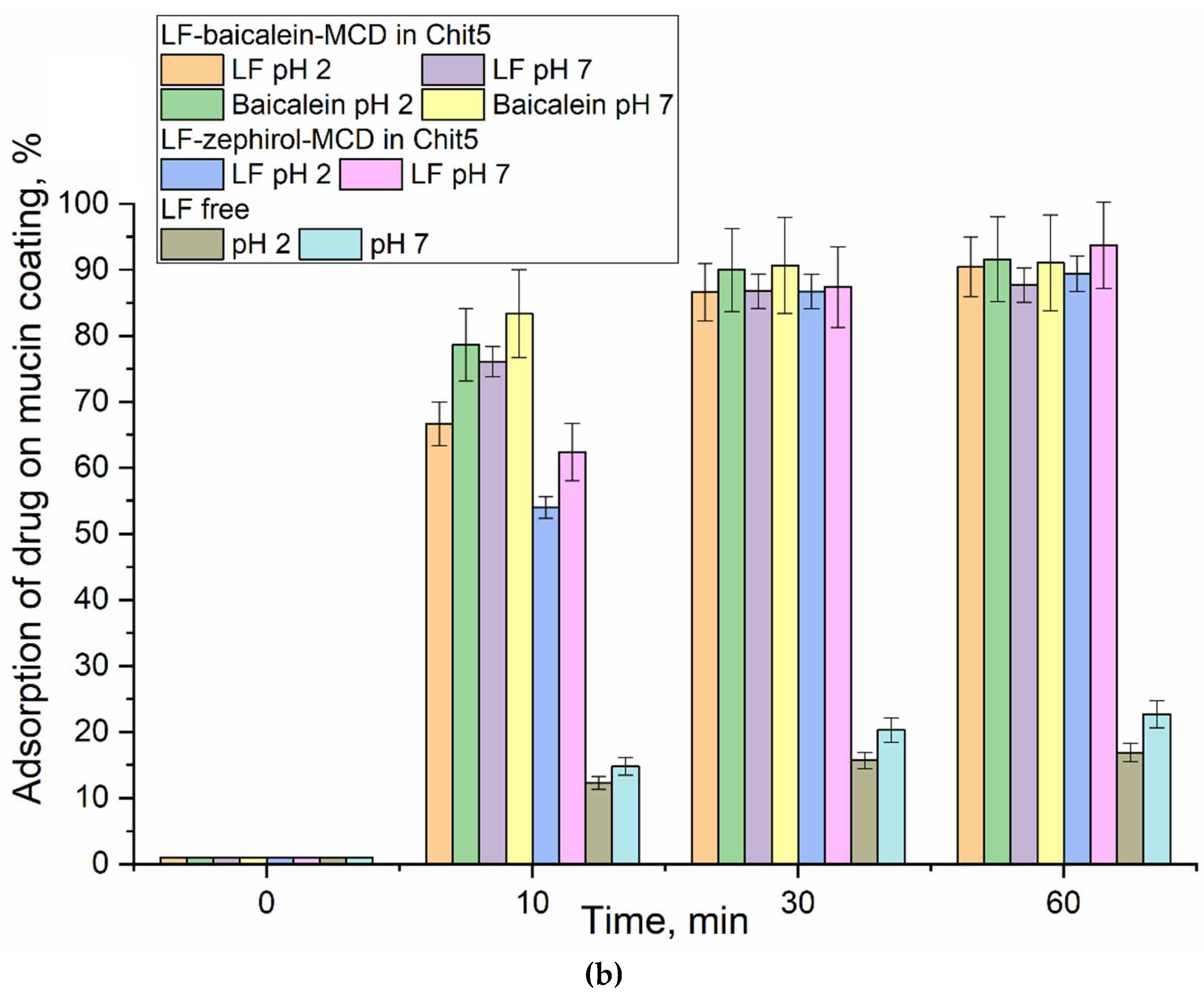

Table 1.
Cell viability (% relative to control) of E. coli cells after two days of incubation with samples in LB medium. Selectivity coefficients (the ratio of activities against different strains) of antibacterial formulations (E. coli vs Lactobacillus). T = 37 °C.
Table 1.
Cell viability (% relative to control) of E. coli cells after two days of incubation with samples in LB medium. Selectivity coefficients (the ratio of activities against different strains) of antibacterial formulations (E. coli vs Lactobacillus). T = 37 °C.
| in MCD | in MCD-Chit5 | in MCD-Chit5-gen | |||||||
| 1 mg/mL | 0.1 mg/mL | Selectivity (E. coli vs Lactobacillus) | 1 mg/mL | 0.1 mg/mL | Selectivity (E. coli vs Lactobacillus) | 1 mg/mL | 0.1 mg/mL | Selectivity (E. coli vs Lactobacillus) | |
| Linalool | 78±6 | 85±8 | 0.6±0.1 | 73±4 | 82±5 | 0.20±0.05 | 58±3 | 76±7 | 0.30±0.06 |
| Menthol | 83±6 | 90±4 | 1.1±0.1 | 81±5 | 90±3 | 0.4±0.1 | 79±6 | 84±5 | 0.4±0.1 |
| Zephirol | 12±2 | 34±3 | 0.8±0.1 | 10±1 | 33±2 | 1.2±0.2 | 10±1 | 23±2 | 1.6±0.2 |
| Quercetin | 85±5 | 92±4 | 1.1±0.1 | 87±4 | 92±3 | 0.9±0.1 | 82±6 | 89±5 | 0.9±0.1 |
| Dihydroquercetin | 86±3 | 91±4 | 1.1±0.1 | 87±5 | 95±2 | 1.0±0.1 | 83±4 | 87±6 | 0.24±0.02 |
| Eugenol | 79±3 | 90±4 | 0.9±0.1 | 70±2 | 85±3 | 1.1±0.1 | 67±5 | 82±4 | 1.2±0.1 |
| Baicalein | 26±3 | 72±7 | 1.8±0.2 | 49±4 | 68±6 | 1.6±0.2 | 66±3 | 77±5 | 0.8±0.1 |
| Myristicin | 80±5 | 87±5 | 1.0±0.1 | 86±4 | 86±7 | 1.2±0.1 | 90±2 | 89±3 | 1.0±0.1 |
| Limonene | 83±4 | 90±2 | 1.0±0.1 | 87±3 | 88±5 | 1.0±0.1 | 87±6 | 89±4 | 0.9±0.1 |
| Azaron | 81±5 | 87±4 | 0.7±0.1 | 86±6 | 91±3 | 0.8±0.1 | 64±3 | 87±2 | 1.0±0.2 |
| 1 μg/mL | 0.1 μg/mL | Selectivity (E. coli vs Lactobacillus) | 1 μg/mL | 0.1 μg/mL | Selectivity (E. coli vs Lactobacillus) | 1 μg/mL | 0.1 μg/mL | Selectivity (E. coli vs Lactobacillus) | |
| LF | 9±1 | 16±3 | 2.3±0.2 | 7±1 | 14±2 | 3.6±0.3 | 7±1 | 13±3 | 4.0±0.3 |
| 10 μg/mL | 1 μg/mL | Selectivity (E. coli vs Lactobacillus) | 10 μg/mL | 1 μg/mL | Selectivity (E. coli vs Lactobacillus) | 10 μg/mL | 1 μg/mL | Selectivity (E. coli vs Lactobacillus) | |
| MN | 41±4 | 77±5 | 0.8±0.1 | 46±7 | 82±8 | 0.7±0.1 | 20±2 | 69±3 | 1.4±0.1 |
| MM | 10±2 | 26±4 | 0.7±0.1 | 9±1 | 18±3 | 2.3±0.3 | 10±1 | 15±1 | 2.4±0.1 |
| SHA | 20±3 | 84±5 | 0.5±0.1 | 20±2 | 85±3 | 0.6±0.1 | 17±2 | 76±8 | 0.7±0.1 |
Table 2.
Synergy coefficients (SYC) of antibacterial activity of LF+X in MCD-Chit5 in comparison with alone LF and X in MCD-Chit5 against E. coli and Lactobacillus cells. The SYC was interpreted as: synergism (> 1.2), additivity (0.8-1.2), or antagonism (< 0.8). Selectivity coefficients (SEC, the ratio of activities against different strains) of antibacterial formulations (E. coli vs Lactobacillus). The SEC was interpreted as: highly specific against E. coli (>2), highly specific against Lactobacillus (<0.5), specific against E. coli (> 1.3), specific against Lactobacillus (<0.85), indifference (0.85<SEC<1.3). T = 37 °C.
| Compound X | Linalool | Menthol | Zephirol | Quercetin | Dihydroquercetin | Eugenol | Baicalein | Myristicin | Limonene | Azaron | MN | MM | SHA |
| E. coli | 1.06±0.13 | 1.49±0.20 | 1.14±0.05 | 1.3±0.1 | 1.3±0.2 | 1.16±0.07 | 1.15±0.05 | 1.08±0.09 | 1.27±0.08 | 1.0±0.1 | 0.23±0.02 | 1.23±0.05 | 1.73±0.24 |
| Lactobacillus | 0.76±0.03 | 1.11±0.05 | 0.26±0.02 | 1.15±0.06 | 1.06±0.08 | 0.91±0.05 | 0.83±0.03 | 1.24±0.12 | 1.02±0.06 | 0.91±0.09 | 0.69±0.05 | 1.06±0.07 | 0.91±0.08 |
| Selectivity E. coli vs Lactobacillus | 1.4±0.1 | 1.3±0.1 | 4.4±0.4 | 1.1±0.1 | 1.2±0.1 | 1.3±0.1 | 1.4±0.1 | 0.9±0.1 | 1.2±0.1 | 1.1±0.1 | 0.33±0.04 | 1.2±0.1 | 1.9±0.2 |
Table 3.
IC50 values of free radical scavenging of the studied substances in nanogels (Chit5-genipin) examined by using ABTS assay.
Table 3.
IC50 values of free radical scavenging of the studied substances in nanogels (Chit5-genipin) examined by using ABTS assay.
| Compound | IC50, mg/mL |
| Linalool | 0.46±0.07 |
| Menthol | >3 |
| Zephirol | 0.27±0.05 |
| Quercetin | <0.01 |
| Dihydroquercetin | |
| Eugenol | |
| Baicalein | |
| Myristicin | |
| Limonene | 1.5±0.2 |
| Azaron | ~0.01 |
| LF | 0.015±0.005 |
| MN | 0.04±0.01 |
| MM | 0.008±0.002 |
| SHA | <0.01 |
Disclaimer/Publisher’s Note: The statements, opinions and data contained in all publications are solely those of the individual author(s) and contributor(s) and not of MDPI and/or the editor(s). MDPI and/or the editor(s) disclaim responsibility for any injury to people or property resulting from any ideas, methods, instructions or products referred to in the content. |
© 2023 by the authors. Licensee MDPI, Basel, Switzerland. This article is an open access article distributed under the terms and conditions of the Creative Commons Attribution (CC BY) license (http://creativecommons.org/licenses/by/4.0/).
Copyright: This open access article is published under a Creative Commons CC BY 4.0 license, which permit the free download, distribution, and reuse, provided that the author and preprint are cited in any reuse.


