Preprint
Review

pH Monitoring in Biological Environment: An Overview of Conventional to Cutting-Edge Sensing Platforms

Altmetrics

Downloads

295

Views

201

Comments

0

A peer-reviewed article of this preprint also exists.

Submitted:

01 August 2023

Posted:

03 August 2023

You are already at the latest version

Alerts
Abstract
pH is considered one of the paramount factors in bodily functions, because most of the cellular tasks exclusively rely on precise pH values. The regulation of pH is a necessary feature of the intracellular atmosphere and can be established as a strong indicator to judge a physiological abnormality in most of the cases. In this context, the current techniques for pH sensing provide us with the futuristic insight to further design therapeutic and diagnostic tools. Thus, pH-sensing (electrochemically and optically) is rapidly evolving toward exciting new applications and expanding researchers’ interests in many chemical contexts, especially in biomedical applications. The adaptation of cutting-edge technology is subsequently producing the modest form of these biosensors as wearable devices, which are providing us the opportunity to target the real-time collection of vital parameters, including pH for improved healthcare systems. The motif of this review is to provide an insight of trending tech-based systems employed in real time or in-vivo pH responsive monitoring. Herein, we briefly go through the pH regulation in the human body to help the beginners and scientific community with quick background knowledge, recent advances in the field, and pH detection in cancerous environments. In the end, we summarize our review by providing an outlook; challenges that need to be addressed and prospective integration of various pH in vivo platforms with modern electronics that can open new avenues of cutting-edge techniques for disease diagnostics and prevention.
Keywords: 
Subject: Chemistry and Materials Science  -   Analytical Chemistry

1. Introduction; scope and importance

pH – firstly associated with Walther Nernst’s theories describing the concentrations of hydrogen (H+)/ hydronium (H3O+) ions that ultimately evolved into the logarithmic term “pH” by Soren Sorenson, analogous to an ionic activity of H+/ H3O+ [1,2]. Since then, pH is of great concern and centrality to all life forms playing extraordinary roles in many chemical and biological processes; therefore, it is exploited in a broad range of applications [3,4]. Explicitly, pH becomes more evident in biological systems by continuously regulating the intracellular/extracellular environment; from protein folding, enzymatic reactions to metabolic proceedings, and ionic transportation, where the changes in pH highly affect these biochemical processes. Even small variations in pH values of blood plasma, intestinal fluid, or cerebrospinal fluid (CSF) that are supposed to be regulated by body’s self-buffering system, can affect cell proliferation and migration, apoptosis, and physiological disorders [4,5]. Similarly in brain homeostasis, pH role is well-understood in regulating the neuronal cells [6]. pH is an integral part to maintain the functioning and morphology of the living organisms in life sciences, as intracellular pH (pHi) is responsible to maintain all the metabolomic processes of a cell [7]. The imbalance in intercellular pH (pHi) values is protected from rapid and local changes by intrinsic buffering capacity [8,9], which is comprehensively discussed in next section. For instance in a cancerous environment, the cancer cells push to maintain pHi at physiological values; consequently extracellular pH (pHe) becomes acidic, which is considered as tumor signature [10]. Also, one can be very prone to have gastric ulcers, atrophic gastritis, and even gastric cancer, once the pH value of gastric juice proceeds higher than 3.5 [11]. Making an early diagnosis and treatment of disease can dramatically reduce the mortality and human suffering, therefore, acid–base disorders remain significant factors in human physiology and the biomarkers detection shed from pH can provide us a potent opportunity to diagnose and treat various disorders at their early stage. Hence, it is very essential to monitor pH variations in vivo and real time to further explore its physiological roles in above said disorders.
Precise pH values are an integral part for the execution of many human physiological functions, therefore, the ionic activities of H+/H3O+ ions are detected with the help of chemical and biochemical tools called “biosensors” [1]. It is of worth to note that the role of pH – regulation in biological systems is very important; and pH-sensing has become an attractive area of research for many researchers, because these sensors are non-destructive tools to detect pH in-situ or in real time environments [12,13,14,15,16]. The progress in emerging biosensing technology have had a great potential and an influential impact for pH monitoring, succeeding to generate an improved, rapid, and an accurate screening of pH by means of biosensors. With the passage of time, pH sensing is becoming more sophisticated with the development of cutting-edge instrumentation. pH measurements, accompanied by some other metabolites, are the most common and frequent evaluates for quality control in biomedical applications. Because, in clinical diagnostics, the detection of pH is the most significant parameter to ensure the normal physiological conditions, hence, ensuring proper bodily functions [17,18,19]. So far, different optical [12,13,20,21,22,23,24,25,26,27,28] and electrochemical [1,29,30,31,32,33,34,35] techniques are adapted to measure pH in complex biological environments, depending on their reliability, feasibility and suitability to the requirement. The frequent research and innovative designs have revolutionized biosensing technology, which is rapidly evolving into the modest form of these biosensors called “wearable-sensors”. A handy work is already done to design such biochemical sensors for pH monitoring in biological field [36,37,38]. Where, the new era of cutting-edge techniques is providing the opportunity to collect real-time physiological parameters in many conditions such as wound management and sweat that makes the healthcare demands more reliable and accessible [39,40,41,42,43,44,45,46]. Wound management is a complex practice, demanding careful considerations of various physiological parameters such as pH, uric acid, ammonium ions, and temperature measurements. pH monitoring with other parameters could be a useful mean in reflecting wound status, assisting in better cure and management [46]. Likewise, sweat is considered to be very useful biological matrix, which contains different biomarkers that help to obtain physiological information, thus pH is an important parameter to be monitored in sweat [47].
Keeping in view the clinical significance of pH, we comprehensively summarize here the pH regulation in physiological and cancerous environments, together with the intracellular and extracellular representative pH sensors. Then, a detailed overview of trending pH sensing techniques, including optical, electrochemical, and their wearable variants are discussed in-vivo or in real life applications. In the end, modern pH sensors with current limitations, challenges and futuristic outlook are concluded. To have the best understanding of this review, we provide here the organizational and thematic components as depicted in Figure 1.

2. pH regulation in human body

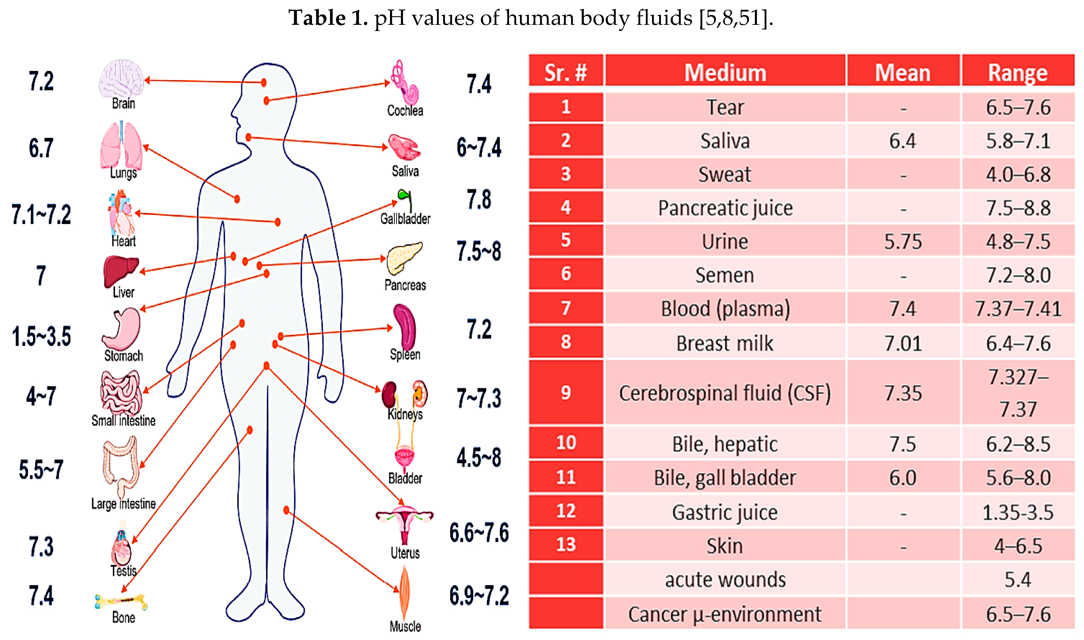
In human physiology, acid-base (pH) balance is responsible for the execution of various biochemical processes [48]. All the metabolic functions are run by enzymes, where the changes in pH highly affect the enzymatic processes. Even, the small variations in blood pH (acidosis; pH < 7.35 and alkalosis pH > 7.45) can cause in impaired enzymatic functioning; which consequent to many physiological disorders [9,49,50,51]. Since, pH is considered one of the most significant factors in human-body, therefore, various chemical–buffering mechanisms mainly containing proteins, work simultaneously to neutralize the solution by absorbing and or releasing H+. The amino side groups in proteins become NH3+ by absorbing excess of H+ and carboxyl groups become –COO– in case of lacking H+, respectively [49]. In this regard, carbonic anhydrase (CA) enzyme has influential role to control the kinetics and location of the pH changes [8]. The pulmonary and the renal are the two main systems that modulate pH using carbon dioxide and bicarbonate, respectively [8,52,53,54]. In former case, the projection of CO2 is utilized to adjust the body’s pH, but in latter case, the pH is regularized through a complex system of nephrons that enable bicarbonate to be reabsorbed and or excreted in urination process. It makes renal system to consume longer time for metabolic compensation rather than minutes or hours, as in former case scenario [8]. Variations in pH of living body is obvious (Figure 2) and different matrices like urine, sweat, saliva, and blood possess different pH values as summarized in tabular form (Table 1). For instance, healthy skin retains the pH values between 4-6, this acidic environment is provided by some amino acids and fatty acids that is helpful in bacterial inhibition. Otherwise, bacterial proliferation may result in various associated diseases, such as hyponatremia and hypernatremia, hypokalemia and hyperkalemia, heart disorders and cystic fibrosis [55].
Similarly at cellular level, proton-activated chloride (PAC) channel acts like a low-pH sensor, and is considered one of the regulators for dynamic-buffering systems. It is also revealed that PAC channel promotes endosomal acidification through chloride ion cross-membranes movement [56]. Additionally, it was further explored that PAC is also involved in acid-induced cell death that leads to various diseases such as ischemia, cancer, inflammation and brain injury.

2.1. pH in cancerous environment

Cancers are considered one of the lethal abnormalities in human evolution and despite improved therapeutics, causing a large death toll all over the world every year [57]. To combat cancer’s losses effectively, the hidden factors which play vital roles during the cancer metastasis must be identified and addressed adequately. Tumor pH and metabolism are intrinsically related, and it is evident from many studies that acidosis is clearly associated with the invasion/metastasis in many systems [52,53,54,58]. It is revealed by Qi, Qi, et al. that deregulation in pHi is a common characteristic of cancer cells and promotes cancer cell proliferation to evade apoptosis and metabolic adaptation [54]. Also, a minute imbalance in pHi values pushes the cancer cells to maintain pHi at physiological values through a complex mechanism of glycolytic process. They suggested that glycolytic metabolism is the one that promotes tumor metabolism effectively, leading to the production of excess acid and protons. Consequently, the extracellular pH (pHe) drops down; thus, pH gradient inverted when compared to biological conditions, defining the gradient as an early event to provide crucial cellular information regarding its aggressiveness and progression. Given the importance of pH-associated cancer cell behaviors, the dynamics of dysregulated pHi help in triggering many cancer cell activities, including apoptosis and metabolic adaptation [3,7,57,59].

2.2. Intracellular/ extracellular pH sensing
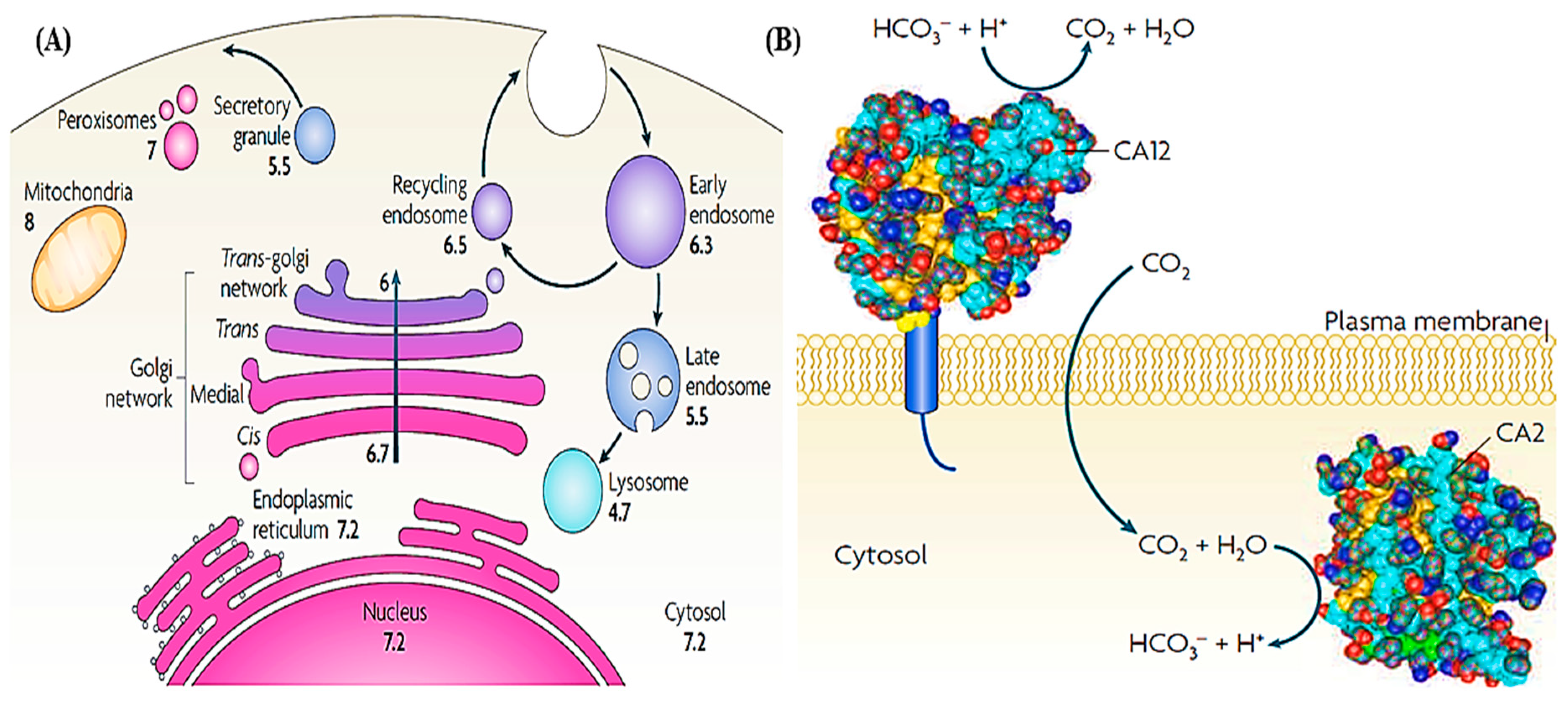
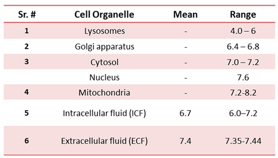
The role of pH is well understood in cellular metabolic processes and considered one of the crucial parameters for the proper functioning and coordination of many intracellular organelles [58]. Overall, the mean intracellular pH (pHi) value is lower than pHe value due to the low concentration of HCO3-. As shown in Figure 2, every cell organelle retains its own pHi values, which are frequently administered due to the continuous inter-conversion of CO2 and HCO3- [60] and can be construed in the following equation (1):
CO2 + H2O ⟶ H2CO3 + HCO3- + H+
The specific – pH values of each organelle changing from 4 to 8.2 are summarized in Table 2. For instance, lysosomes and mitochondria have the lowest and highest pH values of 4.0 – 6 and 8 – 8.2, respectively. Whereas, Golgi apparatus, cytosol, and nucleus possess the pH values of 6.4 – 6.8, 7.0 – 7.2, and 7.6, respectively [61,62]. Similar to that of body’s pH regulation system, the mixture of phosphate ions also serves as a major buffering system in the intracellular fluid. The imbalance in pHi values critically disturbs all the enzymatic reactions and metabolic processes of the cells once it becomes acidic, thus it is neutralized by employing such body's self-buffering systems.
Figure 3. (A) pH illustration of the different cell organelles and (B) CO2, HCO3 and the regulation of pH. Copy right image reprinted with the permission from Ref.[8].
Preprints 81262 g003
The dynamics of pHi or intracellular pH are significant in regulating normal cell physiology and the slight variations in pHi (7.2–7.6) make the cell to be polarized, remodeling actin cytoskeleton, and migration [63]. Since, every organelle in the cell retains its own specific pH values, cell-messengers like Ca2+ and cAMP are affected due to the change in pHi values; which are important in cell activation, growth, and proliferation process [64]. Thus, monitoring of pH in cellular and enzymatic functions could lead as a significant biomarker for many diseases detection [65]. Various in-vivo techniques consisting organic probes and contrast agents are used to measure intracellular and extracellular pH [54,66,67,68]. However, background noise and less sensitivity towards abrupt change in pH values are some of the significant hurdles, which limit these techniques to be used in-vivo for a long time and continuous monitoring of pH-related biological processes. Over the years, researchers have mitigated these issues by developing a variety of pH-sensitive or responsive nanomaterials (NMs), including inorganic, organic, and hybrids, targeting live pH for cell organelle-specific localization monitoring [61,63,69,70]. Based on these NMs, various strategies are employed to construct pH-responsive biosensors to capably distinguish the different acidic divisions in cell organelles, such as early endosomes (pH 6.0−6.5) and lysosomes (pH 4.5−5.5) [53,71,72,73,74,75,76,77,78,79,80,81,82,83,84,85,86,87].

3. An overview of pH sensing techniques

Over the time, pH-measuring devices have been continuously evolved from glass electrodes to test strips or even smarter tools [88]. The small dimensions of the fabricated sensing devices allow them to measure pH in minute sample volumes in confined environments, such as brain fluids, wound fluids, and proteins analytes for POC diagnostics [32]. When we talk about pH monitoring, mainly it has been carried out with different techniques such as electrochemical and optical sensing over the decades to provide quantitative determination of pH for diagnostic purposes. Each technique used for pH monitoring is comprehensively reviewed in the following sections.

3.1. Electrochemical sensors

The development of electrochemical sensors has been implicated since long, pioneering from the early 1900s to today’s high-tech configurations, including glass electrodes to ion-sensitive field effect transistors (ISFET), respectively [1]. Although, the glass electrodes provide accurate and reliable measurements for pH; however, they retain limitations to be miniaturized for in vivo studies, and secondly, they repeatedly require calibrations that hinder their broader applications. ISFET or extended gate (EG) FET based cutting-edge innovations have mitigated these drawbacks by introducing smart nano-sensors. Based on potentiometric phenomena, ISFET or EGFET and chemo-transistors are the most reported pH sensing devices in literature. These types of electrochemical sensors usually interpret the potentials difference at the interface of electrolyte-transistor gate produced by protonation and deprotonation reactions [82,89,90,91,92]. Also, potentiometric ISFET or EGFET-based pH sensors possess huge advantages, including compactness due to cutting-edge technique, durability for pH sensing rendering it promising for biomedical applications. Even ISFETs are now exploited for microbial detection, for instance Chia-Yu et al. reported that bacterial growth can be monitored based on the acidification process during bacterial metabolism [93,94]. Since, ISFET is capable enough to detect ionic strength of an analyte, thus, it retains the potential to be exploited in various diagnostic applications [95]. Principally, the pH shift in some biological reactions triggers the enzymatic activities, which is then transduced by ISFET-based devices.
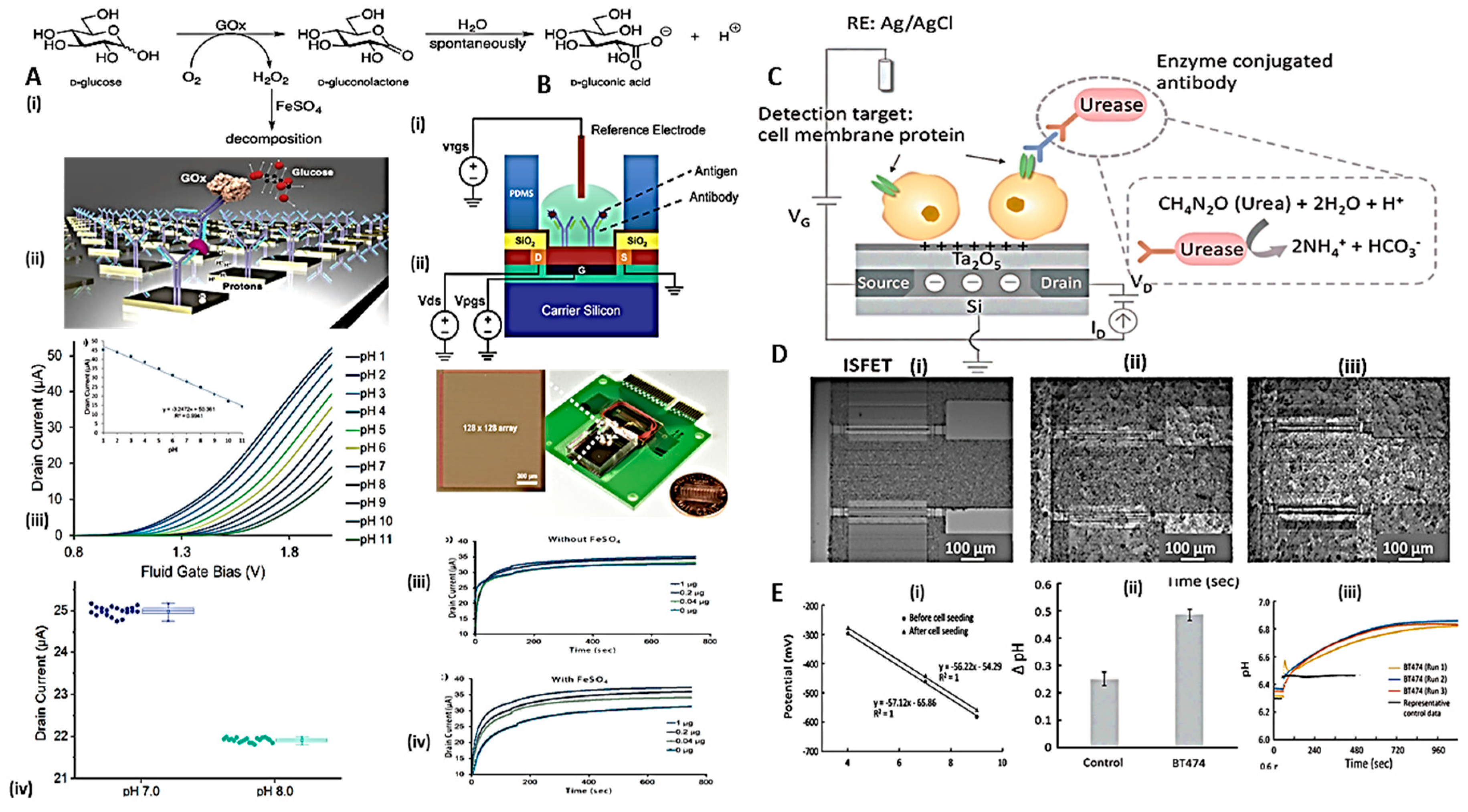
As a proof of concept, it has been used to detect H2O2 in enzymatic reactions in lieu of glucose conversion – by glucose oxidase (Gox) enzyme as shown in Figure 4 (A) to obtain a linear range of pH from 1 to 11. Interestingly, a multiplexing array of 128×128=16,384 comprising dual-gated ISFET (DG-ISFET) sensing devices was fabricated to allow a very high signal throughput. Such configurations possess the ability to chemically amplify the enzymatic reaction’s signal, subjected to the complete consumption of enzyme substrate. In this case, protons were obtained as a result of enzymatic reaction (Gox in KCL) and catalyst FeSO4 addition; where, Gox converted the substrate into H2O2 and FeSO4 further transformed H2O2 through Fenton’s reaction. Thus, providing an excess of protons with 515% enhancement, which is evident in pH change (Figure 4 (B-iii)). As shown, the device exhibited a linear voltage/current response for pH buffers ranging from pH 1 to 11, when biased with top gate from 0 V to 2.0 V. It is of worth to note that all the data acquisition was obtained by full devices -array scan in 2-3 sec, assuring that the platform retains a great promise for time-dependent studies in biomedical applications. This phenomenon is advantageous when minute volumes of analytes need to detect. Following that principle, recently an ISFET-based sensor was developed (Figure 4 (C-E)), demonstrating to detect the human epidermal growth factor receptor (HER2) as a breast cancer biomarker [96]. Herein, poly-L-lysine, a positively charged polymer was used on the gate (Ta2O5) surface to efficiently capture the cells. Images reveal the adherence of the cells on the surface, even after pH calibration experiment. Further, HER2 on the BT474 cell membrane was detected using urea–urease cascade containing secondary antibody. Enzymatic reactions proceeded to produce ammonium ions (NH4+) and HCO3- due to urea hydrolysis. Thus, the potential change due to the NH4+ ions as a result of proton consumption or hydroxide generation of enzymatic metabolic-end product gave rise to increase in sample solution pH. The proposed mechanism was further exploited to quantitatively targeted biomarker subjected to net pH change at the ISFET gate. Moreover, the enzymatic reaction rate at the ISFETs surface depends on the number of immobilized enzymes under constant substrate concentration conditions. As a consequence, high-throughput and highly sensitive detection of such biomarkers is valuable with ISFET-based electrochemical devices.
The use of conducting polymers (CPs), such as polydopamine (PDA), polyvinyl alcohol (PVA), polypropylene (PP), polyaniline (PANI), polypyrrole (PPy), and polyelectrolyte such as poly(sodium 4-styrenesulfonate) (PSS) is trendy to combine with nanoparticles (NPs) for the construction of pH sensor substrates [29].
Flexible fiber in a microfluidic arrangement was used as sensing assembly to produce electrochemical readouts, either potentiometric or amperometric in mice brain [31]. The efficiency of the developed sensing system was validated in-vivo by detecting lactate accompanied by pH sensing. In a similar manner, PANI/MoS2/AN showed a remarkable potentiometric response, specifically towards pH changes in the rat brain while overriding the interfering species[97]. The monitoring of such brain-metabolites can provide useful information about the neurochemical dynamics, hence could be used to monitor complex brain functions subsequent to neurological disorder’s diagnosis.
Shih-Cheng et al. reported flexible and porous pH sensor development by depositing PDA, PVA and PPMMA with IrO2 NPs, revealing an excellent electroanalytical performance towards sensing wide dynamic range of pH (2-12) with a good Nernstian response [29]. Similarly, Zea and colleagues proposed a pH sensing ink formulated with the blend of PANI, PPy and PSS. The developed NMs produced an improved sensitivity towards wider pH window (3-10) with the ability to be miniaturized into a smart pH-sensing device that could be applied in healthcare applications [34].

3.2. Optical-based pH sensors

The second most applicable technique for pH sensing is optical method, which transduces interactive variations at devices – analyte interface; therefore, it is considered one of the safe modalities in biomedical applications, especially for pH monitoring. Optical sensing possesses number of advantages being cheap, rapid, better SNR, more facile in remote sensing, and very effective in miniaturized real-time assessments. Nonetheless, chemical-based optical sensors have the limitations such as temperature dependency, instability due to leaching and photobleaching of the materials, nonlinear response to abrupt change in pH, and very narrow pH range, typically 2−4 pH [25]. However, using blend of various dyes or some reference materials can overcome the above said shortcomings and allow monitoring of wide range pH values [12,20,25]. Following are the details of optical modes that have been explored since long in pH sensing.

3.2.1. Absorption/Visual-based pH sensors
Absorption-based pH sensors are the consequence of selective change of their optical properties when they interact with the analyte. Spectral absorption technique is facile, non-invasive, and non-destructive to evaluate the pH response more rapidly without adding some sophisticated instrumentation in real-time analysis. This allows the variations in pH to be observed with naked–eye corresponding to the pH material responses to the pH changes, resulting in different color. Though, a comprehensive literature is reviewed by Andreas Steinegger and colleagues to provide a complete informative package about optical-based pH sensing [12]. However, we go through the recent updates in trending pH sensors that are typically being employed in biological applications. Especially, the blend of organic-NMs retaining multisensing capabilities could be excellent choices in prospective technologies. For instance, a colorimetric sensor comprising organic molecules boron trifluoride (BF3) with curcumin as the carrier-recognition terminals 2,2-difluoro-4,6-bis(3-methoxy-4-(4-(4,4,5,5-tetramethyl-1,3,2-dioxaborolan-2-yl)benzyl)oxy) styryl)-2H-1,3,2-dioxaborine (DFCB) is recently reported to evaluate pH in live-cell and zebrafish imaging. The quenching mechanism of DFCB on H2O2 was confirmed by adding H2O2 in the solution to cleave the sensing group on DFCB, and the matrix color changings and fluorescent quenching were observed due to protonation/deprotonation phenomenon. The developed sensor successfully detected H2O2 along with a wide (6–11) pH range [98]. Similarly, spiropyran-based cellulose nanocrystals (SP-CNCs) nanosystem exhibited excellent pH-responsive behavior as well as photochromic and photo switching properties. Because, the UV and Vis. irradiation makes SP-based derivatives to behave in reversible color change, which results in displaying different colors at different pH values [99]. Similarly, optodes are another trending class and optically ion selective electrodes, merged as promising sensing devices in optics quite a few years back. Mostly, 3-D scaffolds of optical material are fabricated on a paper to provide real time 3-D resolution maps of the changings in target analyte, such as the pH can be easily visualized in paper-based cultures. Being handy, economical, weightless, and compact; optodes are an excellent choice for pH sensing in real life/in-vivo/or live cell imaging applications [100].
Fluorometric-based sensing is a promising technique retaining various advantages such as real-time and noninvasiveness, and has emerged as a potent tool for sensitive pH monitoring in physiological environment since long [39]. Since, fluorophores are important pH indicative tools for bio-analytical study and organic dyes are one of the classical choices to develop fluorometric-based pH sensors, either in situ or in vivo biological applications. Owing to their resolute scientific importance regarding optical properties, considerable efforts have been made in the past to explore such novel fluorescent molecules in the construction of pH sensors [22,23,101,102,103].
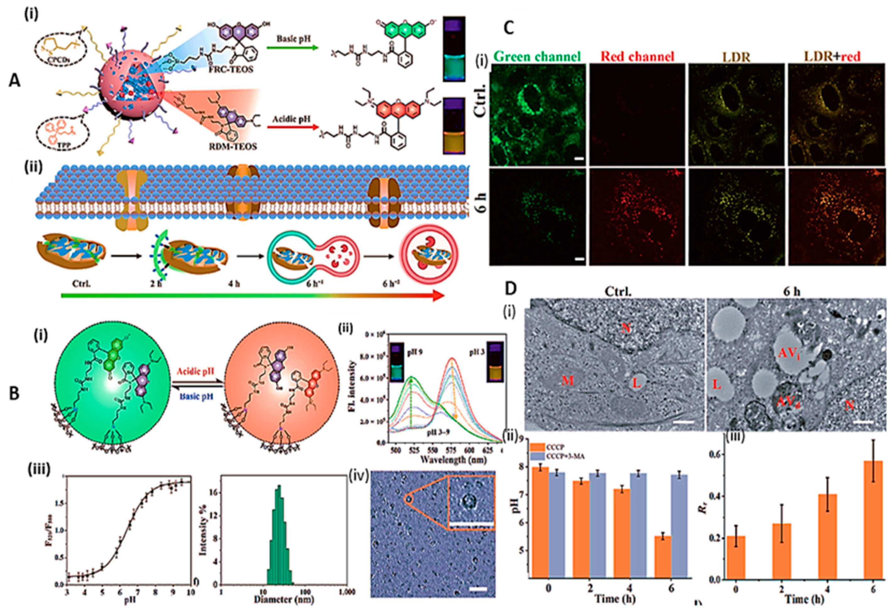
As described earlier, the monitoring of cell-organelles pH is of utmost importance to screen many physiological disorders. For instance, fluctuation in pH provides information about cellular homeostasis like mitochondrial impairment or acidic lysosomes, and fluorescence pH sensors developed for such purposes require concurrent features, like mitochondrial-specificity, biocompatibility, wide pH-sensitive range, and capable enough to track real-time pH variations in cellular functions [61,70,104]. However, fluorescence probes providing quantification of target analyte with single emission, often suffer from a variety of parameters such as interference, photo-bleaching, probe molecule concentration, and optical distance that affect the real sample analysis. One way to combat such factors ensuring reliability is to exploit the “ratiometric” approach, where the variations in two or more excitations or emissions values are measured to detect pH. Owing to its inherent reliability and precision, various ratiometric sensing probes have been developed to monitor pH in cellular quantification [105,106,107,108]. Xin Zhan and colleagues developed a fluorogenic sensing system by developing FR-TPP pH sensitive units (Figure 5) to trace mitophagy process [70]. Where, mitochondria targeting moiety triphenylphosphonium (TPP), a well-known mitochondrial targeting carrier used in many therapeutic and diagnostic studies [105] and cell penetrating cyclic disulfides (CPCDs) were anchored onto the silica NPs surface. Later, xanthene-modified fluorophores (fluorescein and rhodamine B) were encapsulated in silica NPs (Figure 5-A) to provide basic pH sensing at λem ~ 520 and acidic pH sensing at λem ~ 580, respectively.
The developed system retained ratiometric property for both environments; alkaline and acidic pH at a single excitation wavelength (B (i&ii). In addition, broader range pH sensing (4 to 8) allowed the researchers to construct an efficient mitochondrial live tracking ratiometric pH sensor (Figure 5 C & D), covering mitochondrial and lysosomal pH differences associated with the acidification and fusion processes during the whole mitophagy.
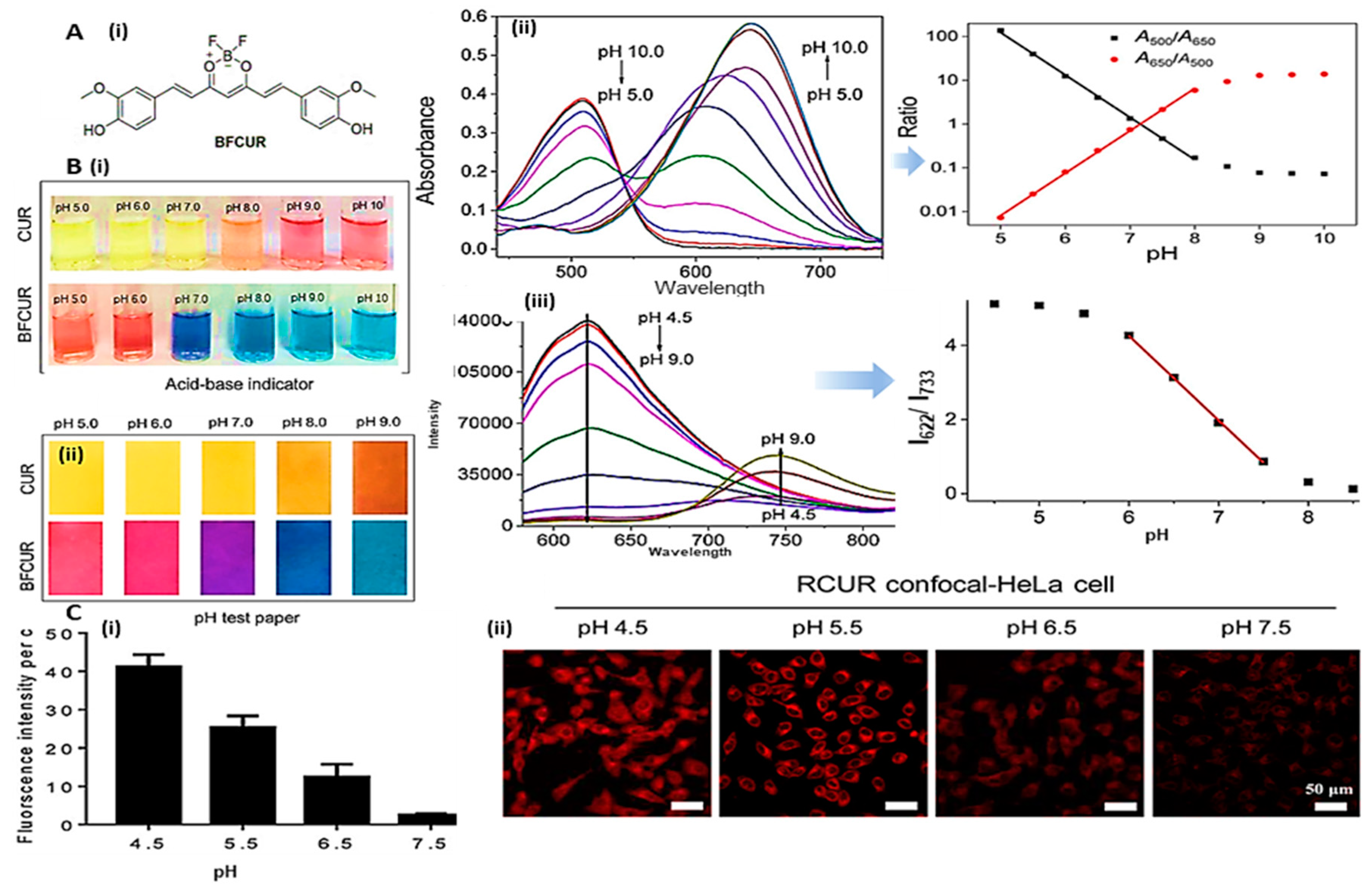
Another study, demonstrated fluorescent dye-based pH detection system, where boron trifluoride (BF) compound was introduced in center of the polyphenol curcumin (CUR). As shown in Figure 6, UV absorption spectra demonstrate a red shift in the peak and a linear response towards pH sensing was observed due to the transfer of [H+] from the phenolic groups of CUR.
BFCUR retained low toxicity during intracellular pH monitoring [109], where the underlying electron donating ability of BFCUR was in response to the increased proton transfer in alkaline environment. Thus, the phenolic hydroxyl groups of BFCUR enhanced the intermolecular charge transfer (ICT) phenomenon that led to a red shift in optical quantification i.e., UV & fluorescence. The authors performed various conformational steps by acquiring quantitative data set (A-B), and subsequently, fluorescent responses of the as synthesized compounds were evaluated in cellular matrix while incubating at various pH values (C, i-ii). The obtained results adequately confirmed the practical suitability of BFCUR compound for biological applications.
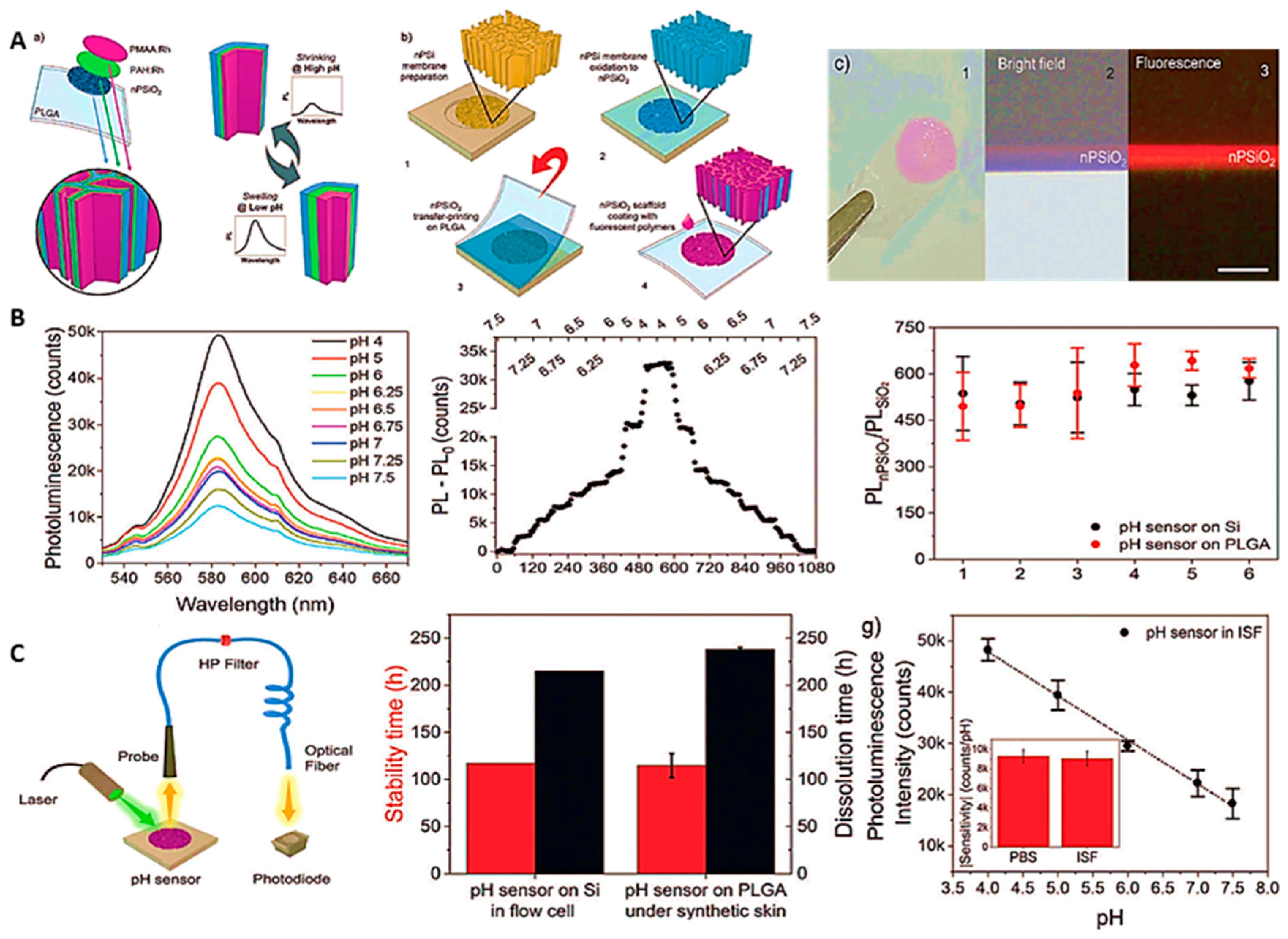
Bioresorbable materials are another important class of NMs being explored in biosensing devices, particularly for in-vivo sensing systems. Because, these devices are directly in contact with the target analyte or biofluids so, there is no need of second device to retrieve the data and hence, excluding the surgery menaces [110]. Figure 7 explains the construction assembly of the sensing system, where the porous silica membrane was fabricated using electrochemical etching of silicon wafer and subsequently, annealed at 1000 ⁰C to produce negative surface charge. Further, alternating stacking was performed with two polyelectrolytes, namely poly(allylamine) (PAH) and poly(methacrylic acid) (PMAA) labeled with pH insensitive rhodamine (Rh) material to develop pH sensor. Here, they optimized and assembled odd layers of co-polymers stacking (PAH being on the outermost layer) in the presence of Tris buffer (pH 8). When the pH decreased, the positive charges attracted the negative charges (Cl- or H2PO4- of PBS buffer) from the stack to regularize the swelling, hence an increase in the photoluminescence intensity was observed. Hence, the change in the solution environment increased or decreased the matrix distance between the polymeric layer and Rh. A linear dynamic range of pH from 4 to 7.5 was detected due to the swelling/ shrinking phenomenon of polymeric layers, where the developed sensor demonstrated accuracy over 100 hr. of continuous operation. Further, the sensor was evaluated for real time pH measurement by implanting in the hypodermis of back of the mice and provided very stable and satisfactory results. The degradability of the developed sensor was examined, which fully degraded in one week during the animal implant experimentation.
Chemically-treated fiber-optic (CT-FO) tip behaves like a smart biosensing device considered an excellent choice in biological diagnostics as it retains the faster response times and potential to be miniaturized in advance biomedical tools (catheter, needle, endoscopy system, etc.), or other technological domains. Due to invulnerability to electromagnetic interferences, FO-based biosensors are potential area in pH drift in out-put signal. Also, it is more adoptable with modern instrumentation, either in sensing. Recently, Junhu Zhou and colleagues reported the development of such type for biosensor for pH monitoring [111].
Figure 7. A (a) Thematic representation and working principle of PAH:Rh and PMAA:Rh coated nPSiO2 fluorescence pH sensor. b) Depicts the main preparation steps for PAH:Rh and PMAA:Rh coated nPSiO2 at PLGA substrate. c) Represents the images of different fabrication steps on PLGA substrate. B) Demonstrates the photoluminescence analysis of the sensor in pH values ranging from 4 to 7.5, corresponding real-time intensity, and pH sensor’s performance on different substrates. C) Representation of pH sensing set-up, stability of the developed system using both the substrates (Si & PLGA) simultaneously underneath the synthetic skin for comparative analysis. Real samples assessment of pH values ranging from 4 to 7.5 at optimized temperature of 37 ⁰C and inset is the comparative analysis in PBS and ISF. Copy right image reprinted with the permission from Ref. [112].
Preprints 81262 g007
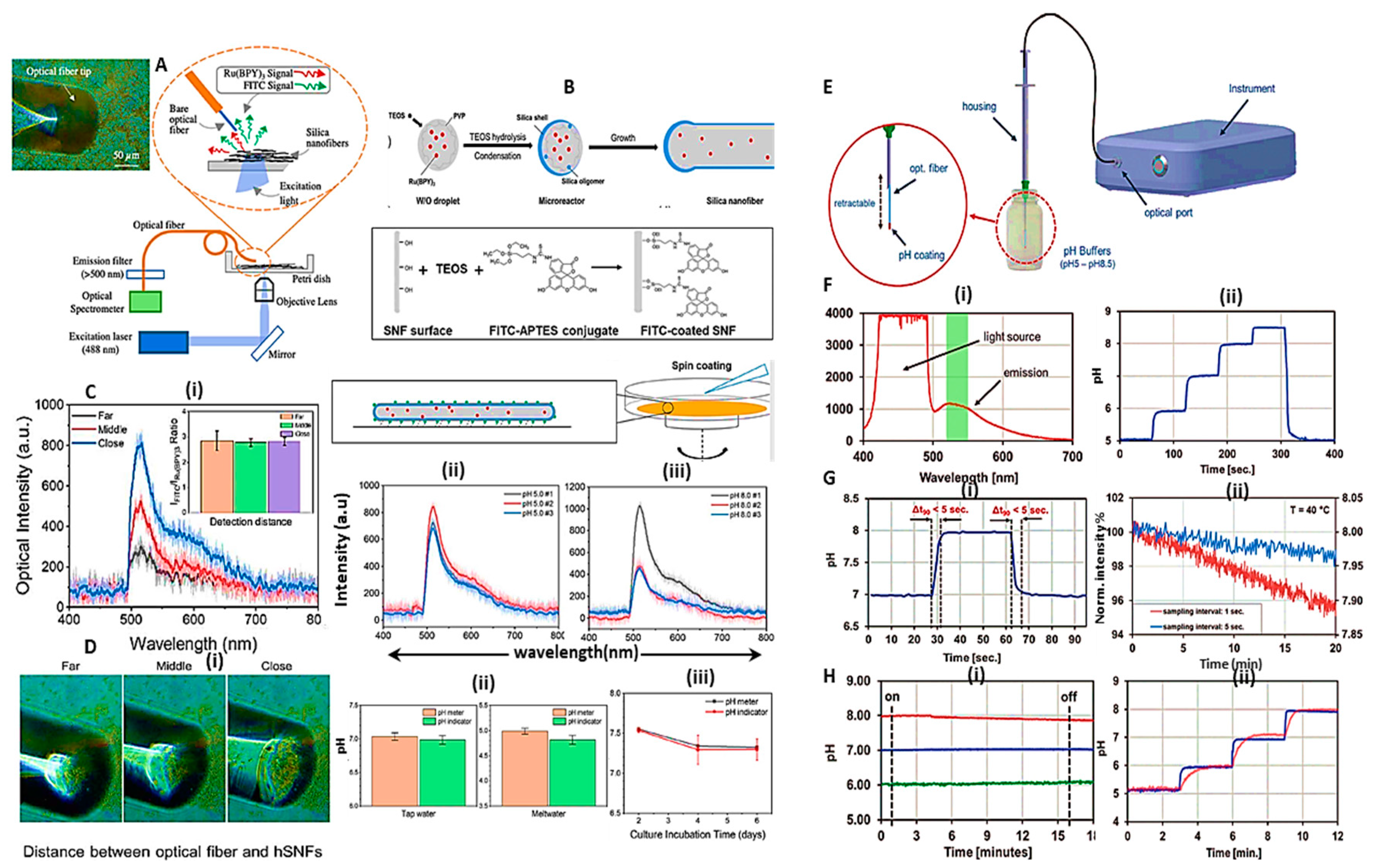
Fluorescein isothiocyanate (FITC) was chosen as an organic moiety to blend with tris(2,2′ -bipyridyl) dichlororuthenium(II) hexahydrate Ru(BPY)3 molecule and doped with hollow silica nanofibers (hSNFs) (Figure 8 (A)). Further, it was integrated with optical-fiber, enabling a facile efficient near-field fluorescence analysis. The developed system showed a linear range between pH 4-9. The sensor retained an excellent response time of less than 10 sec, whereas requiring minute sample volume of 50 µL. The dual fluorescence hSNF biosensor was evaluated in extracellular pH monitoring for practical application using human induced pluripotent stem cell-derived cardiomyocytes (hiPSC-CMs). The pH value was decreased from pH 7.5 to pH 7.3. Based on the demonstrated results, the proposed fluorophores-doped devices are worthy to monitor pH in real samples and complex analysis [111].
While in another study as shown in Figure 8 (B), hydrogel matrix was modified with pH-sensitive dye. In this case, the developed sensor showed very fast response time of <5 sec, and further, CT-FO biosensor was successfully evaluated in optimized and pre-adjusted pH values (between 7.4 to 7.5) of AMES’ medium to study the sensor's applicability in real sample environment [24].
To make the fluorometric biosensor more efficient, new trends in biomaterials are being explored. Conductive hydrogels, for instance, is also another class of biomaterials that possess great prospective in the development of such biosensors.

4. Cutting-Edge Technology; Wearable pH Sensors

On the basis transduction systems, wearable pH sensors are becoming the applied form in real world applications based on the following techniques:
  • Wearable electrochemical pH sensors and
  • Wearable optical pH sensors

4.1. Wearable pH sensors in real life applications

The evolutionary phases in computer technology have served the scientific community to develop algorithmic tools and analyze them in various fields. The computational toolboxes have started a new era of interdisciplinary sciences and gave rise to internet-of-things (IoT) to explore innovative designs and applications. Thus, the synergy of material science and bio-electronics has played a vital role to address the diverse problems in healthcare applications by interlinking the blend of enabling technologies, providing integrated platforms to foreseen wearable devices in POC systems. In the current era, wearable-technologies are revolutionizing biochemical sensors design and remarkably, more in applied form to better help and manage biomedical applications [16,17,18,36,38,113,114,115,116,117,118]. A comprehensive overview is provided somewhere else describing the latest updates regarding the advances in wearable pH sensors. Of them, wound management is becoming clinically an exciting area, and wearable technology is promising to provide cutting-edge solutions in clinical diagnostics and surgical wounds by exploiting various physical, chemical and biological sensors [42,119,120].
  • Wound Management
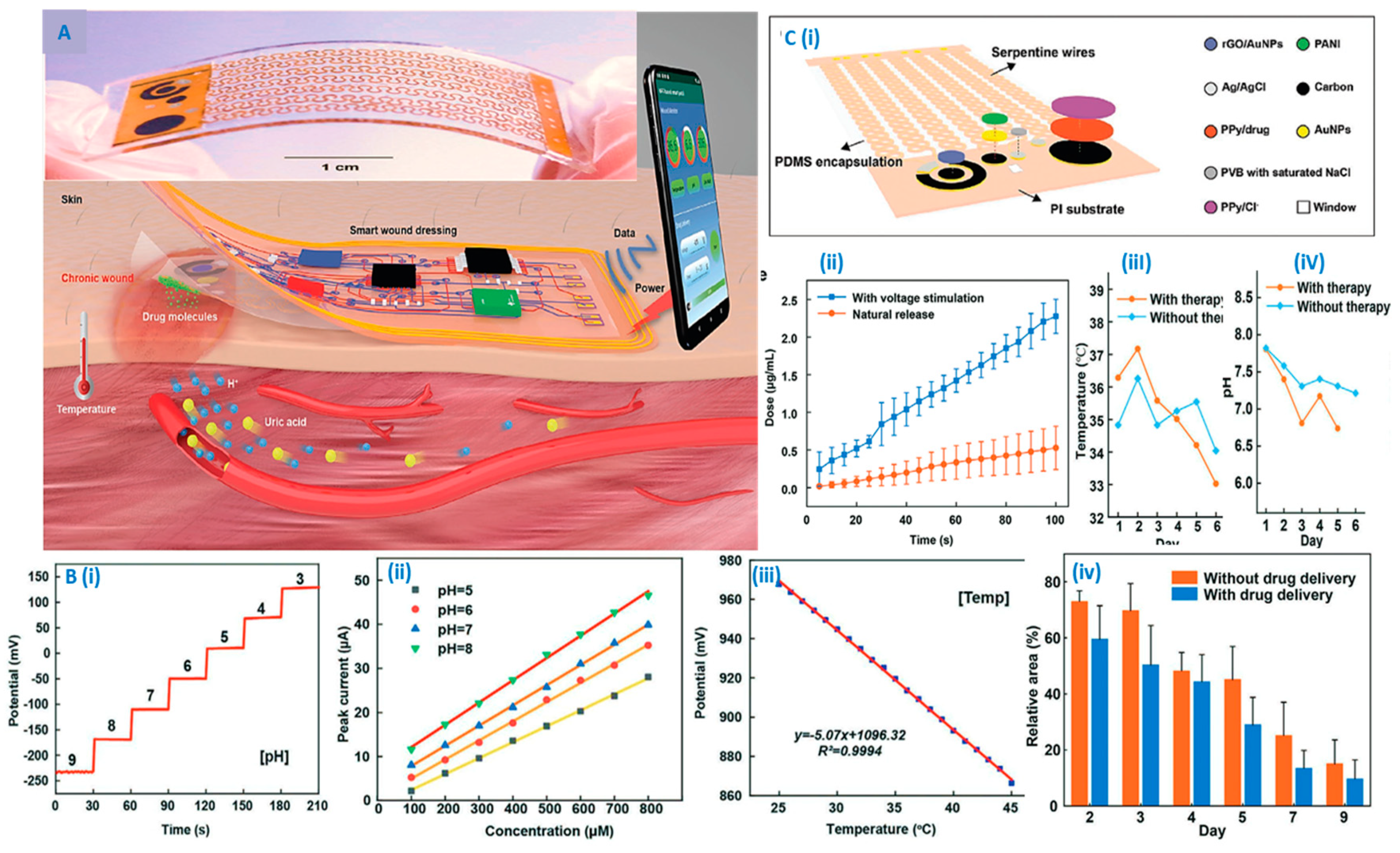
Wound management is another clinical complication growing fast world-wide. Monitoring of wounds by means of elevated levels of various indicators, including temperature (T), pH, uric acid (UA), and ammonium in wound exudate provide very useful information that can be translated to understand wound physiology to deal with it more professionally. Thanks to IoT in the biomedical field, this has been providing more sophisticated and wirelessly distant platform to manage biological complications very effectively. Additionally, the developed systems also performed combinative therapies by means of controlled drug release as well as produced electrical pulses for tissue regeneration in the wound area [42,43,44,45,46]. In this regard, Figure 9 depicts an NFC-enabled smart bioelectronics platform consisting interdisciplinary capabilities, including temperature and pH sensing, drug delivery, acquisition of results, and most importantly, the device was wirelessly powered, wound dressing, multiple sensors and antibiotic coated on polypyrrole (PPy) substrate [44]. While bottom of the system containing polyimide substrate with fabrication and integration of disposable Conductive pads were provided to electrically connect the both layers. All the data regarding change in pH, uric acid, temperature, and infected wound were successfully recorded and wirelessly transmitted and translated by smartphone for analysis. The all-in-one design provided satisfactory outcomes for wound management in terms of drug delivery and sensing various parameters.
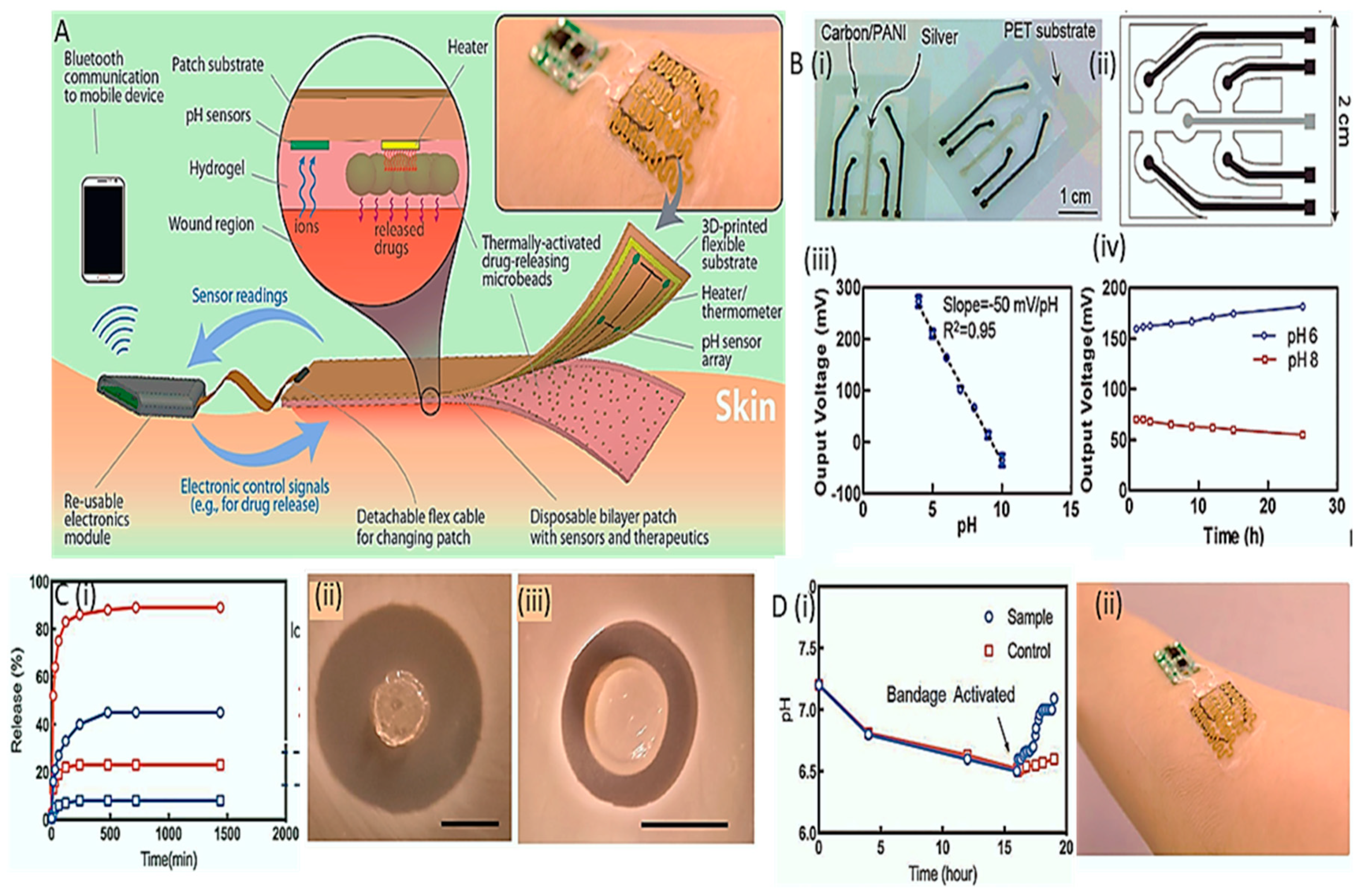
Similarly, Figure 10 (A) depicts the construction of smart bandage comprising various compartments such as pH and temperature sensors, microheater, hydrogel patch encapsulating drug, and IoT to read the data and treat the wound [45]. Here, PANI led to potential output used for further pH translation. The entire system was embedded in bandage, where the sensing and thermo-responsive drug delivery data were obtained via the wireless module in electronic patch. Other parameters, such thermoresponse, drug release, pH and temperature measurements were performed (B-D), where the responses were up to the mark.
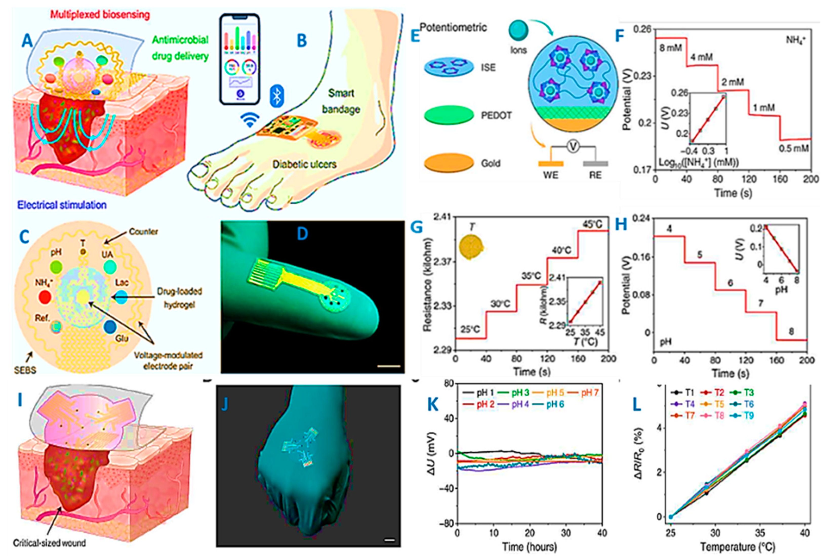
Ehsan Shirzaei et al. [46] developed another smart wearable system to treat and monitor the wound with the help of an integrated multimodal sensor array as shown in Figure 11. The smart wearable patch was comprehensively evaluated for the monitoring of metabolic indicators and antibacterial/ inflammatory activities. with carbon composite constructed as positive exchange membrane while provided. Furthermore, the physical parameters such as mechanical flexibility, stretchability, and adherence to the skin wound were tested in a rodent model, demonstrating that the bioelectronics patch is fully biocompatible and a valuable addition in cutting-edge chronic wound management.
Other than detection, pH responsive hydrogels employed in wound dressings also show a great promise in wound management and regeneration. Because, hydrogels are bioactive materials that encapsulate antimicrobial agents to promote wound healing subjected to pH variations. pH-controlled environment is responsible for antibacterial activities of hydrogel materials by releasing drug to kills proliferated bacteria [121].
  • Wearable sweat pH sensing
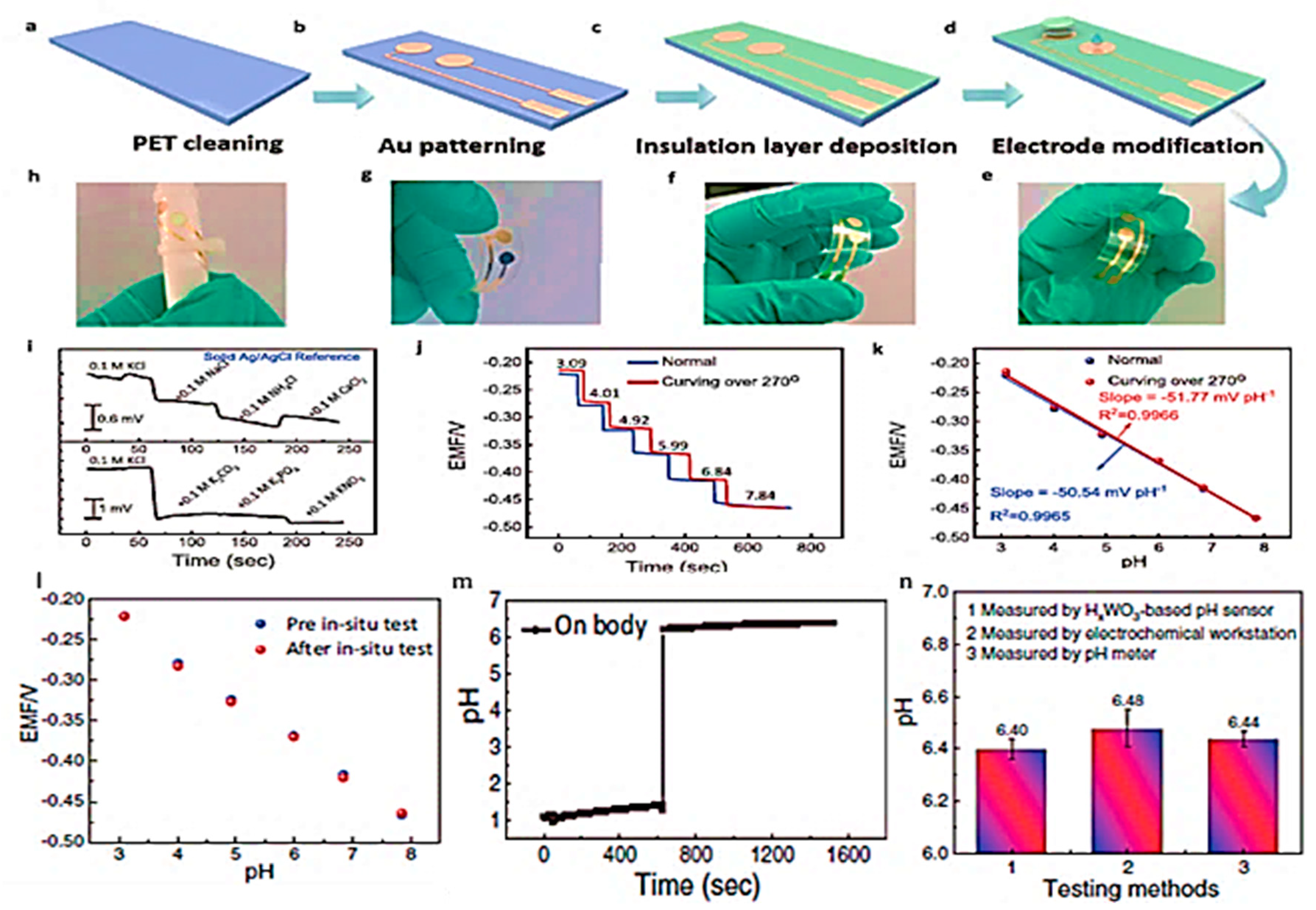
Sweat – is believed to be very useful biological matrix to obtain physiological information and pH is an important parameter to be monitored in sweat [47]. Therefore, wearable technology is rapidly adoptable area due to its promising features in sports health, healthcare and diagnostic applications. From last few years, enormous research is presented in healthcare to target pH along with some other vital parameters to monitor health indications [39,40,41]. For instance, Yitian Tang et al. [41] proposed organic materials – free pH wearable sensor-based on WO3 as an active material. They discovered and exploited the intercalation phenomenon between lattice H+ ions and WO3, which increased the ion-exchange capacity due to the monoclinic to cubic phasal.transitions. This phenomenon was further translated as pH measuring matrix. The typical electrochromic process led to the reduction of W6+ to W5+ subjected to the protons insertion, which turned WO3 into cubic hydrogen tungsten bronze (HxWO3), causing to increase in conductivity as well.
Then, HxWO3-based sensing matrix was used to fabricate miniaturized electrode and used as wearable sweat pH monitoring (Figure 12). As shown, the HxWO3-based matrix produced revocable pH responses vs. time in the pH range of ~ 1 to 11, where the electromotive force (EMF) signal decreased with the increase in pH values as WO3 is dependent on the double injection/extraction of electrons and proton to the structure. The developed sensor showed an excellent behavior towards pH sensing, when used in wearable configurations [41].
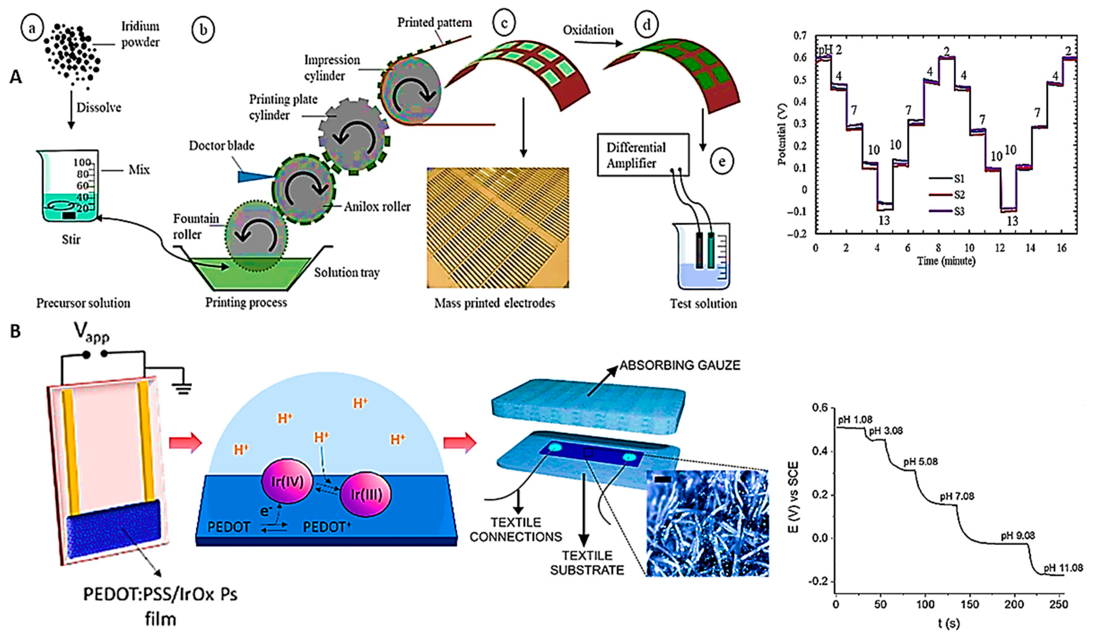
Composites of organic – inorganic materials are also a fruitful substrate choice for such modes of technology, where, textile chemical sensing is another wearable beneficiary in healthcare industry by collecting the vital parameters [122]. IrOx-based composites have been explored in many studies, because these NMs composites possess a high sensitivity to pH in terms of their electrochemistry. Moreover, PANI is used as positive exchange membrane for pH sensitive applications and thus, it provides sufficient analytical performance when composited with metal/metal-oxides. For instance, Federica and colleagues developed smart bandages for the real-time monitoring of wound pH, correlating it with the wound-healing[123]. They fabricated two-terminal pH-sensing devices with the help of polymer and IrO2 NPs, then integrating with the absorbent layer to ensure the continuous delivery of wound exudate flow across the sensor area. Potentiometric transduction against an open-circuit potential (OCP) provided a satisfactory data of 59 ± 4 μA pH−1. In further evaluations, it was found that there was a peak potential shift in Ir-III/Ir-IV redox couple, responsibly enabling the sensor for pH transduction. Later, they developed a two terminal solid-state electrode to elaborate the pH sensing mechanism. BY utilizing IrOx/ PEDOT:PSS electrochemistry, as acidic environment promoted the reduction of Ir-IV sites to Ir-III, causing the associated withdrawal of electrons from the PEDOT:PSS film, leading to current flow across the film. Thus, sensing layer was embedded in absorbent layer to construct a wound bandage, which successfully interpreted as pH range of 6-9, sufficiently monitoring wound with unaffected performance from potential interferences as well as workable in temperature range of 22 to 40 °C.
A flexible pH sensor was reported using IrOx as the sensing film deposited on a PET substrate in a roll-to-roll (R2R) process (Figure 13 (A)). The fabricated electrodes demonstrated a good linear range of pH sensing between 2-13 with a Nernstian response of −60.6 mVpH-1. Other characterizations such as hysteresis, drift, fluctuation, and deviation test were performed to evaluate the stability as well as the repeatability of IrOx-based pH sensor. Further, the fabricated electrodes presented a suitable data from pH 4 to 9 when tested against a synthetic sweat [124].
Similarly, PEDOT:PSS and IrOx NMs provided the pH sensing matrix due to the redox chemistry of IrOx-complex as it exhibited Ir-III/Ir-IV redox couple, which ultimately triggered PEDOT:PSS chain that extracted electrons from the polymeric (PEDOT:PSS) film, causing to current flow across the film (Figure 13 (B)). The developed sensor possessed flexible nature, where it was further fabricated as a fully textile pH sensor embedded in a bandage. Additionally, the sensing evaluations were carried out in simulated wound liquid (exudate) using dynamic flow analysis to validate its performance [125].
Various composites of organic/inorganic (NMs), metal/metal-oxide such as titanium dioxide or titania-carbon (TiO2-carbon), and 2D-NMs (graphene/MXenes/CNTs) play a pivotal role in advancing nanotechnology. Also, the blend of 2D-NMs with PANI retains potentials for pH sensing and capable enough to be adopted in wearable technology. Further doping or laser treatments of these NMs with organic moieties provide wearable gadgets with improved environmental stability, responsiveness and reversibility [47,126,127,128,129]. Hydrogel formulation of such above said composites is another facile approach to obtain the desired outcomes in wearable domain.
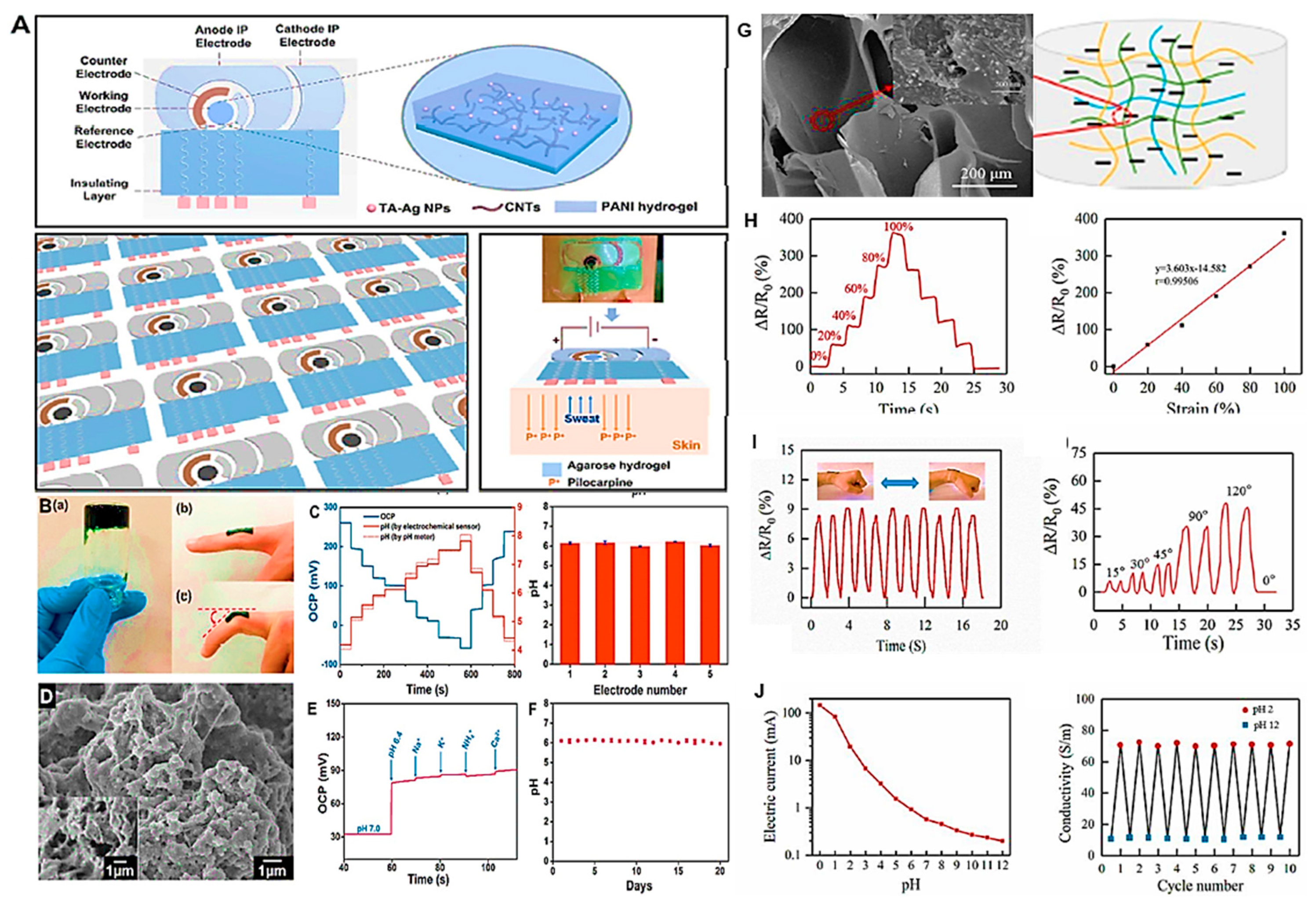
Recently [126], tannic acid-Ag-carbon nanotube-polyaniline (TA-Ag-CNT-PANI)-based hydrogel was used to prepare sweat sensing platform, and employed to detect noninvasively pH and tyrosine as shown in Figure 14 (A-E). Where, a pilocarpine-based iontophoretic system was also embedded into the sensing patch to stimulate sweat secretion from human body. Being sensitive and stable for pH and Tyrosine sensing, the integrated patch was capable enough to successfully detect both the analyzing analytes, simultaneously in sweat matrix. The detected pH results were further correlated to determine Tyr concentrations in various sweat samples, thus providing a reliable mechanism to evaluate the device performance in terms of concerned biomarkers.
It was concluded by the authors that the designed wearable sensing mechanism could be a great addition in healthcare applications.
In another study [127] multiwall carbon nanotubes (MWCNTs)-based hydrogel was prepared by compositing Poly(N-isopropylacrylamide) (PNIPAM), carboxymethyl chitosan (CMCS) and PANI (Figure 14 (F-I)). The resultant CMCS/ MWCNT/PANI hydrogel (F-inset is the magnified image) was thermosensitive as well as efficient in terms of pH sensing. CMCS and MWCNTs provided not only the mechanical properties but also with the better conductivity and temperature sensitivity (G-H). As shown in Figure 14 (I), the pH sensing was performed and the prepared hydrogel offered great response due to the outstanding reversible or doping/de-doping nature of PANI in the matrix, which is known as the best material for pH determinations. The increase in pH values produced doping effects in the PANI matrix and vice versa, thus provided an excellent performance of the hydrogel in terms of pH determinations.
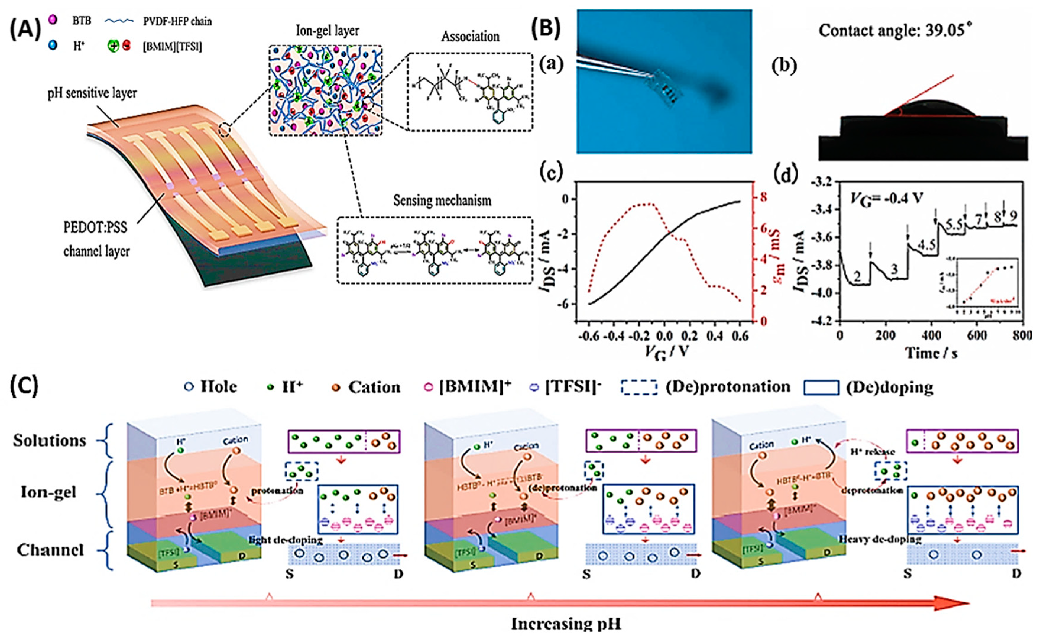
Recently, organic electrochemical transistors (OECTs)-based pH sensors also became the choice in cutting-edge technology owing to their promising capabilities, such as inherent signal amplification and miniaturization ability [130,131,132,133]. Moreover, the modifications with highly pH sensitive materials in gate and channel electrodes provide OECTs an edge to be more sensitive wearable device. However, the proper functioning of OECTs devices solely depend on the availability of enough electrolyte when using in wearable configurations. The probability of human skin not providing enough sweat for OEC working may lead to false results, which limit their broader applications. To mitigate this hinderance, researchers are exploiting the use if ion-gel. Figure 15 shows the pictorial representation of the developed (OECT-based flexible) pH sensor, where PEDOT:PSS was used to modify the gate electrode. The blend of bromothymol blue (BTB), polymeric network ([EMIM][TFSI]) comprising lithium salts (LiTFSI) [BMIM][TFSI]/PVDF-HFP were used to prepare pH sensitive materials for the channel modifications. Further, it was characterized employing electrochemical technique that anionic/ cationic transportation due to the [BMIM]+/[TFSI]-, respectively caused de-doping of the PEDOT:PSS, thus providing pH sensitivity of up to 91 mA dec at -0.4 V gate biasing; which was translated as the pH changes measurements from 2 to 9 [132].
Contrary to electrochemical and optical techniques, piezoelectric mass biosensors are another emerging class to monitor pH. As, the frequency of conducting materials can also be affected subject to pH change in sweat, which could be a facile way to analyze the pH in sweat. However, due to their rigidity, these devices are limited to lab-scale pH detection. Scarpa and colleagues developed a pH responsive sensor based on PEG-DA/CEA hydrogel, demonstrating swelling/shrinking capability at several pH corresponding to resonant frequency shift. The proposed device retained pH range 3–8 in artificial sweat formulation with satisfactory response with 12 kHz/pH unit. Such kinds of devices are rare and could have a great potential to be integrated in modest form, such as wearable devices for the desired applications [134].

5. Conclusion, challenges and future perspectives

We have comprehensively overviewed the pH regulation in physiological and cancerous environments, together with the intracellular and extracellular representative pH sensors in the review. Further, a detailed overview of trending pH sensing techniques, including optical, electrochemical, and their wearable variants are discussed in-vivo or in real life applications. The development of electrochemical sensors for pH sensing has been implicated since long, pioneering from the early 1900s to today’s high-tech configurations, including glass electrodes to ISFET. Similarly, the second most applicable technique, which has been conventionally explored for pH monitoring is optical technique. In current hi-tech era, the advances in nanotechnology due to the computational toolboxes have revolutionized the limits of existing fabrication methodologies of NMs for new applications. It has led the modern science to understand the complex atomic variations, which helped to improve the futuristic diagnostic plans in patients by the virtue of nanotechnology. This has led to the development of hi-tech NMs and thus, various synthetic strategies have been adopted so far to harvest many wearable nanoplatforms for different biological applications. Because, health nexus is the most important to retain the health of society and physiological monitoring can provide detailed information about health conditions at individual level. Therefore, over the times, blend of multidisciplinary toolboxes was employed to design and develop personalized health monitoring systems or miniaturized wearable sensing platforms, which are becoming higher priority for every individual now a day. Thus, great strides in the development of cutting-edge devices for biomedical applications have been made [92], especially, localized-pH monitoring and regulation in biological environment that persists in recent technologies that could open the new avenues in biomedical applications [48,135]

Challenges

Despite of great efforts in the field of nanotechnology to develop state-of-the-art wearable devices for pH monitoring, there are still some key challenges restricting their widespread clinical use. Though, wearable-gadgets possess great potential of being smart, rapid, non-destructive, and non-invasive biological pH monitoring tools, however they have some drawbacks as follow:
In terms of optical read outs, some of the key issues faced by absorptiometric pH devices are: (1) long-term stability of optical-based transduction systems is restricted due to photobleaching of the material corresponding to the temperature dependence. (2) While measuring pH of the analyte, the ionic strength of the sample may have variations, depending on the relation between dye concentrations and H+ activity, causing to generate false response. (3) One of the key challenges is in vivo measurements; performance degradation occurs during real biological samples analysis or even more badly, when they move from lab to bench-side analysis. (4) Scattering effects; can also play a vital role during pH read out and may cause to differ from the actual values. (5) Solubility in immobilization matrices; it may produce inadequate complexation between the probe and target analyte, thus can lead to leaching of material.
Overall, the trending wearable sensing devices face some of the key issues, including adhesion and conformity of the material to the skin, interferences, devices should prone to adopt external factors such as temperature and humidity, and they can counter or compensate these variations in the results, accordingly. The wearable devices should have the comfort level when they have been worn for a longer time. In case of sweat exploration for small molecule monitoring, the composition of these analytes changes over the course of time, hence altering the pH values as well as other electrolytes and biomarkers concentrations. So, there could be a chance for wearable sensors to produce false results while used against sweat analysis.

Future perspectives

As we are living in the era of modern sciences, where researchers have recognized the emergence of the modern sensing techniques by the virtue of nanotechnology, a variety of strategies has been developed to achieve desired goals. The existence of bionic polymeric materials retains many advantages as they are biocompatible and non-toxic, drawing the attention of scientific community to design more facile and novel wearable/ flexible systems. Though, the development of “intelligent-NMs” using modern synthetic approaches have made it possible to effectively achieve the desired outcomes, however, there is still a compelling need in developing such systems possessing fully automation and providing reliable information. This may be achieved by taking the advantages of organic molecules to be blended with excellent inorganic agents that can evenly in contact with the biomarker site to provide maximum effects.
Disruptive technologies like wearable, mHealth, and RWE have paved the way to make diagnostic systems more profound, accurate and reliable. As, these technologies provide administrative commands and sequels to monitor patients through electronic gadgets, e.g., mobile and tablets etc. The growing interests and demands in digital technologies (digi-techs.) providing a smart and effective platform in patients engagements, thus making Mobile health, electronic health records, and patient-generated data more reliable for wearable devices. At the moment, the involvement of digi-techs is about 60%, which will be increased in coming years to influence the performance of wearable tools. Moreover, the adaptation and utilization of such new high-tech brands, including, artificial Intelligence (AI) would be another essential part to take wearable gadgets up to more advanced level. The addition of AI is transforming every sphere of life and in future, it is predicted to be capable enough to fully leverage the wearable technology’s benefits through more R&D penetration, which is 25% at the moment in biomedical industries.

Author Contributions

Conceptualization, writing-original draft preparation, and editing, M.H.A; review, editing and visualization, M.A.H.N; Ning Liu, review, editing and visualization; Arsalan Ahmed Qureshi, review; Ying Li, visualization; Wenzhao Han, review and visualization; Yue Wang, review and visualization; Li Li, review and visualization; Yan Lv, review and visualization. Over all supervision, conceptualization, review editing, and funding acquisition C.Y. All authors have read and agreed to the published version of the manuscript.

Funding

This research was funded by the CAS – President's International Fellowship Initiative (PIFI) (2021PB0093), the Experimental Technology Project of Jilin University (SYXM2023b025), the Jilin Science and Technology Development Program (YDZJ202301ZYTS544, 20230402045GH, 20230402018GH, YDZJ202201ZYTS359), and the Jilin Province Development and Reform Commission (2023C041-8).

Institutional Review Board Statement

Not applicable.

Informed Consent Statement

Not applicable.

Data Availability Statement

Not applicable.

Conflicts of Interest

The authors declare no conflict of interest.

References

  1. Ghoneim, M.; Nguyen, A.; Dereje, N.; Huang, J.; Moore, G.; Murzynowski, P.; Dagdeviren, C. Recent progress in electrochemical pH-sensing materials and configurations for biomedical applications. Chemical Reviews 2019, 119, 5248–5297. [Google Scholar] [CrossRef] [PubMed]
  2. Khan, M.I.; Mukherjee, K.; Shoukat, R.; Dong, H. A review on pH sensitive materials for sensors and detection methods. Microsystem Technologies 2017, 23, 4391–4404. [Google Scholar] [CrossRef]
  3. Kraut, J.A.; Madias, N.E. Metabolic acidosis: pathophysiology, diagnosis and management. Nature Reviews Nephrology 2010, 6, 274–285. [Google Scholar] [CrossRef] [PubMed]
  4. Deitmer, J.W.; Theparambil, S.M.; Ruminot, I.; Noor, S.I.; Becker, H.M. Energy dynamics in the brain: contributions of astrocytes to metabolism and pH homeostasis. Frontiers in Neuroscience 2019, 13, 1301. [Google Scholar] [CrossRef] [PubMed]
  5. Swietach, P. What is pH regulation, and why do cancer cells need it? Cancer Metastasis Reviews 2019, 38, 5–15. [Google Scholar] [CrossRef] [PubMed]
  6. Hopkins, E., Sanvictores, T., Sharma, S.. Physiology, acid base balance. 2022.
  7. White, K.A.; Grillo-Hill, B.K.; Barber, D.L. Cancer cell behaviors mediated by dysregulated pH dynamics at a glance. Journal of Cell Science 2017, 130, 663–669. [Google Scholar] [CrossRef]
  8. Casey, J.R.; Grinstein, S.; Orlowski, J. Sensors and regulators of intracellular pH. Nature Reviews Molecular Cell Biology 2010, 11, 50–61. [Google Scholar] [CrossRef]
  9. Magder, S.; Magder, A.; Samoukovic, G. Intracellular pH regulation and the acid delusion. Canadian Journal of Physiology and Pharmacology 2021, 99, 561–576. [Google Scholar] [CrossRef]
  10. Gatenby, R.A.; Gillies, R.J. Why do cancers have high aerobic glycolysis? Nature Reviews Cancer 2004, 4, 891–899. [Google Scholar] [CrossRef]
  11. Damaghi, M.; Wojtkowiak, J.W.; Gillies, R.J. pH sensing and regulation in cancer. Frontiers in Physiology 2013, 4, 370. [Google Scholar] [CrossRef]
  12. Steinegger, A.; Wolfbeis, O.S.; Borisov, S.M. Optical sensing and imaging of pH values: Spectroscopies, materials, and applications. Chemical Reviews 2020, 120, 12357–12489. [Google Scholar] [CrossRef] [PubMed]
  13. Wang, S.; Fan, Y.; Li, D.; Sun, C.; Lei, Z.; Lu, L.; Wang, T.; Zhang, F. Anti-quenching NIR-II molecular fluorophores for in vivo high-contrast imaging and pH sensing. Nature Communications 2019, 10, 1058. [Google Scholar] [CrossRef] [PubMed]
  14. Wanas, W.; Abd El-Kaream, S.A.; Ebrahim, S.; Soliman, M.; Karim, M. Cancer bioimaging using dual mode luminescence of graphene/FA-ZnO nanocomposite based on novel green technique. Scientific Reports 2023, 13, 27. [Google Scholar] [CrossRef] [PubMed]
  15. Hou, J.-T.; Ren, W.X.; Li, K.; Seo, J.; Sharma, A.; Yu, X.-Q.; Kim, J.S. Fluorescent bioimaging of pH: from design to applications. Chemical Society Reviews 2017, 46, 2076–2090. [Google Scholar] [CrossRef]
  16. Bakker, E. Wearable sensors. ACS Sensors 2023, 8, 1368–1370. [Google Scholar] [CrossRef]
  17. Bariya, M.; Shahpar, Z.; Park, H.; Sun, J.; Jung, Y.; Gao, W.; Nyein, H.Y.Y.; Liaw, T.S.; Tai, L.-C.; Ngo, Q.P. Roll-to-roll gravure printed electrochemical sensors for wearable and medical devices. ACS Nano 2018, 12, 6978–6987. [Google Scholar] [CrossRef]
  18. Zhao, H.; Su, R.; Teng, L.; Tian, Q.; Han, F.; Li, H.; Cao, Z.; Xie, R.; Li, G.; Liu, X. Recent advances in flexible and wearable sensors for monitoring chemical molecules. Nanoscale 2022, 14, 1653–1669. [Google Scholar] [CrossRef]
  19. Min, J.; Tu, J.; Xu, C.; Lukas, H.; Shin, S.; Yang, Y.; Solomon, S.A.; Mukasa, D.; Gao, W. Skin-interfaced wearable sweat sensors for precision medicine. Chemical Reviews 2023, 123, 5049–5138. [Google Scholar] [CrossRef]
  20. Di Costanzo, L.; Panunzi, B. Visual pH sensors: from a chemical perspective to new bioengineered materials. Molecules 2021, 26, 2952. [Google Scholar] [CrossRef]
  21. Venketeswaran, A.; Lalam, N.; Wuenschell, J.; Ohodnicki Jr, P.R.; Badar, M.; Chen, K.P.; Lu, P.; Duan, Y.; Chorpening, B.; Buric, M. Recent advances in machine learning for fiber optic sensor applications. Advanced Intelligent Systems 2022, 4, 2100067. [Google Scholar] [CrossRef]
  22. Zhao, M.; Wang, J.; Lei, Z.; Lu, L.; Wang, S.; Zhang, H.; Li, B.; Zhang, F. NIR-II pH sensor with a FRET adjustable transition point for in situ dynamic tumor microenvironment visualization. Angewandte Chemie International Edition 2021, 60, 5091–5095. [Google Scholar] [CrossRef]
  23. Gond, S.; Yadav, P.; Chauhan, B.S.; Srikrishna, S.; Singh, V.P. Development of an ‘OFF-ON-OFF’colorimetric and fluorometric pH sensor for the study of physiological pH and its bioimaging application. Journal of Molecular Structure 2022, 1252, 132147. [Google Scholar] [CrossRef]
  24. Werner, J.; Belz, M.; Klein, K.-F.; Sun, T.; Grattan, K. Characterization of a fast response fiber-optic pH sensor and illustration in a biological application. Analyst 2021, 146, 4811–4821. [Google Scholar] [CrossRef]
  25. Krivenkov, V.; Samokhvalov, P.; Nabiev, I.; Rakovich, Y.P. pH-Sensing platform based on light–matter coupling in colloidal complexes of silver nanoplates and J-aggregates. The Journal of Physical Chemistry C 2021, 125, 1972–1979. [Google Scholar] [CrossRef]
  26. Wencel, D.; Kaworek, A.; Abel, T.; Efremov, V.; Bradford, A.; Carthy, D.; Coady, G.; McMorrow, R.C.; McDonagh, C. Optical sensor for real-time pH monitoring in human tissue. Small 2018, 14, 1803627. [Google Scholar] [CrossRef]
  27. Kurniawan, D.; Chiang, W.-H. Microplasma-enabled colloidal nitrogen-doped graphene quantum dots for broad-range fluorescent pH sensors. Carbon 2020, 167, 675–684. [Google Scholar] [CrossRef]
  28. Laysandra, L.; Kurniawan, D.; Wang, C.-L.; Chiang, W.-H.; Chiu, Y.-C. Synergistic effect in a graphene quantum dot-enabled luminescent skinlike copolymer for long-term pH detection. ACS Applied Materials Interfaces 2021, 13, 60413–60424. [Google Scholar] [CrossRef] [PubMed]
  29. Chou, S.-C.; Hsieh, Y.-C.; Cheang, W.-H.; Sun, B.-Y.; Chu, C.-Y.; Chen, S.-Y.; Chiao, J.-C.; Wu, P.-W. A flexible IrO2 membrane for pH sensing. Scientific Reports 2022, 12, 11712. [Google Scholar] [CrossRef]
  30. Li, S.; Ferrer-Ruiz, A.; Dai, J.; Ramos-Soriano, J.; Du, X.; Zhu, M.; Zhang, W.; Wang, Y.; Herranz, M.Á.; Jing, L. A pH-independent electrochemical aptamer-based biosensor supports quantitative, real-time measurement in vivo. Chemical Science 2022, 13, 8813–8820. [Google Scholar] [CrossRef]
  31. Booth, M.A.; Gowers, S.A.; Hersey, M.; Samper, I.C.; Park, S.; Anikeeva, P.; Hashemi, P.; Stevens, M.M.; Boutelle, M.G. Fiber-based electrochemical biosensors for monitoring pH and transient neurometabolic lactate. Analytical Chemistry 2021, 93, 6646–6655. [Google Scholar] [CrossRef]
  32. Yoon, J.H.; Kim, S.-M.; Park, H.J.; Kim, Y.K.; Oh, D.X.; Cho, H.-W.; Lee, K.G.; Hwang, S.Y.; Park, J.; Choi, B.G. Highly self-healable and flexible cable-type pH sensors for real-time monitoring of human fluids. Biosensors and Bioelectronics 2020, 150, 111946. [Google Scholar] [CrossRef]
  33. Zamora, M.L.; Domínguez, J.M.; Trujillo, R.M.; Goy, C.B.; Sanchez, M.A.; Madrid, R.E. Potentiometric textile-based pH sensor. Sensors Actuators B: Chemical 2018, 260, 601–608. [Google Scholar] [CrossRef]
  34. Zea, M.; Texidó, R.; Villa, R.; Borrós, S.; Gabriel, G. Specially designed polyaniline/polypyrrole ink for a fully printed highly sensitive pH microsensor. ACS Applied Materials Interfaces 2021, 13, 33524–33535. [Google Scholar] [CrossRef] [PubMed]
  35. García-Guzmán, J.J.; Pérez-Ràfols, C.; Cuartero, M.; Crespo, G.A. Toward in vivo transdermal pH sensing with a validated microneedle membrane electrode. ACS Sensors 2021, 6, 1129–1137. [Google Scholar] [CrossRef]
  36. Han, S.T.; Peng, H.; Sun, Q.; Venkatesh, S.; Chung, K.S.; Lau, S.C.; Zhou, Y.; Roy, V. An overview of the development of flexible sensors. Advanced Materials 2017, 29, 1700375. [Google Scholar] [CrossRef] [PubMed]
  37. He, T.; Wen, F.; Yang, Y.; Le, X.; Liu, W.; Lee, C. Emerging wearable chemical sensors enabling advanced integrated systems toward personalized and preventive medicine. Analytical Chemistry 2023, 95, 490–514. [Google Scholar] [CrossRef]
  38. Sempionatto, J.R.; Jeerapan, I.; Krishnan, S.; Wang, J. Wearable chemical sensors: emerging systems for on-body analytical chemistry. Analytical Chemistry 2019, 92, 378–396. [Google Scholar] [CrossRef]
  39. Bi, Y.; Sun, M.; Wang, J.; Zhu, Z.; Bai, J.; Emran, M.Y.; Kotb, A.; Bo, X.; Zhou, M. Universal fully integrated wearable sensor arrays for the multiple electrolyte and metabolite monitoring in raw sweat, saliva, or urine. Analytical Chemistry 2023, 95, 6690–6699. [Google Scholar] [CrossRef]
  40. Lin, K.; Xie, J.; Bao, Y.; Ma, Y.; Chen, L.; Wang, H.; Xu, L.; Tang, Y.; Liu, Z.; Sun, Z. Self-adhesive and printable tannin–graphene supramolecular aggregates for wearable potentiometric pH sensing. Electrochemistry Communications 2022, 137, 107261. [Google Scholar] [CrossRef]
  41. Tang, Y.; Gan, S.; Zhong, L.; Sun, Z.; Xu, L.; Liao, C.; Lin, K.; Cui, X.; He, D.; Ma, Y. Lattice proton intercalation to regulate WO3-based solid-contact wearable pH sensor for sweat analysis. Advanced Functional Materials 2022, 32, 2107653. [Google Scholar] [CrossRef]
  42. Ates, H.C.; Nguyen, P.Q.; Gonzalez-Macia, L.; Morales-Narváez, E.; Güder, F.; Collins, J.J.; Dincer, C. End-to-end design of wearable sensors. Nature Reviews Materials 2022, 7, 887–907. [Google Scholar] [CrossRef]
  43. Liu, W.; Xie, R.; Zhu, J.; Wu, J.; Hui, J.; Zheng, X.; Huo, F.; Fan, D. A temperature responsive adhesive hydrogel for fabrication of flexible electronic sensors. npj Flexible Electronics 2022, 6, 68. [Google Scholar] [CrossRef]
  44. Xu, G.; Lu, Y.; Cheng, C.; Li, X.; Xu, J.; Liu, Z.; Liu, J.; Liu, G.; Shi, Z.; Chen, Z. Battery-free and wireless smart wound dressing for wound infection monitoring and electrically controlled on-demand drug delivery. Advanced Functional Materials 2021, 31, 2100852. [Google Scholar] [CrossRef]
  45. Mostafalu, P.; Tamayol, A.; Rahimi, R.; Ochoa, M.; Khalilpour, A.; Kiaee, G.; Yazdi, I.K.; Bagherifard, S.; Dokmeci, M.R.; Ziaie, B. Smart bandage for monitoring and treatment of chronic wounds. Small 2018, 14, 1703509. [Google Scholar] [CrossRef]
  46. Shirzaei Sani, E.; Xu, C.; Wang, C.; Song, Y.; Min, J.; Tu, J.; Solomon, S.A.; Li, J.; Banks, J.L.; Armstrong, D.G. A stretchable wireless wearable bioelectronic system for multiplexed monitoring and combination treatment of infected chronic wounds. Science Advances 2023, 9, eadf7388. [Google Scholar] [CrossRef] [PubMed]
  47. Sharifuzzaman, M.; Zahed, M.A.; Reza, M.S.; Asaduzzaman, M.; Jeong, S.; Song, H.; Kim, D.K.; Zhang, S.; Park, J.Y. MXene/fluoropolymer-derived laser-carbonaceous all-fibrous nanohybrid patch for soft wearable bioelectronics. Advanced Functional Materials 2023, 2208894. [Google Scholar] [CrossRef]
  48. Jung, H.S.; Jung, W.-B.; Wang, J.; Abbott, J.; Horgan, A.; Fournier, M.; Hinton, H.; Hwang, Y.-H.; Godron, X.; Nicol, R. CMOS electrochemical pH localizer-imager. Science Advances 2022, 8, eabm6815. [Google Scholar] [CrossRef]
  49. Saladin, K.S.; Porth, C. Anatomy & physiology: the unity of form and function; McGraw-Hill: New York, 2010; Volume 5. [Google Scholar]
  50. Gaohua, L.; Miao, X.; Dou, L. Crosstalk of physiological pH and chemical pKa under the umbrella of physiologically based pharmacokinetic modeling of drug absorption, distribution, metabolism, excretion, and toxicity. Expert Opinion on Drug Metabolism Toxicology 2021, 17, 1103–1124. [Google Scholar] [CrossRef]
  51. Schwalfenberg, G.K. The alkaline diet: is there evidence that an alkaline pH diet benefits health? Journal of Environmental Public Health 2012, 2012. [Google Scholar] [CrossRef]
  52. Gillies, R.J. Cancer heterogeneity and metastasis: Life at the edge. Clinical Experimental Metastasis 2022, 39, 15–19. [Google Scholar] [CrossRef]
  53. Czowski, B.J.; Romero-Moreno, R.; Trull, K.J.; White, K.A. Cancer and pH dynamics: Transcriptional regulation, proteostasis, and the need for new molecular tools. Cancers 2020, 12, 2760. [Google Scholar] [CrossRef]
  54. Qi, Q.; Fox, M.S.; Lim, H.; Sullivan, R.; Li, A.; Bellyou, M.; Desjardins, L.; McClennan, A.; Bartha, R.; Hoffman, L. Glucose infusion induced change in intracellular pH and its relationship with tumor glycolysis in a C6 rat model of glioblastoma. Molecular Imaging Biology 2023, 25, 271–282. [Google Scholar] [CrossRef]
  55. Kovács, A.; Kis, N.; Budai-Szűcs, M.; Gácsi, A.; Csányi, E.; Csóka, I.; Berkó, S. QbD-based investigation of dermal semisolid in situ film-forming systems for local anaesthesia. Drug Design, Development Therapy 2020, 5059–5076. [Google Scholar] [CrossRef]
  56. Osei-Owusu, J.; Kots, E.; Ruan, Z.; Mihaljević, L.; Chen, K.H.; Tamhaney, A.; Ye, X.; Lü, W.; Weinstein, H.; Qiu, Z. Molecular determinants of pH sensing in the proton-activated chloride channel. Proceedings of the National Academy of Sciences 2022, 119, e2200727119. [Google Scholar] [CrossRef] [PubMed]
  57. Zhang, S.-Z.; Xie, L.; Shang, Z.-J. Burden of oral cancer on the 10 most populous countries from 1990 to 2019: Estimates from the Global Burden of Disease study 2019. International Journal of Environmental Research Public Health 2022, 19, 875. [Google Scholar] [CrossRef]
  58. Albatany, M.; Ostapchenko, V.G.; Meakin, S.; Bartha, R. Brain tumor acidification using drugs simultaneously targeting multiple pH regulatory mechanisms. Journal of Neuro-Oncology 2019, 144, 453–462. [Google Scholar] [CrossRef] [PubMed]
  59. Ibrahim-Hashim, A.; Estrella, V. Acidosis and cancer: from mechanism to neutralization. Cancer Metastasis Reviews 2019, 38, 149–155. [Google Scholar] [CrossRef]
  60. Ye, X.; Xiang, Y.; Wang, Q.; Li, Z.; Liu, Z. A red emissive two-photon fluorescence probe based on carbon dots for intracellular pH detection. Small 2019, 15, 1901673. [Google Scholar] [CrossRef]
  61. Chen, Q.; Zhai, J.; Li, J.; Wang, Y.; Xie, X. Exploring ratiometric endolysosomal pH nanosensors with hydrophobic indicators responding at the nanoscale interface and multiple fluorescence resonance energy transfers. Nano Research 2022, 1–8. [Google Scholar] [CrossRef]
  62. Shen, J.; Zeng, Y.; Zhuang, X.; Sun, L.; Yao, X.; Pimpl, P.; Jiang, L. Organelle pH in the arabidopsis endomembrane system. Molecular plant 2013, 6, 1419–1437. [Google Scholar] [CrossRef] [PubMed]
  63. Donahue, C.E.; Siroky, M.D.; White, K.A. An optogenetic tool to raise intracellular pH in single cells and drive localized membrane dynamics. Journal of the American Chemical Society 2021, 143, 18877–18887. [Google Scholar] [CrossRef]
  64. Loiselle, F.B.; Casey, J.R. Measurement of intracellular pH; 2010; pp. 311-331.
  65. Dong, B.; Du, S.; Wang, C.; Fu, H.; Li, Q.; Xiao, N.; Yang, J.; Xue, X.; Cai, W.; Liu, D. Reversible self-assembly of nanoprobes in live cells for dynamic intracellular pH imaging. ACS Nano 2019, 13, 1421–1432. [Google Scholar] [CrossRef] [PubMed]
  66. Igarashi, T.; Kim, H.; Sun, P.Z. Detection of tissue pH with quantitative chemical exchange saturation transfer magnetic resonance imaging. NMR in Biomedicine 2023, 36, e4711. [Google Scholar] [CrossRef]
  67. Boyd, P.S.; Breitling, J.; Korzowski, A.; Zaiss, M.; Franke, V.L.; Mueller-Decker, K.; Glinka, A.; Ladd, M.E.; Bachert, P.; Goerke, S. Mapping intracellular pH in tumors using amide and guanidyl CEST-MRI at 9. 4 T. Magnetic Resonance in Medicine 2022, 87, 2436–2452. [Google Scholar] [CrossRef]
  68. Singh, M. Synthesis of two-dimensional metal, metal oxide and metal hydroxide nanomaterials for biosensing. In Nanozymes in Medicine; Springer, 2023; pp. 161–185. [Google Scholar]
  69. Gu, B.; Zhang, Q. Recent advances on functionalized upconversion nanoparticles for detection of small molecules and ions in biosystems. Advanced Science 2018, 5, 1700609. [Google Scholar] [CrossRef]
  70. Zhang, X.; Chen, J.; Hu, J.; Du Rietz, A.; Wu, X.; Zhang, R.; Zhang, Z.; Uvdal, K.; Hu, Z. Single-wavelength-excited fluorogenic nanoprobe for accurate realtime ratiometric analysis of broad pH fluctuations in mitophagy. Nano Research 2022, 15, 6515–6521. [Google Scholar] [CrossRef]
  71. Wu, Q.; Fu, S.; Xiao, H.; Du, J.; Cheng, F.; Wan, S.; Zhu, H.; Li, D.; Peng, F.; Ding, X. Advances in extracellular vesicle nanotechnology for precision theranostics. Advanced Science 2023, 10, 2204814. [Google Scholar] [CrossRef]
  72. Inobeme, A.; Mathew, J.T.; Adetunji, C.O.; Ajai, A.I.; Inobeme, J.; Maliki, M.; Okonkwo, S.; Adekoya, M.A.; Bamigboye, M.O.; Jacob, J.O. Recent advances in nanotechnology for remediation of heavy metals. Environmental Monitoring Assessment 2023, 195, 111. [Google Scholar] [CrossRef]
  73. Sahoo, M.; Vishwakarma, S.; Panigrahi, C.; Kumar, J. Nanotechnology: Current applications and future scope in food. Food Frontiers 2021, 2, 3–22. [Google Scholar] [CrossRef]
  74. Sim, S.; Wong, N.K. Nanotechnology and its use in imaging and drug delivery. Biomedical Reports 2021, 14, 1–9. [Google Scholar] [CrossRef] [PubMed]
  75. Tran, P.; Lee, S.-E.; Kim, D.-H.; Pyo, Y.-C.; Park, J.-S. Recent advances of nanotechnology for the delivery of anticancer drugs for breast cancer treatment. Journal of Pharmaceutical Investigation 2020, 50, 261–270. [Google Scholar] [CrossRef]
  76. Zhang, G.; Han, W.; Zhao, P.; Wang, Z.; Li, M.; Sui, X.; Liu, Y.; Tian, B.; He, Z.; Fu, Q. Programmed pH-responsive core–shell nanoparticles for precisely targeted therapy of ulcerative colitis. Nanoscale 2023, 15, 1937–1946. [Google Scholar] [CrossRef] [PubMed]
  77. Qin, Y.; Alam, A.U.; Howlader, M.M.; Hu, N.X.; Deen, M.J. Inkjet printing of a highly loaded palladium ink for integrated, low-cost pH sensors. Advanced Functional Materials 2016, 26, 4923–4933. [Google Scholar] [CrossRef]
  78. Erdem, Ö.; Derin, E.; Zeibi Shirejini, S.; Sagdic, K.; Yilmaz, E.G.; Yildiz, S.; Akceoglu, G.A.; Inci, F. Carbon-based nanomaterials and sensing tools for wearable health monitoring devices. Advanced Materials Technologies 2022, 7, 2100572. [Google Scholar] [CrossRef]
  79. Wang, F.-T.; Wang, L.-N.; Xu, J.; Huang, K.-J.; Wu, X. Synthesis and modification of carbon dots for advanced biosensing application. Analyst 2021, 146, 4418–4435. [Google Scholar] [CrossRef] [PubMed]
  80. Yang, G.; Liu, F.; Zhao, J.; Fu, L.; Gu, Y.; Qu, L.; Zhu, C.; Zhu, J.-J.; Lin, Y. MXenes-based nanomaterials for biosensing and biomedicine. Coordination Chemistry Reviews 2023, 479, 215002. [Google Scholar] [CrossRef]
  81. NajafiKhoshnoo, S.; Kim, T.; Tavares-Negrete, J.A.; Pei, X.; Das, P.; Lee, S.W.; Rajendran, J.; Esfandyarpour, R. A 3D Nanomaterials-Printed Wearable, Battery-Free, Biocompatible, Flexible, and Wireless pH Sensor System for Real-Time Health Monitoring. Advanced Materials Technologies 2023, 8, 2201655. [Google Scholar] [CrossRef]
  82. Wang, K.; Liu, X.; Zhao, Z.; Li, L.; Tong, J.; Shang, Q.; Liu, Y.; Zhang, Z. Carbon nanotube field-effect transistor based pH sensors. Carbon 2023, 205, 540–545. [Google Scholar] [CrossRef]
  83. Zhang, R.; Gao, J.; Zhao, G.; Zhou, L.; Kong, F.; Jiang, T.; Jiang, H. Tetrazine bioorthogonal chemistry makes nanotechnology a powerful toolbox for biological applications. Nanoscale 2023, 15, 461–469. [Google Scholar] [CrossRef] [PubMed]
  84. Khademi, R.; Mohammadi, Z.; Khademi, R.; Saghazadeh, A.; Rezaei, N. Nanotechnology-based diagnostics and therapeutics in acute lymphoblastic leukemia: a systematic review of preclinical studies. Nanoscale Advances 2023. [CrossRef]
  85. Zhao, C.; Kang, J.; Li, Y.; Wang, Y.; Tang, X.; Jiang, Z. Carbon-based stimuli-responsive nanomaterials: classification and application. Cyborg Bionic Systems 2023, 4, 0022. [Google Scholar] [CrossRef]
  86. Li, Z.; Liu, W.; Sun, C.; Wei, X.; Liu, S.; Jiang, Y. Gastrointestinal pH-Sensitive Pickering Emulsions Stabilized by Zein Nanoparticles Coated with Bioactive Glycyrrhizic Acid for Improving Oral Bioaccessibility of Curcumin. ACS Applied Materials Interfaces 2023, 15, 14678–14689. [Google Scholar] [CrossRef] [PubMed]
  87. Patel, T.; Itoo, A.M.; Paul, M.; Kondapaneni, L.P.; Ghosh, B.; Biswas, S. Block HPMA-based pH-sensitive gemcitabine pro-drug nanoaggregates for cancer treatment. European Polymer Journal 2023, 186, 111843. [Google Scholar] [CrossRef]
  88. Manjakkal, L.; Dervin, S.; Dahiya, R. Flexible potentiometric pH sensors for wearable systems. RSC Advances 2020, 10, 8594–8617. [Google Scholar] [CrossRef] [PubMed]
  89. Ma, X.; Peng, R.; Mao, W.; Lin, Y.; Yu, H. Recent advances in ion-sensitive field-effect transistors for biosensing applications. Electrochemical Science Advances 2022, e2100163. [Google Scholar] [CrossRef]
  90. Kim, Y.-U.; Cho, W.-J. Self-sensitivity amplifiable dual-gate ion-sensitive field-effect transistor based on a high-k engineered dielectric layer. Japanese Journal of Applied Physics 2023, 62, SC1056. [Google Scholar] [CrossRef]
  91. Chen, H.J.; Lee, T.N.; Tseng, S.-L.; Chen, S.-Z.; Chiu, P.-W. Characterizations of ion-sensitive field-effect transistors with silicon wire array channels and stack-sensing membrane. Journal of The Electrochemical Society 2022, 169, 037511. [Google Scholar] [CrossRef]
  92. Ren, H.; Liang, K.; Li, D.; Chen, Y.; Tang, Y.; Wang, Y.; Li, F.; Liu, G.; Zhu, B. Field-effect transistor-based biosensor for pH sensing and mapping. Advanced Sensor Research 2023, 2200098. [Google Scholar] [CrossRef]
  93. Hsieh, C.-Y.; Huang, N.-T. A proton-selective membrane (PSM)-deposited dual-gate ion-sensitive field-effect transistor (DG-ISFET) integrating a microchamber-embedded filter membrane for bacterial enrichment and antimicrobial susceptibility test. Sensors Actuators B: Chemical 2022, 359, 131580. [Google Scholar] [CrossRef]
  94. Pan, T.-M.; Lin, C.-H.; Pang, S.-T.J.J.o.A.; Compounds. Structural properties and sensing performance of TaOx/Ta stacked sensing films for extended-gate field-effect transistor pH sensors. Journal of Alloys Compounds 2022, 903, 163955. [Google Scholar] [CrossRef]
  95. Juang, D.S.; Lin, C.-H.; Huo, Y.-R.; Tang, C.-Y.; Cheng, C.-R.; Wu, H.-S.; Huang, S.-F.; Kalnitsky, A.; Lin, C.-C. Proton-ELISA: Electrochemical immunoassay on a dual-gated ISFET array. Biosensors and Bioelectronics 2018, 117, 175–182. [Google Scholar] [CrossRef]
  96. Tabata, M.; Khamhanglit, C.; Kotaki, S.; Miyahara, Y. Detection of cell membrane proteins using ion-sensitive field effect transistors combined with chemical signal amplification. Chemical Communications 2022, 58, 7368–7371. [Google Scholar] [CrossRef]
  97. Zhou, J.-X.; Ding, F.; Tang, L.-N.; Li, T.; Li, Y.-H.; Zhang, Y.-J.; Gong, H.-Y.; Li, Y.-T.; Zhang, G.-J. Monitoring of pH changes in a live rat brain with MoS2/PAN functionalized microneedles. Analyst 2018, 143, 4469–4475. [Google Scholar] [CrossRef]
  98. Du, W.; Shen, Z.; Liang, Y.; Gong, S.; Meng, Z.; Li, M.; Wang, Z.; Wang, S. A highly effective “naked eye” colorimetric and fluorimetric curcumin-based fluorescent sensor for specific and sensitive detection of H2 O2 in vivo and in vitro. Analyst 2023, 148, 1824–1837. [Google Scholar] [CrossRef]
  99. Ye, X.; Wang, A.; Zhang, D.; Zhou, P.; Zhu, P. Light and pH dual-responsive spiropyran-based cellulose nanocrystals. RSC Advances 2023, 13, 11495–11502. [Google Scholar] [CrossRef] [PubMed]
  100. Kenney, R.M.; Boyce, M.W.; Whitman, N.A.; Kromhout, B.P.; Lockett, M.R. A pH-sensing optode for mapping spatiotemporal gradients in 3D paper-based cell cultures. Analytical Chemistry 2018, 90, 2376–2383. [Google Scholar] [CrossRef]
  101. Petrunina, N.A.; Shtork, A.S.; Lukina, M.M.; Tsvetkov, V.B.; Khodarovich, Y.M.; Feofanov, A.V.; Moysenovich, A.M.; Maksimov, E.G.; Shipunova, V.O.; Zatsepin, T.S. Ratiometric i-motif-based sensor for precise long-term monitoring of pH micro alterations in the nucleoplasm and interchromatin granules. ACS Sensors 2023, 8, 619–629. [Google Scholar] [CrossRef] [PubMed]
  102. Lesani, P.; Singh, G.; Lu, Z.; Mirkhalaf, M.; New, E.J.; Zreiqat, H. Two-photon ratiometric carbon dot-based probe for real-time intracellular pH monitoring in 3D environment. Chemical Engineering Journal 2022, 433, 133668. [Google Scholar] [CrossRef]
  103. Koide, Y.; Kojima, R.; Hanaoka, K.; Numasawa, K.; Komatsu, T.; Nagano, T.; Kobayashi, H.; Urano, Y. Design strategy for germanium-rhodamine based pH-activatable near-infrared fluorescence probes suitable for biological applications. Communications Chemistry 2019, 2, 94. [Google Scholar] [CrossRef]
  104. Kulkarni, C.A.; Fink, B.D.; Gibbs, B.E.; Chheda, P.R.; Wu, M.; Sivitz, W.I.; Kerns, R.J. A novel triphenylphosphonium carrier to target mitochondria without uncoupling oxidative phosphorylation. Journal of Medicinal Chemistry 2021, 64, 662–676. [Google Scholar] [CrossRef] [PubMed]
  105. Zielonka, J.; Joseph, J.; Sikora, A.; Hardy, M.; Ouari, O.; Vasquez-Vivar, J.; Cheng, G.; Lopez, M.; Kalyanaraman, B. Mitochondria-targeted triphenylphosphonium-based compounds: syntheses, mechanisms of action, and therapeutic and diagnostic applications. Chemical Reviews 2017, 117, 10043–10120. [Google Scholar] [CrossRef]
  106. Zhang, X.; Gu, Y.; Zhang, Y.; Yu, G.-Y.; Liao, Z.-P.; Wu, H.-F.; Shi, C.-G. Ratiometric intracellular pH sensors based on nitrogen-doped graphene oxide quantum dots. Heliyon 2022, 8. [Google Scholar] [CrossRef] [PubMed]
  107. Liu, H.; Li, H.; Xia, S.; Yu, S.; Duan, Y.; Wang, L.; Zhu, H.; He, H. Design of a cellulose nanocrystal-based upconversion ratiometric fluorescent nanoprobe for pH monitoring and imaging. Chemical Engineering Journal 2023, 454, 140456. [Google Scholar] [CrossRef]
  108. Xue, X.-L.; Wang, Y.; Chen, S.; Wang, K.-P.; Niu, S.-Y.; Zong, Q.-S.; Jiang, Y.; Hu, Z.-Q. Monitoring intracellular pH using a hemicyanine-based ratiometric fluorescent probe. Spectrochimica Acta Part A: Molecular Biomolecular Spectroscopy 2023, 284, 121778. [Google Scholar] [CrossRef] [PubMed]
  109. Song, G.; Jiang, D.; Wang, L.; Sun, X.; Liu, H.; Tian, Y.; Chen, M. A series of simple curcumin-derived colorimetric and fluorescent probes for ratiometric-pH sensing and cell imaging. Chinese Chemical Letters 2022, 33, 339–343. [Google Scholar] [CrossRef]
  110. Wang, X.-d.; Wolfbeis, O.S. Fiber-optic chemical sensors and biosensors (2015–2019). Analytical Chemistry 2019, 92, 397–430. [Google Scholar] [CrossRef]
  111. Zhou, J.; Ren, Y.; Nie, Y.; Jin, C.; Park, J.; Zhang, J.X. Dual fluorescent hollow silica nanofibers for in situ pH monitoring using an optical fiber. Nanoscale Advances 2023, 5, 2180–2189. [Google Scholar] [CrossRef]
  112. Corsi, M.; Paghi, A.; Mariani, S.; Golinelli, G.; Debrassi, A.; Egri, G.; Leo, G.; Vandini, E.; Vilella, A.; Dähne, L. Bioresorbable nanostructured chemical sensor for monitoring of pH level In vivo. Advanced Science 2022, 9, 2202062. [Google Scholar] [CrossRef]
  113. Lee, H.; Choi, T.K.; Lee, Y.B.; Cho, H.R.; Ghaffari, R.; Wang, L.; Choi, H.J.; Chung, T.D.; Lu, N.; Hyeon, T. A graphene-based electrochemical device with thermoresponsive microneedles for diabetes monitoring and therapy. Nature Nanotechnology 2016, 11, 566–572. [Google Scholar] [CrossRef]
  114. Nyein, H.Y.Y.; Gao, W.; Shahpar, Z.; Emaminejad, S.; Challa, S.; Chen, K.; Fahad, H.M.; Tai, L.-C.; Ota, H.; Davis, R.W. A wearable electrochemical platform for noninvasive simultaneous monitoring of Ca2+ and pH. ACS Nano 2016, 10, 7216–7224. [Google Scholar] [CrossRef]
  115. Lee, H.; Song, C.; Hong, Y.S.; Kim, M.; Cho, H.R.; Kang, T.; Shin, K.; Choi, S.H.; Hyeon, T.; Kim, D.-H. Wearable/disposable sweat-based glucose monitoring device with multistage transdermal drug delivery module. Science Advances 2017, 3, e1601314. [Google Scholar] [CrossRef]
  116. Lei, Y.; Zhao, W.; Zhang, Y.; Jiang, Q.; He, J.H.; Baeumner, A.J.; Wolfbeis, O.S.; Wang, Z.L.; Salama, K.N.; Alshareef, H.N. A MXene-based wearable biosensor system for high-performance in vitro perspiration analysis. Small 2019, 15, 1901190. [Google Scholar] [CrossRef] [PubMed]
  117. Zhao, J.; Zhang, S.; Sun, Y.; Zhou, N.; Yu, H.; Zhang, H.; Jia, D. Wearable optical sensing in the medical internet of things (MIoT) for pervasive medicine: Opportunities and challenges. ACS Photonics 2022, 9, 2579–2599. [Google Scholar] [CrossRef]
  118. Luo, Y.; Abidian, M.R.; Ahn, J.-H.; Akinwande, D.; Andrews, A.M.; Antonietti, M.; Bao, Z.; Berggren, M.; Berkey, C.A.; Bettinger, C.J. Technology roadmap for flexible sensors. ACS Nano 2023, 17, 5211–5295. [Google Scholar] [CrossRef]
  119. Chandra Kishore, S.; Samikannu, K.; Atchudan, R.; Perumal, S.; Edison, T.N.J.I.; Alagan, M.; Sundramoorthy, A.K.; Lee, Y.R. Smartphone-operated wireless chemical sensors: A review. Chemosensors 2022, 10, 55. [Google Scholar] [CrossRef]
  120. Tang, Y.; Zhong, L.; Wang, W.; He, Y.; Han, T.; Xu, L.; Mo, X.; Liu, Z.; Ma, Y.; Bao, Y. Recent advances in wearable potentiometric pH sensors. Membranes 2022, 12, 504. [Google Scholar] [CrossRef]
  121. Yin, J.; Li, J.; Reddy, V.S.; Ji, D.; Ramakrishna, S.; Xu, L. Flexible textile-based sweat sensors for wearable applications. Biosensors 2023, 13, 127. [Google Scholar] [CrossRef] [PubMed]
  122. Mariani, F.; Serafini, M.; Gualandi, I.; Arcangeli, D.; Decataldo, F.; Possanzini, L.; Tessarolo, M.; Tonelli, D.; Fraboni, B.; Scavetta, E. Advanced wound dressing for real-time pH monitoring. ACS Sensors 2021, 6, 2366–2377. [Google Scholar] [CrossRef]
  123. Han, Z.; Yuan, M.; Liu, L.; Zhang, K.; Zhao, B.; He, B.; Liang, Y.; Li, F. pH-Responsive wound dressings: advances and prospects. Nanoscale Horizons 2023, 8, 422–440. [Google Scholar] [CrossRef]
  124. Chawang, K.; Bing, S.; Chiao, J.-C. Printable and flexible iridium oxide-based pH sensor by a roll-to-roll process. Chemosensors 2023, 11, 267. [Google Scholar] [CrossRef]
  125. Nyein, H.Y.Y.; Tai, L.-C.; Ngo, Q.P.; Chao, M.; Zhang, G.B.; Gao, W.; Bariya, M.; Bullock, J.; Kim, H.; Fahad, H.M. A wearable microfluidic sensing patch for dynamic sweat secretion analysis. ACS Sensors 2018, 3, 944–952. [Google Scholar] [CrossRef]
  126. Xu, Z.; Qiao, X.; Tao, R.; Li, Y.; Zhao, S.; Cai, Y.; Luo, X. A wearable sensor based on multifunctional conductive hydrogel for simultaneous accurate pH and tyrosine monitoring in sweat. Biosensors and Bioelectronics 2023, 234, 115360. [Google Scholar] [CrossRef]
  127. Zhan, T.; Xie, H.; Mao, J.; Wang, S.; Hu, Y.; Guo, Z. Conductive PNIPAM/CMCS/MWCNT/PANI hydrogel with temperature, pressure and pH sensitivity. ChemistrySelect 2021, 6, 4229–4237. [Google Scholar] [CrossRef]
  128. Zhang, Y.; Liao, J.; Li, Z.; Hu, M.; Bian, C.; Lin, S. All fabric and flexible wearable sensors for simultaneous sweat metabolite detection and high-efficiency collection. Talanta 2023, 260, 124610. [Google Scholar] [CrossRef] [PubMed]
  129. Chen, L.; Chen, F.; Liu, G.; Lin, H.; Bao, Y.; Han, D.; Wang, W.; Ma, Y.; Zhang, B.; Niu, L. Superhydrophobic functionalized Ti3C2T x MXene-based skin-attachable and wearable electrochemical pH sensor for real-time sweat detection. Analytical Chemistry 2022, 94, 7319–7328. [Google Scholar] [CrossRef]
  130. Demuru, S.; Kunnel, B.P.; Briand, D. Thin film organic electrochemical transistors based on hybrid PANI/PEDOT: PSS active layers for enhanced pH sensing. Biosensors and Bioelectronics: X 2021, 7, 100065. [Google Scholar]
  131. Fan, J.; Montemagno, C.; Gupta, M. 3D printed high transconductance organic electrochemical transistors on flexible substrates. Organic Electronics 2019, 73, 122–129. [Google Scholar] [CrossRef]
  132. Chen, X.; Ji, J.; Peng, Y.; Gao, Z.; Zhao, M.; Tang, B.; Liu, Y. Flexible pH sensors based on OECTs with a BTB dye-embedded ion-gel gate dielectric. Journal of Materials Chemistry C 2023, 11, 7722–7731. [Google Scholar] [CrossRef]
  133. Mariani, F.; Gualandi, I.; Tessarolo, M.; Fraboni, B.; Scavetta, E. PEDOT: dye-based, flexible organic electrochemical transistor for highly sensitive pH monitoring. ACS applied Materials Interfaces 2018, 10, 22474–22484. [Google Scholar] [CrossRef] [PubMed]
  134. Scarpa, E.; Mastronardi, V.; Guido, F.; Algieri, L.; Qualtieri, A.; Fiammengo, R.; Rizzi, F.; De Vittorio, M. Wearable piezoelectric mass sensor based on pH sensitive hydrogels for sweat pH monitoring. Scientific Reports 2020, 10, 10854. [Google Scholar] [CrossRef] [PubMed]
  135. Shin, Y.S.; Fomina, N.; Johnson, C.; Rocznik, T.; Ahmad, H.; Staley, R.P.-A.; Weller, J.; Lang, C. Toward Rapid and automated immunoassays: using a localized electrochemical pH modulation platform to perform a single-step immunoassay. Analytical Chemistry 2022, 94, 13171–13180. [Google Scholar] [CrossRef] [PubMed]
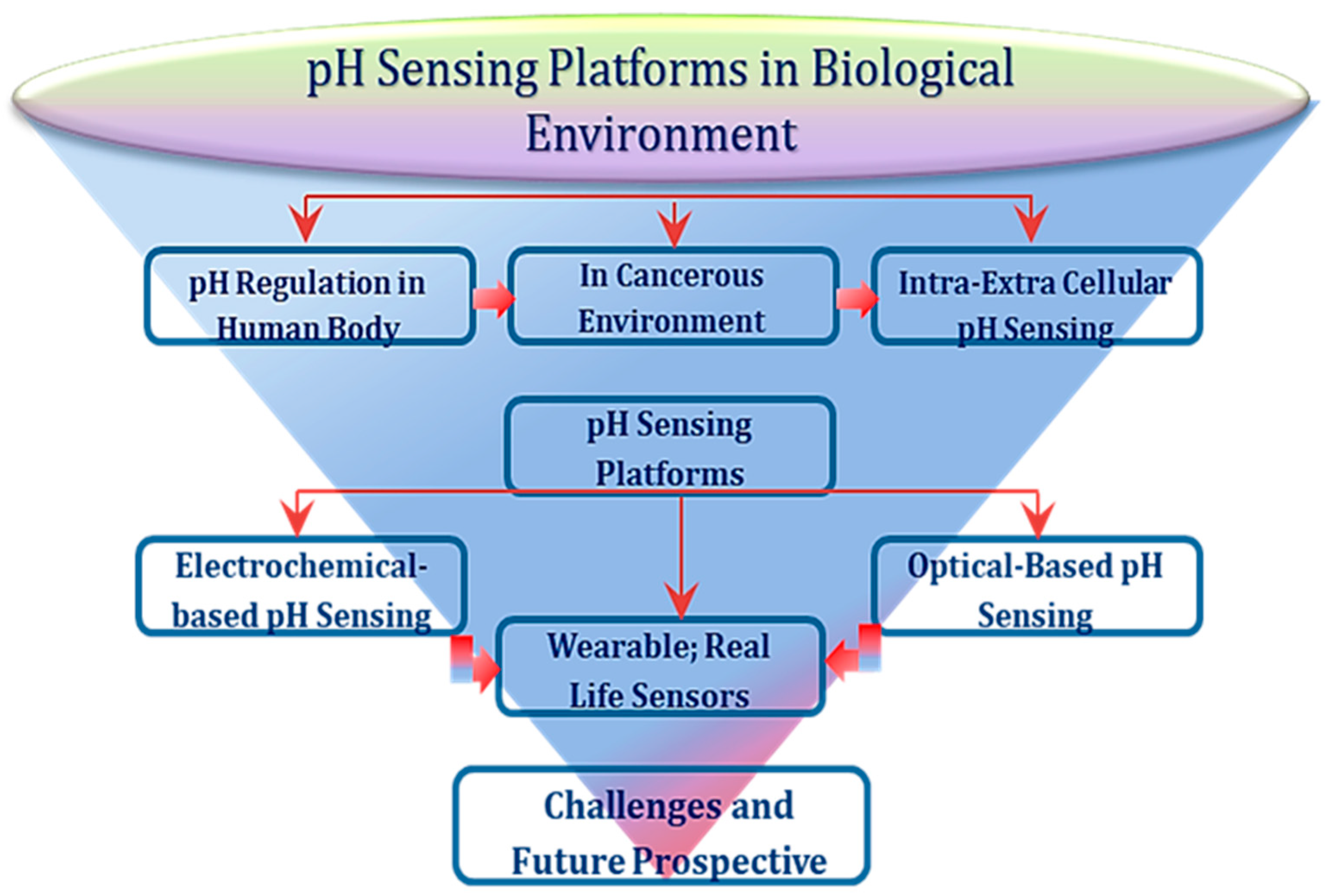
Figure 1. Depicting the organizational and thematic components of pH regulation role and sensing.
Preprints 81262 g001
Figure 2. pH values of the various organs in human body. Copy right image reprinted with the permission from Ref. [50].
Preprints 81262 g002
Figure 4. A (i) Glucose oxidation mechanism and H2O2 production. (ii) working principle image of H-ELISA. (iii) pH sensitivity in terms of voltage-current behavior of the device In set is the calibration plot for pH 1–11. (iv) Reliability and repeatability data of the device in pH 7 and 8 buffer solutions. B (i-iv) Biasing and sensing illustration of the device, chip image, and proton enhancement behavior with and without FeSO4. Copy right image reprinted with the permission from Ref. [95]. (C) Schematic and workin principle of ISFET. D (i-iii) pH evaluation using cell seeding on the device overnight. E (i-iii) Experiment repeatability plot – pH variations vs time data in the presence of urea addition at bare ISFET and ISFET in the presence of BT474 cell line, enzyme with secondary antibody. Copy right image reprinted with the permission from Ref.[96].
Preprints 81262 g004
Figure 5. A Schematic illustration of (i) FR-TPP probe structure and pH sensing mechanism (ii) dual color mitophagy tracking. B – (i) Dual color fluorescence changes with the proposed nanoprobe for simultaneous pH sensing (Exci. 480 nm), ratiometric association between f520/f580 (iii) average size (iv) TEM image of the as prepared probe. Image of the cells (MFC-7) captured by confocal microscope at different time intervals when treated with probe and LDR under CCCR. D (i) TEM images of MCF-7 cytoplasmic region under CCCP treatment (ii) pH variations under different treatments at diverse conditions (iii) R ᷊changes demonstrating mitophagy behavior. Copy right image reprinted with the permission from Ref. [70].
Preprints 81262 g005
Figure 6. A (i, ii) Structures of BFCUR and the UV-vis spectra of the developed probe in the presence of different pH values (5-10). The corresponding ratiometric calibration plot at A500/A650 against pH values ranging from 5 to 8. B (i-iii) Colorimetric analysis between the CUR and BFCUR probes in the aqueous and strip (pH values ranging from 5 to 9). Fluorescence spectra of the developed probe in the presence of different pH values ranging from 5 to 9, where the excitation wavelength was 550nm and the subsequent ratiometric intensities (Is) plot at 622/743 against pH ranging from 5 to 9. C) (i) Fluorescence intensity data analyzed using Image J. (ii) Images of Hela cells in the presence of developed probe (BFCUR) at pH values ranging from 4.5 to 7.5. Copy right image reprinted with the permission from Ref. [109].
Preprints 81262 g006
Figure 8. A The illustration of optical fiber tip set-up modified with hSNF for pH sensing. B) Schematic for the hSNF and Ru(BPY)3 microemulsion synthesis. FITC–APTES conjugation on the SNF surface, hSNFs deposited onto the plasma treated dish C) (i) The optical spectra obtained at various locations. The inserted figure indicates the IFITC/IRu(BPY)3 ratio at each location. The fluorescent spectra for pH (ii) 5.0 and (iii) 8.0 buffer solutions in different cycles. D) (i) Images of the coated fiber collected at different distances. (ii) pH measuring in different samples (i) tap water and hot water, ii) in cell culture media of human cardiomyocytes iii) comparison of the developed system with commercial pH device. Copy right image reprinted with the permission from Ref. [111]. E) The experimental set-up of fiber-optic (FO) sensor for pH measurements. F) (i) The fluorescent spectrum of modified FO sensor (when excited by an LED at ∼470 nm) (ii) pH measuring capability in buffer solutions for pH values ranges from 5 to 8.5 and the illustration of rapid response. G) (i) Time dependent response of the developed system probe from pH 7 to pH 8 and vice versa within ∼1 s. (ii) Luminescence spectra obtained at pH 8 for 20 minutes (when T = 40 °C. with the time intervals of 1 and 5 seconds. H) (i) Stability test for the as prepared device at different pH values (6, 7, 8) after 1 minute and off after 16 minutes. (ii) Comparative measurements in different pH values (5-8) with the developed pH sensor and commercially available meter. Copyright image reprinted with the permission from Ref. [24].
Preprints 81262 g008
Figure 9. (A) Sensing patch and wound management dressing illustration, comprising various sensors for temperature, uric acid and pH monitoring. Smart phone provides the power as well as monitoring and receive the data B (i) The pH sensor with the pH value changing from 9 to 3. (ii) Uric acid linear determinations subjected to variations in pH (iii) temperatures (iv) The wound healing data with respect to drug – present/ or absent during treatment. Copy right image reprinted with the permission from Ref. [44].
Preprints 81262 g009
Figure 10. (A) An illustration of the developed wearable patch for sensing and thermo-responsive delivery of antibiotics, where the heater is shown in the top right corner. The e-module was fabricated to connect the bandage data with electronic gears such as mobiles, computer and smart watch etc. B (i-iv) represents sensor’s fabrication and characterization with pH sensing characteristics. C (i-iii) In vitro antibiotic evaluations and drug release profile of the bandage and D (i-ii) the placement of wearable patch on author’s hand and performance check. Copyright image reprinted with the permission from Ref. [45].
Preprints 81262 g010
Figure 11. (A) An illustration of the developed wearable system for multiple sensing of various parameters and chronic wound treatment. (B) The placement depiction of the developed sensing patch on diabetic foot. (C, D, I, J) show the schematics of various electrodes for pH, uric acid, glucose, ammonium and temperature sensing. Demonstration of drug release upon electrical stimulation and finger-tip sized flexible sensing and therapeutic platform. (G, H, K, L) are the characterization data showing the potentiometric and linear graphs for corresponding pH, temperature, and ammonium sensing. Copy right image used with permission from Ref.[46].
Preprints 81262 g011
Figure 12. (a–d) illustration of the different fabrication steps of the flexible pH sensor. (e-h) The photographs of various modifications steps; sputtered Au, surface modifications, after proton intercalation, and bending test. (i-n) Interference test evaluations, pH response graphs for normal and different bendings of electrode, linear pH sensing curve (before and after sweat test), on-body evaluations for the developed sensor, and a comparative analysis of pH measurement with commercially available pH-meter and with the developed biosensor. Copy right image used with permission from Ref.[41].
Preprints 81262 g012
Figure 13. pH biosensors developed based on IrOx composites showing pH detection. Copy right image used with permission from Ref.[124,125].
Preprints 81262 g013
Figure 14. (A-F) Sketch of the flexible 3-electrodes sensor, mass production of the screen printed electrodes, performance in curved shape, open circuit potential responses (blue line) and pH measurements (with the developed biosensor-solid red line, with pH meter red-dotted line), repeatability & stability, and interferences evaluations.[126]. (G-J) procedural representation of PNIPAM/CMCS/MWCNT/PANI-based hydrogels, the resistivity change in the as synthesized sensing matrix with respect to time, and the material loading/ unloading cycles. Copy right image used with permission from Ref. [127].
Preprints 81262 g014
Figure 15. (A) Schematic illustration of sensor showing the modification of gate with PEDOT:PSS and channel with ionic-liquid gel-based materials. (B) depicts the image of actual chip-based pH sensor, contact angle, electrical characterization showing the gate biasing with the transconductance property, and the pH response of the sensor with standard pH solution. (C) pictorial representation of the OECT device, showing response towards pH variations. Copy right image used with permission from Ref. [132].
Preprints 81262 g015
Table 2. pH values of human cell organelles. [5,8,9,61,62].
Preprints 81262 i001
Disclaimer/Publisher’s Note: The statements, opinions and data contained in all publications are solely those of the individual author(s) and contributor(s) and not of MDPI and/or the editor(s). MDPI and/or the editor(s) disclaim responsibility for any injury to people or property resulting from any ideas, methods, instructions or products referred to in the content.
Copyright: This open access article is published under a Creative Commons CC BY 4.0 license, which permit the free download, distribution, and reuse, provided that the author and preprint are cited in any reuse.
Prerpints.org logo

Preprints.org is a free preprint server supported by MDPI in Basel, Switzerland.

Subscribe

© 2024 MDPI (Basel, Switzerland) unless otherwise stated