1. Introduction
Viruses are a unique class of infectious, obligate intracellular parasites whose genetic material is composed of either DNA or RNA[
1]. The virus particle itself is composed of a nucleocapsid, which contains the genome with the ability to replicate, and a protein shell[
2]. In the case of enveloped viruses, the nucleocapsid is surrounded by a lipid membrane resembling that of the host cell, which is studded with spike structures. The virus is a parasitic entity that relies on the host cell machinery to synthesize its own viral components, allowing for the successful replication and spread of the virus[
3].
Viral infections pose a significant menace to both public health and animal husbandry[
4,
5,
6].
The virus can be transmitted through various means such as water, land, air, body fluids, and excreta, among others[
7], and can rapidly disseminate, culminating in the elimination or death of animals, and thereby engendering significant economic losses[
8,
9,
10].
African Swine Fever virus (ASF) has occurred in 22 countries worldwide since 2016, with Asia being the most affected in terms of animal losses. In China, the outbreak of African swine fever was first confirmed on August 3, 2018[
11]. According to the Chinese government website, a total of 143 cases of African swine fever were reported by July 2019, and more than 1.2 million pigs were culled. The estimated direct economic impact of African swine fever in China amounts to 1 trillion yuan, which is without considering the upstream and downstream of the industrial chain. The outbreak of ASF in Vietnam in 2019 had a severe impact on the Vietnamese pig sector, with more than 20% of the country's pigs being culled or killed[
12].
The impact of the virus is not only in the breeding of pigs, but also in the poultry industry. In 2003, The Netherlands experienced an epidemic of highly pathogenic avian influenza (HPAI) caused by the H7N7 virus subtype. Thirty million birds were culled in this outbreak, about one third of the total poultry population[
13]. The HPAI outbreak in Turkey from 2005 to 2006 resulted in a €28 million loss for broiler producers[
14]. There are other animal viruses, such as blue-ear disease in pigs[
15], foot-and-mouth disease[
16], Marek's disease[
17], etc. This is a significant problem with respect to reduce the economic losses by means of quickly detect the virus.
The rising apprehension within the livestock industry regarding the emergence and dissemination of numerous animal viruses has prompted the adoption of diverse control strategies. These measures are designed to curtail the virus's propagation and mitigate the associated losses[
18]. However, effective viral detection remains a key factor in managing these pathogens. Traditional diagnostic methods, such as nucleic acid amplification-based techniques[
19], antigen or antibody-based assay[
20] and viral isolation[
21], all exhibit a number of limitations, including extended testing times, specialized equipment requirements, and technical expertise[
22]. Therefore, there is an urgent need for fast, user-friendly and field-applicable virus detection modes.
The electrochemical biosensor is an attractive platform for quick virus detection, electrochemical sensors have proven to be an inexpensive and sensitive method, and are used to detect analytes involved in healthcare, environmental monitoring, food packaging by diagnosing the virus before it spreads and cutting off transmission routes[
23]. Traditional virus detection methods have lagged behind and are a significant cause of outbreaks. Electrochemical biosensors have heightened sensitivity and selectivity, affordability, ease of use, portability, and rapid analysis, making them suitable for real-time virus detection and overcoming the limitations of traditional detection methods[
24].
2. Electrochemical Biosensors
2.1. Components of electrochemical biosensor
Electrochemical biosensor is a kind of detection device that uses biological molecules including proteins, nucleic acids, etc., to specifically bind to target analyte[
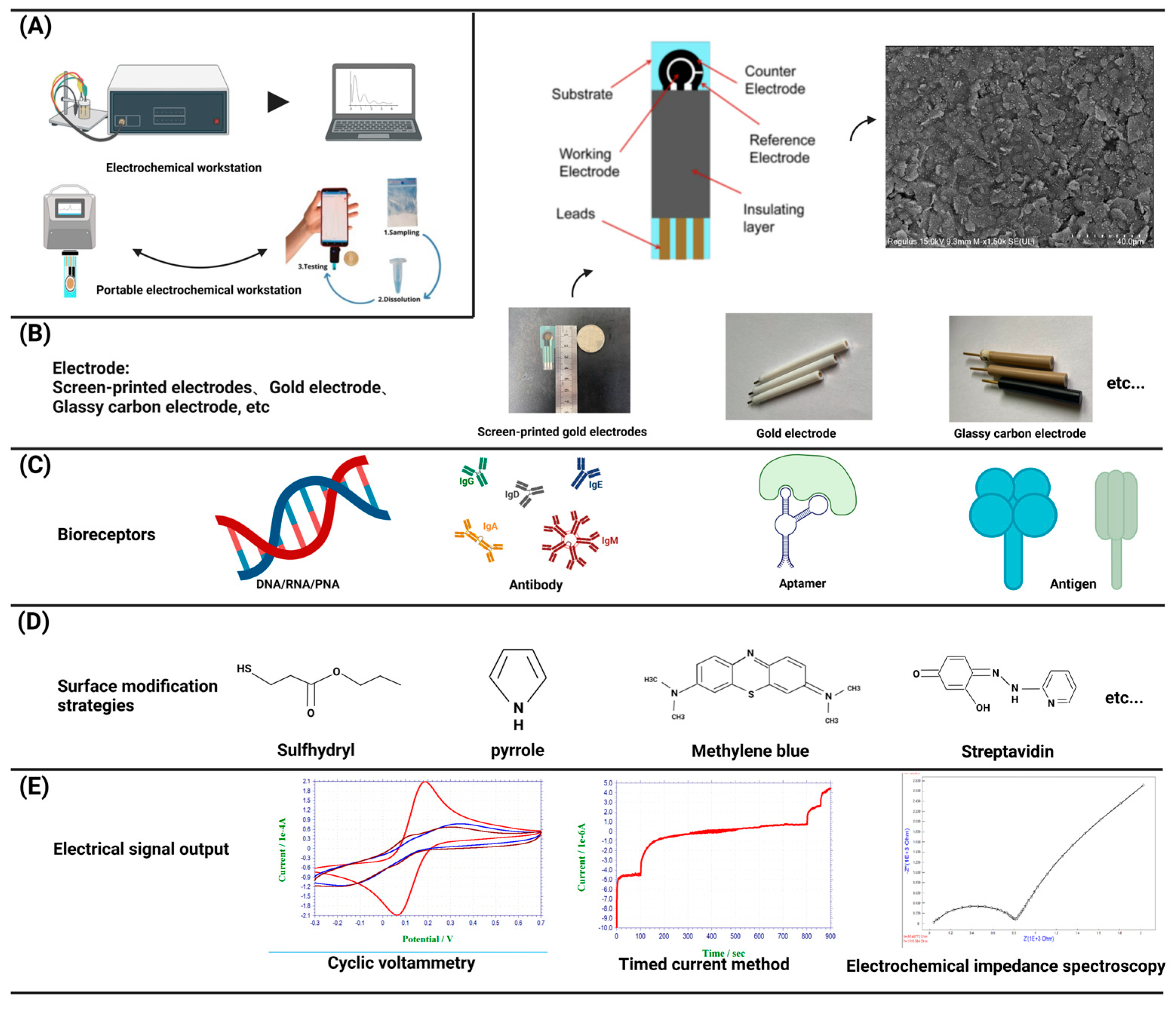
25]. The biological material is used as the sensitive element of the electrochemical biosensor, the electrode is used as the conversion element, the potential or current is the characteristic signal, and the characteristic signal is reflected in the analytical test device on the electrochemical workstation[
26](
Figure 1 A, B, C, E). In electrochemical experiments, a commonly employed configuration is the three-electrode system, comprising a working electrode (WE), a reference electrode (RE), and a counter electrode (CE)[
27]. The WE is the place where the electrochemical reaction occurs and is the object of study, RE is used as a reference to measure and control the system potential, and CE is used to complete the closed circuit to achieve the electrochemical measurement. In order to better connect the biomaterial with the electrode and exert the best performance of the sensor, the help of some compounds, such as thiol compounds[
28] and conductive polymers[
29], is usually needed to make the electrode fully modified (
Figure 1 D). Biomaterials were modified onto WE to form complexes, The electrode-biomaterial complex binds to the analyte, and usually these binding are irreversible, such as paired hybridization of nucleic acids, specific binding of antigens and antibodies, combined with different detection methods of the electrochemical workstation, and the behavior of electrode modification in the above process was recorded on the electrochemical workstation[
30].
2.1.1. Electrode
Commonly used electrode materials are gold electrode, glass carbon electrode, graphene electrode, screen printing electrode and so on. Gold electrode is widely favored due to its inert chemical properties and the ability of non-chemical combination with mercaptan on the surface of gold electrode to form gold-sulfur (Au-S) bonds self-assembled monolayer[
31], which has been widely demonstrated in the field of nucleic acid hybridization. Glassy carbon electrode is one of the most widely used working electrodes. It has good conductivity, high hardness, wide polarization range, and can be used directly as an inert electrode for anodic dissolution. Graphene, characterized by a monolayer of SP2-bonded carbon atoms arranged in a honeycomb lattice, exhibits remarkable attributes such as rapid electron transfer, remarkable thermal conductivity, and favorable biocompatibility. Its superior physical and chemical properties render graphene an ideal electronic material for advanced electrochemical sensing applications[
32].
Screen printed electrodes (SPE) are an ideal component for sensor applications. Compared with the traditional three-electrode system, the sensor surface is modified with nanomaterials to ensure consistency and stability while greatly simplifying the experimental operation process. SPE generally includes a substrate for printed electrodes, the substrate is printed with an external insulation layer and electrode leads, and the substrate is also printed with WE、RE and CE. Each electrode is connected with the corresponding lead to form a three-electrode system[
33]. Because the low-cost manufacturing technology of SPE can be easily extended to mass production, and all types of materials can be added to screen printing slurry, the SPE can be customized for different substrate materials, shapes, and sizes in production to meet the needs of a variety of research. Its low manufacturing cost and ease of manufacture make it possible to mass customize personalized products according to demand, making it an ideal tool in the field of quality-controlled, scientific research[
34].
2.1.2. working station
Electrochemical workstation is the abbreviation of electrochemical detection system. Electrochemical workstation can be directly used to measure the steady state current on the ultra-microelectrode. After special treatment, if the workstation is connected with the micro-current amplifier and shielding box, the current of 1pA or lower can be measured, which provides a solid foundation for electrochemical detection. Two primary categories of electrochemical workstations exist: single-channel workstations and multi-channel workstations. The difference is that multi-channel workstations can be used to detect multiple samples at the same time. At present, the electrochemical workstation has been a commercial product, and different manufacturers provide different models of products with different electrochemical measurement techniques and functions. In order to meet the needs of rapid detection and be convenient to carry, a portable electrochemical workstation has been developed, which has the advantage of being small and light, easy to operate and cheap, and provides researchers with greater flexibility and convenience[
35]. Electrochemical biosensors can detect very small sample sizes and speed up the analysis process, making them powerful and highly sensitive devices[
36]. So far, other types of sensors have also been reported, such as optical sensors[
37], etc. Compared with other types of sensors, electrochemical biosensors do not require expensive equipment, save time, are convenient to carry, and are more user-friendly, especially for areas with backward economic development and scarce resources[
24]. The common detection methods of electrochemical biosensors include voltammetry, amperometry and impedance method.
2.2. Signal analysis and output
2.2.1. Voltammetry
Voltammetry is based on the voltage change between the electrode and the electrolyte solution[
38], on which cyclic voltammetry is the most commonly used electrochemical method and is considered “spectroscopy for electrochemists”. After triangular wave scanning, the electrode completes a reduction and oxidation process. In the range of scanning potential, the electrode can undergo alternate reduction and oxidation reactions, and the resulting current-voltage curve is recorded, reflecting the steady-state response of chemical reactions triggered by electron transfer[
39]. Cyclic voltammetry can be used in a faster time, a wider potential to see the electrode where the Oxidation reduction reaction, to provide a rich signal for the electrode, cyclic voltammetry is generally used for qualitative analysis, rarely as quantitative analysis.
2.2.2. Ampere method
Amperometric detection relies on the alteration of current. A fixed potential was applied to the electrochemical solution, which is enough to oxidize or reduce a certain electroactive substance in the solution. The change of current and time is recorded to obtain the current-time curve[
40,
41]. The ampere method is an electrochemical analysis method to study the kinetics of electrode process.
2.2.3. Impedance method
Electrochemical impedance spectra (EIS) theory and its data interpretation are very complex for researchers who are not familiar with it, such as biologists, biochemists, or materials scientists. The impedance method is an electrochemical measurement technique that employs a low-amplitude sine wave potential (or current) as a perturbation signal[
42]. The reaction rate depends on the frequency, and the frequency change can distinguish the reaction rate of different substances in solution. Due to its broad measurement frequency range, the impedance method provides access to a greater wealth of dynamic information and electrode interface structural details compared to other conventional electrochemical methods[
43].
These basic electrochemical methods all play a crucial role in the signal changes of the working electrode modification process. On the basis of these methods, some more sensitive methods have evolved, such as pulse voltammetry, step voltammetry, multi-point step chronoelectric method, etc., the foundation has been established for more novel electrochemical biosensor models with potential applications.
3. The electrochemical biosensor model
The earliest prototype of electrochemical biosensors can be traced back to the 1970s, when the sensor was based on the specific capture of the analyte by affinity elements, and the subsequent conversion of the capture event into a measurable signal, aiming to broaden the selectivity and sensitivity of the analyte detection in the field of electrochemical detection[
44]. Due to the advantages of low cost, fast and convenient, electrochemical biosensors have been widely concerned, and are more suitable for commercial applications[
45]. The first glucose sensor was reported in 1962, and the glucose sensor using Clark technology was commercialized in 1975[
46]. In the development since then, electrochemical glucose biosensor has generated great commercial value[
47]. Electrochemical biosensors have been widely studied and applied to human society to promote the development of society, including environmental detection[
48,
49], food safety detection[
50,
51], human pathogenic microorganisms detection[
52,
53], etc., but there is little research on animal viruses.
The impact of animal viruses on animal husbandry is huge, and the economic losses caused by animal viruses are inestimable. People are growing increasingly concerned and attempting to apply the established advancements in human virus detection to the study of electrochemical biosensors. The goal is to swiftly detect animal viruses and minimize the economic losses they cause. Electrochemical biosensors can be used to detect animal viruses mainly based on nucleic acids and proteins. Available models for the detection of animal viruses by electrochemical biosensors are described below.
3.1. Nucleic acid based electrochemical biosensor model
Electrochemical biosensor platforms are based on biological molecules that recognize each other through various interactions and cause electron transfer to produce electrical signals to complete detection. Nucleic acids are not only the basic genetic material of organisms, but also the ideal material for the identification of various analytes[
54]. The specificity of the pairing of nucleotide molecules that constitute nucleic acids further shows the advantages of nucleic acid-based electrochemical biosensors.
Reduction of DNA by electrochemical sensors was reported as early as 1960[
55], and subsequently, since DNA purine bases can be oxidized by electrochemistry, Electrochemical sensing methods have been developed for indirect oxidation of DNA by metal complexes acting as electrochemical mediators[
56]. With the development of electrochemical biosensors, nucleic acid has gradually become a bridge between electrode materials and analytes, which also benefits from the easy modification of DNA. Therefore, a promising electrochemical biosensor has been developed, in which the Oxidation reduction reaction of electroactive substances upon the surface of the electrode is caused by specific hybridization events of ssDNA, and the electrochemical signal is generated by transferring electrons. This method is popular in the detection of human pathogens, food guard, environment, etc., and can also be used to detect animal viruses.
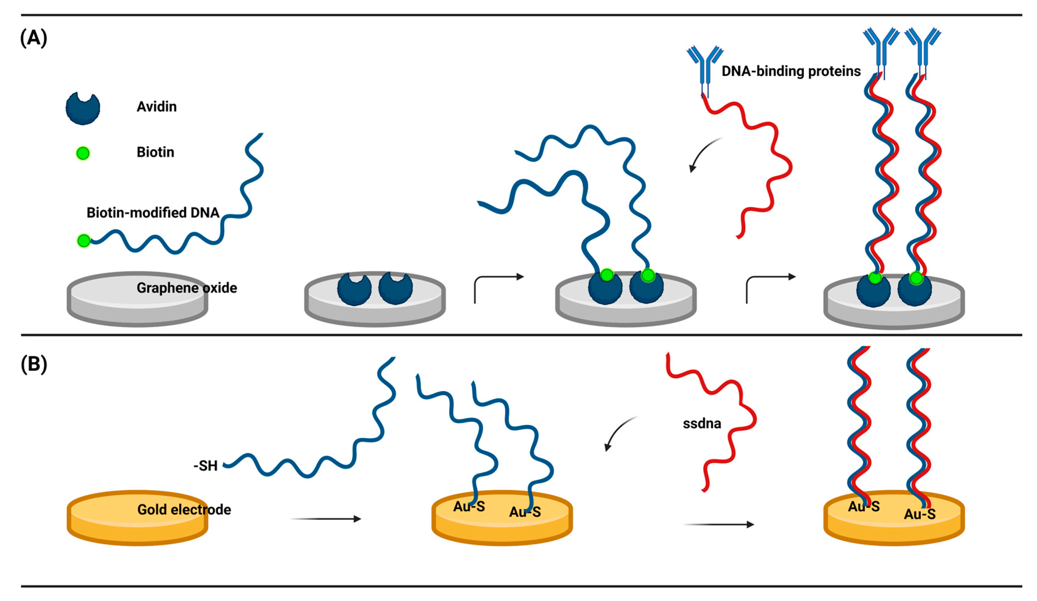
This type of sensor immobilizes ssDNA to the working electrode by both chemical and non-chemical binding, depending on the specificity between the modified nucleic acid and the electrode material. I. Macwan et al. created a DNA electrochemical biosensor with biotin modification to detect biological macromolecules. Biotin-modified DNA forms stable complexes through the hydrophobic force between avidin and graphene, including van der Waals forces, hydrogen bonds, and electrostatic interactions[
57]. The biotin-modified ssDNA hybridized with the complementary ssDNA in the target analyte, and the hybridization signal was converted to an electrical signal to complete the detection of DNA-binding proteins (
Figure 2A). This strategy investigates the relationship between graphene and avidin binding, and although aimed at the detection of DNA-binding proteins, it also indirectly provides a method for the detection of viral nucleic acids. More prevalent is the thiolation modification of DNA, which is attached to the gold working electrode by au-s[
58], There have been many studies on the detection of human viruses[
59,
60]. The ssDNA fixed on the surface of the electrode hybridize with complementary DNA, interact with RNA and protein, or produce corresponding electrical signals through structural changes of DNA (
Figure 2B). The electrical signals can also be generated indirectly by the recognition process of ssDNA using enzymes and Oxidation reduction reaction mediators. The reaction was combined with electrochemical method on electrochemical workstation. The characteristics of these electrochemical DNA biosensors include being portable, simple, cost-effective, having a fast response time, high sensitivity, high selectivity, and compatibility with miniature detection technologies have garnered significant interest in electrochemical DNA biosensors. Such biosensors are deemed feasible for the detection of animal viruses.
3.2. Antibodies and antigens based electrochemical biosensors model
Proteins constitute a vital component of viruses and play a crucial role in their life cycle[
61]. Proteins have a protective effect on the virus, but also determine the specificity of the virus infection, for the host, the virus protein is also an important antigen. There is potential in using viral particles and viral surface proteins to make electrochemical biosensors for the detection of viruses. Several electrochemical biosensors have been reported as exemplary models for detecting SARS-CoV-2 nucleocapsid and spike proteins[
62,
63,
64]. In recent years, such electrochemical biosensors based on mutual recognition of antibodies and antigens have attracted attention, which are related to immunoassay technology and have the basic characteristics of immunoassay. Antibodies and antigens based electrochemical biosensors integrate the high sensitivity and specificity of immunoassay with the conversion of physical signals. When the antibody-antigen immune reaction occurs, this specific binding to form a complex is usually irreversible, and after the transduction of the electrochemical workstation signal, the recognition signal is transformed into an electrical signal, which makes it possible to detect viruses rapidly.
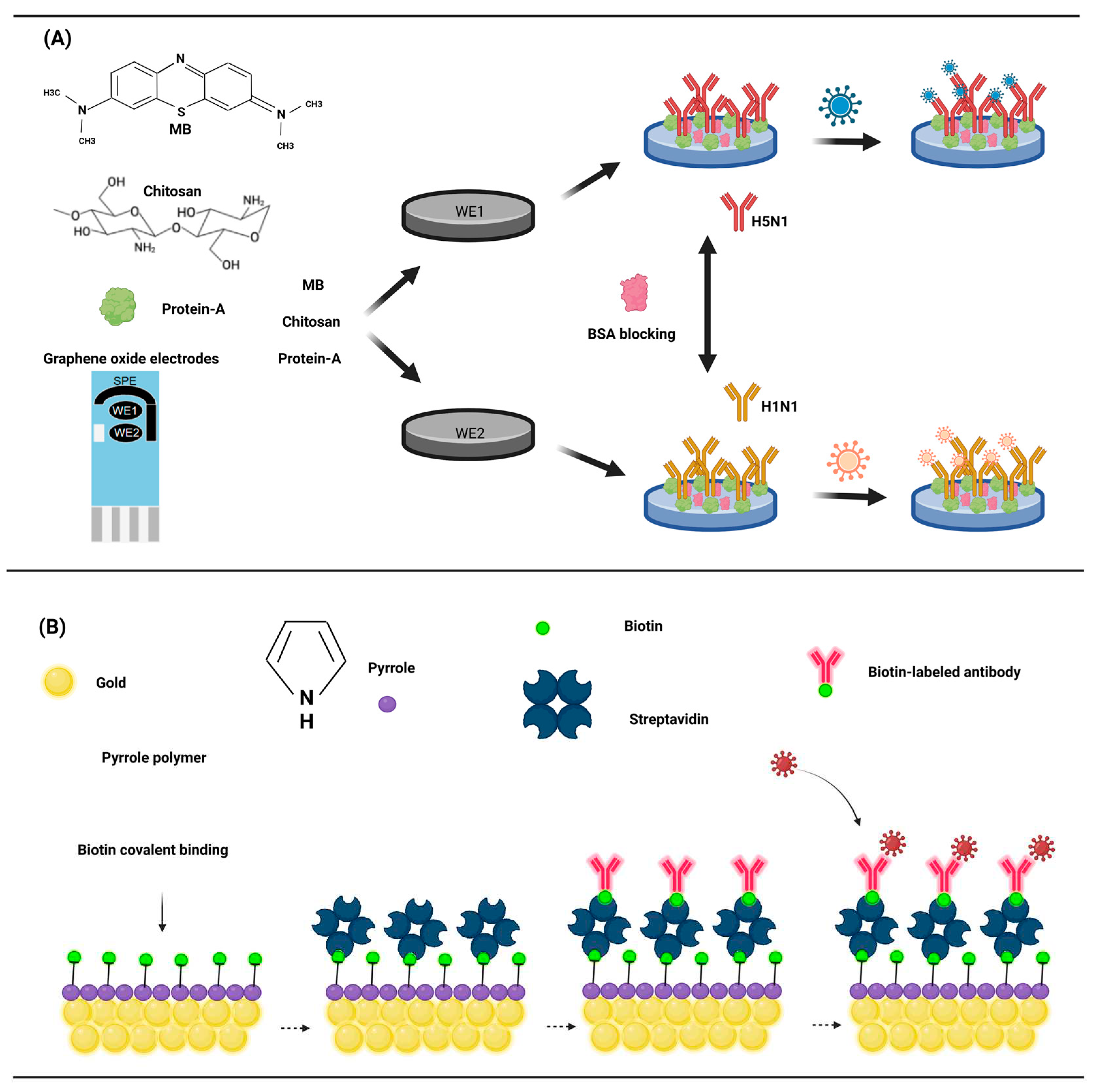
The key to antigen detection is the fixation of the antibody with the electrode, usually the antibody is directly fixed to the electrode surface. Various methods enable the attachment of antibodies to the electrode surface. Adsorption allows direct attachment of antibodies to the electrode surface, M. Veerapandian et al. devised an electrochemical biosensor utilizing methylene blue adsorption of GO (graphene oxide) to detect the antigen of the influenza A virus[
65]. Chitosan (CS) was modified to GO electrodes containing methylene blue for direct adsorption of protein A. Protein A is a specific receptor for fc antibodies and can directly immobilize monoclonal antibodies (
Figure 3A). The sensor's generated current, subsequent to the binding of the antibody and antigen, exhibits a robust correlation with the antigen's concentration, with a detection limit as low as 10 pM for H5N1 and H1N1.
Immobilization of antibodies to electrodes can also be achieved through a matrix of conductive polymers (CPs), which are mainly used as sensors for biological interactions, The ability of high conductivity to monitor the electrical signal generated by the interaction of the probe with the analyte is key to its preference. Polypyrrole is the most commonly used conductive polymer due to its biocompatibility[
66], high hydrophilic properties[
67], and high stability in water. Studies on biotinylated single-chain variable fragment antibody and functionalized polypyrrole has shown that the combination of polypyrrole and biotin-streptavidin is an effective method for immobilizing antibodies[
68]. Biotin was covalently bound to the functionalized polypyrrole, streptavidin was conjugated to biotin, and finally an antibody labeled with biotin was immobilized to the electrode (
Figure 3B). This study showed that the standard curve of the sensor was linearly related to 1pg/ml-100ng/ml antigen concentration, was highly reproducible, and the detection limit has the potential to reach an impressive 1 pg/mL.
There are other ways to immobilize antibodies, such as a special membrane structure called self-assembling monolayers (SAMs)[
69]. Self-assembled monolayers (SAMs) refer to organic layers that spontaneously form upon the electrode's surface when molecules from a solution or gas phase are adsorbed. This membrane structure has a strong choice of terminal functional groups and has the potential to be applied in the construction of nanoscale electrochemical biosensors by immobilized antibodies[
70]. Adsorption of alkane thiols on gold is the most widely used SAMs application. The 5' thiol modified 3' biotin modified ssDNA is connected to the gold surface by self-assembly, and the affinity of biotin and streptavidin is used to form the Au-ssDNA-biotin-streptavidin electrochemical biosensor body. The biotin-labeled antibody is attached to the sensor body, which is also a potential method for immobilizing antibodies. From a protein chemistry point of view, immobilizing proteins on the electrode surface is an extremely complex problem[
71,
72]. Moreover, the protein is sensitive and has low conformational stability, which may be destroyed upon immobilization on the electrode surface. By using SAMs and oligonucleotides to immobilize antibodies, a bond between antibodies and electrodes can be established, which can effectively avoid the problems of antibody ring breaking, inactivation, and non-specific adsorption.
From another point of view, the body will also produce a resistant response to the invasion of the virus, and the corresponding antibodies will be produced in the blood, so the detection of virus antibodies can also provide the basis for the possible existence of the virus. At this time, the antigen of the virus needs to be fixed, and the possible virus can be detected by combining the antigen with the antibody in the blood to generate an electrical signal[
73]. This type of electrochemical biosensor is similar to the antibody-based electroactive biosensor, which will not be described here.
3.3. Nucleic acid aptamer-based electrochemical biosensor model
Nucleic acid aptamer is a kind of artificial biological receptor, which is composed of a small sequence of oligonucleotide nucleotides or short peptides obtained by in vitro screening. Artificial bioreceptor was independently discovered by Ellington and Szostak[
74] and Tuerk and Gold[
75] in 1990, and peptide aptamer was further introduced by Colas et al[
76] in 1996. The discovery of nucleic acid aptamer as a new bioreceptor provided a new research direction for electrochemical biosensor based on nucleic acid aptamer. As a new biological receptor, aptamers have unique advantages. First nucleic acid aptamers were screened and synthesized in vitro by SELEX (systematic evolution of ligands by exponential Enrichment)[
77,
78] technology, and further large-scale production using chemical synthesis methods can reduce costs while also overcoming the use of animals or cells where antibody production is necessary. Due to the short oligonucleotide nucleic acid, nucleic acid aptamer is easier to be modified by chemical groups, easier to connect with the electrode, and has greater openness. At the same time, it also reduces the difficulty of antibody connecting the electrode and simplifies the experimental process. Secondly, aptamers have stronger ability to bind to targets than antibodies, which is due to the fact that aptamers can use multiple action sites to bind to targets through hydrogen bonding, electrostatic interaction, van der Waals forces, etc.[
79]. For some targets that cannot be effectively bound by antibodies, aptamers can effectively solve this problem. Nucleic acid aptamers have the ability to specifically bind to the target of antibodies, and are better than antibodies in some properties, so they are suitable for electrochemical biosensing.
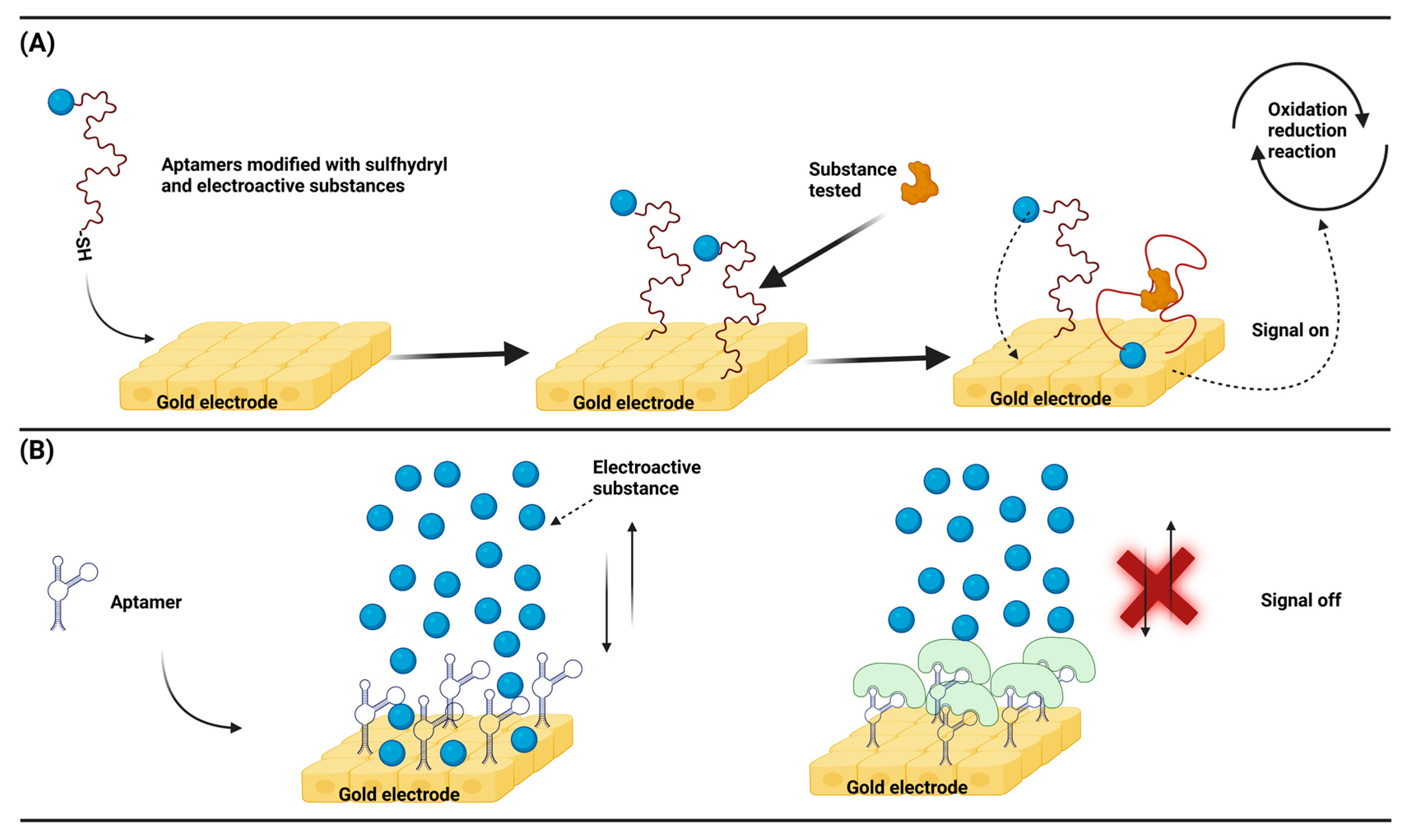
Similar to other types of electrochemical biosensors, nucleic acid aptamer-based electrochemical biosensors also generate signals by transferring electrons from electroactive substances, which has been widely studied for targeted drug delivery and biomarker detection. The same principle can also be applied to the detection of animal viruses. An ingeniously designed sensor model is to take advantage of the structural changes before and after aptamer binding to the target, and use the electroactive probe attached to the aptamer at a distance from the electrode for electron transfer. Aptamer conformation changes lead to a change in the spatial separation between the electrically active probe and the electrode, which in turn produces a change in the signal[
80,
81]. This is a hairpin-like structure design, and the conformational change of the nucleic acid aptamer can meet the rapid detection requirements (
Figure 4A). In another common design, a nucleic acid aptamer acts as a link between the electrode and the target analyte. Binding of the aptamer to the target analyte prevents the electroactive substance from moving closer to the electrode to transfer electrons, which in turn increases the impedance[
82,
83]. This impedance design has faster reaction kinetics, can ignore the interference due to the background current, and has higher sensitivity(
Figure 4B).
4. Advantages and limitation of electrochemical biosensors for virus detection
4.1. Advantages
The advantages of electrochemical biosensor in virus detection are its short detection time, convenience, cheap, simple, and real-time detection[
84]. Usually, the virus is diagnosed in the laboratory by virus isolation, PCR, ELISA, and other methods. The accuracy and practicability of these traditional methods are beyond doubt, which have left a strong mark in the history of human fight against viruses. However, these methods require specialized personnel, expensive equipment, and testing lags[
85]. Due to these limitations, the detection of viruses by electrochemical biosensors has come to the stage of history.
Table 1 shows the comparison of virus detection by conventional methods and electrochemical biosensor.
On the other hand, the popularity of electrochemical biosensors also benefits from the maturity of screen-printed electrode technology. Screen-printed electrodes (SPEs) have long been considered the most promising analytical tool in electrochemical detection. Compared with traditional electrodes, SPE avoids the polishing, cleaning, and activation required for other solid electrodes, and greatly simplifies the experimental process. Commercial SPEs have high versatility, which is also one of the advantages of SPEs[
87,
88]. Because the low-cost manufacturing technology of SPE can be easily extended to mass production, and all types of materials can be added to screen printing slurry, the SPE can be customized for different substrate materials, shapes and sizes in production to meet the needs of a variety of research. At the same time, SPE is small, powerful, low-cost, maintenance-free, and has been widely used in electrochemical research in environmental monitoring[
89,
90,
91], clinical diagnosis[
92,
93], drug analysis[
94,
95] and food detection[
88,
96,
97], which has promoted the development of electrochemical biosensors.
4.2. Limitation
The advantages of electrochemical biosensors such as fast, convenient, and cheap are indeed becoming more and more prominent in the diagnostic industry, although some electrochemical biosensors are still in the development and testing stage. The current problems faced by electrochemical biosensors are mainly the following aspects, the first is accuracy and reproducibility, and analyte concentration becomes increasingly important when moving target analyte testing from a clean laboratory with a controlled environment to the field. Here, many external environmental factors can contribute to significant differences in signal strength, such as temperature, humidity, sample volume, electrode surface area, non-calibrated instruments, and contamination. In response to this problem, corresponding coping strategies have been reported to improve the accuracy and reproducibility of biosensors through ratio electrochemistry[
98]. Secondly, the limited shelf life and stability of the biometric components as well as non-specific binding are still the biggest biosensor limitations, and corresponding strategies have been reported to overcome and reduce this aspect. Finally, The concentration used in the sensor component design is trace and requires precise operation, therefore, only through reasonable design and rigorous testing can the biosensors be transferred from the laboratory to the field.
5. Conclusion
Rapid detection of pathogens, such as viruses, remains a great challenge in analytical medicine due to their quantitative diversity[
99]. The economic loss caused by the virus is inestimable every year. The development of a rapid, cost-effective, user-friendly, and highly specific virus detection strategy is of utmost urgency. Therefore, cost-effective, fast, convenient, and sensitive electrochemical biosensors have been favored[
100]. Due to the rapid detection characteristics of electrochemical biosensors and the characteristics of biological material construction, which will pave the way for the identification of different viruses, the next stage of development should focus on the universality of electrochemical sensors and the detection of viruses with high accuracy, followed by the ability to adapt to work in multiple environments. With the development of materials science, the popularization and application of new materials and the rapid development of electrochemical components, the rapid detection of viruses by electrochemical biosensors will be a valuable depression in the future. In summary, the development of electrochemical biosensors mainly relies on being fast, convenient, specific, and cost-effective. Considering these properties, combining biosensor and biomanufacturing approaches with synthetic biology approaches, or combining all these principles, will be key to successful development of robust biosensors in the future.
Author Contributions
Conceptualization, Y.W, G-R. X, and L.W.; methodology, , Y.W, and G-R.X.; writing—original draft preparation, X.H., S.W., C.M., Y.W; writing—review and editing, S.W., C.M., J.M., W.Z., H.L; supervision, Y.W, G-R. X, and L.W.; funding acquisition, Y.W, G-R. X, and L.W.; All authors have read and agreed to the published version of the manuscript.
Funding
This research was funded by the National Key R&D Program of China (2021YFD1301200), National Natural Science Foundation of China (32172862), Outstanding Youth Foundation of Henan Scientific Committee (222300420043), Leading Talents of Scientific and Technological Innovation in the Central Plains (224200510024), Henan Province science and technology breakthrough project (232102110081).
Institutional Review Board Statement
Not applicable.
Conflicts of Interest
The authors declare no conflict of interest.
References
- Pellett, P.E.; Mitra, S.; Holland, T.C. Basics of virology. Handb Clin Neurol 2014, 123, 45–66. [Google Scholar] [PubMed]
- San, M.C. Virus maturation. Adv.Exp.Med.Biol. 2019, 1215, 129–158. [Google Scholar]
- Kalia, M.; Jameel, S. Virus entry paradigms. Amino Acids 2011, 41, 1147–1157. [Google Scholar] [CrossRef]
- Liu, Q.; Wang, H.Y. Porcine enteric coronaviruses: an updated overview of the pathogenesis, prevalence, and diagnosis. Vet. Res. Commun. 2021, 45, 75–86. [Google Scholar] [CrossRef]
- Abd, E.M.; El-Saadony, M.T.; Alqhtani, A.H.; Swelum, A.A.; Salem, H.M.; Elbestawy, A.R.; Noreldin, A.E.; Babalghith, A.O.; Khafaga, A.F.; Hassan, M.I.; El-Tarabily, K.A. The relationship among avian influenza, gut microbiota and chicken immunity: an updated overview. Poult. Sci. 2022, 101, 102021. [Google Scholar]
- Labadie, T.; Batejat, C.; Leclercq, I.; Manuguerra, J.C. Historical discoveries on viruses in the environment and their impact on public health. Intervirology 2020, 63, 17–32. [Google Scholar] [CrossRef]
- Vidic, J.; Manzano, M.; Chang, C.M.; Jaffrezic-Renault, N. Advanced biosensors for detection of pathogens related to livestock and poultry. Vet. Res. 2017, 48, 11. [Google Scholar] [CrossRef]
- Neumann, E.J.; Kliebenstein, J.B.; Johnson, C.D.; Mabry, J.W.; Bush, E.J.; Seitzinger, A.H.; Green, A.L.; Zimmerman, J.J. Assessment of the economic impact of porcine reproductive and respiratory syndrome on swine production in the united states. JAVMA-J. Am. Vet. Med. Assoc. 2005, 227, 385–392. [Google Scholar] [CrossRef]
- Liu, S.; Zhuang, Q.; Wang, S.; Jiang, W.; Jin, J.; Peng, C.; Hou, G.; Li, J.; Yu, J.; Yu, X.; Liu, H.; Sun, S.; Yuan, L.; Chen, J. Control of avian influenza in china: strategies and lessons. Transbound. Emerg. Dis. 2020, 67, 1463–1471. [Google Scholar] [CrossRef]
- Nakada, S.; Fujimoto, Y.; Kohara, J.; Makita, K. Economic losses associated with mastitis due to bovine leukemia virus infection. J. Dairy Sci. 2023, 106, 576–588. [Google Scholar] [CrossRef]
- Liu, Y.; Zhang, X.; Qi, W.; Yang, Y.; Liu, Z.; An, T.; Wu, X.; Chen, J. Prevention and control strategies of african swine fever and progress on pig farm repopulation in china. Viruses-Basel 2021, 13. [Google Scholar] [CrossRef] [PubMed]
- Nguyen-Thi, T.; Pham-Thi-Ngoc, L.; Nguyen-Ngoc, Q.; Dang-Xuan, S.; Lee, H.S.; Nguyen-Viet, H.; Padungtod, P.; Nguyen-Thu, T.; Nguyen-Thi, T.; Tran-Cong, T.; Rich, K.M. An assessment of the economic impacts of the 2019 african swine fever outbreaks in vietnam. Front. Vet. Sci. 2021, 8, 686038. [Google Scholar] [CrossRef] [PubMed]
- Stegeman, A.; Bouma, A.; Elbers, A.R.; de Jong, M.C.; Nodelijk, G.; de Klerk, F.; Koch, G.; van Boven, M. Avian influenza a virus (h7n7) epidemic in the netherlands in 2003: course of the epidemic and effectiveness of control measures. J. Infect. Dis. 2004, 190, 2088–2095. [Google Scholar] [CrossRef] [PubMed]
- Aral, Y.; Yalcin, C.; Cevger, Y.; Sipahi, C.; Sariozkan, S. Financial effects of the highly pathogenic avian influenza outbreaks on the turkish broiler producers. Poult. Sci. 2010, 89, 1085–1088. [Google Scholar] [CrossRef] [PubMed]
- Mcorist, S.; Khampee, K.; Guo, A. Modern pig farming in the people's republic of china: growth and veterinary challenges. Rev. Sci. Tech. Off. Int. Epizoot. 2011, 30, 961–968. [Google Scholar] [CrossRef]
- Poonsuk, K.; Gimenez-Lirola, L.; Zimmerman, J.J. A review of foot-and-mouth disease virus (fmdv) testing in livestock with an emphasis on the use of alternative diagnostic specimens. Anim. Health Res. Rev. 2018, 19, 100–112. [Google Scholar] [CrossRef]
- Kennedy, D.A.; Cairns, C.; Jones, M.J.; Bell, A.S.; Salathe, R.M.; Baigent, S.J.; Nair, V.K.; Dunn, P.A.; Read, A.F. Industry-wide surveillance of marek's disease virus on commercial poultry farms. Avian Dis. 2017, 61, 153–164. [Google Scholar] [CrossRef]
- Thornton, P.K. Livestock production: recent trends, future prospects. Philos. Trans. R. Soc. B-Biol. Sci. 2010, 365, 2853–2867. [Google Scholar] [CrossRef]
- Monis, P.T.; Giglio, S. Nucleic acid amplification-based techniques for pathogen detection and identification. Infect. Genet. Evol. 2006, 6, 2–12. [Google Scholar] [CrossRef]
- Day, M.J. Introduction to antigen and antibody assays. Top. Companion Anim. Med. 2015, 30, 128–131. [Google Scholar] [CrossRef]
- Leland, D.S.; Ginocchio, C.C. Role of cell culture for virus detection in the age of technology. Clin. Microbiol. Rev. 2007, 20, 49–78. [Google Scholar] [CrossRef] [PubMed]
- Babaei, A.; Rafiee, N.; Taheri, B.; Sohrabi, H.; Mokhtarzadeh, A. Recent advances in early diagnosis of viruses associated with gastroenteritis by biosensors. Biosensors-Basel 2022, 12. [Google Scholar] [CrossRef] [PubMed]
- Aydin, E.B.; Aydin, M.; Sezginturk, M.K. Advances in electrochemical immunosensors. Advan. Clin. Chem. 2019, 92, 1–57. [Google Scholar]
- Manring, N.; Ahmed, M.; Tenhoff, N.; Smeltz, J.L.; Pathirathna, P. Recent advances in electrochemical tools for virus detection. Anal. Chem. 2022, 94, 7149–7157. [Google Scholar] [CrossRef] [PubMed]
- Xiang, Y.; Lu, Y. Portable and quantitative detection of protein biomarkers and small molecular toxins using antibodies and ubiquitous personal glucose meters. Anal. Chem. 2012, 84, 4174–4178. [Google Scholar] [CrossRef] [PubMed]
- Banakar, M.; Hamidi, M.; Khurshid, Z.; Zafar, M.S.; Sapkota, J.; Azizian, R.; Rokaya, D. Electrochemical biosensors for pathogen detection: an updated review. Biosensors-Basel 2022, 12. [Google Scholar] [CrossRef]
- Eom, H.; Kang, J.; Jang, S.; Kwon, O.; Choi, S.; Shin, J.; Nam, I. Evaluating the electrochemical properties of supercapacitors using the three-electrode system. J. Vis. Exp. 2022. [Google Scholar] [CrossRef]
- Lereau, M.; Fournier-Wirth, C.; Mayen, J.; Farre, C.; Meyer, A.; Dugas, V.; Cantaloube, J.F.; Chaix, C.; Vasseur, J.J.; Morvan, F. Development of innovative and versatile polythiol probes for use on elosa or electrochemical biosensors: application in hepatitis c virus genotyping. Anal. Chem. 2013, 85, 9204–9212. [Google Scholar] [CrossRef]
- El-Said, W.A.; Abdelshakour, M.; Choi, J.H.; Choi, J.W. Application of conducting polymer nanostructures to electrochemical biosensors. Molecules 2020, 25. [Google Scholar] [CrossRef]
- Ronkainen, N.J.; Halsall, H.B.; Heineman, W.R. Electrochemical biosensors. Chem. Soc. Rev. 2010, 39, 1747–1763. [Google Scholar] [CrossRef]
- Inkpen, M.S.; Liu, Z.F.; Li, H.; Campos, L.M.; Neaton, J.B.; Venkataraman, L. Non-chemisorbed gold-sulfur binding prevails in self-assembled monolayers. Nat. Chem. 2019, 11, 351–358. [Google Scholar] [CrossRef]
- Lawal, A.T. Synthesis and utilisation of graphene for fabrication of electrochemical sensors. Talanta 2015, 131, 424–443. [Google Scholar] [CrossRef]
- Costa-Rama, E.; Fernandez-Abedul, M.T. Paper-based screen-printed electrodes: a new generation of low-cost electroanalytical platforms. Biosensors-Basel 2021, 11. [Google Scholar] [CrossRef]
- Antuna-Jimenez, D.; Gonzalez-Garcia, M.B.; Hernandez-Santos, D.; Fanjul-Bolado, P. Screen-printed electrodes modified with metal nanoparticles for small molecule sensing. Biosensors-Basel 2020, 10. [Google Scholar] [CrossRef]
- Ji, D.; Liu, L.; Li, S.; Chen, C.; Lu, Y.; Wu, J.; Liu, Q. Smartphone-based cyclic voltammetry system with graphene modified screen printed electrodes for glucose detection. Biosens. Bioelectron. 2017, 98, 449–456. [Google Scholar] [CrossRef]
- Mohamad, N.N.; Ridhuan, N.S.; Abdul, R.K. Progress of enzymatic and non-enzymatic electrochemical glucose biosensor based on nanomaterial-modified electrode. Biosensors-Basel 2022, 12. [Google Scholar] [CrossRef]
- Gruber, P.; Marques, M.; Szita, N.; Mayr, T. Integration and application of optical chemical sensors in microbioreactors. Lab Chip 2017, 17, 2693–2712. [Google Scholar] [CrossRef]
- Venton, B.J.; Cao, Q. Fundamentals of fast-scan cyclic voltammetry for dopamine detection. Analyst 2020, 145, 1158–1168. [Google Scholar] [CrossRef] [PubMed]
- Agrawal, K.; Naik, A.A.; Chaudhary, S.; Parvatalu, D.; Santhanam, V. Prudent practices in ex situ durability analysis using cyclic voltammetry for platinum-based electrocatalysts. Chem.-Asian J. 2021, 16, 3311–3325. [Google Scholar] [CrossRef] [PubMed]
- Yashin, Y.I.; Nemzer, B.V.; Ryzhnev, V.Y.; Yashin, A.Y.; Chernousova, N.I.; Fedina, P.A. Creation of a databank for content of antioxidants in food products by an amperometric method. Molecules 2010, 15, 7450–7466. [Google Scholar] [CrossRef] [PubMed]
- Kumar, H.; Rani, R. Development of biosensors for the detection of biological warfare agents: its issues and challenges. Sci. Prog. 2013, 96, 294–308. [Google Scholar] [CrossRef] [PubMed]
- Magar, H.S.; Hassan, R.; Mulchandani, A. Electrochemical impedance spectroscopy (eis): principles, construction, and biosensing applications. Sensors 2021, 21. [Google Scholar] [CrossRef] [PubMed]
- Khan, M.; Hasan, M.R.; Hossain, S.I.; Ahommed, M.S.; Daizy, M. Ultrasensitive detection of pathogenic viruses with electrochemical biosensor: state of the art. Biosens. Bioelectron. 2020, 166, 112431. [Google Scholar] [CrossRef]
- Wehmeyer, K.R.; White, R.J.; Kissinger, P.T.; Heineman, W.R. Electrochemical affinity assays/sensors: brief history and current status. Annu. Rev. Anal. Chem. 2021, 14, 109–131. [Google Scholar] [CrossRef] [PubMed]
- Mahato, K.; Kumar, A.; Maurya, P.K.; Chandra, P. Shifting paradigm of cancer diagnoses in clinically relevant samples based on miniaturized electrochemical nanobiosensors and microfluidic devices. Biosens. Bioelectron. 2018, 100, 411–428. [Google Scholar] [CrossRef] [PubMed]
- Yoo, E.H.; Lee, S.Y. Glucose biosensors: an overview of use in clinical practice. Sensors 2010, 10, 4558–4576. [Google Scholar] [CrossRef] [PubMed]
- Newman, J.D.; Turner, A.P. Home blood glucose biosensors: a commercial perspective. Biosens. Bioelectron. 2005, 20, 2435–2453. [Google Scholar] [CrossRef]
- Wanjari, V.P.; Reddy, A.S.; Duttagupta, S.P.; Singh, S.P. Laser-induced graphene-based electrochemical biosensors for environmental applications: a perspective. Environ. Sci. Pollut. Res. 2023, 30, 42643–42657. [Google Scholar] [CrossRef]
- Hernandez-Vargas, G.; Sosa-Hernandez, J.E.; Saldarriaga-Hernandez, S.; Villalba-Rodriguez, A.M.; Parra-Saldivar, R.; Iqbal, H. Electrochemical biosensors: a solution to pollution detection with reference to environmental contaminants. Biosensors-Basel 2018, 8. [Google Scholar] [CrossRef]
- Li, Z.; Mohamed, M.A.; Vinu, M.A.; Zhu, Z.; Sharma, V.; Mishra, G.K.; Mishra, R.K. Application of electrochemical aptasensors toward clinical diagnostics, food, and environmental monitoring: review. Sensors 2019, 19. [Google Scholar] [CrossRef]
- Reta, N.; Saint, C.P.; Michelmore, A.; Prieto-Simon, B.; Voelcker, N.H. Nanostructured electrochemical biosensors for label-free detection of water- and food-borne pathogens. ACS Appl. Mater. Interfaces 2018, 10, 6055–6072. [Google Scholar] [CrossRef]
- Cesewski, E.; Johnson, B.N. Electrochemical biosensors for pathogen detection. Biosens. Bioelectron. 2020, 159, 112214. [Google Scholar] [CrossRef] [PubMed]
- Goud, K.Y.; Reddy, K.K.; Khorshed, A.; Kumar, V.S.; Mishra, R.K.; Oraby, M.; Ibrahim, A.H.; Kim, H.; Gobi, K.V. Electrochemical diagnostics of infectious viral diseases: trends and challenges. Biosens. Bioelectron. 2021, 180, 113112. [Google Scholar] [CrossRef] [PubMed]
- Abi, A.; Mohammadpour, Z.; Zuo, X.; Safavi, A. Nucleic acid-based electrochemical nanobiosensors. Biosens. Bioelectron. 2018, 102, 479–489. [Google Scholar] [CrossRef] [PubMed]
- Palecek, E. Oscillographic polarography of highly polymerized deoxyribonucleic acid. Nature 1960, 188, 656–657. [Google Scholar] [CrossRef]
- Yang, I.V.; Thorp, H.H. Modification of indium tin oxide electrodes with repeat polynucleotides: electrochemical detection of trinucleotide repeat expansion. Anal. Chem. 2001, 73, 5316–5322. [Google Scholar] [CrossRef]
- Macwan, I.; Khan, M.; Aphale, A.; Singh, S.; Liu, J.; Hingorani, M.; Patra, P. Interactions between avidin and graphene for development of a biosensing platform. Biosens. Bioelectron. 2017, 89, 326–333. [Google Scholar] [CrossRef]
- Gorodetsky, A.A.; Buzzeo, M.C.; Barton, J.K. Dna-mediated electrochemistry. Bioconjugate Chem. 2008, 19, 2285–2296. [Google Scholar] [CrossRef]
- Cajigas, S.; Alzate, D.; Orozco, J. Gold nanoparticle/dna-based nanobioconjugate for electrochemical detection of zika virus. Microchim. Acta 2020, 187, 594. [Google Scholar] [CrossRef]
- Ilkhani, H.; Farhad, S. A novel electrochemical dna biosensor for ebola virus detection. Anal. Biochem. 2018, 557, 151–155. [Google Scholar] [CrossRef]
- Fan, Y.; Li, X.; Zhang, L.; Zong, Z.; Wang, F.; Huang, J.; Zeng, L.; Zhang, C.; Yan, H.; Zhang, L.; Zhou, F. Sumoylation in viral replication and antiviral defense. Adv. Sci. 2022, 9, e2104126. [Google Scholar] [CrossRef] [PubMed]
- Kumar, N.; Shetti, N.P.; Jagannath, S.; Aminabhavi, T.M. Electrochemical sensors for the detection of SARS-cov-2 virus. Chem. Eng. J. 2022, 430, 132966. [Google Scholar] [CrossRef] [PubMed]
- Bukkitgar, S.D.; Shetti, N.P.; Aminabhavi, T.M. Electrochemical investigations for covid-19 detection-a comparison with other viral detection methods. Chem. Eng. J. 2021, 420, 127575. [Google Scholar] [CrossRef] [PubMed]
- Imran, S.; Ahmadi, S.; Kerman, K. Electrochemical biosensors for the detection of SARS-cov-2 and other viruses. Micromachines 2021, 12. [Google Scholar] [CrossRef]
- Veerapandian, M.; Hunter, R.; Neethirajan, S. Dual immunosensor based on methylene blue-electroadsorbed graphene oxide for rapid detection of the influenza a virus antigen. Talanta 2016, 155, 250–257. [Google Scholar] [CrossRef]
- Yi, J.; Lee, S.; Lee, J.Y. Biomimetic polypyrrole/hyaluronic acid electrodes integrated with hyaluronidase inhibitors offer persistent electroactivity and resistance to cell binding. J. Mat. Chem. B 2022, 10, 1591–1600. [Google Scholar] [CrossRef]
- Huang, H.; Wu, J.; Lin, X.; Li, L.; Shang, S.; Yuen, M.C.; Yan, G. Self-assembly of polypyrrole/chitosan composite hydrogels. Carbohydr. Polym. 2013, 95, 72–76. [Google Scholar] [CrossRef]
- Le HQ, Sauriat-Dorizon, H; Korri-Youssoufi, H. Investigation of spr and electrochemical detection of antigen with polypyrrole functionalized by biotinylated single-chain antibody: a review. Anal. Chim. Acta 2010, 674, 1–8. [CrossRef]
- Wink, T.; van Zuilen, S.J.; Bult, A.; van Bennkom, W.P. Self-assembled monolayers for biosensors. Analyst 1997, 122, 43R–50R. [Google Scholar] [CrossRef]
- Frasconi, M.; Mazzei, F.; Ferri, T. Protein immobilization at gold-thiol surfaces and potential for biosensing. Anal. Bioanal. Chem. 2010, 398, 1545–1564. [Google Scholar] [CrossRef]
- Rusmini, F.; Zhong, Z.; Feijen, J. Protein immobilization strategies for protein biochips. Biomacromolecules 2007, 8, 1775–1789. [Google Scholar] [CrossRef] [PubMed]
- Jonkheijm, P.; Weinrich, D.; Schroder, H.; Niemeyer, C.M.; Waldmann, H. Chemical strategies for generating protein biochips. Angew. Chem.-Int. Edit. 2008, 47, 9618–9647. [Google Scholar] [CrossRef] [PubMed]
- Jarocka, U.; Sawicka, R.; Gora-Sochacka, A.; Sirko, A.; Zagorski-Ostoja, W.; Radecki, J.; Radecka, H. Electrochemical immunosensor for detection of antibodies against influenza a virus h5n1 in hen serum. Biosens. Bioelectron. 2014, 55, 301–306. [Google Scholar] [CrossRef] [PubMed]
- Ellington, A.D.; Szostak, J.W. In vitro selection of rna molecules that bind specific ligands. Nature 1990, 346, 818–822. [Google Scholar] [CrossRef]
- Tuerk, C.; Gold, L. Systematic evolution of ligands by exponential enrichment: rna ligands to bacteriophage t4 dna polymerase. Science 1990, 249, 505–510. [Google Scholar] [CrossRef]
- Colas, P.; Cohen, B.; Jessen, T.; Grishina, I.; Mccoy, J.; Brent, R. Genetic selection of peptide aptamers that recognize and inhibit cyclin-dependent kinase 2. Nature 1996, 380, 548–550. [Google Scholar] [CrossRef]
- Darmostuk, M.; Rimpelova, S.; Gbelcova, H.; Ruml, T. Current approaches in selex: an update to aptamer selection technology. Biotechnol. Adv. 2015, 33, 1141–1161. [Google Scholar] [CrossRef]
- Wu, Y.X.; Kwon, Y.J. Aptamers: the "evolution" of selex. Methods 2016, 106, 21–28. [Google Scholar] [CrossRef]
- Cai, S.; Yan, J.; Xiong, H.; Liu, Y.; Peng, D.; Liu, Z. Investigations on the interface of nucleic acid aptamers and binding targets. Analyst 2018, 143, 5317–5338. [Google Scholar] [CrossRef]
- Yu, Z.G.; Lai, R.Y. A reagentless and reusable electrochemical aptamer-based sensor for rapid detection of ampicillin in complex samples. Talanta 2018, 176, 619–624. [Google Scholar] [CrossRef]
- Das, R.; Dhiman, A.; Mishra, S.K.; Haldar, S.; Sharma, N.; Bansal, A.; Ahmad, Y.; Kumar, A.; Tyagi, J.S.; Sharma, T.K. Structural switching electrochemical dna aptasensor for the rapid diagnosis of tuberculous meningitis. Int. J. Nanomed. 2019, 14, 2103–2113. [Google Scholar] [CrossRef] [PubMed]
- Lum, J.; Wang, R.; Hargis, B.; Tung, S.; Bottje, W.; Lu, H.; Li, Y. An impedance aptasensor with microfluidic chips for specific detection of h5n1 avian influenza virus. Sensors 2015, 15, 18565–18578. [Google Scholar] [CrossRef] [PubMed]
- Karash, S.; Wang, R.; Kelso, L.; Lu, H.; Huang, T.J.; Li, Y. Rapid detection of avian influenza virus h5n1 in chicken tracheal samples using an impedance aptasensor with gold nanoparticles for signal amplification. J. Virol. Methods 2016, 236, 147–156. [Google Scholar] [CrossRef] [PubMed]
- Gattani, A.; Singh, S.V.; Agrawal, A.; Khan, M.H.; Singh, P. Recent progress in electrochemical biosensors as point of care diagnostics in livestock health. Anal. Biochem. 2019, 579, 25–34. [Google Scholar] [CrossRef]
- Brazaca, L.C.; Dos, S.P.; de Oliveira, P.R.; Rocha, D.P.; Stefano, J.S.; Kalinke, C.; Abarza, M.R.; Bonacin, J.A.; Janegitz, B.C.; Carrilho, E. Biosensing strategies for the electrochemical detection of viruses and viral diseases - a review. Anal. Chim. Acta 2021, 1159, 338384. [Google Scholar] [CrossRef]
- Yousefi, H.; Mahmud, A.; Chang, D.; Das, J.; Gomis, S.; Chen, J.B.; Wang, H.; Been, T.; Yip, L.; Coomes, E.; Li, Z.; Mubareka, S.; Mcgeer, A.; Christie, N.; Gray-Owen, S.; Cochrane, A.; Rini, J.M.; Sargent, E.H.; Kelley, S.O. Detection of SARS-cov-2 viral particles using direct, reagent-free electrochemical sensing. J. Am. Chem. Soc. 2021, 143, 1722–1727. [Google Scholar] [CrossRef]
- Couto, R.A.; Lima, J.L.; Quinaz, M.B. Recent developments, characteristics and potential applications of screen-printed electrodes in pharmaceutical and biological analysis. Talanta 2016, 146, 801–814. [Google Scholar] [CrossRef]
- Torre, R.; Costa-Rama, E.; Nouws, H.; Delerue-Matos, C. Screen-printed electrode-based sensors for food spoilage control: bacteria and biogenic amines detection. Biosensors-Basel 2020, 10. [Google Scholar] [CrossRef]
- Perez-Fernandez, B.; Costa-Garcia, A.; Muniz, A.E. Electrochemical (bio)sensors for pesticides detection using screen-printed electrodes. Biosensors-Basel 2020, 10. [Google Scholar] [CrossRef]
- Rebelo, P.; Costa-Rama, E.; Seguro, I.; Pacheco, J.G.; Nouws, H.; Cordeiro, M.; Delerue-Matos, C. Molecularly imprinted polymer-based electrochemical sensors for environmental analysis. Biosens. Bioelectron. 2021, 172, 112719. [Google Scholar] [CrossRef]
- Rishpon, J. Electrochemical biosensors for environmental monitoring. Rev. Environ. Health 2002, 17, 219–247. [Google Scholar] [CrossRef]
- Yunus, M.H.; Yusof, N.A.; Abdullah, J.; Sulaiman, Y.; Ahmad, R.N.; Md, N.S. Simultaneous amperometric aptasensor based on diazonium grafted screen-printed carbon electrode for detection of cfp10 and mpt64 biomarkers for early tuberculosis diagnosis. Biosensors-Basel 2022, 12. [Google Scholar] [CrossRef]
- Pothipor, C.; Aroonyadet, N.; Bamrungsap, S.; Jakmunee, J.; Ounnunkad, K. A highly sensitive electrochemical microrna-21 biosensor based on intercalating methylene blue signal amplification and a highly dispersed gold nanoparticles/graphene/polypyrrole composite. Analyst 2021, 146, 2679–2688. [Google Scholar] [CrossRef]
- Couto, R.A.; Lima, J.L.; Quinaz, M.B. Recent developments, characteristics and potential applications of screen-printed electrodes in pharmaceutical and biological analysis. Talanta 2016, 146, 801–814. [Google Scholar] [CrossRef]
- Wang, C.; Xu, Y.; Zhao, X.; Li, S.; Qian, Q.; Wang, W.; Mi, X. A double-tetrahedral dna framework based electrochemical biosensor for ultrasensitive detection and release of circulating tumor cells. Analyst 2021, 146, 6474–6481. [Google Scholar] [CrossRef] [PubMed]
- Sullivan, C.; Lu, D.; Senecal, A.; Kurup, P. Voltammetric detection of arsenic (iii) using gold nanoparticles modified carbon screen printed electrodes: application for facile and rapid analysis in commercial apple juice. Food Chem. 2021, 352, 129327. [Google Scholar] [CrossRef] [PubMed]
- Abd-Rabboh, H.; Amr, A.; Naglah, A.M.; Almehizia, A.A.; Kamel, A.H. Effective screen-printed potentiometric devices modified with carbon nanotubes for the detection of chlorogenic acid: application to food quality monitoring. RSC Adv. 2021, 11, 38774–38781. [Google Scholar] [CrossRef]
- Spring, S.A.; Goggins, S.; Frost, C.G. Ratiometric electrochemistry: improving the robustness, reproducibility and reliability of biosensors. Molecules 2021, 26. [Google Scholar] [CrossRef] [PubMed]
- Kaya, H.O.; Cetin, A.E.; Azimzadeh, M.; Topkaya, S.N. Pathogen detection with electrochemical biosensors: advantages, challenges and future perspectives. J. Electroanal. Chem. 2021, 882, 114989. [Google Scholar] [CrossRef]
- Soares, R.; Hjort, R.G.; Pola, C.C.; Parate, K.; Reis, E.L.; Soares, N.; Mclamore, E.S.; Claussen, J.C.; Gomes, C.L. Laser-induced graphene electrochemical immunosensors for rapid and label-free monitoring of salmonella enterica in chicken broth. ACS Sens. 2020, 5, 1900–1911. [Google Scholar] [CrossRef]
|
Disclaimer/Publisher’s Note: The statements, opinions and data contained in all publications are solely those of the individual author(s) and contributor(s) and not of MDPI and/or the editor(s). MDPI and/or the editor(s) disclaim responsibility for any injury to people or property resulting from any ideas, methods, instructions or products referred to in the content. |
© 2023 by the authors. Licensee MDPI, Basel, Switzerland. This article is an open access article distributed under the terms and conditions of the Creative Commons Attribution (CC BY) license (https://creativecommons.org/licenses/by/4.0/).


