Preprint
Article

Design, Synthesis, and Biological Evaluation of N'-Phenylhydrazides as Potential Antifungal Agents

Altmetrics

Downloads

86

Views

38

Comments

0

A peer-reviewed article of this preprint also exists.

Submitted:

13 August 2023

Posted:

14 August 2023

You are already at the latest version

Alerts
Abstract
Fifty-two kinds of N’-phenylhydrazides were successfully designed and synthesized. Their anti-fungal activity in vitro against five strains of C. alb. was evaluated. All prepared compounds owned varying degrees of activity and their MIC80, TAI, and TSI values were calculated. The in-hibitory activities of 27/52 compounds against fluconazole-resistant fungi C. alb. 4395 and C. alb. 5272 were much higher than those of fluconazole. The MIC80 values of 14/52 compounds against fluconazole-resistant fungus C. alb. 5122 were less than 4 μg/mL, so it was the most sensitive fungus (TSIB = 12.0). A11 showed the highest inhibitory activity against C. alb. SC5314, 4395, and 5272. The antifungal activities of B14 and D5 against four strains of fluconazole-resistant fungi were higher than those of fluconazole. The TAI values of A11 (2.71), B14 (2.13), and D5 (2.25) are the highest. Further exploring the antifungal mechanism revealed that the fungus treated with compound A11 produced free radicals and reactive oxygen species, and their mycelium mor-phology was damaged. In conclusion, the hydrazide scaffold showed potential in the develop-ment of antifungal lead compounds. Among them, A11, B14, and D5 demonstrated particularly promising antifungal activity and hold potential as novel antifungal agents.
Keywords: 
Subject: Biology and Life Sciences  -   Biochemistry and Molecular Biology

1. Introduction

The drug resistance of fungi shows a mounting trend, on account of the usage of various fungicides frequently for a long time, which made it more difficult to treat infections and led to severe illnesses and death [1]. The World Health Organization has reported the two worst threats to mankind's healthy, that is, the continuous increase of antimicrobial resistance for commercial antimicrobials against fungal infections, and the decreasing availability of effective therapies [2]. Fungal infections in humans are caused by Candida, Aspergillus, and Cryptococcus mainly [3]. Candida infection has emerged as a significant contributor to mortality among patients with malignant tumors, HIV, and organ transplants, which has resulted in severe consequences for the health of individuals worldwide [4].
As one of the four most common pathogens, Candida albicans (C. alb.) causes blood infections and is commonly found in the human gastrointestinal and reproductive tracts [5,6]. For instance, the monitoring results of invasive candidiasis in China show that C. alb. accounts for 44.9% among a total of 32 Candida species [7]. C. alb. is a dimorphic fungus that can grow in both yeast and hyphal forms [8]. In infection, its yeast form is more likely to disseminate in the blood, while the hyphal form penetrates tissues through the production of a fungal biofilm, evades phagocytic attack, and adheres to organs [9]. Approximately half of adults infect C. alb. in their oral and gastrointestinal microbiota, where it normally exists in a symbiotic relationship with the human body [10]. However, under conditions of a compromised immune system, it can overgrow and cause candidiasis [11]. Currently, inhibiting the transition between the two morphologies with drugs is an important way to reduce the risk of C. alb. infection [12]. The results suggest that pathogenic fungi represent a significant and growing risk to human health. It is crucial to address fungal infections in the treatment of patients who have weakened immune systems [13].
Nowadays, various antifungal agents have been developed to cure candidiasis, which can be mainly categorized into four series including azoles, polyenes, echinocandins, and pyrimidines [14]. Among them, fluconazole (FLC) and caspofungin are the most commonly used in clinical (Figure 1) [15,16]. However, some strains of fungi have displayed resistance to these drugs, which results in treatment failure [17,18]. Additionally, these antifungal agents may cause side effects such as headache, fever, and liver damage [19], highlighting the urgent need for a new generation of fungicides [20]. Hydrazide is a common structural fragment found in active natural products and widely used medicines (Figure 2) [21]. Hydrazides not only exhibit various pharmacological activities such as inhibiting lipoxygenase, antiviral, antifungal, antioxidant, and anticancer effects but also own high inhibitory activity against phytopathogenic fungi and insects. Compound 1 displayed a high level of inhibition against lipoxygenase, with a potency of up to 93% observed at a concentration of 10 μM. [22]. A novel acylhydrazine derivative (compound 2) with potent antiviral activity against the histone deacetylase (HDAC), its IC50 values against HDAC1, HDAC2, and HDAC3 were 0.5, 0.1, and 0.06 μM, respectively [23]. Compound 3 demonstrated a minimum inhibitory concentration range of 0.0156-0.125 mg/mL against three strains including C. alb., C. glabrata, and C. tropicalis [24]. Compounds 4 and 5 exhibited potent antifungal activity against C. alb., displaying MIC values of 12.5 μM [25]. Compound 6 owned modulative reactivity towards Cu (II)–amyloid β and free radicals. Furthermore, its ability to mitigate free radicals was found to be similar to that of trolox, which is a Vitamin E analyte [26]. Isoniazid was used clinically to treat depression and inspired the development of other drugs such as isocarboxazid (compound 7), which had irreversible inhibitory effects on monoamine oxidase [27]. Compound 8 could effectively block the T-47D in the G2/M phase of the cell cycle [28]. Compound 9 could efficiently inhibit the growth of Gibberella zeae, Fusium oxporum, Colletotrichum higgensianum, and Score-tiorum and their inhibitory rates were 98%, 91%, 100%, and 99%, respectively, at a concentration of 25 μg/mL [29]. The LD50 value of compound 10 was 220 ng/insect against adult anopheles gambiae [30]. However, studies of N'-phenylhydrazides against the antifungal activity of C. alb. have not been reported.
In this study, N'-phenylhydrazides were designed, synthesized, and structurally characterized. The antifungal activity against five strains of C. alb. was assessed via the broth microdilution (BMD) method, and initial structure-activity relationships (SARs) were established. To probe the antifungal mechanism, investigations were carried out involving free radical scavenging, confocal laser scanning microscopy (CLSM), and scanning electron microscopy (SEM).
Figure 2. Hydrazides with biological activity.
Preprints 82289 g002

2. Results and Discussion

2.1. Design and Synthesis of N'-Phenylhydrazides

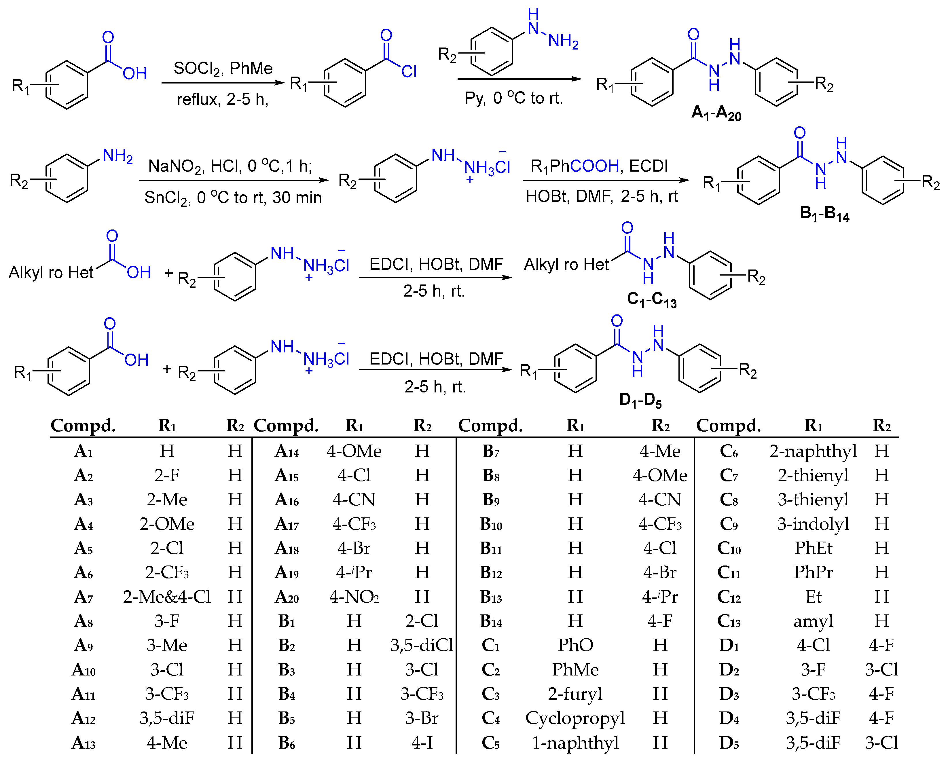
The synthetic routes of the target compounds were shown in Figure 3. Substituted benzoyl chloride was synthesized by the acylation of substituted benzoic acid with SOCl2. And a substitution reaction was occured between it and phenylhydrazine to gain series A compounds. At 0 °C, various substituted phenylhydrazines hydrochloride were achieved through the diazotization reactions. Then, series B compounds were got via the condensation reaction between phenylhydrazine hydrochloride with benzoic acid under EDCI and HOBt. The preparation of series C and D compounds was the condensation reaction between different kinds of carboxylic acids and phenylhydrazine hydrochloride.
A total of 52 N′-phenylhydrazides belonging to four different series were synthesized via concise and efficient methods in up to 96% yield. The structures of target N'-phenylhydrazides were identified by HRMS, 1H NMR, and 13C NMR spectra. It was discovered that five of them (A7, B13, D2, D4, and D5) were unknown. The separated yields, melting points, and the data of HRMS, 1H NMR, and 13C NMR spectra of N′-phenylhydrazides were illustrated in the supporting information.

2.2. Evaluation of Antifungal Activity in Vitro

The in vitro antifungal activities of target candidates against five strains of C. alb. were screened at the concentration of 0.125, 0.25, 0.5, 1, 2, 4, 8, 16, 32, and 64 μg/mL through the broth microdilution (BMD) method. FLC was selected as the positive control. Based on the data of antifungal activity, their MIC80 values and total activity index (TAI) were calculated according to the formulas, MIC80 =   10 log A + log 1 N × a 80 a b and TAI = 1 n 1 / M I C 80 , respectively. The total susceptibility index (TSI) of every stain of fungus was evaluated as well. By analyzing MIC80 and TAI values, the preliminary SRAs were concluded. According to the standards of the Clinical Laboratory Standards Institute, when MIC80 values were distributed in ≤4 μg/mL, 8-16 μg/mL, and≥32 μg/mL, the corresponding compounds were classified as susceptible, intermediate, and resistant candidates, respectively. [31]. To evaluate the potency of the compounds with greater inhibitory activity, time-inhibition rate curves were depicted for seven compounds (A1, A2, A8, A11, A12, B14, and C8) and their respective half-inhibitory time (IT50) were calculated.

2.2.1. Antifungal Activity of Target Compounds

A total of fifty-two kinds of compounds were synthesized, their fungicidal activities in vitro were screened at the concentration in the range of 0.125-64 μg/mL against five strains of fungi, and their MIC80 values and TAI values were calculated (Table 1).
All target compounds displayed definite inhibitory activities against the five test fungi. In terms of series A compounds, A11 held the best inhibitory activity against C. alb. SC5314, 4395, and 5272 and the corresponding MIC80 value was 1.9, 4.0, and 3.7μg/mL, respectively. Thus, A11 owned the highest TAI value (2.71). A8 (MIC80 = 0.7 μg/mL) showed the best inhibitory activity against C. alb. 5122. As far as the C. alb. 5172 was concerned, A16 (MIC80 = 5.8 μg/mL) displayed the best inhibitory activity against it.
As far as series B compounds were concerned, B3 (MIC80 = 6.9 μg/mL) held the most effictive antifungal activity against C. alb. SC5314. B14 had the most potent inhibitory activity against C. alb. 4395 and 5172 and the corresponding MIC80 values were 4.0 and 3.3 μg/mL, respectively. It also owned the highest TAI value (2.13). As far as the C. alb. 5122 was concerned, B3 (MIC80 = 0.7 μg/mL) displayed the best inhibitory activity against it. B5 (MIC80 = 6.3 μg/mL) showed the best inhibitory activity against C. alb. 5272.
As for series C compounds, C2 had the best inhibitory activity against C. alb. SC5314 and 5122 and the corresponding MIC80 values were 3.4 and 1.7 μg/mL, respectively. As far as the C. alb. 4395 and 5272 were concerned, C4 displayed the highest inhibitory activity against them and the corresponding MIC80 values were 8.9 and 3.8 μg/mL, respectively. C4 owned the highest TAI value (1.85). C9 (MIC80 = 1.4 μg/mL) showed the most powerfully antifungal activity against C. alb. 5172.
With respect to series D compounds, D2 exhibited the most potent antifungal activity against C. alb. SC5314 and 4395 with MIC80 values of 12.2 and 5.5 μg/mL, respectively. As far as the C. alb.5122, 5172, and 5272 were concerned, D5 displayed the best inhibitory activity against them and the corresponding MIC80 values were 2.2, 2.7, and 2.4μg/mL, respectively. Hence, D5 owned the highest TAI value (2.25).

2.2.2. The Inhibitory Efficiency of Compounds with Higher Activity

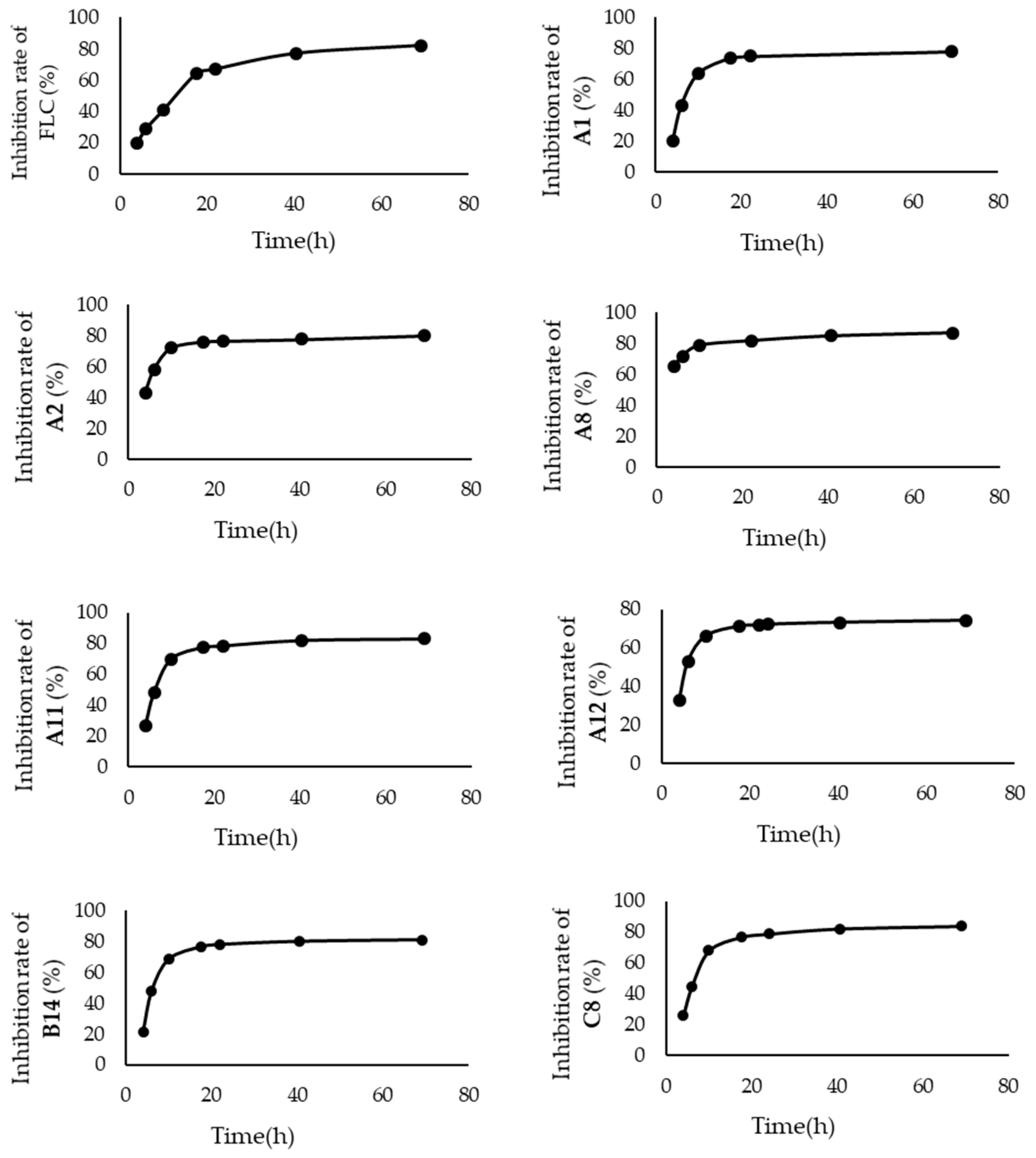
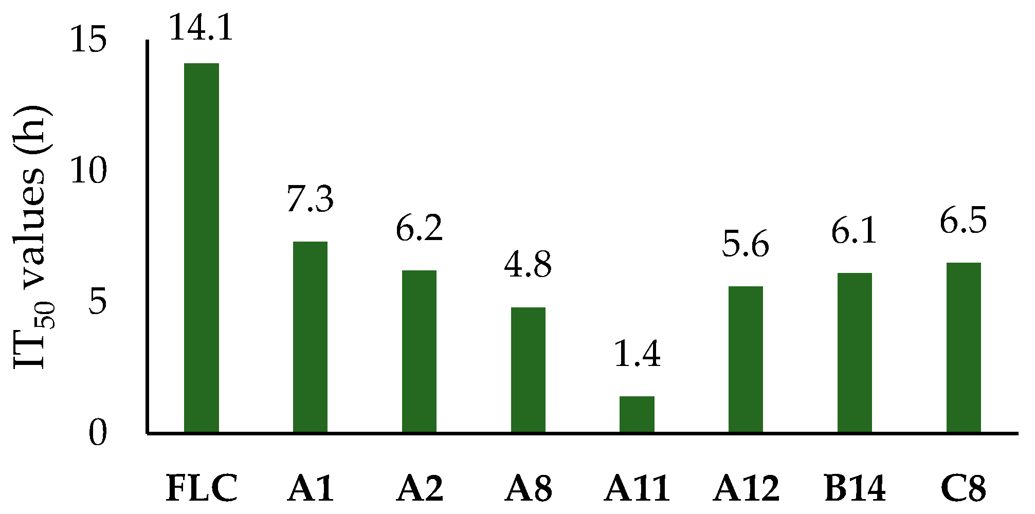
In order to evaluate the inhibitory potential of the compounds exhibiting higher activity, time-inhibition rate curves were depicted for seven compounds (A1, A2, A8, A11, A12, B14, and C8) at the concentration of 3.2 μg/mL against C. alb. SC5314 and their half-inhibitory times (IT50) were calculated (Figure 4 and Figure 5).
The time-inhibition rate curve of FLC showed a relatively smooth overall trend, whereas those of the seven tested compounds exhibited a steep rise in the inhibition rate during the 0-10 hours period and was followed by a gradual flattening after 20 hours. According to calculations, the IT50 value of FLC was 14.1 h. In contrast, the IT50 values of all seven tested compounds were less than 7.5 h. Compound A11, in particular, achieved the lowest IT50 value (1.4 h). Thus, this class of compounds held a more efficient antifungal efficacy, which might be attributed to their simple structure making them easy to enter fungal cells.

2.2.3. The Analysis of Preliminary SARs

In this study, the compounds with TAI values ≥1.19 were defined as susceptible compounds. When 25% of N’-phenylhydrazides owned MIC80 values ≤4 μg/mL against the fungi, the fungi were defined as sensitive ones. Sixteen susceptible compounds and one sensitive fungus, C. alb. 5122 were screened.
According to the above data, the TAI values of twenty-one compounds (A2, A4, A5, A6, A9, A10, A11, A13, A16, B1, B5, B7, B10, B11, B14, C1, C3, C4, C7, D1, and D5) with MIC80 ≤64 μg/mL were calculated according to the formula, TAI = 1 n 1 / M I C 80 , which reflected the antifungal activity of the compounds, and the larger the index, the higher the antifungal activity of the compound [32].
The total susceptibility index (TSI ) of the fungi were calculated according to the formula, TSI = 1 n 1 / M I C 80 , which reflects the sensitivity of every strain of C. alb., and the greater the index, the more sensitive the fungus is to these compounds (Table 2) [32].
Standard C. alb. SC5314 exhibited a high degree of sensitivity to FLC (TSI = 14.8). For series A and B compounds, the most sensitive fungus was C. alb. 5122, the TSI values were 11.6 and 12.0, respectively. C. alb 5172 showed the highest sensitivity to series C and D compounds, the TSI values were 7.8 and 11.2, respectively. Thus, C. alb. 5122 was a sensitive fungus.
The MIC80 and TAI values obtained allowed a preliminary evaluation of SARs. Contrast with A1 to obtain the general trends.
First, the introduction of a chlorine atom to the ortho-, meta-, or para-position of the carbonyl side benzene ring was able to sharply enhance the antifungal activity against all or most of the fungi (A5, A10, and A15). A similar case was observed for ortho-/meta-F, para-Br and meta-/para-CF3 substituted compounds (A2, A8, A6, A11, and A17). The more electron-withdrawing groups on ring A, the better the antifungal activity of the corresponding compounds (A12>A7).
Second, the introduction of nine substituents, identical to those present in the series A compounds, onto the phenyl ring of hydrazine side resulted in compounds with inferior antifungal activity compared to the series A compounds. Among the tested compounds, those with para-CH3, para-Cl, and para-F substituents (B7, B11, and B14) exhibited higher fungicide activities compared to ortho-Cl and meta-Br substitutes (B1 and B5).
Third, the fungicide activity was also influenced by the various heterocycles and alkyl groups. C4 with cyclopropyl significantly improved the fungicide activities toward C. alb. 5172 and 5272. However, the activities were cut down by the naphthyl group consumedly. A similar case was found for C3, C7, and C8 with polar heterocyclic groups (thienyl and furan), which were less effective than A1. The short-chain alkane counterparts (C12) possessed better activities against five strains of fungi than their long-chain alkane counterparts (C13). The longer the carbon chain between the benzene ring and the carbonyl group, the lower the antifungal activity of the corresponding compounds (C11<C10<C2).
Last, combining the pharmacophores of ring A (-F, -Cl, and -CF3) and B (-F and -Cl) sharply enhanced the antifungal activity of series D compounds. However, their antifungal activity still did not reach the level of A11.

2.3. The Investigation of the Antifungal Mechanism

In this research, C. alb. SC5314 was chosen as the tested fungi, and A11 with the highest TAI (2.71) value was used as the tested compound.

2.3.1. Assay of Free Radical Scavenging

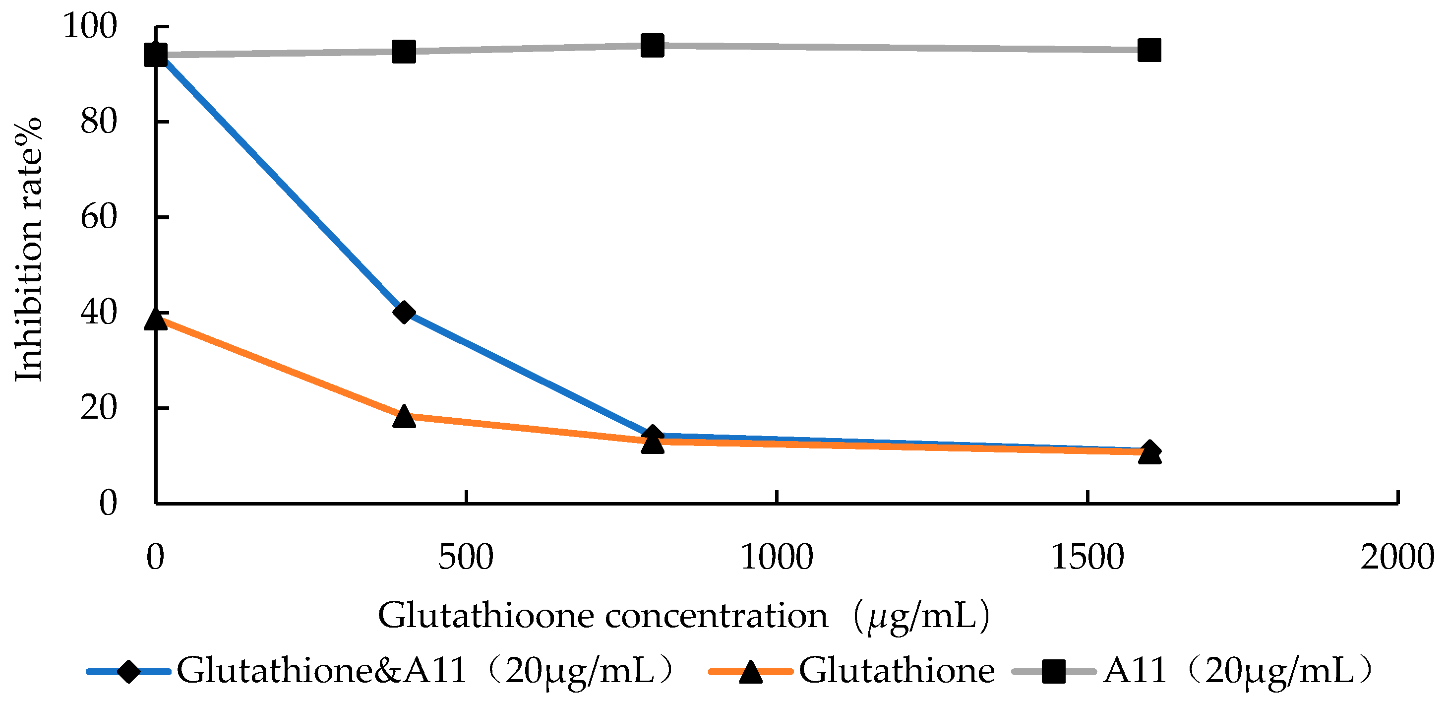
In this assay, glutathione was selected as the free radical scavenger. With an increase in the concentration of glutathione, the inhibition rate of A11 against C. alb. SC5314 decreased continuously. When the concentration of glutathione reached 1600 μg/mL, the antifungal effect of A11 was essentially disappeared (Figure 6).
At concentrations of 1600, 800, and 400 μg/mL, glutathione exhibited low inhibitory effects on the growth of standard C. alb. SC5314 and the inhibition rate of A11 remained consistently stable at around 95%. At the time of the concentrations of glutathione increased, the inhibitory effect of A11 reduced. In the event of the concentration of glutathione was at 400 μg/mL, the inhibition rate of A11 decreased from 94.7% to 18.4%. On the occasion that the concentration of glutathione was raised to 800-1600 μg/mL, the inhibition rate of A11 dropped to below 20%, similar to the inhibition rate observed when glutathione was used alone. This indicated that A11 produced free radicals during the metabolism process within fungal cells.

2.3.2. Production of ROS

To monitor cellular redox processes, the common oxidative stress indicator DCFH-DA was utilized. As shown in Figure 7, treatment with A11 resulted in a significantly higher fluorescence intensity of DCFH-DA (Figure 7A–C) as compared to the control groups (Figure 7D). These results suggested that A11 could impair mitochondrial function and increase the production of large numbers of ROS in C. alb. SC5314.

2.3.3. Effects of A11 on Hyphal Morphology

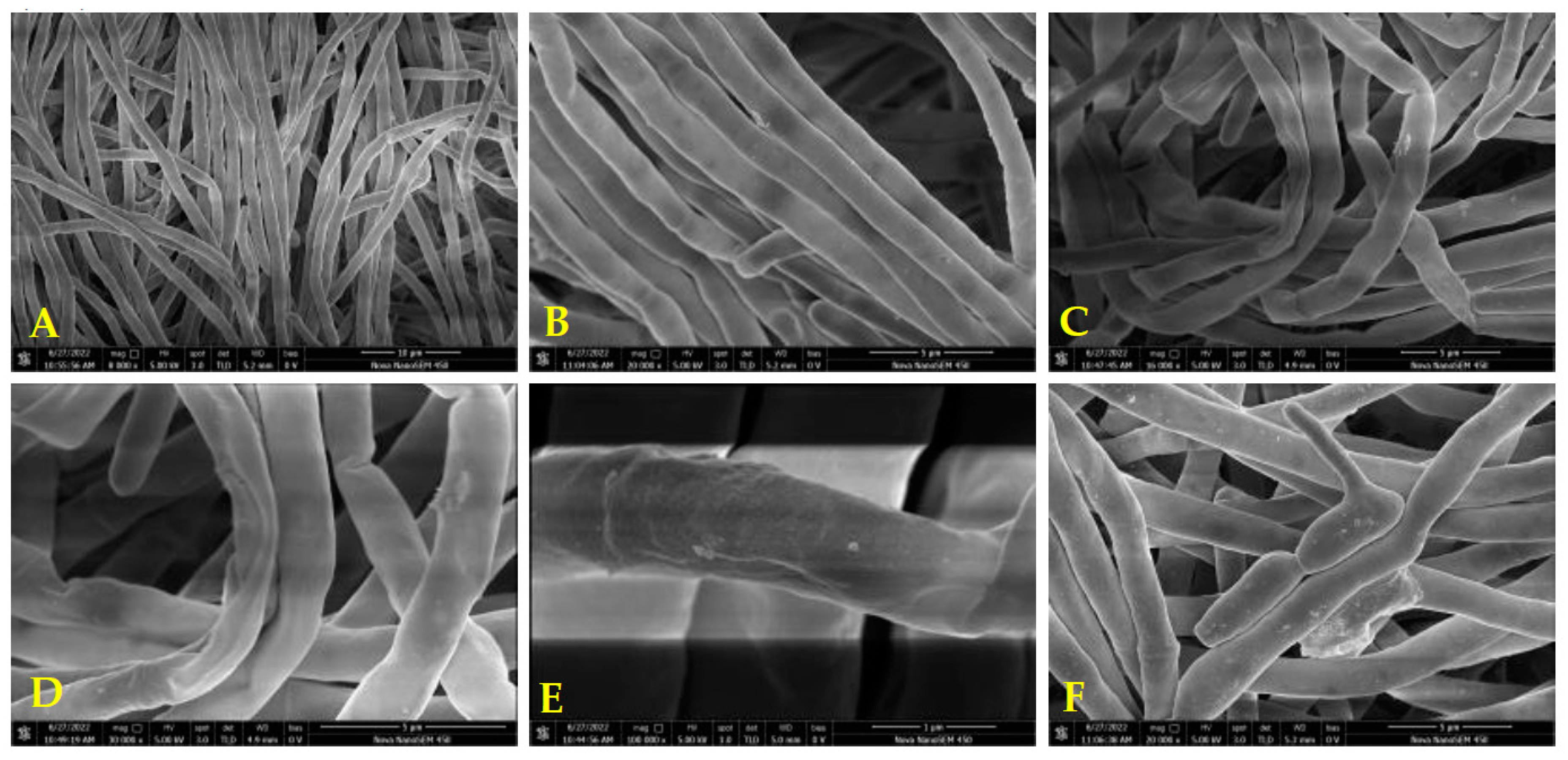
The impact of A11 on the hyphal morphology against C. alb. SC5314 was observed by SEM. In Figure 8A–F, significant morphological changes in the filamentous form of C. alb. were observed after treatment with 20 μg/mL of A11 compared to the control group. The untreated hyphae exhibited dispersed, regular, plump, uniform, and smooth surfaces (Figure 8A,B). In contrast, the treated hyphae appeared shriveled, distorted, with rough surfaces and displayed folding, wrinkling, and invagination (Figure 8C,D). Figure F depicted an abnormal transition of the fungus from the yeast form to the filamentous form.
According to the electron microscopy observations, the integrity of mycelia was damged by A11. The disruptive effect of A11 on hyphal morphology was the main reason for its inhibitory activity.

2.3.4. Preliminary Antifungal Mechanisms

Based on the research results in 2.3.1 and 2.3.2, the metabolic processes of A11 within the cell could be preliminarily analyzed. Firstly, A11 was hydrolyzed by fungal amidase to generate phenylhydrazine. In the structure of hydrazine (H2N-NH2), the lone pairs of electrons on the nitrogen atoms repelled each other strongly and led to the reducibility of phenyl hydrazides. Then, under the action of P450, phenyl hydrazine was oxidized to diazo compounds and then released nitrogen gas and generated free radicals. Their reducibility could lead to the reduction of oxygen within cells, forming reactive oxygen species (ROS) as well such as superoxide anions and hydroxyl radicals. Excessive ROS and free radicals disrupted the normal physiological processes of the fungus and subsequently affected the hyphal morphology of the fungi (Figure 9). This antifungal mechanism was similar to previously reported [33].
The different degrees of inhibition of C. alb. by target compounds stemmed from two factors. Firstly, their antifungal activity was affected by the rate of free radical production. The hydrolysis of amide bonds was the first step in the metabolism of N'-phenylhydrazines and was also a necessary condition for the formation of free radicals. The electron cloud density of the carbonyl group can be reduced and the hydrolysis of the amide bond can be promoted when the electron absorbing group was introduced to the substituted benzoic acid, thus the formation rate of the corresponding free radical was faster. For example, the ortho-fluoride, ortho-trifluoromethyl, meta-chlorine, meta-trifluoromethyl, and para-cyano substituted compounds (A2, A6, A10, A11, and A16) had higher antifungal activity compared to the ortho-, meta- or para-methyl and para-methoxy substituted ones (A2, A9, A13, and A14). The different substituents affected the affinity between the corresponding compounds and hydrolase and the hydrolysis rate and antifungal activity of the compounds.
Secondly, their antifungal activity depend on the stability of the generated free radicals, and the poorer the stability of the free radicals, the higher the antifungal activity of the compound. The electron-withdrawing group reduced the electron cloud density of the benzene ring thereby reducing the stability of substituted phenylhydrazine. For example, compounds substituted with meta or para halogen atoms (B5, B11, B12, and B14) exhibited higher activity compared to compounds substituted with para methyl groups (B7).
The pathogenicity of C. alb. is closely associated with the formation of hyphae, which is also an important factor in the formation of biofilms [34]. Typically, the filamentous form of C. alb. exhibits greater pathogenicity compared to the yeast form [35]. Under normal growth conditions, the hyphae surface appears uniform, plump, and smooth without any folds or wrinkles. In the pathological state, the microscopic appearance of the fungal hyphae exhibits signs of invagination and folding [36]. According to Figure 8, it can be observed that the treatment of C. alb. with compound A11 resulted in an abnormal morphology of the hyphae. This indicated that compound A11 exhibited a certain degree of inhibition on the formation and growth process of C. alb. hyphae. A11 undergone metabolism within the fungal cells and led to the generation of free radicals and ROS disrupted the normal physiological processes of the fungus, and subsequently damaged its hyphal morphology. Next, in vivo antifungal activity and cytotoxicity assay tests will be carried out.

3. Materials and Methods

3.1. Materials

Substituted benzoic acid, substituted phenyl, substituted phenyl hydrazine hydrochloride, 1-Ethyl-3-(3-dimethylaminopropyl) carbodiimide hydrochloride (EDCI), 1-Hydroxybenzotriazole (HOBt), dried N, N-Dimethylformamide (DMF), pyridine, heterocyclic carboxylic acids, alkyl carboxylic acid, phenylhydrazine, fluconazole, and dimethyl sulfoxide (DMSO) were all purchased from Adamas Reagent Co. Ltd. (Shanghai, China). Other analytical pure reagents and solvents were bought from local companies. And before using a number of reagents were purified by standard procedures. Without other explicit direction, water was redistilled and ions were removed before use.
Standard C. alb. SC5314 was donated by Professor Dazhi Zhang of the Second Military Medical University. FLC-resistant fungi C. alb. 4935, 5122, 5172, and 5272 were donated by Professor Changzhong Wang of Anhui University of Chinese Medicine. At 30 °C, the tested fungi were cultured for 48 h on the reasoner's 2A (R2A) agar plates. The dilution of suspension was made with RPMI medium 1640, a volume of 1 mL of these suspensions contained 1×10⁴–1×10⁵ colony forming unit. The Tested compounds were diluted at the concentration of 8 μg/mL with DMSO.
Making use of a mixture of petroleum ether (PE) and ethyl acetate (EtOAc) as the mobile agent and thin-layer chromatography (TLC) using GF254 silica gel to monitor the progress of the reactions. An XT-4 micro melting point instrument was used to measure the melting point of every target compound. A Bruker Advance neo spectrometer (400 MHz) was applied to obtain the 1H NMR and 13C NMR spectrum of every synthesized chemical. Chemical shifts (δ values) and coupling constants (J values) were presented in ppm and Hz, respectively. The high-resolution mass spectra (HRMS) was took advantaged of to characterize every unknown compound. On a mass spectrometer (Thermo Scientific LTQ Orbitrap XL) the positive ion mass spectra of the samples were gained.
Thin-layer chromatography (TLC) precoated GF254 silica gel was employed to monitor the progression of each reaction. The mobile phase consisted of a mixed solution of petroleum ether (PE) and ethyl acetate (EtOAc). The melting points of every target compound was measured using an XT-4 micro melting point instrument. 1H NMR and 13C NMR spectra of all target compounds were obtained at 400 MHz using a Bruker Advance neo spectrometer. Chemical shifts (δ values) were reported in ppm while coupling constants (J values) were presented in Hz. Additionally, high-resolution mass spectra (HRMS) were performed to further characterize all unknown products. Positive ion mass spectrometry of the sample was acquired using a Thermo Scientific LTQ Orbitrap XL instrument.

3.2. The synthetic procedure

3.2.1. Synthesis of Substituted Phenylhydrazine Hydrochloride

Added NaNO2 (1.90 g, 27.5 mmol,1.1 equiv.) to a solution of substituted aniline (2.33 g, 25 mmol,1 equiv.) in hydrochloric acid (60 mL, 20%, V/V), at 0°C and stirred the reaction mixture at this temperature for 1 h. Then added the SnCl2 (9.48 g, 2 equiv., 50mmol) and stirred the mixture for 2 h at room temperature. The precipitate was filtered and washed with brine and Et2O, and dried in vacuo at 40°C overnight [37].

3.2.2. Synthesis of N’-Phenylhydrazides

EDCI (8.8 mmol, 1.1 equiv.) and HOBt (8.8mmol, 1.1 equiv.) were added to a solution of carboxylic acid (8.0 mmol, 1 equiv.) in DMF (20 mL), then phenylhydrazine hydrochloride was added and the reaction mixture was stirred at ambient temperature under nitrogen atmosphere for 3 h, TLC detection. The reaction mixture was poured into H2O (300 mL), filtrated, and recrystallized by ethanol to give the desired light yellow or brownish-yellow product [29].

3.3. Antifungal Activity In Vitro

3.3.1. Determination of MIC80 value

The antifungal activities in vitro of target compounds against five stains of fungi were determined via the BMD method with assays in 96-well plates. FLC was selected as the positive control [31].
To prepare the stock solutions of the tested compounds, they were dissolved in 100% DMSO with a concentration of 0.8 mg/mL. Before the assay the solutions were diluted to the concentration of 8.0 μg/mL with RPMI medium 1640. The final concentrations of tested compounds were 64, 32, 16, 8, 4, 2, 1, 0.5, 0.25, and 0.125μg/mL. After inoculation, the plates were incubated at 30 °C for 24 h. The absorbance of each well was scanned at a wavelength of 625 nm via an ELISA reader, and the inhibition rate was calculated.
Inhibition   rate   ( % ) = [ ( A P A D ) /   ( A   P A   N ) ] × 100 %
Where AP is the absorbance of positive control wells, AD is the absorbance of drug wells and AN is the absorbance of negative control wells.
The MIC80 values were the concentration of tested compounds when the inhibition rate was at 80%. A curve was plotted with the logarithm of the concentration as the horizontal axis and the inhibition rate as the vertical axis, and the MIC80 was calculated.
MIC 80 = 10 log A + log 1 N × a 80 a b
Among them, A refers to the corresponding minimum concentration in the aforementioned gradient concentrations when the inhibition rate is just above 80%. a is the inhibition rate at concentration A, b is the inhibition rate just below a, and N represents the dilution factor.

3.3.2. Time-Inhibition Rate Curves

6.4 mg of the tested compound was dissolved in 0.5 mL of DMSO, then added RPMI medium 1640 to obtain a drug solution with a concentration of 3.2 μg/mL. The specific procedures are the same as those described in section 3.3.1. The absorbances were recorded at 0 h, 4 h, 6 h, 10 h, 17.5 h, 22 h, 24 h, 40.5 h, and 70 h, respectively. Every test was conducted three times. The time-inhibition rate curves were plotted by origin software with the time represented on the x-axis and the corresponding inhibition rate on the y-axis.

3.4. The Investigation of the Antifungal Mechanism

3.4.1. Scavenging of Free Radical Generated from A11

The impact of A11 on the emergence of free radical of C. alb. SC5314 was evaluated. Glutathione with gradient concentrations (400, 800, and 1600 μg/mL) was set as a control group to exclude its effect on fungal growth. A11 at the concentration of 20 μg/mL had pretty antifungal activity, and its inhibition rate against C. alb. SC5314 was close to 100%. A11 was added to gradient concentrations (400, 800, and 1600 μg/mL) of glutathione and achieved a concentration of A11 of 20 μg/mL. As shown in 3.3.1., the assay should be followed.

3.4.2. Production of ROS

The influence of A11 on the generation of ROS of C. alb. SC5314 was ascertained as previously described [38]. DCFH-DA (2',7'-dichlorodihydrofluorescein diacetate, 40 μM, 0.5mL) was used to stain the hyphal tips of the fungal mycelium, followed by incubation in the dark at 25°C for 20 minutes and cleansed with PBS twice, each time for 5 minutes. At an excitation and emission wavelength of 488 nm and 525 nm, respectively, the fluorescence images were observed and captured before and after stimulation by CLSM (LEICA TCS SP8). Images were stored on a computer for further processing and analysis.

3.4.3. Analysis of the Mycelial Morphology

By SEM, the mycelial morphology of C. alb. SC5314 was observed according to the reported method [38]. Compound A11 (TAI = 2.71) and standard C. alb. SC5314 was chosen as the tested specimen and fungus, respectively.
The fungi were cultured in a thermostatic shaker at 37 °C and 200 rpm for a period of 8 to 24 hours. Subsequently, they were soaked in a 4% glutaraldehyde solution for a duration of 4 hours. Following this, the fungi were washed three times using a 0.1 mol/L PBS solution with a pH of 7.2. To initiate dehydration, the fungi were exposed to a series of ethanol concentrations including 30%, 50%, 70%, 80%, and 90% (v/v). Thereafter, the fungi were treated with 100% ethanol thrice, with each exposure lasting fifteen minutes. Finally, the hyphae were subjected to vacuum drying, coated with gold, and examined through scanning electron microscopy (SEM) using a Hitachi S-4800 instrument [39].

4. Conclusion

Fifty-two kinds of target compounds were synthesized efficiently and concisely up to 96% yield, and their structures were characterized by HRMS, 1H NMR, and 13C NMR. It was discovered that five of the compounds (A7, B13, D2, D4, and D5) were unknown.
The target compounds produced different degrees of inhibition against C. alb., among which A11 had the highest antifungal activity for all tested fungi, and its TAI value was 2.71, which was higher than that of the positive control FLC (TAI = 1.19). The presence of an electron-absorbing group could significantly enhance antifungal activity of the compounds, in comparison to those having an electron-giving group. Particularly, compound series D showed considerably higher antifungal activity than A1. The IT50 values of the seven compounds were about half that of FLC, and A11 was only 1.4 h, which had a more efficient antifungal mechanism.
Further exploration of mechanisms revealed that A11 promoted the generation of endogenous ROS and free radicals, disrupted the physiological processes of C. alb. SC5314 and impaired its filamentous morphology, ultimately inhibited fungal growth.
In summary, A11 is expected to be a novel fungicide, and this research held significant importance for the development of antifungal agents. Next, in vivo antifungal activity and cytotoxicity assays will be carried out.

Supplementary Materials

The following supporting information can be downloaded at the website of this paper posted on Preprints.org.

Author Contributions

Conceptualization, Panpan Zhu and Huiling Geng; Data curation, Panpan Zhu; Funding acquisition, Huiling Geng; Investigation, Panpan Zhu; Supervision, Huiling Geng; Writing – original draft, Panpan Zhu, Jinshuo Zheng, Jin Yan, Zhaoxia Li and Xinyi Li; Writing – review & editing, Panpan Zhu, Jinshuo Zheng, Jin Yan, Zhaoxia Li and Xinyi Li.

Funding

This research was funded by the National Natural Science Foundation of China (No. 31572038) and the Key Research and Development Project of Shaanxi Province (2023-YBNY-242).

Institutional Review Board Statement

Not applicable.

Informed Consent Statement

Not applicable.

Data Availability Statement

Not applicable.

Acknowledgments

We extend our sincere appreciation to the Chemical Experiment Teaching and Research Center for their valuable support in conducting nuclear magnetic resonance analysis. Additionally, we are thankful to the Life Science Research Core Service for providing us with access to electron microscopy analysis and ESI-HRMS.

Conflicts of Interest

The authors declare no conflict of interest.

References

  1. Marston, H.D.; Dixon, D.M.; Knisely, J.M.; Palmore, T.N.; Fauci, A.S. Antimicrobial Resistance. JAMA 2016, 316, 1193–1204. [Google Scholar] [CrossRef] [PubMed]
  2. Dartois, V.A.; Rubin, E.J. Anti-Tuberculosis Treatment Strategies and Drug Development: Challenges and Priorities. Nature Reviews Microbiology 2022, 20, 685–701. [Google Scholar] [CrossRef] [PubMed]
  3. Zhang, Y.; Li, Q.; Chao, W.; Qin, Y.; Chen, J.; Wang, Y.; Liu, R.; Lv, Q.; Wang, J. Design, Synthesis and Antifungal Evaluation of Novel Pyrylium Salt in Vitro and in Vivo. Molecules 2022, 27, 4450. [Google Scholar] [CrossRef] [PubMed]
  4. Arendrup, M.C.; Patterson, T.F. Multidrug-Resistant Candida: Epidemiology, Molecular Mechanisms, and Treatment. J Infect Dis 2017, 216, S445–S451. [Google Scholar] [CrossRef] [PubMed]
  5. Wijnants, S.; Vreys, J.; Van Dijck, P. Interesting Antifungal Drug Targets in the Central Metabolism of Candida Albicans. Trends in Pharmacological Sciences 2022, 43, 69–79. [Google Scholar] [CrossRef]
  6. Nobile, C.J.; Johnson, A.D. Candida Albicans Biofilms and Human Disease. Annu. Rev. Microbiol. 2015, 69, 71–92. [Google Scholar] [CrossRef]
  7. Xiao, M.; Sun, Z.Y.; Kang, M.; Guo, D.W.; Liao, K.; Chen, S.C.; Kong, F.; Fan, X.; Cheng, J.W.; Hou, X.; et al. Five-Year National Surveillance of Invasive Candidiasis: Species Distribution and Azole Susceptibility from the China Hospital Invasive Fungal Surveillance Net (Chif-Net) Study. J Clin Microbiol 2018, 56. [Google Scholar] [CrossRef]
  8. Mukaremera, L.; Lee, K.K.; Mora-Montes, H.M.; Gow, N.A.R. Candida Albicans Yeast, Pseudohyphal, and Hyphal Morphogenesis Differentially Affects Immune Recognition. Frontiers in Immunology 2017, 8. [Google Scholar] [CrossRef]
  9. Tits, J.; Cammue, B.P.A.; Thevissen, K. Combination Therapy to Treat Fungal Biofilm-Based Infections. Int. J. Mol. Sci. 2020, 21, 8873. [Google Scholar] [CrossRef]
  10. Mahboub, H.H.; Eltanahy, A.; Omran, A.; Mansour, A.T.; Safhi, F.A.; Alwutayd, K.M.; Khamis, T.; Husseiny, W.A.; Ismail, S.H.; Yousefi, M.; et al. Chitosan Nanogel Aqueous Treatment Improved Blood Biochemicals, Antioxidant Capacity, Immune Response, Immune-Related Gene Expression and Infection Resistance of Nile Tilapia. Comparative Biochemistry and Physiology Part B: Biochemistry and Molecular Biology 2024, 269, 110876. [Google Scholar] [CrossRef]
  11. Li, J.; Buchner, J. Structure, Function and Regulation of the Hsp90 Machinery. Biomed J 2013, 36, 106–117. [Google Scholar] [PubMed]
  12. Lee, K.-H.; Park, S.J.; Choi, S.J.; Park, J.Y. Proteus Vulgaris and Proteus Mirabilis Decrease Candida Albicans Biofilm Formation by Suppressing Morphological Transition to Its Hyphal Form. Yonsei Med. J. 2017, 58, 1135–1143. [Google Scholar] [CrossRef] [PubMed]
  13. White, P.L.; Dhillon, R.; Hughes, H.; Wise, M.P.; Backx, M. Covid-19 and Fungal Infection: The Need for a Strategic Approach. The Lancet Microbe 2020, 1, e196. [Google Scholar] [CrossRef] [PubMed]
  14. Maubon, D.; Garnaud, C.; Calandra, T.; Sanglard, D.; Cornet, M. Resistance of Candida Spp. To Antifungal Drugs in the Icu: Where Are We Now? Intensive Care Medicine 2014, 40, 1241–1255. [Google Scholar] [CrossRef] [PubMed]
  15. Hoang, A. Caspofungin Acetate: An Antifungal Agent. Am. J. Health-Syst. Pharm. 2001, 58, 1206–1214. [Google Scholar] [CrossRef] [PubMed]
  16. Richardson, K.; Cooper, K.; Marriott, M.S.; Tarbit, M.H.; Troke, P.F.; Whittle, P.J. Discovery of Fluconazole, a Novel Antifungal Agent. Rev Infect Dis 1990, 12 Suppl 3, S267–S271. [Google Scholar] [CrossRef]
  17. Boken, D.J.; Swindells, S.; Rinaldi, M.G. Fluconazole-Resistant Candida Albicans. Clin Infect Dis 1993, 17, 1018–1021. [Google Scholar] [CrossRef]
  18. Pasquale, T.; Tomada, J.R.; Ghannoun, M.; Dipersio, J.; Bonilla, H. Emergence of Candida Tropicalis Resistant to Caspofungin. J. Antimicrob. Chemother. 2008, 61, 219. [Google Scholar] [CrossRef]
  19. Sawant, B.; Khan, T. Recent Advances in Delivery of Antifungal Agents for Therapeutic Management of Candidiasis. Biomed. Pharmacother. 2017, 96, 1478–1490. [Google Scholar] [CrossRef]
  20. Carmo, A.; Rocha, M.; Pereirinha, P.; Tomé, R.; Costa, E. Antifungals: From Pharmacokinetics to Clinical Practice. Antibiotics 2023, 12. [Google Scholar] [CrossRef]
  21. Wang, D.; Zetterström, C.E.; Gabrielsen, M.; Beckham, K.S.H.; Tree, J.J.; Macdonald, S.E.; Byron, O.; Mitchell, T.J.; Gally, D.L.; Herzyk, P.; et al. Identification of Bacterial Target Proteins for the Salicylidene Acylhydrazide Class of Virulence-Blocking Compounds*. Journal of Biological Chemistry 2011, 286, 29922–29931. [Google Scholar] [CrossRef] [PubMed]
  22. Tirapegui, C.; Acevedo-Fuentes, W.; Dahech, P.; Torrent, C.; Barrias, P.; Rojas-Poblete, M.; Mascayano, C. Easy and Rapid Preparation of Benzoylhydrazides and Their Diazene Derivatives as Inhibitors of 15-Lipoxygenase. Bioorganic & Medicinal Chemistry Letters 2017, 27, 1649–1653. [Google Scholar]
  23. Kozlov, M.V.; Konduktorov, K.A.; Shcherbakova, A.S.; Kochetkov, S.N. Synthesis of N′-Propylhydrazide Analogs of Hydroxamic Inhibitors of Histone Deacetylases (Hdacs) and Evaluation of Their Impact on Activities of Hdacs and Replication of Hepatitis C Virus (Hcv). Bioorganic & Medicinal Chemistry Letters 2019, 29, 2369–2374. [Google Scholar]
  24. Turan-Zitouni, G.; Altintop, M.D.; Ozdemir, A.; Demirci, F.; Abu Mohsen, U.; Kaplancikli, Z.A. Synthesis and Antifungal Activity of New Hydrazide Derivatives. J Enzyme Inhib Med Chem 2013, 28, 1211–1216. [Google Scholar] [CrossRef] [PubMed]
  25. Pham, V.H.; Phan, T.P.D.; Phan, D.C.; Vu, B.D. Synthesis and Bioactivity of Hydrazide-Hydrazones with the 1-Adamantyl-Carbonyl Moiety. Molecules 2019, 24. [Google Scholar] [CrossRef]
  26. Nam, G.; Suh, J.-M.; Yi, Y.; Lim, M.H. Drug Repurposing: Small Molecules against Cu(Ii)–Amyloid-Β and Free Radicals. Journal of Inorganic Biochemistry 2021, 224, 111592. [Google Scholar] [CrossRef]
  27. Ganrot, P.O.; Rosengren, E.; Gottfries, C.G. Effect of Iproniazid on Monoamines and Monamine Oxidase in Human Brain. Experientia 1962, 18, 260–261. [Google Scholar] [CrossRef]
  28. Zhang, H.-Z.; Drewe, J.; Tseng, B.; Kasibhatla, S.; Cai, S.X. Discovery and Sar of Indole-2-Carboxylic Acid Benzylidene-Hydrazides as a New Series of Potent Apoptosis Inducers Using a Cell-Based Hts Assay. Bioorganic & Medicinal Chemistry 2004, 12, 3649–3655. [Google Scholar]
  29. Wu, Y.-Y.; Shao, W.-B.; Zhu, J.-J.; Long, Z.-Q.; Liu, L.-W.; Wang, P.-Y.; Li, Z.; Yang, S. Novel 1,3,4-Oxadiazole-2-Carbohydrazides as Prospective Agricultural Antifungal Agents Potentially Targeting Succinate Dehydrogenase. Journal of Agricultural and Food Chemistry 2019, 67, 13892–13903. [Google Scholar] [CrossRef]
  30. Clements, J.S.; Islam, R.; Sun, B.; Tong, F.; Gross, A.D.; Bloomquist, J.R.; Carlier, P.R. N′-Mono- and N, N′-Diacyl Derivatives of Benzyl and Arylhydrazines as Contact Insecticides against Adult Anopheles Gambiae. Pesticide Biochemistry and Physiology 2017, 143, 33–38. [Google Scholar] [CrossRef]
  31. de Sousa, E.S.O.; Cortez, A.C.A.; de Souza Carvalho Melhem, M.; Frickmann, H.; de Souza, J.V.B. Factors Influencing Susceptibility Testing of Antifungal Drugs: A Critical Review of Document M27-A4 from the Clinical and Laboratory Standards Institute (Clsi). Brazilian Journal of Microbiology 2020, 51, 1791–1800. [Google Scholar] [CrossRef] [PubMed]
  32. Yang, S.-S.; Lv, Q.-Y.; Fu, J.; Zhang, T.-Y.; Du, Y.-S.; Yang, X.-J.; Zhou, L. New 7-Chloro-9-Methyl-2-Phenyl-3,4-Dihydro-Β-Carbolin-2-Iums as Promising Fungicide Candidates: Design, Synthesis, and Bioactivity. Journal of Agricultural and Food Chemistry 2022, 70, 4256–4266. [Google Scholar] [CrossRef] [PubMed]
  33. Kalgutkar, A.S.; Gardner, I.; Obach, R.S.; Shaffer, C.L.; Callegari, E.; Henne, K.R.; Mutlib, A.E.; Dalvie, D.K.; Lee, J.S.; Nakai, Y.; et al. A Comprehensive Listing of Bioactivation Pathways of Organic Functional Groups. Curr Drug Metab 2005, 6, 161–225. [Google Scholar] [CrossRef] [PubMed]
  34. Kakizaki, T.; Abe, H.; Kotouge, Y.; Matsubuchi, M.; Sugou, M.; Honma, C.; Tsukuta, K.; Satoh, S.; Shioya, T.; Nakamura, H.; et al. Live-Cell Imaging of Septins and Cell Polarity Proteins in the Growing Dikaryotic Vegetative Hypha of the Model Mushroom Coprinopsis Cinerea. Scientific Reports 2023, 13, 10132. [Google Scholar] [CrossRef]
  35. Triastuti, A.; Vansteelandt, M.; Barakat, F.; Amasifuen, C.; Jargeat, P.; Haddad, M. Untargeted Metabolomics to Evaluate Antifungal Mechanism: A Study of Cophinforma Mamane and Candida Albicans Interaction. Natural Products and Bioprospecting 2023, 13, 1. [Google Scholar] [CrossRef]
  36. Zhang, Y.-H.; Yang, S.-S.; Zhang, Q.; Zhang, T.-T.; Zhang, T.-Y.; Zhou, B.-H.; Zhou, L. Discovery of N-Phenylpropiolamide as a Novel Succinate Dehydrogenase Inhibitor Scaffold with Broad-Spectrum Antifungal Activity on Phytopathogenic Fungi. Journal of Agricultural and Food Chemistry 2023, 71, 3681–3693. [Google Scholar] [CrossRef]
  37. Jacob, N.; Guillemard, L.; Wencel-Delord, J. Highly Efficient Synthesis of Hindered 3-Azoindoles Via Metal-Free C-H Functionalization of Indoles. Synthesis 2020, 52, 574–580. [Google Scholar] [CrossRef]
  38. Afri, M.; Frimer, A.A.; Cohen, Y. Active Oxygen Chemistry within the Liposomal Bilayer Part Iv: Locating 2',7'-Dichlorofluorescein (Dcf), 2',7'-Dichlorodihydrofluorescein (Dcfh) and 2',7'-Dichlorodihydrofluorescein Diacetate (Dcfh-Da) in the Lipid Bilayer. Chem. Phys. Lipids 2004, 131, 123–133. [Google Scholar] [CrossRef]
  39. Wang, D.; Zhang, J.; Jia, X.; Xin, L.; Zhai, H. Antifungal Effects and Potential Mechanism of Essential Oils on Collelotrichum Gloeosporioides in Vitro and in Vivo. Molecules 2019, 24. [Google Scholar] [CrossRef] [PubMed]
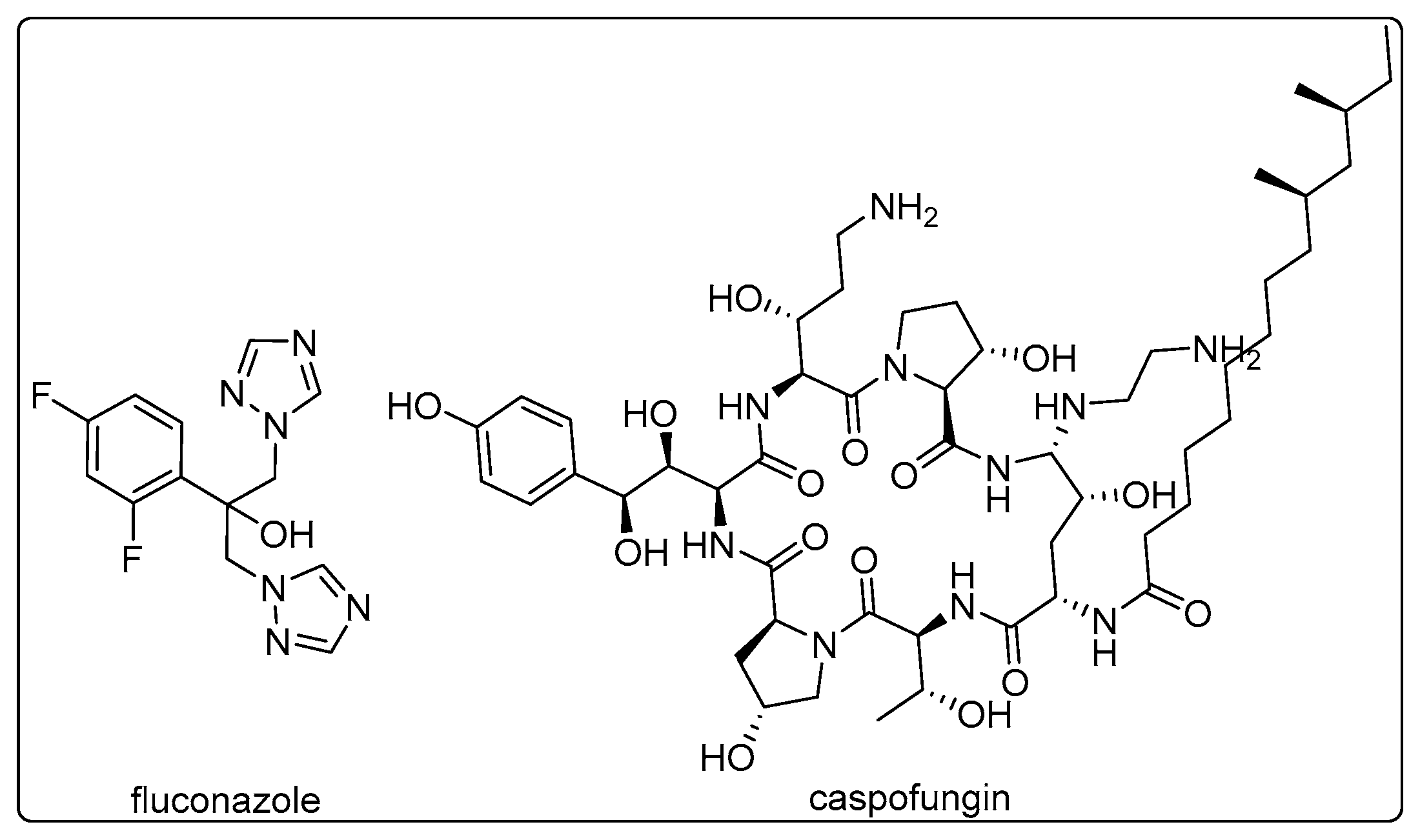
Figure 1. Commercial fungicides.
Preprints 82289 g001
Figure 3. Synthesis of target compounds A1-D5.
Preprints 82289 g003
Figure 4. Curves of time-inhibition rate for each compound.
Preprints 82289 g004
Figure 5. The IT50 values of the seven compounds (h).
Preprints 82289 g005
Figure 6. Effect of glutathione on the inhibition rate.
Preprints 82289 g006
Figure 7. Effects of compound A11 on the ROS of C. alb. SC5314.
Preprints 82289 g007
Figure 8. Microstructure of C. alb. SC5314 under SEM.
Preprints 82289 g008
Figure 9. The metabolic process of A11 in cells.
Preprints 82289 g009
Table 1. MIC80 values (μg/mL) of target compounds against C. alb.
Compd. R1 R2 C. alb.SC5314 C. alb.4395 C. alb.5122 C. alb.5172 C. alb.5272 TAI
A1 H H 26.0 15.0 0.9 >64.0 >64.0 <1.76
A2 2-F H 13.0 4.1 8.5 6.7 12.1 1.79
A3 2-Me H 49.3 >64.0 13.3 21.2 53.0 N. A.d
A4 2-OMe H 14.7 31.0 5.7 7.8 6.4 1.61
A5 2-Cl H 25.3 52.5 10.8 13.8 13.0 1.19
A6 2-CF3 H 11.7 4.5 6.3 12.0 22.1 1.66
A7 2-Me&4-Cl H 26.0 >64.0 >64.0 23.4 63.0 N. A.
A8 3-F H 3.4 9.7 0.7 >64.0 27.0 N. A.
A9 3-Me H 25.3 61.4 10.5 14.7 17.8 1.13
A10 3-Cl H 6.9 6.6 15.0 12.3 59.0 1.44
A11 3-CF3 H 1.9 4.0 2.8 7.4 3.7 2.71
A12 3,5-diF H 3.6 25.0 1.6 >64.0 >64.0 N. A.
A13 4-Me H 13.9 31.0 5.0 14.6 27.0 1.35
A14 4-OMe H 5.9 32.0 1.5 >64.0 >64.0 N. A.
A15 4-Cl H 5.9 32.0 1.5 >64.0 >64.0 N. A.
A16 4-CN H 47.5 15.0 6.3 5.8b 7.2 1.6
A17 4-CF3 H >64.0 15.0 4.6 9.6 9.3 N. A.
A18 4-Br H 24.4 >64.0 7.4 41.4 >64.0 N. A.
A19 4-iPr H >64.0 61.3 >64.0 >64.0 13.5 N. A.
A20 4-NO2 H 6.7 16 0.8 >64.0 >64.0 N. A.
B1 H 2-Cl 26.2 52.3 13.0 14.0 12.5 1.16
B2 H 3,5-diCl 45.3 >64.0 32.2 45.0 >64.0 N. A.
B3 H 3-Cl 6.9 19.0 0.7 >64.0 >64.0 N. A.
B4 H 3-CF3 >64.0 >64.0 31.5 58.6 >64.0 N. A.
B5 H 3-Br 24.6 60.0 3.6 13.9 6.3 1.52
B6 H 4-I 16.7 >64.0 31.8 30.0 19.0 N. A.
B7 H 4-Me 13.4 32.0 21.5 25.0 13.2 1.14
B8 H 4-OMe 9.2 22.0 0.8 >64.0 >64.0 N. A.
B9 H 4-CN >64.0 >64.0 >64.0 >64.0 62.6 N. A.
B10 H 4-CF3 >64.0 >64.0 >64.0 >64.0 >64.0 N. A.
B11 H 4-Cl 10.7 12.3 4.8 7.1 57 1.56
B12 H 4-Br 47.5 14.7 50.4 30 13.1 1.00
B13 H 4-iPr 14.3 >64.0 21.0 52.7 >64.0 N. A.
B14 H 4-F 12.2 4.0 3.5 3.3 14.7 2.13
C1 PhO H 31.7 14.3 11 6.7 7.2 1.50
C2 PhMe H 3.4 42 1.7 >64 >64 N. A.
C3 2-furyl H 17.5 45.8 2.7 7.3 13 1.64
C4 Cyclopropyl H 58.9 8.9 7.7 3.9 3.8 1.85
C5 1-naphthyl H >64.0 >64.0 62.0 >64.0 >64.0 N. A.
C6 2-naphthyl H 14.6 >64.0 51.8 58.7 >64.0 N. A.
C7 2-thienyl H 14.2 60.0 7.0 16.5 42.0 1.17
C8 3-thienyl H 13.9 22.6 11.6 14.7 >64.0 N. A.
C9 3-indolyl H >64.0 >64.0 >64.0 1.4 >64.0 N. A.
C10 PhEt H 51.8 16.1 15.0 >64.0 11.0 N. A.
C11 PhPr H >64.0 >64.0 22.8 >64.0 15.2 N. A.
C12 Et H >64.0 >64.0 13.8 >64.0 12.1 N. A.
C13 amyl H >64.0 42.6 34.4 >64.0 28.5 N. A.
D1 4-Cl 4-F 12.2 5.5 6.4 3.7 29.5 1.81
D2 3-F 3-Cl >64.0 13.8 9.6 >64.0 7.6 N. A.
D3 3-CF3 4-F 63.6 >64 3.8 >64.0 7.6 N. A.
D4 3,5-diF 4-F 30.4 13.5 6.3 >64.0 7.6 N. A.
D5 3,5-diF 3-Cl 64 29.3 2.2 2.7 2.4 2.25
FLC 1.8a >128.0 58.0 >64.0 >128.0 <1.19c
* a: red color refers to susceptible; b: green color refers to the highest activity; c: due to FLC's MIC80 values being greater than 128 μg/mL for C. alb. 4395, thus, the TAI value of FLC was recorded as<1.19; d: N. A. means that their TAI values are not available.
Table 2. TSI values of the specific fungus to all target compounds.
Compd. SC5314 4395 5122 5172 5272
A 6.4 5.4 11.6 5.8 5.2
B 5.0 4.8 12.0 4.8 5.0
C 5.0 4.2 6.4 7.8 5.8
D 3.4 5.8 9.2 11.2 7.6
FLC 14.8 <1.8a 2.6 <2.6 <1.8a
*a: the MIC80 values of FLC for C. alb. 4395 and 5272 were greater than 128 μg/mL, so their TSI values were recorded as<1.8.
Disclaimer/Publisher’s Note: The statements, opinions and data contained in all publications are solely those of the individual author(s) and contributor(s) and not of MDPI and/or the editor(s). MDPI and/or the editor(s) disclaim responsibility for any injury to people or property resulting from any ideas, methods, instructions or products referred to in the content.
Copyright: This open access article is published under a Creative Commons CC BY 4.0 license, which permit the free download, distribution, and reuse, provided that the author and preprint are cited in any reuse.
Prerpints.org logo

Preprints.org is a free preprint server supported by MDPI in Basel, Switzerland.

Subscribe

© 2024 MDPI (Basel, Switzerland) unless otherwise stated