Preprint
Case Report

Pancreatic Neuroendocrine Tumor (P-NET) Presented by Abdominal Pain: Case Report and Literature Review

Altmetrics

Downloads

193

Views

50

Comments

1

Submitted:

14 September 2023

Posted:

18 September 2023

You are already at the latest version

Alerts
Abstract
Pancreatic neuroendocrine tumor (P-NET) is a rare neoplasm originating in the neuroendocrine system. Carcinoid syndrome occurs in approximately 19% of patients with functional P-NETs, typically when liver metastases occur. NETs diagnosis is frequently late, along with symptoms related to hormone hypersecretion. We described the case of a patient with a low-grade non-functional P-NET, but with a typical clinical presentation of a carcinoid syndrome; moreover, we reviewed the literature regarding this topic. An 81-year-old male was admitted to our Department of Internal Medicine at Cannizzaro Hospital (Catania, Italy) because of the onset of abdominal pain with nausea, loose stools and episodic flushing. Firstly, an abdominal contrast-enhanced CT scan showed a small pancreatic hypervascular mass; then a gallium-68 DOTATOC integrated PET/CT revealed an elevated expression of SSTR receptors. Serum Chromogranin A and urinary 5-HIAA measurements resulted negative. Given the small size of the lesion (8 mm), we preferred to perform an endoscopic ultrasonography (EUS) with fine-needle biopsy (EUS-FNB), allowing the diagnosis of low-grade (G1) non-functional P-NET (NF-P-NET). Surgery was waived, while a follow-up strategy was chosen. Early recognition of P-NETs, although rare, is necessary to improve patient’s survival. EUS-FNB should be the protocol of choice for an early characterization of these tumors.
Keywords: 
Subject: Medicine and Pharmacology  -   Oncology and Oncogenics

1. Introduction

Neuroendocrine neoplasms (NENs) are enigmatic malignancies with an increasing incidence and prevalence[1,2,3]. Given the common morphological and immunophenotypical features, all these tumors arise from cells of the diffuse endocrine system.
NENs range from asymptomatic well-differentiated neuroendocrine tumors (NETs) to aggressive neuroendocrine carcinomas (NECs). In fact, nearly 80%-90% of NENs are NETs, while the remaining 10%-20% are carcinomas[4].
NETs can develop in any tissue of the body. Gastrointestinal tract and pancreas are the most common sites of origin, accounting for approximately 60% of the primary sites[5], followed by lungs and other sites.
About 40% of NETs can release hormones responsible for symptoms, depending on the secreted hormone. Carcinoid syndrome is characterized by episodic flushing and diarrhea, due to various vasoactive substances (serotonin, histamine, and other amines.) released into the systemic circulation[6].
Non-functional NETs may often present with subtle and sporadic symptoms, sometimes with gastrointestinal bleeding, abdominal pain, bowel obstruction or unexplained weight loss[7].
Treatment and prognosis depend on the grade and stage of the tumor. NETs diagnosis is frequently late, along with symptoms related to hormone hypersecretion, often after metastases occurred in the liver, where bioactive substances fail to be inactivated. Early diagnosis and recognition are necessary to improve patient’s survival, that did not significantly change over the last 30 years[8].
In this paper, we presented a case of a pauci-symptomatic pancreatic neuroendocrine tumor in a patient with unspecific clinical presentation (abdominal pain) and mild additional symptoms (nausea and loose stools). This was the occasion for a narrative review of the literature on the diagnosis and the management of pancreatic neuroendocrine tumors (P-NETs).

2. Case-report

In May 2023, an 81-year-old man was admitted to our Department of Internal Medicine at Cannizzaro Hospital (Catania, Italy) because of the onset of abdominal pain, especially in the lower abdominal quadrants, with nausea and loose stools (<3 times/day).
The patient’s past medical history included arterial hypertension, type 2 diabetes mellitus, peripheral artery disease (PAD), obesity, hypothyroidism and depressive syndrome. In the past six months he complained of abdominal distension and changes in bowel habits (loose stools). No relevant family history. He was taking levothyroxine, insulin according to HGT, lansoprazole, acarbose, ezetimibe/simvastatin and furosemide. He denied anamnestic consumption of uncooked meat, or fish, or unpasteurized dairy products.
On admission, no fever, arterial hypertension (177/76 mmHg), normal heart rate (86 bpm), glycemia 102 mg/dL and normal SaO2 on room air (98%); no sensorium alterations. Physical examination revealed abdominal distension, with colic pain on deep palpation and hypoactive abdomen sounds. Mucous membranes were normally hydrated. Bedside FAST (Focused Assessment with Sonography in Trauma) scan did not detect peritoneal fluid. Digital rectal examination showed blood traces.
Laboratory tests were performed, showing an increase in serum CRP (17.9 mg/dL), moderate leukocytosis, moderate renal dysfunction (serum Cr: 1,33 mg/dL, eGFR: 54 ml/min/1.73 m2), normal serum potassium (3.6 mEq/L), sodium (139 mEq/L) and chloride (100 mEq/L), mild metabolic acidosis (pH: 7.33, HCO3: 21 mmol/L, pCO2: 42 mmHg) and serum procalcitonin <0.2 ng/mL. Infectious causes of diarrhea were excluded by microbiological and chemical fecal examinations. An abdomen X-ray excluded bowel obstruction or perforation. Moderate intravenous fluid repletion was given.
Few hours from admission, the patient experienced transient states of agitation, with uncontrolled crying spells and temper tantrums. Due to his past medical history of untreated depression, anxiolytic and antipsychotic therapy was prescribed, followed by poor efficacy. During this altered emotional status, a flushing episode was observed in the face and the neck.
A contrast-enhanced abdominal CT scan revealed a pancreatic hypervascular small mass (8 mm) (Figure 1).
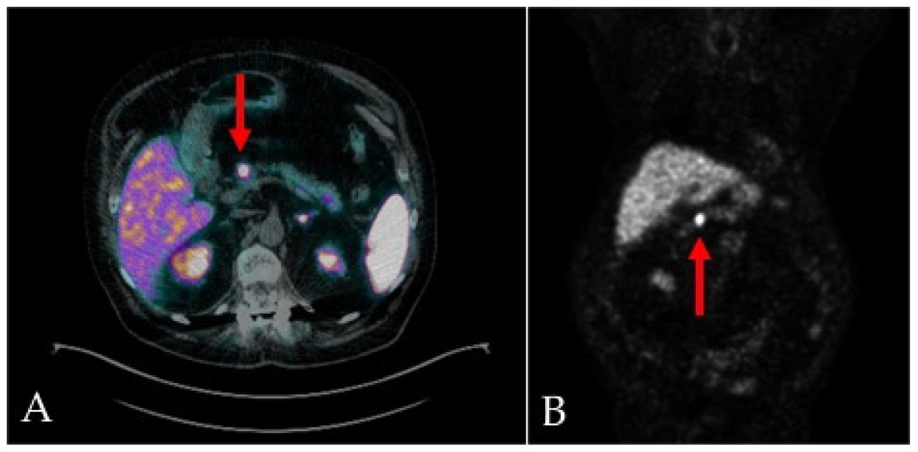
On the fifth day of admission, given the suspicion of a pancreatic neuroendocrine tumor (P-NET), a gallium-68 DOTATOC integrated PET/CT was performed (Figure 2), confirming a small mass between head and body of pancreas, with elevated expression of SSTR2/5 somatostatin receptors. No other sites of disease were detected.
Serum chromogranin A (CgA) measurement resulted within the normal range (98.0 ng/ml, normal values <101.9 ng/ml), as well as did a urine 5-HIAA - test (urinary 5-HIAA: 1.6 mg/24 h; normal values: 1.0 – 8.2 mg/24 h).
A progressive recovery was observed, with no further abdominal pain. In accordance with the remission of symptoms and the normal laboratory values, the patient was discharged with the prescription to undergo an endoscopic ultrasonography with fine-needle biopsy (EUS-FNB), for a targeted diagnostic and therapeutic management.
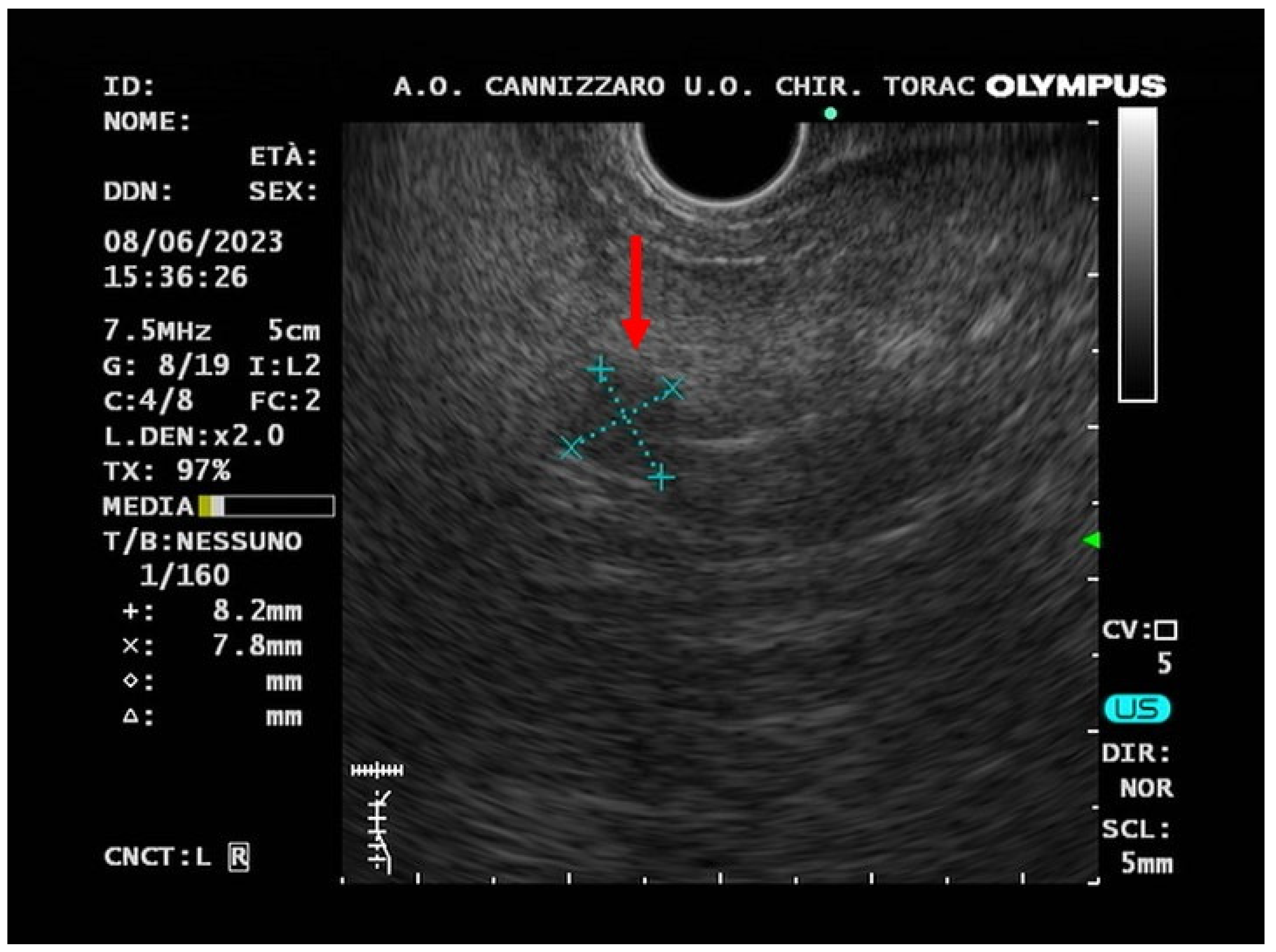
In June 2023, EUS-FNB, performed by a 22 gauge Acquire needle (Boston Scientific, Massachusetts, USA), using a slow-pull technique, visualized the presence of an oval hypo-echogenic mass, with a major axis of 8.9 mm (Figure 3), which was sampled for cyto-histological examination.
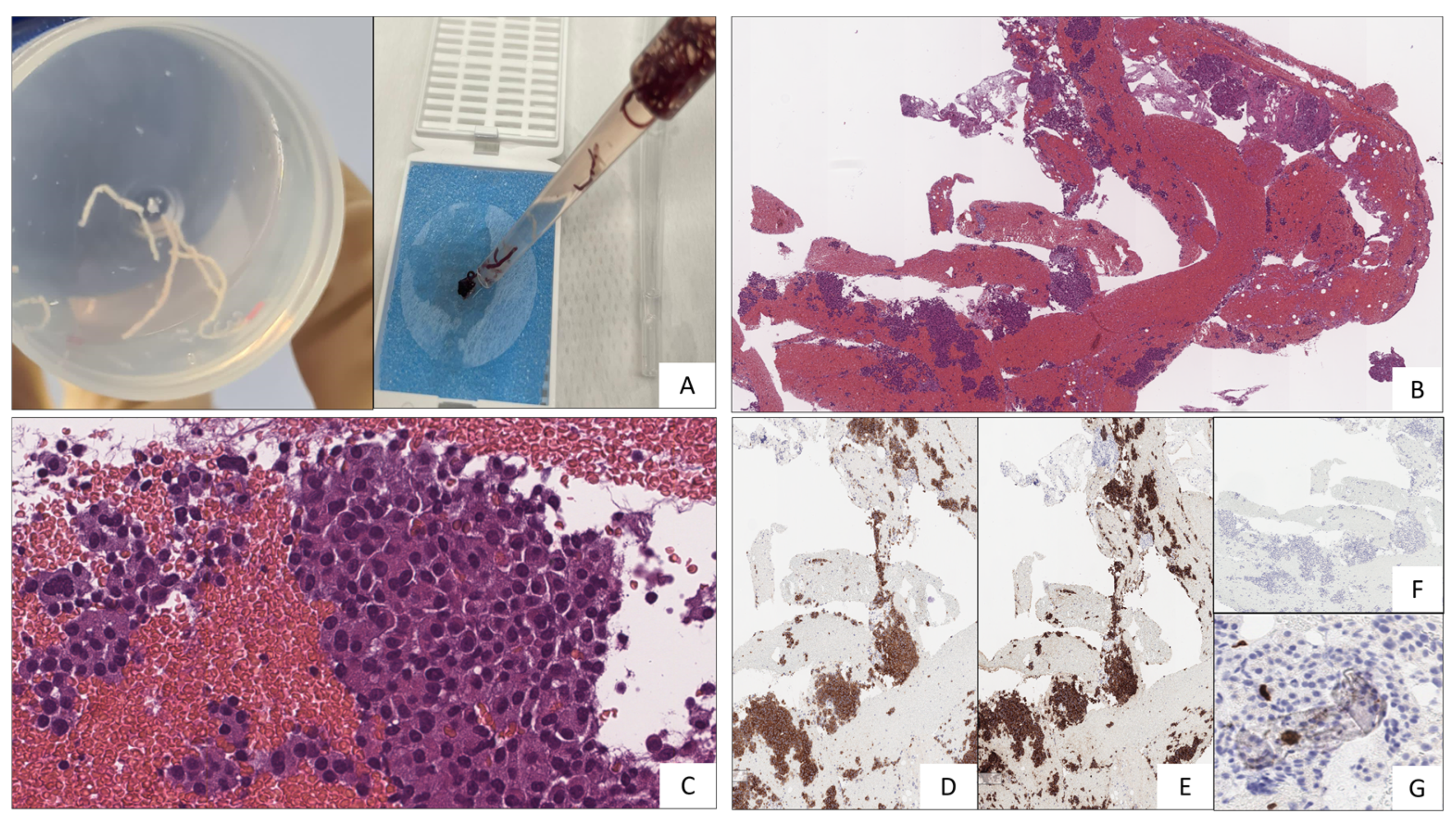
Histological and immunohistochemical examination confirmed the suspicion of P-NET (stage WHO G1, well-differentiated, synaptophysin positive, CgA positive, Ki67 1%,) (Figure 4). Fine needle biopsy allowed to obtain microcores of sample tissue (Figure 4/A); then, using pipette, microcores were picked-up in order to be treated as traditional biopsy. Microcores were composed by abundant blood and entrapped epithelial elements of pancreatic tissue (Figure 4/B). A monomorphic population of epithelial cells, in solid sheets or small nodules, with granular cytoplasm and nuclei with thickened chromatin was also observed (Figure 4/C). Immunochemistry, performed by Bond-Leica immunostainer, revealed positivity for neuroendocrine markers, such as Chromogranin-A (Figure 4/D) and Synaptophysin (Figure 4/E), while that of Serotonin resulted negative (Figure 4/F); the absence of mitosis and necrosis, together with low Ki-67 index (Figure 4/G), allowed to conclude for low grade neuroendocrine neoplasm.
In keeping with current guidelines, these findings suggested the diagnosis of low-grade (G1) non-functional pancreatic neuroendocrine tumor (NF-P-NET) (well-differentiated neoplasm, absence of mythosis, Ki67 ≤2%)[9]. This definition of “non-functional”, based only on negative hormone tests, is finalized to a categorical distinction between “secreting” and “non-secreting” tumors, although it underestimates the importance of clinical presentation.
After the evaluation of stage, grading, symptoms and comorbidities, a conservative approach of watchful waiting was chosen by the surgeon, with a radiological follow-up after one year. We scheduled a clinical follow-up in order to keep symptoms under observation.

3. Review of the Literature

Neuroendocrine neoplasms (NENs) are heterogenous neoplasms arising in secretory cells of the diffuse neuroendocrine system, the so called APUD (Amine Precursor Uptake and Decarboxylation) System. Characterized by amine and neuropeptide hormone production with dense vesicles, these neuroendocrine cells are specialized to receive neuronal inputs and consequentially release message peptides into circulation for regulation and modulation of cell proliferation, growth, and development. NENs are distinguished from pheochromocytomas and paragangliomas (neuroendocrine non-epithelial neoplasms) by the expression of keratin in the former ones, given their epithelial origin[10].
Neuroendocrine tumors (NETs) represent only 0.5% of all malignant conditions and 2% of all malignant tumors of the gastrointestinal tract[11]. Given the continued update in classification of NENs, these epidemiological data are continuously evolving. The prevalence of NETs ranges between 2.5 and 8.35 cases per 10,000, with a recent increase in their incidence rates[1,2,3,12,13,14,15], probably due to imaging improvement, leading to an earlier and more frequent diagnosis of the disease[5].
In the 2019 WHO classification of tumors of the digestive system[16], NENs are divided into well-differentiated neuroendocrine tumors (NETs) and poorly differentiated neuroendocrine carcinomas (NECs), based on their molecular differences. In addition, “mixed neuroendocrine-non-neuroendocrine neoplasms” (MiNENs) were better characterized, according to the simultaneous presence of both neuroendocrine and non-neuroendocrine components, typically poorly differentiated (Table 1).
The most frequent primary sites are gastrointestinal tract (61%), lung (25%), and about 14% remains of unknown origin[17]. Twelve to 22% percent of patients are metastatic at presentation[5].
Recently, abdominal pain was reported as unspecific symptom of a small bowel NET[18]. Our case-report resembles that very recently described by Daraghmeh et al.[18], although in our patient we found a P-NET.
The 2019 WHO classification[16] provided an improved system for determining prognosis and treatment, appliable to all NENs, replacing previous classification based on cell embryologic origin (foregut, midgut, hindgut)[19]. In contrast to 2017 WHO classification of tumors of endocrine organs[20], last classification included pancreatic tumors in gastroenteropancreatic NENs (GEP-NENs)[16].
Gastroenteropancreatic tumors (GEP-NETs) are most commonly located in the gastric mucosa, the small intestine, the rectum, and the pancreas[21]. While a subset of NENs is functional (40%), presenting with characteristic endocrine-related symptoms, the majority of them are non-functional and do not present with symptoms until later stages.
Distant metastases of NF-PNETs are often found at the time of diagnosis, because symptoms of NF-PNETs develop in an advanced stage. Due to these characteristics, NF-PNETs are usually incidentally diagnosed, like GEP-NETS, thanks to the development of imaging techniques, able to identify also very small lesions. In our patient, the presence of flushing, diarrhea and neuropsychiatric symptoms, suggesting a carcinoid syndrome, resulted unrelated to a biochemical elevation of hormonal levels. As a matter of fact, small PNETs without metastases can often remain asymptomatic until they reach a significant dimension, or can present with unspecific symptoms, such as abdominal pain, weight loss, anorexia and nausea.
Up to 90% of P-NETs are hormonally silent, a behavior affecting the prognosis as compared with functioning neoplasms, probably because of late diagnosis[22].
P-NETs may produce a large variety of hormones, such as insulin, gastrin, glucagon, vasoactive intestinal peptide (VIP), serotonin, somatostatin, and others[23]. By contrast, non-functional P-NETs, without hormone overproduction, may present with unspecific symptoms, such as abdominal pain, weight loss, diarrhea and gastrointestinal bleeding[7,24]. Most P-NENs are sporadic, whereas a minority are inherited, associated with Type 1 multiple endocrine neoplasia (MEN-1), von Hippel-Lindau syndrome (VHL), tuberous sclerosis or neurofibromatosis.
Functional pancreatic neuroendocrine tumors, associated with a variety of clinical syndromes, include[25]: insulinomas, the commonest functional P-NETs; gastrinomas, or Zollinger Ellison syndrome; pancreatic polypeptide-secreting tumors; VIPomas, or Verner-Morrison syndrome; glucagonomas, exclusively localized in pancreas; somatostatinomas, the least common NETs.
Carcinoid syndrome is a paraneoplastic syndrome occurring because of the release of bio-active substances, predominantly serotonin (5-HT), but also histamine, bradykinin, prostaglandins E and F, and tachykinins[26]. Recently, Halperin et al.[27] demonstrated, in a population-based analysis carried out on the American “Surveillance, Epidemiology, and End Results-Medicare” database, that 19% of patients with NETs had carcinoid syndrome. Typical symptoms are flushing and diarrhea. Wheezing, palpitations, breathlessness, abdominal pain, telangiectasias and neuropsychiatric symptoms could be also associated to carcinoid syndrome[26,28]. In the majority of cases, tumors are slow-growing and can produce hormonal substances such as serotonin, bradykinins, tachykinins and prostaglandins, with minimal clinical symptoms. When the tumor metastasizes to liver, bioactive substances are no longer inactivated because of the presence of hepatic metastases, therefore reaching systemic circulation. Usually, serotonin is physiologically metabolized by monoamine oxidases in the liver, lungs, and brain to 5-hydroxyindoleacetic acid (5-HIAA)[26,29]. However gastrointestinal NENs, with extensive retroperitoneal nodal involvement, can cause carcinoid syndrome even without hepatic metastases[30]. Recently, long-term complications from carcinoid syndrome, such as mesenteric and/or retroperitoneal fibrosis and carcinoid heart disease (CHD), have been well described[28,31,32]. In particular, CHD is characterized by right heart failure, due to deposition of plaques of fibrotic tissue on the right-side heart valves, caused by vaso-active substances secreted by the tumors (5-HT, histamine, prostaglandins)[33]. The left side of the heart is preserved by the inactivation of bioactive substances in the lung[34]. Otherwise, psychiatric symptoms, such as depression and acute psychosis, have been associated with metastatic carcinoid disease[35,36]. Depressive syndrome could be explained by the reduced levels of tryptophane, due to peripheral consumption by serotonin overproducing NETs[37].
The diagnosis of GEP-NENs is made on the basis of tissue histological examination. Radiological and functional imaging is used to evaluate disease extension (staging) and assess response to therapy, as well as to localize the primary site. Laboratory tests play a diagnostic role only for carcinoid syndrome and hormone-specific syndromes (gastrinomas, insulinomas, glucagonomas), although the assay of either circulating or urinary hormones failed to be highly sensitive and specific, sometimes because blood sampling and urine collection are not made closely to the occurrence of typical symptoms.
The current WHO classification emphasized the role of histological examination in surgically removed neoplasms, in order to establish the morphological characteristics and grading[16]. Three grades (G1, G2, G3) are described for GEP-NETs, based on the proliferation activity assessed by mitotic rate and Ki67 proliferation index[38,39]. For a more specific diagnosis, together with morphology and grading, immunohistochemical staining for chromogranin A (CgA) and synaptophysin should be assessed, as biomarkers of neuroendocrine tumors.
Although WHO histological classifications are specifically intended for surgically removed NENs[9,16], recent studies have investigated the role of endoscopic ultrasound-guided fine-needle aspiration (EUS-FNA) and fine-needle biopsy (EUS-FNB) for the pre-operative evaluation and management of pancreatic NETs (P-NETs)[40,41,42,43,44,45,46,47,48,49]. Despite data about grading agreement between EUS-FNA and surgical specimens highlighted a significant rate of under- or over-grading[48,49,50,51,52,53,54], the recent introduction of needles for EUS-guided fine-needle biopsy (EUS-FNB), as made in our patient, has changed the scenario[44,45]. EUS-FNB, in fact, allows to obtain tissue samples on which immunohistochemical examination can be easily performed, to evaluate Ki67 proliferation index[46,47,48,49,50,51,52]. As a matter of fact, in patients harbouring less than 2 cm P-NETs, management remains still controversial, especially for asymptomatic and non-functional P-NETs[53,54,55,56]. Endoscopy with biopsy is already the gold standard for diagnosing NENs of the stomach, duodenum and colorectum[57,58]. In the diagnosis of pancreatic NENs, EUS is particularly useful in detecting the nature of small lesions. The introduction of EUS-FNB could then overcome the interpretative limits of EUS-FNA, so allowing an early characterization of tumors in which surgery would be destructive of heathy tissue [47,48].
Treatment of patients with small low-grade non-functional pancreatic neuroendocrine tumors (<20 mm) remains still under debate, according to ENETS guidelines. In this respect, Sugawara et al.[59], in a recent metanalysis, demonstrated that surgical resection is recommended in patients with nonmetastatic NF-PNETs measuring between 1.1 and 2.0 cm; on the opposite, those patients with a smaller lesion (< 1 cm) showed greater prognostic benefits with a conservative approach. JNETS [60] suggests a follow-up strategy, with imaging every 6-12 months of < 1 cm asymptomatic tumors without metastases. Moreover, Sadot et al.[61] further reported that, among 104 patients with small, asymptomatic P-NET undergoing a non-operative management, no patient developed evidence of metastases or died because of the tumor, after a median follow-up of 44 months. Of note, however, all these data come from a population much younger (median age 60-65) than our patient (81-yr old).
Paik et al.[62] suggested that patients with P-NETs smaller than1 cm can be managed by observation alone, while P-NETs >1 cm should undergo EUS-FNB to obtain grading and Ki67 immunostaining, to characterize the tumor according WHO classification.
To investigate P-NETs, several imaging can be performed, including computed tomography (CT), magnetic resonance (MRI), ultrasonography and functional imaging with scintigraphy and positron emission tomography (PET). The optimal choice of imaging modality depends on the location of primary and metastatic lesions[63].
Endoscopic ultrasonography (EUS) has become a very useful technique to evaluate pancreatic lesions. On EUS, P-NETs typically appear as well-defined, round, hypoechoic, homogenous vascular lesions[64]. As in our case-report, EUS allows the accurate localization of P-NETs, which would be crucial for surgical interventions. As mentioned before, EUS allows cyto-histological confirmation of neuroendocrine tumors, through guided tissue acquisition for histological procedures[40,41,42,43,44,45,46,47,48,49].
Functional imaging of GEP-NENs is based on the typical expression of somatostatin receptors (SSTR) by neuroendocrine cells[65]. In the past, functional studies were performed with 111indium pentetreotide scintigraphy (Octreoscan®); in recent years, PET/CT with somatostatin analogs tracked with gallium-68 (68Ga-SSA PET/CT) has become the modality of choice for SSTR imaging[9,66,67]. Functional imaging is indicated for staging, localization of the unknown primary tumor in patients with established neuroendocrine metastases, in vivo demonstration of SSTR expression on neuroendocrine cells (for therapeutic planning), as well as the extent of disease after treatment. The most commonly somatostatin analogs used in the clinical practice are 68Ga-DOTA-Tyr3-octreotide (68Ga-DOTA-TOC), 68Ga-DOTA-Tyr3-octreotate (68Ga-DOTA-TATE) and 68Ga-DOTA-Nal3-octreotide (68Ga-DOTA-NOC). Mean sensitivity of 68Ga-DOTA-SSA PET/CT for the diagnosis of P-NETs was 92%, while specificity was 83%[68,69]. In advanced fast-growing G2 and G3 NENs, especially if receptor negativity is demonstrated at 68Ga-SSA PET/CT, 18FDG-PET/CT may be considered in the diagnostic approach[70,71]. Detection of P-NETs with functional imaging could be affected by a physiological uptake, especially in the uncinate process, so suggesting morphological imaging together with histological confirmation as a specific diagnostic process[72]. However, it remains still under debate whether the combined use of 18FDG-PET/CT and 68Ga-DOTA-TOC peptide could improve diagnostic performance of NENs[71].
Nowadays, biochemical diagnosis of NENs has been downsized, due to the high proportion of non-functioning NENs. Considering the high rates of false positive and the heterogeneous serum determinations, Chromogranin A (CgA) should be therefore used in patients with an already documented diagnosis of NEN, in order to establish the treatment response or during the follow-up[73,74], although it results less sensitive for primary diagnosis. On the other hand, Neuron-specific enolase (NSE) is considered an unreliable diagnostic biomarker for NETs, due to low sensitivity and specificity, while no evidence is available regarding its role in follow-up[75].
However, laboratory tests for specific biomarkers (gastrin, insulin, glucagon, VIP, 5-HIAA) are still important tools in certain clinical syndromes. 5-hydroxyindoleacetic acid (5-HIAA), detected in 24-hour urine collection using optimal conditions for assay, is the specific tumor marker of carcinoid syndrome. 5-HIAA has demonstrated a diagnostic sensitivity of 70%, with a specificity of 90%[76]. It is not recommended to use 5-HIAA as a screening test in the presence of diarrhea. Instead, it should be used in patients diagnosed with NEN, to confirm carcinoid syndrome and assess its response to therapy[9,76].
Circulating tumor cells, circulating tumor DNA, circulating micro-RNAs, and NETest (simultaneous measurement of 51 neuroendocrine-specific marker genes in the peripheral blood) are novel biomarkers under validation for NENs. However, this test is not widely available, and needs further validation[77].

4. Discussion

Our case report describes an old patient with an extremely rare pancreatic neuroendocrine tumor (P-NET), diagnosed in presence of unspecific gastrointestinal symptoms and skin flushing. This observation is even much rarer in old people. Despite symptoms suggesting carcinoid syndrome, tumor was well-differentiated and localized in the pancreas without liver metastases. This presentation is extremely rare, with only a few cases reported in the literature[78,79,80]. Biochemical testing for serum CgA and urinary 5-HIAA resulted negative. As emphasized before, laboratory biomarkers have been recently downsized, due to the high rates of false positivity and their pharmacological interference, leading to low sensitivity and specificity[73,74,76].
We confirmed the P-NETs diagnosis through contrast-enhanced CT, followed by functional imaging with a gallium-68 DOTATOC integrated PET/CT. Given the small size of tumor (8 mm), we decided to perform an EUS-FNB, in order to make grading and completing the P-NET management and treatment. EUS-FNB confirmed the diagnosis of well-differentiated, low grade (G1) P-NET (CgA +, Synaptophysin +, Ki67 1%).
The association of NETs and carcinoid syndrome occurs in approximately 19% of patients[26]. Except for patients with primary ovarian or bronchial neuroendocrine tumors, evidence of carcinoid syndrome develops when metastases have occured.25,28]. In patients harbouring P-NETs, carcinoid syndrome is even more rare, accounting for approximately 1 % [12,81]. As a matter of fact, serotonin-producing P-NETs account for 0.58–1.4% of all P-NETs[82]. Only few cases have been previously reported for P-NETs without liver metastases presenting with carcinoid syndrome [79,83]. Some patients with neuroendocrine tumor showed symptoms of flushing with low or normal levels of 5-HIAA[84,85]. Negativity for immunostaining of serotonin found in our tumor biopsy, while is in keeping with normal valuues of 5HIAA, may further support the notion that levels of circulating hormones could increase only in presence of liver metastases[26,29]. Our patient experienced carcinoid symptoms (diarrhea, flushing, unresponsive depression) in absence of documented liver metastases and with negative serum CgA and normal urinary 5-HIAA. Guidelines clearly underscore the concept that negative hormone measurements define NETs as "nonfunctional", even if presenting with suggestive symptoms or positive hormonal expression in NET cells on immunohistochemical staining[86]. This may not always be so, as in our case-report, as well as in other few reports[79,83].
It remains unclear why symptoms resembling carcinoid syndrome developed in our patient, with no evidence of any increase in hormone levels. It may well be that a possible sudden and transient hormone increase in the circulation failed to be detected. Otherwise, some so far unknown mechanisms might have been responsible for abdominal pain, diarrhea and flushing, all together contributing to consider alternative diagnoses regarding bowel diseases, which were definitely excluded by contrast-enhanced CT scan in our patient. In presence of this discrepancy between the presence of symptoms and hormone negativity, however, our case-report would emphasize that clinical presentation should not be disregarded as presentation of carcinoid-like syndrome, therefore leading to complete diagnostic work-up for NETs.
Therefore, despite P-NET of our patient should be defined non-functional according to guidelines[86] because hormone values are within normal range, our case report underscores the concept that imaging and histological examination remain the major tools in the diagnostic process of P-NETs associated with symptoms of carcinoid syndrome. As we reported, performing an EUS-FNB and assessing cyto-histological features could be the protocol of choice in patients with small, low grade, pauci-symptomatic P-NETs, in order to avoid or delay surgical treatment. Of note, we would again underscore the concept that P-NET occurrence without metastases in old patients is really very rare.
P-NET <1. cm could occur in very old people, without metastases, as in our case-report, although median age resulted between 61 and 65 years in a recent metanalysis[59]. Surgical resection in these cases is not warranted. On the contrary, for P-NETs between 1.0 and 2.0 cm, surgical resection provided a better survival, but in patients younger than 65 years, without comorbidities.
In conclusion, the novelty of our case-report can be highlighted as follows: 1) symptoms of carcinoid syndrome can be shown in P-NET <1.0cm occurring in very old people, without metastases and no evidence of an increase in circulating hormones, in agreement with negativity of immunostaining for serotonin shown in tumor tissue. So far, median age-range was found much lower[59]; 2) Although the categorical distinction in "functional" and "nonfunctional" NETs suggested by guidelines[86] on the basis of hormone positivity, clinical presentation could help showing P-NETs with no evidence of hormone release, as in our case-report, so underscoring the concept that physician should take into account the possibility that an atypical pattern of apparently "non-functional" P-NET may occur, although rarely; 3) although not yet included in the latest guidelines, the use of EUS-FNA/B could play an important role in the diagnostic workup, as already reported by a recent multicentre study[62].

List of Acronyms

APUD: Amine Precursor Uptake and Decarboxylation
CgA: Chromogranin A
CHD: Carcinoid heart disease
Cr: Creatinine
CT: Computed tomography
CRP: C-reactive protein
eGFR: Estimated glomerular filtration rate
ENETS: European Neuroendocrine Tumor Society
EUS-FNA: Endoscopic ultrasound with fine-needle aspiration
EUS-FNB: Endoscopic ultrasound with fine-needle biopsy
FAST: Focused Assessment with Sonography in Trauma
68Ga-DOTA-NOC: gallium-68-DOTA-Nal3-octreotide
68Ga-DOTA-TATE: gallium-68-DOTA-Tyr3-octreotate
68Ga-DOTA-TOC: gallium-68-DOTA-Tyr3-octreotide
GEP-NEN: Gastroenteropancreatic neuroendocrine neoplasm
GEP-NET: Gastroenteropancreatic neuroendocrine tumor
HGT: Hemo Glucose Test
5-HIAA: 5-hydroxyindoleacetic acid
5-HT: serotonin
INSM1: insulinoma-associated protein 1
JNETS: Japanese Neuroendocrine Tumor Society
MEN-1: Multiple endocrine neoplasia 1
MiNEN: Mixed neuroendocrine-non-neuroendocrine neoplasm
MRI: Magnetic resonance imaging
NF-P-NET: Non-functional pancreatic neuroendocrine tumor
NEC: Neuroendocrine carcinoma
NEN: Neuroendocrine neoplasm
NET: Neuroendocrine tumor
NSE: Neuron-specific enolase
P-NET: Pancreatic neuroendocrine tumor
PAD: Peripheral artery disease
PET: Positive emission tomography
PPi: Proton pomp inhibitor
SaO2: Oxygen saturation
SSA: Somatostatin analogue
SSTR: Somatostatin receptor
VHL: von Hippel-Lindau syndrome
VIP: Vaso-active intestinal peptide
WHO: World health organization

Author Contributions

Conceptualization, M.R. and L.M.; methodology, M.R. and L.M.; investigation, M.R., N.C., C.S., M.C., A.L., F.M., S.M., S.P., M.I., G.T.; data curation, M.R., N.C., C.S.; writing—original draft preparation, M.R., N.C., C.S., F.M.; writing—review and editing, M.R., L.M.; visualization, M.R.; supervision, L.M., M.I. All authors have read and agreed to the published version of the manuscript.

Funding

This research received no external funding.

Institutional Review Board Statement

The study was conducted according to the guidelines of the Declaration of Helsinki. Ethical review and approval were waived due to the type of the study that is a case report.

Informed Consent Statement

The patient provided written informed consent to publish this manuscript and for material sampling

Data Availability Statement

The data presented in this study are available on request from the corresponding author.

Conflicts of Interest

The authors declare no conflict of interest.

References

  1. Yao, J. C.; Hassan, M.; Phan, A.; Dagohoy, C.; Leary, C.; Mares, J. E.; Abdalla, E. K.; Fleming, J. B.; Vauthey, J. N.; Rashid, A.; Evans, D. B. One hundred years after "carcinoid": epidemiology of and prognostic factors for neuroendocrine tumors in 35,825 cases in the United States. Journal of clinical oncology : official journal of the American Society of Clinical Oncology 2008, 26, 3063–3072. [Google Scholar] [CrossRef] [PubMed]
  2. Das, S.; Dasari, A. Epidemiology, Incidence, and Prevalence of Neuroendocrine Neoplasms: Are There Global Differences? Current oncology reports 2021, 23(4), 43. [Google Scholar] [CrossRef]
  3. Leoncini, E.; Boffetta, P.; Shafir, M.; Aleksovska, K.; Boccia, S.; Rindi, G. Increased incidence trend of low-grade and high-grade neuroendocrine neoplasms. Endocrine 2017, 58(2), 368–379. [Google Scholar] [CrossRef]
  4. Pavel, M.; Öberg, K.; Falconi, M.; Krenning, E. P.; Sundin, A.; Perren, A.; Berruti, A.; ESMO Guidelines Committee. Gastroenteropancreatic neuroendocrine neoplasms: ESMO Clinical Practice Guidelines for diagnosis, treatment and follow-up. Annals of oncology : official journal of the European Society for Medical Oncology 2020, 31(7), 844–860. [Google Scholar] [CrossRef]
  5. Dasari, A.; Shen, C.; Halperin, D.; Zhao, B.; Zhou, S.; Xu, Y.; Shih, T.; Yao, J. C. Trends in the Incidence, Prevalence, and Survival Outcomes in Patients With Neuroendocrine Tumors in the United States. JAMA oncology 2017, 3(10), 1335–1342. [Google Scholar] [CrossRef] [PubMed]
  6. Clement, D.; Ramage, J.; Srirajaskanthan, R. Update on Pathophysiology, Treatment, and Complications of Carcinoid Syndrome. Journal of oncology 2020, 8341426. [Google Scholar] [CrossRef]
  7. Metz, D. C.; Jensen, R. T. Gastrointestinal neuroendocrine tumors: pancreatic endocrine tumors. Gastroenterology 2008, 135(5), 1469–1492. [Google Scholar] [CrossRef]
  8. Garcia-Carbonero, R.; Capdevila, J.; Crespo-Herrero, G.; Díaz-Pérez, J. A.; Martínez Del Prado, M. P.; Alonso Orduña, V.; Sevilla-García, I.; Villabona-Artero, C.; Beguiristain-Gómez, A.; Llanos-Muñoz, M. et al. Incidence, patterns of care and prognostic factors for outcome of gastroenteropancreatic neuroendocrine tumors (GEP-NETs): results from the National Cancer Registry of Spain (RGETNE). Annals of oncology : official journal of the European Society for Medical Oncology 2010, 21(9), 1794–1803. [Google Scholar] [CrossRef] [PubMed]
  9. AIOM for Neuroendocrine Tumors Guide Line 2020. Accessed 15 July 2023 https://www.aiom.it/wp-content/uploads/2020/10/2020_LG_AIOM_Neuroendocrini.pdf.
  10. Ordóñez, N. G. Broad-spectrum immunohistochemical epithelial markers: a review. Human pathology 2013, 44(7), 1195–1215. [Google Scholar] [CrossRef] [PubMed]
  11. Moertel, C. G. Karnofsky memorial lecture. An odyssey in the land of small tumors. Journal of Clinical Oncology 1987, 5(10), 1502–1522. [Google Scholar] [CrossRef] [PubMed]
  12. Ito, T.; Igarashi, H.; Nakamura, K.; Sasano, H.; Okusaka, T.; Takano, K.; Komoto, I.; Tanaka, M.; Imamura, M.; Jensen, R. T.; Takayanagi, R.; Shimatsu, A. Epidemiological trends of pancreatic and gastrointestinal neuroendocrine tumors in Japan: a nationwide survey analysis. Journal of gastroenterology 2015, 50(1), 58–64. [Google Scholar] [CrossRef] [PubMed]
  13. Cho, M. Y.; Kim, J. M.; Sohn, J. H.; Kim, M. J.; Kim, K. M.; Kim, W. H.; Kim, H.; Kook, M. C.; Park, D. Y.; Lee, J. H.; et al. Current Trends of the Incidence and Pathological Diagnosis of Gastroenteropancreatic Neuroendocrine Tumors (GEP-NETs) in Korea 2000-2009: Multicenter Study. Cancer research and treatment 2012, 44(3), 157–165. [Google Scholar] [CrossRef]
  14. Scherübl, H.; Streller, B.; Stabenow, R.; Herbst, H.; Höpfner, M.; Schwertner, C.; Steinberg, J.; Eick, J.; Ring, W.; Tiwari, K.; et al. Clinically detected gastroenteropancreatic neuroendocrine tumors are on the rise: epidemiological changes in Germany. World journal of gastroenterology 2013, 19(47), 9012–9019. [Google Scholar] [CrossRef] [PubMed]
  15. White, B. E.; Rous, B.; Chandrakumaran, K.; Wong, K.; Bouvier, C.; Van Hemelrijck, M.; George, G.; Russell, B.; Srirajaskanthan, R.; Ramage, J. K. Incidence and survival of neuroendocrine neoplasia in England 1995-2018: A retrospective, population-based study. The Lancet regional health. Europe 2022, 23, 100510. [Google Scholar] [CrossRef]
  16. Nagtegaal, I. D.; Odze, R. D.; Klimstra, D.; Paradis, V.; Rugge, M.; Schirmacher, P.; Washington, K. M.; Carneiro, F.; Cree, I. A.; WHO Classification of Tumours Editorial Board. The 2019 WHO classification of tumours of the digestive system. Histopathology 2020, 76(2), 182–188. [Google Scholar] [CrossRef]
  17. Fernandez, C. J.; Agarwal, M.; Pottakkat, B.; Haroon, N. N.; George, A. S; Pappachan, J. M. Gastroenteropancreatic neuroendocrine neoplasms: A clinical snapshot. World journal of gastrointestinal surgery 2021, 13(3), 231–255. [Google Scholar] [CrossRef]
  18. Daraghmeh, L., Shbaita, S., Nassef, O., Melhem, L., & Maqboul, I. (2023). Non-specific Symptoms of Small Bowel Neuroendocrine Tumor and the Diagnostic Challenges: A Case Report. Cureus 15(6), e41080. [CrossRef]
  19. Klöppel, G.; Perren, A.; Heitz, P. U. The gastroenteropancreatic neuroendocrine cell system and its tumors: the WHO classification. Annals of the New York Academy of Sciences 2004, 1014, 13–27. [Google Scholar] [CrossRef]
  20. Lloyd, R.V.; Osamura, R.Y.; Klöppel, G.; Rosai, J. WHO classification of tumours of endocrine organs, 2017 vol. 10. 4th ed. Lyon: International Agency for Research on Cancer.
  21. Modlin, I. M.; Lye, K. D.; Kidd, M. A 5-decade analysis of 13,715 carcinoid tumors. Cancer 2003, 97(4), 934–959. [Google Scholar] [CrossRef] [PubMed]
  22. Cives, M.; Strosberg, J. R. Gastroenteropancreatic neuroendocrine tumors. CA: a cancer journal for clinicians 2018, 68(6), 471–487. [Google Scholar] [CrossRef] [PubMed]
  23. Turaga, K.K.; Kvols, L.K. Recent progress in the understanding, diagnosis, and treatment of gastroenteropancreatic neuroendocrine tumors. CA Cancer J Clin. 2011, 61(2), 113–32. [Google Scholar] [CrossRef]
  24. Zerbi, A.; Falconi, M.; Rindi, G.; Delle Fave, G.; Tomassetti, P.; Pasquali, C.; Capitanio, V.; Boninsegna, L.; Di Carlo, V.; AISP-Network Study Group. Clinicopathological features of pancreatic endocrine tumors: a prospective multicenter study in Italy of 297 sporadic cases. The American journal of gastroenterology 2010, 105(6), 1421–1429. [Google Scholar] [CrossRef]
  25. Jensen, R. T.; Cadiot, G.; Brandi, M. L.; de Herder, W. W.; Kaltsas, G.; Komminoth, P.; Scoazec, J. Y.; Salazar, R.; Sauvanet, A.; Kianmanesh, R.; Barcelona Consensus Conference participants. ENETS Consensus Guidelines for the management of patients with digestive neuroendocrine neoplasms: functional pancreatic endocrine tumor syndromes. Neuroendocrinology 2012, 95(2), 98–119. [Google Scholar] [CrossRef] [PubMed]
  26. Gade, A. K.; Olariu, E.; Douthit, N. T. Carcinoid Syndrome: A Review. Cureus 2020, 12(3), e7186. [Google Scholar] [CrossRef] [PubMed]
  27. Halperin, D. M.; Shen, C.; Dasari, A.; Xu, Y.; Chu, Y.; Zhou, S.; Shih, Y. T.; Yao, J. C. Frequency of carcinoid syndrome at neuroendocrine tumour diagnosis: a population-based study. The Lancet. Oncology 2017, 18(4), 525–534. [Google Scholar] [CrossRef] [PubMed]
  28. Mota, J. M.; Sousa, L. G.; Riechelmann, R. P. Complications from carcinoid syndrome: review of the current evidence. Ecancermedicalscience 2016, 10, 662. [Google Scholar] [CrossRef]
  29. Kvols L., K. Metastatic carcinoid tumors and the malignant carcinoid syndrome. Annals of the New York Academy of Sciences 1994, 733, 464–470. [Google Scholar] [CrossRef]
  30. Ahmed, M. Gastrointestinal neuroendocrine tumors in 2020. World journal of gastrointestinal oncology 2020, 12(8), 791–807. [Google Scholar] [CrossRef]
  31. Bhattacharyya, S.; Davar, J.; Dreyfus, G.; Caplin, M. E. Carcinoid heart disease. Circulation 2007, 116(24), 2860–2865. [Google Scholar] [CrossRef] [PubMed]
  32. Druce, M.; Rockall, A.; Grossman, A. B. Fibrosis and carcinoid syndrome: from causation to future therapy. Nature reviews. Endocrinology 2009, 5(5), 276–283. [Google Scholar] [CrossRef] [PubMed]
  33. Dobson, R.; Burgess, M. I.; Pritchard, D. M.; Cuthbertson, D. J. The clinical presentation and management of carcinoid heart disease. International journal of cardiology 2014, 173(1), 29–32. [Google Scholar] [CrossRef] [PubMed]
  34. Rubin de Celis Ferrari, A. C.; Glasberg, J.; Riechelmann, R. P. Carcinoid syndrome: update on the pathophysiology and treatment. Clinics (Sao Paulo, Brazil) 2018, 73 (suppl 1), e490s. [Google Scholar] [CrossRef] [PubMed]
  35. Russo, S.; Boon, J. C.; Kema, I. P.; Willemse, P. H.; den Boer, J. A.; Korf, J.; de Vries, E. G. Patients with carcinoid syndrome exhibit symptoms of aggressive impulse dysregulation. Psychosomatic medicine 2004, 66(3), 422–425. [Google Scholar] [CrossRef] [PubMed]
  36. Kohen, I.; Arbouet, S. Neuroendocrine carcinoid cancer associated with psychosis. Psychiatry (Edgmont (Pa. : Township)) 2008, 5(6), 29–30. [Google Scholar]
  37. Nobels, A.; Geboes, K.; Lemmens, G. M. May depressed and anxious patients with carcinoid syndrome benefit from treatment with selective serotonin reuptake inhibitors (SSRIs)?: findings from a case report. Acta oncologica (Stockholm, Sweden) 2016, 55(11), 1370–1372. [Google Scholar] [CrossRef] [PubMed]
  38. Tatsumoto, S.; Kodama, Y.; Sakurai, Y.; Shinohara, T.; Katanuma, A.; Maguchi, H. Pancreatic neuroendocrine neoplasm: correlation between computed tomography enhancement patterns and prognostic factors of surgical and endoscopic ultrasound-guided fine-needle aspiration biopsy specimens. Abdominal imaging 2013, 38(2), 358–366. [Google Scholar] [CrossRef]
  39. Grillo, F.; Albertelli, M.; Brisigotti, M. P.; Borra, T.; Boschetti, M.; Fiocca, R.; Ferone, D.; Mastracci, L. Grade Increases in Gastroenteropancreatic Neuroendocrine Tumor Metastases Compared to the Primary Tumor. Neuroendocrinology 2016, 103(5), 452–459. [Google Scholar] [CrossRef]
  40. Atiq, M.; Bhutani, M. S.; Bektas, M.; Lee, J. E.; Gong, Y.; Tamm, E. P.; Shah, C. P.; Ross, W. A.; Yao, J.; Raju, G. S. et al. EUS-FNA for pancreatic neuroendocrine tumors: a tertiary cancer center experience. Digestive diseases and sciences 2012, 57(3), 791–800. [Google Scholar] [CrossRef] [PubMed]
  41. Larghi, A.; Capurso, G.; Carnuccio, A.; Ricci, R.; Alfieri, S.; Galasso, D.; Lugli, F.; Bianchi, A.; Panzuto, F.; De Marinis, L. et al. Ki-67 grading of nonfunctioning pancreatic neuroendocrine tumors on histologic samples obtained by EUS-guided fine-needle tissue acquisition: a prospective study. Gastrointestinal endoscopy 2012, 76(3), 570–577. [Google Scholar] [CrossRef] [PubMed]
  42. Unno, J.; Kanno, A.; Masamune, A.; Kasajima, A.; Fujishima, F.; Ishida, K.; Hamada, S.; Kume, K.; Kikuta, K.; Hirota, M. et al. The usefulness of endoscopic ultrasound-guided fine-needle aspiration for the diagnosis of pancreatic neuroendocrine tumors based on the World Health Organization classification. Scandinavian journal of gastroenterology 2014, 49(11), 1367–1374. [Google Scholar] [CrossRef]
  43. Paiella, S.; Landoni, L.; Rota, R.; Valenti, M.; Elio, G.; Crinò, S. F.; Manfrin, E.; Parisi, A.; Cingarlini, S; D'Onofrio, M. et al. Endoscopic ultrasound-guided fine-needle aspiration for the diagnosis and grading of pancreatic neuroendocrine tumors: a retrospective analysis of 110 cases. Endoscopy 2020, 52(11), 988–994. [Google Scholar] [CrossRef] [PubMed]
  44. Crinò, S. F.; Ammendola, S.; Meneghetti, A.; Bernardoni, L.; Conti Bellocchi, M. C.; Gabbrielli, A.; Landoni, L.; Paiella, S.; Pin, F.; Parisi, A. et al. Comparison between EUS-guided fine-needle aspiration cytology and EUS-guided fine-needle biopsy histology for the evaluation of pancreatic neuroendocrine tumors. Pancreatology 2021, 21(2), 443–450. [Google Scholar] [CrossRef] [PubMed]
  45. Rimbaş, M.; Crino, S. F.; Gasbarrini, A.; Costamagna, G.; Scarpa, A.; Larghi, A. EUS-guided fine-needle tissue acquisition for solid pancreatic lesions: Finally moving from fine-needle aspiration to fine-needle biopsy? Endoscopic ultrasound 2018, 7(3), 137–140. [Google Scholar] [CrossRef] [PubMed]
  46. Eusebi, L. H.; Thorburn, D.; Toumpanakis, C.; Frazzoni, L.; Johnson, G.; Vessal, S.; Luong, T. V.; Caplin, M.; Pereira, S. P. Endoscopic ultrasound-guided fine-needle aspiration vs fine-needle biopsy for the diagnosis of pancreatic neuroendocrine tumors. Endoscopy international open 2019, 7(11), E1393–E1399. [Google Scholar] [CrossRef]
  47. Di Leo, M.; Poliani, L.; Rahal, D.; Auriemma, F.; Anderloni, A.; Ridolfi, C.; Spaggiari, P.; Capretti, G.; Di Tommaso, L.; Preatoni, P. et al. Pancreatic Neuroendocrine Tumours: The Role of Endoscopic Ultrasound Biopsy in Diagnosis and Grading Based on the WHO 2017 Classification. Digestive diseases (Basel, Switzerland) 2019, 37(4), 325–333. [Google Scholar] [CrossRef] [PubMed]
  48. Leeds, J. S.; Nayar, M. K.; Bekkali, N. L. H.; Wilson, C. H.; Johnson, S. J.; Haugk, B.; Darne, A.; Oppong, K. W. Endoscopic ultrasound-guided fine-needle biopsy is superior to fine-needle aspiration in assessing pancreatic neuroendocrine tumors. Endoscopy international open 2019, 7(10), E1281–E1287. [Google Scholar] [CrossRef] [PubMed]
  49. Melita, G.; Pallio, S.; Tortora, A.; Crinò, S. F.; Macrì, A.; Dionigi, G. Diagnostic and Interventional Role of Endoscopic Ultrasonography for the Management of Pancreatic Neuroendocrine Neoplasms. Journal of clinical medicine 2021, 10(12), 2638. [Google Scholar] [CrossRef]
  50. Bernstein, J.; Ustun, B.; Alomari, A.; Bao, F.; Aslanian, H. R.; Siddiqui, U.; Chhieng, D.; Cai, G. Performance of endoscopic ultrasound-guided fine needle aspiration in diagnosing pancreatic neuroendocrine tumors. CytoJournal 2013, 10, 10. [Google Scholar] [CrossRef]
  51. Boutsen, L.; Jouret-Mourin, A.; Borbath, I.; van Maanen, A.; Weynand, B. Accuracy of Pancreatic Neuroendocrine Tumour Grading by Endoscopic Ultrasound-Guided Fine Needle Aspiration: Analysis of a Large Cohort and Perspectives for Improvement. Neuroendocrinology 2018, 106(2), 158–166. [Google Scholar] [CrossRef] [PubMed]
  52. Heidsma, C. M.; Tsilimigras, D. I.; Rocha, F.; Abbott, D. E.; Fields, R.; Smith, P. M.; Poultsides, G. A.; Cho, C.; van Eijck, C.; van Dijkum, E. N. et al. Clinical relevance of performing endoscopic ultrasound-guided fine-needle biopsy for pancreatic neuroendocrine tumors less than 2 cm. Journal of surgical oncology 2020, 122(7), 1393–1400. [Google Scholar] [CrossRef]
  53. Gratian, L.; Pura, J.; Dinan, M.; Roman, S.; Reed, S.; Sosa, J. A. Impact of extent of surgery on survival in patients with small nonfunctional pancreatic neuroendocrine tumors in the United States. Annals of surgical oncology 2014, 21(11), 3515–3521. [Google Scholar] [CrossRef]
  54. Sharpe, S. M.; In, H.; Winchester, D. J.; Talamonti, M. S.; Baker, M. S. Surgical resection provides an overall survival benefit for patients with small pancreatic neuroendocrine tumors. Journal of gastrointestinal surgery 2015, 19(1), 117–123. [Google Scholar] [CrossRef] [PubMed]
  55. Sallinen, V.; Haglund, C.; Seppänen, H. Outcomes of resected nonfunctional pancreatic neuroendocrine tumors: Do size and symptoms matter? Surgery 2015, 158(6), 1556–1563. [Google Scholar] [CrossRef] [PubMed]
  56. Falconi, M.; Eriksson, B.; Kaltsas, G.; Bartsch, D. K.; Capdevila, J.; Caplin, M.; Kos-Kudla, B.; Kwekkeboom, D.; Rindi, G.; Klöppel, G. et al. ENETS Consensus Guidelines Update for the Management of Patients with Functional Pancreatic Neuroendocrine Tumors and Non-Functional Pancreatic Neuroendocrine Tumors. Neuroendocrinology 2016, 103(2), 153–171. [Google Scholar] [CrossRef]
  57. Delle Fave, G.; O'Toole, D.; Sundin, A.; Taal, B.; Ferolla, P.; Ramage, J. K.; Ferone, D.; Ito, T.; Weber, W.; Zheng-Pei, Z. et al. ENETS Consensus Guidelines Update for Gastroduodenal Neuroendocrine Neoplasms. Neuroendocrinology 2016, 103(2), 119–124. [Google Scholar] [CrossRef] [PubMed]
  58. Ramage, J. K.; De Herder, W. W.; Delle Fave, G.; Ferolla, P.; Ferone, D.; Ito, T.; Ruszniewski, P.; Sundin, A.; Weber, W.; Zheng-Pei, Z. et al. ENETS Consensus Guidelines Update for Colorectal Neuroendocrine Neoplasms. Neuroendocrinology 2016, 103(2), 139–143. [Google Scholar] [CrossRef] [PubMed]
  59. Sugawara, T.; Rodriguez Franco, S.; Kirsch, M. J.; Colborn, K. L.; Ishida, J.; Grandi, S.; Al-Musawi, M. H.; Gleisner, A.; Del Chiaro, M. et al. Evaluation of Survival Following Surgical Resection for Small Nonfunctional Pancreatic Neuroendocrine Tumors. JAMA network open 2023, 6(3), e234096. [Google Scholar] [CrossRef] [PubMed]
  60. Ito, T.; Masui, T.; Komoto, I.; Doi, R.; Osamura, R. Y.; Sakurai, A.; Ikeda, M.; Takano, K.; Igarashi, H.; Shimatsu, A.; Nakamura, K. et al. JNETS clinical practice guidelines for gastroenteropancreatic neuroendocrine neoplasms: diagnosis, treatment, and follow-up: a synopsis. Journal of gastroenterology 2021, 56(11), 1033–1044. [Google Scholar] [CrossRef]
  61. Sadot, E.; Reidy-Lagunes, D. L.; Tang, L. H.; Do, R. K.; Gonen, M.; D'Angelica, M. I.; DeMatteo, R. P.; Kingham, T. P.; Groot Koerkamp, B.; Untch, B. R. et al. Observation versus Resection for Small Asymptomatic Pancreatic Neuroendocrine Tumors: A Matched Case-Control Study. Annals of surgical oncology 2016, 23(4), 1361–1370. [Google Scholar] [CrossRef] [PubMed]
  62. Paik, W. H.; Lee, H. S.; Lee, K. J.; Jang, S. I.; Lee, W. J.; Hwang, J. H.; Cho, C. M.; Park, C. H.; Han, J.; Woo, S. M. et al. Malignant potential of small pancreatic neuroendocrine neoplasm and its risk factors: A multicenter nationwide study. Pancreatology 2021, 21(1), 208–214. [Google Scholar] [CrossRef] [PubMed]
  63. Morse, B.; Al-Toubah, T.; Montilla-Soler, J. Anatomic and functional imaging of neuroendocrine tumors. Current treatment options in oncology 2020, 21(9), 75. [Google Scholar] [CrossRef] [PubMed]
  64. Lee, D. W.; Kim, M. K.; Kim, H. G. Diagnosis of Pancreatic Neuroendocrine Tumors. Clinical endoscopy 2017, 50(6), 537–545. [Google Scholar] [CrossRef]
  65. Rust, E.; Hubele, F.; Marzano, E.; Goichot, B.; Pessaux, P.; Kurtz, J. E.; Imperiale, A. Nuclear medicine imaging of gastro-entero-pancreatic neuroendocrine tumors. The key role of cellular differentiation and tumor grade: from theory to clinical practice. Cancer imaging 2012, 12(1), 173–184. [Google Scholar] [CrossRef]
  66. Buchmann, I.; Henze, M.; Engelbrecht, S.; Eisenhut, M.; Runz, A.; Schäfer, M.; Schilling, T.; Haufe, S.; Herrmann, T.; Haberkorn, U. Comparison of 68Ga-DOTATOC PET and 111In-DTPAOC (Octreoscan) SPECT in patients with neuroendocrine tumours. European journal of nuclear medicine and molecular imaging 2007, 34(10), 1617–1626. [Google Scholar] [CrossRef]
  67. Van Binnebeek, S.; Vanbilloen, B.; Baete, K.; Terwinghe, C.; Koole, M.; Mottaghy, F. M.; Clement, P. M.; Mortelmans, L.; Bogaerts, K.; Haustermans, K. et al. Comparison of diagnostic accuracy of (111)In-pentetreotide SPECT and (68)Ga-DOTATOC PET/CT: A lesion-by-lesion analysis in patients with metastatic neuroendocrine tumours. European radiology 2016, 26(3), 900–909. [Google Scholar] [CrossRef]
  68. Treglia, G.; Castaldi, P.; Rindi, G.; Giordano, A.; Rufini, V. Diagnostic performance of Gallium-68 somatostatin receptor PET and PET/CT in patients with thoracic and gastroenteropancreatic neuroendocrine tumours: a meta-analysis. Endocrine 2012, 42(1), 80–87. [Google Scholar] [CrossRef]
  69. Bozkurt, M. F.; Virgolini, I.; Balogova, S.; Beheshti, M.; Rubello, D.; Decristoforo, C.; Ambrosini, V.; Kjaer, A.; Delgado-Bolton, R.; Kunikowska, J. et al. Guideline for PET/CT imaging of neuroendocrine neoplasms with 68Ga-DOTA-conjugated somatostatin receptor targeting peptides and 18F-DOPA. European journal of nuclear medicine and molecular imaging 2017, 44(9), 1588–1601. [Google Scholar] [CrossRef]
  70. Binderup, T.; Knigge, U.; Loft, A.; Federspiel, B.; Kjaer, A. 18F-fluorodeoxyglucose positron emission tomography predicts survival of patients with neuroendocrine tumors. Clinical cancer research 2010, 16(3), 978–985. [Google Scholar] [CrossRef] [PubMed]
  71. Muffatti, F., Partelli, S., Cirocchi, R. et al. Combined 68Ga-DOTA-peptides and 18F-FDG PET in the diagnostic work-up of neuroendocrine neoplasms (NEN). Clin Transl Imaging 2019, 7, 181–188. [Google Scholar] [CrossRef]
  72. Mapelli, P., Tam, H. H., Sharma, R., Aboagye, E. O., & Al-Nahhas, A. Frequency and significance of physiological versus pathological uptake of 68Ga-DOTATATE in the pancreas: validation with morphological imaging. Nuclear medicine communications 2014, 35(6), 613–619. [Google Scholar] [CrossRef]
  73. Leon, A.; Torta, M.; Dittadi, R.; degli Uberti, E.; Ambrosio, M. R.; Delle Fave, G.; De Braud, F.; Tomassetti, P.; Gion, M.; Dogliotti, L. Comparison between two methods in the determination of circulating chromogranin A in neuroendocrine tumors (NETs): results of a prospective multicenter observational study. The International journal of biological markers 2005, 20(3), 156–168. [Google Scholar] [CrossRef] [PubMed]
  74. Welin, S.; Stridsberg, M.; Cunningham, J.; Granberg, D.; Skogseid, B.; Oberg, K.; Eriksson, B.; Janson, E. T. Elevated plasma chromogranin A is the first indication of recurrence in radically operated midgut carcinoid tumors. Neuroendocrinology 2009, 89(3), 302–307. [Google Scholar] [CrossRef]
  75. Sansone, A.; Lauretta, R.; Vottari, S.; Chiefari, A.; Barnabei, A.; Romanelli, F.; Appetecchia, M. Specific and Non-Specific Biomarkers in Neuroendocrine Gastroenteropancreatic Tumors. Cancers 2019, 11, 1113. [Google Scholar] [CrossRef] [PubMed]
  76. Oberg, K.; Couvelard, A.; Delle Fave, G.; Gross, D.; Grossman, A.; Jensen, R. T.; Pape, U. F.; Perren, A.; Rindi, G.; Ruszniewski, P. et al. ENETS Consensus Guidelines for Standard of Care in Neuroendocrine Tumours: Biochemical Markers. Neuroendocrinology 2017, 105(3), 201–211. [Google Scholar] [CrossRef]
  77. Ma, Z. Y.; Gong, Y. F.; Zhuang, H. K.; Zhou, Z. X.; Huang, S. Z.; Zou, Y. P.; Huang, B. W.; Sun, Z. H.; Zhang, C. Z. et al. Pancreatic neuroendocrine tumors: A review of serum biomarkers, staging, and management. World journal of gastroenterology 2020, 26(19), 2305–2322. [Google Scholar] [CrossRef] [PubMed]
  78. Feldman, J. M.; Jones, R. S. Carcinoid syndrome from gastrointestinal carcinoids without liver metastasis. Annals of surgery 1982, 196(1), 33–37. [Google Scholar] [CrossRef] [PubMed]
  79. Zavras, N.; Schizas, D.; Machairas, N.; Damaskou, V.; Economopoulos, N.; Machairas, A. Carcinoid syndrome from a carcinoid tumor of the pancreas without liver metastases: A case report and literature review. Oncology letters 2017, 13(4), 2373–2376. [Google Scholar] [CrossRef]
  80. Famerée, L.; Van Lier, C.; Borbath, I.; Yildiz, H.; Lemaire, J.; Baeck, M. Misleading clinical presentation of carcinoid syndrome. Acta gastro-enterologica Belgica 2021, 84(3), 501–503. [Google Scholar] [CrossRef]
  81. Zandee, W. T.; van Adrichem, R. C.; Kamp, K.; Feelders, R. A.; van Velthuysen, M. F.; de Herder, W. W. Incidence and prognostic value of serotonin secretion in pancreatic neuroendocrine tumours. Clinical endocrinology 2017, 87(2), 165–170. [Google Scholar] [CrossRef]
  82. Depoilly, T.; Leroux, R.; Andrade, D.; Nicolle, R.; Dioguardi Burgio, M.; Marinoni, I.; Dokmak, S.; Ruszniewski, P.; Hentic, O.; Paradis, V. et al. Immunophenotypic and molecular characterization of pancreatic neuroendocrine tumors producing serotonin. Modern pathology 2022, 35(11), 1713–1722. [Google Scholar] [CrossRef] [PubMed]
  83. Haq, A. U.; Yook, C. R.; Hiremath, V.; Kasimis, B. S. Carcinoid syndrome in the absence of liver metastasis: a case report and review of literature. Medical and pediatric oncology 1992, 20(3), 221–223. [Google Scholar] [CrossRef] [PubMed]
  84. Oberg, K.; Janson, E. T.; Eriksson, B. Tumour markers in neuroendocrine tumours. Italian journal of gastroenterology and hepatology 1999, 31 Suppl 2, S160–S162. [Google Scholar]
  85. Eriksson, B., Oberg, K., & Stridsberg, M. Tumor markers in neuroendocrine tumors. Digestion 2000, 62 Suppl 1, 33–38. [Google Scholar] [CrossRef]
  86. Hofland, J., Falconi, M., Christ, E., Castaño, J. P., Faggiano, A., Lamarca, A., Perren, A., Petrucci, S., Prasad, V., Ruszniewski, P., Thirlwell, C., Vullierme, M. P., Welin, S., & Bartsch, D. K. European Neuroendocrine Tumor Society 2023 guidance paper for functioning pancreatic neuroendocrine tumour syndromes. Journal of neuroendocrinology 2023, 35(8), e13318. [Google Scholar] [CrossRef] [PubMed]
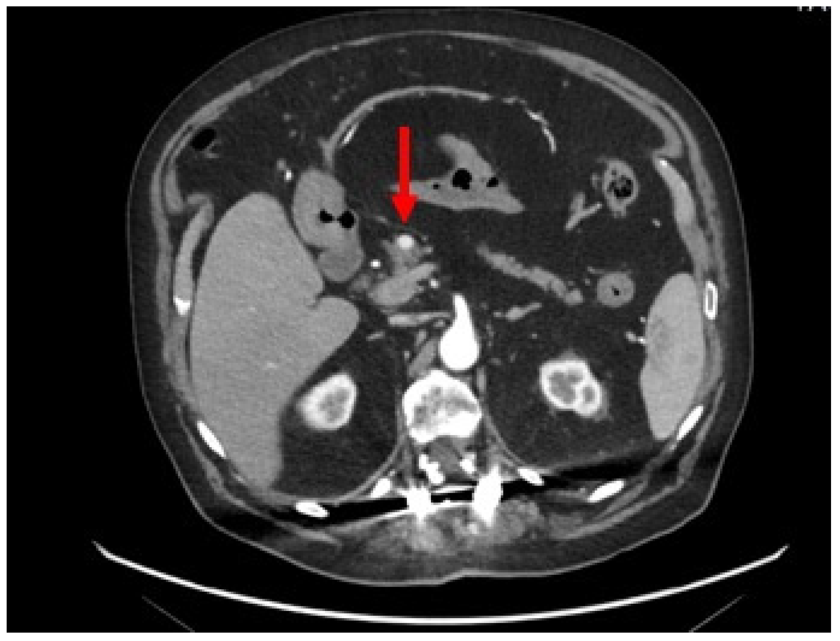
Figure 1. Contrast-enhanced abdominal CT scan: axial section showing a homogeneous and hypervascular mass of 8 mm (red arrow) on arterial phase.
Preprints 85166 g001
Figure 2. 68Ga-DOTA-TOC integrated PET/CT, transaxial (A) and MIP (B), shows focal and intense uptake in the primary pancreatic lesion (red arrows), with elevated expression of SSTR2/5 somatostatin receptors.
Preprints 85166 g002
Figure 3. Endoscopic ultrasound (EUS) image (red arrow) of a small hypo-echogenic lesion with regular margin and a major axis of 8.9 mm.
Preprints 85166 g003
Figure 4. (A) Microcores of sample tissue. (B) Abundant blood and entrapped epithelial elements of pancreatic tissue stained by Hematoxylin-Eosin. (C) Epithelial cells, with granular cytoplasm and nuclei with thickened chromatin (Hematoxylin-Eosin staining). (D) Chromogranin-A (5H7 clone, immunohistochemical staining). (E) Synaptophysin (27G12 clone, immunohistochemical staining). (F) Serotonin (YC5/45 clone, Immunohistochemical staining). (G) Ki67 (MM1 clone, immunohistochemical staining).
Preprints 85166 g004
Table 1. 2019 WHO classification and grading criteria for gastroenteropancreatic neuroendocrine neoplasms (GEP-NENs)[16].
Differentiation Grade Mitotic rate (mitoses/2 mm2) Ki-67 index
NET, G1 Well differentiated Low <2 <3%
NET, G2 Intermediate 2-20 3-20%
NET, G3 High >20 >20%
NEC, small-cell type Poorly differentiated High >20 >20%
NEC, large-cell type >20 >20%
MiNEN Well or poorly differentiated Variable Variable Variable
Disclaimer/Publisher’s Note: The statements, opinions and data contained in all publications are solely those of the individual author(s) and contributor(s) and not of MDPI and/or the editor(s). MDPI and/or the editor(s) disclaim responsibility for any injury to people or property resulting from any ideas, methods, instructions or products referred to in the content.
Copyright: This open access article is published under a Creative Commons CC BY 4.0 license, which permit the free download, distribution, and reuse, provided that the author and preprint are cited in any reuse.
Prerpints.org logo

Preprints.org is a free preprint server supported by MDPI in Basel, Switzerland.

Subscribe

© 2024 MDPI (Basel, Switzerland) unless otherwise stated