Preprint
Article

Neuroprotective Effects of Fermented Blueberry and Black Rice Against Particulate Matter 2.5 μm-Induced Inflammation In Vitro and In Vivo

Altmetrics

Downloads

154

Views

262

Comments

0

A peer-reviewed article of this preprint also exists.

Submitted:

12 September 2023

Posted:

14 September 2023

You are already at the latest version

Alerts
Abstract
The escalating prevalence of particulate matter (PM) has raised serious concerns regarding its detrimental effects on human health. This study aimed to investigate the potential of fermented blueberry and black rice (FBBR) in mitigating the effects of PM2.5, both in SH-SY5Y cells and mice exposed to PM2.5. Various assays, including MTT, NO, western blot, ELISA, and behavioral studies (MWM and Y-maze) were conducted. Our results demonstrated that PM2.5 induced significant cytotoxicity and elevated nitric oxide (NO) production at a concentration of 100μg/mL of PM2.5 in SH-SY5Y cells. Additionally, administration of FBBR effectively attenuated PM2.5-induced cytotoxicity and suppressed NO production in SH-SY5Y cells. In an intranasal instilled mice model, exposure to 10 mg/kg body weight of PM2.5 resulted in cognitive impairments. However, FBBR treatment ameliorated these impairments in both the Y-maze and MWM tests in PM2.5-exposed mice. Furthermore, FBBR administration increased the expression of brain-derived neurotrophic factor (BDNF) and reduced inflammatory markers in the brains of PM2.5-exposed SH-SY5Y cells. These findings underscore the detrimental effects of PM2.5 on the nervous system and highlight the potential of FBBR as a nutraceutical agent for mitigating these effects. Overall, this study emphasizes the urgency of addressing the harmful impact of PM2.5 on the nervous system and suggests the promising role of FBBR as a protective intervention against the adverse effects associated with PM2.5 exposure.
Keywords: 
Subject: Biology and Life Sciences  -   Neuroscience and Neurology
Preprints 84914 i001

Introduction

Particulate matter less than 2.5 micrometers (PM2.5) is a major environmental pollutant with serious health effects. Inhalation of PM2.5 is associated with many adverse health effects, especially on the respiratory and cardiovascular systems [1]. The small size of PM2.5 particles allows them to penetrate deep into the lungs, where they can cause inflammation and oxidative stress. Chronic exposure to PM2.5 is associated with an increased risk of respiratory diseases such as asthma, bronchitis, and lung cancer. In addition, PM2.5 can enter the bloodstream and cause systemic inflammation, leading to cardiovascular disease, including heart attack and stroke. In addition, PM2.5 is associated with adverse pregnancy outcomes, neurodevelopmental disorders in children, and even early death[1,2]. Growing evidence has emerged, indicating that there is a connection between exposure to PM2.5 and adverse effects on the brain and cognitive functions. Inhaling PM2.5 particles can result in their migration from the respiratory system into the bloodstream, subsequently breaching the blood-brain barrier, and directly impacting brain tissue. The inflammatory response triggered by exposure to PM2.5 can incite oxidative stress and neuroinflammation, contributing to neuronal damage and impairing cognitive functions. Research has established a link between prolonged exposure to elevated levels of PM2.5 and an increased risk of neurodegenerative conditions such as Alzheimer's disease and Parkinson's disease [3]. Furthermore, PM2.5 exposure has been associated with cognitive decline, including issues related to attention, memory, and reduced cognitive development in both children and adults.
Neuroinflammation is a grave concern of neuronal cell death and could lead to neurological disorders and neurodegenerative diseases (NDDs) [4]. Inducible nitric oxide synthase (iNOS) is an enzyme responsible for nitric oxide (NO) production is upregulated in neurodegenerative conditions and has been shown to contribute to neuronal damage through oxidative stress and inflammation [3,5,6,7]. Likewise, cyclooxygenase-2 (COX-2) is an inducible enzyme involved in inflammatory prostaglandin synthesis and is known to be the development of neurodegenerative diseases, leading to chronic inflammation and cellular dysfunction in neuronal cells. Tumer necrosis factor-alpha (TNF-α) is a potent inflammatory cytokine, that has been implicated in neuroinflammation, synaptic dysfunction, and neuronal cell death. Dysregulation of various inflammatory interleukins (ILs), such as IL-1β and IL-6, also plays an important role in neurodegeneration by regulating inflammatory responses and contributing to neurotoxicity. In addition, the tumor suppressor protein p53, traditionally implicated in cancer, has become a key player in neurodegeneration [8,9]. In response to cellular stress, p53 can induce apoptosis and promote neuronal cell death, exacerbating neurodegeneration [9,10,11]. BAX, a pro-apoptotic protein, promotes cell death by facilitating the release of mitochondrial proteins. Dysregulation of BAX can result in neuronal death in neurodegenerative conditions. Cleaved Poly (ADP-ribose) polymerase (C. PARP), an enzyme involved in DNA repair, can be a marker of apoptosis when it is cleaved by caspases in response to cellular stress. Its presence indicates ongoing cell death in neurons affected by neurodegenerative diseases, highlighting the role of apoptosis in these conditions and its potential as a therapeutic target [4,12,13]. In contrast, the anti-apoptotic protein B-cell lymphoma 2 (Bcl-2), which regulates mitochondrial integrity and apoptosis, has shown neuroprotective effects by promoting cell survival and inhibiting apoptosis. Overexpression of apoptotic marker level in neuronal cells could be a pivotal cause of NDDs progression.
Black rice (Oryza sativa L.) and blueberries (Vaccinium corymbosum L.) are rich in polyphenolic compounds, including anthocyanins, flavonoids, and so on, which have potent antioxidant activity [13]. The natural antioxidants found in these plants are known to protect the skin from UV light. Previous studies have demonstrated that black rice extract can modulate UV radiation matrix metalloproteinase (MMP) expression in a skin cell model, while blueberry extract shows a protective effect against UV irradiation in a skin cell co-culture system. In our previous study, we identified several bioactive compounds such as gallic acid, chlorogenic acid, rutin, quercetin, resveratrol, syringic acid, and kuromanine in the fermented mixture of black rice and blueberries (FBBR) [13,14]. Considering the potential effects of FBBR to protect against UV-induced skin damage, we sought to investigate the mechanisms underlying its skin protective effects against UVB radiation. Our results demonstrate that the consumption of FBBR improves UVB-induced skin inflammation, improves skin hydration, and strengthens the skin barrier by inhibiting pathways involved in photoaging. reactive oxygen species (ROS), MMP-9, and caspase cascades. In the present study, we investigated the effects of FBBR, prepared with Lactobacillus plantarum MG4221, on neuroinflammation, cognitive functions and neurotoxicity of PM2.5 in vitro and in vivo. FBBR is rich in bioactive compounds that show anti-inflammatory, antioxidant, and anti-aging effects. In this study, we investigated the potential bio-functional activity of neuroprotective effects and alleviation of cognitive impairments by PM2.5-induced mice.

2. Results

2.1. Toxicity of PM2.5 and Protective Effects of FBBR in SH-SY5Y cells

First, we determined a certain dose of PM2.5 for SH-SY5Y cells. At a concentration of 100 µg/mL, PM2.5 significantly reduced cell viability and tripled NO production compared with the control group in SH-SY5Y cells. We then focused on investigating the neuroprotective effects of FBBR by stimulating SH-SY5Y cells with 100 µg/mL PM2.5 (Figure 1). FBBR treatment successfully restored cell viability in PM2.5-induced SH-SY5Y cells and effectively reversed PM2.5 stimulation-induced excessive NO production.

2.2. Effects of FBBR on Inflammatory markers iNOS, COX-2, IL-6 and Apoptotic Proteins Expression in PM2.5-induced SH-SY5Y cells

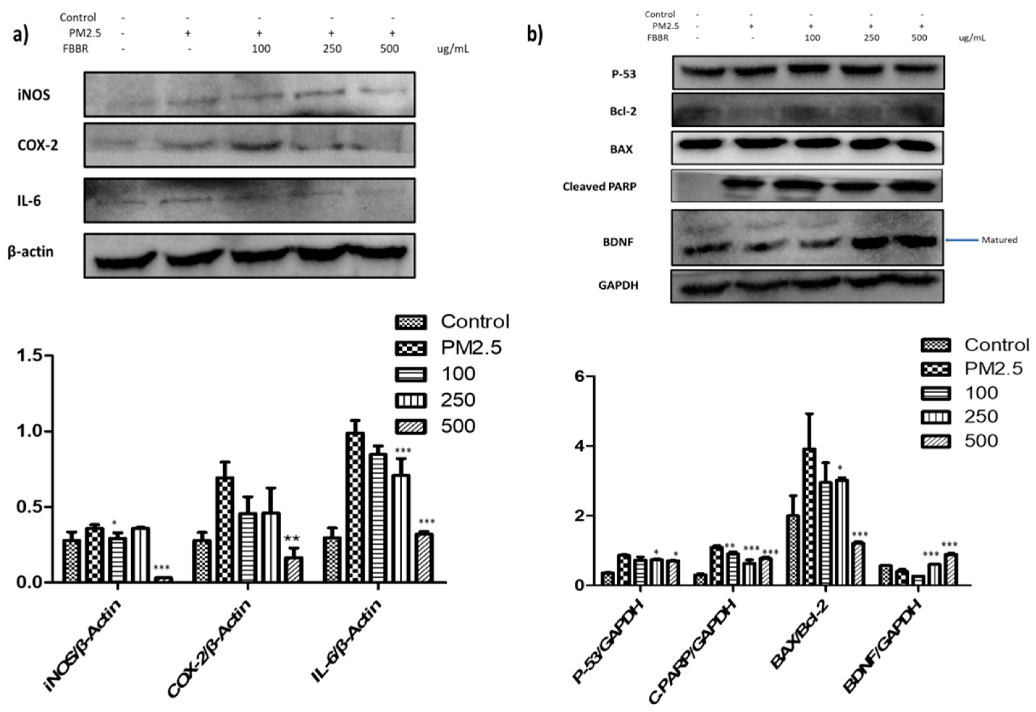
We observed that PM2.5 exposure led to an increase in NO production, which is known to be synthesized by nitric oxide synthases (NOSs). Specifically, PM2.5 upregulated the expression of inducible NOS (iNOS), a potent inflammatory biomarker. However, treatment with FBBR resulted in a significant downregulation of iNOS expression (Figure 2 a). Furthermore, FBBR demonstrated anti-inflammatory effects by suppressing the expressions of COX-2 and IL-6 in PM2.5-induced SH-SY5Y cells. Intriguingly, FBBR also exhibited neuroprotective properties by modulating apoptotic markers. It reduced the levels of pro-apoptotic markers such as p53, BAX, and Cleaved PARP (C. PARP), while simultaneously increasing the expressions of the anti-apoptotic regulator Bcl-2 and brain-derived neurotrophic factor (BDNF) in PM2.5-induced SH-SY5Y cells (Figure 2 b). The BAX/Bcl-2 ratio, a critical determinant of cell survival. [4,10]. BAX/Bcl-2 rapidly increased by PM2.5 and FBBR dose dependently decreased the BAX/Bcl-2 ratio. BDNF has a neuroprotective role by promoting the survival and health of neurons, helping to prevent their degeneration and apoptosis. Moreover, BDNF plays a crucial role in neurogenesis, facilitating the growth and differentiation of new neurons in the nervous system, which is important for learning and memory, as well as overall brain health.

2.3. Effects of FBBR on Cognitive Impairment by PM2.5-induced Mice

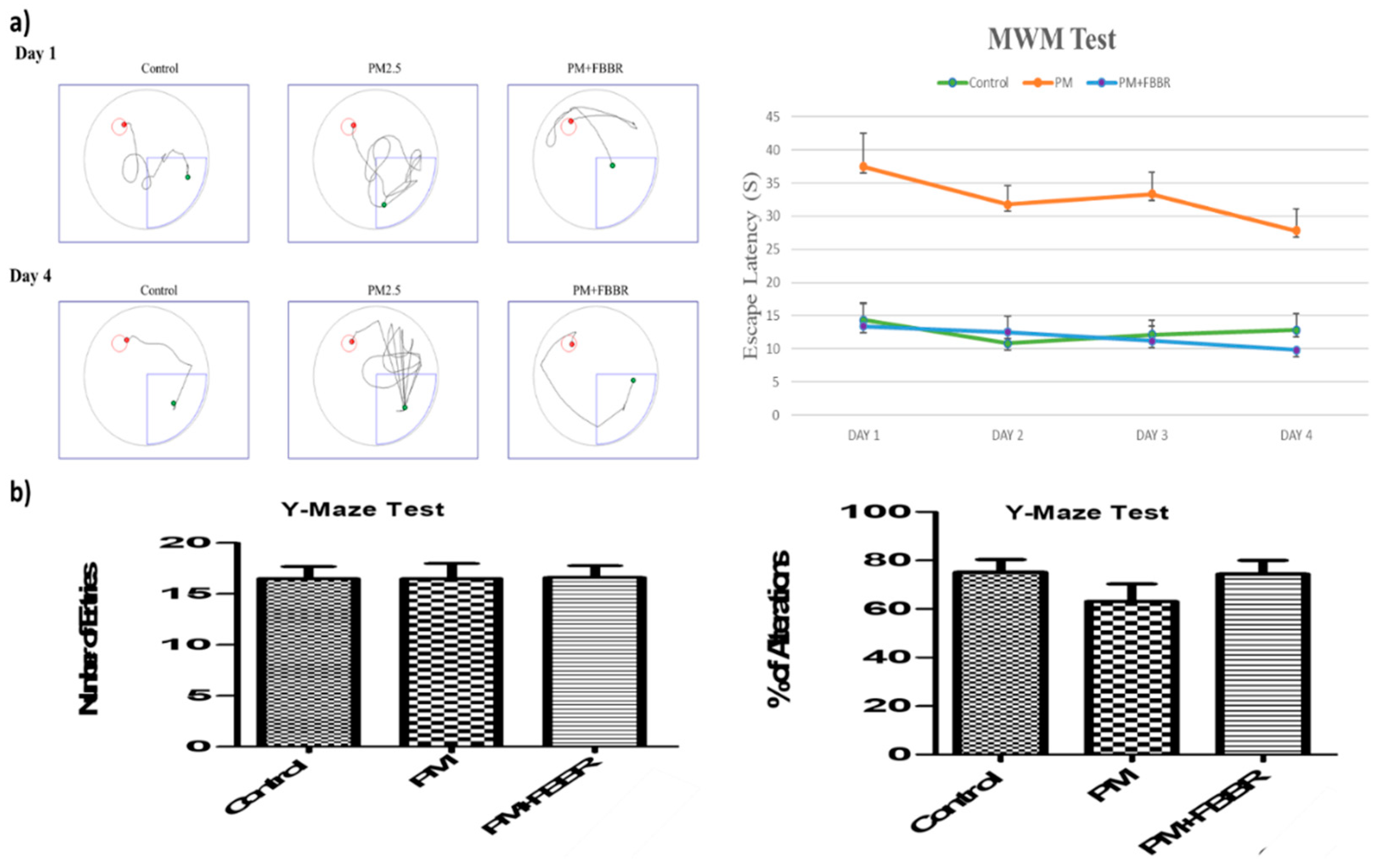
Cognitive function is a multifaceted construct that encompasses a wide range of mental processes, including attention, memory, language, problem-solving, and executive functions [15,16]. We investigated the impact of particulate matter on spatial learning and memory using well-established models such as the Morris Water Maze (MWM) and Y-Maze tests (Figure 3). Previous studies have demonstrated that PM2.5 has a negative effect on cognitive function and is associated with neurological and neurodegenerative diseases [1]. In our study, mice exposed to PM2.5 through intranasal instillation showed nearly twice the impairment compared to the control group in the MWM test. However, treatment with 200 mg/kg body weight of FBBR alleviated the cognitive impairment, bringing it to a level similar to that of healthy control mice. Additionally, FBBR treatment markedly increased the percentage of alteration in PM2.5-induced mice in Y-maze study.

2.4. Effects of FBBR on Neuroinflammation in PM2.5-induced Mice

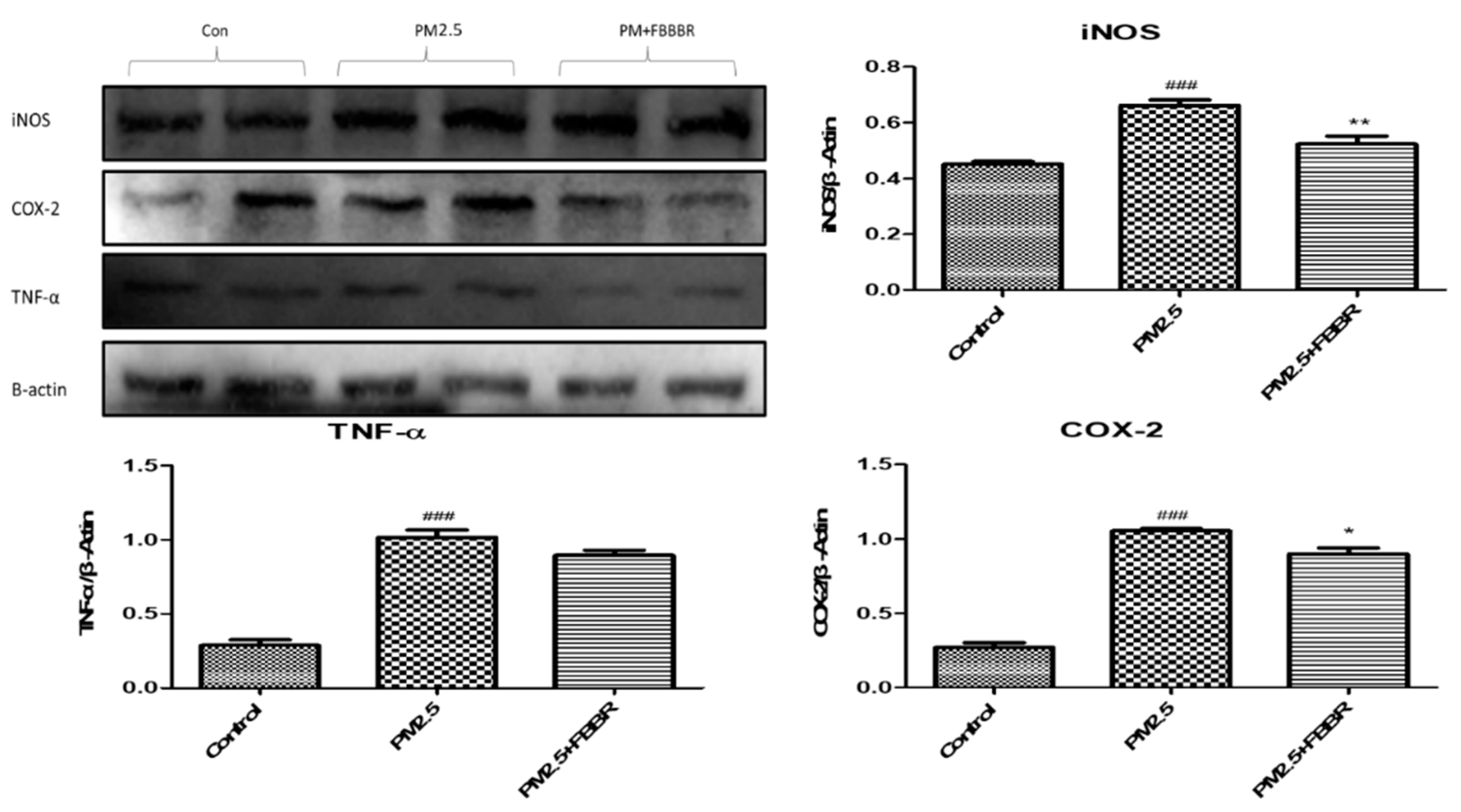
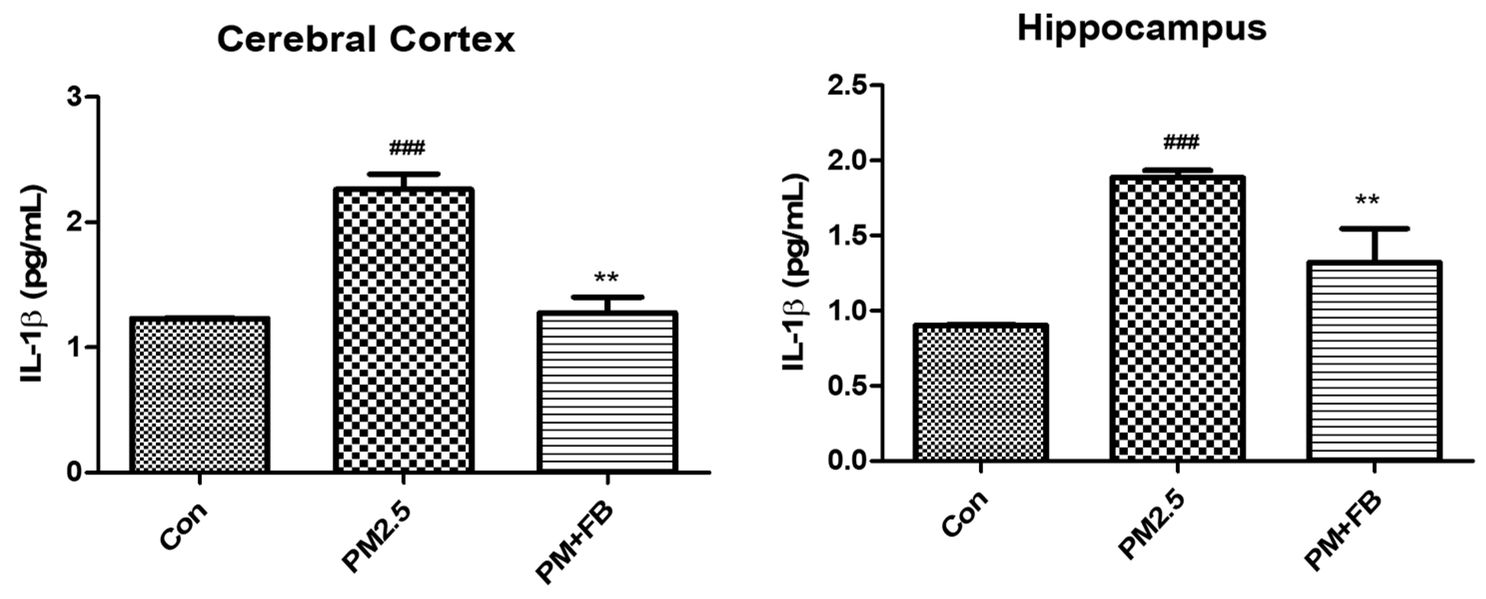
Dementia and NDDs are incapacitating conditions that profoundly impact the brain, resulting in a gradual deterioration of cognitive abilities and various neurological functions. These conditions can be triggered by a multitude of factors, encompassing both genetic predispositions and environmental influences. The advent of the industrial revolution and modern infrastructures has contributed to the escalation of particulate matter (PM) levels in the atmosphere [17]. Among these particles, PM2.5 has garnered particular attention due to its detrimental effects on the nervous system. It has been observed that PM2.5 can instigate adverse reactions in the organs responsible for apoptosis regulation and inflammation, thus posing a toxic threat [1]. Inflammation plays a pivotal role in cell death and leads to NDDs by PM2.5 exposure [3,6,18]. PM2.5 markedly increased pro-inflammatory markers such as, iNOS, COX-2 and TNF-α in the brains of mice and FBBR significantly reversed the inflammatory cytokines and mediators in the brain of PM2.5-induced mice (Figure 4). The cerebral cortex and hippocampus play an important role in cognition and various neurological functions [4]. Disturbances in these regions can lead to chronic diseases such as Alzheimer's disease and psychological disorders. An important inflammatory biomarker associated with these conditions is IL-1β, which exhibits both autocrine and paracrine effects. Elevated levels of IL-1β indicate neuroinflammation. Notably, FBBR effectively restored IL-1β levels in the cerebral cortex and hippocampus of PM2.5-induced mice. This suggests that FBBR has a potential therapeutic benefit to reduce the neuroinflammatory response associated with PM2.5 exposure (Figure 5).

3. Discussion

The primary aim of this research is to delve comprehensively into the promising neuroprotective advantages offered by the combination of fermented blueberry and black rice (FBBR), with the goal of alleviating the detrimental effects caused by exposure to PM2.5 on neuronal cells and cognitive function. PM2.5 is widely recognized for its ability to initiate neuroinflammation, oxidative stress, and cognitive decline, all of which play significant roles in the advancement of neurodegenerative disorders [1,19,20]. This investigation is focused on revealing the intricate underlying mechanisms that give rise to the protective properties exhibited by FBBR. The initiation of this inquiry involved a meticulous evaluation of the impact of PM2.5 exposure on SH-SY5Y cells. At a concentration of 100 µg/mL, PM2.5 notably undermined cell viability and triggered a substantial increase in the production of nitric oxide (NO). This highlights the potent capacity of PM2.5 to induce oxidative stress and inflict significant damage to cellular structures. Encouragingly, treatment with FBBR at the same concentration adeptly restored cell viability to a considerable extent and effectively mitigated the excessive production of NO. These results underscore the considerable potential of FBBR in conferring robust neuroprotection. By effectively countering the adverse effects of PM2.5 on neuronal cells and cognitive function, FBBR holds great promise as a natural intervention to mitigate the progression of neurodegenerative disorders. This study contributes to a deeper understanding of the potential benefits of FBBR and sheds light on its possible role in promoting brain health in the face of environmental challenges like PM2.5 exposure.
The study's scope was expanded to encompass an evaluation of FBBR's effects on mice models exposed to PM2.5. The evident cognitive impairment resulting from PM2.5 exposure was verified through the utilization of both the Morris Water Maze and Y-Maze tests, illuminating the substantial extent of cognitive decline induced by the pollutant [1,21]. However, the administration of FBBR displayed a remarkable ability to ameliorate this cognitive impairment, effectively reinstating the cognitive performance of the treated mice to a level closely resembling that of the control group. In the context of neuroinflammation, a pivotal aspect of the study, mice subjected to PM2.5 exposure exhibited a prominent elevation in pro-inflammatory markers. These markers serve as reliable indicators of heightened inflammation within the brain. Impressively, the implementation of FBBR treatment exerted a significant modulating influence on the escalated levels of these inflammatory cytokines. This compelling outcome underscores the potent capacity of FBBR to effectively alleviate the neuroinflammatory response triggered by exposure to PM2.5, indicating its potential as a valuable intervention in mitigating the adverse neurological effects associated with environmental pollutant exposure [17].
Exposure to PM2.5 resulted in the notable upregulation of key inflammatory markers, including iNOS, COX-2, and IL-6, within SH-SY5Y cells. What adds to the intrigue is the compelling response observed upon the application of FBBR treatment. Demonstrating its anti-inflammatory prowess, FBBR impressively orchestrated the downregulation of iNOS, COX-2, and IL-6 expressions. This intriguing ability underscores FBBR's potential in curbing inflammatory responses. Beyond its anti-inflammatory effects, FBBR revealed its capacity to foster neuroprotection through its modulation of apoptotic markers. In a remarkable display, FBBR not only diminished pro-apoptotic markers but also exerted a positive influence on anti-apoptotic regulators like Bcl-2 and BDNF. The pivotal BAX/Bcl-2 ratio, a decisive factor in determining cellular survival, exhibited a favorable shift under the influence of FBBR. This implies the potential cryoprotective role of FBBR, a notion substantiated by previous studies [2,21,22]. Neuroinflammation is a pivotal contributor to neurodegenerative diseases [3,23]. In fact, PM2.5-induced neuroinflammation was conspicuously observed in brain regions intricately linked with cognition, such as the cerebral cortex and hippocampus. Within these regions, the expression of pivotal inflammatory biomarkers, including iNOS, COX-2, and TNF-α, experienced marked elevation following PM2.5 exposure. Here again, the potential of FBBR shone through, as it adeptly counteracted these neuroinflammatory responses. A significant highlight is the restoration of IL-1β, a crucial inflammatory mediator, to its normal levels through FBBR treatment. This restoration strongly suggests the therapeutic potential of FBBR in mitigating neuroinflammatory responses associated with exposure to PM2.5, a finding with far-reaching implications for addressing the neurodegenerative risks posed by environmental factors [24]. FBBR contains major and effective bioactive compounds as shown in our previous study [14] such as gallic acid, chlorogenic acid, rutin, quercetin, resveratrol, syringic acid, and kuromanine, which showed pivotal role against neurodegeneration[12,25]. Gallic acid, chlorogenic acid, rutin, quercetin, resveratrol, syringic acid, and kuromanine are renowned for their antioxidant and anti-inflammatory properties. They have demonstrated the ability to shield against oxidative stress and neuroinflammation, thereby holding promise in potentially reducing the risk of neurodegenerative diseases [26,27,28].

4. Materials and Methods

4.1. Chemical and Reagents

PM2.5, a type of urban particulate matter with a diameter of ≤2.5 µm, was acquired from NIST in Gaithersburg, MD. MTT (3-(4,5-dimethylthiazol-2-yl)-2,5-diphenyltetrazolium bromide) was obtained from Sigma Aldrich. DMEM (Dulbecco's Modified Eagle's Medium), DPBS (Dulbecco's Phosphate-Buffered Saline), Penicillin Streptomycin (P.S), and Trypsin EDTA were purchased from WELGENE Inc. in Seoul, Korea. Primary and secondary antibodies for Western blot analysis were sourced from Cell Signalling Technology or Abcam (Cambridge, UK). All other chemicals used were of analytical-grade quality and were employed without the need for additional purification.

4.2. Sample Preparations

Black rice and blueberry extracts were prepared by immersing them in 50% ethanol (EtOH) at room temperature for 24 hours. The extracts were then filtered using Whatman No.1 filter paper and this process was repeated three times. Then, the extract was concentrated under reduced pressure using a vacuum rotary evaporator (EYELA, Tokyo, Japan). The resulting extracts were combined in a ratio of 7 parts blueberry extract to 3 parts black rice extract, and the pH was adjusted to 6.0. Next, 2% (v/v) Lactobacillus plantarum MG4221 was added to the mixture, which was then cultured at 37°C for 24 hours. The fermented extracts were subsequently freeze-dried to obtain a powdered form (FBBR), and they were stored at -20°C until they were ready to be used.

4.3. Cell Culture

The human neuroblastoma SH-SY5Y cells were sourced from the Korean cell line bank. To maintain the cells, DMEM (Dulbecco's Modified Eagle's Medium) supplemented with 10% FBS (Fetal Bovine Serum) and 1% P.S (Penicillin Streptomycin) was utilized. The cells were cultured in a controlled environment within an incubator set at 37°C with 5% CO2 and under 95% humidity. Regular subculturing was carried out to maintain the cells, and they were used for experimentation when they reached approximately 80-85% confluency. For the experiments, cells within passage numbers 25-40 were utilized.

4.4. MTT and NO Assay

Cell viability was assessed using the MTT assay, while the NO assay was conducted using the Griess reagent assay [29]. To begin, 2×10⁴ SH-SY5Y cells were seeded each well in a 96-wellplate. Initially, we determined the toxic dosage of PM2.5 in SH-SY5Y cells by measuring NO production. Different concentrations ranging from 10 to 500 μg/mL were tested. Subsequently, the cells were exposed to 100 μg/mL of PM2.5 and treated with FBBR (specific procedure/protocol) for both the MTT and NO assays.

4.5. Animal Model Design

Male C57Bl/6N mice, aged four weeks and weighing approximately 25 ± 2 g, were procured from DBL in Chungcheongbuk-do, Korea. The mice were given a seven-day acclimatization period in a Specific Pathogen Free (SPF) animal room, adhering to controlled conditions including a temperature of 25 °C ± 2 °C, humidity maintained at 55%–65%, and a lighting schedule of 12 hours of light followed by 12 hours of darkness. During this period, the mice had ad libitum access to chow diet and drinking water. Following the acclimation phase, the mice were randomly divided into three groups, each consisting of five mice (n = 6). All animal experiments were carried out in compliance with the guidelines provided by the National Institutes of Health, and the study protocol was approved by the Institutional Animal Care and Use Committee (IACUC) of Konkuk University on 30th May 2022 (approval number: KU22077). Once acclimated, the mice were further divided into three groups: a control group, a PM2.5 group where intranasal administration of PM2.5 at a dosage of 10 mg/kg bodyweight (BW) was conducted for four weeks, and a PM+FBBR group where FBBR was administered as 200 mg/kg BW in addition to PM2.5 induction. Each group contained five mice. After four weeks of treatment, the mice underwent behavioral testing, followed by euthanasia. Brain tissues were collected for subsequent biochemical assays.

4.6. Morris Water Maze Test

The Morris water maze (MWM) is employed to assess the spatial memory capabilities of mice [16,21], as it relies on their navigational skills to locate a submerged escape platform. In this experiment, a circular pool measuring 122 cm in diameter was utilized, with a hidden platform serving as the target. The objective for the animals was to find the shortest and most direct path to the hidden platform from their starting position. To familiarize the mice with the task, four consecutive training sessions were conducted. Following the treatment, the spatial acquisition of the mice was assessed, and the resulting data were analyzed using SMART 3.0 software (Ver. 3.0, Harvard Apparatus, Holliston, MA 01746, USA).

4.7. Y-Maze Test

The Y-maze spontaneous alternation test was developed to assess the inclination of mice to explore unfamiliar surroundings [21]. The experiment involved a Y-shaped maze consisting of three gray plastic arms (each measuring 30 cm) positioned at a 120° angle from each other. Beginning from the center of the maze, the animal was given 5 minutes to freely explore each arm. An entry was logged when all four limbs were inside the arms. Spontaneous alternation occurred when the chosen arm differed from the previous two arms. The test recorded both the number of arm entries and the number of spontaneous changes to determine the percentage of alternation.

4.8. Western Blot Analysis

Total protein was extracted from the cells and brain specimen using PRO-PREP buffer from iNtRON Biotechnology [12,29,30]. The protein was separated from lysate through centrifugation at 14,000 × g at 4°C. An equal amount of protein (30 μg/lane) was loaded onto a 10% SDS polyacrylamide gel for electrophoresis. Following electrophoresis, the protein samples were transferred onto a polyvinylidene fluoride (PVDF) membrane and subsequently blocked using 5% BSA. The membranes were then incubated with the corresponding primary antibodies overnight at 4°C. Subsequently, the membranes were exposed to a secondary antibody (anti-rabbit) at room temperature for 1 hour. The immunosignals on the membranes were visualized using enhanced chemiluminescence (ECL) on an ChemiDoc XRS+ (Bio-Rad). Finally, the band intensity was quantified using ImageJ software.

4.9. IL-1β Level Detection

Protein extraction was conducted from the cerebral cortex and hippocampus of mice in order to assess the levels of IL-1β. This was achieved using an enzyme-linked immunosorbent assay (ELISA) [4,31]. The ELISA assay was carried out following the instructions provided in the kit's manual (ab197742).

4.10. Statistical Analysis

Band intensities were quantified using ImageJ software. The obtained results are reported as means ± SEM for all experimental data. Statistical analysis was performed using Microsoft Excel 2016 and GraphPad Prism 5.0 software (GraphPad Software, Inc., San Diego, CA, USA). One-way analysis of variance (ANOVA) was employed, and Tukey's Multiple Comparison Test was used as a non-parametric test. A p-value of less than 0.05 was considered statistically significant.

5. Conclusion

FBBR showed remarkable attenuation of SH-SY5Y cell death by PM and reduced NO production in PM2.5-induced cells. FBBR expeditiously exerted neuroprotective effects by reducing inflammatory cytokines and mediators in vitro and in vivo. Furthermore, FBBR decreased pro-apoptotic markers and increased anti-apoptotic markers and growth factors (BDNF). FBBR is rich in bioactive and improved cognitive functions in PM2.5-induced mice. Taken all together, FBBR could be a strong neuroprotective agent.

Author Contributions

A.B. Bayazid designed the study, conducted experiments and wrote the manuscript. A.B. Bayazid, S-A Jeong and S. Azam carried out the behavioral study. A.B. Bayazid and S.H. Oh analyzed data. B.O. Lim reviewed and supervised the study. All authors have read and agreed to the published version of the manuscript.

Acknowledgments

This study was supported by Konkuk University 2023.

References

  1. Ku, T., et al., NF-κB-regulated microRNA-574-5p underlies synaptic and cognitive impairment in response to atmospheric PM2.5 aspiration. Particle and Fibre Toxicology, 2017. 14(1): p. 34.
  2. Tseng, C.-Y., et al., The Effect of Ganoderma Microsporum immunomodulatory proteins on alleviating PM2.5-induced inflammatory responses in pregnant rats and fine particulate matter-induced neurological damage in the offsprings. Scientific Reports, 2019. 9(1): p. 6854. [CrossRef]
  3. Block, M.L. and L. Calderón-Garcidueñas, Air pollution: mechanisms of neuroinflammation and CNS disease. Trends in Neurosciences, 2009. 32(9): p. 506-516.
  4. Bayazid, A.B., et al., Sodium butyrate ameliorates neurotoxicity and exerts anti-inflammatory effects in high fat diet-fed mice. Food and Chemical Toxicology, 2022. 159: p. 112743. [CrossRef]
  5. Jabri, M.-A., K. Rtibi, and H. Sebai, Chamomile decoction mitigates high fat diet-induced anxiety-like behavior, neuroinflammation and cerebral ROS overload. Nutritional Neuroscience, 2020: p. 1-12. [CrossRef]
  6. Cavaliere, G., et al., High-fat diet induces neuroinflammation and mitochondrial impairment in mice cerebral cortex and synaptic fraction. Frontiers in cellular neuroscience, 2019. 13: p. 509. [CrossRef]
  7. Song, J., et al., Protective effects of quercetin on traumatic brain injury induced inflammation and oxidative stress in cortex through activating Nrf2/HO-1 pathway. Restorative Neurology and Neuroscience, 2021. 39: p. 73-84.
  8. Sablina, A.A., et al., The antioxidant function of the p53 tumor suppressor. Nature Medicine, 2005. 11(12): p. 1306-1313.
  9. Maor-Nof, M., et al., p53 is a central regulator driving neurodegeneration caused by C9orf72 poly(PR). Cell, 2021. 184(3): p. 689-708.e20. [CrossRef]
  10. Bayazid, A.B. and B.O. Lim, Quercetin Is An Active Agent in Berries against Neurodegenerative Diseases Progression through Modulation of Nrf2/HO1. Nutrients, 2022. 14(23): p. 5132.
  11. Quiñones, M., et al., p53 in AgRP neurons is required for protection against diet-induced obesity via JNK1. Nature Communications, 2018. 9(1): p. 1-16.
  12. Bayazid, A.B., et al., Sodium Butyrate Alleviates Potential Alzheimer’s Disease In Vitro by Suppressing Aβ and Tau Activation and Ameliorates Aβ-induced Toxicity Food and Agricultural Immunology, 2023. 34.
  13. Park, S.H., et al., Fermented black rice and blueberry with Lactobacillus plantarum MG4221 improve UVB-induced skin injury. Food and Agricultural Immunology, 2021. 32(1): p. 499-515. [CrossRef]
  14. Hong, S.M., et al., Fermented blueberry and black rice containing Lactobacillus plantarum MG4221: a novel functional food for particulate matter (PM 2.5)/dinitrochlorobenzene (DNCB)-induced atopic dermatitis. Food&Function, 2021. 12(8): p. 3611-3623.
  15. Wang, D.-M., et al., Effects of Long-Term Treatment with Quercetin on Cognition and Mitochondrial Function in a Mouse Model of Alzheimer’s Disease. Neurochemical Research, 2014. 39(8): p. 1533-1543. [CrossRef]
  16. Xu, J., et al., Melatonin alleviates cognition impairment by antagonizing brain insulin resistance in aged rats fed a high-fat diet. Journal of Pineal Research, 2019. 67(2): p. e12584. [CrossRef]
  17. Lee, C.S.L., et al., Seasonal variation of chemical characteristics of fine particulate matter at a high-elevation subtropical forest in East Asia. Environmental Pollution, 2019. 246: p. 668-677. [CrossRef]
  18. Ansarin, K., M. Khoubnasabjafari, and A. Jouyban, Reliability of malondialdehyde as a biomarker of oxidative stress in psychological disorders. 2015. 5(3): p. 123-127.
  19. Saw, C.L.L., et al., The berry constituents quercetin, kaempferol, and pterostilbene synergistically attenuate reactive oxygen species: Involvement of the Nrf2-ARE signaling pathway. Food and Chemical Toxicology, 2014. 72: p. 303-311. [CrossRef]
  20. Emerit, J., M. Edeas, and F. Bricaire, Neurodegenerative diseases and oxidative stress. Biomedicine&pharmacotherapy, 2004. 58(1): p. 39-46. [CrossRef]
  21. Li, X.-X., et al., Coeloglossum viride var. bracteatum extract improves cognitive deficits by restoring BDNF, FGF2 levels and suppressing RIP1/RIP3/MLKL-mediated neuroinflammation in a 5xFAD mouse model of Alzheimer’s disease. Journal of Functional Foods, 2021. 85: p. 104612.
  22. TANIGAWA, S., M. FUJII, and D.-X. HOU, Stabilization of p53 Is Involved in Quercetin-Induced Cell Cycle Arrest and Apoptosis in HepG2 Cells. Bioscience, Biotechnology, and Biochemistry, 2008. 72(3): p. 797-804. [CrossRef]
  23. Heneka, M.T., et al., Neuroinflammation in Alzheimer's disease. The Lancet Neurology, 2015. 14(4): p. 388-405.
  24. Priprem, A., et al., Anxiety and cognitive effects of quercetin liposomes in rats. Nanomedicine: Nanotechnology, Biology and Medicine, 2008. 4(1): p. 70-78. [CrossRef]
  25. Bayazid, A.B. and B.O. Lim, Quercetin Is An Active Agent in Berries against Neurodegenerative Diseases Progression through Modulation of Nrf2/HO1. 2022. 14(23): p. 5132.
  26. Ajayi, A.M., et al., Repeated social defeat stress exacerbates lipopolysaccharide-induced behavioural deficits in mice: ameliorative role of Chrysophyllum albidum fruit extract through anti-neuroinflammation, antioxidant and neurochemical balance. Metabolic Brain Disease, 2022. 37(7): p. 2467-2481. [CrossRef]
  27. Plazas, E., et al., Natural isoquinoline alkaloids: Pharmacological features and multi-target potential for complex diseases. Pharmacological Research, 2022. 177: p. 106126. [CrossRef]
  28. Hadrich, F., M. Chamkha, and S. Sayadi, Protective effect of olive leaves phenolic compounds against neurodegenerative disorders: Promising alternative for Alzheimer and Parkinson diseases modulation. Food and Chemical Toxicology, 2022. 159: p. 112752. [CrossRef]
  29. Jaradat, N., et al., Chromatography analysis, in light of vitro antioxidant, antidiabetic, antiobesity, anti-inflammatory, antimicrobial, anticancer, and three-dimensional cancer spheroids’ formation blocking activities of Laurus nobilis aromatic oil from Palestine. Chemical and Biological Technologies in Agriculture, 2023. 10(1): p. 25.
  30. Shawahna, R. and N.A. Jaradat, Ethnopharmacological survey of medicinal plants used by patients with psoriasis in the West Bank of Palestine. BMC Complementary and Alternative Medicine, 2017. 17(1): p. 4. [CrossRef]
  31. Jia, G., et al., Neural stem cell-conditioned medium ameliorates Aβ25–35-induced damage in SH-SY5Y cells by protecting mitochondrial function. Bosnian Journal of Basic Medical Sciences, 2021. 21(2): p. 179-186. [CrossRef]
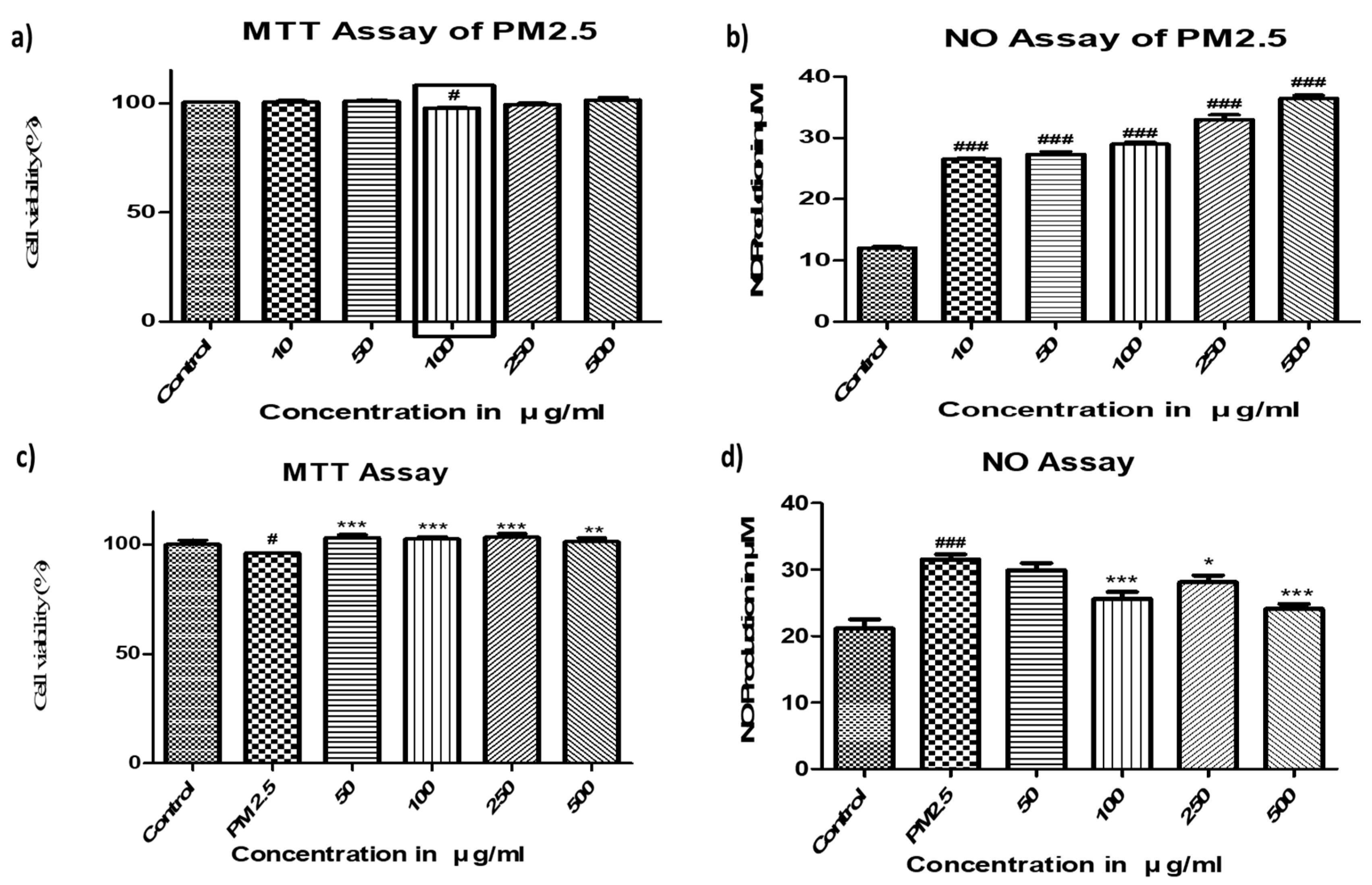
Figure 1. a) Cytotoxic dose of PM2.5 in SH-SY5Y cells and b) NO assay of PM2.5 in SH-SY5Y cells. c), d) Effects of FBBR on Cell viability and NO production in PM2.5 induced SH-SY5Y cells Results were expressed as mean ± SEM from three independent experiments #p < 0.05, ##p < 0.001 ###p < 0.0001 compared to the control group. and *p < 0.05, **p < 0.001, ***p < 0.0001 compared to the PM group.
Preprints 84914 g001
Figure 2. a) Effects of FBBR on pro-inflammatory cytokines (IL-6, iNOS, COX-2) expressions, b) apoptotic regulators and BDNF expressions in PM2.5 induced SH-SY5Y cells. The histograms were expressed as mean ± SEM from three independent experiments and *p < 0.05, **p < 0.001, ***p < 0.0001 compared to the PM group.
Preprints 84914 g002
Figure 3. Effects of FBBR (200mg/kg) on cognition impairments by PM2.5 induced mice in a) MWM test and b) Y-Maze test. Results were expressed as mean ± SEM from three independent experiments.
Preprints 84914 g003
Figure 4. Effects of FBBR (200mg/kg) on iNOS, COX-2 and TNF-α expressions in brains of PM2.5 induced mice. Results were expressed as mean ± SEM from three independent experiments. #p < 0.05, ##p < 0.001 ###p < 0.0001 compared to the control group and *p < 0.05, **p < 0.001, ***p < 0.0001 compared to the PM group.
Preprints 84914 g004
Figure 5. Effects of FBBR (200mg/kg) on IL-1β expressions in cerebral cortex and hippocampus of PM2.5 induced mice. Results were expressed as mean ± SEM from three independent experiments. #p < 0.05, ##p < 0.001 ###p < 0.0001 compared to the control group and *p < 0.05, **p < 0.001, ***p < 0.0001 compared to the PM group.
Preprints 84914 g005
Disclaimer/Publisher’s Note: The statements, opinions and data contained in all publications are solely those of the individual author(s) and contributor(s) and not of MDPI and/or the editor(s). MDPI and/or the editor(s) disclaim responsibility for any injury to people or property resulting from any ideas, methods, instructions or products referred to in the content.
Copyright: This open access article is published under a Creative Commons CC BY 4.0 license, which permit the free download, distribution, and reuse, provided that the author and preprint are cited in any reuse.
Prerpints.org logo

Preprints.org is a free preprint server supported by MDPI in Basel, Switzerland.

Subscribe

© 2024 MDPI (Basel, Switzerland) unless otherwise stated