Preprint
Article

The Presence of the Human Auditory Ossicles -Detected Post-mortem by CT Scan- as a Taphonomic Indicator

Altmetrics

Downloads

163

Views

54

Comments

0

A peer-reviewed article of this preprint also exists.

Submitted:

18 September 2023

Posted:

18 September 2023

You are already at the latest version

Alerts
Abstract
Three tiny bones compose the human ossicular chain: malleus, incus and stapes. Also known as auditory ossicles, they are united by joints in the middle ear cavity of the petrous part of the temporal bone. Completely developed 2 years after birth, the ossicular chain is involved in the physiological process of hearing, by which sound waves from the environment are converted into electrochemical impulses. In the last 500 years, most studies have focussed on the morphogenesis, morphological variability and clinical pathology of the ossicular chain, whilst only a few studies have added relevant knowledge to anthropology and forensic science. The auditory ossicles and the enclosing petrous bone are some of the hardest in the human skeleton. This reflects in a relative resistance to fire. In the post mortem period and in favourable circumstances, the auditory ossicles can fossilize and survive for millions of years. Post mortem loss in skeletal or decomposed remains can be ascribed to their destruction or physical displacement, by either macro-micro-faunal action and/or any other natural or artificial disturbance. Physical displacement is closely connected to the depositional environment of the skeletal remains, such as burial, entombment (sarcophagus, coffin, vault…), submersion or exposure to natural elements. Auditory ossicles can be recovered in situ, or very close to their anatomical location, when the skeletal material has been involved in an archaeological excavation. In the case of accessible or disturbed remains, scavengers may remove the tiny ossicles, and/or they can slip out of the middle ear cavity following skull movements. Entombment offers an effective protection against the displacement of the auditory ossicles, whereas aquatic submersion and aquatic movement almost invariably displace them. The preservation of the human auditory ossicles should be critically considered in the comprehensive context of any forensic investigation on human remains, since it can assist the reconstruction of their taphonomic history. Taphonomic histories of remains can add crucial information to forensic investigations (e.g., the Post Mortem Interval, PMI). The aim of this study, limited by scarce relevant literature, is to discuss the potential role of the ossicular chain, detected by post mortem imaging techniques, as a taphonomical indicator in decomposing and/or skeletonized bodies.
Keywords: 
Subject: Biology and Life Sciences  -   Other

Introduction

The human ossicular chain is an anatomical structure composed of three tiny bones, malleus, incus and stapes, which are completed at birth [1] with refined development by 2 years of life [2]. They are also collectively known as auditory ossicles, and are united by joints in the middle ear cavity of the petrous bone, a part of the temporal bone. Other anatomical structures of the middle ear cavity are the tympanic membrane (eardrum), which seals the medial end of the external acoustic meatus (ear canal), two muscles (tensor tympani and stapedius), six ligaments which reinforce the ossicles joints, vascular vessels (branches of the external and internal carotid arteries) and nerves (branches of the trigeminal -V- and facial -VII- cranial nerves). The middle ear cavity is connected to the pharynx by the Eustachian tube canal. The anatomy of the human ear is displayed in Figure 1. The physiological function of the ossicular chain contributes to the process of hearing, by transmitting sound vibrations from the environment to the oval window of the inner ear, where they are converted into electrochemical impulses [3].
The ossicular chain has been known for 500 years [4], during which most studies have focussed on its morphogenesis, morphological variability and clinical pathology [4,5,6,7,8,9]. In palaeontology and archaeology, the study of the auditory ossicles has been more rarely addressed, nevertheless it has added relevant knowledge to research in paleobiology [10,11,12], paleopathology [13,14], human evolution [9,15,16] and forensics, where it has been defined as a reliable source for DNA extraction [17,18].
Computerized Tomography (CT) Scan and related techniques, such as Micro CT [19], Cone Beam CT [20] and Multislice CT [21], are the principal tools for the imaging study of the human middle ear cavity. In both in-vivo and post mortem contexts, it achieves excellent delineation of soft-tissue and bone alterations, associated with either pathology or trauma. In contrast, the detailed characterization of soft tissue and fluid attained by Magnetic Resonance (MR) limits its post mortem applications to non-skeletonized bodies [22, and references therein].
In the process of decomposition of the human body, the three auditory ossicles are separated from the ligaments and tendons which, in life, ensure their anatomical position in the middle ear cavity. Once they skeletonize, and become dry isolated tiny bones, they lie inside the middle ear cavity which, following the decomposition of the tympanic membrane (eardrum), directly communicates with the external environment through the external acoustic meatus (ear canal). The skeletonized external acoustic meatus is much shorter than in vivo, when soft tissues and the auricle are present, so that in an adult skeleton, the distance separating the auditory ossicles from the external environment is normally within 2 cm.
Such a short distance between the auditory ossicles and the external environment suggests that they can be easily lost. However, they have been found in their anatomical location, or very close, even after millions of years, as has been recorded in palaeontological recoveries [11,15,23]. Contrastingly, one or all auditory ossicles can be missing in more recent skeletons from archaeological sites [24,25] or current forensic cases observed by the authors. Since the quality of the petrous bone and the auditory ossicles is one of the hardest and most dense in the human skeleton [11,17,26], they have been documented to be relatively resistant to fire [27,28,29,30,31] and, in favourable circumstances, can fossilize and survive for millions of years. The taphonomy of bone and the fossilization process are too varied and complex to be discussed in the present context, which focus is the relevance of the presence, or absence, of the auditory ossicles in decomposing and/or completely skeletonized human bodies.
The aim of this study is to discuss the potential relevance of the presence, or absence, of the ossicular chain as a taphonomical indicator in decomposing and/or skeletonized bodies, on the basis of the existing paleontological, archaeological and forensic reports. In the reviewed literature (1975-2022), the ossicular chain was detected either through direct visual observation, or by imaging, whilst only post mortem imaging was applied to present-day forensic casework.

Materials and Methods

Research in the PubMed library was performed by combining the term “ossicles” with “archaeology”, “auditory”, “chain”, “forensic”, “human remains”, “ossicular”, “palaeontology”, “post mortem” and “skeleton”; the search process was completed by hand searching and was not limited by type and year of publication, or by language or country of publication. In the selected studies, the presence/absence of the ossicular chain in decomposing/skeletonized human remains was assessed through direct visual observation or by imaging. In Table 1, the ossicular chain was listed as present and complete if all three auditory ossicles were detectable within either one or both left and right middle ear cavities, left and right temporal bones, left and right petrous bones or the immediate vicinity. If one or more ossicles were missing, the ossicular chain was listed as present and incomplete. If no auditory ossicles could be observed, the ossicular chain was listed as absent.
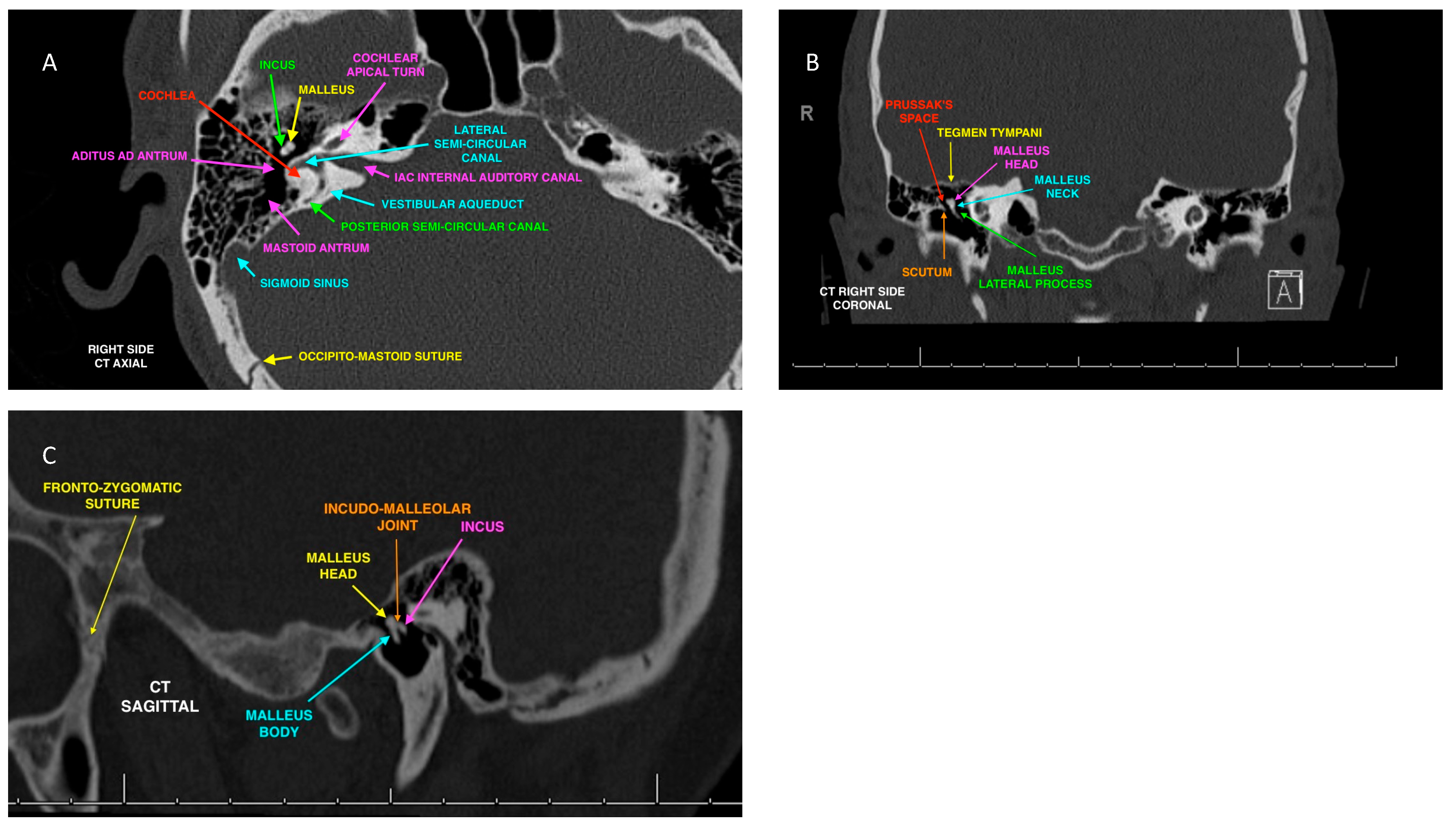
In four present-day forensic cases, the skulls and petrous bones of four individuals, object of medico-legal investigations, were analysed by imaging only. A completely skeletonized skull, colonised by barnacles and recovered on a sea shore in the north of Italy (Scannone Sacca di Goro – Ferrara 44.785458 N, 12.352930 E) in 1999 was analysed by orthopantomography and X-Rays. A second skull, also completely skeletonized and recovered in a forest in the north of Italy (Solignano – Parma 44.613384 N, 9.978914 E) in 2014 was analysed by Head and Face CT Scan with 2D and 3D image reconstructions. As well, two decomposing adult bodies were discovered in Switzerland (Lugano 46.009802 N, 8.960951 E) in the last decade, and were analysed by full-body Multislice CT Scan (MSCT). One body had been exposed outdoors for 15 days, the other was left indoors for three weeks. The CT Scan of the skull, temporal and petrous bones of an adult patient, randomly selected in the hospital database and anonymized, was compared as an example of normal anatomy. The detailed labelling of the middle ear anatomy is presented in Figure 2.

Results

The scholarly disciplines to which most studies belonged were anthropology/archaeology, medicine and palaeontology. Three paleontological studies report the presence of an incomplete and/or fragmented ossicular chain in the analysed samples. Nine archaeological studies show every combination of disturbed and apparently undisturbed remains, in which the ossicular chain can either be absent or present, complete or incomplete, and single ossicles can be whole or damaged. The ossicular chain has been detected as present and complete in the two decomposing bodies of the forensic sample, but absent in the two skeletonized skulls. The summary of results, with notes [15,16,19,24,32,by 33,34-37] are displayed in Table 1.

Discussion

The absence of one or more auditory ossicles from the middle ear cavity of a decomposing or skeletonized human skull can theoretically be connected to any taphonomical process, such as the action of macro-microfaunal scavengers, or the degradation by natural elements (e.g., water movement). However, the destruction (or dissolution) of the auditory ossicles in situ is unlikely in decomposing or skeletonized bodies, because the auditory ossicles and the enclosing petrous bone are hard and prone to long post mortem preservation. Most traces of faunal intervention are likely to be detected on both the soft tissues and the skull bones surrounding the auditory ossicles. Human action can also disturb the remains through excavation, transportation and preparation of the remains, which need to be bagged and placed in conservation boxes in order to be available for investigation. As a consequence, the most likely cause of the absence of auditory ossicles in decomposing or skeletonized bodies is physical displacement.
In one historical study an ossicle (right malleus from the external acoustic meatus) was extracted from the skull of the Nazlet Khater 2 (NK 2) skeleton (dated 35-40.000 years BPE) in 2005, following the skeletons exhumation from the Nile Valley in Upper Egypt in 1980 [38]. It was subsequently transported to a laboratory for analysis, as part of the Belgian Middle Egypt Prehistoric Project [39]. Once extricated from a mixture of sand and glue, the ossicle showed only a slight damage. The role of sand and soil occluding the external acoustic meatus (ear canal) and preventing the loss of the auditory ossicles is known in archaeology [25].
Hagedorn and colleagues [40] performed otoendoscopy on the heads of 250 ancient Egyptian mummies, and found an intact ossicular chain in the 15% of the cases and a post mortem luxation of one or more auditory ossicles in the remaining 85% of the cases. Hoffman and Hudgins [41] detected the bilateral preservation of the ossicular chain in 3 intact 3000 year-old Egyptian mummies, and absence and alterations in the remaining 6 mummies of their study. Hence, it is not unreasonable to assume that the process of artificial mummification, practiced in ancient Egypt on fresh bodies, could result in the loss of the auditory ossicles. However, the process generally favours their persistence in situ [42]. Their study involved the autopsy of an Egyptian mummy wrapped in 12 layers of linen solidified with liquid resin. After removing the temporal bones with a Stryker saw, the bilateral exploration of the middle ear led to the recovery of a complete ossicular chain.
A study of 471 archaeological temporal bones recovered from a British leper cemetery, where the interments of 257 individuals were dated between the 12th and the 17th century Present Era (PE), showed the presence of 136 auditory ossicles in 97 temporal bones of 89 individuals. Most ossicles (78%) were complete, whereas the remaining (22%) displayed various stages of physical damage. Only 4 temporal bones retained a complete ossicular chain. Two interesting standpoints are outlined in the study, firstly the statement “when necessary, soil, small stones and vegetable material were removed carefully from the external auditory canal with the use of fine dental instruments” highlights the condition of most buried bodies, that is with physiological bone cavities obstructed by foreign material [also see [25,43]], secondly, in examining previous research [44] it is apparent that the presence of the ossicular chain in buried bodies was defined as “not related to the type of burial, conditions surrounding it, or the soil”. Finally, the conclusion that the “precision in methods of recovering the skeletons is more likely to influence ossicle recovery” [36] appears to support the hypothesis that the presence of the auditory ossicles in the middle ear cavity of any skeletonized human skull relates to careful excavation and recovery, with the aim of avoiding displacement.
The paleopathological research conducted by Krenz-Niedbała and Łukasik [14] on 435 juvenile (<20 years old) archaeological (14th–17th Century PE) skeletons from two historical burial sites in Poland, revealed only 168 auditory ossicles could from 99 skeletons. One burial site had reportedly been “intensively used with deceased buried in multiple layers”. After the recovery, all skeletons were transferred to an institutional collection. At the time of the examination, the majority of the auditory ossicles were still in situ, and “fine dental instruments” were used to “remove soil and vegetable material from the external auditory canal”. Only “in few cases” were the auditory ossicles “found as loose elements in the storage boxes”. Similar circumstances regarding the presence of auditory ossicles is described by the same authors in 2021.
In a study by Dedouit and colleagues [37], no ossicular chains were found intact in 6 dry skulls with a known PMI (> 10 years). The skulls belonged to an anthropological collection. All skulls had been moved more than once, starting from the location of their original recovery (medieval and contemporary cemeteries, a private garden and forests) to a laboratory, then possibly to a storage facility and, eventually, to the shelves of a collection room. Furthermore, in the same study a body exhumed from an intact metal coffin after 3 years revealed a complete ossicular chain with remnants of joint tissue in the right temporal bone, but no ossicle was present in the left temporal bone. Unfortunately, the authors did not describe the preservation of the exhumed body or how it was positioned inside the coffin. However, they admitted to producing some displacement of the body. In fact, the body appears to have been removed from the coffin, wrapped “in two artefact-free body bags” and placed within the Multislice Computed Tomography to undergo a full-body examination, with the aim of excluding any traumatic lesions. Another recent research suggests that the auditory ossicles are usually present in putrefied bodies, as long as the tympanic membrane, which seals the middle ear and separates it from the external auditory canal, is preserved [18].
Finally, in the examination of the present-day forensic cases, two actively decaying bodies retained a bilaterally complete ossicular chain. This is not surprising, since soft tissue can be present even in the advanced stage of putrefaction, and it is especially preserved in small and secluded anatomical locations with a tortuous access, like the middle ear cavity. Soft tissue remnants encase the tiny ossicles and prevent their displacement. In contrast, no auditory ossicles were detected in any of the two dry skulls. The first dry skull, recovered on a sea shore in 1999, was very well preserved. With the exception of the fracture of both styloid processes of the cranial base, most protruding structures, such as the nasal bones, the nasal sill and 15 teeth in the maxillary dental arch were intact. The macroscopical characterization included abundant sandy material within the cranial sutures, grey-brownish staining and widespread encrustation by barnacles. The investigation ruled out any forensic relevance, concluding that the skull was of archaeological interest and had been only recently displaced after a long period of burial. The forensic investigation of the second dry skull, found in a forest in 2014, concluded that it had been transported from a distinct site and discarded. It was clearly exposed on the ground, very close (5 m) to a popular trekking trail, and a careful search of the surrounding environment failed to produce any other remains. The preservation was excellent, with no sediment embedded in the cranial sutures, or within any other natural bone cavities. A comprehensive interpretation of the findings indicated that the skull had, most likely, never been buried or submerged, but rather it had been stored for a long time in a protected location, such as a crypt, a cemetery vault or a service room. Both skulls had been repeatedly moved before coming to rest in the place of recovery.
While alterations of the ossicular chain in living patients and bodies, fresh and initially decomposing, is usually linked to congenital or acquired pathology, or trauma to the petrous part of the temporal bone, it is becoming clear that the most frequent type of post mortem taphonomic alteration affecting the auditory ossicles of decomposing and skeletonized bodies is physical displacement, rather than destruction. Their hard bone quality, combined with the secluded anatomical location in the middle ear cavity [9] promotes their persistence over time. This only occurs as long as the skull, the temporal bone and its petrous part, where the middle ear cavity is deeply located, remain intact and/or not disturbed, either by faunal scavengers, human action/excavation or natural elements.
During post mortem investigations, the direct examination of the ossicular chain is theoretically only feasible in skeletonized dry remains, although the accumulation and compacting of foreign material (e.g., soil) in the middle ear cavity can complicate the procedure. In any other stage of body decomposition, the use of imaging becomes necessary. CT Scan and related techniques (Micro CT, Cone Beam CT and Multislice CT) constitute the benchmark imaging tools for the study of bone tissue. These techniques have become commonly available for post mortem examination in the last 20 years [22], and are defined by a non-destructive approach, a crucial advantage especially when dealing with tiny bone samples. Adverse factors of potential influence on the examination of the ossicular chain, such as burned bodies, off-centering of the head and image disturbances produced by close metal artefacts (e.g., dental implants), are linked to its anfractuous anatomical location, whereas its tiny dimension can require a related technique, like MSCT, and higher resolutions [21,45].
The physical displacement of the human auditory ossicles is linked to the depositional environment of the remains, which can be summarized as burial, entombment, submersion, or subaerial exposure. The analysed literature confirms that at least some auditory ossicles are likely to be detected in situ, or very close to their anatomical location, in undisturbed burials and/or following the professional handling of excavated skeletal material (both on-field and in the laboratory). On the contrary, they are more easily lost if burials are disturbed by scavengers, natural elements and/or by casual human intervention. In undisturbed bodies, which decompose until they reach the complete skeletonization, the accumulation of soil within the external acoustic meatus encases the ossicles, preventing their displacement. If a deposition is disturbed, scavengers may remove the tiny ossicles, and/or the ossicles can just slip out of the middle ear cavity while the skull is moved around, or carelessly excavated. Entombment in sarcophagi, coffins or vaults offers a more effective protection against the displacement of the auditory ossicles during and after the skeletonization process, both against the natural elements and most scavengers. However, the protection disappears once the tomb is opened and the body exhumed. Mummification, either natural or artificial, can potentially ensure the integrity of the body and the preservation of the auditory ossicles, unless it is the primary cause of their loss, as reported in a few cases of artificial mummification of bodies in ancient Egypt [40,41,42]. Furthermore, while water and sediment movements, typical of aquatic submersion and transport, almost invariably displace the ossicular chain, the exposure to the elements can, in extremely rare circumstances, favour their preservation, for example by fossilization in sheltering sediment, as reported by paleontological studies [15,16,46].
The reconstruction of the taphonomic history of recovered human remains has always played a crucial role in the explanation of many fundamentals of any forensic investigations, including, but not limited to, the generic or specific identification of the individual/s, the cause of death and the PMI [47,48,49]. For instance, any presumptive identification and any specific cause of death might be excluded if the reconstructed taphonomic history of the remains contradicts any witness’ testimony, or contradicts any other evidence collected during the investigation. For instance, exposed remains might be scattered by scavengers [50,51,52], with loss of anatomical regions used to estimate sex and age, barnacles attached to bones indicate marine submersion [53,54], and post mortem taphonomic modifications may mimic, disguise or destroy perimortem trauma [55]. As well, the PMI might be questioned if the preservation and the characterization of the remains is not consistent with the recovery environment, for example burial within acidic soils is known to accelerate decomposition [56,57,58], whereas cold and humid environments promote the formation of adipocere [59] and sphagnan, a polysaccharide resulting from the chemical breakdown of sphagnum mosses in north-west European wetlands, preserves skin and hair but demineralizes bone [60,61]. With respect to this, and considering that the application of post mortem imaging alongside traditional autopsy has become routine [22, and references therein], an imaging study of the ossicular chain is recommended in forensic cases involving decomposing or skeletonized human remains with uncertain post mortem history, because the presence, or the absence, of the ossicular chain could add a meaningful contribution.

Limitations and Future Research

Auditory ossicles have been rarely recorded and reported in archaeological and forensic studies, and have only occasionally been included in paleontological studies. Hence, limited information on their presence and preservation is available in the related literature. This constitutes a systematic bias that, to date, prevents any reliable statistical calculation. Moreover, when the presence and preservation of auditory ossicles is reported, it is usually in the context of different research questions, e.g., paleopathology, with no focus on taphonomy. On this basis, this study presents the role of the detection of auditory ossicles in decomposing or skeletonized bodies, performed by routine post mortem imaging, as that of an ancillary investigation, which needs cautious interpretation in the broader context of any archaeological and forensic casework. Future research should focus on the study of the presence, absence and degree of preservation of the auditory ossicles in decomposing or skeletonized bodies recovered in diverse depositional settings.

Conclusions

The human ossicular chain, located in the middle ear, is less likely displaced in remains which have been lying undisturbed, hardly disturbed or professionally handled, between death and recovery in their final depositional environment. The preservation of the recovered ossicular chain, complete, incomplete, whole or damaged, should be assessed by imaging, specifically by CT Scan and related techniques. Information about the preservation of the ossicular chain should be considered in the comprehensive context of any forensic investigation on decomposing or skeletonized remains, with the aim to assist the reconstruction of their taphonomic history.

Author Contributions

Edda Guareschi conceived and designed the study, acquired, analysed and interpreted data, and drafted the article. Sara Poggesi and Marco Palmesino revised the article adding essential technical content and ethic requirements. Paola Magni revised the article adding important intellectual content. All authors have approved the final article.

Funding

This study received no funding.

Acknowledgments

The authors wish to thank Jelena Bekvalac, MSc, Curator of the Centre for Human Bioarchaeology of the Museum of London, UK, for providing an essential consultation in archaeological studies. The authors also wish to acknowledge four anonymous reviewers for their crucial input on the best research methodology to apply to the uncommon content and focus of this study.

Conflicts of Interest

The authors declare that they have no competing interests.

Ethics

consent was obtained from the patient for the management of personal data, according to the routine procedure applied to every patient upon entering the hospital database (Ente Ospedaliero Cantonale - EOC, Lugano, Switzerland).

References

  1. Scheuer, L.; Black, S.M. Developmental Juvenile Osteology; Academic Press: San Diego, CA, 2000; p. 587. [Google Scholar] [CrossRef]
  2. Leskovar, T.; Beaumont, J.; Lisić, N.; McGalliard, S. Auditory ossicles: a potential biomarker for maternal and infant health in utero. Annals of Human Biology 2019, 46, 367–377. [Google Scholar] [CrossRef] [PubMed]
  3. Møller, A.R. Hearing: anatomy, physiology, and disorders of the auditory system, 3rd; 3; ed.; Plural Pub: San Diego, CA, 2013. [Google Scholar]
  4. Noussios, G.; Chouridis, P.; Kostretzis, L.; Natsisc, K. Morphological and morphometrical study of the human ossicular chain: a review of the literature and a meta-analysis of experience over 50 years. J Clin Med Res 2016, 8, 76–83. [Google Scholar] [CrossRef] [PubMed]
  5. Juliano, A.F.; Ginat, D.T.; Moonis, G. Imaging Review of the Temporal Bone: Part I. Anatomy and Inflammatory and Neoplastic Processes. Radiology 2013, 269, 17–33. [Google Scholar] [CrossRef]
  6. Juliano, A.F.; Ginat, D.T.; Moonis, G. Imaging Review of the Temporal Bone: Part II. Traumatic, Postoperative, and Noninflammatory Nonneoplastic Conditions. Radiology 2015, 276, 655–672. [Google Scholar] [CrossRef]
  7. Ohtsuki, S.; Ishikawa, A.; Yamada, S.; Imai, H.; Matsuda, T.; Takakuwa, T. Morphogenesis of the Middle Ear during Fetal Development as Observed Via Magnetic Resonance Imaging. Anatomical record (Hoboken, N.J. : 2007) 2018, 301, 757–764. [Google Scholar] [CrossRef] [PubMed]
  8. Rolvien, T.; Schmidt, F.N.; Milovanovic, P.; Jähn, K.; Riedel, C.; Butscheidt, S.; Püschel, K.; Jeschke, A.; Amling, M.; Busse, B. Early bone tissue aging in human auditory ossicles is accompanied by excessive hypermineralization, osteocyte death and micropetrosis. Scientific Reports (Nature Publisher Group) 2018, 8, 1–11. [Google Scholar] [CrossRef]
  9. Quam, R.M.; Coleman, M.N.; Martínez, I. Evolution of the auditory ossicles in extant hominids: metric variation in African apes and humans. Journal of Anatomy 2014, 225, 167–196. [Google Scholar] [CrossRef]
  10. Lastella, S.; Siori, M.S.; Ligabue Stricker, F.; Micheletti Cremasco, M. The primate ear bone collection of the University of Turin: revision and improvement. Journal of biological research (Catanzaro) 2012, 85. [Google Scholar] [CrossRef]
  11. Qvist, M. Auditory Ossicles in Archaeological Skeletal Material from Medieval Denmark. Acta Oto-Laryngologica 2000, 120, 82–85. [Google Scholar] [CrossRef]
  12. Sirak, K.; Fernandes, D.; Cheronet, O.; Harney, E.; Mah, M.; Mallick, S.; Rohland, N.; Adamski, N.; Broomandkhoshbacht, N.; Callan, K.; et al. Human auditory ossicles as an alternative optimal source of ancient DNA. Genome research 2020, 30, 427–436. [Google Scholar] [CrossRef]
  13. Quam, R.M.; Rak, Y. Auditory ossicles from southwest Asian Mousterian sites. Journal of Human Evolution 2008, 54, 414–433. [Google Scholar] [CrossRef] [PubMed]
  14. Krenz-Niedbała, M.; Łukasik, S. Skeletal Evidence for Otitis Media in Mediaeval and Post-Mediaeval Children from Poland, Central Europe. International journal of osteoarchaeology 2017, 27, 375–386. [Google Scholar] [CrossRef]
  15. Clarke, R.J.; Rak, Y. Ear ossicle of Australopithecus robustus. Nature 1979, 279, 62–63. [Google Scholar] [CrossRef]
  16. Moggi-Cecchi, J.; Collard, M. A fossil stapes from Sterkfontein, South Africa, and the hearing capabilities of early hominids. Journal of Human Evolution 2002, 42, 259–265. [Google Scholar] [CrossRef]
  17. Kontopoulos, I.; Penkman, K.; McAllister, G.D.; Lynnerup, N.; Damgaard, P.B.; Hansen, H.B.; Allentoft, M.E.; Collins, M.J. Petrous bone diagenesis: a multi-analytical approach. Palaeogeography, palaeoclimatology, palaeoecology 2019, 518, 143–154. [Google Scholar] [CrossRef]
  18. Schwark, T.; Modrow, J.-H.; Steinmeier, E.; Poetsch, M.; Hasse, J.; Fischer, H.; von Wurmb-Schwark, N. The auditory ossicles as a DNA source for genetic identification of highly putrefied cadavers. International journal of legal medicine 2015, 129, 457–462. [Google Scholar] [CrossRef]
  19. Stoessel, A.; David, R.; Gunz, P.; Schmidt, T.; Spoor, F.; Hublin, J.-J. Morphology and function of Neandertal and modern human ear ossicles. Proceedings of the National Academy of Sciences - PNAS 2016, 113, 11489–11494. [Google Scholar] [CrossRef]
  20. Sarment, D.P.; Christensen, A.M. The use of cone beam computed tomography in forensic radiology. Journal of Forensic Radiology and Imaging 2014, 2, 173–181. [Google Scholar] [CrossRef]
  21. Hollinger, A.; Christe, A.; Thali, M.J.; Kneubuehl, B.P.; Oesterhelweg, L.; Ross, S.; Spendlove, D.; Bolliger, S.A. Incidence of auditory ossicle luxation and petrous bone fractures detected in post-mortem multislice computed tomography (MSCT). Forensic Sci Int 2009, 183, 60–66. [Google Scholar] [CrossRef]
  22. Guareschi, E.E. Postmortem imaging in forensic cases. In Forensic Pathology Case Studies, 1st ed.; Elsevier/Academic Press: 2021; pp. 79–93.
  23. Ruiter, D.; Moggi-Cecchi, J.; Masali, M. Auditory ossicles of Paranthropus robustus from Swartkrans, South Africa. American Journal of Physical Anthropology 2002, 60–60. [Google Scholar]
  24. Lisonek, P.; Kutal, M.; Peske, L.; Kubinek, R. AUDITORY OSSICLES FROM ARCHAEOLOGICAL FINDS. Anthropologie (1962-) 1986, 24, 185–188. [Google Scholar]
  25. Krenz-Niedbała, M.; Łukasik, S.; Macudziński, J.; Chowański, S. Morphometry of auditory ossicles in medieval human remains from Central Europe. The Anatomical Record 2022, 305, 1947–1961. [Google Scholar] [CrossRef] [PubMed]
  26. Sigmund, G.; Minas, M. The Trier mummy Paï-es-tjau-em-aui-nu: radiological and histological findings. European radiology 2002, 12, 1854–1862. [Google Scholar] [CrossRef] [PubMed]
  27. Symes, S.A.; Rainwater, C.W.; Chapman, E.N.; Gipson, D.R.; Piper, A.L. 2 - PATTERNED THERMAL DESTRUCTION OF HUMAN REMAINS IN A FORENSIC SETTING. In The Analysis of Burned Human Remains, Schmidt, C.W., Symes, S.A., Eds. Academic Press: San Diego, 2008. [CrossRef]
  28. Correia, P. Fire modification of bone: a review of the literature. Forensic Taphonomy: The Postmortem Fate of Human Remains 1997, 275-293.
  29. Pinhasi, R.; Fernandes, D.; Sirak, K.; Novak, M.; Connell, S.; Alpaslan-Roodenberg, S.; Gerritsen, F.; Moiseyev, V.; Gromov, A.; Raczky, P.; et al. Optimal Ancient DNA Yields from the Inner Ear Part of the Human Petrous Bone. PloS one 2015, 10, e0129102. [Google Scholar] [CrossRef] [PubMed]
  30. Pilli, E.; Vai, S.; Caruso, M.G.; D’Errico, G.; Berti, A.; Caramelli, D. Neither femur nor tooth: petrous bone for identifying archaeological bone samples via forensic approach. Forensic science international 2018, 283, 144–149. [Google Scholar] [CrossRef]
  31. Kulstein, G.; Hadrys, T.; Wiegand, P. As solid as a rock-comparison of CE- and MPS-based analyses of the petrosal bone as a source of DNA for forensic identification of challenging cranial bones. Int J Legal Med 2018, 132, 13–24. [Google Scholar] [CrossRef]
  32. Quam, R.M.; de Ruiter, D.J.; Masali, M.; Arsuaga, J.-L.; Martínez, I.; Moggi-Cecchi, J. Early hominin auditory ossicles from South Africa. Proceedings of the National Academy of Sciences 2013, 110, 8847. [Google Scholar] [CrossRef]
  33. Arensburg, B.; Pap, I.; Tillier, A.M.; Chech, M. The Subalyuk 2 middle ear stapes. International journal of osteoarchaeology 1996, 6, 185–188. [Google Scholar] [CrossRef]
  34. Maureille, B. A lost Neanderthal neonate found. Nature (London) 2002, 419, 33–34. [Google Scholar] [CrossRef]
  35. Ponce De León, M.S.; Zollikofer, C.P.E. New evidence from Le Moustier 1: Computer-assisted reconstruction and morphometry of the skull. The Anatomical record 1999, 254, 474–489. [Google Scholar] [CrossRef]
  36. Bruintje, T.D. The auditory ossicles in human skeletal remains from a leper cemetery in Chichester, England. Journal of archaeological science 1990, 17, 627–633. [Google Scholar] [CrossRef]
  37. Dedouit, F.; Loubes-Lacroix, F.; Costagliola, R.; Guilbeau-Frugier, C.; Alengrin, D.; Otal, P.; Telmon, N.; Joffre, F.; Rougé, D. Post-mortem changes of the middle ear: Multislice computed tomography study. Forensic Science International 2007, 175, 149–154. [Google Scholar] [CrossRef] [PubMed]
  38. Crevecoeur, I. New discovery of an Upper Paleolithic auditory ossicle: The right malleus of Nazlet Khater 2. Journal of human evolution 2007, 52, 341–345. [Google Scholar] [CrossRef] [PubMed]
  39. Vermeersch, P.M.; Paulissen, E.; Gijselings, G.; Otte, M.; Thoma, A.; van Peer, P.; Lauwers, R. 33,000-yr old chert mining site and related Homo in the Egyptian Nile Valley. Nature 1984, 309, 342–344. [Google Scholar] [CrossRef]
  40. Hagedorn, H.G.; Zink, A.; Szeimies, U.; Nerlich, A.G. Macroscopic and endoscopic examinations of the head and neck region in ancient Egyptian mummies. HNO 2004, 52, 413–422. [Google Scholar]
  41. Hoffman, H.; Hudgins, P.A. Head and skull base features of nine Egyptian mummies: evaluation with high-resolution CT and reformation techniques. American journal of roentgenology (1976) 2002, 178, 1367. [Google Scholar] [CrossRef]
  42. Cockburn, A.; Barraco, R.A.; Reyman, T.A.; Peck, W.H. Autopsy of an Egyptian Mummy. Science 1975, 187, 1155–1160. [Google Scholar] [CrossRef]
  43. Spoor, F.; Stringer, C.; Zonneveld, F. Rare temporal bone pathology of the Singa calvaria from Sudan. American journal of physical anthropology 1998, 107, 41–50. [Google Scholar] [CrossRef]
  44. Gregg, J.B.; Gregg, P.S. Dry Bones, Dakota Territory Reflected: An Illustrated Descriptive Analysis of the Health and Well Being of Previous People and Cultures as is Mirrored in Their Remnants University of South Dakota Press: Sioux Fall, S.D., 1987.
  45. Dirnhofer, R.; Jackowski, C.; Vock, P.; Potter, K.; Thali, M.J. VIRTOPSY: minimally invasive, imaging-guided virtual autopsy. Radiographics 2006, 26, 1305–1333. [Google Scholar] [CrossRef]
  46. Quam, R.M.; Darryl, J.d.R.; Masali, M.; Arsuaga, J.-L.; Martínez, I.; Moggi-Cecchi, J. Early hominin auditory ossicles from South Africa. Proceedings of the National Academy of Sciences - PNAS 2013, 110, 8847–8851. [Google Scholar] [CrossRef]
  47. Haglund, W.D.; Sorg, M. Forensic Taphonomy: the postmortem fate of human remains CRC Press: 1997.
  48. Haglund, W.D.; Sorg, M. Advances in Forensic Taphonomy (Method, Theory and Archaeological Perspectives); CRC Press: 2002.
  49. Pokines, J.T.; Symes, S.A. Manual of forensic taphonomy; Taylor & Francis: Boca Raton [Fla.], 2014. [Google Scholar]
  50. Miranker, M.; Giordano, A.; Spradley, K. Phase II Spatial Patterning of Vulture-Scavenged Human Remains. A66. In Proceedings of American Academy of Forensic Sciences, 73rd Annual Scientific Meeting.
  51. Keyes, C.A.; Myburgh, J.; Brits, D. Vulture and Black-Backed Jackal Scavenging: Forensic Implications for the Recovery of Scattered Remains in South Africa. A67. In Proceedings of American Academy of Forensic Sciences, 73rd Annual Scientific Meeting.
  52. Pokines, J.T. Faunal Dispersal, Reconcentration, and Gnawing to Bone in Terrestrial Environments. In Manual of Forensic Taphonomy, CRC Press: 2014.
  53. Magni, P.A.; Tingey, E.; Armstrong, N.J.; Verduin, J. Evaluation of barnacle (Crustacea: Cirripedia) colonisation on different fabrics to support the estimation of the time spent in water by human remains. Forensic Science International 2020. [Google Scholar] [CrossRef] [PubMed]
  54. Pokines, J.T.; Higgs, N.D. Marine Environmental Alterations to Bone. In Manual of Forensic Taphonomy, CRC Press: 2014.
  55. Sorg, M.H. Differentiating trauma from taphonomic alterations. Forensic Science International 2019, 302, 109893. [Google Scholar] [CrossRef] [PubMed]
  56. Berna, F.; Matthews, A.; Weiner, S. Solubilities of bone mineral from archaeological sites: the recrystallization window. Journal of Archaeological Science 2004, 867–882. [Google Scholar] [CrossRef]
  57. Christensen, A.M.; Myers, S.W. Macroscopic Observations of the Effects of Varying Fresh Water pH on Bone. J Forensic Sci 2011, 56. [Google Scholar] [CrossRef]
  58. Forbes, S.L. Decomposition Chemistry in a Burial Environment. In Soil Analysis in Forensic Taphonomy, 1st Edition ed.; CRC Press: Boca Raton, 2008. [Google Scholar]
  59. Magni; Lawn, J.; Guareschi, E.E. A practical review of adipocere: Key findings, case studies and operational considerations from crime scene to autopsy. Journal of Forensic and Legal Medicine 2020, 102109. [CrossRef]
  60. Turner-Walker, G.; Peacock, E.E. Preliminary results of bone diagenesis in Scandinavian bogs. Palaeogeography, palaeoclimatology, palaeoecology 2008, 266, 151–159. [Google Scholar] [CrossRef]
  61. Painter, T.J. Lindow man, tollund man and other peat-bog bodies: The preservative and antimicrobial action of Sphagnan, a reactive glycuronoglycan with tanning and sequestering properties. Carbohydrate Polymers 1991, 15, 123–142. [Google Scholar] [CrossRef]
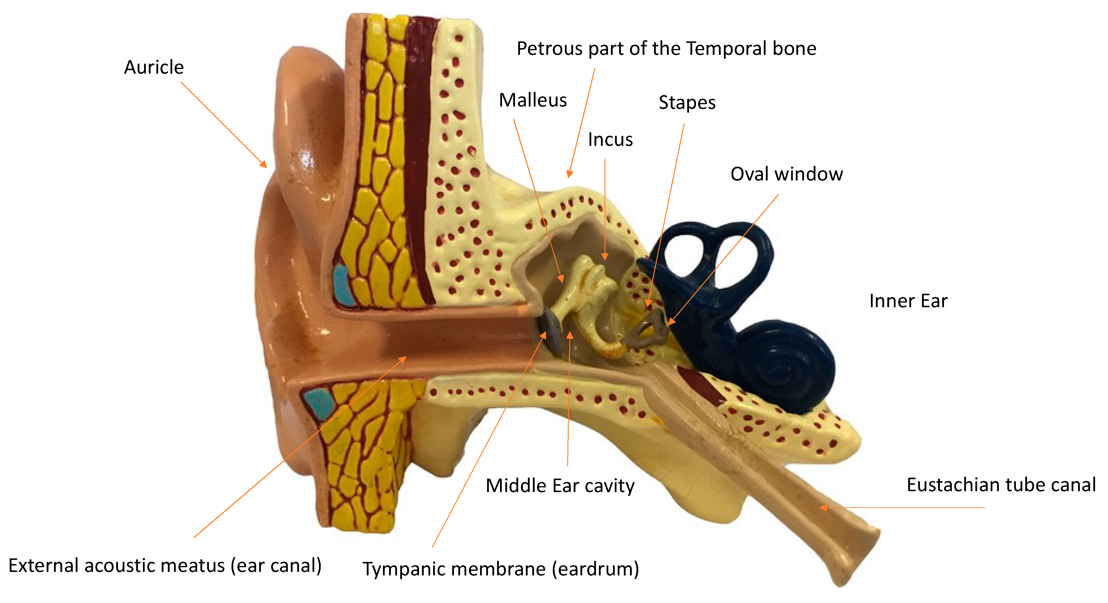
Figure 1. Simplified anatomy of the human ear (adapted from a plastic model, Anatomy Laboratory, Murdoch University).
Preprints 85380 g001
Figure 2. The appearance of the ossicular chain with detailed labelling of the middle ear anatomy, analysed by CT Scan of the skull, temporal and petrous bones of an adult patient was compared as an example of normal anatomy. The patient was randomly selected in the hospital database and anonymized. A) Axial plane; B) Coronal Plane; C) Sagittal Plane.
Preprints 85380 g002
Table 1. Results of the literature search with records of the post mortem presence (complete or incomplete) or absence of the ossicular chain. The CT Scan of the skull, temporal and petrous bones of an adult anonymized patient was compared as an example of normal anatomy.
Sample PMI
and body preservation
Type of analysis Sample size, condition
of the ossicular chain
and notes
References
Australopithecus africanus 2.1-3.3 million years ago (Mya)
Fossilized skeletal remains
Direct visual observation 3
Present, incomplete.
Moggi-Cecchi and colleagues describe a stapes located in the vestibule of the inner ear, “presumably having slipped through the oval window before fossilization”.
(Moggi-Cecchi & Collard, 2002; Quam et al., 2013)
Australopithecus robustus 1.2-1.8 Mya
Fossilized skeletal remains
Direct visual observation 4
Present, complete/incomplete
Quam and colleagues describe the presence of a complete ossicular chain as “an exceptional case of preservation in the human fossil record”.
(Clarke & Rak, 1979; Quam et al., 2013)
Homo neanderthalensis 28-300 kiloyears ago (Kya)
Fossilized skeletal remains
Direct visual observation
CT Scan
MicroCT Scan
3
Present, incomplete
(Arensburg et al., 1996; Maureille, 2002; Ponce De León et al., 1999; Stoessel et al., 2016)
Archaeological excavations 3.3 Mya-Present Era
Fossilized and non-fossilized skeletal remains
Direct visual observation
X-Rays
CT Scan
>1000
Present/Absent/Complete/
Incomplete
Incomplete or absent in most fragmented skulls, temporal and petrous bones.
(Bruintje, 1990; Dedouit et al., 2007; Lisonek et al., 1986)
Present-day forensic cases 1999-2019 Present Era
2 decomposing bodies, 2 skeletonised skulls
Ortho
pantomography
X-Rays
CT Scan
MSCT Scan
Present/Absent
Absent in the skeletonised skulls, present in the decomposing bodies
Hospital patient Normal anatomy of the ossicular chain CT Scan Present, complete
Disclaimer/Publisher’s Note: The statements, opinions and data contained in all publications are solely those of the individual author(s) and contributor(s) and not of MDPI and/or the editor(s). MDPI and/or the editor(s) disclaim responsibility for any injury to people or property resulting from any ideas, methods, instructions or products referred to in the content.
Copyright: This open access article is published under a Creative Commons CC BY 4.0 license, which permit the free download, distribution, and reuse, provided that the author and preprint are cited in any reuse.
Prerpints.org logo

Preprints.org is a free preprint server supported by MDPI in Basel, Switzerland.

Subscribe

© 2024 MDPI (Basel, Switzerland) unless otherwise stated