Preprint
Review

Recent Optimization Methods and Techniques for Medical Image Analysis

Altmetrics

Downloads

505

Views

50

Comments

0

This version is not peer-reviewed

Submitted:

28 September 2023

Posted:

30 September 2023

You are already at the latest version

Alerts
Abstract
Medical image analysis is an important branch in the field of medicine, which mainly uses image processing and analysis techniques to interpret and diagnose medical image data. Medical image data helps doctors to effectively observe and diagnose patients' body structures, tissues and lesions. Medical image analysis has been an important research area in the medical field, and it is important for disease diagnosis, treatment planning, and condition monitoring. In recent years, the rapid development of deep learning and computer vision technologies has contributed greatly to the automation, multimodal data fusion, real-time application, and accuracy improvement of medical image analysis. In addition, the development of deep learning has given rise to some new research areas in medical image analysis, such as Generative Adversarial Networks (GANs) for synthetic medical images, self-supervised learning for unsupervised feature learning, and neural network interpretability. In this paper, we will introduce some optimisation methods for medical images which are effective in improving the accuracy, efficiency and reliability of medical image analysis.
Keywords: 
Subject: Computer Science and Mathematics  -   Artificial Intelligence and Machine Learning

1. Introduction

Medical images [1] are an important form of data in the medical field; they are used to observe, diagnose and treat diseases. Medical images are usually obtained through a variety of imaging techniques that provide detailed information about the internal structure and state of a patient's tissues. Common types of medical images include: x-ray (X-ray) images [2, 3], computed tomography (CT scan) [4], magnetic resonance imaging (MRI), ultrasound images, radiological images, nuclear medicine images [5-7], pathology slide images [8, 9], ophthalmological images [10], and others. Interpretation and analysis of medical images usually requires highly trained medical professionals such as radiologists, neurologists, surgeons and pathologists. At the same time, the professionalism of the doctors is required to be extremely high. However, with the development of deep learning and computer vision technologies, automated medical image analysis tools are becoming increasingly popular [11], and they can assist doctors in making faster and more accurate diagnoses.
Medical image analysis is a specialised area of computer science and medical imaging [12] that involves the application of advanced algorithms and techniques for processing and interpreting medical images such as X-rays[13, 14], MRI scans[15, 16], CT scans and ultrasound images [17, 18]. Its main goal is to assist healthcare professionals in diagnosing diseases, assessing disease progression and planning treatments by extracting valuable information, detecting abnormalities and quantifying relevant features from medical images. Medical image analysis plays a vital role in improving the accuracy and efficiency of medical diagnosis and treatment planning, and ultimately enhancing patient care. At the same time, medical images greatly improve the accuracy of healthcare professionals in diagnosing and treating various medical conditions. By utilising advanced algorithms and techniques, medical image analysis allows for more precise early detection of diseases, quantification of anatomical and pathological features, and monitoring of disease progression. This not only improves diagnostic accuracy, but also facilitates treatment planning and assessment, leading to more personalised and effective care for patients. In addition, it reduces the need for invasive procedures, minimises healthcare costs and ultimately saves lives by facilitating timely intervention. As medical imaging technology [19] continues to advance, medical image analysis continues to be an important tool in modern healthcare.
Optimisation methods are mathematical techniques used to find the best solution among a set of possible alternatives, usually by minimising or maximising an objective function while observing certain constraints. There are a number of optimisation methods that can be applied to medical image analysis, including gradient descent [20, 21] (where parameters are adjusted iteratively to reach optimal values), linear programming (used in resource allocation and scheduling problems), genetic algorithms [22-24] (inspired by biological evolution to find optimal solutions), and integer programming [25, 26] (which is particularly well suited to discrete decisions). These methods are fundamental in many fields such as engineering, economics, machine learning [22, 27, 28] and operations research for efficient decision making, resource allocation and problem solving in complex systems and processes.
Optimisation algorithms for medical image analysis are key components used to improve the efficiency, accuracy and reliability of medical image processing and interpretation. These algorithms can be applied to various areas of medical imaging including image enhancement, segmentation, feature extraction, classification, alignment and visualisation. Common optimization algorithms for medical image analysis include: image enhancement algorithms [29, 30], image segmentation algorithms [31, 32], feature extraction algorithms [33, 34], classification and recognition algorithms [35], image alignment algorithms [36, 37], visualization algorithms [38, 39], deep learning algorithms [40, 41], uncertainty modelling algorithms, and so on. The development and application of these algorithms have made medical image analysis more accurate, automated and efficient.
Optimisation techniques are crucial in medical image analysis. Firstly, optimisation techniques can be used to fine-tune parameters in image processing algorithms to improve the quality of image enhancement, denoising and segmentation, which are essential for accurate diagnosis and treatment planning. Secondly, optimisation methods help in feature selection and extraction, which can identify relevant information in medical images while reducing noise and irrelevant data. In addition, optimisation plays a key role in optimising the allocation of computational resources (e.g. GPU usage) to speed up the analysis of large datasets and complex algorithms.
This article is about optimisation algorithms related to medical image analysis. The rest of the article is structured as follows: section 2 describes the optimisation algorithms related to medical image analysis. Optimisation methods in fine-tuning are discussed and analysed in Section 3. Section 4 introduces the knowledge about optimisation algorithms in feature selection. Section 5 pair presents the optimisation resource allocation. Section 6 concludes the article.

2. Optimisation algorithms related to medical image analysis

This section presents some optimisation algorithms related to medical image analysis. Figure 1 shows the basic flow of optimization algorithm in image processing.
(1)
Image Enhancement Algorithms
Image enhancement is an important task in the field of digital image processing, which aims to improve the visual quality of an image by improving its quality, clarity, contrast and visualisation. Image enhancement algorithms can be classified into many different types and the exact choice depends on the application scenario and the desired enhancement effect. Common image enhancement algorithms are grey scale stretching, histogram equalisation, adaptive histogram equalisation, sharpening filter, noise filter, bilateral filter, super-resolution reconstruction, colour enhancement and deep learning based enhancement methods.
Different image enhancement algorithms are suitable for different application scenarios and requirements for medical image analysis. Choosing the appropriate algorithm usually requires consideration of the trade-off between medical image quality, computational complexity and enhancement effect. In practical applications, algorithm selection and parameter adjustment are usually required according to specific needs to obtain the best medical image enhancement effect.
(2)
Image Segmentation Algorithms
The goal of image segmentation is to divide the image into different regions or objects to identify different objects or regions in the image. Image segmentation algorithms can be classified into types such as threshold segmentation, region growing, edge detection, watershed segmentation, graph-based segmentation, convolutional neural network (CNN) segmentation, instance segmentation, semantic segmentation etc.
The specific choice of image segmentation algorithm selection depends on the specific requirements of the task, such as image complexity, segmentation accuracy and computational resources. In practical applications, it is usually necessary to consider these factors comprehensively and select the most suitable image segmentation algorithm for the task.
(3)
Feature Extraction Algorithms
Feature extraction is a key step in fields such as machine learning and computer vision, where the goal is to extract the most representative and information-rich features from raw data for subsequent pattern recognition, classification or regression tasks. Traditional feature extraction methods include colour features (including colour histogram, colour moments, colour gradient, etc., which are used to describe the colour information in an image), texture features (such as grey scale covariance matrix, local binary pattern and Gabor filter response, etc., which are used to describe the texture information of an image), shape features (including edge detection, corner detection and contour descriptor, etc., which are used to describe the shape information of an object), optical flow features (for describing the motion information of objects in video sequences, including dense optical flow and sparse optical flow, etc.). Deep learning feature extraction, on the other hand, includes Convolutional Neural Network (CNN) features, Autoencoders features, Generative Adversarial Network (GAN) features, as well as feature selection and dimensionality reduction, among others. In deep learning, the different layers of a neural network can often be viewed as feature extractors from lower to higher levels. Lower layers typically extract low-level features such as edges and textures, while higher layers extract more abstract and semantic features.
The choice of feature extraction method depends on the specific task and data type. In the era of deep learning, the use of pre-trained convolutional neural networks is often the method of choice for many computer vision tasks because of the strong generalisation ability of these networks on large-scale datasets. However, in some cases, traditional feature extraction methods can still be useful, especially if the data is limited or needs to be interpretive.
(4)
Classification and Recognition Algorithms
Classification and recognition algorithms are used to classify the input data into different categories or to identify objects. The common classification algorithms are K Nearest Neighbour Algorithm, Decision Tree, Support Vector Machine, Plain Bayesian Classifier, Neural Network. The common recognition algorithms are Target Detection, Face Recognition, Pose Estimation, Image Semantic Segmentation, Optical Character Recognition (OCR). Deep learning methods perform well in many recognition tasks, but they usually require large amounts of labelled data and computational resources for training. Therefore, factors such as data availability, algorithm complexity, and performance requirements need to be considered when selecting an algorithm.
(5)
Image Registration Algorithm
Image Registration aims to align multiple images or images acquired by different sensors to a common coordinate system. Image registration has a wide range of applications in fields such as medical imaging, remote sensing, computer vision and geographic information systems. Common image alignment algorithms and methods include feature-based alignment, mutual information-based alignment, local transformation-based alignment (Local Transformations), deep learning-based alignment, and so on. Among them, feature-based alignment includes point feature alignment and feature matching. Mutual information-based alignment includes mutual information metric and normalised mutual correlation. Local transformation-based alignment includes B-spline alignment and local affine or nonlinear transformation. Deep learning based alignment includes convolutional neural networks and generative adversarial networks.
The choice of an image alignment algorithm depends on the application area, data type, noise level and computational resources. In practical applications, parameter tuning and performance evaluation are usually required to ensure that accurate alignment results are obtained. Image alignment is the basis of many image processing and analysis tasks and is important for subsequent image analysis and processing.
(6)
Visualisation algorithms
Visualisation algorithms are a set of techniques and methods used to visualise data as graphs or images. They help in understanding data, discovering patterns, demonstrating relationships and conveying information. Some of the common visualisation algorithms and methods are scatter plots, line graphs, histograms and histograms, heat maps, box and line plots, scatter matrices, tree diagrams, network diagrams, map visualisations, 3D visualisations.
These visualisation algorithms can be selected and customised according to the type of data, task requirements and target audience. In the field of data science and information visualisation, choosing the right visualisation methods and tools is important for deeper understanding of the data, decision making and communicating the results.
(7)
Deep Learning Algorithms
Deep learning is a branch of machine learning that is based on the idea of artificial neural networks, where data is modelled and interpreted through multi-layer neural networks to achieve various machine learning tasks. Common deep learning algorithms and methods are Multilayer Perceptron (MLP), Convolutional Neural Networks (CNN), Recurrent Neural Networks (RNN), Self-Encoders, Generative Adversarial Networks (GAN), Reinforcement Learning, Attentional Mechanisms, and Transfer Learning.
These deep learning algorithms have a wide range of applications in a variety of fields, including natural language processing, computer vision, speech recognition, recommender systems, medical image analysis, and autonomous driving. The success of deep learning is due to the availability of large-scale datasets and high-performance computing hardware (e.g., GPUs), making it a powerful tool for solving complex problems. At the same time, deep learning also requires careful parameter tuning and significant computational resources to train complex models.

3. Optimization in Fine-tuning

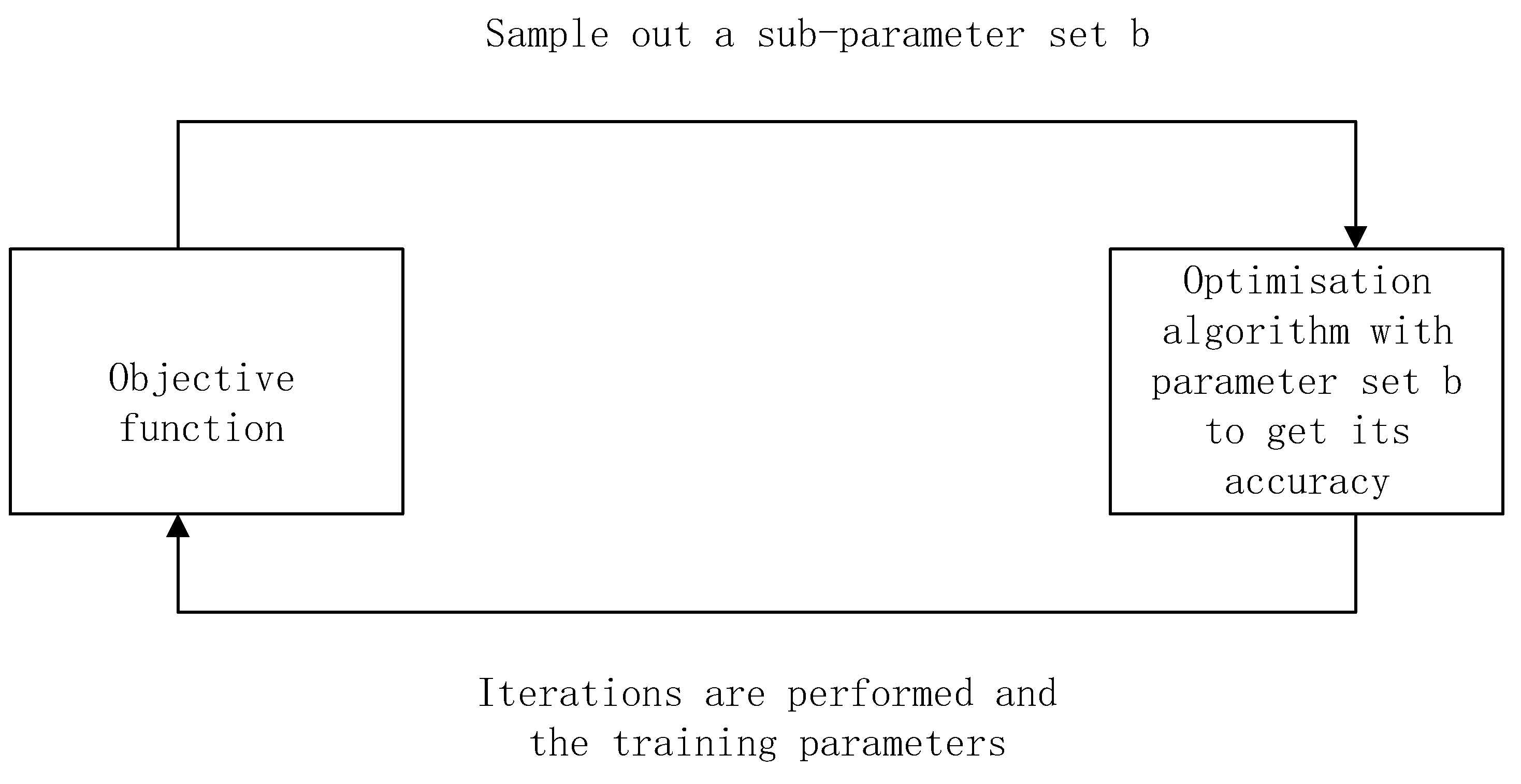
By fine-tuning certain parameters in image processing algorithms during medical image analysis, it is possible to produce an optimal set of parameters for a particular task and improve the accuracy of medical image analysis [42]. Fine-tuning the parameters of an image processing algorithm is a critical step in improving the performance of image processing. Fine-tuning is usually an iterative process that requires careful selection of parameters, monitoring of performance, and adjustments based on results.
Figure 1 illustrates the overall process for parameter optimisation in fine-tuning. The main optimisations in fine-tuning are (1) determining the objective function. In image processing, the objective function is defined as a way to measure the quality or performance of an algorithm under a particular parameter configuration. The function quantifies the effectiveness of the algorithm in accomplishing a specific task such as image enhancement, denoising, or segmentation. Typically, the objective function generates a numerical value that is used as a criterion for evaluating the success of the algorithm. (2) Parameter space generation. Image processing algorithms usually have several tunable parameters, such as filter kernel size, threshold or scaling factor. Different combinations of these parameters form a parameter space that represents all possible configurations of the algorithm. (3) Optimisation algorithms. Optimisation algorithms perform a systematic search of the parameter space using techniques such as gradient descent, genetic algorithms or Bayesian optimisation. The aim is to find parameter values that minimise or maximise the objective function, depending on whether the minimisation or maximisation problem is being solved. (4) Iterative search. The optimisation algorithm iteratively explores the parameter space by evaluating the objective function for different parameter values. Then based on these evaluations, the algorithm adjusts the parameters to bring them closer to the optimal configuration. This process continues until a stopping criterion (e.g., a convergence threshold or a predetermined number of iterations) is met. (5) Parameter tuning. Once the optimisation process has converged or the stopping criteria are met, the algorithm determines a set of parameters that produce the best results based on the defined objective function. These tuned parameters can be used in image processing algorithms to improve performance.
In medical image analysis, optimisation techniques save the time and effort required for manual parameter adjustment by automatically searching for optimal parameter values. This not only improves the efficiency and effectiveness of image processing, but also ensures that the performance of the algorithms is continually optimised across a wide range of medical image analysis tasks, ultimately helping medical professionals to make accurate diagnostic and treatment decisions.

4. Optimization in Feature Selection

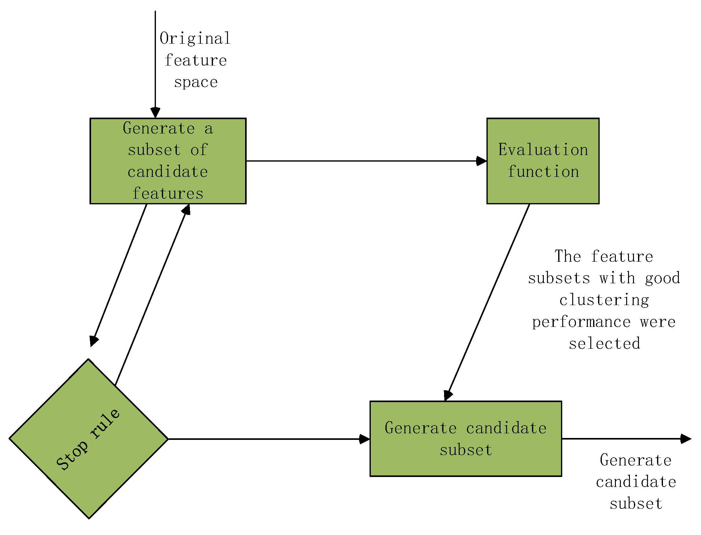
Optimisation of feature selection in medical image analysis is of great significance and impact. The structure of the feature selection algorithm is shown in Figure 2. Medical images usually contain a large number of pixels or features, which without feature selection will result in high dimensional data. High dimensional data not only increases the computational and memory requirements, but may also lead to dimensional catastrophe. By optimising feature selection, computational cost and memory requirements can be reduced and the training and inference process can be accelerated.
Optimisation of feature selection can model helps to identify and select the most relevant features, thus improving the performance and accuracy of machine learning models [43, 44]. By eliminating irrelevant or redundant features, the model focuses more on key information, reducing the risk of overfitting and improving generalisation [45]. In medical image analysis, interpretability [46] is critical because medical decisions require trustworthy interpretations [47]. Optimised feature selection helps to identify which features have the most impact on decision making and helps healthcare professionals to understand the decision making process of the model [48]. Also, optimised feature selection helps to reduce the influence from noisy or unstable features. Medical images can be affected by a variety of factors, including image quality, instrument noise, etc., and feature selection can reduce the negative impact of these factors on the model [49]. Optimised feature selection also helps to reduce data privacy and security risks [50]. By reducing the use of sensitive information, the risk of patient data leakage is reduced.
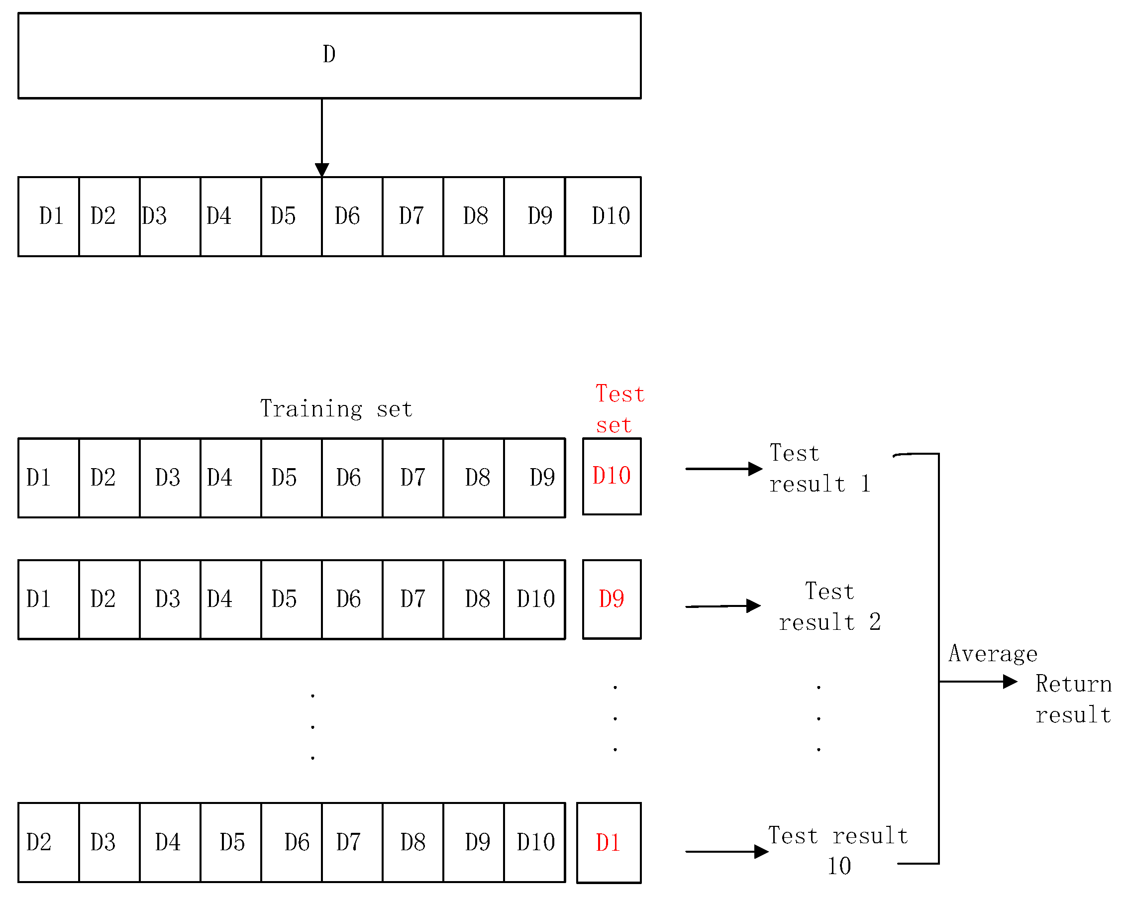
In the feature selection process for medical image analysis, optimisation allows for the systematic identification of the most informative and relevant features while minimising redundancy and noise. The process of optimising features includes (1) feature evaluation metrics. In order to assess the importance of individual features or subsets of features, a quantitative assessment metric (e.g., classification accuracy, information gain, or relevance) needs to be defined. The metric quantifies the ability of a set of features in distinguishing between different medical conditions or accurately describing relevant image attributes. Table 1 gives some indicators for character evaluation. (2) Feature subset search. Feature selection typically involves a combinatorial search of a space of possible feature subsets. This space can be very large, especially when dealing with high-dimensional medical image data. Optimisation techniques such as genetic algorithms, sequential forward selection or recursive feature elimination can be used to efficiently explore this space and select the most informative features. (3) Objective function. The objective function is used to quantify the value of the evaluation metrics for each feature subset. This function can be used to measure the performance of a particular feature set in a given medical image analysis task. (4) Optimisation algorithm. Optimisation algorithms are used to search for subsets of features that maximise or minimise the objective function, depending on whether the objective is feature selection or feature elimination. These algorithms iteratively evaluate different feature subsets and adjust their composition based on performance metrics. (5) Cross-validation[19]. To ensure that the selected features generalise the unseen data well, cross-validation techniques are usually combined with the optimisation process. This helps prevent overfitting and ensures that the selected feature subset is robust and reliable. Figure 3 illustrates the 10-fold cross-validation method. (6) Final feature subset. Once the optimisation process converges or reaches a predefined criterion, it identifies a subset of features that optimise the selected evaluation metrics. This subset can be used as input for subsequent medical image analysis tasks such as classification, segmentation or disease diagnosis.
Optimisation-driven feature selection improves the efficiency and effectiveness of medical image analysis by reducing data dimensionality, improving model interpretability and potentially leading to more accurate and robust results. It also helps mitigate the "curse of dimensionality" often encountered in high-dimensional medical image datasets, where the selection of the right features is critical for improving diagnostic and predictive performance.

5. Optimization in Resource Allocation

Optimisation[51, 52] is also crucial for the efficient allocation of healthcare resources, ensuring that healthcare organisations provide the best possible care to patients while effectively managing limited resources. Optimisation[53] plays an important role in resource planning for healthcare resources, disease detection [54], staff scheduling, patient triage, supply chain management [55], operating theatre scheduling, telemedicine and remote monitoring, emergency response, and resource allocation for rare diseases. Details are described below.
(1)
Resource planning. Optimisation can assist healthcare organisations in long-term resource planning by analysing historical data, patient demographics and forecasting demand. It can help determine the optimal allocation of resources such as hospital beds, medical staff, equipment and medications to meet expected demand while minimising waste and reducing costs.
(2)
Employee Scheduling. For hospitals and clinics, optimisation can be used to create efficient staff schedules that take into account factors such as staff availability [56], skill levels and patient needs. This ensures that the right people are on duty at the right time to provide quality care while maintaining a reasonable workload [57].
(3)
Patient triage. During a surge in patient demand, such as during a pandemic or natural disaster, optimisation algorithms can help healthcare providers prioritise patients based on the severity of their condition and available resources. This ensures that critically ill patients receive immediate attention and appropriate care.
(4)
Supply Chain Management. Optimisation techniques can be used to manage the supply chain [58] of healthcare resources such as medicines, personal protective equipment (PPE) and medical devices. This helps maintain adequate stock levels, minimise waste and avoid shortages.
(5)
Operating theatre scheduling. In the surgical sector, optimisation can optimise surgery scheduling, OR utilisation and staff allocation. This reduces patient wait times, optimises the use of expensive operating theatres and improves overall surgical workflow.
(6)
Telemedicine and remote monitoring. Optimisation can be used to determine the most efficient allocation of resources for telemedicine services and remote patient monitoring, ensuring that patients can access care remotely while minimising pressure on on-site resources.
(7)
Emergency Response. During an emergency or disaster, optimisation models can help emergency responders allocate medical resources efficiently. This includes deploying medical teams, ambulances, and supplies to a disaster area based on real-time data and predicted needs.
(8)
Resource allocation for rare diseases. For diseases with low prevalence, optimisation can help to allocate specialised resources, such as rare disease specialists or specialised equipment, to areas of greatest need [59], ensuring equitable access to care.
(9)
Budget allocation. Healthcare organisations and government agencies can use optimisation to allocate budgets efficiently, ensuring that funds are allocated to the areas of healthcare that are most in need, while meeting the needs of the population.
Optimising healthcare resource allocation [60] helps healthcare organisations make data-driven decisions to maximise the use of available resources, improve patient outcomes and enhance the overall quality and efficiency of healthcare services [61]. It can also play a key role in addressing unforeseen challenges such as pandemics or disasters by rapidly adjusting resource allocation strategies in response to changing circumstances.

6. Conclusion and Future Directions

The field of optimisation is constantly evolving and there are several exciting research directions that may influence its development in the future. These include (1) Robust and uncertainty-aware optimisation. Future research may focus on developing optimisation techniques that are robust to uncertainty and change in real-world data. This is critical in applications such as supply chain management [62], healthcare [63], and finance, where unexpected events can have a significant impact on outcomes. (2) Meta-heuristic and hybrid algorithms. Progress is expected in the development of meta-heuristic algorithms (e.g., genetic algorithms, simulated annealing, particle swarm optimisation) and hybrid methods that combine multiple optimisation techniques. These methods can improve the performance of complex, multi-objective [64] and high-dimensional optimisation problems [65].
Medical image analysis is a key area in the field of medicine that continues to evolve and introduce new technologies to improve the efficiency and accuracy of diagnosis, treatment and disease prevention. Future research directions will continue to drive innovation and development in this field [66]. For the future we can incorporate deep learning to tailor robust and interpretable deep learning models for medical image analysis. At the same time, uncertainty quantification techniques are being incorporated into optimisation-driven analyses to improve diagnosis and treatment planning, advancing real-time and interactive optimisation. In addition, there is an increasing focus on addressing privacy concerns in medical data while optimising the image analysis process, as well as exploring novel optimisation-driven approaches for multimodal and multiscale medical image fusion [67] and analysis to improve the accuracy of disease detection and characterisation. The future of medical image analysis optimisation will be marked by innovations that improve both the clinical utility of image-based healthcare applications and ethical considerations.

Funding

This research did not receive any grants.

Acknowledgment

We thank all the anonymous reviewers for their hard reviewing work.

References

  1. Beutel, J.; Kundel, H.L.; Van Metter, R.L. Handbook of Medical Imaging - Volume I. Physics and Psychophysics; 2000. [Google Scholar]
  2. Rubin; Daniel, L. Informatics in radiology: Measuring and improving quality in radiology: meeting the challenge with informatics. Radiographics 2011, 31, 1511–1527. [Google Scholar] [CrossRef] [PubMed]
  3. Recht, M.P.; Dewey, M.; Dreyer, K.; Langlotz, C.; Niessen, W.; Prainsack, B.; Smith, J.J. Integrating artificial intelligence into the clinical practice of radiology: challenges and recommendations. Eur. Radiol. 2020, 30, 3576–3584. [Google Scholar] [CrossRef] [PubMed]
  4. Song, H.; Yang, S.; Yu, B.; Li, N.; Huang, Y.; Sun, R.; Wang, B.; Nie, P.; Hou, F.; Huang, C.; et al. CT-based deep learning radiomics nomogram for the prediction of pathological grade in bladder cancer: a multicenter study. Cancer Imaging 2023, 23. [Google Scholar] [CrossRef] [PubMed]
  5. Crisci, S.; Piana, M.; Ruggiero, V.; Scussolini, M. A Regularized Affine-Scaling Trust-Region Method for Parametric Imaging of Dynamic PET Data. SIAM J. Imaging Sci. 2021, 14, 418–439. [Google Scholar] [CrossRef]
  6. Schmidtlein, C.R.; Maroun, M.K.; Krol, A.; Gifford, H.; Bodei, L.; O'Donoghue, J.; Häggström, I.; Xu, Y. A deblurring/denoising corrected scintigraphic planar image reconstruction model for targeted alpha therapy. In Proceedings of the Conference on Medical Imaging: Biomedical Applications in Molecular, Structural, and Functional Imaging; 2021. [Google Scholar]
  7. Second Affiliated Hospital of Dalian Medical University Reports Findings in Artificial Intelligence (Application of Artificial Intelligence Nuclear Medicine Automated Images Based on Deep Learningin Tumor Diagnosis). Robotics & Machine Learning Daily News, 2022.
  8. Lu, M.Y.; Williamson, D.F.K.; Chen, T.Y.; Chen, R.J.; Barbieri, M.; Mahmood, F. Data-efficient and weakly supervised computational pathology on whole-slide images. Nat. Biomed. Eng. 2021, 5, 555–570. [Google Scholar] [CrossRef]
  9. Jiang, S.; Suriawinata, A.A.; Hassanpour, S. MHAttnSurv: Multi-head attention for survival prediction using whole-slide pathology images. Comput. Biol. Med. 2023, 158, 106883. [Google Scholar] [CrossRef]
  10. Tatsuo, Y.; Toshibumi, M. Ophthalmological Imager; 2007. [Google Scholar]
  11. Jing, B.; Xie, P.; Xing, E. On the Automatic Generation of Medical Imaging Reports; 2017. [Google Scholar]
  12. Halder, S.; Biswas, G.H.; Samanta, H.; Shahid, S. A Comprehensive Review on Medical Imaging Technologies to Detect Brain Stroke. ITM Web Conf. 2023, 53, 01009. [Google Scholar] [CrossRef]
  13. Pham, V.-T.; Nguyen, Q.-C. Chest X-Rays Abnormalities Localization and Classification Using an Ensemble Framework of Deep Convolutional Neural Networks. Vietnam. J. Comput. Sci. 2022, 10, 55–73. [Google Scholar] [CrossRef]
  14. Rogier, V.D.S.; Bhaskhar, N.; Rubin, D.; Langlotz, C.; Chaudhari, A. Exploring Image Augmentations for Siamese Representation Learning with Chest X-Rays. 2023.
  15. Watkins, K.; Paus, T.; Lerch, J.; Zijdenbos, A.; Collins, D.; Neelin, P.; Taylor, J.; Worsley, K.; Evans, A. Structural Asymmetries in the Human Brain: a Voxel-based Statistical Analysis of 142 MRI Scans. Cereb. Cortex 2001, 11, 868–877. [Google Scholar] [CrossRef]
  16. Franke, K.; Ziegler, G.; Klöppel, S.; Gaser, C.; Alzheimer's Disease Neuroimaging Initiative. Estimating the age of healthy subjects from T1-weighted MRI scans using kernel methods: Exploring the influence of various parameters. NeuroImage 2010, 50, 883–892. [Google Scholar] [CrossRef]
  17. Liu, Y.J.; Ng, W.S.; Teo, M.Y.; Lim, H.C. Computerised prostate boundary estimation of ultrasound images using radial bas-relief method. . Med Biol. Eng. Comput. 1997, 35, 445–454. [Google Scholar] [CrossRef] [PubMed]
  18. Sato, Y.; Nakamoto, M.; Tamaki, Y.; Sasama, T.; Sakita, I.; Nakajima, Y.; Monden, M.; Tamura, S. Image guidance of breast cancer surgery using 3-D ultrasound images and augmented reality visualization. IEEE Trans. Med Imaging 1998, 17, 681–693. [Google Scholar] [CrossRef] [PubMed]
  19. Belaala, A.; Terrissa, L.S.; Zerhouni, N.; Devalland, C. Computer-Aided Diagnosis for Spitzoid Lesions Classification Using Artificial Intelligence Techniques. International Journal of Healthcare Information Systems and Informatics (IJHISI) 2021, 16, 16–37. [Google Scholar] [CrossRef]
  20. Zhao, Q.; Wang, H.; Zhu, X.; Meng, D. Stein variational gradient descent with learned direction. Inf. Sci. 2023, 637. [Google Scholar] [CrossRef]
  21. Wadi, D.; Fredette, M.; Senecal, S. Read the Signs: Towards Invariance to Gradient Descent's Hyperparameter Initialization. arXiv 2023, arXiv:2301.10133. [Google Scholar]
  22. Pérez-Sala, L.; Curado, M.; Tortosa, L.; Vicent, J.F. Deep learning model of convolutional neural networks powered by a genetic algorithm for prevention of traffic accidents severity. Chaos, Solitons Fractals 2023, 169. [Google Scholar] [CrossRef]
  23. Aidi, S.; Mazouzi, M. Optimization Approach for Yard Crane Scheduling Problem using genetic algorithm in Container Terminals. ITM Web Conf. 2023, 52, 02002. [Google Scholar] [CrossRef]
  24. Wang, T.; Qi, Q.; Zhang, W.; Zhan, D. Research on Optimization of Profile Parameters in Screw Compressor Based on BP Neural Network and Genetic Algorithm. Energies 2023, 16, 3632. [Google Scholar] [CrossRef]
  25. Dey, S.; Singh, M.; Williamson, D.P. Special Issue: Integer Programming and Combinatorial Optimization (IPCO) 2021 PREFACE. Mathematical Programming 2023. [CrossRef]
  26. Karuno, Y.; Tomozawa, A.; Tsuji, K. An Integer Programming Formulation of Collecting Weighted Items in Directed Bipartite Graphs with a Budget Constraint. In Proceedings of the 11th IIAE International Conference on Industrial Application Engineering 2023, 2023. [Google Scholar]
  27. Alzubaidi, L.; Zhang, J.; Humaidi, A.J.; Al-Dujaili, A.; Duan, Y.; Al-Shamma, O.; et al. Review of deep learning: concepts, CNN architectures, challenges, applications, future directions. J Big Data 2021, 8, 53. [Google Scholar] [CrossRef] [PubMed]
  28. LeCun, Y.; Bengio, Y.; Hinton, G. Deep learning. Nature 2015, 521, 436–444. [Google Scholar] [CrossRef] [PubMed]
  29. Karthik, K.; Shevgoor, S.K. Improving Clinical Diagnosis Performance with Automated X-ray Scan Quality Enhancement Algorithms. 2022.
  30. Liu, X.; Pedersen, M.; Wang, R. Survey of natural image enhancement techniques: Classification, evaluation, challenges, and perspectives. Digital Signal Processing 2022, 127, 103547. [Google Scholar] [CrossRef]
  31. Kahlouche, S.; Achour, K. A new approach to image segmentation using genetic algorithm with mathematical morphology. 2022.
  32. Wei, T.; Wang, X.; Li, X.; Zhu, S. Fuzzy subspace clustering noisy image segmentation algorithm with adaptive local variance & non-local information and mean membership linking. Eng. Appl. Artif. Intell. 2022, 110, 104672. [Google Scholar] [CrossRef]
  33. Huang, S.; Sallehuddin, M. Multi-scene design analysis of integrated energy system based on feature extraction algorithm. Energy Reports 2022, 8, 466–476. [Google Scholar] [CrossRef]
  34. Li, C.; Li, N.; Qiu, Y.; Peng, Y.; Wang, Y.; Deng, L.; Ma, T.; Li, F.; Yao, D.; Xu, P. Multimodal collaborative BCI system based on the improved CSP feature extraction algorithm. Virtual Real. Intell. Hardw. 2022, 4, 22–37. [Google Scholar] [CrossRef]
  35. Rajesh, S.; Sharanya, B. Recognition and prevention of cyberharassment in social media using classification algorithms. 2021.
  36. Chen, Y.-X.; Xie, R.; Yang, Y.; He, L.; Feng, D.; Shen, H.-B. Fast Cryo-EM Image Alignment Algorithm Using Power Spectrum Features. J. Chem. Inf. Model. 2021, 61, 4795–4806. [Google Scholar] [CrossRef]
  37. Lin, M.; Liu, T.; Li, Y.; Miao, X.; He, C. Image stitching by disparity-guided multi-plane alignment. Signal Process. 2022, 197, 108534. [Google Scholar] [CrossRef]
  38. Perri, V.; Scholtes, I. HOTVis: Higher-Order Time-Aware Visualisation of Dynamic Graphs: Graph Drawing and Network Visualization. In Proceedings of the 28th International Symposium, GD 2020, Revised Selected Papers. Vancouver, BC, Canada, 16–18 September 2020; 2021. [Google Scholar]
  39. Ansari, M.D.; Li, X.; Khan, M. Design and Development of a Data Structure Visualisation System Using the Ant Colony Algorithm. Recent Adv. Electr. Electron. Eng. (Formerly Recent Patents Electr. Electron. Eng. 2023. [Google Scholar] [CrossRef]
  40. Zhang, Z.; Cui, P.; Zhu, W. Deep Learning on Graphs: A Survey. IEEE Trans. Knowl. Data Eng. 2020, 34, 249–270. [Google Scholar] [CrossRef]
  41. Feng, J.; Li, Y.; Zhao, K.; Xu, Z.; Xia, T.; Zhang, J.; Jin, D. DeepMM: Deep Learning Based Map Matching with Data Augmentation. IEEE Trans. Mob. Comput. 2020, PP, 1–1. [Google Scholar] [CrossRef]
  42. Wang, S.; Zhang, Y.; Liu, G.; Phillips, P.; Yuan, T.-F. Detection of Alzheimer’s Disease by Three-Dimensional Displacement Field Estimation in Structural Magnetic Resonance Imaging. J. Alzheimer's Dis 2016, 50, 233–248. [Google Scholar] [CrossRef]
  43. Zhang, Y.; Wu, X.; Lu, S.; Wang, H.; Phillips, P.; Wang, S. Smart detection on abnormal breasts in digital mammography based on contrast-limited adaptive histogram equalization and chaotic adaptive real-coded biogeography-based optimization. Simulation 2016, 92, 873–885. [Google Scholar] [CrossRef]
  44. Zhang, Y.; Wang, S.; Ji, G.; Dong, Z. Exponential wavelet iterative shrinkage thresholding algorithm with random shift for compressed sensing magnetic resonance imaging. IEEJ Trans. Electr. Electron. Eng. 2014, 10, 116–117. [Google Scholar] [CrossRef]
  45. Yuan, G.; Lu, L.; Zhou, X. Feature selection using a sinusoidal sequence combined with mutual information. Eng. Appl. Artif. Intell. 2023, 126. [Google Scholar] [CrossRef]
  46. Moslemi, A. A tutorial-based survey on feature selection: Recent advancements on feature selection. Eng. Appl. Artif. Intell. 2023, 126. [Google Scholar] [CrossRef]
  47. Zhang, Y.; Wang, S.; Huo, Y.; Wu, L.; Liu, A. Feature extraction of brain mri by stationary wavelet transform and its applications. J. Biol. Syst. 2010, 18, 115–132. [Google Scholar] [CrossRef]
  48. Janani, K.; Mohanrasu, S.; Kashkynbayev, A.; Rakkiyappan, R. Minkowski distance measure in fuzzy PROMETHEE for ensemble feature selection. Math. Comput. Simul. 2023. [Google Scholar] [CrossRef]
  49. Zhang, Y.; Wu, L.; Wang, S.; Wei, G. Color image enhancement based on HVS and PCNN. Sci. China Inf. Sci. 2010, 53, 1963–1976. [Google Scholar] [CrossRef]
  50. Touches, T.D.; Munda, M.; Cornet, T.; Gerkens, P.; Hellepute, T. Feature selection with prior knowledge improves interpretability of chemometrics models. Chemom. Intell. Lab. Syst. 2023, 240. [Google Scholar] [CrossRef]
  51. Jiang, S.; Zou, J.; Yang, S.; Yao, X. Evolutionary Dynamic Multi-objective Optimisation: A Survey. ACM Comput. Surv. 2022, 55, 1–47. [Google Scholar] [CrossRef]
  52. Nieminen, M. KALLE KEPLER Optimisation of patient doses and image quality in diagnostic radiology. 2009.
  53. Cristianini, N.; Shawe-Taylor, J. An Introduction to Support Vector Machines and Other Kernel-based Learning Methods: Optimisation Theory. 2000; pp. 79–92. [CrossRef]
  54. Wang, S.-H.; Wu, X.; Zhang, Y.-D.; Tang, C.; Zhang, X. Diagnosis of COVID-19 by Wavelet Renyi Entropy and Three-Segment Biogeography-Based Optimization. Int. J. Comput. Intell. Syst. 2020, 13, 1332–1344. [Google Scholar] [CrossRef]
  55. Choudhury, N.A.; Ramkumar, M.; Schoenherr, T.; Singh, S. The role of operations and supply chain management during epidemics and pandemics: Potential and future research opportunities. Transportation Research Part E: Logistics and Transportation Review 2023, 175, 103139. [Google Scholar]
  56. Kiwanuka, F.N.; Karadsheh, L.; Alqatawna, J.; Amin, A.H.M. Modeling Employee Flexible Work Scheduling As A Classification Problem. Procedia Comput. Sci. 2021, 192, 3281–3290. [Google Scholar] [CrossRef] [PubMed]
  57. Attia, D.; Bürgy, R.; Desaulniers, G.; Soumis, F. A decomposition-based heuristic for large employee scheduling problems with inter-department transfers. Euro J. Comput. Optim. 2019, 7, 325–357. [Google Scholar] [CrossRef]
  58. Risso, L.A.; Ganga, G.M.D.; Filho, M.G.; de Santa-Eulalia, L.A.; Chikhi, T.; Mosconi, E. Present and future perspectives of blockchain in supply chain management: a review of reviews and research agenda. Comput. Ind. Eng. 2023, 179. [Google Scholar] [CrossRef]
  59. Etemad-Sajadi, R.; Heo, C.Y.; Clergue, V. Reprint of: Instilling the core tenets of hospitality in healthcare services: The role of service assurance and social presence. Int. J. Hosp. Manag. 2023, 112. [Google Scholar] [CrossRef]
  60. Wang, S.; Phillips, P.; Yang, J.; Sun, P.; Zhang, Y. Magnetic resonance brain classification by a novel binary particle swarm optimization with mutation and time-varying acceleration coefficients. Biomedical Engineering-Biomedizinische Technik 2016, 61, 431–441. [Google Scholar] [CrossRef]
  61. KhanMohammadi, E.; Talaie, H.; Azizi, M. A healthcare service quality assessment model using a fuzzy best–worst method with application to hospitals with in-patient services. Heal. Anal. 2023, 4. [Google Scholar] [CrossRef]
  62. Abualigah, L.; Hanandeh, E.S.; Abu Zitar, R.; Thanh, C.-L.; Khatir, S.; Gandomi, A.H. Revolutionizing sustainable supply chain management: A review of metaheuristics. Eng. Appl. Artif. Intell. 2023, 126. [Google Scholar] [CrossRef]
  63. Wang, S.; Yang, M.; Zhang, Y.; Li, J.; Zou, L.; Lu, S.; Liu, B.; Yang, J.; Zhang, Y. Detection of Left-Sided and Right-Sided Hearing Loss via Fractional Fourier Transform. Entropy 2016, 18, 194. [Google Scholar] [CrossRef]
  64. Kumar, A.; Dutta, S.; Gandomi, A.H. Chapter Thirteen - Surrogate model-driven bio-inspired optimization algorithms for large-scale and high-dimensional problems. In Biomimicry for Aerospace; Shyam, V., Eggermont, M., Hepp, A.F., Eds., Elsevier: Amsterdam, The Netherlands, 2022; pp. 353–382. [Google Scholar]
  65. MiarNaeimi, F.; Azizyan, G.; Rashki, M. Horse herd optimization algorithm: A nature-inspired algorithm for high-dimensional optimization problems. Knowledge-Based Syst. 2020, 213, 106711. [Google Scholar] [CrossRef]
  66. Zhang, Y.-D.; Zhang, Y.; Lv, Y.-D.; Hou, X.-X.; Liu, F.-Y.; Jia, W.-J.; Yang, M.-M.; Phillips, P.; Wang, S.-H. Alcoholism detection by medical robots based on Hu moment invariants and predator–prey adaptive-inertia chaotic particle swarm optimization. Comput. Electr. Eng. 2017, 63, 126–138. [Google Scholar] [CrossRef]
  67. Wang, S. Advances in data preprocessing for biomedical data fusion: an overview of the methods, challenges, and prospects. Information Fusion 2021, 76, 376–421. [Google Scholar] [CrossRef]
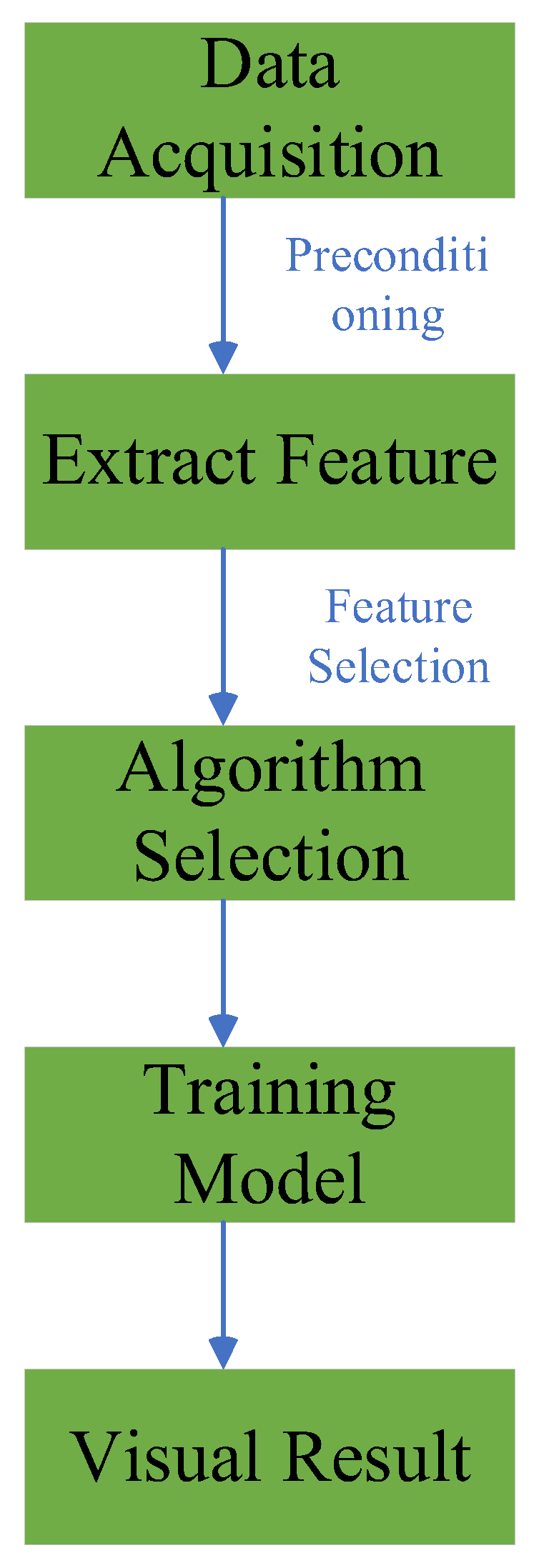
Figure 1. Basic flow of optimization algorithm.
Preprints 86420 g001
Figure 2. Optimisation process for fine-tuning parameters.
Preprints 86420 g002
Figure 3. Structure of feature selection algorithm.
Preprints 86420 g003
Figure 4. Ten-fold cross validation.
Preprints 86420 g004
Table 1. Characterisation Evaluation Indicator Information.
Indicator Descriptions
Information Gain Metrics for decision trees and feature selection that measure the extent to which features affect the target variable by calculating the reduction in uncertainty of the target variable if the features are known.|
Information Gain Ratio The information gain is extended by considering the ratio of entropy and conditional entropy of features to reduce the preference for features with a large number of values.
Chi-Square Test Used to assess the association between the features and the target variable, the chi-square statistic between the features and the target variable was calculated and used to determine if they were independent.
Mutual Information A measure of how much information is shared between two random variables, which can be used to measure the correlation between the feature and the target variable.
Pearson Correlation Coefficient Used to assess the linear relationship between two continuous variables and can be used to identify features that are highly correlated with the target variable.
Spearman Rank Correlation Coefficient Used to assess the monotonic relationship between two variables and can be used to identify the characteristics of the monotonic relationship with the target variable.
Analysis of Variance (ANOVA) Used to compare differences in means between multiple groups and can be used to assess differences in means for different characteristics on a target variable.
Recursive Feature Elimination(RFE) The importance of features is assessed by recursively removing the least important features, using model performance as an evaluation metric.
Relative Importance For tree-based models (e.g., random forests), the metric used to measure the extent to which features contribute to model performance.
AUC-ROC Metrics for biry classification problems that measure the classification performance of features and assess the degree of separation between positive and negative examples.
Disclaimer/Publisher’s Note: The statements, opinions and data contained in all publications are solely those of the individual author(s) and contributor(s) and not of MDPI and/or the editor(s). MDPI and/or the editor(s) disclaim responsibility for any injury to people or property resulting from any ideas, methods, instructions or products referred to in the content.
Copyright: This open access article is published under a Creative Commons CC BY 4.0 license, which permit the free download, distribution, and reuse, provided that the author and preprint are cited in any reuse.
Prerpints.org logo

Preprints.org is a free preprint server supported by MDPI in Basel, Switzerland.

Subscribe

© 2024 MDPI (Basel, Switzerland) unless otherwise stated