Preprint
Communication

PMab-301: An Anti-Giraffe Podoplanin Monoclonal Antibody for Immunohistochemistry

Altmetrics

Downloads

90

Views

67

Comments

0

A peer-reviewed article of this preprint also exists.

Submitted:

29 September 2023

Posted:

09 October 2023

You are already at the latest version

Alerts
Abstract
Immunohistochemistry staining is an essential method in pathological diagnoses. Podoplanin (PDPN) is a specific maker of alveolar epithelium, lymphatic vessels, and glomeruli. In this study, we established a novel anti-giraffe PDPN (girPDPN) mAb, PMab-301, using the Cell-Based Immunization and Screening (CBIS) method. PMab-301 (mouse IgG1, kappa) detected girPDPN in various applications, such as flow cytometry, western blot, and immunohistochemistry. PMab-301 specifically stained type-I alveolar cells using formalin-fixed paraffin-embedded giraffe lung tissues. Our findings suggest the potential usefulness of PMab-301 for the pathophysiological analyses of giraffe tissues.
Keywords: 
Subject: Biology and Life Sciences  -   Animal Science, Veterinary Science and Zoology

1. Introduction

Podoplanin (PDPN) is a mucin-type glycoprotein, [1] which plays an important role in the lymphatic system development [2]. PDPN interacts with C-type lectin-like receptor-2 (CLEC-2) which is an endogenous receptor of PDPN [3,4]. The PDPN-CLEC-2 interaction promotes the embryonic blood and lymphatic vessel separation [5]. PDPN is expressed in human cancers and is associated with malignant progression and metastasis via platelet aggregation [6,7,8,9]. PDPN is expressed in many cell types, including podocytes in glomerulus [1], pulmonary type I alveolar cells [10], and lymphatic endothelial cells [11]. In contrast, PDPN is not expressed in renal tubule cells, pulmonary type-II alveolar cells, and vascular endothelial cells [1]. Therefore, PDPN is a useful maker to distinguish these cells.
We have developed anti-PDPN mAbs for various species including tiger [12], cat [13], pig [14], horse [15], bovine [16], goat [17], sheep [18], alpaca [19], Tasmanian devil [20], bear [21], whale [22], California sea lion [23], golden hamster [24], and ferret [25] using the Cell-Based Immunization and Screening (CBIS) method [26,27,28,29,30]. The Cell-Based Immunization and Screening (CBIS) method is an effective method to develop mAbs for various applications, such as flow cytometry, western blot, and immunohistochemistry. Here, we established a novel anti-giraffe PDPN (girPDPN) mAb using the CBIS method.

2. Materials and Methods

2.1. Cell lines

P3X63Ag8U.1 (P3U1) and Chinese hamster ovary (CHO)-K1 cells were obtained from the American Type Culture Collection (Manassas, VA). P3U1 and CHO-K1 were cultured in a Roswell Park Memorial Institute (RPMI)-1640 medium (Nacalai Tesque, Inc., Kyoto, Japan), with 100 units/mL of penicillin, 0.25 μg/mL of amphotericin B, 100 μg/mL of streptomycin (Nacalai Tesque, Inc.), and 10% heat-inactivated fetal bovine serum (FBS; Thermo Fisher Scientific Inc.). girPDPN coding sequences (cds) were obtained from the WGS database (SJXV01006036.1) by BLAST search using bovine PDPN cds as a query. The girPDPN sequence was registered in DNA Data Bank of Japan (DDBJ, accession number: LC780730). The synthesized DNA of girPDPN (Eurofins Genomics KK), in which the original signal sequence (1-MWKVPVLFFILGSASFWVLAGA-23) was deleted, was subsequently subcloned into a pCAGzeo_ssPA16 or ssMAP16 vector (IL2-signal sequence and PA16 tag or MAP16 tag added to N-terminus). The amino acid sequence of the tag system was as follows: PA16 tag, [31] sixteen amino acids (GLEGGVAMPGAEDDVV); MAP16 tag [32], sixteen amino acids (PGTGDGMVPPGIEDKI). The PA16 tag and the MAP16 tag can be detected by an anti-human PDPN mAb (clone NZ-1) and an anti-mouse PDPN mAb (clone PMab-1), respectively. Using a Neon transfection system (Thermo Fisher Scientific Inc., Waltham, MA), the girPDPN plasmid was transfected into CHO-K1 cells. Using a cell sorter (SH800; Sony Corp., Tokyo, Japan), stable transfectants were established and maintained in the medium, containing 500 µg/mL of Zeocin (InvivoGen, San Diego, CA).
All cells were grown in a humidified incubator at 37oC, in an atmosphere of 5% CO2 and 95% air.

2.2. Hybridomas

To develop mAbs against giraffe PDPN, we intraperitoneally immunized two BALB/c mice (CLEA Japan,Tokyo, Japan) with CHO/MAP16-girPDPN cells (1×10⁸ cells/mouse) plus Imject Alum (Thermo Fisher Scientific, Inc.). The procedure included three additional injections every week (1×10⁸ cells/mouse), which were followed by a final booster intraperitoneal injection (1×10⁸ cells/mouse), two days before harvesting splenocytes. The harvested splenocytes were subsequently fused with P3U1 cells, using PEG1500 (Roche Diagnostics, Indianapolis, IN). For the hybridoma selection, cells were cultured in the RPMI-1640 medium with 10% FBS, 100 units/mL of penicillin, 100 μg/mL of streptomycin, 0.25 μg/mL of amphotericin B, 5 μg/mL of plasmocin, 5% Briclone (NICB, Dublin, Ireland), and hypoxanthine, aminopterin and thymidine (HAT; Thermo Fisher Scientific, Inc.). The supernatants were subsequently screened using flow cytometry using CHO-K1 and CHO/PA16-girPDPN.

2.3. Flow cytometric analysis

CHO/PA16-girPDPN and CHO-K1 cells were harvested after a brief exposure to 0.25% trypsin and 1 mM ethylenediaminetetraacetic acid (EDTA, Nacalai Tesque, Inc.). The cells were treated with 1 μg/mL NZ-1 or PMab-301 for 30 min at 4◦C. The cells were treated with 2 μg/mL of anti-mouse IgG conjugated with Alexa Fluor 488 (Cell Signaling Technology, Inc., Danvers, MA). The fluorescence data were collected using the SA3800 Cell Analyzer (Sony Corporation, Tokyo, Japan).
To determine the dissociation constant (KD), PMab-301 was serially diluted from 10 μg/mL to 0.61 ng/mL. The geometric mean of fluorescence intensity of CHO/PA16-girPDPN at each concentration was calculated by FlowJo v10.8.1 (Becton, Dickinson & Company, Ashland, OR). The KD was estimated by fitting saturation binding curves to the built-in; one-site binding models in GraphPad PRISM 8 (GraphPad Software, Inc., La Jolla, CA).

2.4. Western blotting

Cell lysates were treated with sodium dodecyl sulfate (SDS) sample buffer (Nacalai Tesque, Inc.) at 95◦C for 3 min. The proteins (10 μg) were separated on polyacrylamide gels (5%–20%, FUJIFILM Wako Pure Chemical Corporation, Osaka, Japan) and transferred to polyvinylidene difluoride membranes (Merck KGaA). After blocking with 4% skim milk (Nacalai Tesque, Inc.) in PBS-0.05% Tween 20, membranes were incubated with 5 μg/mL of PMab-301, 1 μg/mL of an anti-PA16 tag mAb (NZ-1), or 1 μg/mL of an anti-IDH1 mAb (RcMab-1). IDH1 is an internal control. Then, they were incubated again with peroxidase-conjugated anti-mouse immunoglobulins (for PMab-301; diluted 1:1000; Agilent Technologies, Inc., Santa Clara, CA) or anti-rat immunoglobulins (for NZ-1 and RcMab-1; diluted 1:10000; Sigma-Aldrich Corporation, St. Louis, MO). Finally, protein bands were detected using ECL Plus Western Blotting Substrate (Thermo Fisher Scientific, Inc.) and a Sayaca-Imager (DRC Co. Ltd., Tokyo, Japan).

2.5. Immunohistochemical analyses

Giraffe lung tissue samples obtained from the laboratory of veterinary pathology, the University of Tokyo. The tissues were fixed in 10% neutral-buffered formalin and processed to make formalin-fixed paraffin-embedded (FFPE) tissue sections. To deparaffinize, rehydrate, and retrieve antigen, the sections were autoclaved in Deparaffinization/Antigen Retrieval Solution pH6 (low pH; Nichirei Biosciences, Tokyo, Japan) 121oC for 20 minutes. Then, sections were blocked using the Super Block T20 (PBS) Blocking Buffer (Thermo Fisher Scientific, Inc.), incubated with 5 μg/mL PMab-301 for 1 h at room temperature, and treated with the Envision + Kit for mouse (Agilent Technologies, Inc.) for 30min. Finally, Color was developed using 3,3′-diaminobenzidine tetrahydrochloride (DAB; Agilent Technologies, Inc.) for 5 min, and counterstaining was performed using hematoxylin (FUJIFILM Wako Pure Chemical Corporation).

3. Results

3.1. Establishment of a novel anti-girPDPN antibody

We immunized mice with girPDPN-overexpressing CHO-K1 (CHO/MAP16-grPDPN) cells (Figure 1A). To produce hybridomas, the splenocytes from these mice were fused with P3U1 cells using polyethylene glycol (Figure 1B). To select anti-girPDPN mAb-producing wells, the reactivities to CHO/PA16-girPDPN cells were observed by flow cytometry (Figure 1C). After limiting dilution, PMab-301 (mouse IgG1, kappa) was finally established (Figure 1D).

3.2. PMab-301 reacted girPDPN-overexpressing CHO-K1 in flow cytometry

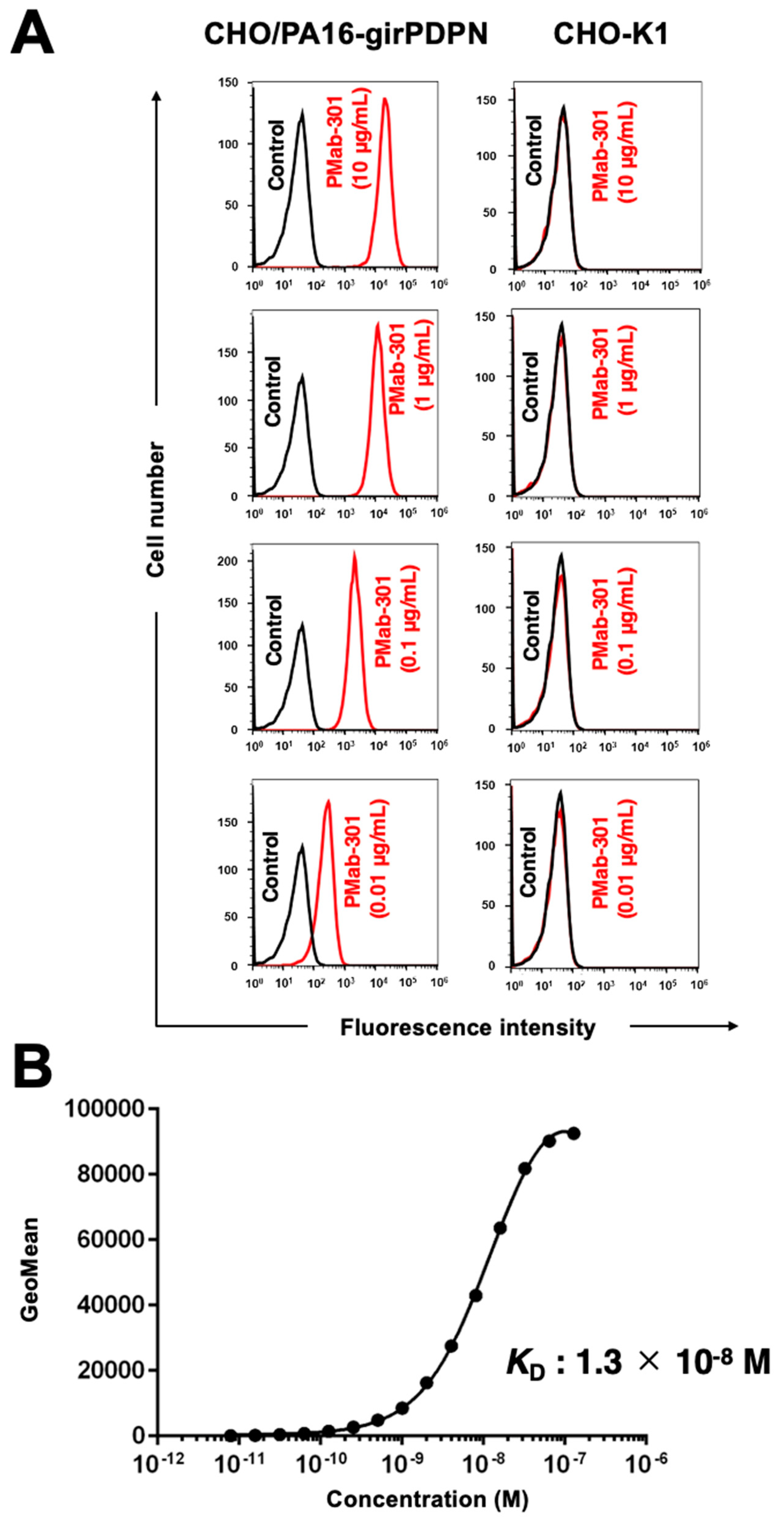
We checked the reactivity of PMab-301 to girPDPN by flow cytometry. PMab-301 reacted to CHO/PA16-girPDPN cells in a dose-dependent manner, but did not react with CHO-K1 cells (Figure 2A). To determine the KD of PMab-301 against CHO/PA16-girPDPN cells, we conducted kinetic analysis by flow cytometry. The geometric mean of the data was plotted versus the concentration of PMab-301. The KD value of PMab-301 for girPDPN was determined as 1.3 × 10⁻⁸ M (Figure 2B).

3.3. girPDPN was detected by western blot by PMab-301

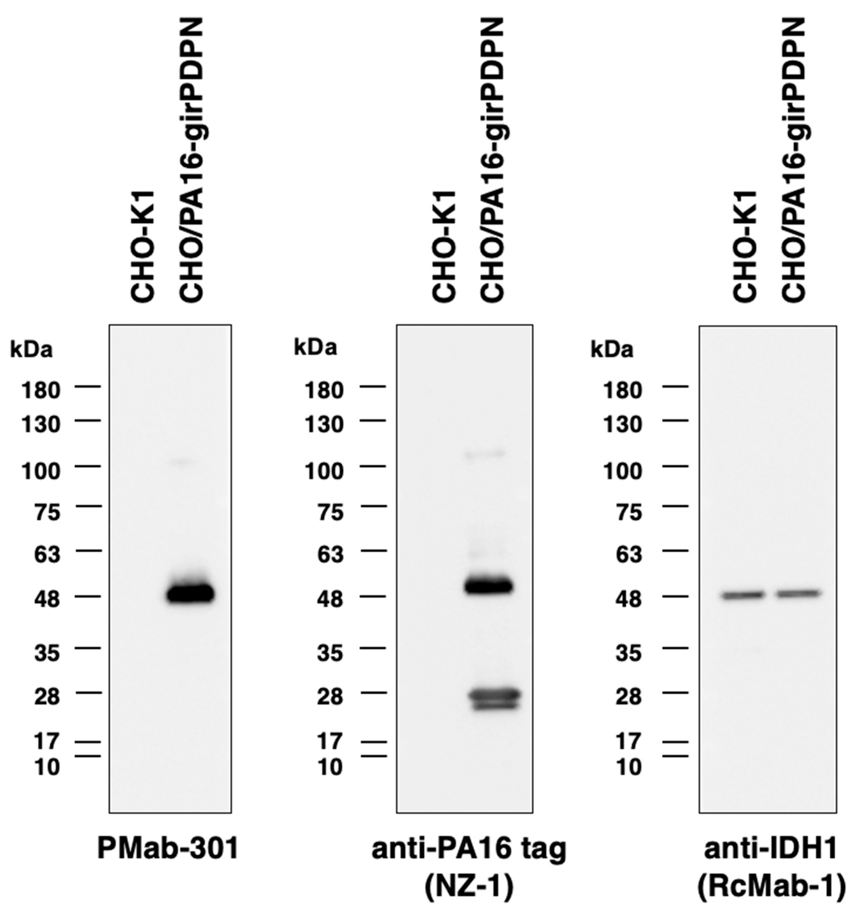
We examined whether PMab-301 is applicable for western blot. Due to glycosylation, PDPN has been detected as about a 48 kDa-band [18,19,33]. As shown in Figure 3, PMab-301 and NZ-1 detected the 48-kDa band of girPDPN in lysates from CHO/PA16-girPDPN cells, whereas this band was not detected in lysates CHO-K1 cells. These data indicate that PMab-301 specifically detects girPDPN in western blot.

3.4. PMab-301 recognized girPDPN in immunohistochemistry

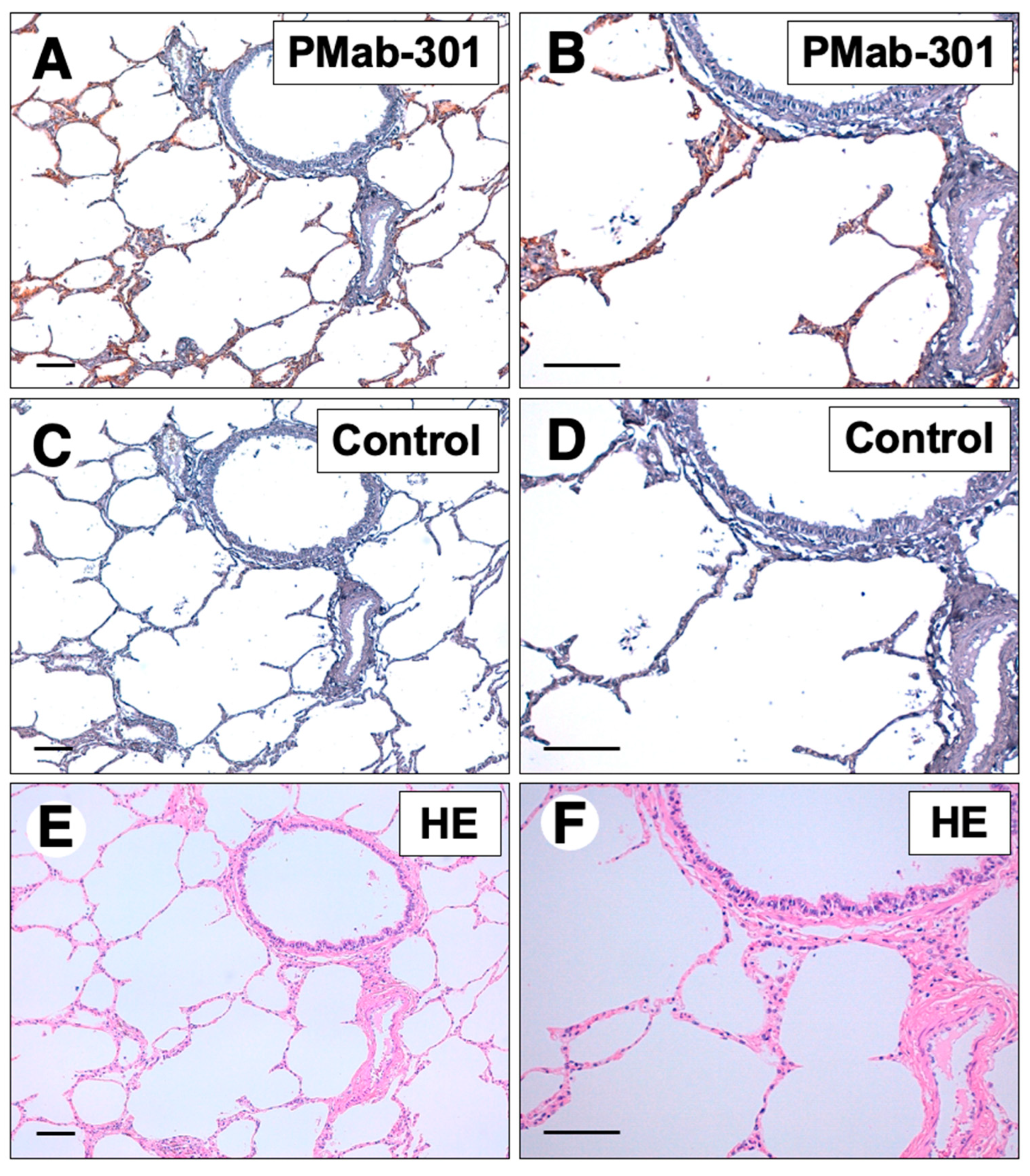
Finally, we investigated whether PMab-301 can be used for immunohistochemical analysis using formalin-fixed paraffin-embedded (FFPE) giraffe lung tissue sections because PDPN is known to be expressed on pulmonary type I alveolar cells of various species [10]. As shown in Figure 4A and 4B, PMab-301 strongly stained alveolar epithelial cells, but did not react with vascular endothelial cells, indicating that PMab-301 is useful for detecting girPDPN in immunohistochemistry.

4. Discussion

We successfully established PMab-301 against girPDPN by the CBIS method and assessed the application of PMab-301. PMab-301 could recognize girPDPN in flow cytometry, western blot, and immunohistochemistry, indicating that PMab-301 can be used in various applications to detect girPDPN.
Several studies have reported that many giraffes died from unknown causes [34,35,36]. Immunohistochemistry is a commonly used method in pathological examination to find out the cause of death. In this study, PMab-301 could stain alveolar epithelium cells, but did not react with vascular endothelial cells in giraffe lung tissues (Figure 4). These data indicate that PMab-301 could bind to girPDPN specifically in immunohistochemistry of giraffe tissue. Because PDPN is a maker of not only alveolar epithelium but also lymphatic endothelial cells in various animals [16,17,18,19], PMab-301 will be a helpful antibody to distinguish lymphatic endothelial cells from vascular endothelial cells in giraffe tissues.

References

  1. Breiteneder-Geleff, S.; Matsui, K.; Soleiman, A.; Meraner, P.; Poczewski, H.; Kalt, R.; Schaffner, G.; Kerjaschki, D. Podoplanin, novel 43-kd membrane protein of glomerular epithelial cells, is down-regulated in puromycin nephrosis. Am J Pathol 1997, 151, 1141–1152. [Google Scholar]
  2. Schacht, V.; Ramirez, M.I.; Hong, Y.K.; Hirakawa, S.; Feng, D.; Harvey, N.; Williams, M.; Dvorak, A.M.; Dvorak, H.F.; Oliver, G.; et al. T1alpha/podoplanin deficiency disrupts normal lymphatic vasculature formation and causes lymphedema. EMBO J 2003, 22, 3546–3556. [Google Scholar] [CrossRef]
  3. Suzuki-Inoue, K.; Kato, Y.; Inoue, O.; Kaneko, M.K.; Mishima, K.; Yatomi, Y.; Yamazaki, Y.; Narimatsu, H.; Ozaki, Y. Involvement of the snake toxin receptor CLEC-2, in podoplanin-mediated platelet activation, by cancer cells. J Biol Chem 2007, 282, 25993–26001. [Google Scholar] [CrossRef]
  4. Kato, Y.; Kaneko, M.K.; Kunita, A.; Ito, H.; Kameyama, A.; Ogasawara, S.; Matsuura, N.; Hasegawa, Y.; Suzuki-Inoue, K.; Inoue, O.; et al. Molecular analysis of the pathophysiological binding of the platelet aggregation-inducing factor podoplanin to the C-type lectin-like receptor CLEC-2. Cancer Sci 2008, 99, 54–61. [Google Scholar] [CrossRef]
  5. Bertozzi, C.C.; Schmaier, A.A.; Mericko, P.; Hess, P.R.; Zou, Z.; Chen, M.; Chen, C.Y.; Xu, B.; Lu, M.M.; Zhou, D.; et al. Platelets regulate lymphatic vascular development through CLEC-2-SLP-76 signaling. Blood 2010, 116, 661–670. [Google Scholar] [CrossRef]
  6. Ochoa-Alvarez, J.A.; Krishnan, H.; Pastorino, J.G.; Nevel, E.; Kephart, D.; Lee, J.J.; Retzbach, E.P.; Shen, Y.; Fatahzadeh, M.; Baredes, S.; et al. Antibody and lectin target podoplanin to inhibit oral squamous carcinoma cell migration and viability by distinct mechanisms. Oncotarget 2015, 6, 9045–9060. [Google Scholar] [CrossRef]
  7. Kato, Y.; Kaneko, M.; Sata, M.; Fujita, N.; Tsuruo, T.; Osawa, M. Enhanced expression of Aggrus (T1alpha/podoplanin), a platelet-aggregation-inducing factor in lung squamous cell carcinoma. Tumour Biol 2005, 26, 195–200. [Google Scholar] [CrossRef]
  8. Kato, Y.; Kaneko, M.K.; Kuno, A.; Uchiyama, N.; Amano, K.; Chiba, Y.; Hasegawa, Y.; Hirabayashi, J.; Narimatsu, H.; Mishima, K.; et al. Inhibition of tumor cell-induced platelet aggregation using a novel anti-podoplanin antibody reacting with its platelet-aggregation-stimulating domain. Biochem Biophys Res Commun 2006, 349, 1301–1307. [Google Scholar] [CrossRef]
  9. Suzuki, H.; Kaneko, M.K.; Kato, Y. Roles of Podoplanin in Malignant Progression of Tumor. Cells 2022, 11. [Google Scholar] [CrossRef]
  10. Dobbs, L.G.; Williams, M.C.; Gonzalez, R. Monoclonal antibodies specific to apical surfaces of rat alveolar type I cells bind to surfaces of cultured, but not freshly isolated, type II cells. Biochim Biophys Acta 1988, 970, 146–156. [Google Scholar] [CrossRef]
  11. Hirakawa, S.; Hong, Y.K.; Harvey, N.; Schacht, V.; Matsuda, K.; Libermann, T.; Detmar, M. Identification of vascular lineage-specific genes by transcriptional profiling of isolated blood vascular and lymphatic endothelial cells. Am J Pathol 2003, 162, 575–586. [Google Scholar] [CrossRef]
  12. Furusawa, Y.; Kaneko, M.K.; Nakamura, T.; Itai, S.; Fukui, M.; Harada, H.; Yamada, S.; Kato, Y. Establishment of a Monoclonal Antibody PMab-231 for Tiger Podoplanin. Monoclon Antib Immunodiagn Immunother 2019, 38, 89–95. [Google Scholar] [CrossRef]
  13. Yamada, S.; Itai, S.; Nakamura, T.; Yanaka, M.; Saidoh, N.; Chang, Y.W.; Handa, S.; Harada, H.; Kagawa, Y.; Ichii, O.; et al. PMab-52: Specific and Sensitive Monoclonal Antibody Against Cat Podoplanin for Immunohistochemistry. Monoclon Antib Immunodiagn Immunother 2017, 36, 224–230. [Google Scholar] [CrossRef]
  14. Kato, Y.; Yamada, S.; Furusawa, Y.; Itai, S.; Nakamura, T.; Yanaka, M.; Sano, M.; Harada, H.; Fukui, M.; Kaneko, M.K. PMab-213: A Monoclonal Antibody for Immunohistochemical Analysis Against Pig Podoplanin. Monoclon Antib Immunodiagn Immunother 2019, 38, 18–24. [Google Scholar] [CrossRef]
  15. Kato, Y.; Yamada, S.; Itai, S.; Kobayashi, A.; Konnai, S.; Kaneko, M.K. Anti-Horse Podoplanin Monoclonal Antibody PMab-219 is Useful for Detecting Lymphatic Endothelial Cells by Immunohistochemical Analysis. Monoclon Antib Immunodiagn Immunother 2018, 37, 272–274. [Google Scholar] [CrossRef]
  16. Honma, R.; Ogasawara, S.; Kaneko, M.K.; Fujii, Y.; Oki, H.; Nakamura, T.; Takagi, M.; Konnai, S.; Kato, Y. PMab-44 Detects Bovine Podoplanin in Immunohistochemistry. Monoclon Antib Immunodiagn Immunother 2016, 35, 186–190. [Google Scholar] [CrossRef]
  17. Furusawa, Y.; Yamada, S.; Nakamura, T.; Sano, M.; Sayama, Y.; Itai, S.; Takei, J.; Harada, H.; Fukui, M.; Kaneko, M.K.; et al. PMab-235: A monoclonal antibody for immunohistochemical analysis against goat podoplanin. Heliyon 2019, 5, e02063. [Google Scholar] [CrossRef]
  18. Kato, Y.; Furusawa, Y.; Sano, M.; Takei, J.; Nakamura, T.; Yanaka, M.; Okamoto, S.; Handa, S.; Komatsu, Y.; Asano, T.; et al. Development of an Anti-Sheep Podoplanin Monoclonal Antibody PMab-256 for Immunohistochemical Analysis of Lymphatic Endothelial Cells. Monoclon Antib Immunodiagn Immunother 2020, 39, 82–90. [Google Scholar] [CrossRef]
  19. Kato, Y.; Furusawa, Y.; Yamada, S.; Itai, S.; Takei, J.; Sano, M.; Kaneko, M.K. Establishment of a monoclonal antibody PMab-225 against alpaca podoplanin for immunohistochemical analyses. Biochem Biophys Rep 2019, 18, 100633. [Google Scholar] [CrossRef]
  20. Furusawa, Y.; Yamada, S.; Itai, S.; Nakamura, T.; Takei, J.; Sano, M.; Harada, H.; Fukui, M.; Kaneko, M.K.; Kato, Y. Establishment of a monoclonal antibody PMab-233 for immunohistochemical analysis against Tasmanian devil podoplanin. Biochem Biophys Rep 2019, 18, 100631. [Google Scholar] [CrossRef]
  21. Takei, J.; Furusawa, Y.; Yamada, S.; Nakamura, T.; Sayama, Y.; Sano, M.; Konnai, S.; Kobayashi, A.; Harada, H.; Kaneko, M.K.; et al. PMab-247 Detects Bear Podoplanin in Immunohistochemical Analysis. Monoclon Antib Immunodiagn Immunother 2019, 38, 171–174. [Google Scholar] [CrossRef]
  22. Yamada, S.; Itai, S.; Nakamura, T.; Takei, J.; Sano, M.; Konnai, S.; Kobayashi, A.; Nakagun, S.; Kobayashi, Y.; Kaneko, M.K.; et al. Immunohistochemical Analysis of the Harbor Porpoise Using Antipodoplanin Antibody PMab-237. Monoclon Antib Immunodiagn Immunother 2019, 38, 104–107. [Google Scholar] [CrossRef]
  23. Tanaka, T.; Asano, T.; Sano, M.; Takei, J.; Hosono, H.; Nanamiya, R.; Nakamura, T.; Yanaka, M.; Harada, H.; Fukui, M.; et al. Development of Monoclonal Antibody PMab-269 Against California Sea Lion Podoplanin. Monoclon Antib Immunodiagn Immunother 2021, 40, 124–133. [Google Scholar] [CrossRef]
  24. Nanamiya, R.; Suzuki, H.; Takei, J.; Li, G.; Goto, N.; Harada, H.; Saito, M.; Tanaka, T.; Asano, T.; Kaneko, M.K.; et al. Development of Monoclonal Antibody 281-mG(2a)-f Against Golden Hamster Podoplanin. Monoclon Antib Immunodiagn Immunother 2022, 41, 311–319. [Google Scholar] [CrossRef]
  25. Goto, N.; Suzuki, H.; Tanaka, T.; Asano, T.; Kaneko, M.K.; Kato, Y. Development of a Monoclonal Antibody PMab-292 Against Ferret Podoplanin. Monoclon Antib Immunodiagn Immunother 2022, 41, 101–109. [Google Scholar] [CrossRef]
  26. Asano, T.; Nanamiya, R.; Takei, J.; Nakamura, T.; Yanaka, M.; Hosono, H.; Tanaka, T.; Sano, M.; Kaneko, M.K.; Kato, Y. Development of Anti-Mouse CC Chemokine Receptor 3 Monoclonal Antibodies for Flow Cytometry. Monoclon Antib Immunodiagn Immunother 2021, 40, 107–112. [Google Scholar] [CrossRef]
  27. Nanamiya, R.; Takei, J.; Asano, T.; Tanaka, T.; Sano, M.; Nakamura, T.; Yanaka, M.; Hosono, H.; Kaneko, M.K.; Kato, Y. Development of Anti-Human CC Chemokine Receptor 9 Monoclonal Antibodies for Flow Cytometry. Monoclon Antib Immunodiagn Immunother 2021, 40, 101–106. [Google Scholar] [CrossRef]
  28. Sayama, Y.; Kaneko, M.K.; Kato, Y. Development and characterization of TrMab-6, a novel anti-TROP2 monoclonal antibody for antigen detection in breast cancer. Mol Med Rep 2021, 23. [Google Scholar] [CrossRef]
  29. Sayama, Y.; Kaneko, M.K.; Takei, J.; Hosono, H.; Sano, M.; Asano, T.; Kato, Y. Establishment of a novel anti-TROP2 monoclonal antibody TrMab-29 for immunohistochemical analysis. Biochem Biophys Rep 2021, 25, 100902. [Google Scholar] [CrossRef]
  30. Yamada, S.; Itai, S.; Nakamura, T.; Yanaka, M.; Kaneko, M.K.; Kato, Y. Detection of high CD44 expression in oral cancers using the novel monoclonal antibody, C(44)Mab-5. Biochem Biophys Rep 2018, 14, 64–68. [Google Scholar] [CrossRef]
  31. Fujii, Y.; Kaneko, M.; Neyazaki, M.; Nogi, T.; Kato, Y.; Takagi, J. PA tag: a versatile protein tagging system using a super high affinity antibody against a dodecapeptide derived from human podoplanin. Protein Expr Purif 2014, 95, 240–247. [Google Scholar] [CrossRef]
  32. Fujii, Y.; Kaneko, M.K.; Kato, Y. MAP Tag: A Novel Tagging System for Protein Purification and Detection. Monoclon Antib Immunodiagn Immunother 2016, 35, 293–299. [Google Scholar] [CrossRef]
  33. Yamada, S.; Honma, R.; Kaneko, M.K.; Nakamura, T.; Yanaka, M.; Saidoh, N.; Takagi, M.; Konnai, S.; Kato, Y. Characterization of the Anti-Bovine Podoplanin Monoclonal Antibody PMab-44. Monoclon Antib Immunodiagn Immunother 2017, 36, 129–134. [Google Scholar] [CrossRef]
  34. Potter, J.S.; Clauss, M. Mortality of captive giraffe (Giraffa camelopardalis) associated with serous fat atrophy: a review of five cases at Auckland Zoo. J Zoo Wildl Med 2005, 36, 301–307. [Google Scholar] [CrossRef]
  35. Sullivan, K.; van Heugten, E.; Ange-van Heugten, K.; Poore, M.H.; Dierenfeld, E.S.; Wolfe, B. Analysis of nutrient concentrations in the diet, serum, and urine of giraffe from surveyed North American zoological institutions. Zoo Biol 2010, 29, 457–469. [Google Scholar] [CrossRef]
  36. Garijo, M.M.; Ortiz, J.M.; Ruiz de Ibanez, M.R. Helminths in a giraffe (Giraffa camelopardalis xgiraffa) from a zoo in Spain. Onderstepoort J Vet Res 2004, 71, 153–156. [Google Scholar] [CrossRef]
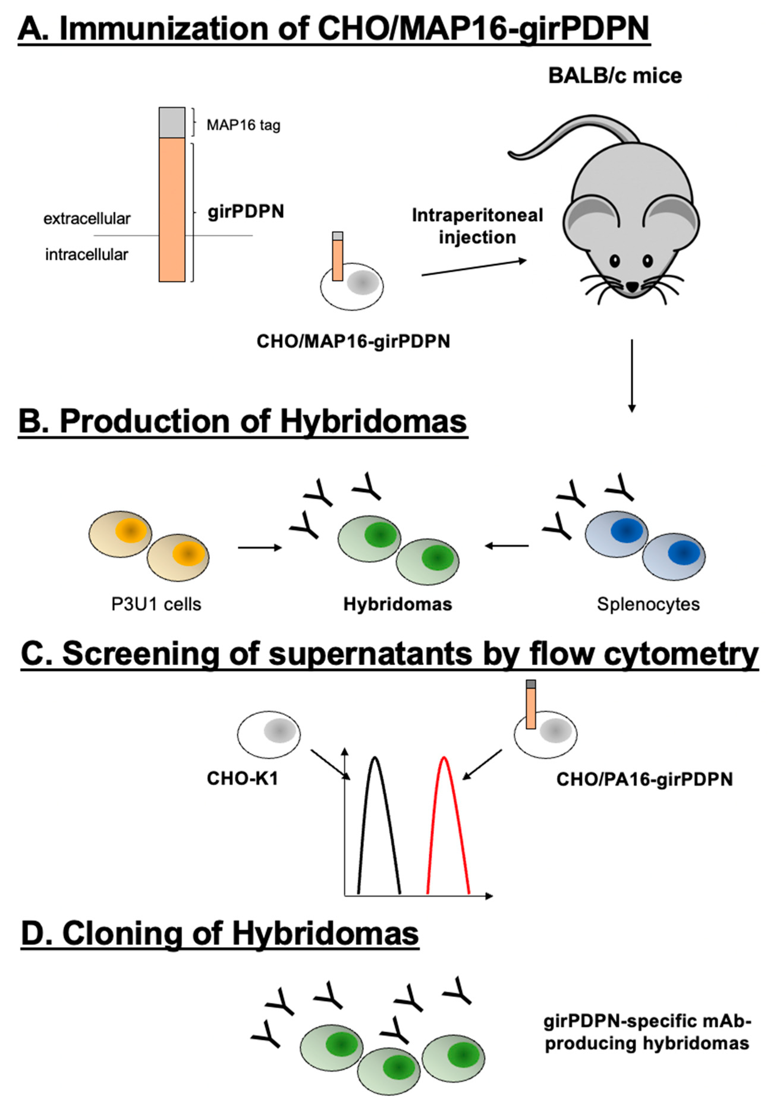
Figure 1. The establishment of PMab-301 using the CBIS method. (A) MAP16-tagged girPDPN-overexpressing CHO-K1 (CHO/MAP16-girPDPN) cells were immunized into two BALB/c mice. (B) The splenocytes were fused with P3U1 cells. (C) The culture supernatants were screened using flow cytometry to select anti-girPDPN mAb-producing hybridomas. (D) PMab-301 was established through limiting dilution and some additional screenings.
Preprints 86484 g001
Figure 2. Flow cytometry of girPDPN-overexpressing cells using PMab-301. (A) CHO/PA16-girPDPN and CHO-K1 cells were treated with 0.01–10 µg/mL of PMab-301 (redline) or blocking buffer (negative control, blackline), followed by treatment with AlexaFluor488-conjugated anti-mouse IgG. (B) The binding affinity of PMab-301 was determined against CHO/PA16-girPDPN cells by flow cytometry.
Preprints 86484 g002
Figure 3. Western blot with PMab-301. Cell lysates (10 μg) of CHO/PA16-girPDPN and CHO-K1 cells were electrophoresed and transferred to PVDF membranes. After blocking, membranes were incubated with PMab-301, anti-PA16 tag mAb (NZ-1), or anti-IDH1 mAb (RcMab-1). RcMab-1 was used to detect an internal control.
Preprints 86484 g003
Figure 4. Immunohistochemical staining of giraffe lung with PMab-301. Immunohistochemical analysis for the giraffe lung. The sections of the giraffe lung were incubated with 5 μg/mL of PMab-301 (A, B) or a blocking buffer (C, D), followed by the EnVision+Kit. (E, F) Hematoxylin and eosin (HE) staining was performed. Scale bars = 100 μm.
Preprints 86484 g004
Disclaimer/Publisher’s Note: The statements, opinions and data contained in all publications are solely those of the individual author(s) and contributor(s) and not of MDPI and/or the editor(s). MDPI and/or the editor(s) disclaim responsibility for any injury to people or property resulting from any ideas, methods, instructions or products referred to in the content.
Copyright: This open access article is published under a Creative Commons CC BY 4.0 license, which permit the free download, distribution, and reuse, provided that the author and preprint are cited in any reuse.
Prerpints.org logo

Preprints.org is a free preprint server supported by MDPI in Basel, Switzerland.

Subscribe

© 2024 MDPI (Basel, Switzerland) unless otherwise stated