1. Introduction
Entrapment neuropathies of the lower limb are an often misunderstood and overlooked group of conditions, often leading to chronic pain and other disabilities if not timely treated. Among the most frequent of these syndromes encountered in clinical practice are fibular nerve entrapment, proximal tibial neuropathy, sural nerve neuropathy, deep gluteal syndrome or sciatic nerve entrapment, and lateral femoral cutaneous nerve entrapment, also known as meralgia paresthetica. The challenge in diagnosing these conditions often arises from a lack of standardized diagnostic tests and clear clinical criteria present in literature, coupled with a deficiency in clinician confidence. This narrative review aims to provide a clearer overview of these common entrapment neuropathies by reviewing the anatomy, pathophysiology, clinical presentation, and Electrodiagnostic (EDX) findings in patients with these pathologies. By focusing on these prevalent syndromes, this article intends to bridge the gap in understanding, leading to more effective diagnosis, management, and improved patient outcomes. A comprehensive literature search was conducted using the databases from Pubmed (which includes the MEDLINE database), Cochrane Library, Google Scholar and EMBASE with literature published from 1970 to 2022.
2. Fibular nerve entrapment (Peroneal nerve entrapment)
2.1. Introduction
Fibular or peroneal nerve entrapment neuropathy is the most common mononeuropathy of the lower extremity and the third most common after median and ulnar neuropathies [
1,
2,
3]. The most frequently affected site is at the fibular head, where the nerve’s superficial trajectory predisposes it to injury [
4,
5].
2.2. Anatomy, Etiology and Pathophysiology
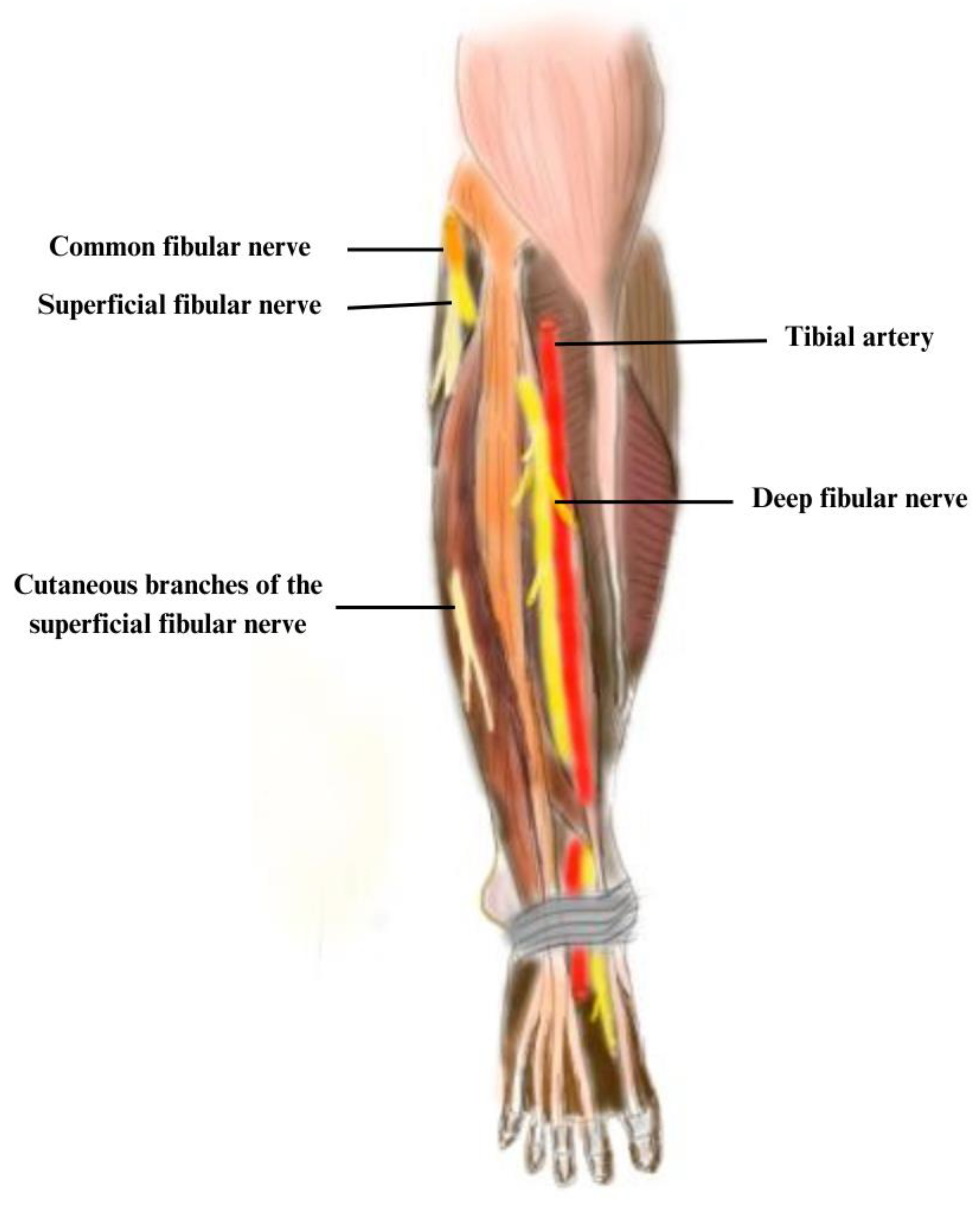
The common fibular nerve (fibular nerve or external sciatic nerve) branches off from the lateral division of the sciatic nerve [
6]. At the superior popliteal fossa, it branches into the superficial fibular nerve and the deep fibular nerve [
1,
3,
7] (
Figure 1). The superficial fibular nerve’s terminal divisions are the medial dorsal and the intermediate cutaneous nerves [
1,
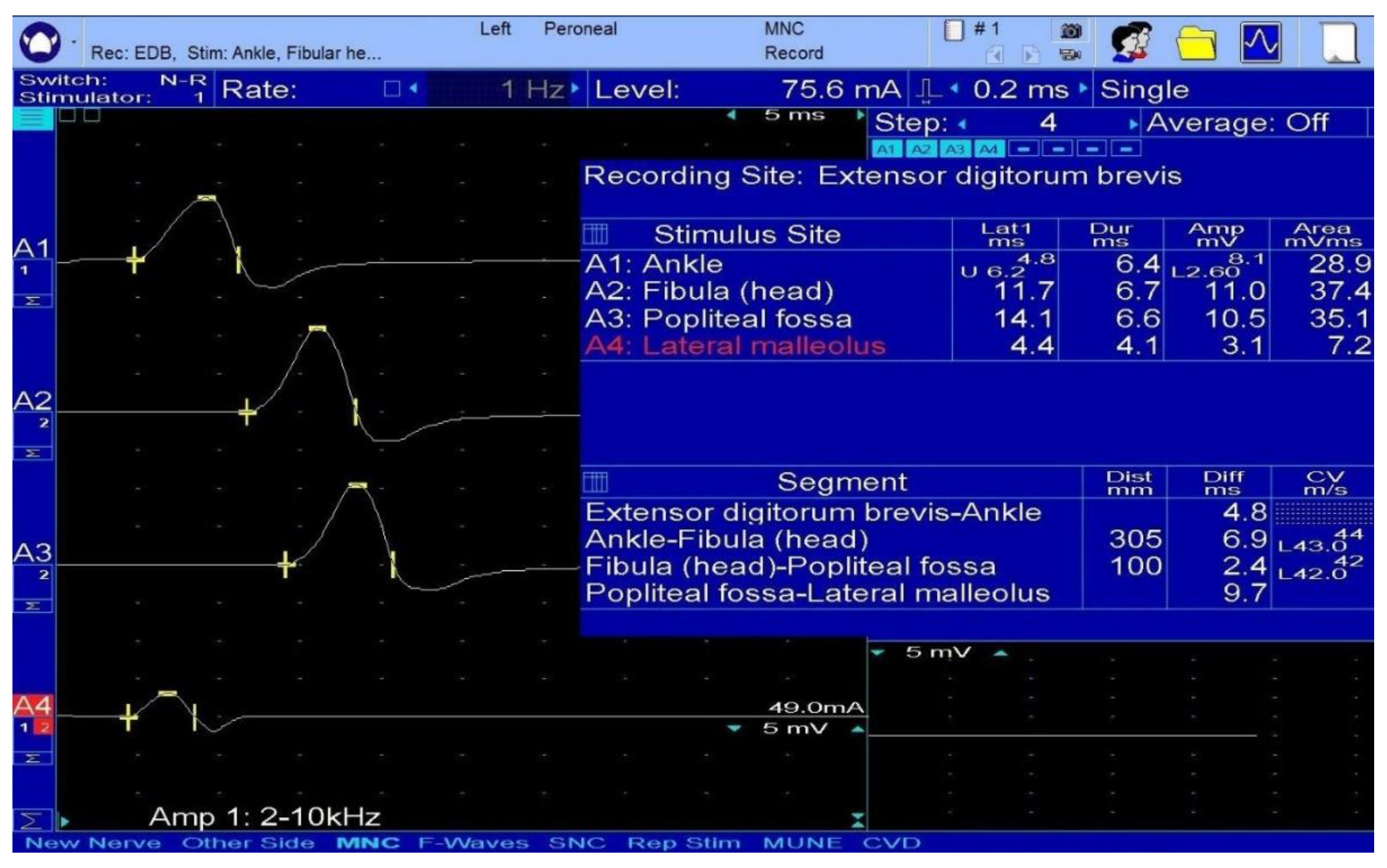
3]. A common anatomical variant is represented by the accessory fibular nerve, present in approximately one quarter of the general population, which supplies the innervation of the extensor digitorum brevis muscle [
3] (
Figure 2).
For the common fibular nerve, the most typical site for compression is at the fibular head [
7,
8]. Other compression sites are: 1) the exit of the lateral leg compartment, as it pierces through the crural fascia, for the superficial fibular nerve and 2) in the tight tunnel formed by the extensor retinaculum muscle above and the navicular and talus bones underneath for the deep fibular nerve [
1].
Acute presentations are usually related to high energy trauma, such as knee dislocations, fibular fractures and ankle trauma, while chronic presentations are a result of behavioural causes or masses, such as ganglion cysts and neuromas [
1]. Moreover, knee arthroplasty represents a risk of fibular nerve entrapment [
2] as well as prolonged immobilisation [
7]. Diabetes is also a predisposing condition due to the edema caused by sorbitol in the nerve tissue [
9]. Likewise, behavioral causes can precipitate a neuropathy through repetitive cross-legged position, prolonged periods of squatting and extrinsic compression due to bed rest [
3,
7]. Weight loss may also induce fibular neuropathy, associated secondary with a loss of subcutaneous tissue and therefore greater risk of compression [
7].
2.3. Clinical Presentation and Physical Examination
Symptoms occur acutely or insidiously, depending on the precipitating cause [
1,
3]. Fibular neuropathy is the most frequently reported lower extremity mononeuropathy in athletes [
3], dancers [
1] or occupations requiring squatting or kneeling for an extended period of time, for instance carpentry or farm work [
4].
A typical patient suffering from fibular nerve entrapment presents with foot drop, resulting in steppage gait, pain and numbness of the lateral lower leg and foot dorsum [
1,
7], symptoms typically aggravated by plantar flexion and foot inversion [
6].
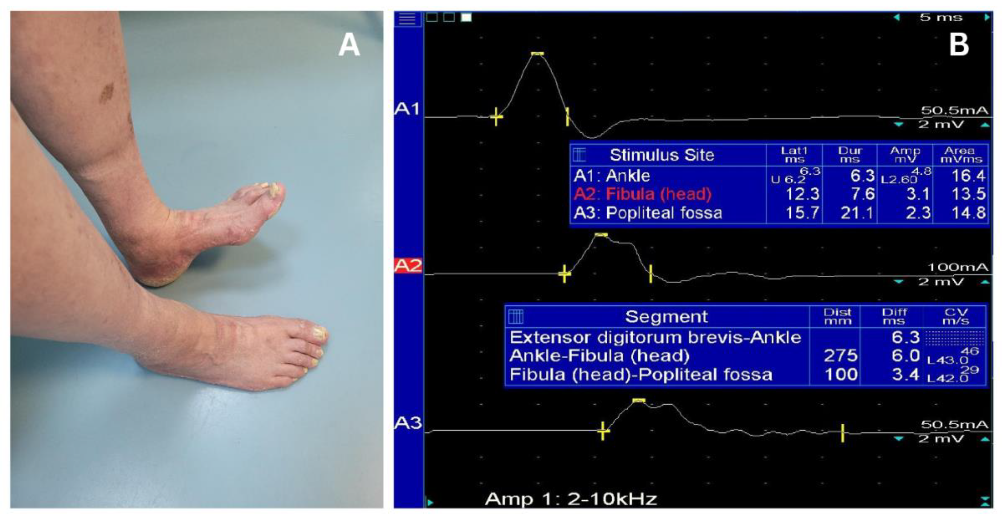
Common fibular nerve entrapment neuropathy presents with weakness of foot eversion, dorsiflexion of the ankle, together known as foot drop, weakness of great toe extension and sensory loss, burning, tingling, pain in the anterolateral distal leg and foot dorsum [
1]. Patients may report pain waking them up at night, especially when the blanket touches the anterior part of the leg or the foot dorsum [
9] (
Figure 3).
Superficial fibular nerve entrapment neuropathy symptoms are rarely present in isolation and are most commonly exacerbated during exercise [
6]. Weakness of foot eversion is similar to the previous entrapment neuropathy, but ankle dorsiflexion and great toe extension are normal. The sensory abnormalities are the same as with common fibular nerve entrapment neuropathy, present at the foot dorsum and lateral aspect of the shin, with sparing of the dorsal space of the first web space and fifth toe, without severe paresis [
1]. It occurs more commonly in soccer players [
3].
Deep fibular nerve entrapment neuropathy or anterior tarsal tunnel syndrome grants few sensory symptoms, or the patient could be asymptomatic [
1]. Numbness and paresthesias in the first web space, that awaken the patient from sleep [
3], together with pain or dull aching in the anterior ankle and dorsal foot that worsens with tight shoe wearing could be present [
1]. Tibialis anterior weakness may lead to reduced ability for plantar flexion of the foot [
9].
The differential diagnosis of foot drop with proximal neuropathies such as sciatic mononeuropathy, lumbosacral plexopathy and lumbar radiculopathy is vital. In addition to the signs and symptoms of fibular neuropathy described above, these pathologies present with foot inversion and plantar flexion weakness, medial, lateral and plantar foot sensory loss and a reduced Achilles reflex. The L4-S1 lumbar radiculopathies also present with hip abduction weakness [
4,
7]. For the definitive diagnosis, however, electrodiagnostic studies are needed.
2.4. Electrodiagnostic and Imaging Techniques
To differentiate between common fibular nerve, superficial fibular nerve and deep fibular nerve entrapment neuropathy and other neuropathies, EDX studies are the definitive assessment tool. EDX studies are also necessary for injury severity, specifically to discern between an axonal or demyelinating injury, thus guiding prognosis for potential nerve function recovery [
1,
5,
7]. The presence of compound muscle action potential (CMAP) in the affected territory is associated with a good prognosis [
3]. Contralateral comparison is useful for quantifying the severity of axonal loss [
4].
For the common fibular nerve evaluation, motor nerve conduction studies (NCS) are carried out with the G1 recording electrode placed on the midpoint of the extensor digitorum brevis muscle and the G2 electrode in the fifth metatarsophalangeal joint [
10]. The stimulating electrode has three separate sites: at ankle level, laterally to the tibialis anterior tendon, above and 10 cm below the fibular head [
3,
10]. A stimulation sweep speed of 5 ms/division and gain at 5 mV/div are used. The frequency filter is set between the low 2 Hz and high 10 kHz [
10]. The conduction block can be identified when decrease in CMAP amplitude is less than 50% [
11].
EMG should be conducted on the tibialis anterior at least. Additionally, peroneus longus and, less often, the short head of the biceps femuralis muscles can be studied [
1]. The tibialis anterior muscle, innervated by the deep fibular branch, is the most likely to show abnormal findings (denervation activity, neurogenic motor unit action potentials (MUAP)) [
3]. The short head of the biceps femoris is studied because it is the only fibular-innervated muscle above the knee, thus it will help localising the lesion above or below the knee [
3,
4].
Antidromic sensory NCS of the superficial fibular nerve’s two branches is recommended [
12]. The recording electrode for sensory response is placed at the inframalleolar line, over the intermediate and medial dorsal cutaneous nerves, or 3 cm proximally to the bimalleolar line. The stimulating electrode is placed at the anterior edge of the fibula, proximal to the recording electrode with a distance of 12-14 cm [
3,
12]. A stimulation sweep speed of 1 ms/division and gain of 20 microV are applied. The frequency filter was set between the low 30 Hz and high 2.000 Hz [
12]. Moreover, in case of unknown etiology, ultrasound could be useful in the visualisation of scarring, bone infiltrations, mass lesions, with contralateral comparison rendering the most accurate results [
1,
7]. Magnetic Resonance Imaging (MRI) can also be used [
7]. It identifies T2 hyperintensity lesions in the nerve trajectory and denervation signs of the anterior and lateral muscle compartments for the common fibular nerve and for the anterior compartment for the deep fibular nerve. For the deep fibular nerve, it can also show fascial defects and muscle herniation, with axial imaging in dorsiflexion being recommended [
6]. However, for the common fibular nerve, ultrasound has been shown to be higher in specificity and sensitivity, even considering the combination of MRI and ultrasound [
8].
3. Proximal Tibial Neuropathy
3.1. Subsection
Proximal tibial neuropathy (PTN) is defined as entrapment of the tibial nerve in the popliteal fossa by impinging masses or by the fibrous sling of the soleus muscle. The latter is also known as soleal sling syndrome (SSS) [
13,
14].
3.2. Anatomy, Etiology and Pathophysiology
As the sciatic nerve branches into the common perineal nerve and the tibial nerve at distal thigh level, the tibial branch continues its tract through the popliteal fossa. It passes over the popliteal muscle and, in order to enter the deep posterior compartment of leg muscles, it has to pass under the tendinous arch of the soleus muscle. Along its tract, the nerve is accompanied by the tibial artery and vein [
13] (
Figure 1). Due to its deep location, the nerve is usually not affected by external traumatic accidents. The main etiological reason of PTN entrapments is space occupying lesions. We found several cases in the literature: tibial arterial aneurysm [
15] and pseudoaneurysm [
16], intraneural ganglion cysts [
17], Baker's cysts [
18], tibial bone exostosis [
19] and popliteal muscle enlargement [
20]. In these cases, the space occupying lesions either shifted the nerve to the fibrous sling of the soleus muscle or were the primary reason of entrapment by compressing the nerve with mass effect. Although space occupying lesions are more common, primary nerve entrapment by the fibrous sling of the soleus muscle can be the reason for PTN [
14].
3.3. Clinical Presentation and Physical Examination
The dominant symptoms are pain and weakness in popliteal, calf and plantar muscles. These can get worse with active plantar flexion or passive dorsiflexion of the foot and ankle [
14]. Moderate weakness of toe flexor muscles can also be present. The dominant sensory symptoms are numbness, paresthesia, hypersensitivity and tingling in the sole and heel of the foot. Physical examination plays a huge role in the diagnosis and can be easily assessed. Many patients will have a positive Tinel's sign, which can be evoked by gently pressing approximately 8-9 cm below the tibial plateau, which will provoke severe pain. It is also characteristic for PTN for Tinel’s sign to radiate to the medial sole of the foot. In more advanced cases, muscle atrophies in the calf and plantar region can be seen as well [
13,
14,
21].
3.4. Electrodiagnostic and Imaging Techniques
The deep location of the tibial nerve makes EDX evaluation very challenging. Therefore, many physicians rely on physical examination for diagnosis and MRI and/or ultrasonography (US) for positive diagnosis.
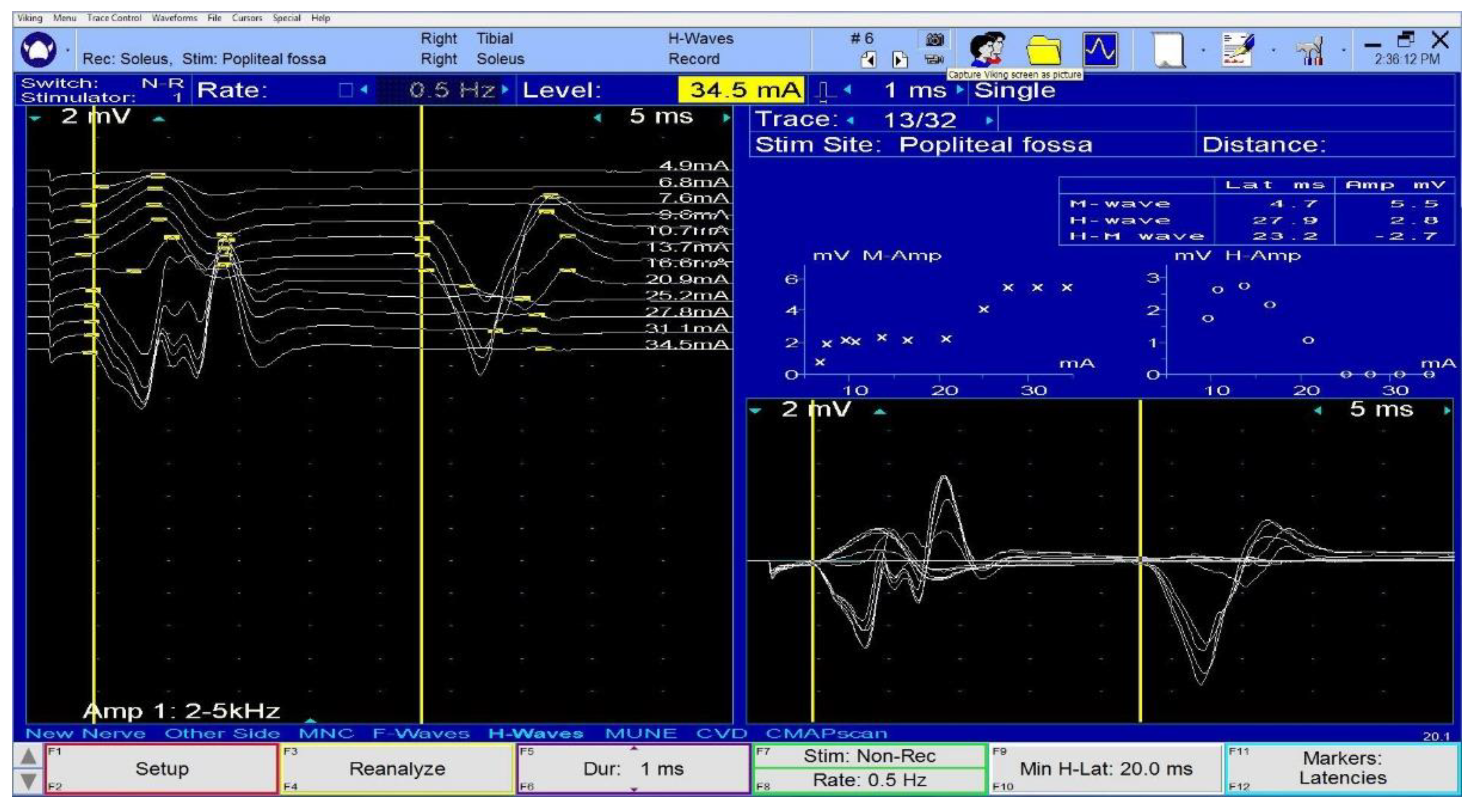
Even so, D.C. Preston et al. described the recommended procedure of tibial motor study. The proximal stimulation site is located in the popliteal fossa and the stimulator should be placed just over the popliteal pulse in mid-posterior knee. The distal stimulation site is located in the medial ankle, positioning the stimulator slightly proximal and posterior to the medial malleolus. The recording site is the abductor hallucis brevis muscle. The G1 electrode is placed at equal distance of 1 cm proximal and inferior to the navicular prominence while G2 is placed over the metatarsal-phalangeal joint of the hallux. The tibial CMAP is monitored and often high intensity stimulations are required when stimulated at the popliteal fossa to ensure supramaximal stimulation. Moreover, the soleus H-reflex study can be helpful in PTN diagnosis. This reflex is absent or delayed in PTN . The stimulation site is identical with the proximal tibial motor study protocol, with the cathode being placed rostrally. The G1 recording site is 2-4 cm distal to the locus where the soleus meets the two bellies of the gastrocnemius, and the G2 recording site is just over the Achilles tendon. The H-reflex occurs at low intensity stimulations without (
Figure 4) a direct muscle response [
22].
High Resolution UltraSound (HRUS) is a good tool to investigate the presence or absence of space occupying lesions. Moreover, we can have the dynamic image of structures surrounding the nerve and help to differentially diagnose venous claudication [
23]. On the other hand, MRI will show proximal T2 nerve hyperintensity, which indicates venous congestion and blockage of normal axoplasmic flow. In addition, distally the nerve will show T2 hyperintensity due to Wallerian degeneration. At the site of entrapment, the nerve will show abnormal flattening. Moreover, MRI can indirectly indicate neural entrapment by showing regional pathological changes associated with muscle denervation. In some cases, it was even possible to confirm the thickening of the fibrous sling of the soleus muscle [
24,
25,
26].
4. Sural Nerve Neuropathy
4.1. Introduction
Because of its superficial location and extensive usage as a nerve graft, the sural nerve is one of the most well studied nerves in human body. Moreover, it is one of the nerves with numerous anatomical variations, therefore any type of compression of the nerve along its long trajectory will lead to sural nerve neuropathy (SNN) [
27].
4.2. Anatomy, Etiology and Pathophysiology
The sural nerve is derived from S1 and S2 nerve roots. In the most common anatomical variation, the sural nerve arises from two branches of the tibial nerve and common fibular nerve. The medial sural cutaneous branch emerges from the tibial nerve at the level of the distal popliteal fossa. It traverses the two gastrocnemius heads and is usually joined at mid-calf level by the communicating branch of the fibular nerve and forms the proper sural nerve. The trajectory of the nerve travels down the posterolateral side of the foot and lateral to the Achilles tendon, accompanied by the small saphenous vein (SSV). At the ankle, it passes to the lateral malleolus and posterior to the fibular tendons and bifurcates into the lateral calcaneal nerve and lateral dorsal digital nerve of the V finger [
27,
28,
29].
The etiology of SNN damage can be distributed in three groups: a) traumatic; b) atraumatic and c) iatrogenic. Therefore, due to its superficial tract and close relationship with bone structures, any traumatic impact has a high risk of causing SNN. Ankle sprains, fractures of distal fibula, talus, calcaneus, and base of the fifth metatarsal are the most common traumatic causes. Atraumatic causes are myositis ossificans in the gastrocnemius, fibular or Achilles tendon tendinosis, osteochondroma, or space occupying lesions, such as Baker’s cyst, local schwannoma, and neuroma or intraneural ganglia.Nevertheless, the main cause of SSN remains iatrogenic. The superficial location and anatomical variations make the nerve susceptible to direct intra-operational traumas. Likewise, the close relationship with the anatomical structures represent a risk of nerve injury during their manipulation, such as: SSV varices ablation, achilles or fibular tendon repair, gastrocnemius recession surgery, arthoscopies [
28,
30,
31,
32,
33].
4.3. Clinical Presentation and Physical Examination
The sural nerve is a purely sensory nerve and provides sensation for the lateral part of ankle, heel and foot up to the base of the fifth metatarsal bone. Thus, patients with SSN will complain about paresthesias, tingling, radiating pain or/and tenderness in the above-mentioned areas. The pain and discomfort are exacerbated by physical activity and during nighttime. The physical examination is often unremarkable, with normal reflexes and unaffected motor components. Tinel’s sign may be positive but is unreliable in patients with good physical condition. Physicians can perform provocative maneuvers such as inversion and plantar flexion of the foot, which is expected to be positive [
27,
28,
29].
4.4. Electrodiagnostic and Imaging Techniques
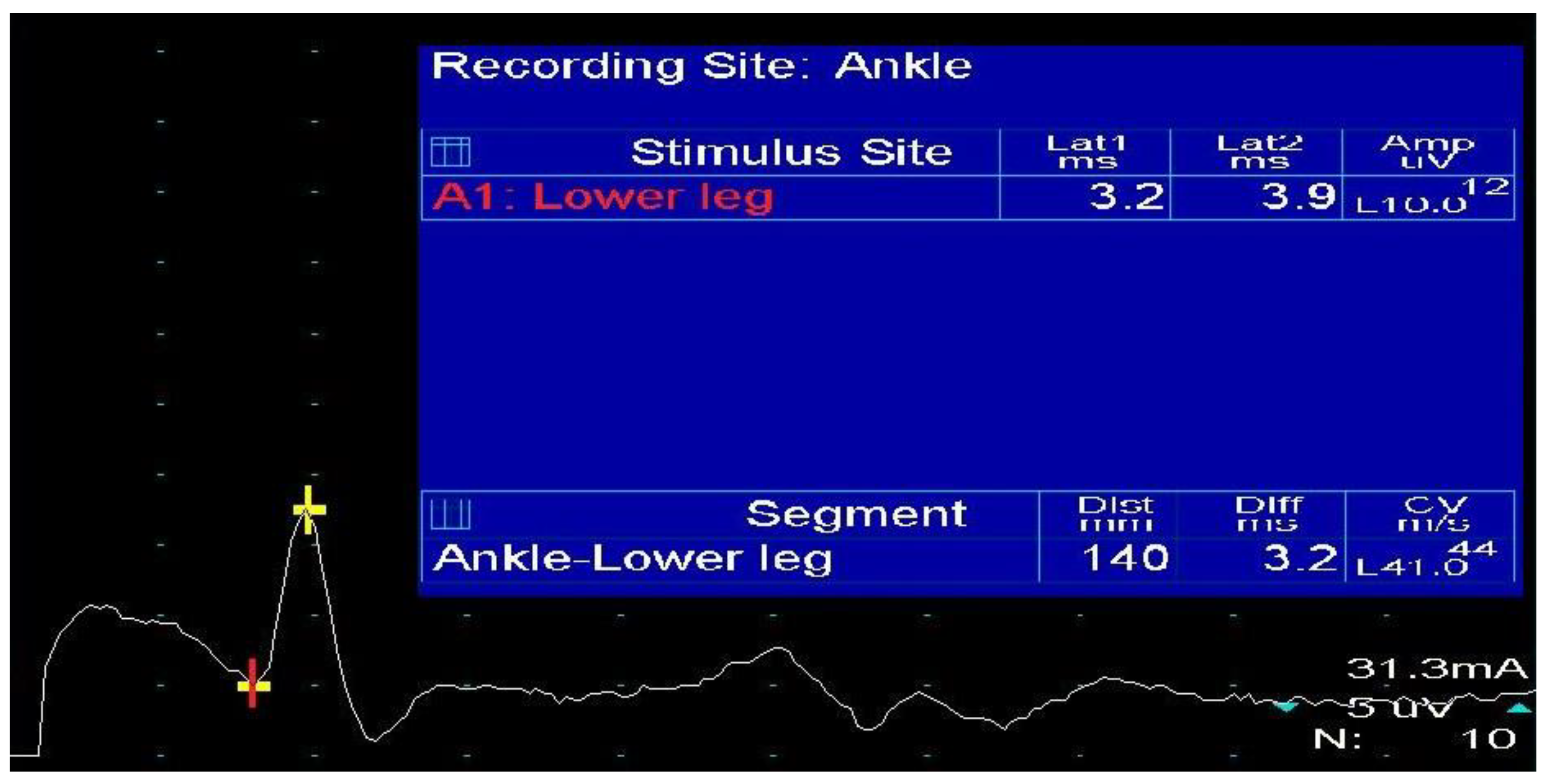
D.C. Preston and his team explain in detail the procedure of NCS of the sural nerve. The posterior-lateral calf represents the stimulation site and two points at the posterior ankle are the recording sites. G1 is located just posterior to the lateral malleolus while G2 is located 3-4 cm distal to G1. Low stimulation intensities (5-25 mA) and a duration of 0.1 ms are usually enough to achieve supramaximal response (
Figure 5). The optimal position for patients is lying on their side with the studied leg facing up. It is advised that the examination is performed on both feet and that the results are compared [
22]. However, clinical presentation and anamnesis of the patient is usually enough to suspect potential SNN. The imaging studies are used to confirm the suspicion and to locate the exact site of entrapment and evaluate its severity. Ultrasound or HRUS in particular is often very efficient for this purpose. Plain radiographs are also useful, since fractures, osteochondromas and myositis ossificans can also be the cause of SSN. MRI is opportune to investigate the presence of space occupying lesions in soft tissues and localize the entrapment site with high specificity [
31,
32,
33].
5. Deep Gluteal Syndrome (Sciatic Nerve Entrapment)
5.1. Introduction
Deep Gluteal Syndrome (DGS) is characterized by the entrapment of the sciatic nerve (SN) in the subgluteal area. This complex includes several types of variants—piriformis syndrome (PS), gemelli-internal obturator complex syndrome, ischiofemoral impingement syndrome and hamstring syndrome—and correlates with non-discogenic and extrapelvic etiologies having clinical presentations of pain and dysesthesias in the posterior region of the hip and buttocks [
34,
35]. PS is the most common explanation for DGS and is caused by sciatic nerve impingement by the piriformis muscle (PM) [
36,
37,
38]. It is often overlooked and can vary from 6% to up to 17,8% of cases of chronic low back pain or sciatica [
39,
40,
41]. The mean age of diagnosis is 43 years old, with a slight female predominance [
40,
42].
Gemelli-internal obturator complex syndrome involves the compression of the SN by the internal obturator muscle. The ischiofemoral impingement syndrome refers to the entrapment of the sciatic nerve by the quadratus femoris in the ischiofemoral space, whereas proximal hamstring syndrome refers to the impingement of this nerve by the semitendinosus, semimembranosus and biceps femoris muscles, respectively [
43].
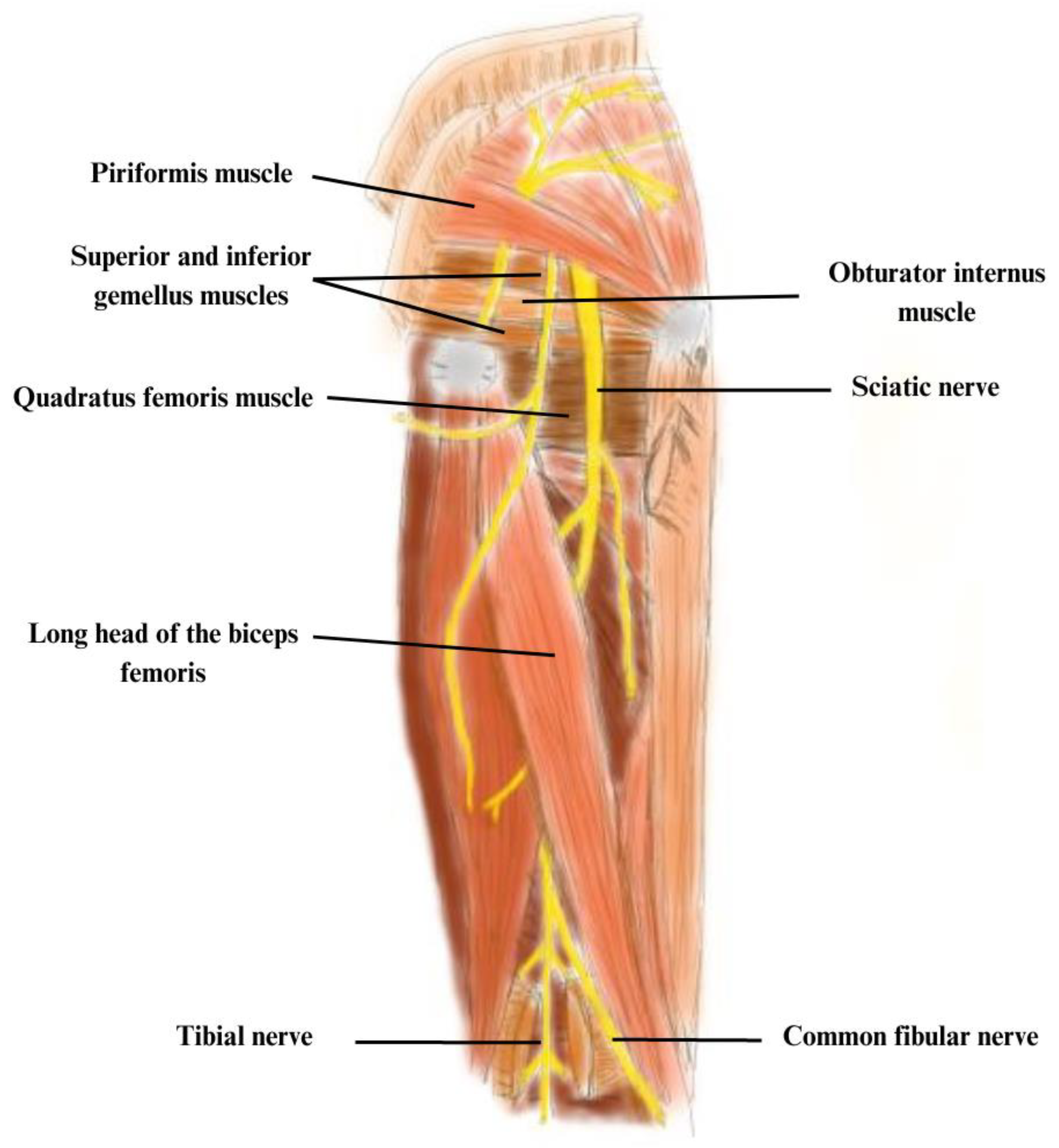
5.2. Anatomy, Etiology and Pathophysiology
The SN derives from the L4-S3 ventral branch of the sacral plexus [
37]. It can be trapped at the level of the lumbar spine, but also due to intrapelvic and extrapelvic pathologies in the subgluteal space or deep gluteal space. The subgluteal space is located anterior to the gluteus maximus muscle. It is positioned lateral to the tensor fasciae latae muscle and linea aspera, medial to the sacrotuberous ligament and inferior to the ischial tuberosity at the level of the proximal insertion of the hamstring muscles. The femur trochanters and neck are anterior [
34,
44]. The PM has the proximal insertion at the level of the anterolateral surface of the sacrum and the superior margin of the greater sciatic notch. Distal insertion is situated at the level of the superior greater trochanter (
Figure 6). Its contraction determines the external rotation of the hip and, secondarily, abduction when it is flexed [
41,
45]. The trajectory of the SN nerve has several characteristics. After the passage out to the greater sciatic notch, the nerve is lying down inferiorly to the PM, over the obturator internus muscle. Thus, this typical anatomical pattern causes a scissor effect with entrapment of the nervous structure [
43,
46]. At the thigh, it is situated posterior to the adductor magnus and anterior to the long head of the biceps femoris. Likewise, when it enters the popliteal fossa, it passes between the biceps and the semimembranosus muscles [
37].
The etiologies that trigger DGS symptoms are varied. Pathologies which diminish SN mobility during joint movement—iatrogenic, traumatic, inflammatory, tumoral or mechanical overuse etiologies—can cause nerve damage [
43]. Moreover, several structures determine SN entrapment through acute edema: musculotendinous, osseous, neurovascular or capsular tissue [
37]. Vascular causes and endometriosis compress the SN as a result of the intimate relationship with the iliac vessels, ovaries and sacral plexus [
37]. Moreover, several congenital and acquired anomalies of the PM, internal obturator muscle and sciatic nerves are reported. Nerve branches passing through one of the muscles, trajectory variants of the SN, high variability in the insertions of the PM and accessory PM can cause SN entrapment [
37,
47]. Other causes include hypertrophy secondary to overuse, infection of the PM and leg length discrepancy [
36,
40,
48]. PM hematoma or fibrosis following trauma is a frequent etiology but in some cases the cause is unidentified [
42]. In addition, several conditions such as strains, avulsions and tendinopathies encountered in running and jumping sports predispose individuals to hamstring syndrome [
38].
5.3. Clinical Presentation and Physical Examination
Some clinical features can explain the DGS manifestation: posterior thigh and hip area pain, unilateral buttock discomfort with low back pain, sometimes with a radicular distribution, accompanied by tenderness [
38,
39,
40,
43]. These symptoms are exacerbated by walking and running, hip flexion with knee extension—when the nerve is maximally stretched—and worsens at night [
38,
44]. The patient walks with a limp due to muscular weakness, cannot stand for more than 30 minutes and adopts an antalgic position, with the healthy ischium supporting the weight [
34,
37,
44]. In patients with ischiofemoral impingement and hamstring syndrome, the pain is exacerbated when taking bigger steps or at the initial heel strike [
38].
Furthermore, in several reviews, the PS is associated with a quartet of symptoms: 1) buttock pain—consistently present 2) pain aggravation by sitting, 3) external tenderness near the greater sciatic notch and 4) any PS provocative maneuver that determines PS symptoms [
39,
40]. Thus, some provocative maneuvers simulate the symptoms: Lasègue test or straight leg raise (SLR); Freiberg sign—in supine position, active internal rotation of the hip; Pace sign—resisted hip abduction; FAIR test—narrowing of the space between the PM and internal obturator muscle by Flexion of the hip, Adduction and Internal Rotation [
43,
44]. Additionally, Michel et al. devised a 12-point clinical assessment score for the diagnosis and treatment standardisation of PS. The test’s sensitivity and specificity were 96.4% and 100%, respectively, for a score over 8 points, where the diagnosis was considered “Probable” [
36]. Additionally, the PM muscle can also generate deep myofascial pain, exacerbated by prolonged walking or through squatting [
42,
48,
49]. The palpation of the structures in the gluteal region can reveal a sensitive mass in the PS or tenderness to the IT, between the ischium and the femoral head: 1) lateral and at the IT level—characteristic of hamstring syndrome and ischiofemoral impingement; 2) medial to the IT—pudendal nerve entrapment. Moreover, the differential diagnosis of deep gluteal region pain with lumbosacral radiculopathies, sacroiliac or hip articular pathologies and gynecological disease must be performed [
37,
38,
50]. US or CT-guided injections provide relief of symptoms and have diagnostic and prognostic, post-operative significance [
37]. Two systematic reviews concluded that the diagnostic pathway must include anamnesis, physical examination, imaging: pelvic radiographs and MRIs, lidocaine or corticosteroid injection and EDX findings [
34,
38].
5.4. Electrodiagnostic and Imaging Techniques
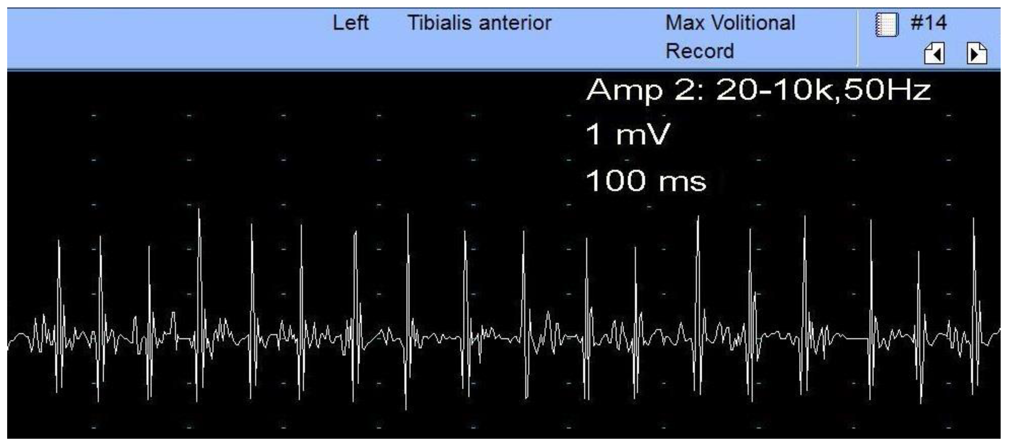
For SN assessment, NCS and EMG should be bilaterally conducted. The NCS studies of the fibular and tibial nerve, respectively fibular superficial nerve and sural nerve are described in their chapters. The F responses of the tibial and fibular nerves and the H-reflexes are usually prolonged and should be studied. The fibular nerve is more severely affected than the tibial nerve and, where there is axonal loss, the CMAP amplitudes are reduced. The EMG study narrows the lesion site and quantifies the severity of the disease. The study protocol involves the examination of two fibular and tibial innervated muscles, the long and short heads of the biceps femoris muscle and one muscle innervated by the superior and inferior gluteal nerves [
22]. Thus, if muscles innervated by the fibular and tibial nerves and the biceps femoris muscle present EMG reduced recruitment of MUAPs, a SN lesion can be confirmed (
Figure 7). Moreover, if an anomaly is found in the muscles innervated by the superior and inferior gluteal nerves, a plexopathy or radiculopathy is more likely. However, the L5 and S1 paraspinal muscles must also be assessed for the differential diagnosis between radiculopathy and plexopathy [
22,
41,
47,
51].
Likewise, in the assessment of PS, the dynamic examination is mandatory, since NCS and EMG are frequently normal in the early stages of nerve injury (4). Spontaneous EMG activity and amplitude decrease of sensory and motor nerve action potential may appear after several weeks [
41,
47,
51]. Therefore, at the onset of symptoms, the EDX studies may be normal and they must be repeated three to four weeks later [
38]. However, Fishman and Wilkins [
22] test the reliability of the H-reflex between the anatomical and FAIR (Flexion Adduction Internal Rotation) position, with a prolonged H-reflex in case of SN neuropathy [
42,
45,
49]. At the same time, EDX studies exclude myopathies or neuropathies with overlapping symptoms, such as radiculopathies, sciatic nerve palsy, and hip joint-mediated pain [
37,
41,
47,
48].
US, CT and MRI are used to exclude other pathologies, as well as for establishing the etiology. In addition, magnetic resonance neurography (MRN) can visualise PM and SN modifications [
45,
51]. Thereby, a study conducted by Filler et al. found MRN to have a specificity of 93% and sensitivity of 64% in diagnosing PS in patients with chronic sciatica and normal MRI results. PS was the major diagnosis established following MRN and interventional MRI imaging. The criteria consisted of asymmetry and SN hyperintensity [
35]. Moreover, for DGS, MRI is mandatory, excluding discogenic causes of sciatica [
34,
38]. CT and US visualise hematomas, tumours, and abscesses which could cause buttock pain. Periarticular endoscopic examination of the subgluteal space detect the cause of entrapment and simultaneously decompresses the nerve [
37,
42,
44,
50].
6. Meralgia paresthetica (Lateral Femoral Cutaneous Nerve Entrapment)
6.1. Introduction
Meralgia paresthetica (MP), also known as Bernhardt-Roth syndrome, is a purely sensory mononeuropathy which involves compression of the lateral femoral cutaneous nerve (LFC) at different sites along its trajectory [
52,
53,
54,
55]. This syndrome is commonly encountered in active male individuals, with a mean age diagnosis of 50 years. Having a higher prevalence in diabetes mellitus patients (247 cases per 10.000 patients per year), the incidence rate in the general population is approximately 4.3 cases per 10.000 patients per year [
52,
56].
6.2. Anatomy, Etiology and Pathophysiology
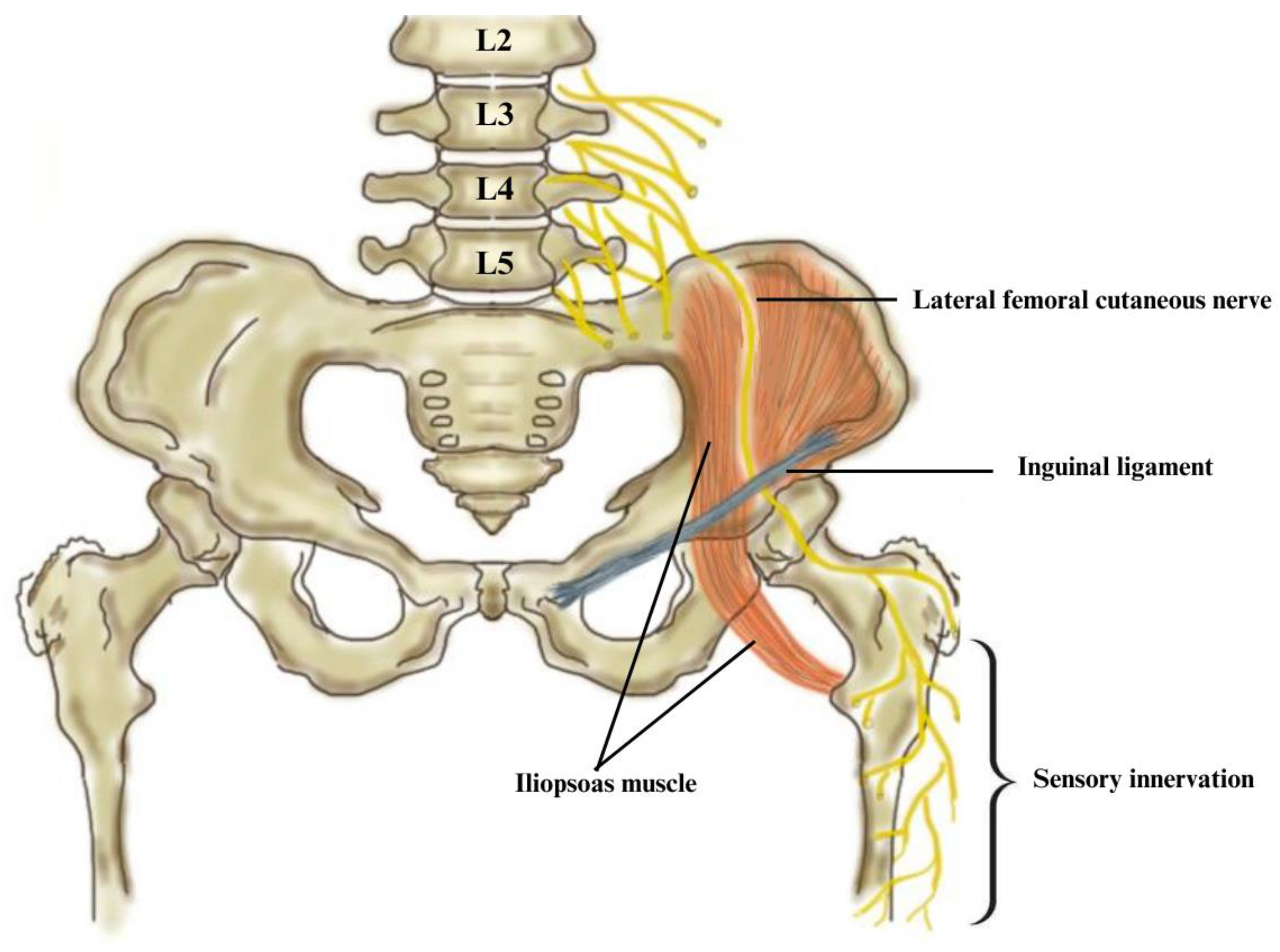
The LFC is a sensory nerve originating from the lumbar plexus (L1-L3). It emerges at the lateral border of the psoas major muscle and follows an oblique path from the anterior surface of the iliacus muscle to the superior iliac spine, under the inguinal ligament, anterior and medial to the sartorius muscle [
52,
54]. As it enters the thigh, it divides into an anterior and posterior branch [
55] (
Figure 8). The most common location for chronic compression or entrapment is at the exit from the pelvis. Moreover, at this level, five documented anatomical variations or exit variants are identified, which explains the variability of clinical presentation [
52,
54].
MP can be idiopathic or spontaneous, caused by: 1) mechanical factors including obesity, pregnancy, restrictive clothing (military armor, police uniforms, jeans), and/or 2) metabolic factors such as diabetes mellitus and alcoholism [
56]. However, iatrogenic cases like: post-surgical complication as a result of hip joint surgery or prone position during spine surgery were reported [
57]. Other procedures such as iliac bone graft extraction, appendicectomy, and caesarean section concern isolated cases [
52,
54].
6.3. Clinical Presentation and Physical Examination
The typical presentation of patients with MP is unilateral pain, paresthesia and numbness in the lateral or anterolateral thigh. In addition, due to high interindividual variability as a result of the nerve’s anatomical variations, burning sensation, muscle aches or buzzing complete the clinical symptomatology [
52,
53]. Symptoms are usually relieved with sitting, as a result of tension reduction in the inguinal ligament, and occur or are intensified by prolonged standing or walking. Either leg seems to be affected, without preference for the dominant lower limb (51). Motor dysfunctions are absent and deep tendon reflexes are preserved [
54,
55]. Furthermore, a number of provocative maneuvers such as pelvic compression, neurodynamic testing, and Tinel’s sign improve the positive diagnosis [
53].
6.4. Electrodiagnostic and Imaging Techniques
LFC nerve stimulation is performed with a frequency of 1 Hz at the ASIS level, the electrical stimulus having a duration of 0.1 ms and an intensity of 30 mA. The recording electrode is placed 14 cm distally on the oblique line created by the ASIS and patella [
53]. Motor NCS are useful to rule out other neuropathies and are usually within normal limits [
54]. The EDX study is considered abnormal if the sensory nerve action potential amplitude on the affected side is 50% shorter than on the healthy side [
53]. Limitations of sensory NCS include obese patients and the technical difficulties related to anatomical variation [
52,
53,
54]. However, in obese patients where direct stimulation of the nerve is impaired, somatosensory evoked potentials (SSEP) and pain-related evoked potentials (PREP) are recommended. Nevertheless, SSEP accuracy is controversial. Some studies report a relatively low specificity of 76% and extremely low sensitivity of 52%, having a limited indication in MP diagnosis [
58]. Contrary, other authors have obtained a sensitivity of 81.3%, achieving accurate findings [
59]. However, both studies recommend the association of SSEP and sensory NCS for a comprehensive and accurate assessment of MP patients. PREP is a relatively new technique, having an AUC = 0.97, with a sensitivity of 91.7% and a specificity of 100% for MP diagnosis [
60]. Thus, although sensory NCS studies were considered to be superior EDX methods, recent studies have concluded that modern techniques such as SSEP and PREP provide better accuracy [
59,
60].
US and MRN ar two other important investigations which visualise the nerve trajectory, anatomical variations and morphologic changes. US is useful to evaluate the nerve and the tissue masses located in the retroperitoneal cavity. Thus, for the LFC nerve, a retrospective study yielded a cross-sectional cut-off value of 5 mm
2 for a positive diagnosis. However, this relatively small diameter makes it difficult to discriminate it from soft tissue [
53]. Supplementarily, LFC ultrasound-guided nerve blocks can be used as a additional diagnostic tool and to differentiate from a lumbosacral radiculopathy [
55]. In this technique, lidocaine is injected approximately 1 cm inferior and slightly medial to the ASIS [
52,
61]. MRN is also used with high accuracy in etiology, nerve injury detection and for preoperative evaluation. Its limitations include cost burdens and lack of experience in recognizing anatomical variations [
62].
7. Take-Home Messages
Lower limb entrapment neuropathies, particularly the most frequent syndromes encountered in clinical practice such as fibular nerve entrapment, proximal tibial neuropathy, sural nerve neuropathy, deep gluteal syndrome or sciatic nerve entrapment, and lateral femoral cutaneous nerve entrapment, also known as meralgia paresthetica, are conditions that demand attention. The patients exhibit symptoms such as dysesthesia, territory-specific pain, muscle weakness, and physical signs identified through examination and pain-inducing maneuvers, which should be prioritized for suspicion over radiculopathies, plexopathies, orthopedic conditions, or gynecological pathologies in certain circumstances. These include instances when patients have held abnormal body postures (potentially during surgical procedures), carried out repetitive movements, experienced trauma, or are in postoperative stage. Professional athletes represent a high-risk demographic.
EDX studies are the primary investigative approach when nerve entrapment is suspected, especially applicable to the entrapment syndrome discussed in this narrative review. This analysis requires a specialist with vast experience, as entrapment neuropathies can produce ambiguous results if inadequately performed. For more complex cases where EDX results are inconclusive or if a secondary entrapment cause is suspected, implementing MRN, MRI or ultrasound when MRN is not available, becomes crucial for confirming the diagnosis, excluding other conditions, and identifying the cause. Additionally, ultrasound or CT-guided injections of lidocaine at the nerve site, which can bring symptomatic relief, hold diagnostic and prognostic value.
With modern advancements in non-invasive procedures, such as endoscopic decompression, conditions like these have become easily manageable. Therefore, by following a systematic diagnostic approach focused on the prevalent and often complex entrapment neuropathies, treatment could be started early in the natural evolution of the disease, leading to halted nerve atrophy progression. This can result in a dramatic quality of life improvement for patients, who typically present with chronic debilitating pain in the course of the disease.
Author Contributions
“Conceptualization, N.C.D. and R.B.; methodology, N.C.D., R.B. and T.D.L.; validation, V.V. and T.D.L.; formal analysis, S.I., and L.L.P.; data curation, N.C.D.; writing—original draft preparation, N.C.D., R.B. and A.B.; writing—review and editing, N.C.D., R.B. and A.B.; visualization, D.M.D.; supervision, V.V. and D.F.M.; project administration, D.F.M. All authors have read and agreed to the published version of the manuscript.”.
Funding
This research received no external funding.
Acknowledgments
We would like to thank Sigrid Crasnean for her proofreading and technical support in writing this paper.
Conflicts of Interest
The authors declare no conflict of interest.
References
- Fortier, L.M. , Markel, M., Thomas, B.G., Sherman, W.F., Thomas, B.H., Kaye, A.D.: An Update on Peroneal Nerve Entrapment and Neuropathy. Orthop Rev (Pavia). 13, (2021). [CrossRef]
- Carender, C.N. , Bedard, N.A., An, Q., Brown, T.S.: Common Peroneal Nerve Injury and Recovery after Total Knee Arthroplasty: A Systematic Review. Arthroplast Today. 2020, 6, 662–667. [Google Scholar] [CrossRef]
- Marciniak, C. : Fibular (Peroneal) Neuropathy. Electrodiagnostic Features and Clinical Correlates. Phys Med Rehabil Clin N Am. 2013, 24, 121–137. [Google Scholar] [CrossRef]
- Craig, A. : Entrapment neuropathies of the lower extremity. PM and R. 5, (2013). [CrossRef]
- Marciniak, C. , Armon, C., Wilson, J., Miller, R.: Practice parameter: utility of electrodiagnostic techniques in evaluating patients with suspected peroneal neuropathy: an evidence-based review. Muscle Nerve. 2005, 31, 520–527. [Google Scholar] [CrossRef]
- Beltran, L.S. , Bencardino, J., Ghazikhanian, V., Beltran, J.: Entrapment neuropathies III: lower limb. Semin Musculoskelet Radiol. 2010, 14, 501–511. [Google Scholar] [CrossRef]
- Bowley, M.P. , Doughty, C.T.: Entrapment Neuropathies of the Lower Extremity. Med Clin North Am. 2019, 103, 371–382. [Google Scholar] [CrossRef]
- Bignotti, B. , Assini, A., Signori, A., Martinoli, C., Tagliafico, A.: Ultrasound versus MRI in common fibular neuropathy. Muscle Nerve. 2017, 55, 849–857. [Google Scholar] [CrossRef] [PubMed]
- Anderson, J.C. : Common Fibular Nerve Compression: Anatomy, Symptoms, Clinical Evaluation, and Surgical Decompression. Clin Podiatr Med Surg. 2016, 33, 283–291. [Google Scholar] [CrossRef]
- Chen, S. , Andary, M., Buschbacher, R., Del Toro, D., Smith, B., So, Y., Zimmermann, K., Dillingham, T.R.: Electrodiagnostic reference values for upper and lower limb nerve conduction studies in adult populations. Muscle Nerve. 2016, 54, 371–377. [Google Scholar] [CrossRef] [PubMed]
- Cruz-Martinez, A. , Arpa, J., Palau, F.: Peroneal neuropathy after weight loss. J Peripher Nerv Syst. 2000, 5, 101–105. [Google Scholar] [CrossRef] [PubMed]
- Saffarian, M.R. , Condie, N.C., Austin, E.A., Mccausland, K.E., Andary, M.T., Sylvain, J.R., Mull, I.R., Zemper, E.D., Jannausch, M.L.: Comparison of four different nerve conduction techniques of the superficial fibular sensory nerve. Muscle Nerve. 2017, 56, 458–462. [Google Scholar] [CrossRef]
- Pomeroy, G. , Wilton, J., Anthony, S.: Entrapment neuropathy about the foot and ankle: an update. J Am Acad Orthop Surg. 2015, 23, 58–66. [Google Scholar] [CrossRef]
- Williams, E.H. , Williams, C.G., Rosson, G.D., Dellon, L.A.: Anatomic site for proximal tibial nerve compression: a cadaver study. Ann Plast Surg. 2009, 62, 322–325. [Google Scholar] [CrossRef]
- Toncev, S. , Sretenovic, S., Mitrovic, S., Toncev, G.: An unusual case of aneurysmal tibial nerve compression detected in ED. Am J Emerg Med. 2013, 31, 1155.e1–1155.e3. [Google Scholar] [CrossRef]
- Rahimizadeh, A. , Davaee, M., Shariati, M., Rahimizadeh, S.: Posterior Tibial Neuropathy Secondary to Pseudoaneurysm of the Proximal Segment of the Anterior Tibial Artery with Delayed Onset. J Brachial Plex Peripher Nerve Inj. 2018, 13, e15–e19. [Google Scholar] [CrossRef]
- Spinner, R.J. , Hébert-Blouin, M.N., Rock, M.G., Amrami, K.K.: Extreme intraneural ganglion cysts. J Neurosurg. 2011, 114, 217–224. [Google Scholar] [CrossRef] [PubMed]
- Moon, S.H. , Im, S., Park, G.Y., Moon, S.J., Park, H.J., Choi, H.S., Jo, Y.R.: Compressive neuropathy of the posterior tibial nerve at the lower calf caused by a ruptured intramuscular baker cyst. Ann Rehabil Med. 2013, 37, 577–581. [Google Scholar] [CrossRef] [PubMed]
- van den Hurk, L. , van den Besselaar, M., Scheltinga, M.: Exercise induced neuropathic lower leg pain due to a tibial bone exostosis. Phys Sportsmed. 2021, 49, 363–366. [Google Scholar] [CrossRef] [PubMed]
- Cho, K.J. , Kang, S., Ko, S., Baek, J., Kim, Y., Park, N.K.: Neurovascular Compression Caused by Popliteus Muscle Enlargement Without Discrete Trauma. Ann Rehabil Med. 2016, 40, 545–550. [Google Scholar] [CrossRef]
- Williams, E.H. , Rosson, G.D., Hagan, R.R., Hashemi, S.S., Dellon, A.L.: Soleal sling syndrome (proximal tibial nerve compression): results of surgical decompression. Plast Reconstr Surg. 2012, 129, 454–462. [Google Scholar] [CrossRef]
- Electromyography and Neuromuscular Disorders: Clinical-Electrodiagnostic-Ultrasound Correlations, Fourth Edition. J Clin Neurophysiol. 38, e19 (2021). [CrossRef]
- Gruber, L. , Loizides, A., Klien, S., Trieb, T., Koller, M., Löscher, W., Gruber, H.: A rare case of an acute soleus arcade syndrome complicated by a ganglion cyst: diagnosis by dynamic ultrasound. Med Ultrason. 2017, 19, 447–450. [Google Scholar] [CrossRef]
- Chhabra, A. , Williams, E.H., Subhawong, T.K., Hashemi, S., Soldatos, T., Wang, K.C., Carrino, J.A.: MR neurography findings of soleal sling entrapment. AJR Am J Roentgenol. 196, (2011). [CrossRef]
- Chalian, M. , Soldatos, T., Faridian-Aragh, N., Williams, E.H., Rosson, G.D., Eng, J., Carrino, J.A., Chhabra, A.: 3T magnetic resonance neurography of tibial nerve pathologies. J Neuroimaging. 2013, 23, 296–310. [Google Scholar] [CrossRef] [PubMed]
- Ladak, A. , Spinner, R.J., Amrami, K.K., Howe, B.M.: MRI findings in patients with tibial nerve compression near the knee. Skeletal Radiol. 2013, 42, 553–559. [Google Scholar] [CrossRef] [PubMed]
- Flanigan, R.M. , DiGiovanni, B.F.: Peripheral nerve entrapments of the lower leg, ankle, and foot. Foot Ankle Clin. 2011, 16, 255–274. [Google Scholar] [CrossRef] [PubMed]
- Hirose, C.B. , McGarvey, W.C.: Peripheral nerve entrapments. Foot Ankle Clin. 2004, 9, 255–269. [Google Scholar] [CrossRef] [PubMed]
- Roy, P.C. : Electrodiagnostic evaluation of lower extremity neurogenic problems. Foot Ankle Clin. 2011, 16, 225–242. [Google Scholar] [CrossRef] [PubMed]
- Donovan, A. , Rosenberg, Z.S., Cavalcanti, C.F.: MR Imaging of Entrapment Neuropathies of the Lower Extremity. RadioGraphics. 2010, 30, 1001–1019. [Google Scholar] [CrossRef] [PubMed]
- Fader, R.R. , Mitchell, J.J., Chadayammuri, V.P., Hill, J., Wolcott, M.L.: Percutaneous Ultrasound-Guided Hydrodissection of a Symptomatic Sural Neuroma. Orthopedics. 2015, 38, e1046–e1050. [Google Scholar] [CrossRef] [PubMed]
- Damarey, B. , Demondion, X., Wavreille, G., Pansini, V., Balbi, V., Cotten, A.: Imaging of the nerves of the knee region. Eur J Radiol. 2013, 82, 27–37. [Google Scholar] [CrossRef]
- Jackson, L.J. , Serhal, M., Omar, I.M., Garg, A., Michalek, J., Serhal, A.: Sural nerve: imaging anatomy and pathology. Br J Radiol. 96, (2023). [CrossRef]
- Kizaki, K. , Uchida, S., Shanmugaraj, A., Aquino, C.C., Duong, A., Simunovic, N., Martin, H.D., Ayeni, O.R.: Deep gluteal syndrome is defined as a non-discogenic sciatic nerve disorder with entrapment in the deep gluteal space: a systematic review. Knee Surg Sports Traumatol Arthrosc. 2020, 28, 3354–3364. [Google Scholar] [CrossRef]
- Filler, A.G. , Haynes, J., Jordan, S.E., Prager, J., Villablanca, J.P., Farahani, K., McBride, D.Q., Tsuruda, J.S., Morisoli, B., Batzdorf, U., Johnson, J.P.: Sciatica of nondisc origin and piriformis syndrome: diagnosis by magnetic resonance neurography and interventional magnetic resonance imaging with outcome study of resulting treatment. J Neurosurg Spine. 2005, 2, 99–115. [Google Scholar] [CrossRef]
- Michel, F. , Decavel, P., Toussirot, E., Tatu, L., Aleton, E., Monnier, G., Garbuio, P., Parratte, B.: Piriformis muscle syndrome: diagnostic criteria and treatment of a monocentric series of 250 patients. Ann Phys Rehabil Med. 2013, 56, 371–383. [Google Scholar] [CrossRef] [PubMed]
- Martin, H.D. , Reddy, M., Gomez-Hoyos, J.: Deep gluteal syndrome. J Hip Preserv Surg. 2015, 2, 99–107. [Google Scholar] [CrossRef] [PubMed]
- Park, J.W. , Lee, Y.K., Lee, Y.J., Shin, S., Kang, Y., Koo, K.H.: Deep gluteal syndrome as a cause of posterior hip pain and sciatica-like pain. Bone Joint J. 2020, 102-B, 556–567. [Google Scholar] [CrossRef]
- Hopayian, K. , Song, F., Riera, R., Sambandan, S.: The clinical features of the piriformis syndrome: a systematic review. Eur Spine J. 19, (2010). [CrossRef]
- Hopayian, K. , Danielyan, A.: Four symptoms define the piriformis syndrome: an updated systematic review of its clinical features. Eur J Orthop Surg Traumatol. 2018, 28, 155–164. [Google Scholar] [CrossRef]
- Kirschner, J. S.; Foye, P. M.; Cole, J. L. Piriformis Syndrome, Diagnosis and Treatment. Muscle Nerve 2009, 40(1), 10–18. [Google Scholar] [CrossRef]
- Jankovic, D. , Peng, P., Van Zundert, A.: Brief review: piriformis syndrome: etiology, diagnosis, and management. Can J Anaesth. 2013, 60, 1003–1012. [Google Scholar] [CrossRef]
- Hernando, M.F. , Cerezal, L., Pérez-Carro, L., Abascal, F., Canga, A.: Deep gluteal syndrome: anatomy, imaging, and management of sciatic nerve entrapments in the subgluteal space. Skeletal Radiol. 2015, 44, 919–934. [Google Scholar] [CrossRef]
- Carro, L.P. , Hernando, M.F., Cerezal, L., Navarro, I.S., Fernandez, A.A., Castillo, A.O.: Deep gluteal space problems: piriformis syndrome, ischiofemoral impingement and sciatic nerve release. Muscles Ligaments Tendons J. 2016, 6, 384–396. [Google Scholar] [CrossRef]
- Miller, T. , White, K., Ross, D.: The diagnosis and management of Piriformis Syndrome: myths and facts. Can J Neurol Sci. 2012, 39, 577–583. [Google Scholar] [CrossRef] [PubMed]
- Natsis, K. , Totlis, T., Konstantinidis, G.A., Paraskevas, G., Piagkou, M., Koebke, J.: Anatomical variations between the sciatic nerve and the piriformis muscle: A contribution to surgical anatomy in piriformis syndrome. Surgical and Radiologic Anatomy. 2014, 36, 273–280. [Google Scholar] [CrossRef] [PubMed]
- Probst, D. , Stout, A., Hunt, D.: Piriformis Syndrome: A Narrative Review of the Anatomy, Diagnosis, and Treatment. PM R. 11 Suppl 1, (2019). [CrossRef]
- Cass, S.P. : Piriformis syndrome: a cause of nondiscogenic sciatica. Curr Sports Med Rep. 2015, 14, 41–44. [Google Scholar] [CrossRef] [PubMed]
- Kean Chen, C. , Nizar, A.J.: Prevalence of Piriformis Syndrome in Chronic Low Back Pain Patients. A Clinical Diagnosis with Modified FAIR Test. Pain Practice. 2013, 13, 276–281. [Google Scholar] [CrossRef] [PubMed]
- Martin, H.D. , Kivlan, B.R., Palmer, I.J., Martin, R.R.L.: Diagnostic accuracy of clinical tests for sciatic nerve entrapment in the gluteal region. Knee Surg Sports Traumatol Arthrosc. 2014, 22, 882–888. [Google Scholar] [CrossRef] [PubMed]
- Fishman, L.M. , Wilkins, A.N.: Piriformis Syndrome: Electrophysiology vs. Anatomical Assumption. Functional Electromyography. 77–93 (2011). [CrossRef]
- Cheatham, S. W.; Kolber, M. J.; Salamh, P. A. Meralgia Paresthetica: A Review of the Literature. Int. J. Sports Phys. Ther. 2013, 8, 883–893. [Google Scholar] [PubMed]
- Suh, D.H. , Kim, D.H., Park, J.W., Park, B.K.: Sonographic and electrophysiologic findings in patients with meralgia paresthetica. Clin Neurophysiol. 2013, 124, 1460–1464. [Google Scholar] [CrossRef] [PubMed]
- Sanjaya,A. : Meralgia paresthetica: finding an effective cure. Postgrad Med. 2020, 132, 1–6. [Google Scholar] [CrossRef]
- Coffey, R.; Gupta, V. Meralgia Paresthetica. In StatPearls; StatPearls Publishing: Treasure Island, FL, 2023; Available online: https://www.ncbi.nlm.nih.gov/books/NBK557735/ (updated 1 May 2023).
- Parisi, T.J. , Mandrekar, J., Dyck, P.J.B., Klein, C.J.: Meralgia paresthetica: relation to obesity, advanced age, and diabetes mellitus. Neurology. 2011, 77, 1538–1542. [Google Scholar] [CrossRef]
- Goulding, K. , Beaulé, P.E., Kim, P.R., Fazekas, A.: Incidence of lateral femoral cutaneous nerve neuropraxia after anterior approach hip arthroplasty. Clin Orthop Relat Res. 2010, 468, 2397–2404. [Google Scholar] [CrossRef]
- Seror, P. : Somatosensory evoked potentials for the electrodiagnosis of meralgia paresthetica. Muscle Nerve. 2004, 29, 309–312. [Google Scholar] [CrossRef]
- El-tantawi, G.A.Y. : Reliability of sensory nerve-conduction and somatosensory evoked potentials for diagnosis of meralgia paraesthetica. Clin Neurophysiol. 2009, 120, 1346–1351. [Google Scholar] [CrossRef]
- Ahmed Shaikh, A. , Das, M., Roy, A., Baby, B., Dhar, D., Abigayil, J., Varghese, V., Mathew, V., Aaron, S., Sivadasan, A., Premkumar, P., Suresh, T., Prabhakar, A.: Role of Pain-Related Evoked Potential in the Diagnosis of Meralgia Paresthetica. Ann Indian Acad Neurol. 2021, 24, 379–382. [Google Scholar] [CrossRef] [PubMed]
- Tumber, P.S. , Bhatia, A., Chan, V.W.: Ultrasound-guided lateral femoral cutaneous nerve block for meralgia paresthetica. Anesth Analg. 2008, 106, 1021–1022. [Google Scholar] [CrossRef] [PubMed]
- Chhabra, A. , Madhuranthakam, A.J., Andreisek, G.: Magnetic resonance neurography: current perspectives and literature review. Eur Radiol. 2018, 28, 698–707. [Google Scholar] [CrossRef] [PubMed]
|
Disclaimer/Publisher’s Note: The statements, opinions and data contained in all publications are solely those of the individual author(s) and contributor(s) and not of MDPI and/or the editor(s). MDPI and/or the editor(s) disclaim responsibility for any injury to people or property resulting from any ideas, methods, instructions or products referred to in the content. |
© 2023 by the authors. Licensee MDPI, Basel, Switzerland. This article is an open access article distributed under the terms and conditions of the Creative Commons Attribution (CC BY) license (http://creativecommons.org/licenses/by/4.0/).
