Preprint
Review

Advances in Modulating Mechanical Properties of Gelatin-based Hydrogel in Tissue Engineering

Altmetrics

Downloads

460

Views

97

Comments

0

A peer-reviewed article of this preprint also exists.

Submitted:

15 October 2023

Posted:

18 October 2023

You are already at the latest version

Alerts
Abstract
In the last two decades, gelatin-based hydrogels have been widely used as tissue engineering scaffolds due to their excellent biocompatibility, biodegradability, easy processability, transparency, non-toxicity, and reasonable structural similarity to the natural extracellular matrix (ECM). However, intrinsic low mechanical properties of gelatin are not structurally and mechanically suitable to support cell growth and proliferation. That’s why various crosslinking strategies including physical, chemical, enzymatic and combination of them as well as networking patterns including double network, interpenetrating network and nano reinforcing mechanism have been utilized to enhance the structural stability and mechanical integrity of gelatin. In this review, the advances in modulating the mechanical properties of gelatin-based hydrogels for the design and development of structurally stable scaffolds for tissue engineering are discussed. The optimized crosslinking parameters with the adequate mechanical properties of gelatin-based hydrogels are reviewed. Gelatin-based scaffolds for a wide range of tissue engineering applications, such as bone, cartilage, cardiac, skin, and nerve tissue engineering are also outlined. Lastly, current challenges and future perspectives in this research field are presented.
Keywords: 
Subject: Chemistry and Materials Science  -   Biomaterials

1. Introduction

Gelatin, derived from collagen—the most abundant protein in mammals—is extensively used for tissue engineering due to its proficient mimicry of native tissues, capacity to establish a favorable milieu for cellular proliferation, and adaptability for customization in accordance with the distinct requirements of diverse tissues. Incorporating gelatin into a hydrogel system not only allows for the inclusion of bioactive agents but also provides exceptional biocompatibility and the flexibility to fine-tune its mechanical characteristics [1,2,3]. Gelatin can be obtained through partial hydrolysis facilitated by acids, bases, enzymes, or their combinations [4]. It can be sourced from various biological materials, including animal collagen, particularly from bovine or porcine origins, as well as from marine sources like fish, with each source offering distinct properties and applications [5]. The origin of gelatin, the methodologies applied for its extraction, and the specific conditions maintained during the extraction process [6] collectively play a pivotal role in defining its properties, including parameters such as molecular weights and isoelectric points [7]. Type A gelatin (isoionic point 6-9) is produced by treating collagen with acid, while type B gelatin (isoionic point 5-9) is produced by treating collagen with alkali, and acidic treatment respectively [7]. Acidic treatment is optimal for collagens with less covalent crosslinking, whereas alkaline treatment is optimal for collagens with stronger covalent crosslinking [7]. The distinctive amino acid profiles of gelatin, sourced from a variety of sources including fish, sea urchins, jellyfish, cow hides, and bones, contribute to its unique physical and chemical qualities [8,9]. For example, the tightly packed chains are a result of an amino acid called glycine's contribution, inhibition of conformational freedom due to the presence of amino acid proline [10].
Gelatin hydrogel holds immense importance in the field of tissue engineering due to a multitude of compelling reasons. Firstly, gelatin is biocompatible, non-cytotoxic, and has reduced immunogenicity relative to native collagen, making it the Food and Drug Administration generally recognized as safe (FDA-GRAS) [11]. The biodegradability of gelatin stems from its matrix metalloproteinase (MMP) enzymatic degradation sensitive sites, and as it breaks down, the resulting degradation products remain biocompatible [12]. Secondly, unlike collagen, it has limited or no antigenicity, but its chemical composition is nearly identical, with cell-binding sites such as arginine-glycine-aspartic acid (RGD) peptide and enzyme-mediated breakdown sites in its backbone [13]. Thirdly, the mechanical properties of gelatin hydrogel can be finely tuned to match the mechanical characteristics of target tissues [14]. By adjusting gelatin concentration or cross-linking density, it is possible to create hydrogels with stiffness and elasticity that closely resemble native tissues, thereby providing appropriate mechanical cues for cells to thrive and differentiate [15]. Fourthly, gelatin is one of the many adhesive proteins found in extracellular matrix and blood, where the tripeptide arginine-glycine-aspartic acid serves as the cell recognition site (RGD) [16,17]. Fifthly, The porous nature of gelatin hydrogel allows for efficient nutrient and oxygen transport to encapsulated cells and facilitates the removal of waste products [18]. This ensures a conducive environment for cell proliferation and differentiation within the hydrogel matrix. Sixthly, it is considerably less expensive and more practical than collagen for preparing concentrated solution [19]. Seventhly, the capability to create a thermally reversible network in water is one of the most essential features of gelatin [20]. This unique property allows it to form gels that can be easily melted and reformed upon changes in temperature, adding to its versatility in various applications.
However, gelatin does have limited mechanical characteristics and is prone to dissolving under physiological conditions, much like most natural polymers. The susceptibility of gelatin's triple helix structure to thermal denaturation leads to the formation of a random-coiled structure, making gelatin less stable. The ratio of triple helix to random coil ultimately influences the mechanical and swelling behavior of gelatin: a higher triple helix content increases Young’s modulus while decreasing swelling properties [21,22]. Gelatin also forms physical gels in all hydrogen-bond-friendly solvents above its chain overlap concentration (2% w/v), and during the gelation process, gelatin chains partially recover the triple helix collagen structure, although not in the appropriate register [23].
The mechanical properties of hydrogels are important for their applications in tissue engineering, drug delivery, and biosensing, as they can affect the behavior of cells and tissue. For instance, within a specific tissue, the elasticity of the extracellular matrix (ECM) can produce dynamic changes during different developmental and pathological stages, leading to simultaneous alterations in tissue mechanics [24]. It is also reported that mechanical properties of hydrogel influence cell fate and function (Figure 1) [25]. Soft tissues, including blood vessels, cartilage, tendons, and ligaments, operate under physiological conditions where their mechanical environment is complex, highlighting the intricate connection between tissue structure and biomechanics. This intricate relationship highlights the need for hydrogels to closely replicate such mechanical contexts to accurately emulate native tissue behaviors and responses, making enhanced hydrogel mechanical properties an imperative for successful tissue engineering and regenerative medicine endeavors.
Mechanical properties of gelatin hydrogel vary on factors such as the concentration of gelatin [26] used, crosslinking density [27], and the presence of reinforcing agents or additives [28]. Modification of these properties can be accomplished through a variety of methods, including but not limited to controlling water percentage [29], modifying physical texture [30], varying cross-linker content [31], with an appropriate solution such as potassium sulfate [32], via 3D bio printing technologies [33] and other pertinent approaches, in accordance with specific requirements and ease of processing. For instance, the mechanical properties of a gelatin-based hydrogel are influenced by its water content, with improved capabilities observed as the water percentage decreases [29]. Although gelatin-based hydrogel is extensively studied for has been extensively studied for diverse applications including tissue engineering [34] , nano medicine [35], wound management [36], therapeutics [37], controlled drug delivery [1,38], 3D bioprinting [33], ocular tissue engineering [39], fracture healing and bone regeneration [40], bioink [41], and cancer gene therapy delivery system [42], an exhaustive investigation into mechanical properties remains lacking.
Herein, we will explore the advancements and innovations that have contributed to enhancing the mechanical strength and performance of gelatin-based hydrogels, unlocking their potential for diverse applications in tissue engineering, biomedicine, drug delivery and beyond. Furthermore, an extensive table presenting data on the mechanical properties, method of mechanical testing, crosslinking techniques, and types of crosslinkers employed in gelatin-based hydrogels is also provided in this review.

2. Strategies to improve mechanical properties of gelatin-based hydrogel

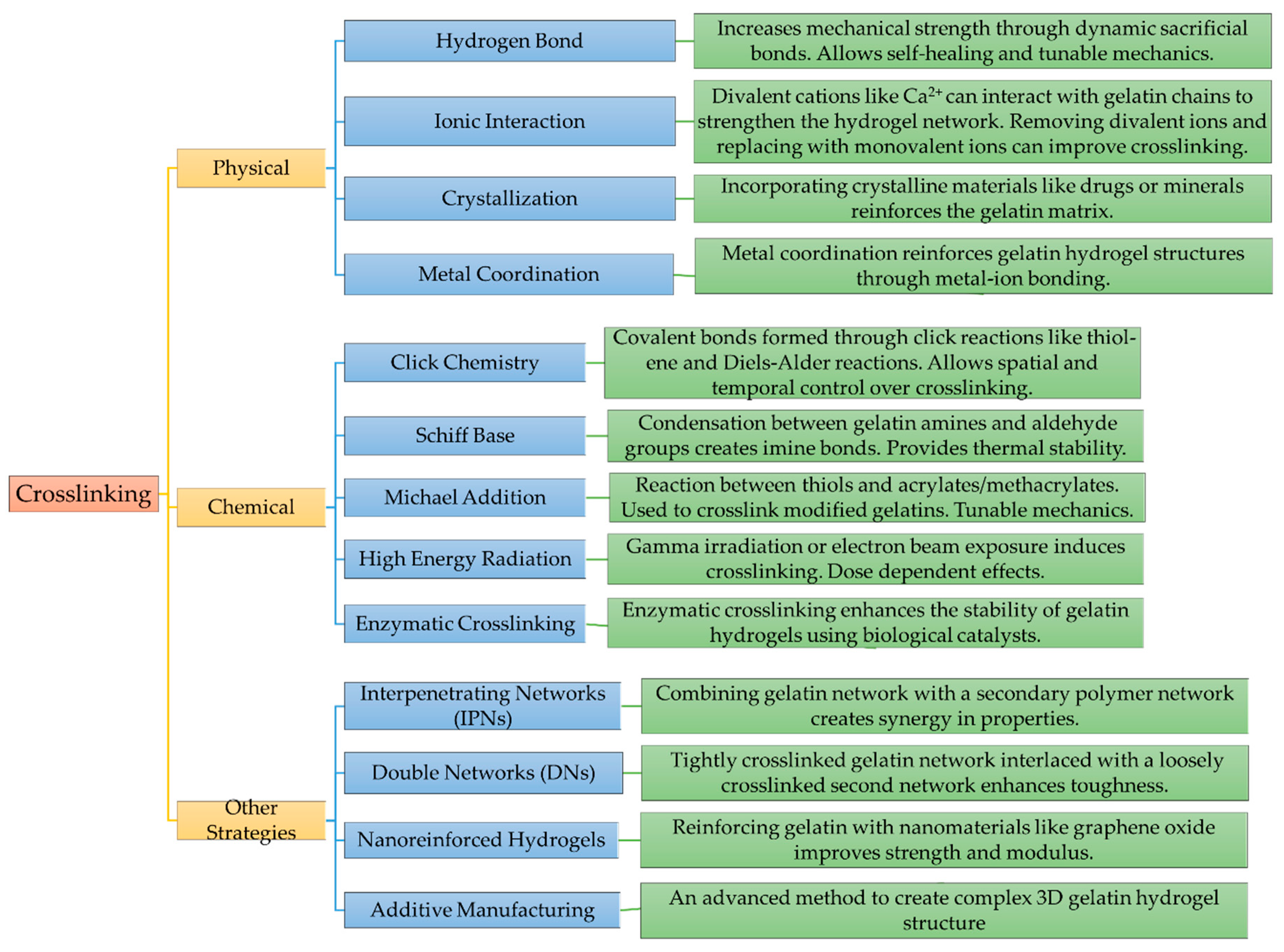
Cross-linking of resilient polymeric networks is a common approach in the creation of numerous hydrogels. Physical and chemical crosslinks are two of the most fundamental crosslinking methods [43,44,45]. While physical cross-linking encompasses mechanisms such as ionic interactions, crystallization, amphiphilic copolymer associations, hydrogen bonds, and protein interactions, chemical cross-linking involves processes like radical polymerizations, chemical reaction of complementary groups, high-energy irradiation-induced cross-linking, and enzymatic cross-linking (Figure 2).
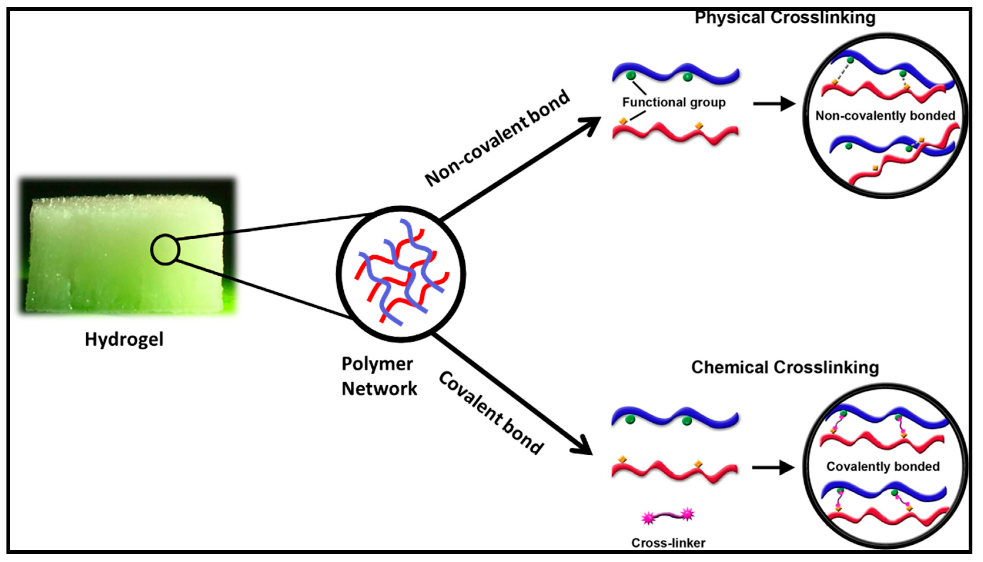
Physically cross-linked hydrogels are typically formed through reversible intermolecular interactions, characterized by non-covalent bonding [46]. The lack of chemical crosslinking agents is the primary advantage of a physical crosslink in terms of biomedical safety, hence reducing the risk of cytotoxicity from unreacted chemical crosslinkers [47]. Compared to physically crosslinked hydrogels, covalent bonds often occur between polymer chains in chemically crosslinked hydrogels, and most of these bonds are strong and permanent (Figure 3) [48]. The structural and mechanical integrity of gelatin can be enhanced mainly by either physical or chemical crosslinking strategies through the reactions between functional groups contained in water-soluble macromonomers [49,50]. Additionally, different crosslinking techniques like interpenetrating polymer networks, double networking, nano reinforcement, additive manufacturing, and others have seen increased utilization.

2.1. Physical crosslinking

Physically crosslinked hydrogels are a class of hydrogel materials formed without the use of potential cytotoxic chemical crosslinkers [52]. Instead, they rely on intermolecular reversible interactions [53], such as hydrogen bonding, intra or inter polymer chain entanglement (Figure 4), ionic/electrostatic interactions, crystallization, metal coordination, stereo-complex formation, hydrophobic/hydrophilic interactions, to create a network structure within the gel.

2.1.1. By Hydrogen Bond

Hydrogen bonds are crucial for the functioning and structure of living beings, as they play essential roles in stabilizing protein structure [55], providing unique solvent properties [56], facilitating enzyme-substrate interactions [57], and enabling cell-cell recognition and adhesion [58]. A hydrogen bond is formed when a hydrogen atom bonds with an electronegative element such as fluorine (F), oxygen (O), or nitrogen (N) and another electronegative atom with lone electron pairs. Gelatin, with its side chains containing amino (-NH2) and carboxyl (-COOH) groups, aids in the formation of hydrogen bonding in gelatin-based hydrogel systems [59]. Vigata et al. demonstrated that gelatin based GelMA hydrogels swelled in water, primarily relied on hydrogen bonds for water-hydrogel interactions [60]. In another study, Wang et al. synthesized three types of DNA hydrogels with differing degrees of hydrogen bonding, through the utilization of ultralong single-stranded DNA (ssDNA) produced via dual rolling circle amplification (RCA), revealing a direct correlation between hydrogen bonding degree and the mechanical strength and entrapment efficiency of the hydrogels [54]. The utilization of such attributes of hydrogen bonds in the preparation of hydrogels sheds light on the development of improved biomaterials. For instance, Zhang et al. utilized sacrificial hydrogen bond by Incorporating linear poly(methacrylic acid) (PMAA) into gelatin-based hydrogels to make them extremely stiff and tough with a Young's modulus (11 MPa) and a fracture energy (8.5 kJ m-2) similar to those of tough synthetic hydrogels, rubber, cartilage, and skin [61]. Additionally, the gel demonstrates excellent recovery and healing capabilities, along with being biocompatible and stable in physiological saline solutions, making it a highly prospective material for various load-bearing medical applications.
Besides high toughness and stiffness, dynamic hydrogen bonds also impart various functions, including self-recovery, self-healing, rebuild ability, and shape memory into hydrogel. For instance, Hui Jie Zhang et al. synthesized the gelatin-tannic acid-based hydrogels through a hot-pressing process (50 °C,) where gelatin crosslinked with tannic acid in solution via hydrogen bonding [62]. By tuning the tannic acid to gelatin ratio in the gel, it is possible to control the mechanical properties of the gelatin/tannic acid hydrogel (G/T gel). The G/T gel with optimum mechanical properties possesses high Young's modulus, fracture strain, and fracture energy of ∼60 MPa, ∼10, and ∼24 kJ m-2, respectively. Owing to the dynamic nature of hydrogen bonds, the mechanical properties of the G/T gels have strain rate dependence. When heated to 40 °C however, the G/T hydrogel self-healed within several seconds. In a similar study, Jie Wang et al. also showed that gelatin/tannic acid hydrogel possessed a rapid self-healing ability (within 0.65 s) and high self-healing efficiency (95%) [63]. In another study, Zhang et al. prepared self-healing gelatin-UPy-Fe (ureido-pyrimidinone (UPy) dimers) hydrogel with great injectable potential via dual cross-linking approaches of quadruple hydrogen bonding and ionic coordination [64]. The damaged hydrogel self-healed relatively fast within the initial 0.5 h and completely self-healed without any trail after 5 h, thanks to the irreversibility of both the hydrogen bonding and the ionic coordination.
Liu et al. presented another novel and pioneering method, utilizing multi-functional hydrogen bonds (provided by tannic acid) to improve the characteristics of gelatin-based hydrogels [65]. The researchers successfully obtained a substantial enhancement in ultimate stress in GelMA-TA hydrogels (4.6 MPa for 20% GelMA-TA and 3.2 MPa for 10% GelMA-TA), representing 3.5 and 4.3 times the respective values of pristine GelMA, accompanied by comparable trends in compressive modulus and tensile properties. In addition, GelMA-TA hydrogel incorporated with carbon nanotubes demonstrated mechanical strength, flexibility, and adhesiveness, making it a potential strain-sensor, capable of real-time strain detection and self-reconnection through hydrogen bonds. Through this research a GelMA-based double-network (DN) hydrogel is introduced to overcome the challenge of combining high stiffness, super-elasticity, enormous deformability, superior adhesion properties, and self-healing capability in one single hydrogel where Low GelMA concentrations provided high deformability, while high TA to GelMA ratios improved structural stiffness and adhesion properties.
Rather than self-healing property, hydrogen bonding also helps to bring adhesive property in gelatin-based hydrogel [66]. Hydrogel prepared with gelatin and chondroitin sulfate (CS) in the presence of borax showed excellent adhesive strength (30.64 ± 0.9 kPa) which can be optimized by tuning the gelatin concentration and degree of oxidation of the CS. Lap-shear adhesion test showed that weakening of hydrogen bond reduced the adhesive strength [66].
Another bioinspired gelatin-based sticky hydrogel is designed by George et al. which incorporates strong hydrogen bonding interactions, resulting in impressive mechanical properties, with a storage modulus (G') of 4.37 ± 0.14 kPa and a loss modulus (G") of 0.46 ± 0.08 kPa [67]. These numerical values indicate the hydrogel's ability to withstand deformation and provide sufficient support for wound healing. The presence of hydrogen bonds in the hydrogel's composition improves its adhesion properties, making it highly effective in sticking to diverse surfaces, a critical aspect for its successful use in burn wound care. This was further validated through its ability to promote efficient wound healing in rat models with second-degree burn wounds, confirming its potential for pre-clinical applications.

2.1.2. By Ionic/Electrostatic Interactions

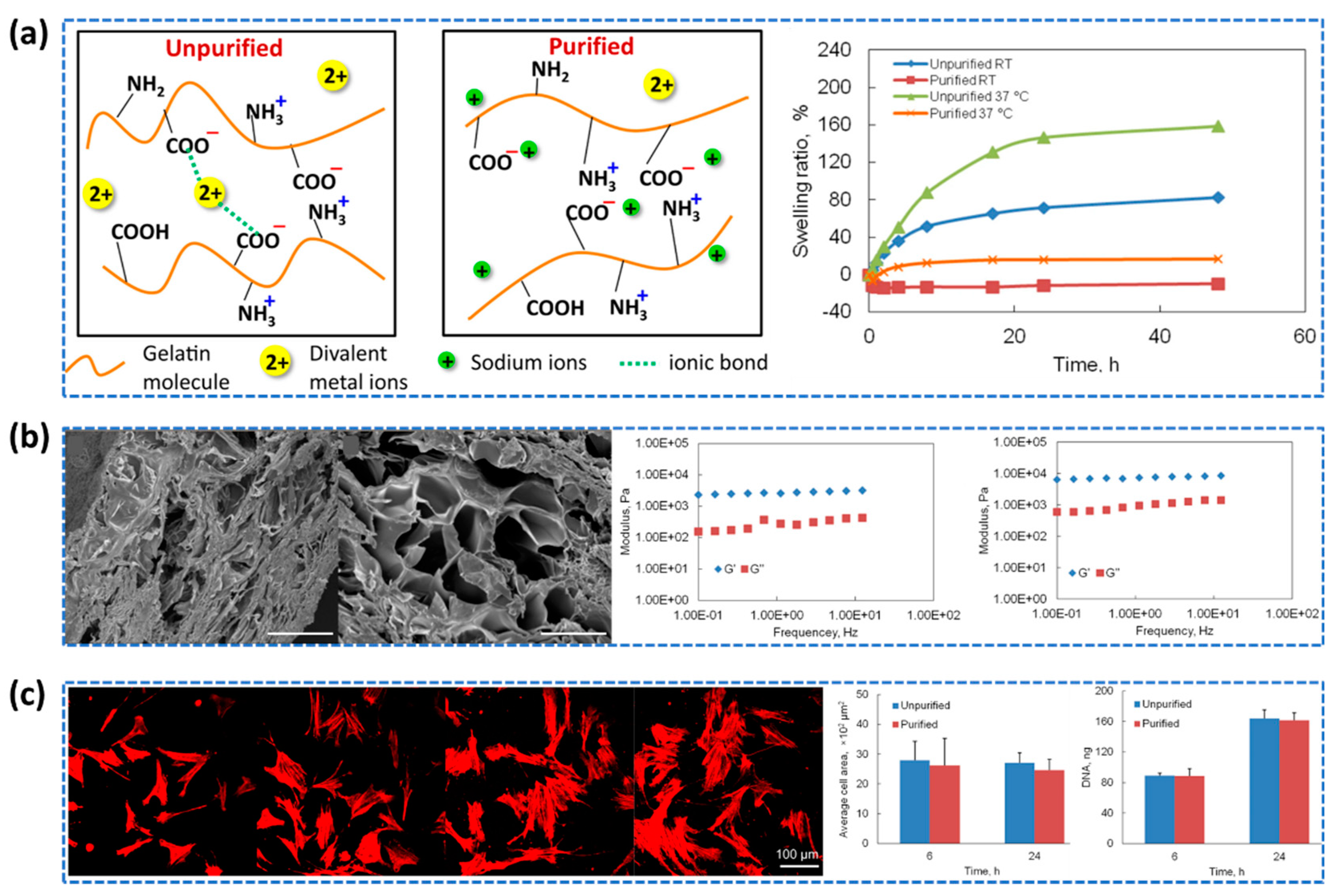
Gelatin naturally contains small quantities of divalent metal ions, including Ca2+, Fe2+ and Cu2+ (Figure 5), which are essential for the formation of the gelatin gel network [68,69,70]. The amount of divalent metal ions in gelatin can vary depending on the source of the collagen and the manufacturing process. The presence of divalent metal ions in gelatin facilitates ionic interactions with carboxylic acid groups on the gelatin molecule chains [71]. These ionic interactions play a crucial role in gel formation and gelatin aggregation [72], contributing to the overall stability and structure of the gel network. The removal of divalent metal ions alters the ionic environment, leading to changes in the electrostatic interactions and subsequent properties of the gelatin hydrogel [71].
Xing et al. aimed to investigate the stability and mechanical properties of gelatin hydrogels by removing divalent metal ions (Ca2+ and Fe2+) and replacing them with sodium ions [71]. After purifying these gelatin hydrogels with Chelex resin, they exhibited better crosslinking when treated with 1-ethyl-3-(3-dimethylaminopropyl) carbodiimide (EDC). The purification process eliminated divalent metal ions from the gelatin, subsequently replacing them with an equivalent amount of Na+ ions. Consequently, these hydrogels exhibited increased strength, improved stability, and the development of a larger-pore network (Figure 5). These enhancements render them well-suited for biomedical applications, as substantiated by successful in vitro cell culture experiments.
Ionic strength of the surrounding environment also plays an important role in designing gelatin methacryloyl (GelMA) hydrogels for specific applications. Vigata et al. showed that by manipulating the ionic strength, it is possible to control the swelling behavior and mechanical properties of the hydrogels [60]. Additionally, ionic strength influenced water retention, pH affected swelling capacity, drug loading impacted release profiles, and characteristics hydrogel network regulate water uptake in GelMA hydrogels. Increasing ionic strength in GelMA hydrogels resulted in a decrease in the water fraction (Wf) content from approximately 97% to 83.5%, while favoring an increase in the first-bound water fraction (Wnfb).
Zhang et al. synthesized a unique self-healing hydrogel having multiple types of cross-linking interactions within the gelatin hydrogel network, utilizing both hydrogen bonding and ionic coordination [73]. By utilizing multiple cross-linking mechanisms, hydrogel displayed excellent mechanical properties, with a storage modulus (G') of 1 kPa at 25 °C with no apparent shear-thinning property, and the ability to withstand large deformations up to 1000% strain, while also demonstrating an outstanding self-healing property. According to author, this is maybe due to the presence of multiple hydrogen bonds and ionic coordination interactions that allowed the gelatin chains to reassemble and reconnect after being mechanically damaged or broken. The improved mechanical strength and self-healing properties make it a promising material for creating scaffolds for tissue regeneration and drug delivery systems.

2.1.3. By Crystallization

Gelatin hydrogels are a promising material for local drug delivery. Incorporating various crystal materials in gel network can vastly improve mechanical properties such as sustain release. Using crystallized bupivacaine in making hydrogels can help create a better way to deliver drugs. These hydrogels can release drugs slowly and steadily, which means patients won't need as many injections, making it more comfortable for them [74].
The mechanical properties of gelatin hydrogels can be adjusted by incorporating calcium carbonate crystals into the gel network [75]. The size and morphology of calcite (CaCO3) crystals changed significantly with varying gelatin content in the hydrogel, forming blocks, radial aggregates, and elongated crystals with rough and curved surfaces. The study found that the presence of CaCO3 crystals within the different sized pore of the gel matrix enhanced the mechanical strength of the hydrogels. Furthermore, the size and distribution of the CaCO3 crystals within the gelatin hydrogel influenced the extent of mechanical reinforcement. Hydrogels with smaller pore sizes exhibited a higher density of CaCO3 crystals, leading to a more pronounced reinforcement effect. In contrast, hydrogels with larger pores had a lower concentration of crystals and therefore a lesser enhancement in mechanical strength.
Xiao et al. demonstrated that the presence of crystallinity in silk fibroin brings multiple advantages such as enhanced mechanical strength, structural stability, tunable properties to the gelatin and silk fibroin interpenetrating polymer network hydrogels (GelMA–SF IPN hydrogel) [76]. The compressive modulus of produced GelMA hydrogels was notably affected by the concentration of silk fibroin (SF), the treatment with MeOH, and their combined impact. MeOH treatment was observed to induce the formation of β-sheet structures, strengthening the microstructure, and leading to a substantial five-fold increase in the compressive modulus when the SF concentration was raised from 0.5 to 2 wt.%. The presence of crystallinity in silk fibroin contributes to the hydrogels' exceptional mechanical properties, particularly their high tensile strength, as the well-defined crystalline regions enhance overall mechanical integrity and stability, making them suitable for load-bearing applications.

2.1.4. By Metal Coordination

Metal coordination is another way to improve the weak mechanical properties and thermostability of gelatin. Zheng et al. combined gelatin with zirconium ions in a controlled way [77]. This caused the gelatin molecules to connect with the zirconium ions and create a sort of framework inside the hydrogel. This made the hydrogel stronger by making the molecules stick together better, which increased its strength and toughness without compromising the biocompatibility. Upon immersion in a 0.06 M Zr4+ solution, the hydrogel demonstrated significantly improved mechanical properties, with elastic modulus, compressive modulus, and compressive strength measuring approximately 400 kPa, 1192 kPa, and 476 kPa, respectively, representing a remarkable enhancement of approximately 100-fold, 11-fold, and 5-fold compared to pure gelatin.
Ge et al. developed a gelatin-based hydrogel by coordinating oxidized tannic acid (TA) (oxidized by using sodium periodate, NaIO4) and ferric ions, which led to the formation of covalent cross-links and intermolecular hydrogen bonds, consequently enhancing its mechanical properties [78]. It is found that different molar ratios of NaIO4 to TA significantly affected the cross-link density of developed hydrogels, with maximum compressive stress values reaching 12.1 kPa, demonstrating improved mechanical stiffness with increasing concentrations of oxidized TA.
Yi et al. introduced a multifunctional hydrogel by the modification of gelatin methacrylate (GelMA) hydrogels with histidine and Zn2+ ion (GelMA-His-Zn(II)) that combines metal coordination mechanisms with covalent bonds [79]. The incorporation of metal coordination bonds and covalent crosslinking enables the formation of a dual network structure in the hydrogels, offering tunable mechanical properties for tailoring to specific application needs. The GelMA-His-Zn(II) hydrogels exhibited significantly higher compression moduli compared to GelMA-Zn(II) hydrogels (without histidine), with GelMA-His-Zn(II)-1, GelMA-His-Zn(II)-2, and GelMA-His-Zn(II)-3 hydrogels having approximately 3.4 fold, 3.3 fold, and 5.8 fold greater moduli than GelMA-Zn(II)-1, GelMA-Zn(II)-2, and GelMA-Zn(II)-3 hydrogels, respectively.
Wang et al. also developed a gelatin hydrogel with exceptional mechanical strength through the self-assembly process assisted by metal ions and hydrogen [80] . The introduction of Fe3+ metal ions significantly improved the mechanical strength of gelatin hydrogels. It is also observed that trivalent ions show stronger coordination with carboxyl groups in gelatin compared to bivalent ions. Particularly, hydrogels treated with ferric ions demonstrated the highest compression strength of 11.81 MPa, which was 295 times higher than that of pure gelatin hydrogels soaked in deionized water. The robustness of the hydrogels depended on the interaction strength between metal ions and carboxyl groups present in gelatin. The bonding capabilities were significantly influenced by the arrangement of outer electronic structures of the metal ions, ultimately affecting the mechanical strength of the hydrogels.
Jing et al. demonstrated another similar utilization of metal coordination with Fe3+ ions in the synthesis of poly(acrylic acid)–Fe3+/gelatin/poly(vinyl alcohol) (PAA-Fe3+/Gelatin/PVA) triple-network supramolecular hydrogels, resulting in hydrogels possessing remarkable toughness, strength, and self-healing properties [81]. By adjusting the compositions, hydrogels with customizable mechanical properties are produced. Among them the PAA-Fe3+/Gelatin5%/PVA10% triple-network hydrogel demonstrated a significantly higher tensile strength (186.1 kPa) compared to single-network hydrogels (62.5 kPa) and double-network hydrogels PAA-Fe3+/Gelatin5% (103.5 kPa) and PAA-Fe3+/PVA10% (86.8 kPa). Additionally, the PAA-Fe3+/Gelatin/PVA triple-network hydrogel with 3% gelatin reached a tensile strength of 194.5 kPa and compressive strength of 16.4 MPa, while the one with 15% PVA showed a tensile strength of 208.4 kPa and compressive strength of 13.9 MPa respectively. The concentration of Fe3+ in the PAA-Fe3+/Gelatin3%/PVA15% triple-network hydrogel influenced the mechanical properties, with the hydrogel containing 0.20 mmol Fe3+ exhibiting a tensile strength of 239.6 kPa and compressive strength of 16.7 MPa.

2.1.5. By Stereo Complex Formation

The combination of different stereo complexes and gelatin in a hybrid hydrogel has considerable promise for a wide range of biomedical applications. It offers a distinctive amalgamation of mechanical robustness, biological functionality, and compatibility with living cells. Wang et al. developed a mechanically strong hybrid hydrogel scaffold composed of stereocomplex PDLA-PEG-PDLA and PLLA-PEG-PLLA and gelatin/nano hydroxyapitite (nHA), demonstrating its potential for bone regeneration by stimulating tissue growth [82]. The scaffold's structure consisted of an inner core formed by a stereocomplex of PDLA and PLLA, while the outer layer was composed of gelatin and nHA. The hybrid scaffold consisting of PDLA/PLLA/Gel/nHA/Gen exhibited rapid bone formation in defects, improved tissue integration, and outstanding biocompatibility in contrast to both the empty scaffold and the Gel/nHA/Gen scaffold groups. Importantly, these benefits were achieved without causing tissue necrosis or inflammation.

2.1.6. Supramolecular Hydrogel

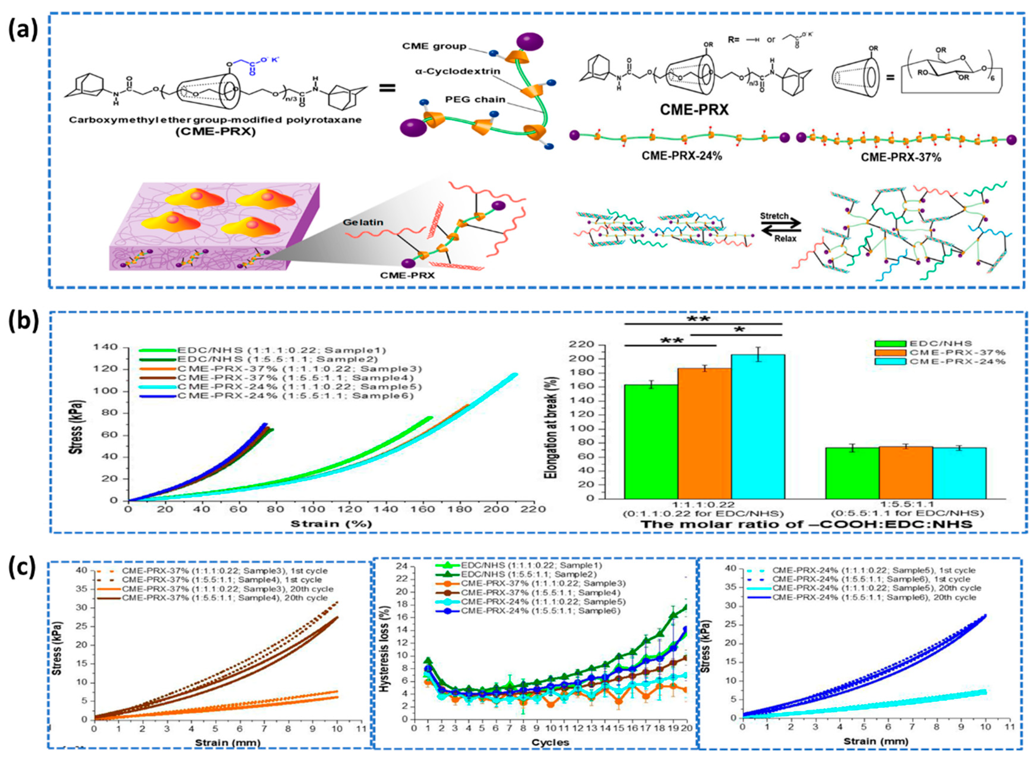
Supramolecular hydrogels, a solid three-dimensional network of hydrophilic molecules with reversible noncovalent bonds, such as hydrogen bond, hydrophobic interaction, and cation–π and π–π interactions, are anticipated to exhibit self-healing properties [83], stimuli responsiveness [84], and superior mechanical properties compared to conventional hydrogels [85]. Due to their adaptable synthesis approaches, adjustable characteristics, and biocompatibility, supramolecular hydrogels have garnered significant appeal in the fields of regenerative medicine, drug delivery, tissue engineering, and biosensing [86]. In contrast to typical hydrogels, where they only serve as weak links, the ionic bonds in supramolecular hydrogel serve as both strong and weak bonds where the strong bonds form a primary network, and the weak bonds make up a sacrificial network [87,88,89]. The enhancement of gelatin hydrogel mechanical characteristics can be achieved by incorporating supramolecular cross-linkers that possess the ability to move within the structure, resulting in an overall improvement in the hydrogel's strength and flexibility (Figure 6) [90].
The mechanism of supramolecular hydrogel mainly depends on hydrogen bonds [91,92], hydrophobic interactions [93], ionic interactions [94], metal ion co-ordination [95] and inclusion complexes or guest-host interactions [96]. In supramolecular hydrogels, intramolecular interactions between two polyelectrolytes with opposing charges may be multivalent or several non-covalent interactions may also be formed which can greatly enhance the mechanical characteristics of the hydrogel [97]. This kind of hydrogels is often produced by random co-polymerization, resulting in a variety of ionic bonds of variable strengths [98].
In defining the physiochemical characteristics and macroscopic behaviors of the resultant supramolecular hydrogel materials, the thermodynamic and molecular dynamic parameters of the supramolecular cross-linkages are critical. In particular, the equilibrium constant has a direct influence on the cross-linking density of supramolecular hydrogels, while the kinetics has a direct influence on the dynamic character of the cross-linkages between polymer chains [97].
Shin et al. successfully synthesized gallol-derived, hyaluronic acid and gelatin-based supramolecular bioinks that exhibit impressive mechanical properties, including temporal shear-thinning during printing and rapid stabilization after printing [99]. The mechanical tests unveiled a substantial difference in adhesive strength between the gallol-containing hydrogels (18.7 ± 2.4 kPa) and the non-gallol mixtures (12.3 ± 0.4 kPa). Additionally, the storage modulus (G') of the gallol ECM hydrogels was notably higher at 121.9 ± 7.8 Pa, in contrast to the non-gallol mixtures with a G' value of 5 Pa.
Another High-strength supramolecular hydrogels, incorporating cellulose and gelatin, renowned for their remarkable pH-responsive attributes, were successfully synthesized through a streamlined one-pot tandem procedure [100]. The research indicates that manipulating the amount of gelatin provides a method for customizing the mechanical characteristics of these hydrogels. Utilizing the depolymerization of the supramolecular structure of cellulose and gelatin within the ethylene diamine/potassium thiocyanate (EDA/KSCN) system, these composite hydrogels were intricately engineered. This process entailed the amalgamation of these two natural polymers via physical cross-linking, which was subsequently followed by a series of cyclic freezing and thawing treatments. The exceptional toughness exhibited by supramolecular hydrogels, surpassing that of both hydrogel with cellulose but gelatin (CH) and gelatin hydrogels, is underpinned by the intensified physical crosslinking mechanism enabled by the freezing-thawing process which enhanced the formation of hydrogen bonds. This phenomenon culminates in a substantial increase in the compressive elastic modulus within supramolecular hydrogels (CH-G-2.0), boasting an augmentation of nearly 1.9 times compared to CH and an impressive 9.6 times in relation to the gelatin hydrogel.

2.2. Chemical Crosslinking

In hydrogels, chemical cross-linking involves the creation of covalent bonds, which usually connect polymer chains. These covalent bonds are significantly stronger and more enduring in comparison to the connections observed in physically cross-linked hydrogels. A variety of crosslinking approaches have been described, including but not limited to the Diels–Alder Click reaction, the formation of Schiff bases, Michael-type additions, exposure to high-energy radiation, and enzymatically induced crosslinking [101].
  • Necessity of crosslinking in gelatin-based scaffolds
Biopolymers are vulnerable to water dissolution which restricts their widespread application, without overcoming this limitation, in tissue engineering [102]. Gelatin, being a biopolymer, is not the exception. Crosslinking is a straightforward strategy to improve the water resistance of the gelatin-based biomaterials [102]. Numerous crosslinkers and crosslinking methods are being employed depending on the intended outcome [102,103,104]. Crosslinkers bridge polymeric chains leading to interconnected 3D network which imparts improved structural stability and mechanical integrity. However, it reduces polymeric chain mobility and biodegradability, functional groups availability and increases the chance of potential cytotoxicity. Keeping all these in mind, finding a suitable crosslinker and crosslinking technique is the utmost important for gelatin based scaffolding materials.
  • Biocompatibility of crosslinkers
Cross-linking agents or crosslinkers are the chemical substances with two or more reactive ends that are capable of chemically attaching to specific functional groups on polymer chains. By forming chemical bonds with myriad polymer chains, they improve the physical properties of the hydrogel polymers. In addition, crosslinkers serve as the primary contributors to the favorable mechanical properties exhibited by hydrogel polymers. While crosslinkers are essential for enhancing the mechanical properties of hydrogels, it is essential to acknowledge that they may entail potential toxicity to biological systems [103]. For instance, glutaraldehyde, known for its potent cross-linking capabilities, can be detrimental to living organisms due to its hazardous properties [105]. The hazardous characteristics arise from unreacted residues of the crosslinking agent [106]. As a multidisciplinary field, tissue engineering prioritized biocompatibility at first place for usage of hydrogel like materials. The biocompatibility of chemically crosslinked gelatin hydrogel for ophthalmic applications has been assessed in other studies [107]. Researchers are working on non-toxic, biocompatible crosslinkers development so that hydrogel having high mechanical properties can be used in living system [108,109]. For example, Shi et al. synthesized poly(amidoamine) crosslinker with improved cell adhesion and variable stiffness [110]. Using poly(ethylene glycol) diglycidyl ether (PEG-DE) as crosslinker, Hao et al. has an interesting evaluation in vitro blood compatibility of an artificial blood vessel made of silk fibroin [111].

2.2.1. Crosslinking by Click Chemistry

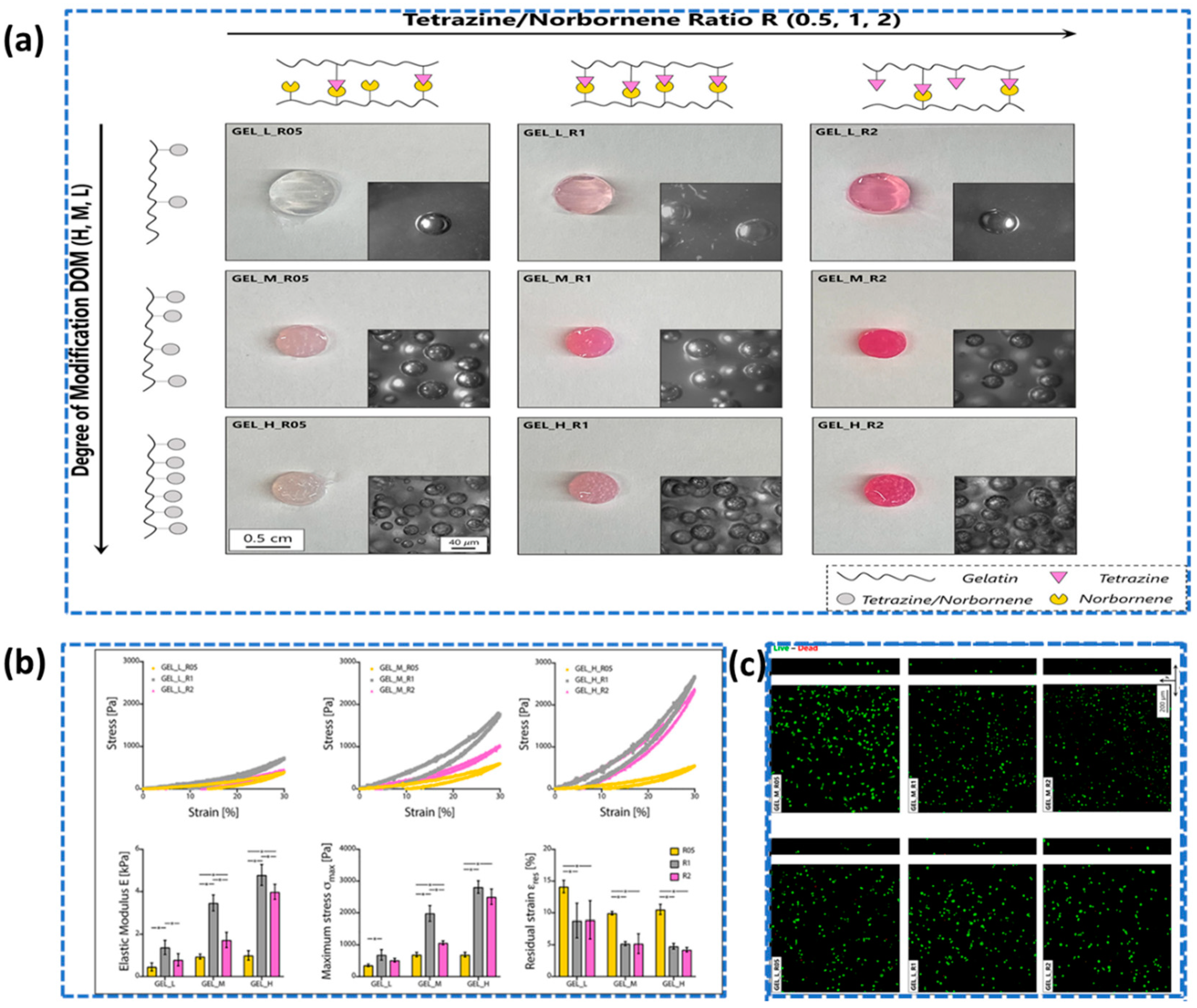
In the context of chemical synthesis, click chemistry emerges as a potent and indispensable technique. It adeptly facilitates a diverse spectrum of chemical reactions, demonstrating its proficiency in constructing intricate molecular architectures [112]. Following the introduction of click chemistry, it has gained significant attention as a powerful methodology in the field of mechanically robust hydrogel synthesis, generating considerable interest in its application [113,114,115,116]. For instance, Contessi Negrini et al. described a prospective technique for enhancing the mechanical robustness of gelatin hydrogels via the utilization of bioorthogonal click chemistry and a cross-linking technique involving tetrazine (Tz) and norbornene (Nb), enabling the control of characteristics by adjusting the modification degree (DOM) and Tz/Nb ratio (R) [117]. Hydrogels with elevated DOM demonstrated swifter cross-linking and enhanced stability. The mechanical attributes were customized by changing DOM and R, yielding elastic moduli spanning 0.5 kPa to 5 kPa. This indicates the ability to manipulate the mechanical strength of gelatin hydrogels by altering the concentration of click reagents, resulting in greater cross-linking density, improved mechanical properties, and the possibility for spatial cross-linking control. In addition, the researchers successfully introduce adhesive properties to gelatin hydrogels by incorporating adhesive molecules, enabling strong adhesion to various substrates, including biological tissues, which holds significant relevance for tissue engineering applications (Figure 7).
Another investigation demonstrated the capacity of "click" crosslinked gelatin hydrogels to serve as a robust platform for cell culture, highlighting their suitability for various tissue engineering applications [118]. This study introduces a method where gelatin is modified with norbornene, crosslinked with a polyethylene glycol (PEG) linker using nitrile oxide-norbornene click reaction. Similar to Contessi Negrini et al.’s study, elevated concentrations of the click reagents result in heightened cross-linking density, thereby augmenting the mechanical properties of the hydrogels. Consequently, this enables the researchers to fabricate hydrogels with tailored mechanical strength suitable for specific applications.
Daniele et al. also described the implementation of click chemistry to develop intricately designed scaffolds with precise control over their microarchitecture [119]. Here, gelatin methacrylamide (GelMA) polymerized inside a poly(ethylene glycol) (PEG) framework resulted in development of bio/synthetic interpenetrating network (BioSINx), to generate a mechanically strong network capable of supporting both internal cell encapsulation and surface cell adhesion. By combining these two materials via thiol click chemistry, the researchers achieved interpenetrating networks within the hydrogel, where thiol groups on both GelMA and PEG molecules readily undergo a click reaction, forming covalent bonds that crosslink the polymers and create a unified structure. The study compared different hydrogel networks, demonstrating that BioSINx displayed a broad tunable range of elastic modulus (10.8 to 327.7 kPa) and a substantial increase (500%) in upper limit compared to BioSINp, PEG-co-GelMA, PEGTYC, and GelMA hydrogels. The enhanced mechanical strength of BioSINx was achieved through a multi-mode crosslinking approach, leading to a more uniform and interconnected network, while maintaining favorable cytocompatibility. In addition, the study elegantly underscored how the hydrogels' mechanical attributes were intricately shaped by composition, functional group ratio, and network ideality, with the incorporation of tetra-alkyne PEG within BioSINx notably augmenting both covalent and physical crosslinking, ultimately yielding enhanced mechanical robustness, a synergy that transcended mere additive effects.
Xu & Bratlie discussed the adjustment of mechanical characteristics of hydrogels produced by click reactions, including diels-Alder reactions, thiol-ene reactions [120]. However, information about azide and alkyne cycloaddition (AAC) click reaction for similar properties cannot be found in the study.

2.2.2. Crosslinked by Schiff Base Reaction

Schiff base reactions can be utilized in hydrogel systems to introduce functional groups, enhance crosslinking, or promote specific chemical reactions within the hydrogel matrix [121]. A Schiff base is formed through the condensation of an aldehyde or ketone with a primary amine or hydrazine, resulting in the formation of an imine or hydrazone linkage, respectively. For example, the reaction between the amine group present in gelatin and the aldehyde groups of succinoglycan dialdehyde (SGDA) [122]. This reaction leads to the formation of a Schiff base, specifically an imine bond, which exhibits reversible characteristics and significantly impacts the stability of the resulting hydrogel. SGDA-reinforced gelatin hydrogels (SGDA/Gels) exhibited an impressive 11-fold boost in compressive stress relative to PG hydrogels under identical deformation strain, coupled with a notable 1040% increase in storage modulus (G′). The chemical crosslinking introduced by SGDA additionally bolstered the thermal resilience of SGDA/Gels, guaranteeing their structural integrity at elevated temperatures of 60°C, positioning them as compelling contenders for long-term use in controlled drug delivery systems and as three-dimensional scaffolds in tissue engineering.
Another high mechanical strength gelatin composite hydrogels were prepared by incorporating cellulose nanofibrils with a unique beads-on-a-string morphology and utilizing a Schiff-base reaction for covalent bonding [123]. This composite hydrogel is much stronger than pure gelatin hydrogel. It can withstand a compressive stress of 3.398 MPa, which is 59 times as much as pure gelatin hydrogel. The Schiff-base reaction also employed to create mechanically enhanced nanocomposite gelatin-based hydrogels [124]. The study examined the viscoelastic and mechanical properties of nanocomposite hydrogels, revealing that the introduction of both nanohydroxyapatite (nHA) and bisphosphonate-modified hydroxyapatite (nHABP) nanoparticles reinforced the network structure, with stronger enhancements observed in nHABP-containing hydrogels, leading to improved mechanical properties and denser structures. Additionally, nanocomposite hydrogels with reversible bonds exhibited consistent mechanical properties across multiple loading cycles, and the introduction of nanoparticles increased hysteresis, indicating enhanced toughness, with nanoparticles containing nHABP particles showing higher energy dissipation than those with nHA particles.

2.2.3. Michael Addition Crosslinking

The Michael-type addition reaction is a versatile and concerted chemical process involving the nucleophilic attack of a nucleophile on an α,β-unsaturated carbonyl compound's electron-deficient double bond, widely utilized for building complex molecules, materials, and bioconjugates [125]. To illustrate, thiolate-modified chitosan (TCS) and methacrylate gelatin (GelMA) were successfully combined through the thiol-Michael addition reaction which is widely recognized and referred to as a "click" reaction [112], resulting in the fabrication of hydrogels with enhanced properties [126]. The study involved creating a composite hydrogel by crosslinking thiolate-modified chitosan (TCS) and methacrylate gelatin (GelMA), and tensile testing demonstrated that hydrogels with increasing TCS content exhibited rising tensile strength, with the 50 wt% TCS hydrogels achieving a tensile strength of 1.2 MPa and a Young's modulus of 10 kPa, comparable to natural skin's mechanical properties. Additionally, these hydrogels displayed remarkable extensibility, making them promising candidates for wound dressings capable of maintaining a moist environment while withstanding mechanical stresses during bodily movement.
By utilizing thiol Michael addition reactions, a network structure is achieved when glycidylmethacrylated gelatin (GMA-gelatin) is crosslinked with either hexane 1,6-dithiol or nonane 1,9-dithiol, thereby enabling the modulation of material characteristics in this gelatin-based hydrogel [127]. Mechanical testing was conducted to evaluate the tensile strength, compressive modulus, and elasticity of the hydrogels, revealing their favorable mechanical properties that indicate their potential suitability as scaffolds for tissue engineering and other applications where mechanical stability is crucial. The discovery underscores the remarkable effectiveness of the thiol Michael addition reaction in seamlessly crosslinking suitably functionalized biopolymers with concise hydrophobic crosslinkers, bestowing meticulous command over the resultant network properties and promising a paradigm shift for crafting an array of biopolymer-based hydrogels.
The synthesis of a hybrid hydrogel, combining graphene oxide reinforcement with alginate and gelatin, was also achieved through the utilization of Schiff-base bond formation and thiol-Michael addition reactions [128]. In this study, Ding et al. used aldehyde methylene sodium alginate (AMSA), amino gelatin (aminoG), and graphene oxide modified with dithiothreitol (DGO) elements and were employed these elements to create DN-hydrogels, namely AMSA/aminoG (AG) and DGO/AMSA/aminoG (GSG), using different proportions. The relationship between compressive strengths and porosity was opposite for AG and GSG hydrogels, while both hydrogels showed positive cell viability and proliferation, GSG hydrogels exhibited greater osteogenic potential compared to AG hydrogels, underscoring the promising role of novel GSG hydrogels in tissue engineering. These hydrogels serve as scaffolds that offer improved mechanical support, promote cell proliferation, and enhance bone regeneration.

2.2.4. Crosslinking by Radiation (Photocrosslinking)

High energy electron irradiation offers a reagent-free and controlled method for photo-crosslinking of gelatin, enabling the tuning of material properties for tissue engineering applications. Photocrosslinking of gelatin-based hydrogel offers several advantages including temporal and spatial control of crosslinking process, precisely light dosage regulation by switching the light on and off to achieve functional tunability, cellular process stimulation in vivo by light exposure, etc [129]. Mechanical property of photocrosslinkable hydrogels is directly related to the degree of photo-crosslinking – More photo-crosslinking imparts higher mechanical properties. Again, the degree of photo-crosslinking depends on many factors including photosensitive polymer concentration, photoinitiator efficiency, photoinitiator concentration, light exposure duration, and light intensity [130]. In the field of tissue engineering, selecting a proper photoinitiator is utmost important in order to achieve optimum functionality with minimal cytotoxicity [129]. A gelatin-based hydrogel can be synthesized with radical or cationic photoinitiators, both of which have their benefits and drawbacks. However, radical photoinitiator finds its wider application in biomedical field due to its better biocompatibility in contrast to cationic photoinitiator which may produce protonic acids that might be harmful to cells [129]. Among the most widely used UV light-sensitive photoinitiators, Irgacure 2959 (1-[4-(2-hydroxyethoxy)-phenyl]- 2-hydroxy-2-methyl-1-propane-1-one) is used for tissue engineering, since it has low cytotoxicity, moderate water solubility, and minimal immunogenicity [131]. Of note, photo-crosslinking gelatin hydrogel using visible light (400-800 nm) is more favorable and safer than UV light (200-400 nm) irradiation in the field of tissue engineering because the overexposure of UV irradiation causes cell death [132,133].
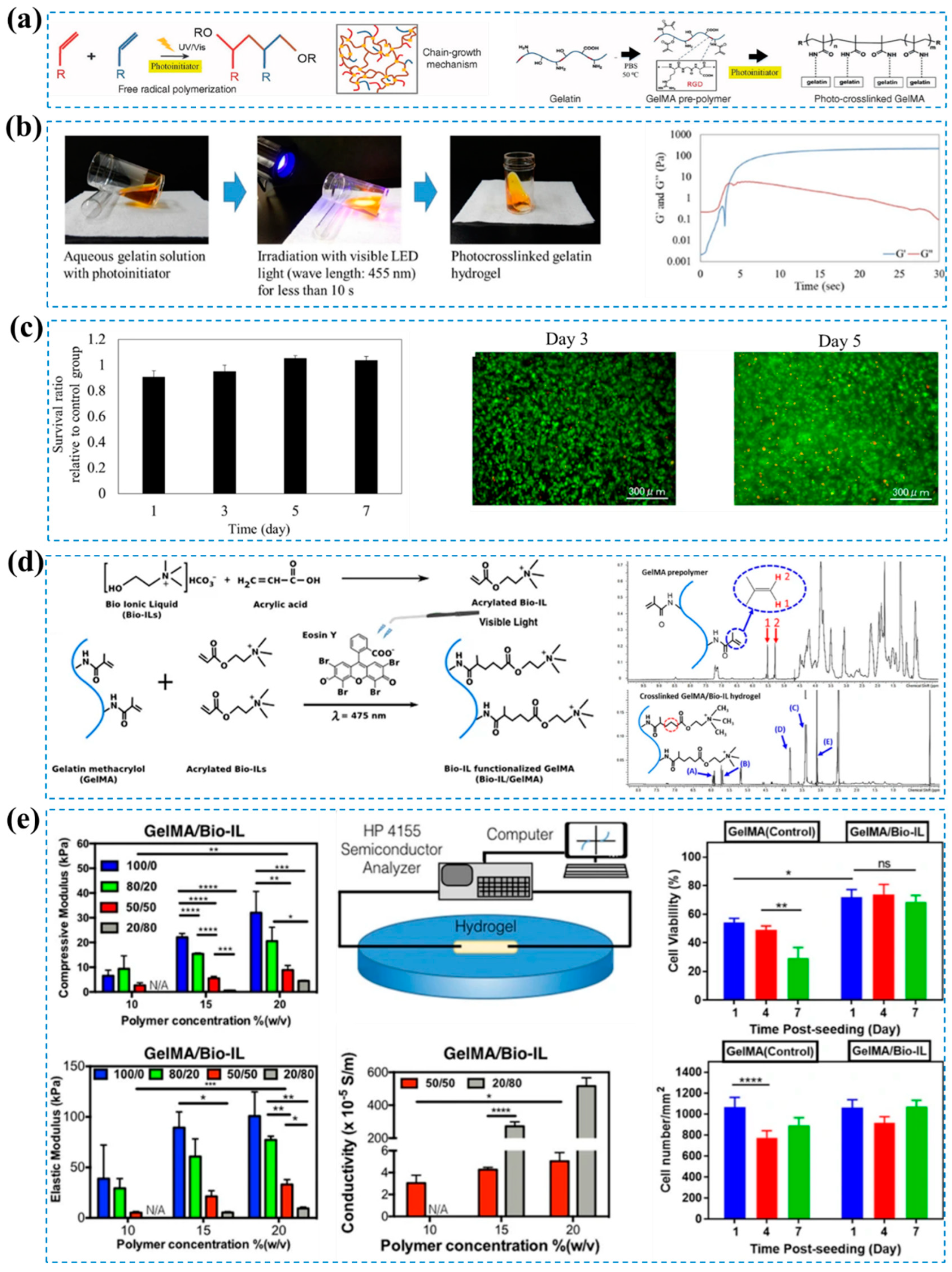
Because of gelatin's functional versatility, it can be easily modified with different polymerizable motifs, such as methacrylamide, thiol, and norbornene to enable photopolymerization using a suitable photoinitiator in the presence of visible light or UV-light. GelMA, a photocrosslinkable functionalized gelatin with methacrylamide groups via chemical reaction, can be photocrosslinked in the presence of Irgacure 2959 photoinitiator upon UV light exposure [49,134]. Mechanical properties of GelMA hydrogel can be tuned by varying the degree of methacrylation (approximately 20%, 54% and 81%) and gel percentage (5%, 10% and 15%). For instance, hydrogel prepared with 15% GelMA showed around three-fold higher compressive modulus compared to hydrogel with 5% GelMA at a specific degree of methacrylation [49]. Again, at constant gel percentage (15% w/v), GelMA hydrogel exhibited around three-times higher compressive modulus when the degree of methacrylation increased from 20% to 81%. Interestingly, all GelMA hydrogels showed a reasonable level of cell viability though it decreased with increasing gel percentage [49]. As a result of varying the hydrogel concentration, Xin Zhao et al. was also able to modify the mechanical and degradation properties of the GelMA hydrogels, with elastic and compressive moduli ranging from a few hundred kPa to a few thousand kPa and degradation times varying from a few days to several months [135]. Moreover, GelMA hydrogel can be tuned mechanically by adjusting the photoinitiator concentration and light exposure duration [2,136]. However, Irgacure 2959 is prone to DNA and tissue damage due to its UV-excitation requirement, which can potentially damage cellular DNA and tissues [137]. This has led to the investigation of several visible light photoinitiating systems for the manufacture of GelMA hydrogels, including camphorquinone, fluorescein, rose bengal, riboflavin, and lithium phenyl-2,4,6-trimethylbenzoylphosphinate (LAP) [138,139,140,141,142,143,144]. For example, Tim B. F. Woodfield and his team prepared cross-linked GelMA hydrogels using ruthenium (Ru)/ sodium persulfate (SPS) visible light photo-initiating system and showed that the developed hydrogel experienced comparable physico-mechanical and biological properties as GelMA hydrogel photopolymerized using Irgacure 2959 and LAP [138]. On the other hand, Toshihiro Kushibik et al. synthesized photocrosslinked gelatin hydrogel in presence of fibroblast growth factor (bFGF) using visible light photoinitiator system, pentamethylcyclopentadienyl triphenylphosphine ruthenium chloride and sodium persulfate, for wound healing purpose [133]. Based on their results, bFGF-containing photocrosslinked gelatin hydrogels showed sustained release of bFGF, were biocompatible, and wet tissue adhesive. Considering their ease of use, high efficacy, and sustainability, the developed hydrogel in this study may be a promising biomaterial for clinical wound healing. Nasim Annabi and her group developed conductive choline-based bio-ionic liquid (Bio-IL) conjugated GelMA hydrogel (GelMA/Bio-IL) which showed tunable compressive modulus in the range of 0.60 ± 0.20 kPa to 32.07 ± 8.61 kPa [145]. The tensile and compressive mechanical properties of Bio-IL functionalized GelMA hydrogel could be modulated by varying the final polymer concentration as well as the ratio of polymer to Bio-IL. Importantly, their developed hydrogel system showed high electrical conductivity without using any additional conductive components, and they exhibited excellent biocompatibility both in in vitro and in vivo [145]. Their engineered hydrogels supported primary cardiomyocyte growth and function in both two- and three-dimensional cultures (Figure 8).
Besides, commonly available GelMA, Lana Van Damme et al. developed thiol-norbornene crosslinkable gelatin-based hydrogel (GelNB/SH) to be used for cell encapsulation purposes [146]. The mechanical properties of GelNB/SH hydrogel could be tuned not only by varying the degree of substitution (GelNB DS55%, GelSH DS75%) but also by varying the crosslinking chemistry (step-growth vs chain-growth/single vs dual crosslinking). Comparatively to benchmark GelMA, equimolar GelNB DS55% + GelSH DS75% resulted in comparable biocompatibility and superior differentiation of the encapsulated cells into the adipogenic lineage [146]. As the primary goal of tissue engineering is to heal damaged tissue using cells (such as stem cells or progenitor cells) and bioactive molecules (such as growth factors, cytokines, proteins etc.), it may be necessary to photpolymerize hydrogel networks directly in the presence of cells and bioactive molecules. In such cases, it is crucial to maintain viability of entrapped cells and functionality of bioactive molecules under photocrosslinking environment.
In addition to visible light and UV irradiation, gelatin can be crosslinked using gamma irradiation. For instance, Liu et al. prepared an interpenetrating gelatin/γ-PGA hydrogels by hot-pressing pre-gelation and synergistic with gamma irradiation [147]. The process begins with the hot-pressing preassembly of gelatin and γ-polyglutamic acid (γ-PGA), which facilitates intermolecular interactions and establishes a distinct structure. Following this, the radiation crosslinking, utilizing high energy gamma radiation enhances the hydrogel's strength through the creation of covalent bonds between the polymer chains. The gelatin/γ-PGA hydrogel exhibited good biocompatibility, biodegradability, and mechanical properties. Cataldo et al. used γ-radiation to convert gelatin, a low molecular weight collagen derivative from porcine skin, into a stable durable hydrogel [27]. Here, high-energy radiation is used to crosslink gelatin hydrogels, forming a stable three-dimensional network that enhances mechanical strength and stability. The crosslinking density can be controlled by adjusting the radiation dose, allowing customization of swelling behavior and mechanical properties for various biomedical applications. Noteworthy that higher doses of radiation energy may degrade gelatin hydrogel. For instance, Wisotzki et al. examined the impact of high energy electron irradiation on gelatin hydrogel's network structure via small-angle X-ray scattering (SAXS), aiming to elucidate the changes induced by this sterilization method [148]. It was found that the network structure of gelatin hydrogels was affected by high energy electron irradiation, with lower doses leading to cross-linking and higher doses resulting in degradation. These changes were detected through alterations in scattering intensity and characteristic length scale. Therefore, the impact of high energy electron irradiation on gelatin hydrogels can vary, encompassing both cross-linking and degradation, contingent on the dose applied. Consequently, optimization of radiation doses under a specific operating condition is essential to maintain the intrinsic chemical and biological properties of gelatin. In another study, Şener Raman et al. used electron beam irradiation to enhance the mechanical strength of gelatin hydrogels where polyethylene glycol diacrylate (PEGDA) is incorporated into gelatin matrix [149]. The researchers successfully employed electron beam irradiation to crosslink PEGDA/gelatin hybrid hydrogels, resulting in the formation of a robust three-dimensional network structure. By carefully adjusting the composition of PEGDA and gelatin, the material properties of the hybrid hydrogels could be finely tailored, rendering them highly robust for applications involving cellular interactions and tissue regeneration. The storage modulus of the P21G9 (a hybrid hydrogel where PEGDA 26 wt%, Gelatin type A 4 wt%) exhibited a substantial enhancement, rising from 0.13 MPa (in pure gelatin) to 1.40 MPa, indicating an impressive elasticity improvement of 1,078%. Notably, higher doses of irradiation were found to correlate with increased crosslinking density, leading to substantial improvements in stiffness and tensile strength, all while preserving desirable water absorption and swelling characteristics in the hydrogels. However, no cell viability test was done in this work.

2.2.5. Enzymatic Crosslinking

Enzymatically crosslinked hydrogels are a class of biomaterial that is gaining traction in tissue engineering. These hydrogels are formed by the use of enzymes, which are biological catalysts that can connect polymer chains to create a three-dimensional network structure that can hold a large amount of water, giving rise to hydrogels with remarkable swelling and mechanical properties. The use of enzymes in the crosslinking process allows for precise control over the gel formation under mild conditions [150]. Horseradish peroxidase (HPR), Elastase, trans-glutaminase (TGlu), tyrosinase (Tyr) are some commonly utilized enzymes in the development and production of hydrogels Unlike other crosslinking methods, which may involve harsh chemicals or high temperatures, enzymatic crosslinking can take place at physiological temperatures and pH levels, making it particularly well-suited for applications where biocompatibility and biodegradability are main concern [151]. Therefore, enzymatically crosslinked gelatin-based hydrogel system is commonly employed in the field of tissue engineering [152,153,154,155]. Echave et al. developed microbial transglutaminase (mTG) crosslinked gelatin-based 3D scaffolds for bone tissue engineering [154]. The preparation processes include dissolving Type B gelatin in water, adding mTG solution, forming crosslinked hydrogels with varying gelatin concentrations and enzymatic activity, and finishing with ethanol treatment, PBS washing, and freeze-drying. Different gelatin concentrations led to differences in swelling capacity and stiffness in the scaffolds, and greater enzymatic activity resulted in decreased water absorption and increased stiffness. The highest Young Modulus value (41.9 ± 8.0 kPa) was observed in the formulations that included 20% gelatin and 30 U/g of mTG. The produced hydrogel demonstrated elasticity values resembling those of osteoid tissue and were found to be non-cytotoxic in all cases, suggesting their potential suitability for future in vivo bone regeneration applications.

2.3. Various Hybrid Crosslinked Hydrogels

Both physical and chemical crosslinking exhibit their strengths and weaknesses. Chemical approaches are ideal for producing a stable hydrogel with the desired mechanical characteristics, whereas physical approaches offer biocompatibility due to the absence of chemical crosslinking agents. However, physically crosslinked hydrogel possesses weak mechanical integrity. On the other hand, during the degradation process, the inclusion of aldehydes, polyepoxides, and isocyanates that form chemical interactions with gelatin molecules might release reactive and hazardous compounds [156,157,158]. Therefore, a mix of physical and chemical techniques would be preferable for creating hydrogels with balanced characteristics [104]. For instance, a double-crosslinked hydrogel, achieved through the combination of physical crosslinking involving hydrogen bonds and chemical crosslinking via imine bonds, demonstrated extraordinary mechanical robustness and outstanding resistance to degradation [159]. This unique amalgamation of crosslinking mechanisms has rendered the hydrogel exceptionally durable and stable. Table 1 presents a comprehensive compilation of mechanical property data for various gelatin hydrogels, encompassing details regarding their crosslinking methodologies, modes of testing, and the specific crosslinkers employed during their preparation.

2.3.1. Interpenetrating polymer network (IPN)

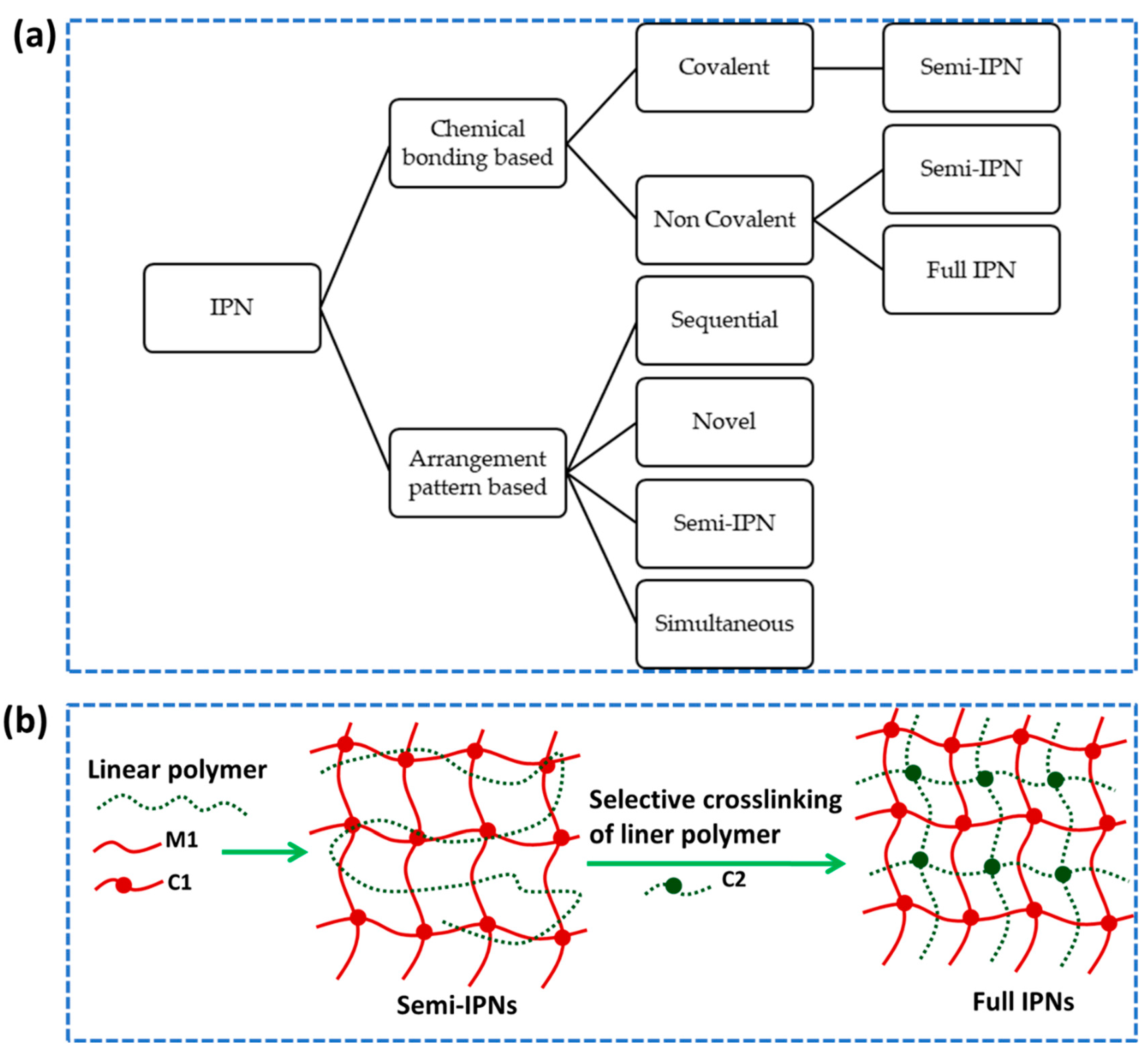
Interpenetrating polymer networks, or IPNs, are a subclass of multicomponent polymer materials that are made up of a closer combination of two polymer networks that have been individually crosslinked (Figure 9) [179]. The individual polymers within the IPN synergistically combine their advantageous properties, giving rise to novel systems that often exhibit distinct characteristics not found in any of the constituent polymers alone. An IPN is considered semi-IPN if only one of its components is crosslinked, while a full IPN has crosslinks between both of its components. Interpenetrating polymer networks are distinct from semi-interpenetrating polymer networks by the fact that the constituent linear or branched polymers can, in theory, be separated from the constituent polymer network(s) without breaking chemical interactions [180]. It is possible to adjust and customize the qualities of the produced material to fulfill particular needs by utilizing the combination and interplay of its properties [181]. Although IPNs are very similar to blends, blocks, or graft polymers, they have distinct properties [180]. For instance, IPN swells, but does not dissolve in solvents and have reduced creep and flow characteristics [182]. One significant advantage of IPNs is their ability to create dense hydrogel matrices with controllable physical properties that are stiffer and tougher, making them more efficient for drug loading compared to other hydrogels. IPNs can have diverse forms based on chemical bonding and arrangement pattern [183]. Different forms of IPN and their formation strategies are illustrated in Figure 9.
The concept of interpenetrating polymer network (IPN) systems brings together polymers possessing diverse properties, presenting an intriguing avenue to engineer materials that surpass the limitations of individual hydrogel networks. This advanced strategy, involves integrating two distinct polymer networks into a single IPN hydrogel to simultaneously achieve biocompatibility and mechanical resilience, which is especially important in tissue engineering [181]. For instance, Jain et al. synthesized gelatin based supermacroporous IPN hydrogel with a high Young's modulus at subzero temperature [160]. The IPN cryogel network comprising PAN (polyacrylonitrile) and gelatin exhibits significant mechanical robustness, with Young's modulus values spanning from 123 kPa to 819 kPa based on polymer concentration, alongside a demonstration of biocompatibility and the ability to foster cell growth within the scaffold structures. Likewise, the interpenetrating polymer network (IPN) hydrogel based on dextran and gelatin represents a promising biomaterial for vascular tissue engineering, combining enhanced mechanical strength, biocompatibility, and bioactivity to support vascular tissue regeneration and cell adhesion [184]. The hydrogel demonstrates a distinct blend of properties from synergistic dextran-gelatin polymers. Mechanical tests indicate a notable Young's modulus (75-480 kPa), offering tunable strength via polymer ratio adjustment. Another study showcased the development of mechanically robust gelatin-alginate IPN hydrogels, achieved through a novel combination of enzymatic and ionic crosslinking approaches [185]. The mechanical evaluation demonstrates impressive results, where IPN hydrogels exhibit a tunable Young modulus ranging from 60 kPa to 500 kPa through a novel combination of enzymatic and ionic crosslinking, showcasing enhanced mechanical properties with promising applications in resilient hydrogel materials.
  • Mechanism of IPN
The most widely used techniques for producing IPN composite hydrogels are a) a simultaneous approach, b) a sequential approach, and c) selective cross-linking of a linear polymer trapped within a semi-IPN structure [186]. For the formation of simultaneous interpenetrating networks (SINs), two distinct, non-interfering interactions are necessary. Consequently, chain and step polymerizations have become the polymerization techniques of choice for several applications [187,188]. Chen et al. reported on an in-situ development of simultaneous IPN hydrogel based on natural bio macromolecular building blocks that are collagen and hyaluronic acid (HA) [188]. In similar cases gelatin could be a good fit due to its exceptional biocompatibility.
In the case of IPN formation through a sequential strategy, polymer networks are made by crosslinking or polymerizing distinct polymer networks consecutively. This method typically starts with the formation of the first polymer network, followed by introducing the second set of monomers or prepolymers and initiating their crosslinking or polymerization within the structure of the initial network [189]. Between a sequential IPN and a SIN, there are a few other polymerization strategies that are worthwhile to consider. In sequential in situ prepared IPNs, both monomers are polymerized by free radical reaction, and the two monomers must have very distinct reactivities toward free radicals [187,190]. In contrast, In a semi-Interpenetrating Polymer Network (semi-IPN), selective cross-linking of an entrapped linear polymer involves targeting and linking the linear polymer chains without disrupting the intertwined polymer network structure.
  • Preparation of IPNs
All interconnected polymer networks (IPNs) consist of two distinct polymers. The exception is homo-IPNs, in which the two polymers are identical [191]. Since these polymers may be generated by any of the known techniques of polymer synthesis, certain methods are substantially more effective than others for achieving specific goals. Chain and step polymerization are the most commonly used techniques [182].
Crosslinking is crucial for fabrication of IPNs. Heating-cooling, enzymatic, and ionic methods are the major crosslinking strategies used in present days. There is another crosslinking technique also available which is called photo crosslinking. This method requires a photo-initiator to initiate the crosslinking and polymerization of the monomers to create a network, and both the photo-initiator and the monomer are often hazardous substances. Additionally, the photo-initiator can generate hazardous and undesirable byproducts, such as benzene and toluene [192].
Numerous sequential IPNs have utilized two chain polymerizations, in which monomer mix I is polymerized, and monomer mix II is swollen in, followed by the polymerization of monomer mix II and I [193,194]. Interpenetrating Network Hydrogels (INHs) represent an innovative and revolutionary approach to wound care, boasting exceptional attributes of strength and transparency, which render them highly suitable for use as external dressings. These hydrogels feature a unique dual-network structure that effectively improves their mechanical properties and overall stability, providing unparalleled support and protection during the wound healing process. The remarkable transparency of INHs allows medical practitioners to closely monitor wound healing progress, eliminating the necessity for frequent dressing changes and contributing to enhanced wound management [195]. Gelatins exceptional biocompatibility and ability to maintain a moist wound environment also hold potential in designing IPNs for faster recovery and reduced scarring.

2.3.2. Double Network Hydrogels

The double network (DN) gels consist of two interpenetrating polymer network (IPN) structures where the properties of the IPN structures such as network density, rigidity, molecular weight, and cross-linking density remain in sharp contrast [196]. For the manufacture of tough hydrogels, the DN gels are of pivotal interest due to their superior mechanical features [197]. The mechanical properties of DN gels made from numerous different polymer pairs are significantly better than those of the individual components. For instance, Wang et al. developed a DN hydrogel by the direct inclusion of a synthetic gel into a biological gel derived straight from an animal body, which has compressive and tensile strengths several to several tens of times higher than its constituent components [198]. Also, it has been shown that DN hydrogels made with radiation-induced polymerization and crosslinking are much stronger than those made with thermal polymerization and a crosslinking agent [199]. By combining a physically cross-linked gelatin network with a covalently cross-linked poly (γ-glutamic acid) (γ-PGA) network, Dou et al. successfully developed a hybrid DN hydrogel, which possesses outstanding mechanical properties and biocompatibility [169]. The created γ-PGA-GEL DN hydrogel exhibited exceptional compression performance, reaching 38 MPa, surpassing all previously reported γ-PGA-based hydrogels. Another study explored a cell-laden DN hydrogel, utilizing photocrosslinkable gelatin and gellan gum biomacromolecules, to investigate their mechanical properties and cytotoxicity [164]. The results demonstrated their potential as biocompatible materials with favorable mechanical characteristics, making them promising candidates for tissue engineering and regenerative medicine applications.
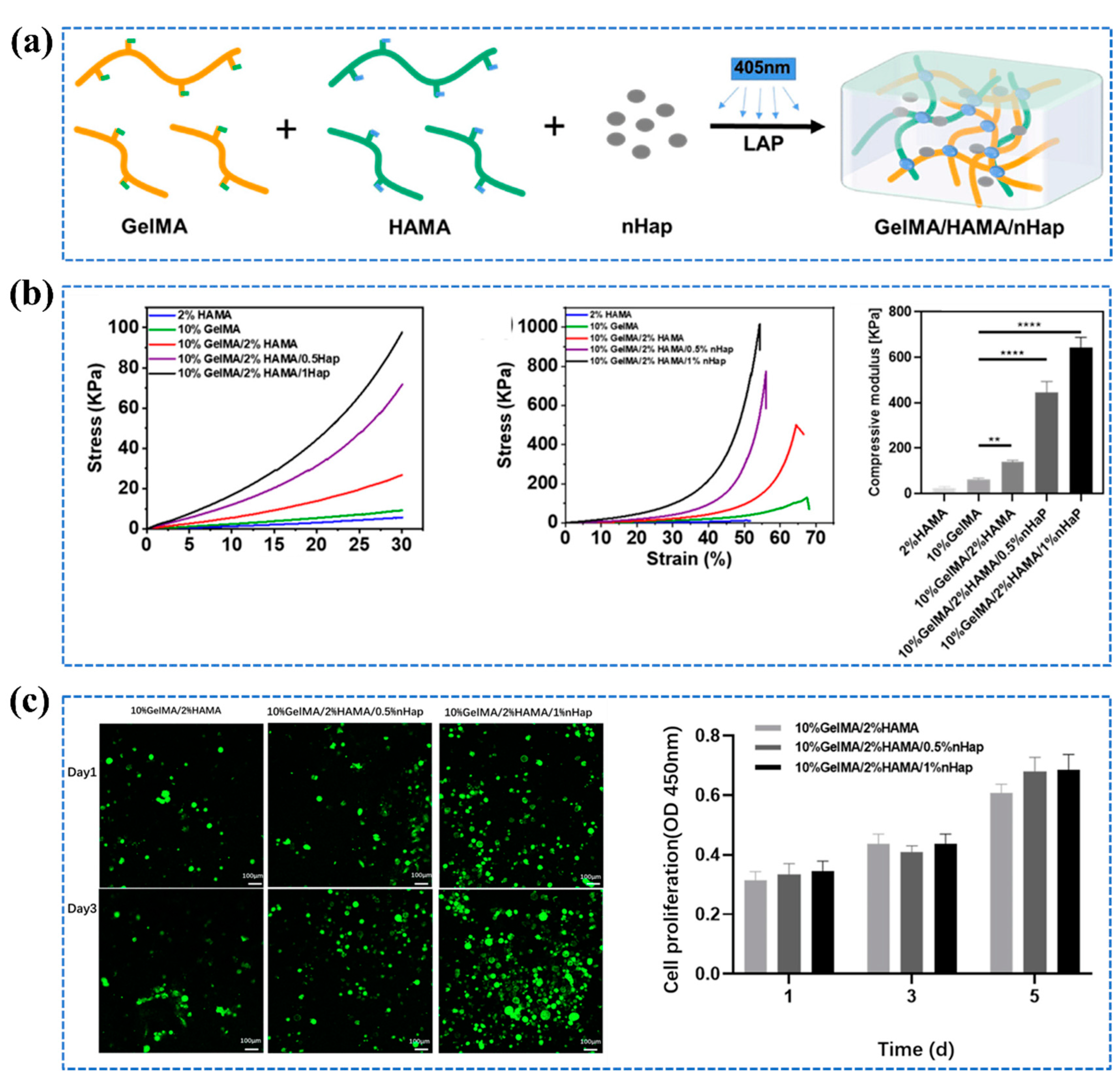
In another study, utilizing double network strategy, Zheng, et al. fabricated GelMA/HAMA/nHap composite hydrogel, composed of methylacrylylated gelatin (GelMA), methylacrylylated hyaluronic acid (HAMA), and nano-hydroxyapatite (nHap) with the purpose of addressing bone repair applications [200]. The resulted hydrogel exhibited significantly enhanced mechanical strength with a compressive elastic modulus 20 times greater than HAMA SN hydrogel, near 1 MPa compressive fracture strength, while maintaining high swelling ratio, water content (>88%). Moreover, the findings from the in vitro cytocompatibility assessment demonstrated that the composite hydrogel effectively facilitated the viability and growth of BMSCs on its surfaces, indicating strong compatibility with these cells (Figure 10).
  • Toughening Mechanism
As suggested by its name, Double network hydrogel consists of two interpenetrating polymer networks, first network of which is tightly crosslinked and the second network of which is loosely crosslinked [201]. When the molar ratio of the second network to the first network is increased, the strength of DN gel goes up [202]. However, When the second polymer was synthesized without the cross-linker, it was baffling why the DN gel became the hardest . To address this, Nakajima et al. conducted a study and made an interesting discovery. They observed that some crosslinkers reacted only on one side of the first gel during synthesis, leaving unreacted double bonds within the gel [203]. When the second network was formed within the first network, these remaining unreacted double bonds joined the co-polymerization process. As a result, the second network became chemically cross-linked with the first network. This concept is also applicable to ordinary DN gels, which possess an inter-cross-linked structure. Importantly, these gels demonstrate impressive strength even without requiring an additional crosslinker for the second network. This is attributed to the robust covalent interconnection between the two networks. It was explained further that totally independent DN gels, which lack covalent linkages between the first and second networks, cannot be toughened by the uncrosslinked second network [202].
  • Preparation Methods of DN Hydrogels
The DN hydrogels are generally manufactured by a two-step sequential free-radical polymerization method in which a neutral second polymer network of high relative molecular mass is integrated into a swelling heterogeneous polyelectrolyte first network [204]. This process can be achieved using two primary approaches: postaddition and preaddition. In the postaddition method, a pre-synthesized neutral network is immersed in a monomeric electrolyte solution. Within this solution, electrolyte monomers undergo polymerization, forming linear polyelectrolytes within the neutral hydrogels. Conversely, the preaddition approach commences with the synthesis of linear polyelectrolytes, which are subsequently mixed with neutral first-network monomers. This results in the creation of a first network comprising linear polyelectrolytes. Ultimately, the highly swollen hydrogel is placed into a second precursor solution. This step triggers a second network polymerization process, culminating in the formation of robust DN gels.
With the help of two-step photo crosslinking polymerization, Shin et al. developed mechanically strong DN hydrogels from two modified bio-macromolecules, gellan gum methacrylate (GGMA) and gelatin methacrylamide (GelMA) which are photoreactive versions of gellan gum and gelatin [164]. The cell-laden double-network hydrogel demonstrated promising mechanical properties, positioning it as a potential material for tissue engineering and regenerative medicine, while its low cytotoxicity further validates its suitability for biomedical applications involving cell encapsulation and delivery.
Nakajima et al. introduced a novel technique called molecular stent, to produce a robust DN hydrogel with two neutral polymer networks [205]. According to the author, the molecular stent approach offers an efficient way of toughening any kind of functional hydrogel, which was previously regarded as challenging.
Serafim et al. introduced an innovative one-pot synthesis method for producing superabsorbent hybrid hydrogels [206]. This technique combines methacrylamide gelatin with polyacrylamide to create these advanced materials. The combination of these two polymers in a single-step process results in hydrogels with outstanding water-absorbing capabilities. While methacrylamide gelatin offers biocompatibility, the incorporation of polyacrylamide enhances the hydrogel's mechanical strength and resilience.
3D Bioprinting is another technique for the preparation of DN hydrogels. Typical organ 3D bioprinting involves four steps: blueprint predesign, 3D printer and bioink preparation, processing, and post-printing maturation [207]. In "organ manufacturing" field, multi-nozzle extrusion-based organ 3D bioprinting methods have shown a few notable advantages such as automation, sophistication, and integration. For instance, gelatin-based hydrogels such as gelatin/fibrinogen, gelatin/alginate/fibrinogen, and gelatin/alginate/fibrinogen/hyaluronan, with outstanding physical, chemical, biological, and medicinal properties, have played a crucial role in each of the successful extrusion-based organ 3D bioprinting methods [207].
In addition to possessing excellent mechanical capabilities, the formability of DN gels becomes a crucial attribute when precise three-dimensional shapes are essential. This is particularly important for artificial tissues such as cartilage, tendon, and muscle, where achieving both strong mechanical performance and accurate geometrical configurations is imperative. Li et al., in their work, introduced a noteworthy innovation: a free-shapable TOCN/PAM/Gelatin hydrogel based on gelatin [208]. This hydrogel has shown remarkable potential in significantly enhancing the mechanical resilience of hydrogel materials. Furthermore, the hydrogel displays rapid shape memory, enabling it to swiftly revert to its original form following deformation or stretching. TEMPO-oxidized cellulose nanofibers (TOCN) were used in preparing this hydrogel by TEMPO-mediated oxidation described in [209].

2.3.3. Nanoreinforced Hydrogels

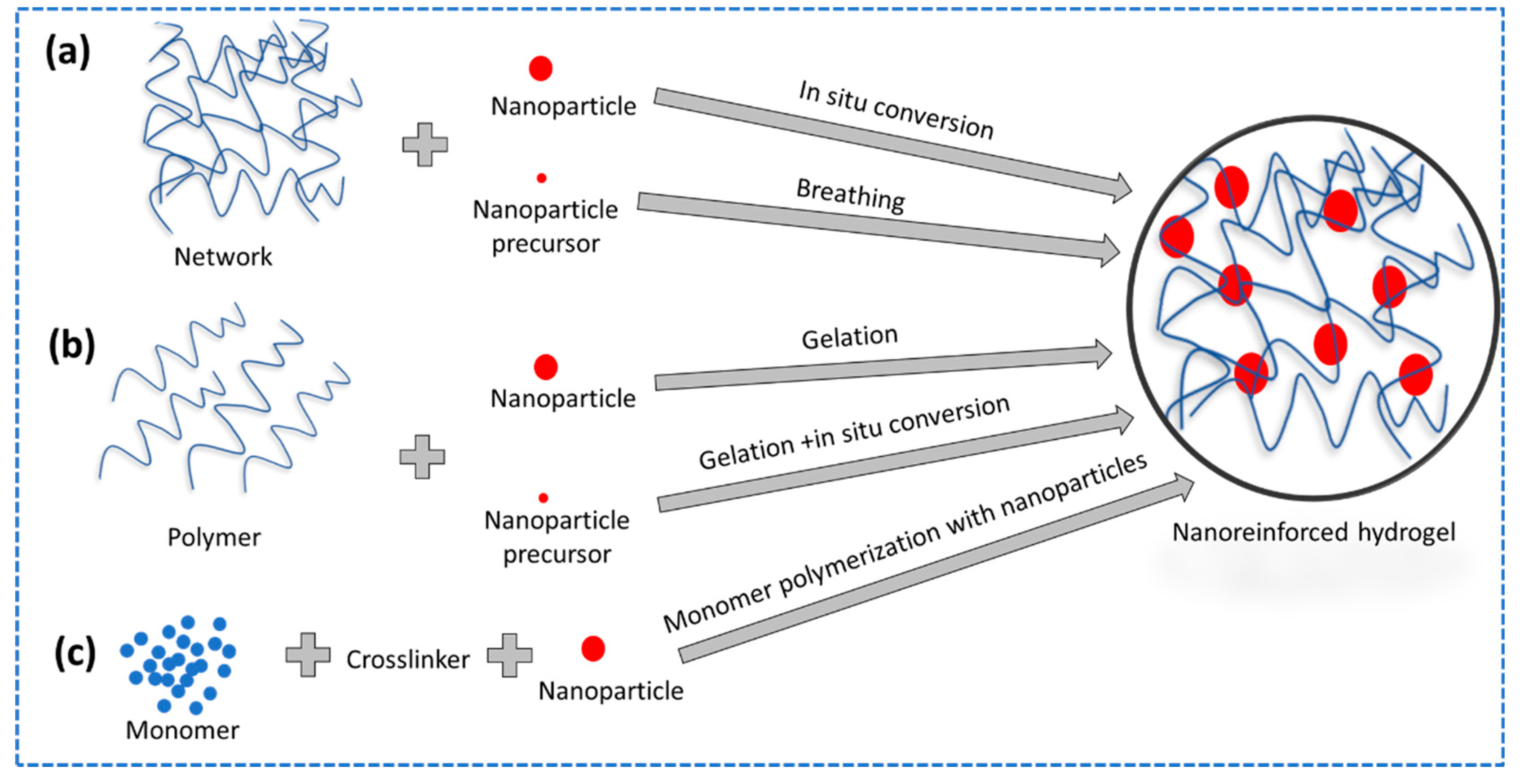
The advent of nanocomposite (NC) hydrogels marked a transformative phase by seamlessly integrating the remarkable mechanical, optical, and swelling attributes of nanoparticles into hydrogel frameworks [210]. However, achieving the optimum potential offered by nanoparticles necessitates more than merely introducing them into a chemically crosslinked network. Instead, a novel approach emerged where nanoparticles were harnessed as multifunctional crosslinkers, playing a pivotal role in the fabrication of NC hydrogels [211]. Nanoparticles can be incorporated into hydrogels through three main approaches: mixing them into pre-formed hydrogels, combining them with polymer solutions before gelation, or growing them within the polymer network from incorporated precursors (Figure 11) [212].
Gelatin-based nanocomposite hydrogel is an excellent solution to overcome the poor mechanical properties of gelatin incorporated conventional hydrogel. Lee et al. constructed hybrid nanocomposite hydrogels with better mechanical features using gelatin and intercalated hydrotalcite (IHT) crosslinked with glutaraldehyde [213]. Hydrotalcite incorporation enhances swelling and significantly improves the mechanical properties of gelatin-based hybrid nanocomposite hydrogels, promising advanced biomedical applications requiring robust load-bearing capacity. The addition of IHT up to 2 wt % to gelatin-based gels significantly enhances their mechanical properties, including shear modulus and crosslinking density, attributed to uniform IHT dispersion and strong gelatin-IHT interaction. However, IHT content exceeding 2 wt % leads to reduced shear modulus and crosslinking density due to increased water affinity and looser gel networks.
Piao et al. synthesized another gelatin-based reduced graphene oxide (RGO)–gelatin nanocomposite hydrogel with a high storage modulus by exploiting a one-pot method [173]. By combining the effects of RGO and gelatin, these hydrogels exhibit a synergistic behavior that enhances mechanical strength, stability, and multifunctionality within a single material. Notably, as the amount of gelatin in the hydrogel increases, its stiffness also rises, while maintaining a consistent water content of approximately 98.0–98.8%. For instance, when measured at a frequency of 10 rad/s, the stiffness of RGG10H reaches 172.3 kPa, surpassing the stiffness of RGG5H (91.1 kPa) by 89%, and that of RGG2H (64.4 kPa) by 169% ( the number in the hydrogel system denotes gelatin concentration in mg mL-1). This increase in stiffness is attributed to the greater number of sites where gelatin chains and GO nanosheets chemically bind, resulting in a stronger and more stable structure with reduced chain movement. In a related study, Piao et al. developed GO-gelatin hydrogels that were physically interconnected, yielding stiffness values of 114.5 kPa for RGG10H and 3.2 kPa for RGG5H, using the same composition as the chemically interconnected hydrogels. Consequently, the stiffness of chemically connected RGG10H surpasses its physically linked counterpart by 50%, while RGG5H demonstrates an impressive 27-fold increase in stiffness compared to its GO-based counterpart. These findings underscore the substantial role of chemical bonds in augmenting the strength of the RGGHs, particularly in comparison to physically linked hydrogels, which is especially pronounced for the weaker hydrogels featuring lower gelatin concentrations.
Gelatin/PAAm/Laponite Nanocomposite hydrogel exhibits improved tensile and stretching properties [172]. In situ radical polymerization of the monomer (AAm) in an aqueous solution of Laponite RDS and gelatin with a trace quantity of BIS as a co-crosslinker was used to construct the hydrogels. The resulted hydrogel reveals the significant role of gelatin in enhancing mechanical properties, including high compressive stress (208.4 ± 5.9 kPa), rendering them promising candidates for various applications requiring robust mechanical performance and flexibility, especially in the biomedical field.

2.3.4. Additive Manufacturing of Hydrogels

Additive manufacturing, commonly known as 3D printing, is a transformative process that involves the creation of intricate components by building them layer by layer using a digital design file as a blueprint (Figure 12). With additive manufacturing, it is possible to produce complicated, three-dimensional geometries with hydrogels and to alter the composition of the material throughout the manufactured pieces [214]. Typically, liquid precursors are cross-linked layer by layer during the additive manufacturing of hydrogels. Filament/paste, powder, solid sheet can also be used as starting materials which facilitates the incorporation of diverse materials during production [215]. There is an extensive assessment available elsewhere, offering a thorough examination of additive manufacturing technologies, including a range of 3D printing techniques and materials, along with valuable insights into their theoretical underpinnings, real-world uses, and future potential for both researchers and practitioners in this domain [216]. A typical 3D printing process of scaffolds involves the synthesis of a solution, computer-aided design (CAD) for scaffold geometry, computer-controlled extrusion of the scaffold material, and subsequent crosslinking to create the final printed design.
Tytgat et al. utilized additive manufacturing techniques to produce gelatin-based Gel-NB/Gel-SH scaffolds for adipose tissue engineering [220]. The hydrogel was prepared by combining norbornene-functionalized gelatin (Gel-NB) with thiolated gelatin (Gel-SH). The 3D printing method used here allows for the creation of precise gelatin scaffolds, which can be tailored to mimic the natural environment of adipose tissue. Photo-crosslinking strengthens these scaffolds, providing vital mechanical support for tissue regeneration and preventing them from collapsing.
Numerous investigations into two-photon polymerization (2PP) processing of modified gelatins have been published, with the majority so far using gelatin-methacrylamide (Gel-MOD) [30,221,222]. For instance, utilizing 2PP technique, 3D scaffolds were produced by starting with photosensitive gelatin [221]. Finally, the scaffolds were manufactured using direct laser writing utilizing two-photon polymerization with a femtosecond laser emitting about 515 nm. Considering biocompatibility, the photoinitiator was chosen.
However, modified gelatin used in 2PP technique has limitations associated with mechanical properties. Nonetheless, Hoorick et al. addressed this issue by creating a gelatin derivative in which all primary amines were transformed into methacrylamides (0.38 mmol/g gelatin), and additional methacrylates were introduced to the carboxylic acids to overcome this constraint [223]. The resultant hydrogel exhibits remarkable mechanical characteristics, rendering it adaptable substance for high-resolution additive manufacturing in the fields of tissue engineering and regenerative medicine. Its capability to establish stable covalent bonds upon specific light exposure enhances structural robustness, facilitating precise fabrication of scaffolds.
The printability and processability of hydrogels in additive manufacturing are important as they enable the precise fabrication of complex structures critical for applications such as tissue engineering and regenerative medicine. Distler et al. examined the printability and processability of the electrically conductive and 3D-printable oxidized alginate-gelatin polypyrrole:PSS (ADA-GEL-PPy:PSS) hydrogels using a commercial extrusion-based 3D printer [218]. These hydrogels were found to be very compatible with 3D printing, having low viscosity and behaving well under shear stress (Figure 12). This made it possible to produce scaffolds with intricate designs and effective pore connections. The scaffolds were robust and easy to manage. Moreover, apart from being easily printed, the ADA-GEL-PPy:PSS hydrogels were also simple to process. Furthermore, the hydrogels could be easily sterilized and seeded with cells without affecting their properties (Figure 12).

3. Mechanical Characterization Techniques of Hydrogel

For mechanical characterization of hydrogels, the fundamental mechanical testing methods and equipment used for polymeric materials more generally are also applied [224]. Hydrogels, like other polymers, display time-dependent mechanical characteristics due to the inherent viscoelasticity of the polymer network [225]. As a result, time plays a significant role in the design and execution of mechanical experiments on hydrogels, which can be described in terms of either time or frequency. Here we will provide a concise overview of the predominant testing techniques commonly employed, encompassing tensile testing, compression testing (both unconfined and confined variations), localized indentation utilizing a probe, and frequency-based testing methodologies such as shear rheometry and dynamic mechanical analysis (DMA) (Figure 13).
  • Tensile Testing
Tensile strength is a fundamental material property that characterizes a material's capacity to resist stretching or pulling forces until it reaches the point of rupture or experiences irreversible deformation, typically denoted in units of force per unit area such as pounds per square inch (psi) or pascals (Pa). Understanding the tensile strength of hydrogel biomaterials is crucial because it determines their ability to withstand mechanical stresses and deformations in biological applications, ensuring the integrity and functionality of these materials in physiological environments [226]. In a tensile test for soft materials, universal testing machine or materials testing machine is used [227]. A representative sample is clamped between specialized grips to prevent slipping. A tensile testing machine applies a controlled, steadily increasing pulling force while measuring the material's deformation, usually in the form of elongation. This data is used to create a stress-strain curve, revealing how the material responds to the applied force.
  • Compression Testing
Compression testing of hydrogel materials, in contrast to tensile testing, assesses how these materials respond to compressive forces rather than tensile or pulling forces. Compression testing of hydrogel materials can be conducted in both confined and unconfined variations [224]. In confined compression testing, the hydrogel sample is subjected to compressive forces within a defined space, often using specialized fixtures to prevent lateral expansion. This helps evaluate how the hydrogel behaves under compression while maintaining its shape within constraints. Unconfined compression testing, on the other hand, involves applying compressive forces without containment, allowing the hydrogel to expand laterally as it compresses. Both methods provide valuable insights into the hydrogel's compressive strength, deformation characteristics, and its ability to recover its original shape after compression.
  • Indentation
Indentation testing is a mechanical testing method used to evaluate the mechanical properties of soft materials such as hydrogels, specifically their response to localized compressive forces. Instruments used for indentation testing include hardness testers for measuring resistance to penetration, universal testing machines with indentation accessories, specialized nanoindenters for nanoscale testing, microindenters for microscale testing, instrumented indentation testers for detailed property measurements, durometers for measuring the hardness of soft materials, and atomic force microscopes for nanoscale indentation testing. Key parameters measured in indentation testing include hardness, which quantifies resistance to penetration; stiffness, representing how the hydrogel responds to the force; elastic modulus, indicating elastic behavior; and creep, the gradual deformation under constant force, assessing viscoelastic properties. In this test, a controlled and precisely measured force is applied to the surface of a hydrogel sample using a specialized indenter or probe. The probe penetrates into the hydrogel, creating an indentation mark, and the force-depth relationship is recorded.
  • Frequency-based Testing
Frequency-based testing methodologies such as shear rheometry and dynamic mechanical analysis (DMA) serve as integral tools for the comprehensive characterization of mechanical properties across a spectrum of frequencies. Shear rheometry assesses material responses to varying shear forces, elucidating properties encompassing viscosity, elasticity, and viscoelastic tendencies. In contrast, DMA investigates how materials react to dynamic loading at different frequencies, yielding critical data related to parameters like storage and loss moduli, damping behavior, and relaxation times. Instruments for frequency-based testing of hydrogel materials include rheometers for assessing their response to shear forces, dynamic mechanical analyzers (DMA) to study mechanical behavior at different frequencies, and other tools like ultrasonic testers and electromagnetic resonance spectrometers to explore various properties at different frequency ranges.

4. Application of Mechanically Improved Gelatin-based Hydrogels in Tissue Engineering

Gelatin hydrogels have demonstrated their versatility and potential in various tissue engineering applications, including bone tissue regeneration, cartilage tissue regeneration, cardiac tissue regeneration, nerve tissue regeneration, ocular tissue engineering, and scaffold tissue engineering, offering promising solutions for the development of advanced biomaterials (Figure 14). Their biocompatibility, tunable properties, and capacity to support cell growth and tissue-specific functions make them valuable candidates for a wide range of regenerative medicine approaches.

4.1. In bone Tissue Regeneration

In an effort to repair or reconstruct bone, approximately 2.2 million bone-grafting procedures are performed annually on the pelvis, spine, and other body extremities [228]. Bone grafts or substitute materials are frequently used by surgeons to stimulate bone growth [229]. Bone graft research is now concentrating on bone tissue engineering, which uses a combination of cells, scaffolds, and bioactive agents to develop new workable tissues that can replace the injured tissues [230]. Biological bone is a nanocomposite made up of a wide variety of inorganic and organic components [231]. Due to the complexity of the bone extracellular matrix (ECM), a scaffold for bone regeneration made of a single material, such as collagen, is unable to provide the vital signals for cellular growth [232]. Nonetheless, if two or more materials are combined for scaffold fabrication, a synergistic effect may be produced to enhance the scaffold's mechanical strength and facilitate cell adhesion, proliferation, and differentiation [231,233,234,235]. Numerous mechanically robust gelatin-based hydrogels have been developed for the purpose of bone tissue regeneration, capitalizing on gelatin's inherent biocompatibility [236,237,238]. Injectable hydrogels find frequent use in bone and cartilage tissue engineering, especially in cases involving challenging access or the regeneration of minor defects, primarily due to their ease of application [239].
Gelatin is combined with chitosan, alginate, and nano-hydroxyapatite (nHAp) to produce a scaffold with favorable mechanical strength, including adequate strength and porosity, supporting cellular growth and bone regeneration within the bone tissue environment [231]. The combination of chitosan, gelatin, alginate, and nHAp in a scaffold result in increased mechanical stability due to electrostatic interactions and crosslinking, contributing to a compact and stable structure. Another novel gelatin-based hydrogel incorporating metformin-loaded mesoporous silica nanoparticles (MF-MSNs-COOH/GelMA) shows potential for bone regeneration, offering controlled drug release, enhanced mechanical properties, cytocompatibility, and osteogenic differentiation capability [240]. With a compressive strength of 110 kPa, the hydrogel exhibits robustness suitable for application in bone regeneration.
A combination of gelatin and chitosan in the creation of 3D hybrid hydrogels, supplemented with human platelet lysate exhibit exceptional properties in promoting the growth and specialized bone-forming transformation of human mesenchymal stem cells (hMSCs) [178]. The use of naturally derived and biocompatible materials such as gelatin and chitosan, in conjunction with human platelet lysate, underscores the promise of secure and efficient therapeutic approaches in the realm of bone regeneration and repair with a strong emphasis on their resilience and strength. The result showed that hMSCs proliferated and differentiated into osteoblasts more effectively in the hybrid hydrogels with hPL than in the hydrogels without hPL (Figure 15). hPL is a blood-derived product that contains a variety of growth factors and other signaling molecules that can promote cell proliferation and differentiation. The hybrid scaffold exhibited satisfactory mechanical robustness with a tensile modulus of 680 kPa, tensile strength of 250 kPa, and elongation at break of 29%. However, increased chitosan content in the network reduced stiffness (280 kPa) and strength (80 kPa), resulting in a softer material, but no data on gelatin's effect was provided.

4.2. Cartilage Tissue Regeneration

Cartilage tissue engineering employs a combination of biocompatible scaffold material, cells, and growth factors to create cartilage-like tissue that has similar biomechanical properties to native cartilage, with the goal of repairing articular cartilage defects. Hydrogels have attracted considerable attention as potential biomaterials for cartilage tissue regeneration due to their potential to facilitate cartilage repair [241]. Numerous mechanically robust gelatin-based hydrogels have been developed for the purpose of bone tissue regeneration, capitalizing on gelatin's inherent biocompatibility [242,243,244].
In cartilage tissue engineering, the mechanical strength of a designed scaffold is a crucial factor because it gives chondrocytes the ability to grow unhindered and to withstand mechanical loads if the scaffold needs to be implanted in the joint as a tissue replacement. Bhat et al. has synthesized a chitosan–agarose–gelatin (CAG) cryogel with excellent porosity and mechanical properties [171]. This CAG cryogel can offer substantial mechanical strength and a compliant compression modulus, allowing it to be used as a transitory porous architecture for the growing of chondrocytes in three-dimensional environments.
Another gelatin-based scaffold with dynamic reciprocity for cartilage tissue engineering was reported by [245]. This tri-copolymer of gelatin, chondoitin-6-sulfate, and hyaluronan contained regenerated collagen fibers with a hierarchical structure and anisotropic properties. This hydrogel offers biomimetic platform for cartilage tissue engineering, providing an ideal microenvironment for chondrocytes and promoting cartilage regeneration. In addition, its porous structure facilitates nutrient diffusion and offers mechanical properties suitable for withstanding physiological loads, making it a promising solution for addressing cartilage defects and degenerative joint diseases. Sartore et al. investigated the impact of various polysaccharides on gelatin-based hydrogels in relation to their influence on the chondrogenic differentiation of human mesenchymal stromal cells [246]. The findings highlight the differential effects of polysaccharides on guiding the development of cartilage-like tissue from these cells within hydrogel environments (Figure 16). This research contributes to an understanding of how the choice of polysaccharides in gelatin-based hydrogels can modulate chondrogenic differentiation.

4.3. Cardiac Tissue Regeneration

One American dies per minute from a myocardial infarction (MI), making it the leading cause of death from cardiovascular illness in the country [247]. There aren't enough healthy donors available for heart transplants, and the procedure comes with a high risk of complications. As a result, cell-based therapies [248,249] and tissue engineering [250,251,252] have been suggested as promising options for treating MI. In order to successfully regenerate cardiac tissue, it is necessary to use hydrogels that are both bioactive and biocompatible, and that resemble the biochemical and biomechanical microenvironments found in native tissue. These hydrogels should keep cells in the infarcted area and sustain myocardial wall stress, cell survival, and function. For cardiac tissue regeneration, injectable hydrogels, which use liquid biomaterials to form solid gels after injection, offer a number of benefits, including the potential to self-assemble in situ, minimally invasive delivery capacity (in contrast to other methods such as in vitro designed tissue or epicardial fix implantation), and the ability to promote host tissue regeneration [253,254]. Gelatin finds application in cardiac tissue engineering, manifesting in the form of scaffolds for constructing cardiac tissue constructs [255,256].
Designing biomaterial scaffolds that can enhance the maturation, contractility, and electrophysiological function of cardiomyocytes for cardiac tissue regeneration may benefit greatly from gelatin-based hydrogel scaffolds that are embedded with GO (graphene oxide) and cross-linked by genipin, providing the favorable mechanical property and topographic surface characteristics for generating functional cardiac tissue in vitro [257]. This user-friendly hydrogel platform provides a biocompatible environment that supports the long-term growth and function of cardiomyocytes, making it a valuable tool for cardiac tissue engineering and regenerative medicine research.
For the purpose of myocardial regeneration and repair, Navaei et al. looked into Gelatin methacryloyl embedded on gold nanorod (GelMA-GNR) hybrid hydrogels as an improved biomaterial [258]. The gelatin-based conductive hydrogels, incorporating gold nanorods, offer a biocompatible and supportive environment for cardiac cell growth and organization. These hydrogels exhibit outstanding mechanical properties, providing both strength and flexibility, enabling them to maintain structural stability and integrity while allowing for essential physiological stretching and contraction, closely resembling natural cardiac tissue behavior.
Navaei et al. engineered cardiac tissues for heart regeneration by gelatin-based hydrogels incorporated with electrically conductive gold nanorods (GNRs) to emulate the natural structure and function of the heart [259]. Their approach seamlessly combined micro- and nano-technologies, resulting in the development of gelatin methacrylate (GelMA) hydrogel constructs featuring surface micro-topographies, specifically microgrooves enhanced with GNRs. When cardiac cells seeded on GelMA-GNR hydrogels and cultured in a bioreactor, they aligned along microgrooves, leading to the formation of dense and highly organized cardiac tissues that displayed spontaneous contractility (Figure 17). The GelMA-GNR cardiac tissues, which were electrically conductive, consistently responded to external stimulation by changing their beat rate, demonstrating improved formation of cardiac tissues with enhanced cellular organization, connectivity, and electrical properties, suggesting their potential as functional cardiac patches for treating damaged heart tissue.

4.4. Nerve Tissue Regeenration

Gelatin-based hydrogels have also been used in peripheral nerve tissue engineering to release vascular endothelial growth factor [260]. This VEGF–A165 incorporated Gelatin/Gelatin-Genipin (A/GL–GP) hydrogel would be a suitable carrier for encapsulating and gradually releasing VEGF–A165 over time. Bioactive VEGF-A165 release, as observed in vitro, can induce capillary-like tube development and axonal outgrowth ex vivo, suggesting that gelatin-based drug-releasing hydrogels may be appropriate for this purpose.
In order to evaluate the biocompatibility and biodegradation of 3D gelatin and gelatin/hyaluronan gels, as well as their ability to enhance brain tissue reconstruction, Zhang et al. synthesized and implanted 3D gelatin and gelatin/hyaluronan system into traumatic rat brains using cell assembling equipment that showed potential in the repair of injured brain tissue [261]. Even though gelatin structures degrade at a slower rate, the infiltration of cells around gelatin stents was expedited.
Ye et al. introduced an innovative approach for the production of nerve guidance conduits (NGCs) through the utilization of 3D printing technology and gelatin methacrylate (GelMA) [262]. The researchers used a Digital Light Processing (DLP) printing technology to create neural NGCs for healing large-gap nerve damage (Figure18). These GelMA-based conduits are meticulously engineered to closely emulate the characteristics of natural neural tissue. The employment of DLP printing techniques enables the creation of conduits with multiple channels, designed to significantly augment the process of nerve regeneration. The NGCs were found to promote the survival, growth, and movement of neural cells within their channels. Additionally, the study revealed that these NGCs could stimulate neural crest stem cells to develop into neurons (Figure 18).

5. Conclusion and Future Prospective

In conclusion, gelatin-based hydrogels have emerged as promising candidates for tissue engineering scaffolds, primarily owing to their remarkable biocompatibility, biodegradability, processability, transparency, non-toxicity, and structural similarity to the natural extracellular matrix. However, their inherent low mechanical properties pose a significant challenge in supporting cell growth and proliferation effectively. To address this limitation, a variety of crosslinking strategies, including physical, chemical, enzymatic, and combinations thereof, as well as innovative networking patterns such as double networks, interpenetrating networks, and nano-reinforcing mechanisms, have been employed to enhance the structural stability and mechanical integrity of gelatin-based hydrogels.
This review has provided valuable insights into the recent advancements in modulating the mechanical properties of gelatin-based hydrogels, with a focus on designing structurally stable and robust scaffolds for tissue engineering applications. The optimized crosslinking parameters necessary to achieve adequate mechanical properties in gelatin-based hydrogels have been thoroughly discussed. Additionally, we have outlined the diverse applications of gelatin-based scaffolds in tissue engineering, spanning various fields such as bone, cartilage, cardiac, skin, and nerve tissue engineering.
Looking ahead, it is essential to recognize current challenges and identify areas for further research and improvement. To unlock the full potential of gelatin-based hydrogels in tissue engineering, we must overcome existing limitations and optimize fabrication processes. Advances in materials science, bioengineering, and biotechnology will play a pivotal role in addressing these challenges, leading to innovative solutions for diverse problems in the field of tissue engineering. In essence, the field of gelatin-based hydrogels for tissue engineering holds significant promise, and ongoing research efforts are poised to make substantial contributions to the field in the years ahead.

Author Contributions

Conceptualization, K.D. and M.S.N.A.A.; software, K.D. and M.S.N.A.A.; validation, K.D. and M.S.N.A.A.; formal analysis, K.D.; M.S.N.A.A.; N.M.; A.G. and G.R.; data curation, K.D. and M.S.N.A.A.; writing—original draft preparation, K.D.; M.S.N.A.A.; N.M.; A.G. and G.R.; writing—review and editing, K.D.; M.S.N.A.A.; N.M.; A.G. and G.R.; supervision, K.D.; project administration, K.D.; funding acquisition, K.D.

Funding

This research received funding from Bio-Nanomaterials and Tissue Engineering Laboratory (BNTELab), Department of Applied Chemistry and Chemical Engineering, Faculty of Science, University of Chittagong, Chittagong-4331, Bangladesh. Grant Number: BNTELab-Fund-01-2023.

Conflicts of Interest

The authors declare no conflict of interest.

References

  1. Young, S.; Wong, M.; Tabata, Y.; Mikos, A.G. Gelatin as a Delivery Vehicle for the Controlled Release of Bioactive Molecules. Journal of Controlled Release 2005, 109, 256–274. [Google Scholar] [CrossRef] [PubMed]
  2. Van Den Bulcke, A.I.; Bogdanov, B.; De Rooze, N.; Schacht, E.H.; Cornelissen, M.; Berghmans, H. Structural and Rheological Properties of Methacrylamide Modified Gelatin Hydrogels. Biomacromolecules 2000, 1, 31–38. [Google Scholar] [CrossRef] [PubMed]
  3. Pierce, B.F.; Pittermann, E.; Ma, N.; Gebauer, T.; Neffe, A.T.; Hölscher, M.; Jung, F.; Lendlein, A. Viability of Human Mesenchymal Stem Cells Seeded on Crosslinked Entropy-Elastic Gelatin-Based Hydrogels. Macromol Biosci 2012, 12, 312–321. [Google Scholar] [CrossRef] [PubMed]
  4. Croome, R.J. Acid and Alkaline Hydrolysis of Gelatin. Journal of Applied Chemistry 1953, 3, 280–286. [Google Scholar] [CrossRef]
  5. Alam, M.R.; Shahid, M.A.; Alimuzzaman, S.; Khan, A.N. Sources, Extractions and Applications of Bio-Maker Collagen–A Review. Biomedical Engineering Advances 2022, 4, 100064. [Google Scholar] [CrossRef]
  6. Bogue, R.H. Conditions Affecting the Hydrolysis of Collagen to Gelatin. Ind Eng Chem 1923, 15, 1154–1159. [Google Scholar] [CrossRef]
  7. Keenan, T.R. Gelatin. Kirk-Othmer Encyclopedia of Chemical Technology, 2000. [Google Scholar]
  8. Kharkar, P.M.; Kiick, K.L.; Kloxin, A.M. Designing Degradable Hydrogels for Orthogonal Control of Cell Microenvironments. Chem Soc Rev 2013, 42, 7335–7372. [Google Scholar] [CrossRef]
  9. Nur Hanani, Z.A.; Roos, Y.H.; Kerry, J.P. Use and Application of Gelatin as Potential Biodegradable Packaging Materials for Food Products. Int J Biol Macromol 2014, 71, 94–102. [Google Scholar] [CrossRef]
  10. Slade, L.; Levine, H. Polymer-chemical properties of gelatin in foods. Advances in meat research (USA), 1987. [Google Scholar]
  11. Santoro, M.; Tatara, A.M.; Mikos, A.G. Gelatin carriers for drug and cell delivery in tissue engineering. Journal of controlled release 2014, 190, 210–218. [Google Scholar] [CrossRef] [PubMed]
  12. Vandooren, J.; Van den Steen, P.E.; Opdenakker, G. Biochemistry and molecular biology of gelatinase B or matrix metalloproteinase-9 (MMP-9): the next decade. Critical reviews in biochemistry and molecular biology 2013, 48, 8222–272. [Google Scholar] [CrossRef] [PubMed]
  13. Alizadeh, M.; Abbasi, F.; Khoshfetrat, A.B.; Ghaleh, H. Microstructure and Characteristic Properties of Gelatin/Chitosan Scaffold Prepared by a Combined Freeze-Drying/Leaching Method. Materials Science and Engineering: C 2013, 33, 3958–3967. [Google Scholar] [CrossRef]
  14. Pamplona, R.; González-Lana, S.; Romero, P.; Ochoa, I.; Martín-Rapún, R.; Sánchez-Somolinos, C. Tuning of Mechanical Properties in Photopolymerizable Gelatin-Based Hydrogels for In Vitro Cell Culture Systems. ACS Appl Polym Mater 2023, 5, 1487–1498. [Google Scholar] [CrossRef]
  15. Vanderhooft, J.L.; Alcoutlabi, M.; Magda, J.J.; Prestwich, G.D. Rheological Properties of Cross-Linked Hyaluronan–Gelatin Hydrogels for Tissue Engineering. Macromol Biosci 2009, 9, 20–28. [Google Scholar] [CrossRef]
  16. Davidenko, N.; Schuster, C.F.; Bax, D.V.; Farndale, R.W.; Hamaia, S.; Best, S.M.; Cameron, R.E. Evaluation of Cell Binding to Collagen and Gelatin: A Study of the Effect of 2D and 3D Architecture and Surface Chemistry. J Mater Sci Mater Med 2016, 27, 1–14. [Google Scholar] [CrossRef] [PubMed]
  17. Ramorino, G.; Gobetti, A.; Cornacchia, G.; Roca, E. Effect of Static Offsets on the Nonlinear Dynamic Mechanical Properties of Human Brain Tissue. J Mech Behav Biomed Mater 2022, 130, 105204. [Google Scholar] [CrossRef]
  18. Shao, L.; Gao, Q.; Xie, C.; Fu, J.; Xiang, M.; He, Y. Synchronous 3D Bioprinting of Large-Scale Cell-Laden Constructs with Nutrient Networks. Adv Healthc Mater 2020, 9, 1901142. [Google Scholar] [CrossRef]
  19. Rosellini, E.; Cristallini, C.; Barbani, N.; Vozzi, G.; Giusti, P. Preparation and Characterization of Alginate/Gelatin Blend Films for Cardiac Tissue Engineering. J Biomed Mater Res A 2009, 91A, 447–453. [Google Scholar] [CrossRef]
  20. Babin, H.; Dickinson, E. "Influence of transglutaminase treatment on the thermoreversible gelation of gelatin. Food hydrocolloids 2001, 15, 271–276. [Google Scholar] [CrossRef]
  21. Karim, A.A.; Bhat, R. Fish Gelatin: Properties, Challenges, and Prospects as an Alternative to Mammalian Gelatins. Food Hydrocoll 2009, 23, 563–576. [Google Scholar] [CrossRef]
  22. Bigi, A.; Panzavolta, S.; Rubini, K. Relationship between Triple-Helix Content and Mechanical Properties of Gelatin Films. Biomaterials 2004, 25, 5675–5680. [Google Scholar] [CrossRef] [PubMed]
  23. Klotz, B.J.; Gawlitta, D.; Rosenberg, A.J.W.P.; Malda, J.; Melchels, F.P.W. Gelatin-Methacryloyl Hydrogels: Towards Biofabrication-Based Tissue Repair. Trends Biotechnol 2016, 34, 394–407. [Google Scholar] [CrossRef]
  24. Dey, K.; Roca, E.; Ramorino, G.; Sartore, L. Progress in the Mechanical Modulation of Cell Functions in Tissue Engineering. Biomater Sci 2020, 8, 7033–7081. [Google Scholar] [CrossRef] [PubMed]
  25. DeForest, C.A.; Anseth, K.S. Advances in bioactive hydrogels to probe and direct cell fate. Annual review of chemical and biomolecular engineering 2012, 3, 421–444. [Google Scholar] [CrossRef] [PubMed]
  26. Wisotzki, E.I.; Hennes, M.; Schuldt, C.; Engert, F.; Knolle, W.; Decker, U.; Käs, J.A.; Zink, M.; Mayr, S.G. Tailoring the Material Properties of Gelatin Hydrogels by High Energy Electron Irradiation. J Mater Chem B 2014, 2, 4297–4309. [Google Scholar] [CrossRef] [PubMed]
  27. Cataldo, F.; Ursini, O.; Lilla, E.; Angelini, G. Radiation-Induced Crosslinking of Collagen Gelatin into a Stable Hydrogel. J Radioanal Nucl Chem 2007, 275, 125–131. [Google Scholar] [CrossRef]
  28. Ge, S.; Liu, Q.; Li, M.; Liu, J.; Lu, H.; Li, F.; Zhang, S.; Sun, Q.; Xiong, L. Enhanced Mechanical Properties and Gelling Ability of Gelatin Hydrogels Reinforced with Chitin Whiskers. Food Hydrocoll 2018, 75, 1–12. [Google Scholar] [CrossRef]
  29. Manish, V.; Arockiarajan, A.; Tamadapu, G. Influence of Water Content on the Mechanical Behavior of Gelatin Based Hydrogels: Synthesis, Characterization, and Modeling. Int J Solids Struct 2021, 233, 111219. [Google Scholar] [CrossRef]
  30. Vlierberghe, S. van; Schacht, E.; Dubruel, P. Reversible Gelatin-Based Hydrogels: Finetuning of Material Properties. Eur Polym J 2011, 47, 1039–1047. [Google Scholar] [CrossRef]
  31. Jaipan, P.; Nguyen, A.; Narayan, R.J. Gelatin-Based Hydrogels for Biomedical Applications. MRS Commun 2017, 7, 416–426. [Google Scholar] [CrossRef]
  32. Jiang, Y.; Wang, H.; Wang, X.; Yu, X.; Li, H.; Tang, K; Li, Q. Preparation of gelatin-based hydrogels with tunable mechanical properties and modulation on cell–matrix interactions. Journal of Biomaterials Applications 2021, 36, 902–911. [Google Scholar] [CrossRef]
  33. Wang, Xiaohong, et al. Gelatin-based hydrogels for organ 3D bioprinting. Polymers 2017, 9, 401. [Google Scholar] [CrossRef] [PubMed]
  34. Lukin, I.; Erezuma, I.; Maeso, L.; Zarate, J.; Desimone, M.F.; Al-Tel, T.H.; Dolatshahi-Pirouz, A.; Orive, G. Progress in Gelatin as Biomaterial for Tissue Engineering. Pharmaceutics 2022, 14, 1177. [Google Scholar] [CrossRef] [PubMed]
  35. Salahuddin, B.; Wang, S.; Sangian, D.; Aziz, S.; Gu, Q. Hybrid Gelatin Hydrogels in Nanomedicine Applications. ACS Appl Bio Mater 2021, 4, 2886–2906. [Google Scholar] [CrossRef] [PubMed]
  36. Kang, J. Il; Park, K.M. Advances in Gelatin-Based Hydrogels for Wound Management. J Mater Chem B 2021, 9, 1503–1520. [Google Scholar] [CrossRef] [PubMed]
  37. Echave, Mari Carmen, et al. Recent advances in gelatin-based therapeutics. Expert opinion on biological therapy 2019, 19, 773–779. [Google Scholar] [CrossRef] [PubMed]
  38. Elzoghby, A.O. Gelatin-Based Nanoparticles as Drug and Gene Delivery Systems: Reviewing Three Decades of Research. Journal of Controlled Release 2013, 172, 1075–1091. [Google Scholar] [CrossRef]
  39. Rose, J.B.; Pacelli, S.; El Haj, A.J.; Dua, H.S.; Hopkinson, A.; White, L.J.; Rose, F.R.A.J. Gelatin-Based Materials in Ocular Tissue Engineering. Materials 2014, 7, 3106–3135. [Google Scholar] [CrossRef] [PubMed]
  40. Gibbs, D.M.R.; Black, C.R.M.; Dawson, J.I.; Oreffo, R.O.C. A Review of Hydrogel Use in Fracture Healing and Bone Regeneration. J Tissue Eng Regen Med 2016, 10, 187–198. [Google Scholar] [CrossRef]
  41. Asim, S.; Tabish, T.A.; Liaqat, U.; Ozbolat, I.T.; Rizwan, M. Advances in Gelatin Bioinks to Optimize Bioprinted Cell Functions. Adv Healthc Mater 2023, 12, 2203148. [Google Scholar] [CrossRef]
  42. Nezhadi, Somayeh Hallaj, et al. "Gelatin-based delivery systems for cancer gene therapy." Journal of Drug Targeting 2009, 17, 731–738.
  43. Liu, Mei, et al. Injectable hydrogels for cartilage and bone tissue engineering. Bone research 2017, 5, 1–20. [Google Scholar]
  44. Liu, Shao Qiong, et al. Synthetic hydrogels for controlled stem cell differentiation. Soft Matter 2010, 6, 67–81. [Google Scholar] [CrossRef]
  45. Hennink, W.E.; van Nostrum, C.F. Novel crosslinking methods to design hydrogels. Advanced drug delivery reviews 2012, 64, 223–236. [Google Scholar] [CrossRef]
  46. Voorhaar, L; Hoogenboom, R. Supramolecular polymer networks: hydrogels and bulk materials. Chemical Society Reviews 2016, 45, 4013–4031. [Google Scholar] [CrossRef] [PubMed]
  47. Berger, J.; Reist, M.; Mayer, J.M.; Felt, O; Gurny, R. Structure and interactions in chitosan hydrogels formed by complexation or aggregation for biomedical applications. European journal of pharmaceutics and biopharmaceutics 2004, 57, 35–52. [Google Scholar] [CrossRef]
  48. Xue, K.; Liow, S.S.; Karim, A.A.; Li, Z.; Loh, X.J. A Recent Perspective on Noncovalently Formed Polymeric Hydrogels. The Chemical Record 2018, 18, 1517–1529. [Google Scholar] [CrossRef] [PubMed]
  49. Nichol, J.W.; Koshy, S.T.; Bae, H.; Hwang, C.M.; Yamanlar, S.; Khademhosseini, A. Cell-Laden Microengineered Gelatin Methacrylate Hydrogels. Biomaterials 2010, 31, 5536–5544. [Google Scholar] [CrossRef] [PubMed]
  50. Naahidi, S.; Jafari, M.; Logan, M.; Wang, Y.; Yuan, Y.; Bae, H.; Dixon, B.; Chen, P. Biocompatibility of Hydrogel-Based Scaffolds for Tissue Engineering Applications. Biotechnol Adv 2017, 35, 530–544. [Google Scholar] [CrossRef]
  51. Ramachandran, R.; Jung, D.; Spokoyny, A.M. Cross-Linking Dots on Metal Oxides. NPG Asia Materials 2019, 11, 1–4. [Google Scholar] [CrossRef]
  52. Berger, J.; Reist, M.; Mayer, J.M.; Felt, O.; Peppas, N.A.; Gurny, R. Structure and Interactions in Covalently and Ionically Crosslinked Chitosan Hydrogels for Biomedical Applications. European Journal of Pharmaceutics and Biopharmaceutics 2004, 57, 19–34. [Google Scholar] [CrossRef]
  53. Wu, M.; Peng, Q.Y.; Han, L.B.; Zeng, H.B. Self-Healing Hydrogels and Underlying Reversible Intermolecular Interactions. Chinese Journal of Polymer Science 2021, 39, 1246–1261. [Google Scholar] [CrossRef]
  54. Wang, X.; Wang, H.; Zhang, H.; Yang, T.; Zhao, B.; Yan, J. Investigation of the Impact of Hydrogen Bonding Degree in Long Single-Stranded DNA (SsDNA) Generated with Dual Rolling Circle Amplification (RCA) on the Preparation and Performance of DNA Hydrogels. Biosensors 2023, 13, 755. [Google Scholar] [CrossRef]
  55. Bhattacharjee, A; Bansal, M. ; Collagen structure: the Madras triple helix and the current scenario. IUBMB life 2005, 57, 161–172. [Google Scholar] [CrossRef]
  56. Chang, M.; Choi, D.; Fu, B.; Reichmanis, E. Solvent Based Hydrogen Bonding: Impact on Poly(3-Hexylthiophene) Nanoscale Morphology and Charge Transport Characteristics. ACS Nano 2013, 7, 5402–5413. [Google Scholar] [CrossRef]
  57. Namchuk, M.N.; Withers, S.G. Mechanism of Agrobacterium β-Glucosidase: Kinetic Analysis of the Role of Noncovalent Enzyme/Substrate Interactions. Biochemistry 1995, 34, 16194–16202. [Google Scholar] [CrossRef]
  58. Paleos, C.M.; Tsiourvas, D.; Sideratou, Z. Hydrogen Bonding Interactions of Liposomes Simulating Cell-Cell Recognition. A Review. Origins of Life and Evolution of the Biosphere 2004, 34, 195–213. [Google Scholar] [CrossRef]
  59. Wang, Y.J.; Zhang, X.N.; Song, Y.; Zhao, Y.; Chen, L.; Su, F.; Li, L.; Wu, Z.L.; Zheng, Q. Ultrastiff and Tough Supramolecular Hydrogels with a Dense and Robust Hydrogen Bond Network. Chemistry of Materials 2019, 31, 1430–1440. [Google Scholar] [CrossRef]
  60. Vigata, M.; Meinert, C.; Bock, N.; Dargaville, B.L.; Hutmacher, D.W. Deciphering the Molecular Mechanism of Water Interaction with Gelatin Methacryloyl Hydrogels: Role of Ionic Strength, Ph, Drug Loading and Hydrogel Network Characteristics. Biomedicines 2021, 9, 574. [Google Scholar] [CrossRef] [PubMed]
  61. Zhang, H.J.; Wang, L.; Wang, X.; Han, Q.; You, X. Developing Super Tough Gelatin-Based Hydrogels by Incorporating Linear Poly(Methacrylic Acid) to Facilitate Sacrificial Hydrogen Bonding. Soft Matter 2020, 16, 4723–4727. [Google Scholar] [CrossRef] [PubMed]
  62. Zhang, H.J.; Wang, X.; Wang, L.; Sun, T.L.; Dang, X.; King, D.R.; You, X. Dynamic bonds enable high toughness and multifunctionality in gelatin/tannic acid-based hydrogels with tunable mechanical properties. Soft Matter, 2021, 17, 9399–9409. [Google Scholar] [CrossRef] [PubMed]
  63. Wang, J.; Tang, F.; Wang, Y.; Lu, Q.; Liu, S.; Li, L. Self-Healing and Highly Stretchable Gelatin Hydrogel for Self-Powered Strain Sensor. ACS Appl Mater Interfaces 2020, 12, 1558–1566. [Google Scholar] [CrossRef]
  64. Zhang, G.; Lv, L.; Deng, Y.; Wang, C.; Zhang, G.Z.; Wang, C.Y.; Lv, L.; Deng, Y.H. Self-Healing Gelatin Hydrogels Cross-Linked by Combining Multiple Hydrogen Bonding and Ionic Coordination. Macromol Rapid Commun 2017, 38, 1700018. [Google Scholar] [CrossRef] [PubMed]
  65. Liu, B.; Wang, Y.; Miao, Y.; Zhang, X.; Fan, Z.; Singh, G.; Zhang, X.; Xu, K.; Li, B.; Hu, Z.; et al. Hydrogen Bonds Autonomously Powered Gelatin Methacrylate Hydrogels with Super-Elasticity, Self-Heal and Underwater Self-Adhesion for Sutureless Skin and Stomach Surgery and E-Skin. Biomaterials 2018, 171, 83–96. [Google Scholar] [CrossRef] [PubMed]
  66. Zhou, L.; Dai, C.; Fan, L.; Jiang, Y.; Liu, C.; Zhou, Z.; Guan, P.; Tian, Y.; Xing, J.; Li, X.; et al. Injectable Self-Healing Natural Biopolymer-Based Hydrogel Adhesive with Thermoresponsive Reversible Adhesion for Minimally Invasive Surgery. Adv Funct Mater 2021, 31, 2007457. [Google Scholar] [CrossRef]
  67. George, B.; Bhatia, N.; Kumar, A.; Gnanamani, A.; Thilagam, R.; Shanuja, S.K.; Vadakkadath Meethal, K.; Shiji, T.M. Bioinspired Gelatin Based Sticky Hydrogel for Diverse Surfaces in Burn Wound Care. Scientific Reports 2022, 12, 1–19. [Google Scholar] [CrossRef] [PubMed]
  68. Hsu, W.; Chen, Y.L.; Horng, J.C. Promoting Self-Assembly of Collagen-Related Peptides into Various Higher-Order Structures by Metal-Histidine Coordination. Langmuir 2012, 28, 3194–3199. [Google Scholar] [CrossRef] [PubMed]
  69. Pires, M.M.; Lee, J.; Ernenwein, D.; Chmielewski, J. Controlling the Morphology of Metal-Promoted Higher Ordered Assemblies of Collagen Peptides with Varied Core Lengths. Langmuir 2012, 28, 1993–1997. [Google Scholar] [CrossRef] [PubMed]
  70. Vogt, C.; Xing, Q.; He, W.; Li, B.; Frost, M.C.; Zhao, F. Fabrication and Characterization of a Nitric Oxide-Releasing Nanofibrous Gelatin Matrix. Biomacromolecules 2013, 14, 2521–2530. [Google Scholar] [CrossRef] [PubMed]
  71. Xing, Q.; Yates, K.; Vogt, C.; Qian, Z.; Frost, M.C.; Zhao, F. Increasing mechanical strength of gelatin hydrogels by divalent metal ion removal. Scientific reports, 2014, 4, 4706. [Google Scholar] [CrossRef]
  72. Dahl, S.L.M.; Rucker, R.B.; Niklason, L.E. Effects of Copper and Cross-Linking on the Extracellular Matrix of Tissue-Engineered Arteries. Cell Transplant 2005, 14, 367–374. [Google Scholar] [CrossRef]
  73. Zhang, G.; Lv, L.; Deng, Y.; Wang, C. Self-Healing Gelatin Hydrogels Cross-Linked by Combining Multiple Hydrogen Bonding and Ionic Coordination. Macromol Rapid Commun 2017, 38, 1700018. [Google Scholar] [CrossRef]
  74. Steverink, J.G.; van Tol, F.R.; Oosterman, B.J.; Vermonden, T.; Verlaan, J.J.; Malda, J.; Piluso, S. Robust Gelatin Hydrogels for Local Sustained Release of Bupivacaine Following Spinal Surgery. Acta Biomater 2022, 146, 145–158. [Google Scholar] [CrossRef]
  75. Nindiyasari, F.; Fernández-Díaz, L.; Griesshaber, E.; Astilleros, J.M.; Sánchez-Pastor, N.; Schmahl, W.W. Influence of Gelatin Hydrogel Porosity on the Crystallization of CaCO 3. Cryst Growth Des 2014, 14, 1531–1542. [Google Scholar] [CrossRef]
  76. Xiao, W.; He, J.; Nichol, J.W.; Wang, L.; Hutson, C.B.; Wang, B.; Du, Y.; Fan, H.; Khademhosseini, A. Synthesis and Characterization of Photocrosslinkable Gelatin and Silk Fibroin Interpenetrating Polymer Network Hydrogels. Acta Biomater 2011, 7, 2384–2393. [Google Scholar] [CrossRef]
  77. Zheng, F.; Yang, X.; Li, J.; Tian, Z.; Xiao, B.; Yi, S.; Duan, L. Coordination with Zirconium: A Facile Approach to Improve the Mechanical Properties and Thermostability of Gelatin Hydrogel. Int J Biol Macromol 2022, 205, 595–603. [Google Scholar] [CrossRef]
  78. Ge, S.; Ji, N.; Cui, S.; Xie, W.; Li, M.; Li, Y.; Xiong, L; Sun, Q. Coordination of covalent cross-linked gelatin hydrogels via oxidized tannic acid and ferric ions with strong mechanical properties. Journal of agricultural and food chemistry 2019, 67, 11489–11497. [Google Scholar] [CrossRef]
  79. Yi, X.; He, J.; Wang, X.; Zhang, Y.; Tan, G.; Zhou, Z.; Chen, J.; Chen, D.; Wang, R.; Tian, W.; et al. Tunable Mechanical, Antibacterial, and Cytocompatible Hydrogels Based on a Functionalized Dual Network of Metal Coordination Bonds and Covalent Crosslinking. ACS Appl Mater Interfaces 2018, 10, 6190–6198. [Google Scholar] [CrossRef] [PubMed]
  80. Wang, J.; Fan, X.; Liu, H.; Tang, K. Self-assembly and metal ions-assisted one step fabrication of recoverable gelatin hydrogel with high mechanical strength. Polymer-Plastics Technology and Materials 2020, 59, 1899–1909. [Google Scholar] [CrossRef]
  81. Jing, Z.; Zhang, Q.; Liang, Y.Q.; Zhang, Z.; Hong, P.; Li, Y. Synthesis of Poly(Acrylic Acid)–Fe3+/Gelatin/Poly(Vinyl Alcohol) Triple-Network Supramolecular Hydrogels with High Toughness, High Strength and Self-Healing Properties. Polym Int 2019, 68, 1710–1721. [Google Scholar] [CrossRef]
  82. Wang, H.; Wang, E.; Huang, Y.; Li, X. Hybrid Hydrogel Based on Stereocomplex PDLA/PLLA and Gelatin for Bone Regeneration. J Appl Polym Sci 2020, 137, 49571. [Google Scholar] [CrossRef]
  83. Strandman, S.; Zhu, X.X. Self-Healing Supramolecular Hydrogels Based on Reversible Physical Interactions. Gels 2016, 2, 16. [Google Scholar] [CrossRef] [PubMed]
  84. Guo, M.; Pitet, L.M.; Wyss, H.M.; Vos, M.; Dankers, P.Y.W.; Meijer, E.W. Tough Stimuli-Responsive Supramolecular Hydrogels with Hydrogen-Bonding Network Junctions. J Am Chem Soc 2014, 136, 6969–6977. [Google Scholar] [CrossRef] [PubMed]
  85. Du, X.; Zhou, J.; Shi, J.; Xu, B. Supramolecular Hydrogelators and Hydrogels: From Soft Matter to Molecular Biomaterials. Chem Rev 2015, 115, 13165–13307. [Google Scholar] [CrossRef] [PubMed]
  86. Dong, R.; Pang, Y.; Su, Y.; Zhu, X. Supramolecular Hydrogels: Synthesis, Properties and Their Biomedical Applications. Biomater Sci 2015, 3, 937–954. [Google Scholar] [CrossRef]
  87. Henderson, K.J.; Zhou, T.C.; Otim, K.J.; Shull, K.R. Ionically Cross-Linked Triblock Copolymer Hydrogels with High Strength. Macromolecules 2010, 43, 6193–6201. [Google Scholar] [CrossRef]
  88. Haque, M.A.; Kurokawa, T.; Kamita, G.; Gong, J.P. Lamellar Bilayers as Reversible Sacrificial Bonds to Toughen Hydrogel: Hysteresis, Self-Recovery, Fatigue Resistance, and Crack Blunting. Macromolecules 2011, 44, 8916–8924. [Google Scholar] [CrossRef]
  89. Sun, J.Y.; Zhao, X.; Illeperuma, W.R.; Chaudhuri, O.; Oh, K.H.; Mooney, D.J.; Vlassak, J.J; Suo, Z. Highly stretchable and tough hydrogels. Nature 2012, 489, 133–136. [Google Scholar] [CrossRef]
  90. Lee, D.H.; Tamura, A.; Arisaka, Y.; Seo, J.H.; Yui, N. Mechanically Reinforced Gelatin Hydrogels by Introducing Slidable Supramolecular Cross-Linkers. Polymers 2019, 11, 1787. [Google Scholar] [CrossRef]
  91. Zhang, G.; Chen, Y.; Deng, Y.; Ngai, T.; Wang, C. Dynamic Supramolecular Hydrogels: Regulating Hydrogel Properties through Self-Complementary Quadruple Hydrogen Bonds and Thermo-Switch. ACS Macro Lett 2017, 6, 641–646. [Google Scholar] [CrossRef]
  92. Ye, E.; Chee, P.L.; Prasad, A.; Fang, X.; Owh, C.; Yeo, V.J.J.; Loh, X.J. Supramolecular soft biomaterials for biomedical applications, In-Situ Gelling Polym, Springer 2015, 17, 194–202. 17.
  93. Mihajlovic, M.; Staropoli, M.; Appavou, M.S.; Wyss, H.M.; Pyckhout-Hintzen, W.; Sijbesma, R.P. Tough Supramolecular Hydrogel Based on Strong Hydrophobic Interactions in a Multiblock Segmented Copolymer. Macromolecules 2017, 50, 3333–3346. [Google Scholar] [CrossRef]
  94. Huang, S.C.; Xia, X.X.; Fan, R.X.; Qian, Z.G. Programmable Electrostatic Interactions Expand the Landscape of Dynamic Functional Hydrogels. Chemistry of Materials 2020, 32, 1937–1945. [Google Scholar] [CrossRef]
  95. Miyazaki, T.; Yomota, C; Okada, S. Development and release characterization of hyaluronan–doxycycline gels based on metal coordination. Journal of controlled release 2001, 76, 337–347. [Google Scholar] [CrossRef] [PubMed]
  96. Rowland, M.J.; Atgie, M.; Hoogland, D.; Scherman, O.A. Preparation and Supramolecular Recognition of Multivalent Peptide-Polysaccharide Conjugates by Cucurbit[8]Uril in Hydrogel Formation. Biomacromolecules 2015, 16, 2436–2443. [Google Scholar] [CrossRef] [PubMed]
  97. Appel, E.A.; del Barrio, J.; Loh, X.J.; Scherman, O.A. Supramolecular Polymeric Hydrogels. Chem Soc Rev 2012, 41, 6195–6214. [Google Scholar] [CrossRef]
  98. Sun, T.L.; Kurokawa, T.; Kuroda, S.; Ihsan, A. bin; Akasaki, T.; Sato, K.; Haque, M.A.; Nakajima, T.; Gong, J.P. Physical Hydrogels Composed of Polyampholytes Demonstrate High Toughness and Viscoelasticity. Nature Materials 2013, 12, 932–937. [Google Scholar] [CrossRef]
  99. Shin, M.; Galarraga, J.H.; Kwon, M.Y.; Lee, H.; Burdick, J.A. Gallol-Derived ECM-Mimetic Adhesive Bioinks Exhibiting Temporal Shear-Thinning and Stabilization Behavior. Acta Biomater 2019, 95, 165–175. [Google Scholar] [CrossRef]
  100. Lu, Q.; Zhang, S.; Xiong, M.; Lin, F.; Tang, L.; Huang, B.; Chen, Y. One-Pot Construction of Cellulose-Gelatin Supramolecular Hydrogels with High Strength and PH-Responsive Properties. Carbohydr Polym 2018, 196, 225–232. [Google Scholar] [CrossRef]
  101. Li, X.; Cai, J.; Lois, K.; Taaca, M.; Prieto, E.I.; Vasquez, M.R. Current Trends in Biomedical Hydrogels: From Traditional Crosslinking to Plasma-Assisted Synthesis. Polymers 2022, 14, 2560. [Google Scholar]
  102. Reddy, N.; Reddy, R.; Jiang, Q. Crosslinking Biopolymers for Biomedical Applications. Trends Biotechnol 2015, 33, 362–369. [Google Scholar] [CrossRef] [PubMed]
  103. Oryan, A.; Kamali, A.; Moshiri, A.; Baharvand, H.; Daemi, H. Chemical Crosslinking of Biopolymeric Scaffolds: Current Knowledge and Future Directions of Crosslinked Engineered Bone Scaffolds. Int J Biol Macromol 2018, 107, 678–688. [Google Scholar] [CrossRef] [PubMed]
  104. Hu, W.; Wang, Z.; Xiao, Y.; Zhang, S; Wang, J. Advances in crosslinking strategies of biomedical hydrogels. Biomaterials science, 2019, 7, 843–855. [Google Scholar] [CrossRef] [PubMed]
  105. Zimmer, C; McKinley, D. New approaches to pollution prevention in the healthcare industry. Journal of Cleaner Production 2008, 16, 734–742. [Google Scholar] [CrossRef]
  106. Speer, D.P.; Chvapil, M.; Eskelson, C.D.; Ulreich, J. Biological Effects of Residual Glutaraldehyde in Glutaraldehyde-Tanned Collagen Biomaterials. J Biomed Mater Res 1980, 14, 753–764. [Google Scholar] [CrossRef] [PubMed]
  107. Lai, J.Y. Biocompatibility of Chemically Cross-Linked Gelatin Hydrogels for Ophthalmic Use. J Mater Sci Mater Med 2010, 21, 1899–1911. [Google Scholar] [CrossRef] [PubMed]
  108. Zafar, S.; Hanif, M.; Azeem, M.; Mahmood, K.; Gondal, S.A. Role of Crosslinkers for Synthesizing Biocompatible, Biodegradable and Mechanically Strong Hydrogels with Desired Release Profile. Polymer Bulletin 2022, 79, 9199–9219. [Google Scholar] [CrossRef]
  109. Sánchez-Cid, P.; Jiménez-Rosado, M.; Romero, A.; Pérez-Puyana, V. Novel Trends in Hydrogel Development for Biomedical Applications: A Review. Polymers 2022, 14, 3023. [Google Scholar] [CrossRef]
  110. Shi, J.; Ouyang, J.; Li, Q.; Wang, L.; Wu, J.; Zhong, W; Xing, M. M. Cell-compatible hydrogels based on a multifunctional crosslinker with tunable stiffness for tissue engineering. Journal of Materials Chemistry 2012, 22, 23952–23962. [Google Scholar] [CrossRef]
  111. Hao, Y.; Sun, D.; Wang, Q.; Dong, F.; Zhang, G.; Wang, J. In Vitro Blood Compatibility Evaluation of Silk Fibroin by Chemical Crosslinking. Materials Technology 2015, 30, 327–331. [Google Scholar] [CrossRef]
  112. Kolb, H.C.; Finn, M.G; Sharpless, K.B. Click chemistry: diverse chemical function from a few good reactions. Angewandte Chemie International Edition 2001, 40, 2004–2021. [Google Scholar] [CrossRef] [PubMed]
  113. Bao, Z.; Xian, C.; Yuan, Q.; Liu, G.; Wu, J. Natural Polymer-Based Hydrogels with Enhanced Mechanical Performances: Preparation, Structure, and Property. Adv Healthc Mater 2019, 8, 1900670. [Google Scholar] [CrossRef]
  114. Macdougall, L.J.; Truong, V.X.; Dove, A.P. Efficient in Situ Nucleophilic Thiol-Yne Click Chemistry for the Synthesis of Strong Hydrogel Materials with Tunable Properties. ACS Macro Lett 2017, 6, 93–97. [Google Scholar] [CrossRef]
  115. Yu, F.; Cao, X.; Zeng, L.; Zhang, Q.; Chen, X. An Interpenetrating HA/G/CS Biomimic Hydrogel via Diels–Alder Click Chemistry for Cartilage Tissue Engineering. Carbohydr Polym 2013, 97, 188–195. [Google Scholar] [CrossRef] [PubMed]
  116. Wang, G.; Zhu, J.; Chen, X.; Dong, H.; Li, Q.; Zeng, L.; Cao, X. Alginate Based Antimicrobial Hydrogels Formed by Integrating Diels–Alder “Click Chemistry” and the Thiol–Ene Reaction. RSC Adv 2018, 8, 11036–11042. [Google Scholar] [CrossRef] [PubMed]
  117. Contessi Negrini, N.; Angelova Volponi, A.; Sharpe, P.T.; Celiz, A.D. Tunable Cross-Linking and Adhesion of Gelatin Hydrogels via Bioorthogonal Click Chemistry. ACS Biomater Sci Eng 2021, 7, 4330–4346. [Google Scholar] [CrossRef] [PubMed]
  118. Truong, V.X.; Hun, M.L.; Li, F.; Chidgey, A.P.; Forsythe, J.S. In Situ-Forming Click-Crosslinked Gelatin Based Hydrogels for 3D Culture of Thymic Epithelial Cells. Biomater Sci 2016, 4, 1123–1131. [Google Scholar] [CrossRef] [PubMed]
  119. Daniele, M.A.; Adams, A.A.; Naciri, J.; North, S.H.; Ligler, F.S. Interpenetrating Networks Based on Gelatin Methacrylamide and PEG Formed Using Concurrent Thiol Click Chemistries for Hydrogel Tissue Engineering Scaffolds. Biomaterials 2014, 35, 1845–1856. [Google Scholar] [CrossRef]
  120. Xu, Z.; Bratlie, K.M. Click Chemistry and Material Selection for in Situ Fabrication of Hydrogels in Tissue Engineering Applications. ACS Biomater Sci Eng 2018, 4, 2276–2291. [Google Scholar] [CrossRef] [PubMed]
  121. Xu, J.; Liu, Y.; Hsu, S. hui Hydrogels Based on Schiff Base Linkages for Biomedical Applications. Molecules 2019, 24, 3005. [Google Scholar] [CrossRef]
  122. Kim, S.; Jeong, D.; Lee, H.; Kim, D.; Jung, S. Succinoglycan Dialdehyde-Reinforced Gelatin Hydrogels with Toughness and Thermal Stability. Int J Biol Macromol 2020, 149, 281–289. [Google Scholar] [CrossRef]
  123. Liu, Q.; Liu, J.; Qin, S.; Pei, Y.; Zheng, X.; Tang, K. High Mechanical Strength Gelatin Composite Hydrogels Reinforced by Cellulose Nanofibrils with Unique Beads-on-a-String Morphology. Int J Biol Macromol 2020, 164, 1776–1784. [Google Scholar] [CrossRef]
  124. Emami, Z.; Ehsani, M.; Zandi, M.; Daemi, H.; Ghanian, M.-H.; Foudazi, R. Modified Hydroxyapatite Nanoparticles Reinforced Nanocomposite Hydrogels Based on Gelatin/Oxidized Alginate via Schiff Base Reaction. Carbohydrate Polymer Technologies and Applications 2021, 2, 100056. [Google Scholar] [CrossRef]
  125. Mather, B.D.; Viswanathan, K.; Miller, K.M.; Long, T.E. Michael Addition Reactions in Macromolecular Design for Emerging Technologies. Prog Polym Sci 2006, 31, 487–531. [Google Scholar] [CrossRef]
  126. Wu, Q.; Wang, L.; Ding, P.; Deng, Y.; Okoro, O.V.; Shavandi, A.; Nie, L. Mercaptolated Chitosan/Methacrylate Gelatin Composite Hydrogel for Potential Wound Healing Applications. Composites Communications 2022, 35, 101344. [Google Scholar] [CrossRef]
  127. Neffe, A.T.; Löwenberg, C.; Lendlein, A. Hydrogel Networks by Aliphatic Dithiol Michael Addition to Glycidylmethacrylated Gelatin. MRS Adv 2021, 6, 796–800. [Google Scholar] [CrossRef]
  128. Ding, P.; Okoro, O.V.; Sun, Y.; Wang, L.; Wei, X.; Liu, S.; Deng, Y.; Fan, L.; Jiang, G.; Wang, L.; et al. Graphene Oxide-Reinforced Alginate/Gelatin Hydrogel via Schiff-Base Bond and Thiol-Michael Addition for Bone Regeneration. Mater Today Commun 2022, 33, 104904. [Google Scholar] [CrossRef]
  129. Choi, J.R.; Yong, K.W.; Choi, J.Y.; Cowie, A.C. Recent Advances in Photo-Crosslinkable Hydrogels for Biomedical Applications. Biotechniques 2019, 66, 40–53. [Google Scholar] [CrossRef] [PubMed]
  130. Ifkovits, J.L.; Burdick, J.A. Review: Photopolymerizable and Degradable Biomaterials for Tissue Engineering Applications. https://home.liebertpub.com/ten 2007, 13, 2369–2385. [Google Scholar] [CrossRef] [PubMed]
  131. Rouillard, A.D.; Berglund, C.M.; Lee, J.Y.; Polacheck, W.J.; Tsui, Y.; Bonassar, L.J.; Kirby, B.J. Methods for photocrosslinking alginate hydrogel scaffolds with high cell viability. Tissue Engineering Part C: Methods 2011, 17, 173–179. [Google Scholar] [CrossRef] [PubMed]
  132. Yang, D.H.; Chun, H.J. Visible Light-Curable Hydrogel Systems for Tissue Engineering and Drug Delivery. Adv Exp Med Biol 2020, 1249, 85–93. [Google Scholar] [PubMed]
  133. Kushibiki, T.; Mayumi, Y.; Nakayama, E.; Azuma, R.; Ojima, K.; Horiguchi, A.; Ishihara, M. Photocrosslinked Gelatin Hydrogel Improves Wound Healing and Skin Flap Survival by the Sustained Release of Basic Fibroblast Growth Factor. Scientific Reports 2021, 11, 1–12. [Google Scholar] [CrossRef] [PubMed]
  134. Benton, J.A.; Deforest, C.A.; Vivekanandan, V.; Anseth, K.S. Photocrosslinking of Gelatin Macromers to Synthesize Porous Hydrogels That Promote Valvular Interstitial Cell Function. Tissue Eng Part A 2009, 15, 3221–3230. [Google Scholar] [CrossRef]
  135. Zhao, X.; Lang, Q.; Yildirimer, L.; Lin, Z.Y.; Cui, W.; Annabi, N.; Ng, K.W.; Dokmeci, M.R.; Ghaemmaghami, A.M.; Khademhosseini, A. Photocrosslinkable Gelatin Hydrogel for Epidermal Tissue Engineering. Adv Healthc Mater 2016, 5, 108–118. [Google Scholar] [CrossRef]
  136. Soucy, J.R.; Shirzaei Sani, E.; Portillo Lara, R.; DIaz, D.; DIas, F.; Weiss, A.S.; Koppes, A.N.; Koppes, R.A.; Annabi, N. Photocrosslinkable Gelatin/Tropoelastin Hydrogel Adhesives for Peripheral Nerve Repair. Tissue Eng Part A 2018, 24, 1393–1405. [Google Scholar] [CrossRef]
  137. Greene, T.; Lin, T.Y.; Andrisani, O.M.; Lin, C.C. Comparative Study of Visible Light Polymerized Gelatin Hydrogels for 3D Culture of Hepatic Progenitor Cells. J Appl Polym Sci 2017, 134, 44585. [Google Scholar] [CrossRef]
  138. Lim, K.S.; Klotz, B.J.; Lindberg, G.C.J.; Melchels, F.P.W.; Hooper, G.J.; Malda, J.; Gawlitta, D.; Woodfield, T.B.F. Visible Light Cross-Linking of Gelatin Hydrogels Offers an Enhanced Cell Microenvironment with Improved Light Penetration Depth. Macromol Biosci 2019, 19, 1900098. [Google Scholar] [CrossRef] [PubMed]
  139. Hu, J.; Hou, Y.; Park, H.; Choi, B.; Hou, S.; Chung, A.; Lee, M. Visible Light Crosslinkable Chitosan Hydrogels for Tissue Engineering. Acta Biomater 2012, 8, 1730–1738. [Google Scholar] [CrossRef] [PubMed]
  140. Yamane, K.; Mazaki, T.; Shiozaki, Y.; Yoshida, A.; Shinohara, K.; Nakamura, M.; Yoshida, Y.; Zhou, D.; Kitajima, T.; Tanaka, M.; et al. Collagen-Binding Hepatocyte Growth Factor (HGF) Alone or with a Gelatin- Furfurylamine Hydrogel Enhances Functional Recovery in Mice after Spinal Cord Injury. Scientific Reports 2018, 8, 1–12. [Google Scholar] [CrossRef]
  141. Fairbanks, B.D.; Schwartz, M.P.; Bowman, C.N.; Anseth, K.S. Photoinitiated Polymerization of PEG-Diacrylate with Lithium Phenyl-2,4,6-Trimethylbenzoylphosphinate: Polymerization Rate and Cytocompatibility. Biomaterials 2009, 30, 6702–6707. [Google Scholar] [CrossRef] [PubMed]
  142. Lin, H.; Zhang, D.; Alexander, P.G.; Yang, G.; Tan, J.; Cheng, A.W.M.; Tuan, R.S. Application of Visible Light-Based Projection Stereolithography for Live Cell-Scaffold Fabrication with Designed Architecture. Biomaterials 2013, 34, 331–339. [Google Scholar] [CrossRef] [PubMed]
  143. Shih, H.; Lin, C.C. Visible-Light-Mediated Thiol-Ene Hydrogelation Using Eosin-Y as the Only Photoinitiator. Macromol Rapid Commun 2013, 34, 269–273. [Google Scholar] [CrossRef] [PubMed]
  144. Wei, S.M.; Pei, M.Y.; Pan, W.L.; Thissen, H.; Tsai, S.W. Gelatin Hydrogels Reinforced by Absorbable Nanoparticles and Fibrils Cured In Situ by Visible Light for Tissue Adhesive Applications. Polymers 2020, 12, 1113. [Google Scholar] [CrossRef] [PubMed]
  145. Noshadi, I.; Walker, B.W.; Portillo-Lara, R.; Sani, E.S.; Gomes, N.; Aziziyan, M.R.; Annabi, N. Engineering Biodegradable and Biocompatible Bio-Ionic Liquid Conjugated Hydrogels with Tunable Conductivity and Mechanical Properties. Scientific Reports 2017, 7, 1–18. [Google Scholar] [CrossRef] [PubMed]
  146. Van Damme, L.; Van Hoorick, J.; Blondeel, P.; Van Vlierberghe, S. Toward Adipose Tissue Engineering Using Thiol-Norbornene Photo-Crosslinkable Gelatin Hydrogels. Biomacromolecules 2021, 22, 2408–2418. [Google Scholar] [CrossRef]
  147. Liu, B.; Huang, W.; Yang, G.; An, Y.; Yin, Y.; Wang, N.; Jiang, B. Preparation of Gelatin/Poly (γ-Glutamic Acid) Hydrogels with Stimulated Response by Hot-Pressing Preassembly and Radiation Crosslinking. Materials Science and Engineering: C 2020, 116, 111259. [Google Scholar] [CrossRef]
  148. Wisotzki, E.I.; Tempesti, P.; Fratini, E.; Mayr, S.G. Influence of High Energy Electron Irradiation on the Network Structure of Gelatin Hydrogels as Investigated by Small-Angle X-Ray Scattering (SAXS). Physical Chemistry Chemical Physics 2017, 19, 12064–12074. [Google Scholar] [CrossRef] [PubMed]
  149. Şener Raman, T.; Kuehnert, M.; Daikos, O.; Scherzer, T.; Krömmelbein, C.; Mayr, S.G.; Abel, B.; Schulze, A. A Study on the Material Properties of Novel PEGDA/Gelatin Hybrid Hydrogels Polymerized by Electron Beam Irradiation. Front Chem 2023, 10, 1094981. [Google Scholar] [CrossRef]
  150. Kobayashi, S.; Uyama, H.; Ohmae, M. Enzymatic polymerization for precision polymer synthesis. Bulletin of the Chemical Society of Japan, 2001, 74, 613–635. [Google Scholar] [CrossRef]
  151. Elham Badali; Hosseini, M. ; Mohajer, M.; Hassanzadeh, S.; Saghati, S.; Hilborn, J.; Khanmohammadi, M. Enzymatic Crosslinked Hydrogels for Biomedical Application. Polymer Science, Series A 2022, 63, S1–S22. [Google Scholar]
  152. Besser, R.R.; Bowles, A.C.; Alassaf, A.; Carbonero, D.; Claure, I.; Jones, E.; Reda, J.; Wubker, L.; Batchelor, W.; Ziebarth, N.; et al. Enzymatically Crosslinked Gelatin–Laminin Hydrogels for Applications in Neuromuscular Tissue Engineering. Biomater Sci 2020, 8, 591–606. [Google Scholar] [CrossRef]
  153. He, J.; Chen, R.; Lu, Y.; Zhan, L.; Liu, Y.; Li, D.; Jin, Z. Fabrication of Circular Microfluidic Network in Enzymatically-Crosslinked Gelatin Hydrogel. Materials Science and Engineering: C 2016, 59, 53–60. [Google Scholar] [CrossRef]
  154. Echave, M.C.; Pimenta-Lopes, C.; Pedraz, J.L.; Mehrali, M.; Dolatshahi-Pirouz, A.; Ventura, F.; Orive, G. Enzymatic Crosslinked Gelatin 3D Scaffolds for Bone Tissue Engineering. Int J Pharm 2019, 562, 151–161. [Google Scholar] [CrossRef] [PubMed]
  155. Sakai, S.; Hirose, K.; Taguchi, K.; Ogushi, Y.; Kawakami, K. An Injectable, in Situ Enzymatically Gellable, Gelatin Derivative for Drug Delivery and Tissue Engineering. Biomaterials 2009, 30, 3371–3377. [Google Scholar] [CrossRef] [PubMed]
  156. Maji, K.; Dasgupta, S.; Pramanik, K.; Bissoyi, A. Preparation and evaluation of gelatin-chitosan-nanobioglass 3D porous scaffold for bone tissue engineering. International journal of biomaterials, 2016, 2016, 9825659. [Google Scholar] [CrossRef] [PubMed]
  157. Sisson, K.; Zhang, C.; Farach-Carson, M.C.; Chase, D.B.; Rabolt, J.F. Evaluation of Cross-Linking Methods for Electrospun Gelatin on Cell Growth and Viability. Biomacromolecules 2009, 10, 1675–1680. [Google Scholar] [CrossRef] [PubMed]
  158. Poursamar, S.A.; Lehner, A.N.; Azami, M.; Ebrahimi-Barough, S.; Samadikuchaksaraei, A.; Antunes, A.P.M. The effects of crosslinkers on physical, mechanical, and cytotoxic properties of gelatin sponge prepared via in-situ gas foaming method as a tissue engineering scaffold. Materials Science and Engineering: C 2016, 63, 1–9. [Google Scholar] [CrossRef] [PubMed]
  159. Wang, R.; Li, Q.; Chi, B.; Wang, X.; Xu, Z.; Xu, Z.; Chen, S; Xu, H. Enzyme-induced dual-network ε-poly-L-lysine-based hydrogels with robust self-healing and antibacterial performance. Chemical Communications 2017, 53, 4803–4806. [Google Scholar] [CrossRef]
  160. Jain, E.; Srivastava, A.; Kumar, A. Macroporous Interpenetrating Cryogel Network of Poly(Acrylonitrile) and Gelatin for Biomedical Applications. Journal of Materials Science: Materials in Medicine 2008, 20, 173–179. [Google Scholar] [CrossRef]
  161. Hago, E.E.; Li, X. Interpenetrating Polymer Network Hydrogels Based on Gelatin and PVA by Biocompatible Approaches: Synthesis and Characterization. Advances in Materials Science and Engineering 2013, 2013, 328763. [Google Scholar] [CrossRef]
  162. Ma, C.; Choi, J.B.; Jang, Y.S.; Kim, S.Y.; Bae, T.S.; Kim, Y.K.; Park, J.M.; Lee, M.H. Mammalian and Fish Gelatin Methacryloyl-Alginate Interpenetrating Polymer Network Hydrogels for Tissue Engineering. ACS Omega 2021, 6, 17433–17441. [Google Scholar] [CrossRef] [PubMed]
  163. Miao, T.; Miller, E.J.; McKenzie, C.; Oldinski, R.A. Physically Crosslinked Polyvinyl Alcohol and Gelatin Interpenetrating Polymer Network Theta-Gels for Cartilage Regeneration. J Mater Chem B 2015, 3, 9242–9249. [Google Scholar] [CrossRef] [PubMed]
  164. Shin, H.; Olsen, B.D.; Khademhosseini, A. The Mechanical Properties and Cytotoxicity of Cell-Laden Double-Network Hydrogels Based on Photocrosslinkable Gelatin and Gellan Gum Biomacromolecules. Biomaterials 2012, 33, 3143–3152. [Google Scholar] [CrossRef]
  165. Yan, X.; Chen, Q.; Zhu, L.; Chen, H.; Wei, D.; Chen, F.; Tang, Z.; Yang, J.; Zheng, J. High Strength and Self-Healable Gelatin/Polyacrylamide Double Network Hydrogels. J Mater Chem B 2017, 5, 7683–7691. [Google Scholar] [CrossRef]
  166. Nakayama, A.; Kakugo, A.; Gong, J.P.; Osada, Y.; Takai, M.; Erata, T.; Kawano, S. High Mechanical Strength Double-Network Hydrogel with Bacterial Cellulose. Adv Funct Mater 2004, 14, 1124–1128. [Google Scholar] [CrossRef]
  167. Lv, B.; Bu, X.; Da, Y.; Duan, P.; Wang, H.; Ren, J.; Lyu, B.; Gao, D.; Ma, J. Gelatin/PAM Double Network Hydrogels with Super-Compressibility. Polymer 2020, 210, 123021. [Google Scholar] [CrossRef]
  168. Yan, X.; Yang, J.; Chen, F.; Zhu, L.; Tang, Z.; Qin, G.; Chen, Q.; Chen, G. Mechanical Properties of Gelatin/Polyacrylamide/Graphene Oxide Nanocomposite Double-Network Hydrogels. Compos Sci Technol 2018, 163, 81–88. [Google Scholar] [CrossRef]
  169. Dou, C.; Li, Z.; Luo, Y.; Gong, J.; Li, Q.; Zhang, J.; Zhang, Q.; Qiao, C. Bio-Based Poly (γ-Glutamic Acid)-Gelatin Double-Network Hydrogel with High Strength for Wound Healing. Int J Biol Macromol 2022, 202, 438–452. [Google Scholar] [CrossRef] [PubMed]
  170. Wang, Y.; Chen, Y.; Zheng, J.; Liu, L.; Zhang, Q. Three-Dimensional Printing Self-Healing Dynamic/Photocrosslinking Gelatin-Hyaluronic Acid Double-Network Hydrogel for Tissue Engineering. ACS Omega 2022, 7, 12076–12088. [Google Scholar] [CrossRef]
  171. Bhat, S.; Tripathi, A.; Kumar, A. Supermacroprous Chitosan–Agarose–Gelatin Cryogels: In Vitro Characterization and in Vivo Assessment for Cartilage Tissue Engineering. J R Soc Interface 2011, 8, 540–554. [Google Scholar] [CrossRef] [PubMed]
  172. Li, C.; Mu, C.; Lin, W.; Ngai, T. Gelatin Effects on the Physicochemical and Hemocompatible Properties of Gelatin/PAAm/Laponite Nanocomposite Hydrogels. ACS Appl Mater Interfaces 2015, 7, 18732–18741. [Google Scholar] [CrossRef]
  173. Piao, Y.; Chen, B. One-Pot Synthesis and Characterization of Reduced Graphene Oxide–Gelatin Nanocomposite Hydrogels. RSC Adv 2016, 6, 6171–6181. [Google Scholar] [CrossRef]
  174. Li, C.; Mu, C.; Lin, W. Novel Hemocompatible Nanocomposite Hydrogels Crosslinked with Methacrylated Gelatin. RSC Adv 2016, 6, 43663–43671. [Google Scholar] [CrossRef]
  175. Piao, Y.; Chen, B. Self-Assembled Graphene Oxide–Gelatin Nanocomposite Hydrogels: Characterization, Formation Mechanisms, and PH-Sensitive Drug Release Behavior. J Polym Sci B Polym Phys 2015, 53, 356–367. [Google Scholar] [CrossRef]
  176. Ge, S.; Li, M.; Ji, N.; Liu, J.; Mu, H.; Xiong, L.; Sun, Q. Preparation of a Strong Gelatin-Short Linear Glucan Nanocomposite Hydrogel by an in Situ Self-Assembly Process. J Agric Food Chem 2018, 66, 177–186. [Google Scholar] [CrossRef] [PubMed]
  177. Ghanbari, M.; Salavati-Niasari, M.; Mohandes, F.; Firouzi, Z. Modified Silicon Carbide NPs Reinforced Nanocomposite Hydrogels Based on Alginate-Gelatin by with High Mechanical Properties for Tissue Engineering. Arabian Journal of Chemistry 2022, 15, 103520. [Google Scholar] [CrossRef]
  178. Re, F.; Sartore, L.; Moulisova, V.; Cantini, M.; Almici, C.; Bianchetti, A.; Chinello, C.; Dey, K.; Agnelli, S.; Manferdini, C.; Bernardi, S. 3D gelatin-chitosan hybrid hydrogels combined with human platelet lysate highly support human mesenchymal stem cell proliferation and osteogenic differentiation. Journal of tissue engineering 2019, 10, 2041731419845852. [Google Scholar] [CrossRef] [PubMed]
  179. Kato, K. Polymer Letters, 1955, 4, 35. 4.
  180. Jenkins, A.D.; Stepto, R.F.T.; Kratochvíl, P.; Suter, U.W. Glossary of Basic Terms in Polymer Science (IUPAC Recommendations 1996). Pure and Applied Chemistry 1996, 68, 2287–2311. [Google Scholar] [CrossRef]
  181. Matricardi, P.; Di Meo, C.; Coviello, T.; Hennink, W.E; Alhaique, F. Interpenetrating polymer networks polysaccharide hydrogels for drug delivery and tissue engineering. Advanced drug delivery reviews 2013, 65, 1172–1187. [Google Scholar] [CrossRef] [PubMed]
  182. Sperling, L.H.; Hu, R. Interpenetrating Polymer Networks. In Polymer Blends Handbook; Springer: Dordrecht, Netherlands, 2014; pp. 677–724. [Google Scholar]
  183. Shivashankar, M; Mandal, B. K. A review on interpenetrating polymer network. International Journal of Pharmacy and Pharmaceutical Sciences 2012, 4, 1–7. [Google Scholar]
  184. Liu, Y.; Chan-Park, M.B. Hydrogel Based on Interpenetrating Polymer Networks of Dextran and Gelatin for Vascular Tissue Engineering. Biomaterials 2009, 30, 196–207. [Google Scholar] [CrossRef] [PubMed]
  185. Wen, C.; Lu, L.; Li, X. Mechanically Robust Gelatin–Alginate IPN Hydrogels by a Combination of Enzymatic and Ionic Crosslinking Approaches. Macromol Mater Eng 2014, 299, 504–513. [Google Scholar] [CrossRef]
  186. Dragan, E.S. Design and applications of interpenetrating polymer network hydrogels. A review. Chemical Engineering Journal 2014, 243, 572–590. [Google Scholar] [CrossRef]
  187. Hourston, D.J; Schäfer, F.U. Poly (ether urethane)/poly (ethyl methacrylate) interpenetrating polymer networks: morphology, phase continuity and mechanical properties as a function of composition. Polymer 1996, 37, 3521–3530. [Google Scholar] [CrossRef]
  188. Chen, F.; Le, P.; Lai, K.; Fernandes-Cunha, G.M.; Myung, D. Simultaneous Interpenetrating Polymer Network of Collagen and Hyaluronic Acid as an in Situ-Forming Corneal Defect Filler. Chemistry of Materials 2020, 32, 5208–5216. [Google Scholar] [CrossRef] [PubMed]
  189. Silverstein, M.S. Interpenetrating Polymer Networks: So Happy Together? Polymer 2020, 207, 122929. [Google Scholar] [CrossRef]
  190. Rouf, C.; Derrough, S.; André, J.-J.; Widmaier, J.-M.; Meyer, G.C. Methacrylic—Allylic Interpenetrating Polymer Networks 1994, 239, 143–156.
  191. Sperling, L.H. Recent Advances in Interpenetrating Polymer Networks. Polym Eng Sci 1985, 25, 517–520. [Google Scholar] [CrossRef]
  192. Du, M.; Lu, W.; Zhang, Y.; Mata, A; Fang, Y. Natural polymer-sourced interpenetrating network hydrogels: Fabrication, properties, mechanism and food applications. Trends in Food Science & Technology 2021, 116, 342–356. [Google Scholar]
  193. Curtius, A.J.; Covitch, M.J.; Thomas, D.A; Sperling, L.H. Poly (Butadiene)/poly (styrene) interpenetrating polymer networks. Rubber Chemistry and Technology 1972, 45, 1582–1596. [Google Scholar] [CrossRef]
  194. Huelck, V.; Thomas, D.A.; Sperling, L.H. Interpenetrating Polymer Networks of Poly (Ethyl Acrylate) and Poly(Styrene-Comethyl Methacrylate). I. Morphology via Electron Microscopy. Macromolecules 1972, 5, 340–347. [Google Scholar] [CrossRef]
  195. Wang, J; Wei, J. Interpenetrating network hydrogels with high strength and transparency for potential use as external dressings. Materials Science and Engineering: C 2017, 80, 460–467. [Google Scholar] [CrossRef]
  196. Haque, M.A.; Kurokawa, T; Gong, J. P. Super tough double network hydrogels and their application as biomaterials. Polymer 2012, 53, 1805–1822. [Google Scholar] [CrossRef]
  197. Dong, W.; Huang, C.; Wang, Y.; Sun, Y.; Ma, P.; Chen, M. Superior Mechanical Properties of Double-Network Hydrogels Reinforced by Carbon Nanotubes without Organic Modification. International Journal of Molecular Sciences 2013, 14, 22380–22394. [Google Scholar] [CrossRef] [PubMed]
  198. Wang, X.; Wang, H; Brown, H. R. Jellyfish gel and its hybrid hydrogels with high mechanical strength. Soft Matter 2011, 7, 211–219. [Google Scholar] [CrossRef]
  199. Jeong, J.-O.; Park, J.-S.; Kim, Y.-A.; Yang, S.-J.; Jeong, S.-I.; Lee, J.-Y.; Lim, Y.-M. Gamma Ray-Induced Polymerization and Cross-Linking for Optimization of PPy/PVP Hydrogel as Biomaterial. Polymers 2020, 12, 111. [Google Scholar] [CrossRef] [PubMed]
  200. Zheng, J.; Wang, Y.; Wang, Y.; Duan, R; Liu, L. Gelatin/Hyaluronic Acid Photocrosslinked Double Network Hydrogel with Nano-Hydroxyapatite Composite for Potential Application in Bone Repair. Gels, 2023, 9, 742. [Google Scholar] [CrossRef] [PubMed]
  201. Gong, J.P.; Katsuyama, Y.; Kurokawa, T; Osada, Y. Double-network hydrogels with extremely high mechanical strength. Advanced materials 2003, 15, 1155–1158. [Google Scholar] [CrossRef]
  202. Gong, J.P. Why are double network hydrogels so tough? Soft Matter 2010, 6, 2583–2590. [Google Scholar] [CrossRef]
  203. Nakajima, T.; Furukawa, H.; Tanaka, Y.; Kurokawa, T.; Osada, Y.; Gong, J.P. True Chemical Structure of Double Network Hydrogels. Macromolecules 2009, 42, 2184–2189. [Google Scholar] [CrossRef]
  204. Chen, Q.; Chen, H.; Zhu, L; Zheng, J. Fundamentals of double network hydrogels. Journal of Materials Chemistry B 2015, 3, 3654–3676. [Google Scholar] [CrossRef]
  205. Nakajima, T.; Sato, H.; Zhao, Y.; Kawahara, S.; Kurokawa, T.; Sugahara, K; Gong, J. P. A universal molecular stent method to toughen any hydrogels based on double network concept. Advanced functional materials 2012, 22, 4426–4432. [Google Scholar] [CrossRef]
  206. Serafim, A.; Tucureanu, C.; Petre, D.G.; Dragusin, D.M.; Salageanu, A.; van Vlierberghe, S.; Dubruel, P.; Stancu, I.C. One-Pot Synthesis of Superabsorbent Hybrid Hydrogels Based on Methacrylamide Gelatin and Polyacrylamide. Effortless Control of Hydrogel Properties through Composition Design. New Journal of Chemistry 2014, 38, 3112–3126. [Google Scholar] [CrossRef]
  207. Wang, X.; Ao, Q.; Tian, X.; Fan, J.; Tong, H.; Hou, W; Bai, S. Gelatin-based hydrogels for organ 3D bioprinting. Polymers 2017, 9, 401. [Google Scholar] [CrossRef]
  208. Li, N.; Chen, W.; Chen, G.; Tian, J. Rapid Shape Memory TEMPO-Oxidized Cellulose Nanofibers/Polyacrylamide/Gelatin Hydrogels with Enhanced Mechanical Strength. Carbohydr Polym 2017, 171, 77–84. [Google Scholar] [CrossRef] [PubMed]
  209. Saito, T.; Kimura, S.; Nishiyama, Y.; Biomacromolecules, A.I.-. ; 2007, undefined Cellulose Nanofibers Prepared by TEMPO-Mediated Oxidation of Native Cellulose. ACS Publications 2007, 8, 2485–2491. [Google Scholar]
  210. Gaharwar, A.K.; Peppas, N.A.; Khademhosseini, A. Nanocomposite Hydrogels for Biomedical Applications. Biotechnol Bioeng 2014, 111, 441–453. [Google Scholar] [CrossRef] [PubMed]
  211. Haraguchi, K; Takehisa, T. Nanocomposite hydrogels: A unique organic–inorganic network structure with extraordinary mechanical, optical, and swelling/de-swelling properties. Advanced materials 2002, 14, 1120–1124. [Google Scholar] [CrossRef]
  212. Dannert, C.; Stokke, B.T.; Dias, R.S. Nanoparticle-Hydrogel Composites: From Molecular Interactions to Macroscopic Behavior. Polymers 2019, 11, 275. [Google Scholar] [CrossRef]
  213. Lee, W.F; Lee, S.C. Effect of hydrotalcite on the swelling and mechanical behaviors for the hybrid nanocomposite hydrogels based on gelatin and hydrotalcite. Journal of applied polymer science 2006, 100, 500–507. [Google Scholar] [CrossRef]
  214. Wong, K.V.; Hernandez, A. A Review of Additive Manufacturing. ISRN Mechanical Engineering 2012, 2012, 1–10. [Google Scholar] [CrossRef]
  215. Guo, N.; Leu, M.C. Additive Manufacturing: Technology, Applications and Research Needs. Frontiers of Mechanical Engineering 2013, 8, 215–243. [Google Scholar] [CrossRef]
  216. Gibson, I.; Rosen, D.; Stucker, B. Additive Manufacturing Technologies: 3D Printing, Rapid Prototyping, and Direct Digital Manufacturing, 2nd ed.; Springer: New York, NY, USA, 2015. [Google Scholar]
  217. Urruela-Barrios, R.; Ramírez-Cedillo, E.; de León, A.D.; Alvarez, A.J.; Ortega-Lara, W. Alginate/Gelatin Hydrogels Reinforced with TiO2 and β-TCP Fabricated by Microextrusion-Based Printing for Tissue Regeneration. Polymers 2019, 11, 457. [Google Scholar] [CrossRef] [PubMed]
  218. Distler, T.; Polley, C.; Shi, F.; Schneidereit, D.; Ashton, M.D.; Friedrich, O.; Kolb, J.F.; Hardy, J.G.; Detsch, R.; Seitz, H.; et al. Electrically Conductive and 3D-Printable Oxidized Alginate-Gelatin Polypyrrole:PSS Hydrogels for Tissue Engineering. Adv Healthc Mater 2021, 10, 2001876. [Google Scholar] [CrossRef] [PubMed]
  219. Kara, A.; Distler, T.; Polley, C.; Schneidereit, D.; Seitz, H.; Friedrich, O.; Tihminlioglu, F.; Boccaccini, A.R. 3D Printed Gelatin/Decellularized Bone Composite Scaffolds for Bone Tissue Engineering: Fabrication, Characterization and Cytocompatibility Study. Mater Today Bio 2022, 15, 100309. [Google Scholar] [CrossRef] [PubMed]
  220. Tytgat, L.; van Damme, L.; van Hoorick, J.; Declercq, H.; Thienpont, H.; Ottevaere, H.; Blondeel, P.; Dubruel, P.; van Vlierberghe, S. Additive Manufacturing of Photo-Crosslinked Gelatin Scaffolds for Adipose Tissue Engineering. Acta Biomater 2019, 94, 340–350. [Google Scholar] [CrossRef] [PubMed]
  221. Van Hoorick, J.; Gruber, P.; Markovic, M.; Rollot, M.; Graulus, G.J.; Vagenende, M.; Tromayer, M.; van Erps, J.; Thienpont, H.; Martins, J.C.; et al. Highly Reactive Thiol-Norbornene Photo-Click Hydrogels: Toward Improved Processability. Macromol Rapid Commun 2018, 39, 1800181. [Google Scholar] [CrossRef]
  222. Ovsianikov, A.; Deiwick, A.; Van Vlierberghe, S.; Dubruel, P.; Möller, L.; Dräger, G; Chichkov, B. Laser fabrication of three-dimensional CAD scaffolds from photosensitive gelatin for applications in tissue engineering. Biomacromolecules 2011, 12, 851–858. [Google Scholar] [CrossRef]
  223. Van Hoorick, J.; Ovsianikov, A.; Dubruel, P; Van Vlierberghe, S. Photo-crosslinkable gelatin hydrogels: versatile materials for high-resolution additive manufacturing. Mater Matters 2018, 13, 75–82. [Google Scholar]
  224. Oyen, M.L. Mechanical characterisation of hydrogel materials. International Materials Reviews 2014, 59, 44–59. [Google Scholar] [CrossRef]
  225. Ferry, J.D. Viscoelastic Properties of Polymers, 3rd ed. Wiley, New York, 1980.
  226. Vedadghavami, A.; Minooei, F.; Mohammadi, M.H.; Khetani, S.; Rezaei Kolahchi, A.; Mashayekhan, S.; Sanati-Nezhad, A. Manufacturing of Hydrogel Biomaterials with Controlled Mechanical Properties for Tissue Engineering Applications. Acta Biomater 2017, 62, 42–63. [Google Scholar] [CrossRef]
  227. Huerta, E.; Corona, J.E.; Oliva, A.I.; Avilés, F.; González-Hernández, J. Universal Testing Machine for Mechanical Properties of Thin Materials. Revista mexicana de física 2010, 56, 317–322. [Google Scholar]
  228. Costa-Pinto, A.R.; Reis, R.L; Neves, N.M. Scaffolds based bone tissue engineering: the role of chitosan. Tissue Engineering Part B: Reviews 2011, 17, 331–347. [Google Scholar] [CrossRef] [PubMed]
  229. Agarwal, A. Advent and Maturation of Regenerative Medicine. Tissue Engineering and Regenerative Medicine 2013, 10, 155–159. [Google Scholar] [CrossRef]
  230. Lickorish, D.; Guan, L.; Davies, J.E. A Three-Phase, Fully Resorbable, Polyester/Calcium Phosphate Scaffold for Bone Tissue Engineering: Evolution of Scaffold Design. Biomaterials 2007, 28, 1495–1502. [Google Scholar] [CrossRef] [PubMed]
  231. Sharma, C.; Dinda, A.K.; Potdar, P.D.; Chou, C.F.; Mishra, N.C. Fabrication and Characterization of Novel Nano-Biocomposite Scaffold of Chitosan–Gelatin–Alginate–Hydroxyapatite for Bone Tissue Engineering. Materials Science and Engineering: C 2016, 64, 416–427. [Google Scholar] [CrossRef] [PubMed]
  232. Lin, X.; Patil, S.; Gao, Y.G.; Qian, A. The Bone Extracellular Matrix in Bone Formation and Regeneration. Front Pharmacol 2020, 11, 521497. [Google Scholar] [CrossRef] [PubMed]
  233. Sharma, C.; Dinda, A.K.; Mishra, N.C. Synthesis and Characterization of Glycine Modified Chitosan-Gelatin-Alginate Composite Scaffold for Tissue Engineering Applications. J Biomater Tissue Eng 2012, 2, 133–142. [Google Scholar] [CrossRef]
  234. Gautam, S.; Chou, C.F.; Dinda, A.K.; Potdar, P.D.; Mishra, N.C. Surface Modification of Nanofibrous Polycaprolactone/Gelatin Composite Scaffold by Collagen Type I Grafting for Skin Tissue Engineering. Materials Science and Engineering: C 2014, 34, 402–409. [Google Scholar] [CrossRef]
  235. Sharma, C.; Gautam, S.; Dinda, A.; Lett, N.M.-. Adv.Mater.; 2011, undefined Cartilage Tissue Engineering: Current Scenario and Challenges. aml.iaamonline.org 2011, 2, 90–99. [Google Scholar]
  236. Rottensteiner, U.; Sarker, B.; Heusinger, D.; Dafinova, D.; Rath, S.N.; Beier, J.P.; Kneser, U.; Horch, R.E.; Detsch, R.; Boccaccini, A.R.; et al. In Vitro and in Vivo Biocompatibility of Alginate Dialdehyde/Gelatin Hydrogels with and without Nanoscaled Bioactive Glass for Bone Tissue Engineering Applications. Materials 2014, 7, 1957–1974. [Google Scholar] [CrossRef]
  237. Nie, L.; Wu, Q.; Long, H.; Hu, K.; Li, P.; Wang, C.; Sun, M.; Dong, J.; Wei, X.; Suo, J.; Hua, D. Development of chitosan/gelatin hydrogels incorporation of biphasic calcium phosphate nanoparticles for bone tissue engineering. Journal of Biomaterials Science, Polymer Edition 2019, 30, 1636–1657. [Google Scholar] [CrossRef]
  238. Sreekumaran, S.; Radhakrishnan, A.; Rauf, A.A.; Kurup, G.M. Nanohydroxyapatite Incorporated Photocrosslinked Gelatin Methacryloyl/Poly(Ethylene Glycol)Diacrylate Hydrogel for Bone Tissue Engineering. Prog Biomater 2021, 10, 43–51. [Google Scholar] [CrossRef]
  239. Salgado, A.J.; Coutinho, O.P.; Reis, R.L. Bone Tissue Engineering: State of the Art and Future Trends. Macromol Biosci 2004, 4, 743–765. [Google Scholar] [CrossRef]
  240. Qu, L.; Dubey, N.; Ribeiro, J.S.; Bordini, E.A.F.; Ferreira, J.A.; Xu, J.; Castilho, R.M.; Bottino, M.C. Metformin-Loaded Nanospheres-Laden Photocrosslinkable Gelatin Hydrogel for Bone Tissue Engineering. J Mech Behav Biomed Mater 2021, 116, 104293. [Google Scholar] [CrossRef] [PubMed]
  241. Balakrishnan, B.; Banerjee, R. Biopolymer-Based Hydrogels for Cartilage Tissue Engineering. Chem Rev 2011, 111, 4453–4474. [Google Scholar] [CrossRef] [PubMed]
  242. Asti, A; Gioglio, L. Natural and synthetic biodegradable polymers: different scaffolds for cell expansion and tissue formation. The International journal of artificial organs 2014, 37, 187–205. [Google Scholar] [CrossRef]
  243. Ghanbari, M.; Salavati-Niasari, M.; Mohandes, F.; Dolatyar, B.; Zeynali, B. In Vitro Study of Alginate–Gelatin Scaffolds Incorporated with Silica NPs as Injectable, Biodegradable Hydrogels. RSC Adv 2021, 11, 16688–16697. [Google Scholar] [CrossRef]
  244. Zhao, W.; Jin, X.; Cong, Y.; Liu, Y.; Fu, J. Degradable Natural Polymer Hydrogels for Articular Cartilage Tissue Engineering. Journal of Chemical Technology & Biotechnology 2013, 88, 327–339. [Google Scholar]
  245. Chang, C.H.; Liu, H.C.; Lin, C.C.; Chou, C.H.; Lin, F.H. Gelatin–Chondroitin–Hyaluronan Tri-Copolymer Scaffold for Cartilage Tissue Engineering. Biomaterials 2003, 24, 4853–4858. [Google Scholar] [CrossRef]
  246. Sartore, L.; Manferdini, C.; Saleh, Y.; Dey, K.; Gabusi, E.; Ramorino, G.; Zini, N.; Almici, C.; Re, F.; Russo, D.; Mariani, E. Polysaccharides on gelatin-based hydrogels differently affect chondrogenic differentiation of human mesenchymal stromal cells. Materials Science and Engineering: C, 2021; 126, 112175. [Google Scholar]
  247. Mozaffarian, D.; Benjamin, E.J.; Go, A.S.; Arnett, D.K.; Blaha, M.J.; Cushman, M.; de Ferranti, S.; Després, J.P.; Fullerton, H.J.; Howard, V.J.; et al. Heart Disease and Stroke Statistics-2015 Update : A Report from the American Heart Association. Circulation 2015, 131, e29–e39. [Google Scholar] [CrossRef]
  248. Orlic, D.; Kajstura, J.; Chimenti, S.; Jakoniuk, I.; Anderson, S.M.; Li, B.; Pickel, J.; McKay, R.; Nadal-Ginard, B.; Bodine, D.M.; et al. Bone Marrow Cells Regenerate Infarcted Myocardium. Nature 2001, 410, 701–705. [Google Scholar] [CrossRef]
  249. Williams, A.R.; Hatzistergos, K.E.; Addicott, B.; McCall, F.; Carvalho, D.; Suncion, V.; Morales, A.R.; Da Silva, J.; Sussman, M.A.; Heldman, A.W.; et al. Enhanced Effect of Combining Human Cardiac Stem Cells and Bone Marrow Mesenchymal Stem Cells to Reduce Infarct Size and to Restore Cardiac Function After Myocardial Infarction. Circulation 2013, 127, 213–223. [Google Scholar] [CrossRef]
  250. Engelmayr, G.C.; Cheng, M.; Bettinger, C.J.; Borenstein, J.T.; Langer, R.; Freed, L.E. Accordion-like Honeycombs for Tissue Engineering of Cardiac Anisotropy. Nature Materials 2008, 7, 1003–1010. [Google Scholar] [CrossRef] [PubMed]
  251. Zimmermann, W.H.; Melnychenko, I.; Wasmeier, G.; Didié, M.; Naito, H.; Nixdorff, U.; Hess, A.; Budinsky, L.; Brune, K.; Michaelis, B.; et al. Engineered Heart Tissue Grafts Improve Systolic and Diastolic Function in Infarcted Rat Hearts. Nature Medicine 2006, 12, 452–458. [Google Scholar] [CrossRef] [PubMed]
  252. Leor, J.; Aboulafia-Etzion, S.; Dar, A.; Shapiro, L.; Barbash, I.M.; Battler, A. ; Granot, Y; Cohen, S. ; 2000. Bioengineered cardiac grafts: a new approach to repair the infarcted myocardium?. Circulation, 2000, 102, 56–61. [Google Scholar]
  253. Ungerleider, J.L.; Johnson, T.D.; Rao, N.; Christman, K.L. Fabrication and Characterization of Injectable Hydrogels Derived from Decellularized Skeletal and Cardiac Muscle. Methods 2015, 84, 53–59. [Google Scholar] [CrossRef] [PubMed]
  254. Wang, H.; Zhou, J.; Liu, Z.; Wang, C. Injectable Cardiac Tissue Engineering for the Treatment of Myocardial Infarction. J Cell Mol Med 2010, 14, 1044–1055. [Google Scholar] [CrossRef] [PubMed]
  255. Hasan, A.; Khattab, A.; Islam, M.A.; Hweij, K.A.; Zeitouny, J.; Waters, R.; Sayegh, M.; Hossain, M.M.; Paul, A. Injectable Hydrogels for Cardiac Tissue Repair after Myocardial Infarction. Advanced Science 2015, 2, 1500122. [Google Scholar] [CrossRef] [PubMed]
  256. Navaei, A.; Truong, D.; Heffernan, J.; Cutts, J.; Brafman, D.; Sirianni, R.W.; Vernon, B.; Nikkhah, M. PNIPAAm-Based Biohybrid Injectable Hydrogel for Cardiac Tissue Engineering. Acta Biomater 2016, 32, 10–23. [Google Scholar] [CrossRef]
  257. Zhang, F.; Zhang, N.; Meng, H.X.; Liu, H.X.; Lu, Y.Q.; Liu, C.M.; Zhang, Z.M.; Qu, K.Y; Huang, N.P. Easy applied gelatin-based hydrogel system for long-term functional cardiomyocyte culture and myocardium formation. ACS Biomaterials Science & Engineering 2019, 5, 3022–3031. [Google Scholar]
  258. Navaei, A.; Saini, H.; Christenson, W.; Sullivan, R.T.; Ros, R.; Nikkhah, M. Gold Nanorod-Incorporated Gelatin-Based Conductive Hydrogels for Engineering Cardiac Tissue Constructs. Acta Biomater 2016, 41, 133–146. [Google Scholar] [CrossRef]
  259. Navaei, A.; Moore, N.; Sullivan, R.T.; Truong, D.; Migrino, R.Q; Nikkhah, M. Electrically conductive hydrogel-based micro-topographies for the development of organized cardiac tissues. RSC advances 2017, 7, 3302–3312. [Google Scholar] [CrossRef]
  260. Gnavi, S.; di Blasio, L.; Tonda-Turo, C.; Mancardi, A.; Primo, L.; Ciardelli, G.; Gambarotta, G.; Geuna, S.; Perroteau, I. Gelatin-Based Hydrogel for Vascular Endothelial Growth Factor Release in Peripheral Nerve Tissue Engineering. J Tissue Eng Regen Med 2017, 11, 459–470. [Google Scholar] [CrossRef] [PubMed]
  261. Zhang, T.; Yan, Y.; Wang, X.; Xiong, Z.; Lin, F.; Wu, R; Zhang, R. Three-dimensional gelatin and gelatin/hyaluronan hydrogel structures for traumatic brain injury. Journal of bioactive and compatible polymers 2007, 22, 19–29. [Google Scholar] [CrossRef]
  262. Ye, W.; Li, H.; Yu, K.; Xie, C.; Wang, P.; Zheng, Y.; Zhang, P.; Xiu, J.; Yang, Y.; Zhang, F.; He, Y. 3D printing of gelatin methacrylate-based nerve guidance conduits with multiple channels. Materials & Design 2020, 192, 108757. [Google Scholar]
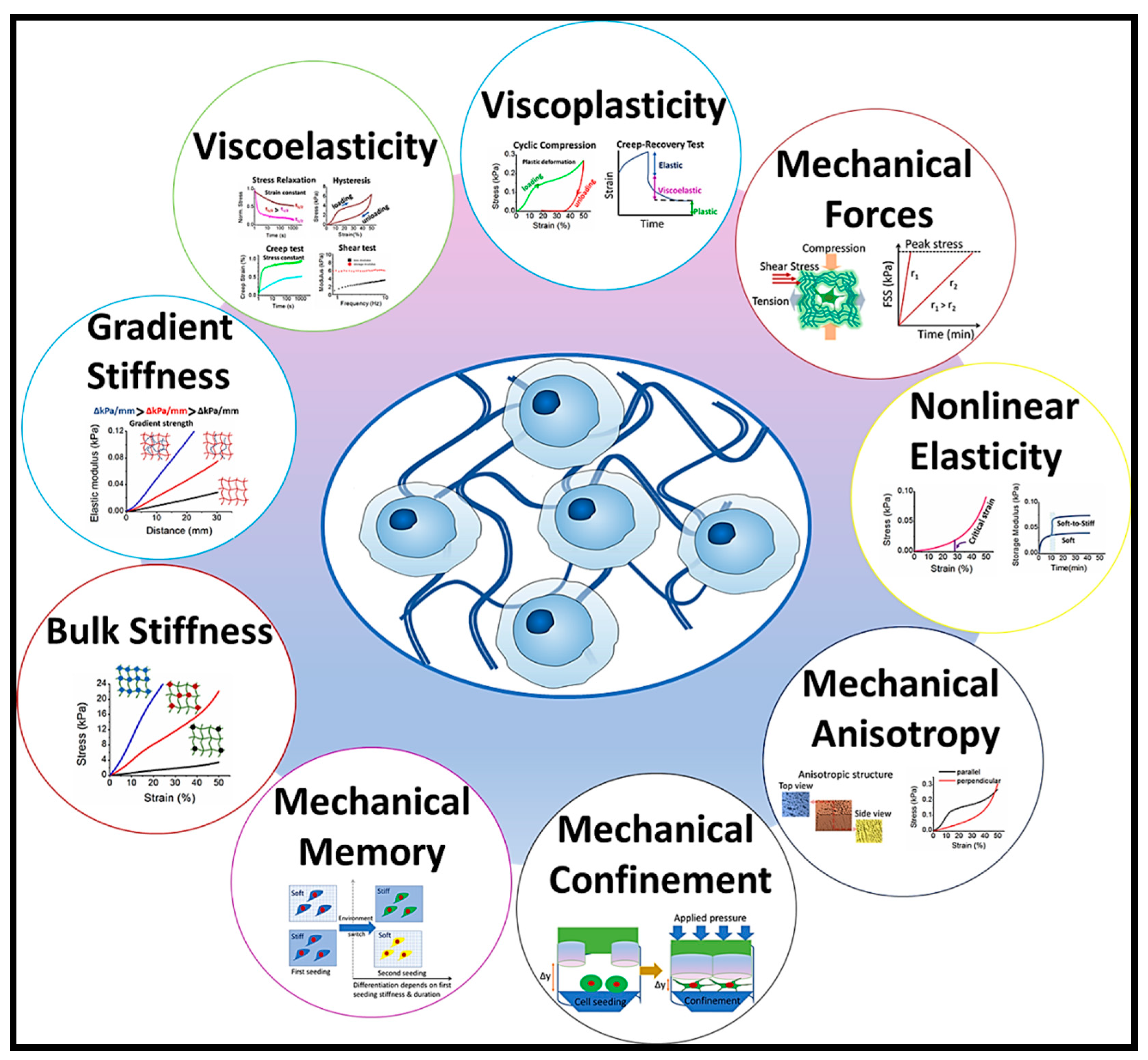
Figure 1. Visual representation depicting the mechanical variables influencing cellular activities such as cell spreading, proliferation, migration, and differentiation.
Preprints 87848 g001
Figure 2. Different types of crosslinking strategies.
Preprints 87848 g002
Figure 3. A visual depiction showcasing the bonding types for both physical and chemical cross-linking. Reproduced from ref. [51] under a Creative Commons Attribution 4.0 International License.
Preprints 87848 g003
Figure 4. Illustration of hydrogen bond and intra and intermolecular polymer chain entanglement. Reproduced from ref. [54] under a Creative Commons Attribution 4.0 International License.
Preprints 87848 g004
Figure 5. (a) Possible ionic and electrostatic interactions and swelling ratio (%) of unpurified (more divalent ions) and purified (less divalent ions) gelatin hydrogels; reproduced from ref. [71] under a Creative Commons Attribution 3.0 Unported License. (b) Pore morphologies and mechanical properties of unpurified and purified gelatin hydrogels; reproduced from ref. [71] under a Creative Commons Attribution 3.0 Unported License. (c) Cell attachment and morphology of EDC-crosslinked purified and unpurified gelatin hydrogels; reproduced from ref. [71] under a Creative Commons Attribution 3.0 Unported License.
Preprints 87848 g005
Figure 6. Mechanical properties of supramolecular hydrogel. (a) Supramolecular structure of CME-PRXs and the cell cultivation on supramolecular gelatin hydrogels crosslinked by CME-PRXs. (b) Stress–strain curves and Elongation at break of gelatin hydrogels cross-linked by CME-PRXs. (c) Cyclic tensile test graphs for gelatin hydrogels cross-linked by CME-PRX-37% and CME-PRX-24%, hysteresis loss for each cycle (sample size: n = 3). Reproduced from ref. [90].
Preprints 87848 g006
Figure 7. (a) Gelatin hydrogels synthesized via bioorthogonal cross-linking involving tetrazine (Tz) and norbornene (Nb) compounds. (b) The assessment of their mechanical characteristics. (c) Culturing human dental pulp stem cells (DPSCs) in fabricated 3D hydrogel matrices under in vitro conditions. Reproduced from ref. [117] under a Creative Commons Attribution 4.0 International License.
Preprints 87848 g007
Figure 8. Photocrosslinking mechanisms and associated mechanical, electrical and biocompatibility assessments of GelMA and GelMA-Bio-IL hydrogels. (a) Photopolymerization of GelMA, reproduced from ref. [129] under the Attribution-NonCommercial-NoDerivatives 4.0 Unported License. (b) Formation and mechanical characterization of photocrosslinked gelatin hydrogel. Reproduced from ref. [133] under a Creative Commons Attribution 4.0 International License (CC-BY), copyright 2019, Springer Nature. (c) Evaluation of in vitro and in vivo biocompatibility of photocrosslinked gelatin hydrogels. Reproduced from ref. [133] under a Creative Commons Attribution 4.0 International License (CC-BY), copyright 2019, Springer Nature. (d) Synthesis and 1H-NMR analysis of Bio-IL functionalized GelMA hydrogels prepared by visible light irradiation. Reproduced from ref. [145] under a Creative Commons Attribution 4.0 International License. (e) Mechanical, electrical and cell viability assessments of GelMA/Bio-IL hydrogels. Reproduced from ref. [145] under a Creative Commons Attribution 4.0 International License.
Preprints 87848 g008
Figure 9. (a) Different forms of interpenetrating polymer network and (b) Semi-IPN and full IPN formation strategies.
Preprints 87848 g009
Figure 10. (a) The preparation scheme of GelMA/HAMA/nHap double network hydrogel.; (b) Stress-strain curves and compressive modulus of GelMA/HAMA/nHap double network hydrogel (c) cell viability of double network hydrogel. Reproduced from ref. [200] under a Creative Commons Attribution 4.0 International License.
Preprints 87848 g010
Figure 11. Illustration of the typical methods employed to incorporate nanoparticles into hydrogel system. (a) Nanoparticle addition to pre-formed hydrogels; (b) Nanoparticle addition to polymer solutions and (c) Monomer polymerization in the presence of NPs.
Preprints 87848 g011
Figure 12. (a) Illustration of Additive or 3D Manufacturing process. Reproduced from ref. [170], [217]. (b) Stereomicroscope pictures of scaffolds created using Gelatin-Hyaluronic Acid Double-Network Hydrogel, a 3D bioprinting system with a temperature-controlled printhead and a cooled substrate, and graphs showing compressive modulus and stress-strain curves for the printed hydrogel scaffolds. Reproduced from ref. [170]. (c) Optical microscope pictures of 3D-printed pyrrole (Py)-modified oxidized alginate-gelatin (ADA-GEL) hydrogel prepared with temperature pre-treated gelatin, the printability factor of the hydrogel prints, light microscope images of a single-layer 3D-printed ADA-GEL, and rheological assessment of Py-infused ADA-GEL respectively. Reproduced from ref. [218]. (d) Stress-strain and initial elastic modulus curves of pyrrole (Py)-modified oxidized alginate-gelatin (ADA-GEL) hydrogel. Reproduced from ref. [218]. (e) Compressive stress and young modulus of Gelatin(Gel)/ Decellularized bone particles (dbPTs) scaffold. Reproduced from ref. [219]. (f) Cytocompatibility investigation of 3D printed alginate-gelatin/polypyrrole:polystyrenesulfonate (ADA-GEL-PPy:PSS) hydrogel with cells. Reproduced from ref. [218].
Preprints 87848 g012
Figure 13. Frequently used mechanical testing methods for hydrogel. (a) Tensile testing, (b) Compressive testing, (c) Constrained compression evaluation, (d) Indentation analysis, (e) Shear rheological assessment, (f) Dynamic mechanical analysis (DMA) investigation.
Preprints 87848 g013
Figure 14. Gelatin hydrogel in wide range of tissue engineering applications.
Preprints 87848 g014
Figure 15. (a) Cyclic compressive stress–strain curves of 3D gelatin-chitosan hybrid hydrogels. Reproduced from ref. [178]. (b) Proliferation of bone marrow mesenchymal stem cells (BM-hMSCs) and adipose tissue-derived human mesenchymal stem cells (AT-hMSCs) cultivated in gelatin hydrogel, and Immunofluorescence images show OCN expression in BM-hMSCs and AT-hMSCs cultured in hydrogel. Reproduced from ref. [178]. (c) Assessment of mineral deposition. Reproduced from ref. [178].
Preprints 87848 g015
Figure 16. (a) Mechanical characteristics of hybrid gelatin hydrogels. Reproduced from ref. [246]. (b) Markers for chondrogenesis, fibrosis, and hypertrophy in human bone marrow-derived mesenchymal stem cells (hBM-MSCs) undergoing chondrogenic differentiation within hybrid gelatin hydrogel. Reproduced from ref. [246]. (c) Chondrogenic differentiation of hBM-MSCs within gelatin hybrid hydrogel. Reproduced from ref. [246].
Preprints 87848 g016
Figure 17. Electrically conductive gelatin-based hydrogel promotes development of organized cardiac tissues. (a) The fabrication procedure of gelatin methacrylate (GelMA) hydrogel constructs comprised of surface micro-topographies incorporated with electrically conductive gold nanorods (GNRs) (GelMA–GNR); reproduced from ref. [259] under Creative Commons Attribution 3.0 Unported Licence. (b) SEM micrographs, porosity percentage, electrical impedance measurements and mechanical properties of GelMA and GelMA–GNR hydrogels; reproduced from ref. [259] under Creative Commons Attribution 3.0 Unported Licence. (c) Immunostained images of SATN (green) and Cx43 gap junctions (red) and quantified area coverage of cardiac specific markers within GelMA and GelMA–GNR microgrooved cardiac tissues on day 7 of culture; reproduced from ref. [259] under Creative Commons Attribution 3.0 Unported Licence. (d) Synchronous spontaneous beating behavior (beats per minute; BPM), voltage excitation thresholds and of cardiac tissues on GelMA and GelMA–GNR constructs. (B) and (C) extracted beating signals of cardiac tissues on GelMA and GelMA–GNR constructs; reproduced from ref. [259] under Creative Commons Attribution 3.0 Unported Licence.
Preprints 87848 g017
Figure 18. (a) Nerve guidance conduits (NGCs) using GelMA-based materials through digital light processing-based (DLP) printing. Reproduced from ref. [262]. (b) Neural crest stem cells cultured on GelMA nerve guidance conduits exhibited successful neuronal differentiation marked by extended neurites and the formation of cell junctions. Reproduced from ref. [262]. (c) Viscosity-shear rate and shear modulus of GelMA with water and GelMA. Reproduced from ref. [262].
Preprints 87848 g018
Table 1. Comparative analysis of mechanical properties of various types of gelatin hydrogels.
Hydrogel system Mode of testing/Testing instrument Type of crosslinking Crosslinker Young’s modulus (kPa) Tensile strength
(kPa)
Failure strength (kPa) Compressive modulus (kPa) Storage modulus
(kPa)
Reference
PAN-gelatin cryogel Dynamic mechanical analyzer (DMA) Free radical polymerization N,N’-methylene bis(acrylamide) and glutaraldehyde 123-819 [160]
GelMA–SF Instron 5542 mechanical tester UV irradiation ≈ 70 [76]
PVA/GE Tensile Enzymatic and physical mTG 980±2.45 750±0.18 [161]
GelMA with alginate Compression UV irradiation Ca2+ ion 201.2±5.5 [162]
GelMA with alginate (fish gelatin) Unconfined compression UV irradiation Ca2+ ion 48.6±4.7 [162]
Gelatin/PVA/PEG Unconfined compression Physical crosslinking PVA 189-351 [163]
GGMA/GelMA DN Unconfined, uniaxial compression test Photo-crosslinking 110 6900±100 [164]
Gelatin/PAAm Uniaxial tensile test Physical and covalent crosslinking 84 268 [165]
Bacterial cellulose-gelatin Tensile and compression Chemical crosslinking N-(3-dimethylaminopropyl)-N’-ethylcarbodiimide hydrochloride (EDC) 6700 610 [166]
Gelatin/PAM DN Compression Initiator-induced polymerization 1.76 150 3.52 [167]
Gelatin/PAAm/GO NC-DN gel Free radical polymerization N, N′-methyl-bis-acrylamide 187.3 324 [168]
Gelatin/PAAm DN gel Free radical polymerization N, N′-methyl-bis-acrylamide 76.4 306 [168]
γ-PGA-GEL DN hydrogel Compression Physical and covalent crosslinking 270 38000 [169]
GelMA/HA-HYD DN hydrogel Compression and tensile Photo-crosslinking 101±6.41 [170]
CAG cryogels Unconfined compression Chemical crosslinking Glutaraldehyde 120 44 [171]
Gelatin/PAAm/Laponite Nanocomposite Compression and Tensile Radical polymerization N,N′-Methylenebis-
(acrylamide)
20.5±0.2 337.9±4.9 208.4±5.9 [172]
RGO-gelatin hydrogel DMA Chemical crosslinking Graphene oxide 172.3 [173]
MA-gelatin/PAAm hydrogel Compressive and tensile Free radical
copolymerization
22.4±0.3 15.5±0.1 [174]
Graphene Oxide–Gelatin Nanocomposite hydrogel Oscillatory shear
measurement
Physical crosslinking 114.5 [175]
Gelatin−short linear glucan (SLG) nanocomposite hydrogels Texture profile analysis 50 1.9 [176]
Oxidized Alginate/Grlatin/Silicon Carbide Nanoparticle hydrogel (OA/GEL/SiC NPs) Compressive and tensile Chemical or/and physical crosslinking N-hydroxysuccinimide (NHS) and 1-ethyl-3-(3-dimethylaminopropyl) carbodiimide (EDC) 9.25±0.05 [177]
3D gelatin-chitosan hybrid hydrogel Compressive Chemical and physical PEG 680 250 [178]
Disclaimer/Publisher’s Note: The statements, opinions and data contained in all publications are solely those of the individual author(s) and contributor(s) and not of MDPI and/or the editor(s). MDPI and/or the editor(s) disclaim responsibility for any injury to people or property resulting from any ideas, methods, instructions or products referred to in the content.
Copyright: This open access article is published under a Creative Commons CC BY 4.0 license, which permit the free download, distribution, and reuse, provided that the author and preprint are cited in any reuse.
Prerpints.org logo

Preprints.org is a free preprint server supported by MDPI in Basel, Switzerland.

Subscribe

© 2024 MDPI (Basel, Switzerland) unless otherwise stated