Preprint
Review

Movement Asymmetries: from their Molecular Origin to the Analysis of Movement Asymmetries in Sportsmen

Altmetrics

Downloads

143

Views

141

Comments

0

A peer-reviewed article of this preprint also exists.

Submitted:

18 October 2023

Posted:

18 October 2023

You are already at the latest version

Alerts
Abstract
Asymmetry plays a major role in biology at all scales. This can be seen examining the helix of DNA, the fact that the human heart is on the left side, or that most people use their right hand. A single protein such as Myosin 1D can induce helical motion in another molecule. This causes cells, organs, and even entire bodies to twist in a domino effect, causing left-right behaviour. More in general, athlete movements are often asymmetric and, during the physical rehabilita-tion after injury the asymmetry is visually discernible. Herein we review the molecular basis of movement asymmetries and report on the available knowledge on the few therapeutics inves-tigated so far such as meloxicam. From a more rehabilitative perspective, it is very important to use effective methods to control the process of resolving the injury-related movement asym-metry through the complex use of specialized exercises, measurements and gait analysis which all can provide useful information on the effectiveness of rehabilitation plans. If for each athlete the normal range of asymmetry is known, the asymmetry can be treated individually and the evolution can be monitored over time. Appropriate measures should be taken if the movement asymmetry is outside this range. In addition, genetic, physiological, and psychological factors relevant to athlete health should be considered in the process of assessing and improving exer-cise asymmetry as we also discuss in this review. The main proposal of this work is that move-ment asymmetries in athletes should be treated individually, taking into account the athlete’s genetics, physical condition, and previous injuries.
Keywords: 
Subject: Public Health and Healthcare  -   Physical Therapy, Sports Therapy and Rehabilitation

1. Introduction

Asymmetry is one of the most important concepts in science in general and in biology in particular [1,2,3]. Movement asymmetries are correlated to molecular aspects, including the activity of particular proteins that determine the macroscopic behavioural asymmetric effects observed in organisms. This also holds true for the presence of asymmetries in sport [4,5]. Traditionally, sport science specialists have treated bilateral movement asymmetries in athletes as a sign of poor biomechanics and as a possible risk of injury [6,7,8,9,10]. This is true during injury rehabilitation when an athlete’s movements are restricted as a result of the injury [11,12,13]. However, the situation differs during exercise and training: there are many obviously asymmetric sports such as tennis, fencing, shot put and javelin. Some sports are only partially asymmetric (e. g. high jump, long jump, football and basketball), while some seem to be mostly symmetrical such as running and rowing [5]. It is also well known that asymmetries depend not only on the type of sport but on each individual and the applied test as well. Of course, asymmetric sports involve a certain amount of asymmetric movement. This leads to the uneven development of muscles on either side of an athlete’s body. If the asymmetries between the movements of the dominant and non-dominant limbs decrease, this can be a sign of fatigue in the dominant limbs or of the presence of micro-injuries [14,15]. At the same time, the majority of experts conclude that sports performance, risk of injury, and the ability to adapt to exercise and recover post injury depends on such biological factors as genetic predisposition to injury, age, muscle elasticity, physical condition and history of injury [16,17,18,19,20,21]. This suggests that the athlete's biomechanical, kinematic and kinetic parameters are tightly connected with the biomolecular processes in his/her muscles, ligaments, tendons, and bones. Without proper genetics, a good nutrition plan, and effective methods of bio-diagnostics, the athlete will fail to achieve good physical condition. Another aspect to consider is that there may also be perception-driven movement asymmetries caused by asymmetries in visual, audio, or other senses. For example, athletes whose attention is unevenly distributed in different directions are more likely to also have reduced reaction and movement asymmetries in certain directions, and, as a result, will be more prone to injury [22,23]. In this paper we explore the current knowledge on the molecular basis of the movement asymmetries and the role of movement asymmetries in sport, in order to define the line between positive and negative aspects related to asymmetries and find out how biomechanical and biological processes in an athlete’s organism are connected to each other. The main applicative purpose of our work is to summarise modern strategies and methods adopted for the measurement, evaluation and interpretation of movement asymmetries in sport and sports rehabilitation. This review also aims to understand the role of genetics in the formation and development of movement asymmetries in humans and especially in athletes. As for the methodology used in this work, we explored the recent scientific literature relating to themes reported in the Introduction section, as well as the keywords ‘movement asymmetry; sports; physical rehabilitation; genetics; nutrition’. For this purpose, we used scientific search engines such as PubMed and Google Scholar. In our search we excluded retracted publications and non-English articles.

2. Biomechanical aspects of movement asymmetries in sport.

Specialists agree that asymmetry is relatively normal in sport, since the vast majority of people have a certain degree of asymmetry within their physiognomy. Functional lower-limb asymmetries can be assessed using jumping tests including single-leg vertical jumps, countermovement jumps and drop jumps [24,25,26,27,28,29,30]. Lower-limb strength asymmetries can be assessed using back squats, isometric squats, and isokinetic knee extensions [31,32,33,34]. According to massive studies of the acceptable thresholds of asymmetry, 10–15% asymmetry is associated with abnormal differences between limbs [35,36]. Moreover, a ≤10% asymmetry has been suggested as a goal for athletes returning to sport [36]. At the same time, experts agree that coaches and physical rehabilitation specialists should treat movement asymmetries individually, taking into account the athlete’s genetic, physiological and physical data [37,38,39,40]. It is very important to know the athlete’s optimal percentage of movement asymmetries related to the bilateral asymmetry characteristics of his/her sport. If the difference between the normal and observable asymmetries is too large (more than 10%) or if it is too small (for example, less than half of the normal asymmetry coefficient) then some measures should be taken to return to a normal level. In the first case, the difference should be normalised by improving the non-dominant side biomechanics, while in the second case, we should improve the dominant side biomechanics. Unfortunately, there is no general agreement on how to estimate the athlete’s movement asymmetries coefficient. For this purpose, we can use some standard exercises and measurements – for example, left- and right-hand force measurements, single-hand exercises with balls and dumbbells, vertical jumps on a single leg and ROMs (ranges of motion) measurements of active joints. Knowing the normal parameters for these exercises, coaches can detect abnormalities in an athlete’s performance and conduct a more detailed analysis of their condition. For more accurate estimation of movement asymmetries, specialists use Motion Capture (MoCap) methods [41,42,43,44,45,46] and techniques for registering and measuring ground forces such as force plates and treadmills [47,48,49,50]. These methods allow us to estimate movement asymmetries during walking and running. Of course, measurement results depend on the physical condition of the athlete: rested or tired, having muscle imbalance or dysfunction or not, etc. In order to study the athlete’s muscle activity, specialists usually use electromyography (EMG), which is useful to help detect neuromuscular abnormalities [51,52,53]. Usually, movement asymmetries caused by an injury can be seen with the naked eye. In this case, athletes' movements may be constrained by effusion or pain leading to reduced muscle activation and weakness and can be easily detected without the use of special techniques[54]. We have a similar situation during an athlete’s rehabilitation after injury where compensations strategies can be observed in the injured limb and other areas of the body when compared with healthy controls[55]. In this case, an effective rehabilitation plan should be implemented by physical rehabilitation specialists taking into account the character of the injury and abnormalities observed[55]. Not less important is the detection of movement asymmetries associated with microtrauma that do not cause pain. In this case, the athlete can maintain a high level of performance by using compensatory movements and thereby increasing the risk of injury. For the estimation of movement asymmetries caused by hidden microtrauma, specialists often use screening tools such as the Functional Movement Screen (FMS), which was identified to be the most popular field-based injury screening tool for identifying at-risk athletes. FMS consists of 7 basic exercises for detecting potential risks of injury. The main purpose of this screening tool is to determine whether the athlete is limited in fundamental movements [56,57]. Athletes with microtrauma often demonstrate good performance during sporting events because they use compensatory movements to maintain a high level of performance. The use of FMS may help the coach or physical medicine specialist detect movement asymmetries during control exercises (Deep Squat, Hurdle Step, In-line Lunge, Active Straight-leg Raise, Trunk Stability Push-up, Rotary Stability, Shoulder Mobility). Only two exercises (Deep Squat and Trunk Stability Push-up ) are symmetrical, while the other five include asymmetric movements and can reveal asymmetries in the movements of the athlete’s limbs. It has been shown that participating in multiple sports throughout multiple sporting seasons was associated with higher total FMS scores and fewer asymmetries, which may decrease subsequent risk of injury [58]. Some authors recommend that coaching/medical staff apply collective training programs with emphasis on trunk stabilisation improvements (especially in younger players), while other specific deficits evidenced by FMS may be looked at on an individual basis or in sub-groups [59]. According to the work of Mokha et al., asymmetry or a low FMS individual test score, was a better predictor of musculoskeletal injury than the composite FMS score [60]. Specialists also use FMS to obtain additional information about functional inter-limb asymmetries [61]. Australian scientists have proved FMS efficiency when combined with single-leg vertical jumps and previous injury [61]. For a more detailed analysis, we can use Motion Capture techniques: the parallel use of FMS and Motion Capture is recommended when the athlete has complex movement asymmetries which cannot be easily identified. Motion Capture allows us to measure flexion and extension angles more accurately [62,63,64]. In many cases, the use of two-dimensional programs is enough [65,66]. One such program is the 2D motion analysis Kinovea. It features tools to compare, slow down, measure and annotate motion in videos. The methods described above may be used in different combinations or separately, but in all cases, one should take into consideration the athlete’s physical condition - rested or tired, pre-existing bilateral asymmetries, movement asymmetries related to their sport and their history of injury. During rehabilitation, physical rehabilitation specialists can use simple physical exercises and measurements to control the injured athlete’s condition. Alternatively, they can use motion capture techniques to acquire more detailed information about biomechanical parameters responsible for the athlete’s movement asymmetries. For each measurement, it is possible to calculate the corresponding movement asymmetries coefficient.

3. Biological aspects of movement asymmetries

From the outside, most organisms appear symmetrical. However, many organs are asymmetrical and overall our world is fundamentally asymmetric. An example is given by the double helix of DNA, the asymmetric division of stem cells, or the fact that the human heart is positioned on the left (Figure 1a). However, the mechanism by which these asymmetries emerge and how they are linked to one another are aspects that still need to be elucidated. For example, whether macroscopic asymmetry is directly related to the chirality observed at the molecular level remains an open question. Working with Drosophila, Lebreton et al. found that a protein acting as the conserved molecular motor in the human body, such as myosin (Figure 1b), induces stereotypic chirality at all biological scales, from its F-actin transduction in vitro to the organ level, till to organismal behaviour [67]. Thus, this protein can produce de novo changes in shape and orientation not only at a nanometric but also macroscopic scale via chiral interactions with the actin cytoskeleton [67]. A molecular basis of lateral defects was also proven in humans [68]. In fact an extremely rare novel missense variant in MYO1D gene (Pro765Ser) was observed in correlation with visceral heterotaxy and left isomerism in polysplenia syndrome in humans. More in detail, the heterotaxy was found to result from the failure of the developing embryo to establish normal left-right asymmetry [68]. Remarkably, typical manifestations following the above-mentioned biomolecular alterations include an abnormal symmetry in the resulting organism. Surprisingly, a single mutated amino acid, Ser765, causes minor structural drifts and stability changes, that potentially affect the overall biophysical and functional properties of the resulting myosin including those connected with the microfilament motor activities. Importantly, MYO1D is ubiquitously expressed across several human tissues and is reported to induce severe phenotypes in knockout mouse models [68]. The above-described study constitutes the first report that a MYO1D genetic variant may cause left-right axis abnormalities in humans, adding to already known evidences from zebrafish, frog and Drosophila models [67]. Interestingly, the ability of Ser765 to induce small structural deviations and changes altering the stability and the function of myosin was proven using a bioinformatics-based multilevel structural analysis (structural modelling of protein variants, divergence, and stability). This study resulted an important expansion of the previous knowledge on the genetic origin of human lateral defects, potentially opening the door to the identification of new drug targets and the development of personalized medicine for high-risk families. [68]

3.1. Genetic factors and asymmetry

When dealing with movement asymmetries we cannot ignore that apart from the particular sport, the elasticity and quality of their muscles (density, separation and definition), the quality of their nutritional plan and physical activity environments as well as their history of previous injuries, such biological factors as the athlete’s DNA and consequently his/her genetic predisposition play a crucial role. Genetics influence all areas of fitness and performance and has a large influence over strength, muscle size and muscle fiber composition (fast or slow twitch), anaerobic threshold (AT), lung capacity, flexibility, and, to some extent, endurance. The following genetic factors should be given attention during training:
  • Genetic sensitivity to pain: according to the latest experimental data, a deficiency in the AMPD1 gene can cause symptoms such as pain and muscle weakness after exercise [16,17]. In asymmetric sports, dominant limbs are under more pressure than non-dominant limbs and, therefore, an athlete with higher pain tolerance will continue training with micro-injuries in the dominant limbs without feeling pain. This may lead to more serious injuries. In this case, the only way to detect abnormal changes in the athlete’s behaviour is to perform performance tests in time. If the dominant limbs move slower or have less force, then this may be a sign of a hidden injury. In the case of athletes with lower pain tolerance, even non-significant injuries will cause pain and impair movement performance. For such athletes, specialists recommend using yoga, biofeedback therapy and exercises to increase pain tolerance.
  • Genetic predisposition to injuries: it was found that the genes AMPD1, GDF5, COL5A1 and IGF2 might be associated with increased injury occurrence in soccer players [16,18]. According to the latest research, the TT genotype of the GDF5 gene may be associated with an increased number of ankle and knee injuries as well as an increase in the total number of injuries and a decrease in the number of matches played [16]. This suggests that athletes who are genetically prone to certain types of injuries should be tested for such undetected injuries, especially in the dominant limbs.
  • Genetic need for increased recovery after hard activity: The AMPD1 gene has been shown to be linked to such a need [17]. It was suggested that special attention should also be paid to the dominant limbs which will be under excessive physical load in asymmetric sports. The athlete should be given enough time for recovery. Obviously, the athletes with more endurance and a shorter recovery time will be favoured for such sports.
  • Genetic deficit of muscle elasticity or power: There is evidence suggesting that titin (encoded by the TTN gene), a protein that is expressed in cardiac and skeletal muscles, is one of the factors responsible for the muscles' elasticity [19]. In some asymmetric sports, athletes with insufficient muscle elasticity may have issues with movements requiring a larger range of motion. Such athletes are recommended individual plans for training and nutrition [20,21].
In the next section we will discuss on the importance of assessing movement asymmetries in athletes through physical tests and on the rehabilitation. It is obvious that the results of the tests depend on the athlete’s physical and psychological condition. Tests may be taken after a period of rest in order to estimate whether the athlete is prepared for training and competition, or, alternatively, the athlete’s movement asymmetries can be tested after training in order to estimate how physical load and fatigue affect his physical condition and the bilateral asymmetries of his movements. Great relevance is given also to rehabilitation. If a non-dominant limb is injured, then the difference between non-dominant and dominant limbs will increase. Otherwise, if a dominant limb is injured, then the difference between limbs may decrease in case of minor injury, or the non-dominant limb may become even stronger in case of a more serious injury. In both cases, the injured limb should be trained with the purpose to regain its strength. The effectiveness of rehabilitation will depend on the athlete's genetic ability to recover. Athletes with higher pain tolerance will recover faster than athletes with higher sensitivity to pain. The rehabilitation process can be hampered by poor nutrition or previous injuries.

4. Detection of movement asymmetries and physical rehabilitation

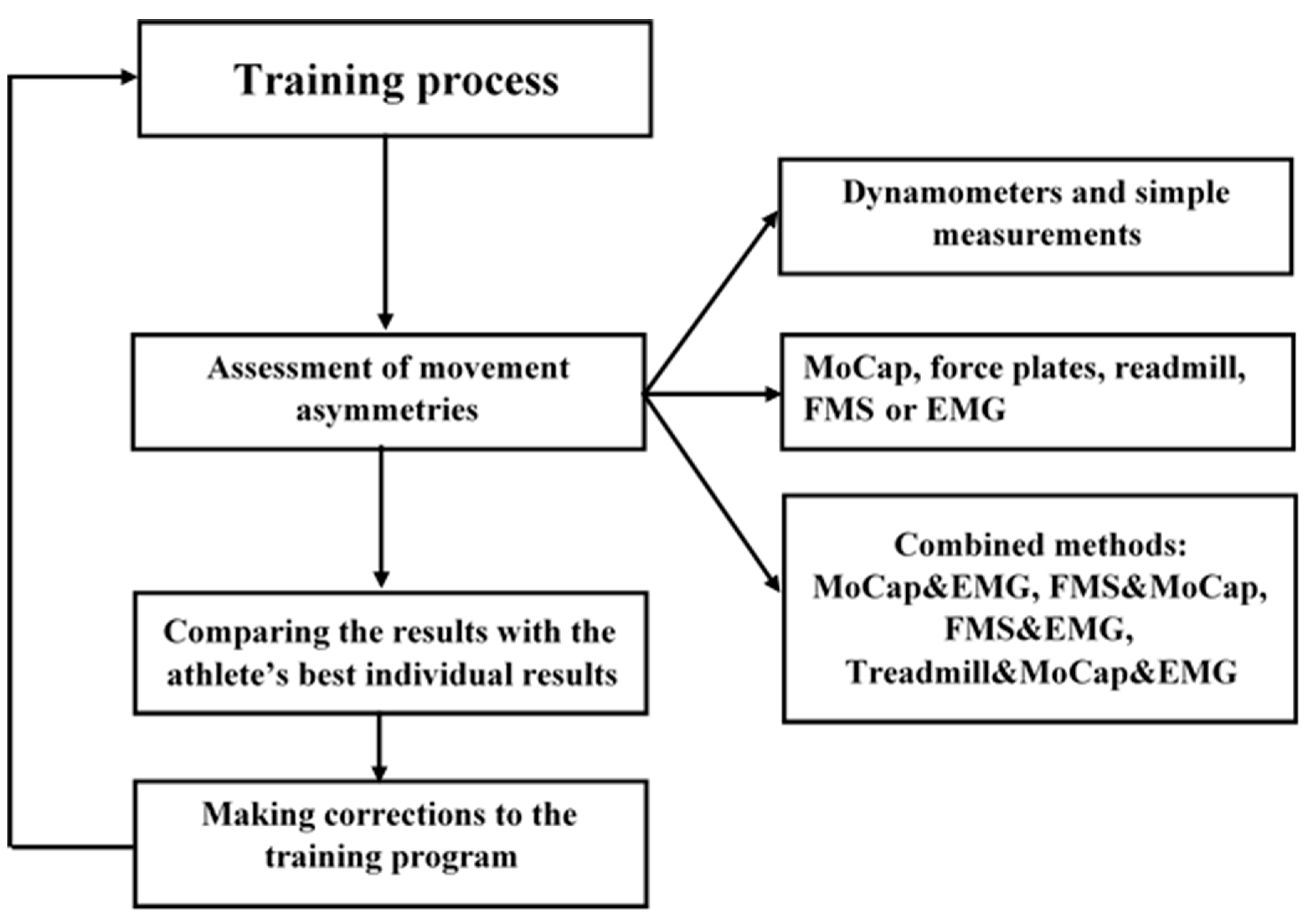
Knowledge of the above-mentioned biomechanical and biological aspects related to movement asymmetries can help us elaborate recommendations on how coaches and physical rehabilitation specialists should detect negative movement asymmetries and restore the normal state of the athlete. In this review, we consider two different types of negative movement asymmetries: i) the first one is related to negative movement asymmetries, which emerge during the training process and may be caused by factors such as improper training, overload, and poor nutrition. In this case, it is very important to detect such asymmetries in time and take the necessary measures to prevent their further development into more serious changes in the athlete's organism; ii) the second one is related to the post-injury negative movement asymmetries, which are usually present during injury rehabilitation. In this case, physical rehabilitation specialists should provide an effective rehabilitation plan involving not only the optimal treatment plan but methods for evaluating its effectiveness as well. The detection of movement asymmetries of the first type will happen during the training process. During this stage, we suggest the following scheme consisting of three stages (see Figure 2). The first stage involves the estimation of the athlete’s basic physical and physiological parameters. Two procedures will be appropriate at this stage: i) during exercises, coaches should keep control of the optimal parameters of physical condition for each athlete. For example, force measurements using dynamometers - best results for both hands, single-leg jumps (SLJ) - best results for both legs, 100-meter sprint results, and others; ii) physical rehabilitation specialists should keep track of the athlete’s physiological parameters such as heartbeat frequency, blood pressure and blood oxygen saturation. Deterioration in the values of these parameters may be caused by fatigue. At this stage, we can detect slight negative changes in the athlete’s condition and make corrections to the training process.
The second stage aims to detect more hidden movement asymmetries and deviations in the athlete's physical and physiological parameters. This stage includes the following procedures: i) the use of more sophisticated techniques for the estimation of movement asymmetries such as motion capture, force plates and treadmills; ii) the use of screening tools, such as FMS. This will help one predict possible trauma risks; iii) the assessment of the athlete’s muscle activity using EMG. EMG-based FMS can effectively detect early sports injuries and plays an effective role in reducing sports injuries. In the third stage, we can use different combinations of the above-mentioned methods, for example, MoCap and EMG, FMS and MoCap, MoCap with force plate and EMG, etc. This will increase the data integrity and consistency, making it more flexible and accurate.
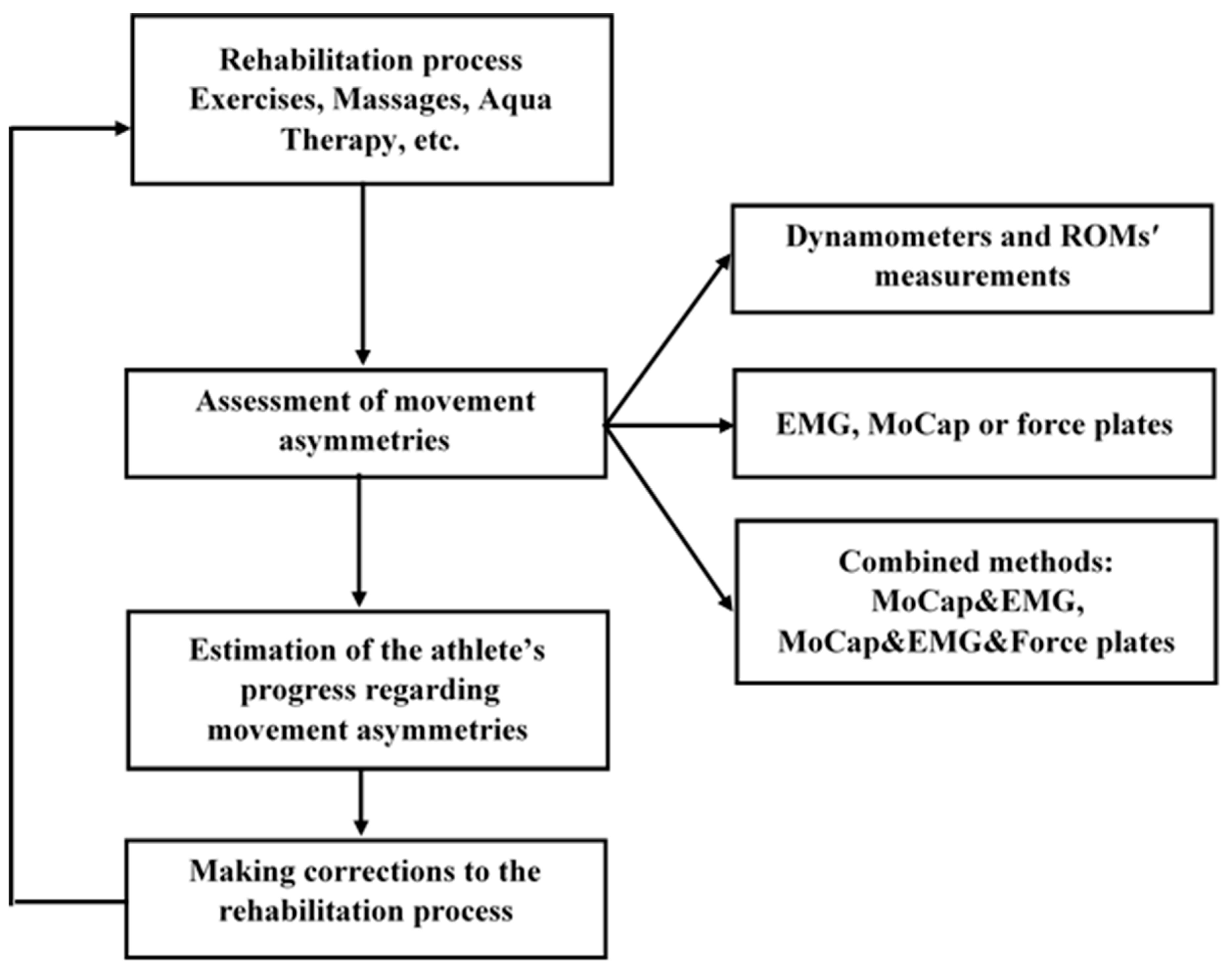
During injury rehabilitation, the main task for physiotherapists is to normalise the condition of the injured joints and muscles, which in most cases restricts the athlete’s ability to move and manifest themselves through movement asymmetries. For this purpose, specialists of physical rehabilitation use kinesiotherapy, massages, physiotherapy and aqua therapy among other methods (see Figure 3). The common practice for monitoring the progress of treatment is to use special control exercises, goniometers, gait analysis and electromyography. These techniques allow one to estimate muscle strength and range of movement (ROMs) of the injured muscles and joints. If the process of treatment does not bring noticeable changes to the athlete’s condition, the physiotherapist should modify the methods and techniques used during the treatment.
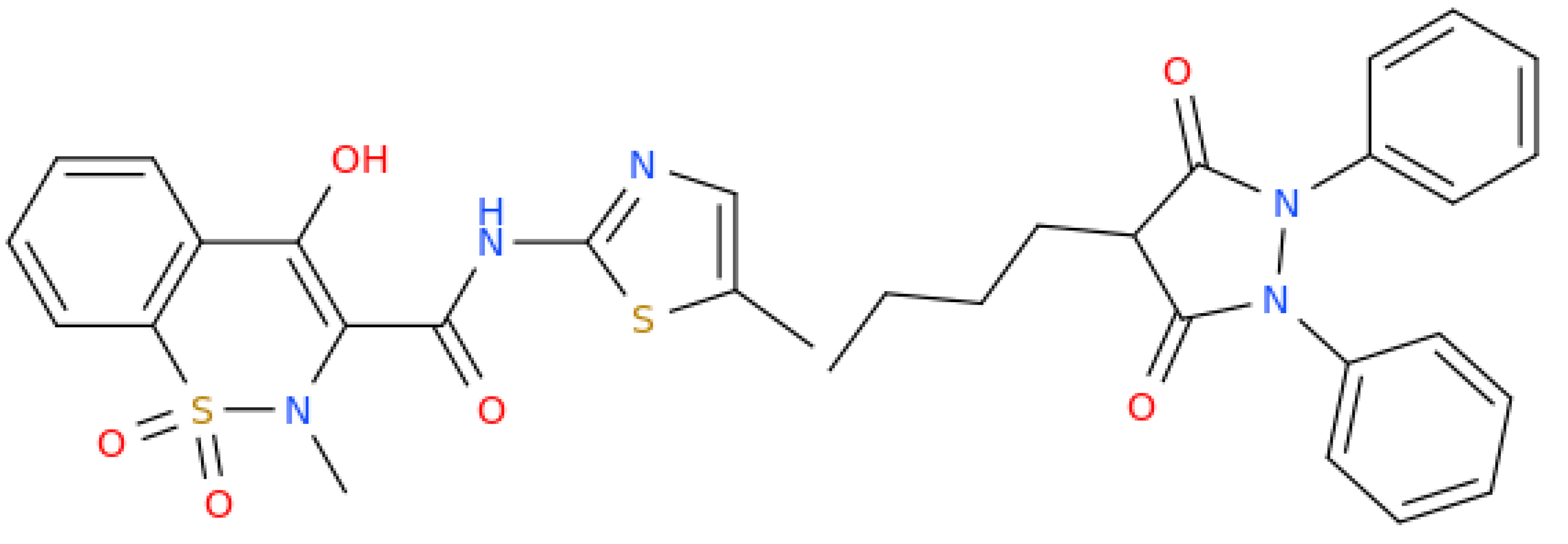
Alternatively, more sophisticated techniques for movement analysis should be considered. This can be a combination of different techniques such as MoCap, EMG and force plates (see Figure 3). The parallel use of these methods makes it possible to understand the real connection between movement asymmetries and muscle activity. This will help the physiotherapist to find out which muscles are responsible for the athlete’s weakness and slow recovery. In some cases, injuries may be caused by asymmetries in the athlete’s reactions: they may have different reaction times in different directions or their attention may be mostly concentrated in certain directions over others. Such situations often occur in team sports such as football or hockey, where the player has a certain position on the playing field that determines the focus of his attention. In sports, where attention is distributed unevenly in different directions, specialists recommend athletes pass special cognitive tests to check and improve their reaction time [22,23]. Another important factor determining an athlete’s susceptibility to injury may be the rules of the sport in question. According to recent works, similar to wrestling, sidedness has an impact on tactics and fighting techniques [69,70,71,72]. For example, the study of the lateralization of injuries in judo is significant since determining the side of injury helps to identify technical factors affecting the risk of injury [69]. Overall, several methods are commonly used for detecting and improving the negative movement asymmetries in athletes during the training and the rehabilitation. During training, in fact, it is important to identify the asymmetries in an athlete's movements that may be caused by overload and micro-injuries. To accomplish this there are a variety of methods at our disposal, including physical exercises and measurements to estimate muscle strength and joint range of motion. In addition, screening tools such as Functional Movements Screen, Motion Capture technology and electromyography can be used. The in depth information captured by these tools will help us decrease the risk of injury and optimise the training process. During injury rehabilitation, an athlete’s movements are often asymmetrical, where asymmetries can be identified visually. In this case, it is important to use effective methods in order to control the process of elimination of movement asymmetries related to the injury. The combined use of special exercises, measurements, and gait analysis may provide us with useful information about the effectiveness of the rehabilitation plan. We state that movement asymmetries should be treated individually, where each athlete will know his/her normal range of movement asymmetries. If movement asymmetries are out of this range, appropriate action must be taken. During the process of assessment and improvement of movement asymmetries, also genetic, physiological and psychological factors regarding the athlete’s health should be taken into account. Few studies were conducted on therapeutics able to treat movement asymmetries. Meloxicam (use name for 4-hydroxy-2-methyl-N-(5-methyl-1,3-thiazol-2-yl)-1,1-dioxo-1lambda6,2-benzothiazine-3-carboxamide, Figure 4) tested together with other non-steroidal anti-inflammatory drugs such as phenylbutazone (4-butyl-1,2-diphenylpyrazolidine-3,5-dione, Figure 4) was shown to result in a reduction in head movement asymmetry [73], however the same activity was not observed in a subsequent study conducted on horses [74]. This latter finding seems to indicate that the movement asymmetries can be expressions of biological variation and are not necessarily related to pain/dysfunction that would be responsive to anti-inflammatory treatment.

5. Conclusions

Asymmetry is found at all levels in our world starting from chiral compounds to living organisms endowed with asymmetrical behaviours. From a biomolecular and mechanistic perspective, single residue mutations of motion-related proteins such as myosin can determine functional changes of the biological structures that lead to the macroscopic movement determining asymmetry as seen in insects and other organisms, including humans. Movement asymmetries are at least to some extent normal in humans and in particular in sportsmen. These could anyway show too significant asymmetries in some cases that need to be carefully assessed and treated by using physical therapy, which is the best weapon we can use to counteract the negative effects of asymmetries, lacking so far any valid therapeutics to be used for the same purpose.
After a detailed analysis of the possible impacts of movement asymmetries on sports performance and the athlete’s physical condition, we can make the following conclusions. Movement asymmetries should be treated differently during training and rehabilitation. During training, knowing an athlete’s normal movement asymmetries will help coaches identify abnormal asymmetries in a timely manner and allow them to take appropriate action. In asymmetric sports, special attention should be paid to the most active limbs that experience greater physical stress. Coaches can use simple control exercises and measurements to estimate the condition of the dominant limbs of athletes. If the results of testing are significantly worse than normal indicators, then we can look into pathological changes in the athlete’s organism. During rehabilitation, physical rehabilitation specialists should put together an effective rehabilitation plan to eliminate movement asymmetries caused by injury. The progress of rehabilitation depends not only on the methods and technologies used, but on genetic factors and previous injuries as well. In both cases, the effect of physical activity on the athlete’s movement asymmetries will depend on genetic factors such as sensitivity to pain, predisposition to injury, injury recovery rate as well as muscle strength and elasticity. Other important factors include nutrition [75,76], and previous injuries. Health/fitness professionals are likely to face future challenges in how to optimise exercise training for individuals with desirable genes, as well as for motivating individuals without them to begin and/or continue exercising [77]. To meet these challenges, health/fitness professionals will need to become familiar with exercise genomics (i.e., studying how genetic and environmental factors affect exercise performance) and how it affects muscle strength, aerobic capacity and other exercise-related phenotypes.

Author Contributions

All authors are contributed equally to this work. All authors have read and agreed to the published version of the manuscript.

Funding

This research received no external funding.

Institutional Review Board Statement

Not applicable.

Informed Consent Statement

Not applicable.

Data Availability Statement

Not applicable.

Acknowledgments

We also acknowledge the Italian National Council for Research (CNR) and Compete Physiotherapy and Rehabilitation Center (www.competephysio.co.uk, accessed on 02 October 2023) for their support of this research.

Conflicts of Interest

The authors declare no conflict of interest.

References

  1. Ocklenburg, S.; Mundorf, A. Symmetry and Asymmetry in Biological Structures. Proc Natl Acad Sci USA 2022, 119(28), e2204881119. [Google Scholar] [CrossRef]
  2. Mundorf, A.; Peterburs, J.; Ocklenburg, S. Asymmetry in the Central Nervous System: A Clinical Neuroscience Perspective. Front. Syst. Neurosci. 2021, 15, 733898. [Google Scholar] [CrossRef]
  3. Paillard, T. Asymmetry of Movement and Postural Balance and Underlying Functions in Humans. Symmetry 2023, 15(3), 759. [Google Scholar] [CrossRef]
  4. Kadri, M.A.; Noé, F.; Maitre, J.; Maffulli, N. Paillard, T. Effects of Limb Dominance on Postural Balance in Sportsmen Practicing Symmetric and Asymmetric Sports: A Pilot Study. Symmetry 2021, 13(11), 2199. [CrossRef]
  5. Afonso, J.; Peña, J.; Sá, M.; Virgile, A.; García-de-Alcaraz, A.; Bishop, C. Why Sports Should Embrace Bilateral Asymmetry: A Narrative Review. Symmetry 2022, 14(10), 1993. [Google Scholar] [CrossRef]
  6. Bishop, C.; Turner, A.; Read, P. Training Methods and Considerations for Practitioners to Reduce Inter-Limb Asymmetries. Strength Cond J 2018, 40, 40–46. [Google Scholar] [CrossRef]
  7. Carpes, F.P.; Mota, C.B.; Faria, I.E. On the Bilateral Asymmetry during Running and Cycling - a Review Considering Leg Preference. Phys Ther Sport. 2010, 11(4), 136–42. [Google Scholar] [CrossRef] [PubMed]
  8. Fort-Vanmeerhaeghe, A.; Bishop, C.; Buscà, B.; Aguilera-Castells, J.; Vicens-Bordas, J.; Gonzalo-Skok, O. Inter-Limb Asymmetries are Associated with Decrements in Physical Performance in Youth Elite Team Sports Athletes. PLoS One 2020, 15(3), e0229440. [Google Scholar] [CrossRef] [PubMed]
  9. Fort-Vanmeerhaeghe, A.; Gual, G.; Romero-Rodriguez, D.; Unnitha, V. Lower Limb Neuromuscular Asymmetry in Volleyball and Basketball Players. J Human Kinet 2016, 50, 135–144. [Google Scholar] [CrossRef]
  10. Hart, N.; Nimphius, S.; Spiteri, T.; Newton, R. Leg Strength and Lean Mass Symmetry Influences Kicking Performance in Australian Football. J Sports Sci Med 2014, 13, 157–165. [Google Scholar]
  11. Bishop, C.; Turner, A.; Gonzalo-Skok, O.; Read, P. Inter-Limb Asymmetry during Rehabilitation: Understanding Formulas and Monitoring the "Magnitude" and "Direction". Aspetar 2020, 18–22. [Google Scholar]
  12. Noyes, F.; Barber, S.; Mangine, R. Abnormal Lower Limb Symmetry Determined by Function Hop Tests after Anterior Cruciate Ligament Rupture. Amer J Sports Med 1991, 19, 513–518. [Google Scholar] [CrossRef] [PubMed]
  13. Rohman, E.; Steubs, J.; Tompkins, M. Changes in Involved and Uninvolved Limb Function during Rehabilitation after Anterior Cruciate Ligament Reconstruction: Implications for Limb Symmetry Index Measures. Amer J Sports Med 2015, 43, 1391–1398. [Google Scholar] [CrossRef] [PubMed]
  14. Svensson, K.; Eckerman, M.; Alricsson, M.; Magounakis, T.; Werner, S. Muscle injuries of the dominant or non-dominant leg in male football players at elite level. Knee Surg. Sports Traumatol. Arthrosc. 2018, 26, 933–937. [Google Scholar] [CrossRef] [PubMed]
  15. DeLang, D.M.; Salamh, P.A.; Farooq, A.; Tabben, M.; Whiteley, R.; van Dyk, N.; Chamari, K. The dominant leg is more likely to get injured in soccer players: Systematic review and meta-analysis. Biol. Sport 2021, 38, 397–435. [Google Scholar] [CrossRef] [PubMed]
  16. McCabe, K.; Collins, C. Can Genetics Predict Sports Injury? The Association of the Genes GDF5, AMPD1, COL5A1 and IGF2 on Soccer Player Injury Occurrence. Sports (Basel) 2018, 6(1), 21. [Google Scholar] [CrossRef]
  17. Collins, C. Resistance Training, Recovery and Genetics: AMPD1 the Gene for Recovery. J. Athl. Enhanc. 2017, 6. [Google Scholar] [CrossRef]
  18. Posthumus, M.; September, A.V.; O’Cuinneagain, D.; van der Merwe, W.; Schwellnus, M.P.; Collins, M. The COL5A1 Gene Is Associated with Increased Risk of Anterior Cruciate Ligament Ruptures in Female Participants. Am. J. Sports Med. 2009, 37, 2234–2240. [Google Scholar] [CrossRef]
  19. Maciejewska-Skrendo, A.; Leźnicka, K.; Leońska-Duniec, A.; Wilk, M.; Filip, A.; Cięszczyk, P.; Sawczuk, M. Genetics of Muscle Stiffness, Muscle Elasticity and Explosive Strength. J Hum Kinet. 2020, 74, 143–159. [Google Scholar] [CrossRef]
  20. Guest, N.S.; Horne, J.; Vanderhout, S.M.; El-Sohemy, A. Sport Nutrigenomics: Personalized Nutrition for Athletic Performance. Front Nutr. 2019, 6, 8. [Google Scholar] [CrossRef]
  21. Guest, N.; Corey, P.; Vescovi, J.; El-Sohemy, A. Caffeine, CYP1A2 Genotype, and Endurance Performance in Athletes. Med Sci Sports Exerc. 2018, 50, 1570–8. [Google Scholar] [CrossRef]
  22. Egoyan, A.; Khipashvili, I. Use of Psychophysiological Computer Tests during the Process of Sportsmen’s Preparation, Physical Education. Sport and Science (PSS) 2017, 3, 8–17. [Google Scholar] [CrossRef]
  23. Wilkerson, G.B.; Simpson, K.A.; Clark, R.A. Assessment and Training of Visuomotor Reaction Time for Football Injury Prevention. J Sport Rehabil. 2017, 26(1), 26–34. [Google Scholar] [CrossRef]
  24. Impellizzeri, F.M.; Rampinini, E.; Maffiuletti, N.; Marcora, S.M. A Vertical Jump Force Test for Assessing Bilateral Strength Asymmetry in Athletes. Med. Sci. Sport Exerc. 2007, 39, 2044–2050. [Google Scholar] [CrossRef] [PubMed]
  25. Madruga-Parera, M.; Bishop, C.; Read, P.; Lake, J.; Brazier, J.; Romero-Rodriguez, D. Jumping-based Asymmetries are Negatively Associated with Jump, Change of Direction, and Repeated Sprint Performance, but not Linear Speed, in Adolescent Handball Athletes. J. Hum. Kinet. 2020, 71, 47–58. [Google Scholar] [CrossRef] [PubMed]
  26. Hewit, J.; Cronin, J.; Hume, P. Asymmetry in Multi-Directional Jumping Tasks. Phys Ther Sport 2012, 13, 238–42. [Google Scholar] [CrossRef]
  27. Menzel, H.J.; Chagas, M.H.; Szmuchrowski, L.A.; Araujo, S.R.S.; de Andrade, A.G.P.; de Jesus-Moraleida, F.R. Analysis of Lower Limb Asymmetries by Isokinetic and Vertical Jump Tests in Soccer Players. J Strength Cond Res 2013, 27, 1370–7. [Google Scholar] [CrossRef]
  28. Paterno, M.V.; Ford, K.R.; Myer, G.D.; Heyl, R.; Hewett, T.E. Limb Asymmetries in Landing and Jumping 2 Years Following Anterior Cruciate Ligament Reconstruction. Clin J Sport Med 2007, 17, 258–62. [Google Scholar] [CrossRef]
  29. Roso-Moliner, A.; Lozano, D.; Nobari, H.; Bishop, C.; Carton-Llorente, A.; Mainer-Pardos, E. Horizontal Jump Asymmetries are Associated with Reduced Range of Motion and Vertical Jump Performance in Female Soccer Players. BMC Sports Sci Med Rehabil. 2023, 15(1), 80. [Google Scholar] [CrossRef]
  30. Bishop, C.; Pereira, L.A.; Reis, V.P.; Read, P.; Turner, A.N.; Loturco, I. Comparing the Magnitude and Direction of Asymmetry during the Squat, Countermovement and Drop Jump Tests in Elite Youth Female Soccer Players. J Sports Sci. 2020, 38, 1296–1303. [Google Scholar] [CrossRef]
  31. Costa Silva, J.R.L.; Detanico, D.; Dal Pupo, J.; Freitas, C. Bilateral Asymmetry of Knee and Ankle Isokinetic Torque in Soccer Players u20 Category. Braz J Kinanthro Hum Perf 2015, 17, 195–204. [Google Scholar] [CrossRef]
  32. Dos'Santos, T.; Thomas, C.; Jones, P.; Comfort, P. Asymmetries in Single and Triple Hop are not Detrimental to Change of Direction Speed. J Trainology 2017, 6, 35–41. [Google Scholar] [CrossRef]
  33. Dos'Santos, T.; Thomas, C.; Jones, P.A.; Comfort, P. Assessing Muscle Strength Asymmetry via a Unilateral Stance Isometric Mid-Thigh Pull. Int J Sports Physiol Perf 2017, 12, 505–511. [Google Scholar] [CrossRef] [PubMed]
  34. Bishop, C.; Turner, A.; Jarvis, P.; Chavda, S.; Read, P. Considerations for Selecting Field-Based Strength and Power Fitness Tests to Measure Asymmetries. J Strength Cond Res. 2017, 31(9), 2635–2644. [Google Scholar] [CrossRef] [PubMed]
  35. Bishop, C.; Turner, A.; Read, P. Effects of Inter-Limb Asymmetries on Physical and Sports Performance: A Systematic Review. J. Sport. Sci. 2018, 36, 1135–1144. [Google Scholar] [CrossRef] [PubMed]
  36. Read, P.J.; McAuliffe, S.; Bishop, C.; Oliver, J.L.; Graham-Smith, P.; Farooq, M.A. Asymmetry Thresholds for Common Screening Tests and Their Effects on Jump Performance in Professional Soccer Players. J Athl Train. 2021, 56(1), 46–53. [Google Scholar] [CrossRef] [PubMed]
  37. Barrera-Domínguez, F.J.; Carmona-Gómez, A.; Tornero-Quiñones, I.; Sáez-Padilla, J.; Sierra-Robles, Á.; Molina-López, J. Influence of Dynamic Balance on Jumping-Based Asymmetries in Team Sport: A Between-Sports Comparison in Basketball and Handball Athletes. Int J Environ Res Public Health. 2021, 18(4), 1866. [Google Scholar] [CrossRef] [PubMed]
  38. Maloney, S.J. The Relationship Between Asymmetry and Athletic Performance: A Critical Review. J. Strength Cond. Res. 2019, 33, 2579–2593. [Google Scholar] [CrossRef] [PubMed]
  39. Bishop, C.; Turner, A.; Maloney, S.; Lake, J.; Loturco, I.; Bromley, T.; Read, P. Drop Jump Asymmetry is Associated with Reduced Sprint and Change-of-Direction Speed Performance in Adult Female Soccer Players. Sports (Basel). 2019, 7(1), 29. [Google Scholar] [CrossRef]
  40. Bishop, C.; Lake, J.; Loturco, I.; Papadopoulos, K.; Turner, A.; Read, P. Interlimb Asymmetries: The Need for an Individual Approach to Data Analysis. J Strength Cond Res. 2021, 35(3), 695–701. [Google Scholar] [CrossRef]
  41. Auvinet, E.; Multon, F.; Meunier, J. Lower Limb Movement Asymmetry Measurement with a Depth Camera. Annu Int Conf IEEE Eng Med Biol Soc. 2012, 6793–6. [Google Scholar] [CrossRef]
  42. Auvinet, E.; Multon, F.; Meunier, J. New Lower-Limb Gait Asymmetry Indices Based on a Depth Camera. Sensors 2015, 15, 4605–4623. [Google Scholar] [CrossRef]
  43. Thomas, C.; Dos'Santos, T.; Comfort, P.; Jones, P.A. Effect of Asymmetry on Biomechanical Characteristics During 180° Change of Direction. J Strength Cond Res. 2020, 34(5), 1297–1306. [Google Scholar] [CrossRef]
  44. Wu, J.; Liu, Y.; Wu, X. Early Identification of Gait Asymmetry Using a Dual-Channel Hybrid Deep Learning Model Based on a Wearable Sensor. Symmetry 2023, 15, 897. [Google Scholar] [CrossRef]
  45. Steinmetzer, T.; Wilberg, S.; Bönninger, I. Analyzing Gait Symmetry with Automatically Synchronized Wearable Sensors in Daily Life. Microprocess. Microsyst. 2020, 77, 103118. [Google Scholar] [CrossRef]
  46. Gulgin, H.; Hall, K.; Luzadre, A. 3D Gait Analysis with and without an Orthopedic Walking Boot. Gait Posture 2018, 59, 76–82. [Google Scholar] [CrossRef] [PubMed]
  47. Viteckova, S.; Kutilek, P.; Svoboda, Z. Gait Symmetry Measures: A Review of Current and Prospective Methods. Biomed. Signal Process. Control 2018, 42, 89–100. [Google Scholar] [CrossRef]
  48. Karimpour, R.; Krupenevich, R.L.; Miller, R.H.; Shim, J.K. Evaluation of Gait Asymmetry using Force Plates versus Accelerometer. J Mech Med Biol 2018, 18(2), 1850015. [Google Scholar] [CrossRef]
  49. Hamill, J.; Bates, B.T.; Knutzen, K.M. Ground Reaction Force Symmetry during Walking and Running. Res Q Exerc Sport 2013, 55(3), 289–293. [Google Scholar] [CrossRef]
  50. Dai, B.; Butler, R.J.; Garrett, W.E.; Queen, R.M. Using Ground Reaction Force to Predict Knee Kinetic Asymmetry Following Anterior Cruciate Ligament Reconstruction. Scand J Med Sci Sports 2014, 24, 974–981. [Google Scholar] [CrossRef] [PubMed]
  51. Wojdala, G.; Trybulski, R.; Bichowska, M.; Krzysztofik, M. A Comparison of Electromyographic Inter-Limb Asymmetry During a Standard Versus a Sling Shot Assisted Bench Press Exercise. J Hum Kinet. 2022, 83, 223–234. [Google Scholar] [CrossRef] [PubMed]
  52. Abdul Halim, H.N.; Azaman, A.; Manaf, H.; Saidin, S.; Zulkapri, I.; Yahya, A. Gait Asymmetry Assessment using Muscle Activity Signal: A Review of Current Methods. J Phys: Conference Series 2019, 1372, 012075. [Google Scholar] [CrossRef]
  53. Aedo-Muñoz, E.; Sanchez-Ramirez, C.; Moya-Jofre, C.; Bustamante-Garrido, A.; Araya-Ibacache, M.; Dal Bello, F.; Brito, C.J.; Miarka, B. Electromyographic Inter-Limb Asymmetry in Bench Press Exercise in Elite Paralympics Weightlifters. ISBS Proceedings Archive 2019, 37, 67. [Google Scholar]
  54. Palmieri-Smith, R.M; Villwock, M.; Downie, B.; Hecht, G.; Zernicke, R. Pain and Effusion and Quadriceps Activation and Strength. Journal of Athletic Training 2013, 48, 186–191. [Google Scholar] [CrossRef]
  55. Ithurburn, M. P.; Paterno M., V.; Ford, K. R.; Hewett, T. E.; Schmitt, L. C. Young Athletes with Quadriceps Femoris Strength Asymmetry at Return to Sport After Anterior Cruciate Ligament Reconstruction Demonstrate Asymmetric Single-Leg Drop-Landing Mechanics. American Orthopaedic Society for Sports Medicine 2015, 43. [Google Scholar] [CrossRef]
  56. Cook, G.; Burton, L.; Hoogenboom, B.J.; Voight, M. Functional Movement Screening: the Use of Fundamental Movements as an Assessment of Function - Part 1. Int J Sports Phys Ther. 2014, 9(3), 396–409. [Google Scholar]
  57. Cook, G.; Burton, L.; Hoogenboom, B.J.; Voight, M. Functional Movement Screening: the Use of Fundamental Movements as an Assessment of Function - Part 2. Int J Sports Phys Ther. 2014, 9(4), 549–63. [Google Scholar]
  58. Triplett, C.R.; Dorrel, B.S.; Symonds, M.L.; Selland, C.A.; Jensen, D.D.; Poole, C.N. Functional Movement Screen Detected Asymmetry & Normative Values Among College-Aged Students. Int J Sports Phys Ther. 2021, 16(2), 450–458. [Google Scholar] [CrossRef] [PubMed]
  59. Marques, V.B.; Medeiros, T.M.; de Souza Stigger, F.; Nakamura, F.Y.; Baroni, B.M. The Functional Movement Screen (FMS™) in Elite Young Soccer Players Between 14 and 20 Years: Composite Score, Individual-Test Scores and Asymmetries. Int J Sports Phys Ther. 2017, 12(6), 977–985. [Google Scholar] [CrossRef] [PubMed]
  60. Mokha, M.; Sprague, P.A.; Gatens, D.R. Predicting Musculoskeletal Injury in National Collegiate Athletic Association Division II Athletes from Asymmetries and Individual-Test Versus Composite Functional Movement Screen Scores. J Athl Train. 2016, 51(4), 276–82. [Google Scholar] [CrossRef] [PubMed]
  61. Jones, S.C.; Fuller, J.T.; Chalmers, S.; Debenedictis, T.A.; Zacharia, A.; Tarca, B.; Townsley, A.; Milanese, S. Combining Physical Performance and Functional Movement Screen Testing to Identify Elite Junior Australian Football Athletes at Risk of Injury. Scand J Med Sci Sports 2020, 30(8), 1449–1456. [Google Scholar] [CrossRef] [PubMed]
  62. Mrozek, A. et al. Assessment of the Functional Movement Screen Test with the Use of Motion Capture System by the Example of Trunk Stability Push-Up Exercise Among Adolescent Female Football Players. Vibrations in Physical Systems 2020, 31, 2020220-1. [Google Scholar]
  63. Chang, H.Y. et al. Automatic Image-Capture and Angle Tracking System Applied on Functional Movement Screening for Athletes. 1st IEEE International Conference on Knowledge Innovation and Invention (ICKII) 2018, 106–107. [Google Scholar]
  64. Whiteside, D.; Deneweth, J.M.; Pohorence, M.A.; Sandoval, B.; Russell, J.R.; McLean, S.G.; et al. Grading the Functional Movement Screen: A Comparison of Manual (Real-Time) and Objective Methods. J Strength Cond Res. 2016, 30(4), 924–33. [Google Scholar] [CrossRef]
  65. Saki, F. Functional Movement Screen in Elite Boy Basketball Players: A Reliability Study. PTJ 2017, 6(4), 211–216. [Google Scholar] [CrossRef]
  66. Jurak, I.; et al. Assessing Young Gymnasts’ Dynamic Posture: A Comparison of Methods. J. Appl. health sci. 2020, 6, 129–135. [Google Scholar] [CrossRef]
  67. Lebreton, G.; Géminard, C.; Lapraz, F.; Pyrpassopoulos, S.; Cerezo, D.; Spéder, P.; Ostap, E.M.; Noselli, S. Molecular to Organismal Chirality is Induced by the Conserved Myosin 1D. Science 2018, 362(6417), 949–952. [Google Scholar] [CrossRef]
  68. Alsafwani, R.S.; Nasser, K.K.; Shinawi, T.; Banaganapalli, B.; Elsokary, H.A.; Zaher, Z.F.; Shaik, N.A.; Abdelmohsen, G.; Al-Aama, J.Y.; Shapiro, A.J.; Al-Radi, O.; Elango, R.; Alahmadi, T. Novel MYO1D Missense Variant Identified Through Whole Exome Sequencing and Computational Biology Analysis Expands the Spectrum of Causal Genes of Laterality Defects. Front. Med. 2021, 8, 724826. [Google Scholar] [CrossRef]
  69. Błach, W.; Rydzik, Ł.; Stanula, A.; Cynarski, W.J.; Ambroży, T. Injury Symmetry in Judo. Symmetry 2023, 15, 13. [Google Scholar] [CrossRef]
  70. Sterkowicz, S.; Lech, G.; Blecharz, J. Effects of Laterality on the Technical/Tactical Behaviour in View of the Results of Judo Fights. Arch. Budo 2010, 6, 173–177. [Google Scholar]
  71. Korobeinikova, L.; Korobeynikov, G.; Cynarski, W.; Borysova, O.; Kovalchuk, V.; Matveev, S.; Vorontsov, A.; Novak, V. Tactical Styles of Fighting and Functional Asymmetry of the Brain Among Elite Wrestlers. Ido Mov. Cult. J. Martial Arts Anthropol. 2020, 20, 433–443. [Google Scholar]
  72. Ruas, C.V.; Brown, L.E.; Pinto, R.S. Lower-extremity side-to-side strength asymmetry of professional soccer players according to playing position. Kinesiology 2015, 2, 188–192. [Google Scholar]
  73. Banse, H. , & Cribb, A. E.. Comparative efficacy of oral meloxicam and phenylbutazone in 2 experimental pain models in the horse. Canad Vet J. 2017, 58(2), 157. [Google Scholar]
  74. Persson-Sjodin, E., Hernlund, E., Pfau, T., Haubro Andersen, P., Holm Forsström, K., & Rhodin, M. Effect of meloxicam treatment on movement asymmetry in riding horses in training. PLoS One 2019, 14, e0221117.
  75. Ricci, A.; Roviello, G. N. Exploring the Protective Effect of Food Drugs against Viral Diseases: Interaction of Functional Food Ingredients and SARS-CoV-2, Influenza Virus, and HSV. Life 2023, 13, 402. [Google Scholar] [CrossRef]
  76. Baker, S.; Gilhen-Baker, M.; Roviello, G. N. The Role of Nutrition and Forest-Bathing in the Physical Rehabilitation of Physically Inactive Patients: From the Molecular Aspects to New Nature-Inspired Techniques. International Journal of Environmental Research and Public Health 2022, 20, 793. [Google Scholar] [CrossRef]
  77. Roviello, V.; Gilhen-Baker, M.; Vicidomini, C.; Roviello, G. N. Forest-bathing and physical activity as weapons against COVID-19: A review. Environmental chemistry letters 2022, 20, 131–140. [Google Scholar] [CrossRef]
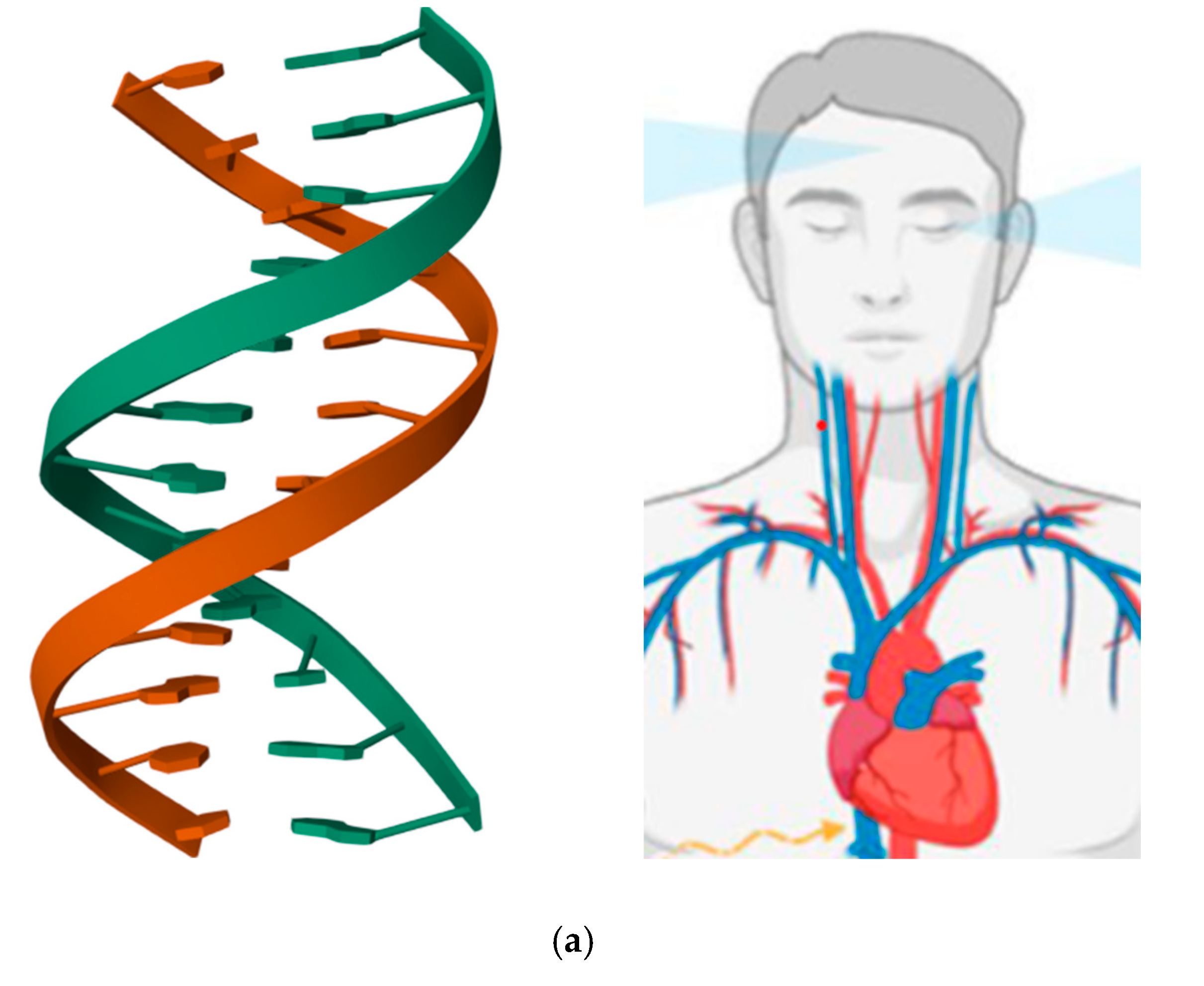
Figure 1. (a) Asymmetry of the double helix of DNA (the structure corresponds to the DNA dodecamer d(GGCAAAAAACGG)/d(CCGTTTTTTGCC) and is freely available at the link https://www.rcsb.org/3d-view/1FZX/0, accessed on 2d October 2023) and human body (adapted from https://www.mdpi.com/ijms/ijms-23-15150/article_deploy/html/images/ijms-23-15150-g001.png, accessed on 2d October 2023); (b) 3D views of the molecular structure of the human myosin I (the structure is freely available at the link https://www.rcsb.org/3d-view/4L79/1 , accessed on 2d October 2023).
Preprints 88071 g001aPreprints 88071 g001b
Figure 2. A schematic view of movement asymmetry assessment during training.
Preprints 88071 g002
Figure 3. A schematic view of movement asymmetries assessment during rehabilitation.
Preprints 88071 g003
Figure 4. Structural representation of meloxicam (left) and phenylbutazone (right).
Preprints 88071 g004
Disclaimer/Publisher’s Note: The statements, opinions and data contained in all publications are solely those of the individual author(s) and contributor(s) and not of MDPI and/or the editor(s). MDPI and/or the editor(s) disclaim responsibility for any injury to people or property resulting from any ideas, methods, instructions or products referred to in the content.
Copyright: This open access article is published under a Creative Commons CC BY 4.0 license, which permit the free download, distribution, and reuse, provided that the author and preprint are cited in any reuse.
Prerpints.org logo

Preprints.org is a free preprint server supported by MDPI in Basel, Switzerland.

Subscribe

© 2024 MDPI (Basel, Switzerland) unless otherwise stated